International Research Journal of Engineering and Technology (IRJET)
e-ISSN: 2395-0056
Volume: 09 Issue: 05 | May 2022
p-ISSN: 2395-0072
www.irjet.net
A REVIEW PAPER ON THE DETECTION OF DIABETIC RETINOPATHY Revathy A1, Hema S2 1PG
Student Dept of Electronics & communication Engineering LBSITW, Kerala, India Professor, Dept of Electronics & Communication Engineering, LBSITW, Kerala, India ---------------------------------------------------------------------***--------------------------------------------------------------------2Assistant
Abstract - Diabetic retinopathy (DR) is a major issue in the
soft exudates are the severe stage of exudates appear as grey-white colour. It is also referred to as a cotton wool spot. In the case of diabetic retinopathy, early detection is very important. Analysing the fundus image manually is a difficult task, so choosing automated detection is better than manual detection.
medical field. DR can damage the blood vessels in the tissue at the retina. In severe cases, DR leads to vision loss. Early detection is very important in the case of diabetic retinopathy but it is a major challenge in the medical field. The two types of DR are non-proliferative diabetic retinopathy (NPDR) and proliferative diabetic retinopathy (PDR). Blurred vision, sudden vision and flashes are the main symptoms of DR. This paper focuses on the different techniques used in the detection of diabetic retinopathy. Automated detection help to detect diabetic retinopathy easily compared to manual detection.
Key Words: Non-proliferative diabetic retinopathy, Proliferative diabetic retinopathy
1. INTRODUCTION Diabetic retinopathy (DR) is a retinal disease caused due to the excess sugar level in the blood. In severe cases, it may lead to sudden vision loss. So early detection and treatment are very important in the case of diabetic retinopathy. According to the WHO, there are 31.7 million people affected by diabetes in India and it is expected to rise to 79.4 million by 2.30. National Diabetes and diabetic retinopathy survey 2019, says that the prevalence of diabetes is 11.8% in the last four years from 2015 to 2019 [1]. When detecting diabetic retinopathy in a clinal way requires more time and effort. But when choosing the automated detection of diabetic retinopathy it is less time consuming compared to the clinical method. The main symptoms of DR are blurred vision, floaters and flashes and vision loss. Blood vessels, optic disc and macula are the main components of a healthy retina any changes in these components occur in eye disease. Diabetic retinopathy has two stages non-proliferative diabetic retinopathy (NPDR) and proliferative diabetic retinopathy (PDR).Microaneurysms(Mas), hemorrhage (H), hard exudates (HE) and soft exudates are the different signs of NPDR. According to the presence and quantity of these lesions, NPDR is again classified into mild, moderate and severe. The microaneurysm is considered the early stage of DR. These appear as tiny red dots. The next stage of DR is hemorrhages. Dot hemorrhages are the bright red dot and the blot hemorrhages are larger blood lesions. Hemorrhages lead to blurred vision. Exudates are considered the third stage of the DR. The leakage of lipids and proteins from damaged blood is called exudates. Depending on their edge, energy and threshold exudates are classified into hard and soft. Hard exudates appeared as bright yellow colour and
© 2022, IRJET
|
Impact Factor value: 7.529
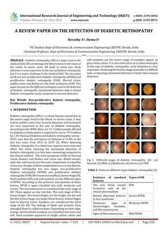
Fig 1: Different stages of diabetic retinopathy [2] : (a) Normal; (b) Mild; (c) Moderate; (d) Severe; (e) PDR Table 1: Features different types diabetic retinopathy [3] Existence features
of
standard
The new blood vessels formation with all the retinal abnormalities Retinal abnormal features in four quadrants Numerous signs of Hemorrhages, Exudates, and Microaneurysms Signs of Microaneurysms
|
Types of DR PDR
Severe NPDR Moderate NPDR
Mild NPDR
ISO 9001:2008 Certified Journal
|
Page 255