International Research Journal of Engineering and Technology (IRJET) e-ISSN: 2395-0056

Volume: 09 Issue: 12 | Dec 2022 www.irjet.net p-ISSN: 2395-0072
International Research Journal of Engineering and Technology (IRJET) e-ISSN: 2395-0056
Volume: 09 Issue: 12 | Dec 2022 www.irjet.net p-ISSN: 2395-0072
1Assistant Professor, Dept. of Computer Science and Engineering, VNR Vignana Jyothi Institute of Engineering and Technology, Hyderabad, India 2,3,4,5Student, Dept. of Computer Science and Engineering, VNR Vignana Jyothi Institute of Engineering and Technology, Hyderabad, India ***
Abstract - The most prevalent and deadly disease, brain tumors,haveanextremelylowlifeexpectancy.Thisstudyuses deep learning algorithms to categorize and identify human braintumorsWhichweareusingtotrainthedatasetfordeep neural networks. Since new data cannot be trained, the accuracy level of the current analysis is insufficient. Here, the braintumorisdetectedusingtheRegionConvolutionalNeural Network (R-CNN) VGG16 model and the DensNet model and segmented from the MRI images using Particle Swarm Optimization(PSO)(normal,benign,malignant).Thedataset has undergone extensive feature analysis and segmentation training. We achieve the highest accuracy by combining PSO with R-CNN.
Key Words: VGG16, DenseNet, RCNN, PSO
Brain tumors are essentially the uncontrolled expansionofmalignantthecellsinthebrain,asopposedtoa tumor, essentially the unchecked expansion of malignant cells in any region of the body. Different types of brain tumors exist. The word "benevolent" refers to a group of aberrant,non-cancerousbraincells.Adenomasandfibroids are two examples of benign tumors. Pre-malignant brain tissueisagroupofaberrantcellsthatarenotcancerousbut havethecapacitytodevelopintomalignantcells.Themost severe sort of tumor is malignant, which is a collection of diseasedcells.Sarcomaandcarcinomaaretwoexamplesof malignant tumors. A person's life can be saved if a brain tumorisfoundandtreatedproperly.
In India, the frequency of brain tumors is gradually increasing, and more instances are reported each year amongpeopleofallages.Accordingtoreports,braintumors are India's tenth most prevalent cancer form. The International Association of Cancer Registries claims that (IARC),Indiareportsabout28000casesofbraincancereach year,andsadly,Braintumourscause24000deathsannually. Braintumoursareadangerousmedicalcondition.Iffound andtreatedimproperly,canbefatal.Inasingleyear,over 330,000 children and adults received diagnoses of CNS cancer worldwide, and this figure is continuing to climb along with the rising mortality rate of brain tumors. CNS
cancer accounts for 2% of all cancers in India, and the incidencerangesfrom5to10per100,000people,withan increasing trend. A person's chance of surviving for five yearsis36%,whiletheirchanceofsurvivingfortenyearsis 31%.
A patient's life expectancy can be extended by a timely diagnosisofabraintumor.Typically,imagingdataanalysis of brain tumor pictures is used to make the diagnosis of brain tumors. In order to accurately assess brain tumor photos,thereareseveralimportantstepsthatmustbetaken. Non-invasive imaging technique magnetic resonance imaging (MRI) generates intricate, three-dimensional anatomical images. It is frequently used for disease detection, diagnosis and therapeutic monitoring image. Consequently,thevariousimaginingauxiliarycircumstances, therearefourdifferenttypesofbrainMRIimagingmodes: weightedT1mode,Flairmode,T1cemode,andT2weighted mode.Differentmodescanshowvariousaspectsofabrain tumor.Theclassificationanditispossibletoidentifybrain tumors accomplished using a variety of techniques, most notablyCNNmodelsEfficientNetB0,InceptionV3,Xception, GaussianConvolutionalNeuralNetwork,andFuzzyCMeans Clustering.
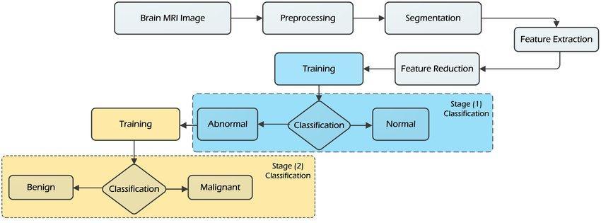
The BRATS2019 dataset, which covers the four modes of MRI images, is being used in this research as part of a method as the detection of brain cancer. Images of brain cancerswereusedstrategiesforprocessingmedicalimages, suchaspre-processingandtheoptimizationtechnique,were utilizedtosegmentthemedicalimageimages.Thedatawas then trained with two CNN models, which assisted in classifyingbraintumorsandidentifyingtheirvariousforms. Enhancement, filter operation, segmentation, and feature extraction are all included in the pre-processing, while featureidentificationandextractionareincludedinthepostprocessing.
[1]Hasnainalishah,FaisalSaeed,(2022)thispaperdoneby utilizingamodelwithinaconvolutionalneuralnetwork, "A RobustApproachforBrainTumorDetectioninMagnetic ResonanceImagesUsingFinetunedEfficientNet" named
International Research Journal of Engineering and Technology (IRJET) e-ISSN: 2395-0056
Volume: 09 Issue: 12 | Dec 2022 www.irjet.net p-ISSN: 2395-0072
EfficientNet B0, we have identified a brain tumor in the magneticresonancescans.Theyhaveemployedmethodsfor processingimagestransformpoor-qualityphotosintohighquality ones. Image segmentation is accomplished using dilationanderosiontechniques.TheEfficientNetB0modelis trained on a dataset of 3000 MR images to identify brain tumors. The validation accuracy of the suggested method was98.87%.Theaccuracyofthismodeliscomparedinthis studytothatoftheVGG16,InceptionV3,Xception,ResNet50, andInceptionResNetV2models.Lessdatawastrained,and thetOnewoulddividethethreegliomagradesintogrades two, three, and four; the other would classify tumors into meningioma,pituitary,andglioma.Thesedatasetsinclude 233and73.Inthispapertypeoftumorwasnotidentified.
[2]MohammadRizwan,AyshaShabbir,(2022)intheirpaper titled “Brain Tumor and Glioma Grade Classification usingGaussianConvolutionalNeuralNetwork”deviseda method to employ a gaussian brain cancer classification usingconvolutionalneuralnetworks.Inthisinvestigation, threedifferentdatasetswereused:onetoOnewoulddivide thethreegliomagradesintogradestwo,three,andfour;the otherwouldclassifytumorsintomeningioma,pituitary,and glioma. These datasets contain 233 and 73 patients' T1weighted MR scans, for a total of 3064 and 516 pictures, respectively.Thismethodhadaccuracyratesof99.8%and 97.14 percent. To identify the type of tumor and glioma grade, this system pre-processed the data using gaussian filtersandtrainedthedata usingagaussianconvolutional neuralnetwork.Gaussianfiltersreducenoiseandblurthe image, but they don't improve the image's quality or accuratelysegmentit.
[3]SyedAliNawaz,DostMuhammadKhan,(2022)intheir papertitled“BrainTumorclassificationBasedonhybrid optimized multi feature analysis using magnetic resonanceimagingdataset”Thestudy[1][2][3]y’sgoalis to use brain magnetic resonance imaging to construct a machine-vision-basedclassificationmodelforbraintumors. Forglioblastoma,meningioma,andmetastaticbraintumor classification,see,asystemknownasaclassificationsystem forhybridbraintumors(HBTC)developed.Inthissystem, images are first pre-processed using K-S-L image enhancementtoimproveimagequality.Thepre-processed imagesarethensegmentedusingthreshold-andclusteringbased segmentation. Following the extraction of features usingtherunlengthmatrixandco-occurrencematrix,nine optimizedfeaturesarechosenusingtheFisherco-efficient technique. After selecting the optimized features, four differentclassifiers RandomTree,MetaBagging,Decision Tree, and Multilayer Perceptron are used to classify the type of tumor. However, the machine learning algorithms used
[4] Ahmed S. Musallam, Ahmed S. Sharif, (2022) in their paper titled “A New Convolutional Neural Network ArchitectureforAutomaticDetectionofBrainTumorsin
Magnetic Resonance Imaging Images”Acomputationally light model with few Max pooling, convolutional, and trainingiterationswasusedinthisstudy'sarchitecture.This study suggests a three-step preprocessing method to improve image quality, along with a deep convolutional neuralnetwork.isusedtoaccuratelyidentifytumorsofthe pituitary, meningiomas, and gliomas. This report also illustratedhowtheproposedarchitectureandthecurrent approaches were compared. This system's accuracy was 98.22% overall, 99% in glioma detection, 99.13% in meningioma detection, 97.3% in pituitary detection, and 97.14%innormalimagedetection.3394MRimageswere usedinthisworktotrainonandidentifythevarioustumor types.Withminimaltrainingdataandnospecifictechniques, imagesegmentationisperformed.
[5] Nellum Norren, Sellapan Palnappan, (2020) in their paper titled “A deep learning model based on concatenation approach for the diagnosis of brain tumor”ThismodelcombinesthetwoseparateCNNmodels, Inception-v3andDenseNet201,todetectbraintumorsinMR images.First,thedatasetispre-trainedusingfollowingthe Inception-v3model,thetrainingandfeatureextractionare doneusingtheDenseNet201model.Theaccuracyprovided by the suggested method is 99.34% and 99.51%, respectively.Thetypeoftumorisnotidentifiedinthispaper, andthereislesstraineddata.
[6]ShariarSazzad,Misba UI Hoque, (2019)intheirpaper titled “Development of Automated Brain Tumor IdentificationUsingMRIImages”Thestrategyusedinthis studyisusinggrayscalephotostodetectbraintumors.Color in grayscale fluctuations are reduced before training the dataset,andnoisesareeliminatedusingafilteroperation. The color segmentation in this method is dependent on thresholds. Due to our inability to adequately extract the features,thereisnoguaranteeforsegmentation.Theresults alsoincludeundesirableplaces.
[7]SergioPereira,AdrianoPinto,(2019)intheirpapertitled “BrainTumorSegmentationusingConvolutionalNeural Networks in MRI Images” The amount of data that an imagingtechniquelikeMRIproduceslimitsclinicalpractice using exact quantitative evaluations, making it difficult to manuallysegmenttheimagesquickly.Duetothesubstantial spatial and anatomical variation among brain tumors, automaticsegmentationisadifficulttask;asaresult,reliable andautomaticsegmentationmethodsarerequired.Inthis study, we investigate 33 tiny kernels as a part of a selfcontained convolutional neural network segmentation technique.Giventhefewerweightsthereareinthenetwork, using small kernels enables the design of more intricate architectures and helps prevent overfitting. In addition, despiteitsrarityinpre-processing,welookedattheuseof intensitynormalization.
International Research Journal of Engineering and Technology (IRJET) e-ISSN: 2395-0056
Volume: 09 Issue: 12 | Dec 2022 www.irjet.net p-ISSN: 2395-0072
[8]MingLi,LishanKuang,(2019)intheirpapertitled“Brain tumor detection based on multimodal Information fusion and CNN [5]” This strategy's primary goal is to increasetheaccuracyofitsearlierstrategies.Convolutional neuralnetworksandmultimodalinformationfusionareboth usedinthismethod.Toimproveaccuracyandperformance, theyusedanormalizationlayerbetweenthepoolinglayer andtheconvolutionallayerinthisapproach.Incomparison tothetwo-dimensionaldetectionnetwork,theaccuracywas muchhigher.First,thisstudyconvertsmultimodal2D-CNNs to 3D-CNNs, allowing for the detection of brain lesions severalmodalitiesinthreedimensions.Therawinputofthe 2D-CNN can be resolved., which calls for a wide neighborhood of defects, while also better extracting the modaloftheinformationaldifferences.Tumortypesarenot identified,andimproperimagesegmentationisused.
[9]KeerthiTK,ShobaXavier(2018)intheirpapertitled“An Intelligent System for Early Assessment and Classification of Brain Tumor” Theproposedsystemcan beusedfortumortypecategorizationandearlydiagnosis. Therearefourphasesinthesystem.1.imageprocessing2. segmentationthresholdsforsegmentation3.GLCMcanbe used to extract characteristics from brain MRI data. SVM classificationisthelaststep.WiththeGA-SVMclassifier,the systemwilloffergreateraccuracyandexpanditscapacity forgeneratingdecisions.SVMismoreproductiveanduses lessmemory.Thedevicealsodeterminesthetumor'sstage ofgrowthandoffershealthysuggestions.SVMonlyworks well for smaller datasets; hence, it is not appropriate for largerdatasets.
[10] Dr A Jagan (2017) in their paper titled “A New Approach for Segmentation and Detection of Brain Tumorin3DBrainMRImaging”ThisstudyusesClustering usingfuzzyC-meansandtheimprovedEMtechnique.The decisionwastakenduringdetection,andbothapproaches wereprocessedandcalculatedbasedonwhichhadahigher accuracyrate.Themaingoalofthemethodistoincrease3D Accuracy,sensitivity,andspecificitybrainMRimagesusing fluid-attenuated inversion recovery-based segmentation. Thefluidattenuationinversionrecoveryinthe3DbrainMR image segmentation was the major focus of this investigation.Fuzzyc-meansEuclideandistancemeasures giveunderlyingfactorsunequalweight.
[11]HaochengShenandJianguoZhang(2017)intheirpaper titled “Fully connected CRF with data-driven prior for multi-class brain tumor segmentation” The automatic segmentation of brain tumors described in this study is basedonFC-CRF.Thispaper'sprimarycontributionsareas follows: 1) Apply FC-CRF to segment brain tumors into multipleclassesusingahierarchicalapproach.2)compared grid CRF with FC, demonstrating that the latter greatly enhanced tumor border segmentation. Tumor boundaries that are segmented have greatly improved. They use a histogramforanalysis.Processingtimeincreaseswhenthe
borders are widened and each image pixel is processed separately.
[12] R Lavanya Devi, Nivedita (2017) in their paper titled “ClassificationandSegmentationofBrainTumorsinMRI Images using PNN” .Theproposedprocessisbrokendown into four fundamental components. Preprocessing comes initially, followed by feature extraction using a gray-level cooccurrence matrix (GLCM). GLCM is the second matrix. Phase 3 is PNN-based categorization., followed by segmentationusingtheK-meansclusteringtechniqueasthe finalstep.PrincipalComponentAnalysisisusedforpicture recognition and compression (PCA). PNN has the best classification accuracy and is the fastest method. PCA successfully decreases the data's dimensionality. MRI imaging is more effective than CT scanning. First, a systematic review unearths a total of 18,725 potentially importantscientificpapers.Studiesthatfocusontheactual observed behavior of app users rather than behavior reported via questionnaires have been found to have a research gap. This study looks at how notifications' frequency,content,andpresentationinteract.
[13]HarishChetty,MonitShah(2017)intheirpapertitled “A Survey on Brain Tumor Extraction Approach from MRI Images using Image processing”. The proposed process Initially t2 weighted MRI images will be given as input.Thenthepre-processingschemewillbedoneonthe image by using an average filter the image will be smoothened.Skullstrippingmethodisappliedtoremovethe fattytissueandskullinimages.Watershedtechniqueisused for segmentation. Erosion technique in morphological methodisappliedtodetecttumor.ThisapproachwasNot suitableforlargedatasets.
[14]HussnaElnoorMohammedAbdalla,M.Y.Esmail(2018) in their paper titled “Brain tumor detection using Artificial Neural Network” The system is first provided withabrainMRIsampleasinputintheproposedprocess, and the image is then improved using the histogram equalisation technique. This method uses an image histogramtoadjusttheimagecontrastbeforesegmenting theimageusingaglobalthresholdingmethodandextracting features. By using Neuro fuzzy classifier, the image gets classifiedandshowstheresultasnormalandabnormal.This approachhastoo muchcomputation time andtreatslocal pixels same as pixels for apart, sensitive to location of an objectinanimage.
[15]YaminiSharma,YogeshK.Meghrajani(2015)titledthe paperas“BrainTumorExtractionFromMRIImageUsing Mathematical Morphological Reconstruction”. In this study, labelled MRI images that have been affected by impulsive noise were analyzed, and a method for locating brain tumours was proposed. A binary picture is created from a grayscale image using the global thresholding approach. Then, an operation known as "morphological
value: 7.529 | ISO 9001:2008 Certified Journal |
International Research Journal of Engineering and Technology (IRJET) e-ISSN: 2395-0056
Volume: 09 Issue: 12 | Dec 2022 www.irjet.net p-ISSN: 2395-0072
openingbyreconstruction" whichinvolvestwosteps is used.Thefirstiserosionbyadiscstructuralelement(SE), anda morphological reconstructionmakesupthesecond. Thefirstphase'serodedimageisusedasamarkerimagefor thesecondphaseandoffersamarkerpoint atthetumour location.Thismorphologicalprocesscreatesabinaryimage with a tumour in the foreground, labels the image, and removesthesaltnoise.Thismethodonlyeliminatednoise.
The primary goals of the study are to use Particle Swarm Optimization(PSO)tosegmenttheMRIimageandregionbased convolutional neural networks (VGG16 and the DenseNet model) to predict the tumor. identifying many tumor kinds, including benign (non-cancerous) and its subtypes, such as adenomas and fibroids, as well as malignant(cancer).TheBRATS2019dataset,whichincludes fourMRIimagemodalities,istheoneusedintheproposed system.
Since they are one of the leading causes of death among humans, brain tumors must be found early and treated. Basically,differentimageprocessingalgorithmsareapplied to achieve this. In this study, we investigated two distinct methods for MRI image processing-based brain tumor detection.Afterreadingthestudy,itisclearthattheregionexpansion technique is quite promising for the process's detectionphaseinthefuture.Thesetwoalgorithmsenable us to identify brain tumors using a variety of detection methods.Bothimageprocessingmethodshaveahighrateof tumoridentificationsuccess.However,bothalgorithmshave somedrawbacksthatcouldberesolvedbycreatinganew algorithm that considers a variety of factors and the comparisonofthealgorithmsdiscussedinthispaper

Specialthankstoourteam guide, G.LaxmiDeepthi,for all the technical support and guidance which led to the completionoftheliteraturesurveyphaseoftheprojectwith agoodresultintheend.
[1] Shah,HasnainAli,etal."ARobustApproachforBrain TumorDetectioninMagneticResonanceImagesUsing FinetunedEfficientNet." IEEEAccess 10(2022):6542665438.
Thismodelallowsforthetrainingofadditionaldata.Using an optimization technique, the images are segmented to distinguishbetweennormalandpathologicalcells.Following optimization,thedatasetistrainedtoextractfeaturesand identify the type of tumor using the VGG16 and DenseNet models.
[2] Rizwan, Muhammad, et al. "Brain tumor and glioma gradeclassificationusinggaussianconvolutionalneural network." IEEE Access 10(2022):29731-29740.
[3] Nawaz, Syed Ali, Dost Muhammad Khan, and Salman Qadri. "Brain Tumor Classification Based on Hybrid Optimized Multi-Features Analysis Using Magnetic Resonance Imaging Dataset." Applied Artificial Intelligence (2022):1-27.
[4] Musallam,AhmedS.,AhmedS.Sherif,andMohamedK. Hussein. "A New Convolutional Neural Network ArchitectureforAutomaticDetectionofBrainTumorsin Magnetic Resonance Imaging Images." IEEE Access 10 (2022):2775-2782.
[5] Noreen,Neelum,etal."Adeeplearningmodelbasedon concatenation approach for the diagnosis of brain tumor." IEEE Access 8(2020):55135-55144.
[6] Sazzad,TMShahriar,etal."Developmentofautomated brain tumor identification using MRI images." 2019 International Conference on Electrical, Computer and Communication Engineering (ECCE).IEEE,2019.
[7] Pereira,Sérgio,etal."Braintumorsegmentationusing convolutional neural networks in MRI images." IEEE
International Research Journal of Engineering and Technology (IRJET) e-ISSN: 2395-0056
Volume: 09 Issue: 12 | Dec 2022 www.irjet.net p-ISSN: 2395-0072
transactions on medical imaging 35.5 (2016): 12401251.
[8] Li, Ming, et al. "Brain tumor detection based on multimodalinformationfusionandconvolutionalneural network." IEEE Access 7(2019):180134-180146.
[9] Keerthana, T. K., and Sobha Xavier. "An intelligent systemforearlyassessmentandclassificationofbrain tumor." 2018 Second International Conference on Inventive Communication and Computational Technologies (ICICCT).IEEE,2018.
[10] Jagan, A. "A new approach for segmentation and detectionofbraintumorin3dbrainmrimaging." 2018 Second International Conference on Electronics, CommunicationandAerospaceTechnology(ICECA).IEEE, 2018.
[11] Shen, Haocheng, and Jianguo Zhang. "Fully connected CRFwithdata-drivenpriorformulti-classbraintumor segmentation." 2017 IEEE International Conference on Image Processing (ICIP).IEEE,2017.
[12] Lavanyadevi, R., et al. "Brain tumor classification and segmentation in MRI images using PNN." 2017 IEEE International Conference on electrical, instrumentation and communication engineering (ICEICE).IEEE,2017.
[13] Chetty,Harish,etal."Asurveyonbraintumorextraction approach from MRI images using image processing." 2017 2nd International Conference for Convergence in Technology (I2CT).IEEE,2017.
[14] Abdalla,Hussna ElnoorMohammed, andM. Y.Esmail. "Brain tumor detection by using artificial neural network." 2018 International Conference on Computer, Control, Electrical, and Electronics Engineering (ICCCEEE).IEEE,2018.
[15] Sharma,Yamini,andYogeshK.Meghrajani."Braintumor extraction from MRI image using mathematical morphologicalreconstruction." 2014 2nd International Conference on Emerging Technology Trends in Electronics,CommunicationandNetworking.IEEE,2014.
2022, IRJET | Impact Factor value: 7.529 | ISO 9001:2008 Certified Journal | Page346