

DIRECTIONAL CLASSIFICATION OF BRAIN TUMOR IMAGES FROM MRI USING
CNN-BASED DEEP LEARNING
G MAHESH CHALLARI 1 , J PRASHANTHI 21 Assistant Professor in Department of cse at Sree Dattha Institute of Engineering And Science,
2 Assistant Professor in Department of cse at Sree Dattha Group of Institutions.,
Abstract - A brain tumour is a dangerous development of unnatural cells in the brain. If not treated in time, it might prove fatal. It is therefore crucial to find the tumor early and start therapy as soon as possible. People with brain tumours had a significant fatality rate before the discovery of early diagnosis. The mortality rate lowers considerably after an early diagnosis is established. Accurate early diagnosis of a brain tumour increases a patient's chance of survival. The customary system used to detect tumors involved physicians studying the MRI scans and analyzing the abnormalities in the image. However, with the increase in the size of data and limited amount of time it becomes extremely strenuous for the physicians to analyze the image. Our research has resulted in the release of a convolutional neural network model for the detection of brain tumours, whichfurther classifies the tumour into glioma, pituitary, or meningioma. This automated model is improving the detection and classification accuracy of tumours, demonstrating that it is a useful tool for physicians. The same brain MRI database is used to train and test all the types under consideration, including CNN, ResNet50, MobileNetV2, and VGG19. The effectiveness of each type is reviewed. Accuracy, error rate, and time to train are just few of the criteria used to evaluate the results from each CNN variant
Key Words: Convolutional Neural Networks, Brain Tumor, MRI, Medical Disorder, Healthcare.
1. INTRODUCTION
Analyzing biomedical data is growing significantly and is crucial in diagnosing and properly treating disease. Brain tumorcontainsmorecompleximagedatathatneedsimage processing to analyze. There is a higher death rate and impropertreatmentofbraintumors,asthestatisticstaken bytheNationalBrainTumorFoundation(NBTF)worldwide [1]. Several approaches (or) frameworks have been developedtoconsiderbraintumorsinrecentyears.Itcomes across data classification, proper treatment planning and predicting outcomes. Images of tumours in the brain's structure are difficult to classify because of issues with contrast,noise,andmissingboundaries.MagneticResonance (MR)imaging[2,3],PositronEmissionTomography(PET) [3,4],andComputedTomography(CT)scan[5,6]areused to assess the efficacy of the diagnostic procedure by analysingtheaforementionedelements.Scannersuseimage processingtolookforsignsofillness.Thescanningprocess
is done to diagnose and treat the brain tumours better, which identifies tumor images to be analyzed effectively. Supposetheidentificationofpredictingthetumorcellsand locating them helps to diagnosis the disease properly. Makingtheanalysesmorequalitativeandamountquantity improvesthecharacteristicsofatumordiagnosisthrough data segmentation. Based on the development of the MR images generation, it can be processed manually, semiautomatically, and fully automatically [5]. Based on the image processing relatedto themedical field,information shouldbeaccuratewhileprocessingtheimagebasedonthe segmentation and classification process. While processing theimages,theexecutionshouldbetime-consuming[6].
2. LITERATURE REVIEW
Incomputervision,imagesegmentationisahighlyeffective technological procedure. In the field of image processing, segmentation is a useful tool. Pixels are organized into groups called segments, which are themselves artefacts. When an image is segmented, it is no longer necessary to analyzetheentirethingatonce.Thethreestepsinvolvedin theprocessingofanimagearedepictedinoveralldiagnosis of "skull," "brain," or "tumour" might be possible by labelling. One application of object detection is to identify certainobjectsinsideapicture.Inordertoproperlyidentify andcategorizeobjects,segmentationisessential
Inthissection,wetakealookatthevarioussegmentation methodscurrentlyavailableintheliteratureforMRimage processing.Theauthor[66]proposesanappropriate,novel approach to tissue segmentation from MRI brain imaging. The "WM and GM and CSF" segmentation is useful for studyingdiseasesanddesigningtreatments.Toeliminatethe graininess and sharpen the image, anisotropic diffusion filteringisused.Testsoftheproposedtechniqueon10MRI images have been conducted, and the results have been compared to those obtained using existing methods (including "Otsu MT" "fuzzy C-means". The results of the experiments show that, in comparison to the existing approaches,theaveragesegmentationaccuracyisimproved by96.79%,thespecificityis96.55%,andthesensitivityis 96.55%
Brain tumour identification and increased breast cancer detectioninMRIimageswerebothtargetedbytheauthorof [82],whoproposedatemplate-basedKmeansandenhanced
International Research Journal of Engineering and Technology (IRJET) e-ISSN: 2395-0056 Volume: 11 Issue: 01 | Jan 2024 www.irjet.net p-ISSN: 2395-0072 © 2024, IRJET | Impact Factor value: 8.226 | ISO 9001:2008 Certified Journal | Page342
***
fuzzyCmeans(TKFCM)system.Thisalgorithmoutperforms the competition even when presented with a noisy MR image. In comparison to thresholding, area expanding, regionsplittingandmerging,ANN,TK-means,andFCM,this algorithmperformsbetter.Itusedasimulatortotestoutits proposed solution and see how well it worked. Besides, it hasprovenaconsensusreachedtobemorereliablethanthe conventionalstatisticalclassificationresults.However,the time required for the TKFCM algorithm to detect brain tumorsisconcisecomparedtoothertraditionalmethods.
3. SYSTEM ANALYSIS
3.1.1 DETECTION OF BRAIN TUMOR USING RESUNET ARCHITECTURE
Using a classification technique, a brain tumour detection system aids in diagnosing and treating patients recently diagnosedwithatumourinthebrain.Earlydiagnosisand treatmentcan be provided byusingtheMRI classification method, which helps identify the brain's tumour and determine the tumor’s density. U-Net architecture is associated with a variety of classification methods in the literature.Basedonacomparisonoflow-levelandhigh-level feature information, this work proposes Residual U-NET with improved local feature information for improved medical image segmentation. As part of the modified ResidualU-Netmodel,thedropoutandwidecontextlayers are addressed in addition to the residual module and attention gate. Large sensitive scaled information and small-scale images benefit from adding salient feature information. After making certain adjustments, the gate attention model performed better than state-of-the-art methodslikeU-NetandCNNDensely
3.1.2 LGG Dataset
Thisdataset hasMR images of the brain and manual flare abnormality segmentation masks. In total, the Cancer Genome Atlas (TCGA) collection contains 3929 images, including2556non tumorimagesand1373cancerimages from110individuals

Theratiooftrainingandtestimagesaretested70:30.The sizeofallimagesinthisdatasetis256x256pixels.Access
thedatasetat'https://www.kaggle.com/mateuszbuda/lggmri-segmentation'...
4. PROPOSED METHODOLOGIES
4.1.1 Image Pre-processing
Thedeeplearningmodel iscrucialinmakingthenetwork lesssensitivetothenoiseinthedataandimagesitisgivento analyze. Because of how consistently it analyses images acrosstheboard,theN4ITKmethodisutilizedforcorrecting bias-basedimageprocessing.Severalalgorithmsexistthat performthedatapre-processingforbraintumor.Fromthe existingN4ITKalgorithmprovidesreliabledataasitcollects thedatabasedonthecapabilityofbiasfieldforMRIImages.
4.1.2 Modified Residual U-Net
AsresidualnetworkassociatetheU-Netbackbonewithmore residual blocks in Deep framework to gradient mitigation problem.InthisU-Netarchitecture,theconvolutionalneural network is related to performing image segmentation into threeelements,Encoderanddecoder,basedontheprocessof contractionandexpansion.Forthepurposeofexpandingthe blocks, the suggested architecture takes into account the Convolutional2D,Maxpooling2D,WideContext,Transpose 2D,ResidualBlock,andConcatenatemetrics.
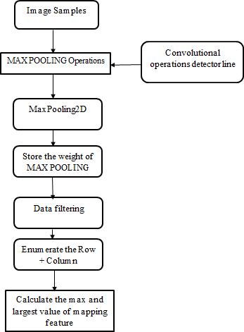
4.1.3 Max Pooling Layer
Layer will supply the down feature extraction mapping, highlighting the feature patches depending on the feature map approach, in addition to max and average pooling. Maximum presence pooling uses extracted features to calculateanaveragepresenceandthenusesthattoactivate thepresenceofthosefeatures.Simplysaid,thisconvolutional layeristheneuralnetwork-basedconvolutionallayerthatis usedtogeneratethemappingfeatureextractionbyfiltering learning based on input images. It is determined through research that the layer functions best when extracting simplefeatures.
4.1.4 Dropout Layer
Itis themethodusedtostayaway fromtheconvolutional neuralnetworkmodel'soverfittingissue.Whenthetraining phase is updated, the main cause of dropout is a random settingonthehiddenedgeunitsbeingsetto0asneurons. Using a sigmoid activation function and a numerical representationoftheresidualu-net
International Research Journal of Engineering and Technology (IRJET) e-ISSN: 2395-0056 Volume: 11 Issue: 01 | Jan 2024 www.irjet.net p-ISSN: 2395-0072 © 2024, IRJET | Impact Factor value: 8.226 | ISO 9001:2008 Certified Journal | Page343
Figure3.1.2BrainTumorimageandMaskimage
4.1.5 Attention Gate with Residual U-Net
Thecomingstepofdatapreprocessingistohandlemissing
Two parallel connections are used as input for the two convolutionallayersinabroadcontextblock.Useboth1xN andNx1filtersontheinitiallink. Theconnectionuses1xN filter with a convolutional model and N x 1 filter with a convolutional model. In this process, the information’sare extractedtoformthedatasimilaritybyconsideringthewide context. It is further classified into different convolutional sub-classnetworksforbraintissue.
4.1.6 Wide Context Information
Two parallel connections are used as input for two convolutionallayersinalargecontextblock.Useboth1xN andNx1filtersontheinitiallink.Theconnectionuses1xN filter with a convolutional model and N x 1 filter with a convolutional model. In this process, the information’sare extractedtoformthedatasimilaritybyconsideringthewide context. It is further classified into different convolutional sub-classnetworksforbraintissue.


5. RESULTS


The quantity of photos given careful consideration is one indicator of performance in the analysis of results. Tensor Flow, along with the proper modules, can be used as a checkpointandschedulerforthelearningprocess.Patient, RNSseqcluster,laterality,tumourlocation,andethnicityare justsomeofthegranularcharacteristicsthatmaybefoundin theKaggledataset.Thosedatasetswithphotosaretheresult ofacheckpoint-drivensortingprocedure.Patientid,picture path,andmask pathareallincludedinthefinal datasetas showninTable3.1.Tocomparethecountvaluetothemask andfindthemaskcountplot.
To locate the tumour onthemask, weuse photos of brain tumours with varying pixel colours. Using parameterslike imagepath,maskpath,andmask,anewdatasetisgenerated. Brain_df_train, test size = 0.15 is used to test and divide traineddataaccordingonthechosenmodel.Afterthat,those picturesareconfirmed
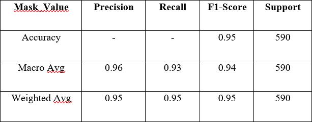
UsingaclassifiermodeltrainedwithdatafromtheResidual NetworkResNet50,weexaminethephotos.Conv2D,Input, ZeroPadding2D, Batch Normalization, Activation, MaxPooling2D,andbatchnormalizedimagesareclassifiedin the input layer. Using an image classification model to determine whether or not the tumour is present, the classifier model achieved a loss in data of 0.2353, with an accuracyof94.745%.Tables3.2and3.3depictthemetrics usedtogeneratetheconfusionmatrixfromtheoriginaland forecast images: accuracy score, confusion matrix, and classificationreport
International Research Journal of Engineering and Technology (IRJET) e-ISSN: 2395-0056 Volume: 11 Issue: 01 | Jan 2024 www.irjet.net p-ISSN: 2395-0072 © 2024, IRJET | Impact Factor value: 8.226 | ISO 9001:2008 Certified Journal | Page344
Figure 4.1.3 Max pooling layer
Figure 4.1.4 Dropout layer
Figure 4.1.5 Residual U-Net Architecture with Gate Attention Mechanism
S.No. Mask Dtype 1 0 2556 2 1 1373
Table 5.1 Mask Count part
In p ut ou tp ut 1x9 Residu al UNet 9x 1 Re sid ual UNe t 1x 1 1 R es id u al UN et 11 x1 Re sid ual UNe t 1x 1 3 R es id u al UN et 13x 1 Res idu al UNe t 1x 1 5 R es id u al UN et 15x 1 Res idu al UNet

Training and validation loss values, or how well they predicted, areplottedagainst epochs(theX-axisinFigure 3.6).Thelossrangesfromzerotoone.FortheproposedRes U-Netarchitecture,thelossiscalculatedusingdatafromthe first 30 epochs. Since the validation set is probably not representative of the complete dataset, the validation loss variesgreatly.Thevalidationsampleshouldberandomized orresampled,inmyopinion

Figure5.2
from0to30forbothtrainingandtestingdata.The value of the dice rises steadily with the number ofepochs Accuracy for the suggested Residual U-Net architecture is showninFigure3.8,whereepochrangesfrom0to30are usedforthetraineddataandVal.Asmoretimehaspassed (moreepochshavebeenadded),theaccuracyvaluehasrisen steadily

Datapre-processing,encoding,anddecodingonMRIimage datasetsareallcarriedoutusingthesuggestedarchitecture. Segmentationofimprovedimagesisusedasthebasisforthis analysis.LossandAccuracyaredeterminedbycomparingthe valuesofthetrainingandtestepochs,respectively.Thedata categorizationmodelusestheEpochsvaluetocalculatedata lossandaccuracy.TheproposedResU-Netachievesbetter accuracythanthecurrentResU-Netmethodasthenumberof iterationsincreases,asmeasuredbythelossfunction,Dice co-efficientvalue,95%Accuracy.

6. CONCLUSION
Intherecentera,healthcareplaysasignificantrolebasedon biomedicaldatasetsrelatedtobraintumorimagesandother classificationandsegmentationprocesses.Varioustypesof tumorarediscussedbasedondifferentgrades.Atfirst,we talkabouthowthedatasetinfluences thepreprocessingof images.Afterthat,asetoffeaturephotosisgeneratedand categorizedusingestablishedmethods.Tumourdetectionand image segmentation processing are described. To better segmentmedicalimagesandfocusontheattentionmodulein tumourimages,wefirstpresentthedesignofResidualU-NET withsomeimprovedlocalfeatureinformation.Inthis,local informationonimagefeatureareconsideredbyimproving theimagesegmentationthrough theproposedResidualUNetasitintegratesbothresidualandattentiongatemodules and considers both dropout and wide context layer. Focal Trversky Loss and Trversky Accuracyare computed using training and test data, and Epochs value variation is evaluated for large-scale images through whole, core, and enhancingimagesegmentation.
International Research Journal of Engineering and Technology (IRJET) e-ISSN: 2395-0056 Volume: 11 Issue: 01 | Jan 2024 www.irjet.net p-ISSN: 2395-0072 © 2024, IRJET | Impact Factor value: 8.226 | ISO 9001:2008 Certified Journal | Page345
Table 5.2 Attribute metrics based on mask value
Table 5.3 Attribute metrics based on mask value (Accuracy; Macro Avg; Weighted Avg)
Figure 5.1 Epochs Vs Training &Validation loss
depictshowthedicecoefficientfortheproposed Residual U-Net architecture is calculated using epochs ranging
Figure 5.3 Epochs Vs Dice coefficient
Figure 5.4 Epochs Vs Accuracy
BraintumorsegmentationusingSpatialAttentionResU-Net givesbestresultscomparedtoexistingU-Netmodels.With increasingtrainingdata,wecanfurtherimprovediceandIoU scores.Andalsowithmoreresearchontheclasseslabelled on the data we can predict the stage and severity of the disease. After segmenting the MR Image, it is possible to measurethetumour'ssizeinbraintissues
REFERENCES
[1].RobertTufel,“TheNationalBrainTumorFoundation”: GivingHelp,GivingHope”; JournalofNeoplasia, 3(3):264265,2001.
[2]. Soltaninejad,Mohammadreza,Zhang,Lei,Lambrou, Tryphon,Yang,Guang,Allinson,NigelandYe,Xujiong,”MRI BrainTumorSegmentationandPatientSurvivalPrediction UsingRandomForestsandFullyConvolutionalNetworks”, ComputerMethodsandProgramsinBiomedicine,157,69-84, 2018.
[3].LangenKJ,BartensteinP,BoeckerH,BrustP,CoenenHH, DrzezgaA,GrünwaldF,KrauseBJ,KuwertT,SabriO,Tatsch K, Weber WA, Schreckenberger M, “German guidelines for brain tumour imaging by PET and SPECT using labelled aminoacids”,NuclearMedicine,50(4),167-173,2011.
[4]. Irene Neuner, Joachim B. Kaffanke, Karl-Josef Langen, Elena Rota Kops, LutzTellmann,GabrieleStoffels,Christoph Weirich,ChristianFilss,JürgenScheins,HansHerzog&N.Jon Shah, “Multimodal imaging utilising integrated MR-PET for human brain tumour assessment”, European Radiology volume22,2568–2580,2012.
[5]. B. Johnston; M.S. Atkins; B. Mackiewich; M. Anderson, “Segmentation of multiple sclerosis lesions in intensity correctedmultispectralMRI”,IEEETransactionsonMedical Imaging,15(2),154-169,1996.
[6].ErenaSiyoumBiratu;FriedhelmSchwenker;Yehualashet Megersa Ayanoand TayeGirmaDebelee, “A Survey of Brain Tumor Segmentation and Classification Algorithms”, J. Imaging,7(9),2021.
[7].LimJiaQi,NormaAlias,FarhanaJohar,“Detectionofbrain tumour in 2D MRI: implementation and critical review of clustering-basedimagesegmentationmethods”,Oncologyand Radiotherapy,1(52),1-10,2020.
[8]. Dr. Thejaswini P; Ms. Bhavya Bhat; Kushal Prakash, “Detection and Classification of Tumour in Brain MRI”, I.J. EngineeringandManufacturing,2019.
[9].G.Preethi;V.Sornagopal,“MRIimageclassificationusing GLCM texture features”, International Conference on Green Computing Communication and Electrical Engineering (ICGCCEE),2014.
[10]. Sindhu Devunooru, AbeerAlsadoon, P. W. C. Chandana&Azam Beg, “Deep learning neural networks for medicalimagesegmentationofbraintumoursfordiagnosis”, JournalofAmbient Intelligenceand Humanized Computing, 12,455-483,2021.
International Research Journal of Engineering and Technology (IRJET) e-ISSN: 2395-0056 Volume: 11 Issue: 01 | Jan 2024 www.irjet.net p-ISSN: 2395-0072 © 2024, IRJET | Impact Factor value: 8.226 | ISO 9001:2008 Certified Journal | Page346