
Augmentation of Multimodal 3D Magnetic Resonance Imaging using Generative Adversarial Network for Brain Tumor Segmentation
Bhavesh Parmar1, 2 ,
Mehul Parikh21Research Scholar, Gujarat Technological University, Gujarat, India
2L. D. College of Engineering, Ahmedabad, Gujarat, India ***
Abstract – Gliomas are the most common malignancy in brain. Magnetic Resonance Imaging (MRI) images are widely used for medical imaging applications. MultimodalMRIcanbe used effectively for segmentation of brain tumors using Convolutional Neural Network (CNN). CNN accuracy depends on the large amount of data. Medical imaging datasets are mostly smaller in size. There is a chance of missingmodalityin MRI due to clinical settings or in case of patient not available for prognosis. This paper proposed a novel generator –discriminator architecture for development of cohort of MRI modality. Modified 3D UNet architectures were used as generator and PatchGAN for discriminator. In BraTS2020 dataset, there are four MRI modalities present. The modified 3D Unet provided with different three modalities as input and it produce the remaining modality. Discriminator block find the MSE, PSNR, and SSIM values to check before adopting as cohort. The generated modalities are used as input alongwith the original images in dataset for input to the modified 3D Unet. The model performance in segmentation improvedup to 2 to 3 points in each tumor sub regions. The mean dice values achieved using the proposed work is 0.87. 0.81. and 0.78 for WT, TC, and ET respectively.
1. INTRODUCTION
Magnetic Resonance Imaging (MRI) is the widely used imaging modality in the clinical practice. MRI can be very usefulinassessingdifferentinsightsindiagnosisandclinical planning. Multimodal characteristics of MRI scans can be very useful in neuroimaging, specifically brain tumor classification and segmentation. Gliomas are the most frequently occurring central nervous system (CNS) malignancy accounting 30% to all other CNS tumors [1]. WorldHealthOrganisation(WHO)classifiedbraintumorsin grade I to IV considering aggression of malignancy. With amplifying order of antagonism, Grade I and II are Low GradeGlioma(LGG),while,gradeIIIandIVareHighGrade Glioma(HGG)[2].Duetoveryhighvariationsinsize,shape, structure, infiltrative nature of growth and peritumoral edema, brain tumors are hard to delineate from its surrounding healthy tissues. Further, the large volume of MRI scan, Manual border segmentation with visual inspectionistimeconsumingandpronetohumanerror[3].
Recently, deep learning methods have shown promising resultsinmedicalimaging[4].3DUNet[5]andvariationsof
3DUNet[6-9]arethemostadoptedmethodforbraintumor segmentationapplications.
Model accuracy in segmentation for the deep learning networkisdatacentric.MedicalImagingdatasetareusually smaller in comparison to other computer vision datasets. Therequirementofmultimodalpatientimagingmightalso not possible due to various clinical requirements. Additionally,prognosisandfollowupofsingularsubjectmay notbepossible. Tocaterthe need,unavailableimagedata can be augmented using Generative Adversarial Network (GAN).
Inthispaper,generativeanddiscriminativemethodofGAN isproposedandcompareditssuitabilityandimprovements in outcomes on existing methods of brain tumor segmentationapplication.
2. RELATED WORKS
Due to success of deep learning in compute vision applications,datademandincreasingdaybyday[10].Unlike othercomputervisiondataset,medicalimagingdatasetsare limited.Theneedofaugmenteddatacansolvetheproblem ofinadequatedataset.VariousapplicationofGANincludes imagetoimagetranslation[11],imagesynthesisfromnoise [12],styletransfer[13],andimagesegmentation[14].Dueto smallersizeofmedicalimagedatasets,GANalgorithmsare gettingpopularity.
The Generative Adversarial Network (GAN) method was originallyintroducedin2014,byGoodfellowetal[10],for imagegeneration.InMedicalimagesynthesis,CycleGANcan be helpful for cohort of absent modalities [15]. GANS can alsobeusedforsegmentationofmedicalimages.Hanetal., has suggested GAN method for multiple spinal structure segmentationfromMRIscans. Several methodssuggested possibilityofutilizingGANinmedicalimagesegmentation [11,16,17,18].Another2DGANbasedmethodRescueNet was proposed for brain tumor segmentation from MRI images [19]. The detailed review of application of GAN is reviewedbyYietal[20].
This work is motivated from the MM-GAN [21], a 3D extension of Pix2Pix GAN [22]. The method synthesise missingimageofmodalityfromthebytheavailableimages of another three modalities in multimodal brain tumor
International Research Journal of Engineering and Technology (IRJET) e-ISSN: 2395-0056 Volume: 11 Issue: 01 | Jan 2024 www.irjet.net p-ISSN: 2395-0072 © 2024, IRJET | Impact Factor value: 8.226 | ISO 9001:2008 Certified Journal | Page175
segmentationapplication.Themodifiedpatchbased3DUNet [23]alongwithPatchGAN[24]proposedhereforGeneratorDiscriminator framework to improve the segmentation accuracy in brain tumor segmentation application using BraTS2020[25-29]dataset.
3. METHOD
3.1 Data
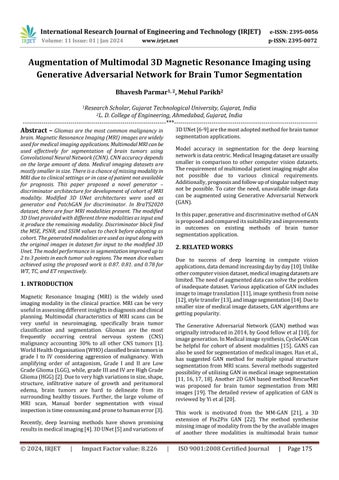
TheBraTS2020datasetwereusedforthiswork.Multimodal BrainTumorSegmentation(BraTS}challengeconsistingof MRIscansof240x240sizewith155scansineachslice[25] BraTS2020 dataset includes 369 training data of different subjects collected from different scanner and protocols at various institutions [26-29]. It includes four modalities of MRInamely;(i)T1–nativescan,(ii)T1ce–PostcontrastT1 weighted,(iii)T2–T2weighted,and(iv)FLAIR–T2Fluid Attenuated Inversion Recovery. Another 125 and 166 previouslyunknownsubjectdataofallfourmodalitieswere providedforvalidationandtesting[29]. Trainingdatawere manually segmentated and approved by experienced neurologist.Figure1ShowthesamplescanofdifferentMRI modalitiesfromthedataset.

3.2 Data Pre-processing
BraTS2020 provides rigidly registered scans with skullstrippingand,resampleat1mm3 isotropicresolutions The multi institutional and different protocol in MRI image acquisitioncreatesnonlinearityinimages.Thereisintensity variation in MRI scans while recording. These inherent intensityinhomoginitiescanberemovedusingN4ITkinsight toolkit [30]. Min-Max scaling applied to standardise the dataset in the range of 10 to 110 voxel intensity values. Further, using mean and standard deviation values of intensity of voxel, z-score normalisation were applied between 0 and 1 for each voxel before feeding to the network.
To prevent overfitting of the model during training, 3D mirroring,random3Drotationbetween0to45°andrandom elasticdeformationwereappliedwith50%probabilityof combinationofaugmentation.
3.3 Model Architecture
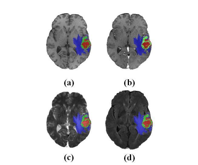
Theoverallarchitectureoftheproposedmethodisdisplayed infigure3.Theframeworkincludestwoblocks;Generatorfor development of synthetic image and discriminator for comparingtheoutputwithgroundtruth.
MultimodalnatureofMRIcanbeusefulintumorsubregion segmentationandthequalitativevisualsofeachmodalityare infigure1.MRIvolumesintransverse,coronalandsagittal anatomical planes are displayed in figure 2 with correspondinggroundtruthoftumorregions.
Thegeneratorisanencoder-decoderbasedarchitecture,a modified 3D UNet architecture with concatenated skip connections [23]. In the Downsample block consist of convolution, batch Normalization and leaky ReLU. Convolution is performed on input through several filters with fixed kernel size 4 and stride 2. Batch normalization prevents vanishing gradient and leaky ReLU used as an activation function. The Upsampling block made of four layers, transposed Convolution, Batch Normalization, Dropout, and ReLU. In transposed convolution, image is stitchedandconvolvedtoenlargethesize.Dropuoutisuseful in preventing overfitting, and ReLU is used as activation function which stops the saturation. The framework of generatorblockisshowninfigure3,withallthesub-blocks includedinDownsamplingandUpsamplingblocks.
International Research Journal of Engineering and Technology (IRJET) e-ISSN: 2395-0056 Volume: 11 Issue: 01 | Jan 2024 www.irjet.net p-ISSN: 2395-0072 © 2024, IRJET | Impact Factor value: 8.226 | ISO 9001:2008 Certified Journal | Page176
Fig -1:MRImodalitiesintransverseplaneoverlappedwith groundtruthsegmentationfor(a)T1,(b)T1ce,(c)T2,and (d) FLAIR, indicated with colour blue, green and red for Peritumoraledema(ED),necroticandnon-enhancingtumor core(NCR/NET),andGD-enhancingtumor(ET)respectively.
Fig -2:MRIscansin(a)transverse,(b)coronal,and(c) sagittalplaneforatypicalsampleimagesfromthe BraTS2020dataset.
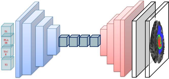


and are L1 norm(absoluteerror)and L2 norm(mean squareerror)respectively isusedasnulltensor.

4. RESULTS
4.1 Implementation Details
The discriminator is PatchGAN [24] consisting of four discriminating blocks followed by zero padding and convolution. Each discriminating blocks composed of Convolution, Batch Normalization, and Leaky ReLU. The convolutionindiscriminatorblockhavingfiltersfromsize60 to512andBatchnormalizationappliedafterthefirstblock. Convolutingisperformedafterdiscriminativeblockfollowed by zero padding, with kernel size 4 and stride 1. The discriminatorframeworkisdisplayedinfigure4.

3.4 Losses
Pytorch[31]andTensorFlow [32]areimplementedusing python 3.6 to implement the proposed algorithm. NVIDIA QuadroP5000GPU64bitRAMcomputingsystemusedfor training, validation and testing of model. The 3D UNet network was initially trained on 295 images with 80:20 rations for trainingand validation.Later the network was trainedon369images,followedbyvalidationon125images andtestedon166unknownimagesfromBraTSdataset.Dice Similarity score, simply known as dice, and Hausdorff distance(95thpercentile)wereevaluated.
4.2 Evaluation Parameters
Augmentation Quality
Thefirstmetricsweconsideredaimatassessingthequality ofthewholeimagesgenerated.Tocomputethesemetricswe first had to crop and normalize the images as follows. We center-croppedthegeneratedimagesto155x194,whichis thesizeofthelargestboundingboxtocontaineachbrainin the BraTS2015 dataset [26]. Then, we applied mean normalization[33]toeachimageforgeneratedorreal,and computedthefollowingthreemetrics:(i)themeansquared error(MSE),(ii)thePeakSignal-to-NoiseRatio(PSNR)[34], and(iii)theStructuralSimilarity(SSIM)[35]:



The generator and discriminator architecture use loss functionassuggestedin[22],writtenasloss; Where x isconcatenationofthreeinputssequenceand G(x) symbolize guess generated by GAN. Dummy loss function generated with all values one, used by generator loss

Where, W and H arethewidthandheightoftheimages I and aretherealandgeneratedimagerespectively ,and are the pixel values of respectively the real and generated images; μ istheaverageofpixelvalues, σ2 isthevarianceof pixel values, and is the covariance of and I pixel values. Both MSE and SSIM have values between 0 and 1. LowervaluesofMSEmeanabetterqualityofthegenerated image.Instead,thegreatertheSNRandSSIMare,thebetteris thequality.

International Research Journal of Engineering and Technology (IRJET) e-ISSN: 2395-0056 Volume: 11 Issue: 01 | Jan 2024 www.irjet.net p-ISSN: 2395-0072 © 2024, IRJET | Impact Factor value: 8.226 | ISO 9001:2008 Certified Journal | Page177
Fig -3:Generatorframeworkwithencoderdecoder architecture,consistingofdownsamplingandupsampling blocksonbothsiderespectively.
Fig -4:Discriminatorframeworkwithfourdiscriminator blocksfollowedbyzeropaddingandconvolution,giving binaryoutput
Model Performance
Evaluationofsegmentationperformancecanbemeasured with the Sorensen–Dice index also known as the Dice Similarity Coefficient (DSC) of simply Dice [35]. DSC representsthedegreeofoverlapbetweenpredictedregion mapandgroundtruthandcalculatedusingequation5as;
Where,notationsTP,FPandFNareTruePositive(TP),False Positive(FP)andFalseNegative(FN)respectively.
Hausdorffdistancemeasuresthemaximumdistanceofone settothenearestpointintheotherset[18],definedas:
Where, sup and inf represents the supremum and the infimumamongtheconsideredsets.Hausdorffdistanceat 95th percentile (HD95) is considered to avoid noisy segmentation and achieve more robust results the evaluationschemeusesthe95thpercentile.
4.3 Augmentation Evaluation
Three MRI sequences given as input to the generator network and the output are passed through the discriminator. i.e. T1ce, T2, and FLAIR input given for generation of T1 scan. Four such experiments were performedtofindallthemissingmodalitygenerationinthe currentscenario.ThepassesoutcomeisevaluatedwithMSE, PSNR, and SSIM for whole brain MRI scan. The MSE and PSNR parameters also been calculated for tumor regions. Table 1 and 2 demonstrates the results for different modalities for values of evaluation parameters for whole imageandtumorregionrespectively.
LowervaluesofMSEmeanabetterqualityofthegenerated image.Instead,thegreatertheSNRandSSIMare,thebetteris thequality.MeanvalueofMSEforthegeneratedmodalities asdisplayedintable1isevidentthattheperformanceofGAN method proposed here is acceptable. All three parameters indicate almost equal behaviour for all modalities. The similarityofT1andT1ceconsideredbeingbettertoT2and FLAIRimages.Thevariationintheresultsmaybeduetoclass imbalance in the input dataset and the inherent radiomic properties of images. The results might be improved on considerationofonlytworelatedmodalitiesinsteadofthree inputchannelconsideredhere.
Thedicesimilaritycoefficient(DSC)alsoreferredasdiceand Hausdorffdistance(95thPercentile)werecalculatedfrom ground truth and the synthesized image to find the applicability of proposed method in the brain tumor segmentation. Table 3 demonstrate the performance of model trained with synthetic MRI scans in training, validation and Testing and table 4 compared with the existingstate-of-theartmethods.
Table-3:PerformanceofmodelusingsyntheticMRIscans,
The mean values of Dice and Hausdorff represents the performance of modified 3D Unet network model of synthetic images of each modality generated using the proposedmethod.Thevaluesintestphasearelow,which canbeconsideredastheactual performanceonunknown dataset. The dice and Hausdorff values are comparatively declining in TC and ET region respectively, due to tissue infiltrationintheinputimages.
Table-4:Performancecomparisonwithstate-of-theart methods.
The proposed method is compared with most adopted Convolutional Network (CNN) architecture basic 3D Unet andmodified3DUnet.Themethodconsideredinthiswork alsousemodified3DUnet.Forcomparison,inthe3DUnet and modified 3D Unet, the input provided without any modification of original dataset. Whereas the proposed
International Research Journal of Engineering and Technology (IRJET) e-ISSN: 2395-0056 Volume: 11 Issue: 01 | Jan 2024 www.irjet.net p-ISSN: 2395-0072 © 2024, IRJET | Impact Factor value: 8.226 | ISO 9001:2008 Certified Journal | Page178
Modality MSE PSNR SSIM T1 0:0041±0:0038 25:2569±3:6512 0:8472±0:0830 T1CE 0:0054±0:0040 23:9242±3:6958 0:8027±0:1003 T2 0:0077±0:0061 22:3719±3:5290 0:7835±0:1141 FLAIR 0:0072±0:0050 22:5524±3:5655 0:7610±0:1175
Table-1:Evaluationofaugmentedwholeimage
Modality MSE PSNR T1 0:0113±0:0099 20:8938±3:6111 T1CE 0:0168±0:0172 19:8441±4:6258
0:0207±0:0167 18:1305±3:5930 FLAIR 0:0221±0:0375 19:0374±4:1582
Table-2:Evaluationoftumorregionfromaugmentedimage
T2
Method Dice Hausdorff(mm) WT TC ET WC TC ET Train 0.91 0.89 0.79 3.66 3.52 30.04 Valid 0.89 0.79 0.75 6.39 14.07 36.00 Test 0.87 0.81 0.78 6.44 24.36 18.95
Method Dice Hausdorff(mm) WT TC ET WC TC ET 3DUNET[6] 0.84 0.83 0.76 7.37 11.15 26.78 Modified3DUnet[23] 0.83 0.82 0.74 5.34 11.34 23.29 ProposedMethod 0.87 0.81 0.78 6.44 24.36 18.95
methodtakesinputoforiginaldatasetalongwithsynthetic data, which makes the data size double fold here. The improvement in the results for dice values0.87,0.81,and 0.78forWT,TC,andETrespectively,duetoincreaseinthe datasize.
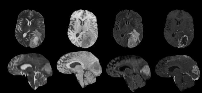
Thequalitativeresultofeachofthemodalityisrepresenting in figure 5 as a sample for one of the subject from the dataset.
5. CONCLUSIONS
Generator-discriminatorframeworkfromsynthesisofMRI imaging modality has been discussed. IN the suggested method,thegeneratorusemodified3DUnetarchitecture.In BraTS2020dataset,fourmodalitiesareincludednamely;T1, T1CE,T2,andFLAIR.Thegeneratornetworkfedwithany three images of different modality and it produce the remainingmodalityimageasanoutput.Thediscriminator usePatchGANframeworkanddistinguishtheoutputfrom theavailablegroundtruth. Three parameters,MSE, PSNR, andSSIMusedtofindthesuitabilityofoutcome.Theimage developedwiththisframeworkshowlowmeanvalueofMSE andhighervaluesofPSNRandSSIM.Theresultssuggestto adopt the synthetic image can be considered as cohort of input image. Further, the generated images of different modalitiesareusedalongwiththeoriginaldatasetforbrain tumorsegmentationapplicationusingModified3DUnet.By makingthedatavolumedouble,withoriginalandsynthetic data,performanceofthemodelimprovedwithdicevalueof 0.87.0.81.and0.78forWT,TC,andETrespectively.
REFERENCES
[1] Sontheimer, H. (2020). Brain Tumors. Diseases of the Nervous System (Second Edition), 207-233.
https://doi.org/10.1016/B978-0-12-821228-8.00009-3
[2] Wesseling, P., & Capper, D. (2018). WHO 2016 Classification of gliomas. Neuropathology and applied neurobiology, 44(2), 139–150.
https://doi.org/10.1111/nan.12432
[3] Blystad,I.,Warntjes,J.B.,Smedby,O.,Landtblom,A.M., Lundberg,P.,&Larsson,E.M.(2012).SyntheticMRIof the brain in a clinical setting. Acta radiologica (Stockholm, Sweden : 1987), 53(10), 1158–1163. https://doi.org/10.1258/ar.2012.120195
[4] Illimoottil, M., & Ginat, D. (2023). Recent Advances in DeepLearningandMedicalImagingforHeadandNeck Cancer Treatment: MRI, CT, and PET Scans. Cancers, 15(13).https://doi.org/10.3390/cancers15133267
[5] Çiçek, Ö., Abdulkadir, A., Lienkamp, S.S., Brox, T., Ronneberger, O. (2016). 3D U-Net: Learning Dense Volumetric Segmentation from Sparse Annotation., Springer LNCS,vol9901.https://doi.org/10.1007/9783-319-46723-8_49
[6] Myronenko, A. (2019). 3D MRI Brain Tumor Segmentation Using Autoencoder Regularization. BrainLes 2018 Springer LNCS, vol 11384., 311 – 320, https://doi.org/10.1007/978-3-030-11726-9_28
[7] Isensee, F., Jaeger, P. F., Kohl, S. A. A., Petersen, J., & Maier-Hein, K. H. (2021). nnU-Net: a self-configuring method for deep learning-based biomedical image segmentation. Nature methods, 18(2), 203–211. https://doi.org/10.1038/s41592-020-01008-z.
[8] McKinley,R.,Meier, R.,Wiest,R.(2019),Ensemblesof Densely Connected CNNs with Label Uncertainty for Brain Tumor Segmentation. BrainLes2018, Springer LNCS, 11384, vol. 456–465, https://doi.org/10.1007/ 978-3-030-11726-9_40
[9] Zhou, C., Chen, S., Ding, C., Tao, D. (2019), Learning ContextualandAttentiveInformationforBrainTumor Segmentation. BrainLes2018, SpringerLNCS, vol., 11384, 497–507, https://doi.org/10.1007/978-3-030-117269_44.
[10] Goodfellow, I., Pouget-Abadie, J., Mirza, M., Xu, B., Warde-Farley, D., Ozair, S., Courville, A., Bengio, Y, (2020),Generativeadversarialnets. Commun. ACM, 63, 11,139–144.https://doi.org/10.1145/3422622
[11] Isola,P.,Zhu,J.Y.,Zhou,T.,Efros,A.A,(2017),Imageto-Image Translation with Conditional Adversarial Networks, IEEE Conference on Computer Vision and Pattern Recognition (CVPR 2017), Honolulu,USA,59675976.https://doi.org/10.1109/CVPR.2017.632
[12] Karras, T., Aila, T., Laine, S., Lehtinen, J.: Progressive growing of GANs for improved quality, stability, and variation. ICLR (2018) ArXiv preprint https://arxiv.org/abs/1710.10196
[13] Gatys, L.A., Ecker, A.S., Bethge, M.,(2016),Image style transfer using convolutional neural
International Research Journal of Engineering and Technology (IRJET) e-ISSN: 2395-0056 Volume: 11 Issue: 01 | Jan 2024 www.irjet.net p-ISSN: 2395-0072 © 2024, IRJET | Impact Factor value: 8.226 | ISO 9001:2008 Certified Journal | Page179
Fig -5:MRImodalitiesgeneratedfromtheproposed framework,displayedintransverseandcoronalplane.
networks. IEEE Conference on Computer Vision and Pattern Recognition (CVPR),LasVegas,NV,USA,2016, pp. 2414-2423, https://doi.org/10.1109/CVPR.2016. 265
[14] SatoM.,HottaK.,ImanishiA.,MatsudaM.andTerai K. (2018). Segmentation of Cell Membrane and Nucleus by Improving Pix2pix. 11th International Joint Conference on Biomedical EngineeringSystems and Technologies (BIOSTEC 2018,)BIOSIGNALS, Vol. 4: 216-220. https://doi.org/10.5220/00066483021 60220.
[15] Zhu,J.,Park,T.,Isola,P.,andEfros,A.,(2017), Unpaired Image-to-Image Translation Using Cycle-Consistent AdversarialNetworks," IEEEInternationalConferenceon Computer Vision (ICCV 2017), Venice, Italy, pp. 22422251,https://doi.org/10.1109/ICCV.2017.244
[16] Han,Z.,Wei,B.,Mercado,A.,Leung,S.,Li,S.:Spine-GAN: Semanticsegmenta-tionofmultiplespinalstructures. Medical image analysis 50,23–35(2018)
[17] Dong,X.,Lei,Y.,Wang,T.,Thomas,M.,Tang,L.,Curran, W.J., Liu, T., Yang, X.: Automatic multiorgan segmentation in thorax CT images using U-net-GAN. Medical physics 46(5),2157–2168(2019)
[18] He,K.,Zhang,X.,Ren,S.,Sun,J.:Deepresiduallearning for image recognition. In: Proceedings of the IEEE conference on computer vision and pattern recognition. pp.770–778(2016)
[19] Nema,S.,Dudhane,A.,Murala,S.,Naidu,S.:RescueNet: An unpaired GAN for brain tumor segmentation. Biomedical Signal Processing and Control 55, 101641 (2020)
[20] Yi,X.,Walia,E.,Babyn,P.,(2019),Generativeadversarial network in medical imaging: A review, Medical Image Analysis, Volume 58, 101552, ISSN 1361-8415, https://doi.org/10.1016/j.media.2019.101552
[21] Alogna,E.,Giacomello,E.,andLoiacono,D.,(2020)Brain Magnetic Resonance Imaging Generation using Generative Adversarial Networks, IEEE Symposium Series on Computational Intelligence (SSCI), Canberra, ACT, Australia, 2020, pp. 2528-2535, https://doi.org/ 10.1109/SSCI47803.2020.9308244
[22] Cirillo,M.,Abramian,D.,Eklund,A.(2021).Vox2Vox:3DGAN for Brain Tumour Segmentation. BrainLes2020, SpringerLNCS.vol12658.https://doi.org/10.1007/9783-030-72084-1_25.
[23] Parmar, B., Parikh, M. (2021). Brain Tumor SegmentationandSurvivalPredictionUsingPatchBased Modified3DU-Net.BrainLes2020. SpringerLNCS, vol 12659.https://doi.org/10.1007/978-3-030-72087-2_35
[24] Li,C.,Wand,M.(2016).PrecomputedReal-TimeTexture Synthesis with Markovian Generative Adversarial Networks. Computer Vision, ECCV 2016. SpringerLNCS. vol 9907. https://doi.org/10.1007/978-3-319-464879_43
[25] Menze, B.H., Jakab, A., Bauer, S., Kalpathy-Cramer, J., Farahani,K.,Kirby,J.,Burren,Y.,Porz,N.,Slotboom,J., Wiest, R., et al.: The multimodal brain tumor image segmentationbenchmark(BRATS).IEEEtransactionson medical imaging,34(10),1993–2024(2014)
[26] S.Bakas,H.Akbari,A.Sotiras,M.Bilello,M.Rozycki,J. Kirby, et al., "Segmentation Labels and Radiomic FeaturesforthePre-operativeScansoftheTCGA-GBM collection", The Cancer Imaging Archive, 2017. https://doi.org/10.7937/K9/TCIA.2017.KLXWJJ1Q
[27] S.Bakas,H.Akbari,A.Sotiras,M.Bilello,M.Rozycki,J. Kirby, et al.,(opens in a new window) "Segmentation Labels and Radiomic Features for the Pre-operative ScansoftheTCGA-LGGcollection", The Cancer Imaging Archive, 2017.https://doi.org/10.7937/K9/TCIA.2017. GJQ7R0EF
[28] Bakas,S.,Akbari,H.,Sotiras,A.,Bilello,M.,Rozycki,M., Kirby, J.S., Freymann, J.B., Farahani, K., Davatzikos, C.: Advancing the cancer genome atlas glioma MRI collections with expert segmentation labels and radiomicfeatures. Scientific data 4,170117(2017)
[29] Bakas, S., Reyes, M., Jakab, A., Bauer, S., Rempfler, M., Crimi,A.,Shinohara,R.T.,Berger,C.,Ha,S.M.,Rozycki,M., etal.:Identifyingthebestmachinelearningalgorithms forbraintumorsegmentation,progressionassessment, andover-allsurvivalpredictionintheBRATSchallenge. arXiv preprint arXiv:1811.02629(2018)
[30] Tustison,N.J.,Avants,B.B.,Cook,P.A.,Zheng,Y.,Egan, A., Yushkevich, P. A., & Gee, J. C. (2010). N4ITK: improved N3 bias correction. IEEE transactions on medical imaging, 29(6),1310–1320.https://doi.org/10. 1109/TMI.2010.2046908
[31] Paszke, Adam et al. “Automatic differentiation in PyTorch,” NIPS-W 2017
[32] 26.D.Jhaetal.,"ResUNet++:AnAdvancedArchitecture for Medical Image Segmentation," 2019 IEEE InternationalSymposiumonMultimedia(ISM),SanDiego, CA, USA, 2019,pp.225-2255,https://doi.org/10.1109/ ISM46123.2019.00049.
[33] A.Ng,“GradientdescentinpracticeI-featurescaling,” Coursera. [Online]. Available: https://www.coursera .org/learn/machine-learning/lecture/xx3Da/gradientdescent-in-practice-i-feature-scaling
International Research Journal of Engineering and Technology (IRJET) e-ISSN: 2395-0056 Volume: 11 Issue: 01 | Jan 2024 www.irjet.net p-ISSN: 2395-0072 © 2024, IRJET | Impact Factor value: 8.226 | ISO 9001:2008 Certified Journal | Page180
[34] “Psnr,”Mathworks.[Online].Available:https://it.Math works.com/help/vision/ref/psnr.html
[35] Z.Wang,A.Bovik,H.Sheikh,andE.Simoncelli,“Image qualityassessment:From errorvisibility tostructural similarity,” Image Processing, IEEE Transactions on,vol. 13, pp. 600 – 612, 05 2004. [Online]. Available: https://ieeexplore.ieee.org/document/1284395
International Research Journal of Engineering and Technology (IRJET) e-ISSN: 2395-0056 Volume: 11 Issue: 01 | Jan 2024 www.irjet.net p-ISSN: 2395-0072 © 2024, IRJET | Impact Factor value: 8.226 | ISO 9001:2008 Certified Journal | Page181