
International Research Journal of Engineering and Technology (IRJET) e-ISSN:2395-0056
Volume: 11 Issue: 12 | Dec 2024 www.irjet.net p-ISSN:2395-0072

International Research Journal of Engineering and Technology (IRJET) e-ISSN:2395-0056
Volume: 11 Issue: 12 | Dec 2024 www.irjet.net p-ISSN:2395-0072
Mr. Basant Rawot1, Dr. Heli Shah2 ,
1Parul Institute of Technology, Parul University, India
2HOD of The Department, Parul Institute of Technology, Parul University, India
Abstract:
Inthepastyear,thenovelcoronavirushasaffectedover1millionindividualsandresultedinmorethan50,000deaths.COVID19infectionhasthepotentialtoprogressintopneumonia,aconditionthatcanbeidentifiedthroughachestx-rayandshould be managed appropriately. This study presents a novel approach for automatically detecting COVID-19 infection using chest radiographs. The dataset compiled for this study comprises 480 X-rays obtained from patients diagnosed with coronavirus and from healthy patients. Due to the limited availability of COVID-19 patient images, we will utilize transfer learning principles for this undertaking. We employ various architectures of convolutional neural networks (CNNs) that have been trainedonImageNet.WethenmodifythesenetworkstofunctionasfeatureextractorsspecificallyforX-rayimages.CNNsare subsequentlyfusedwithintegratedmachinelearningmethodologies,includingSupportvectormachines(SVM)andLSTM.The results indicate 9375% accuracy with an F1 score of 9386%. Hence, the suggested method exhibits efficacy in identifying pneumonia
Keywords: Convolutionneuralnetworks,ImageNet,Multilayerperceptron,Supportvectormachine,X-rayimages
1. Introduction
Alungairsacinfectioncausespneumonia.Thelungstructureismadeupofpulmonaryalveoli,bronchus,andlobesconnected to the main veins. Every part has to work correctly to guarantee an effective exchange of carbon dioxide and oxygen. Along with fluids in their air sacs, pneumonia patients will have alveolar inflammation. There are only a few of the medical examination technologies developed such as CT scans, X-ray and ultrasound as a result of medical technology progress. PneumoniadiagnosisisregardedasbestachievedwithCTscans.VGGandResNetnetworksareutilizedtoaccuratelyclassify lungultrasoundimagesofpneumoniaaccordingtodifferentclinicalphasesusingself-generatedLUSdatasets.
The introduction of deep learning in recent years has resulted in a paradigm shift in medical image analysis. Convolutional neural networks (CNNs) are highly effective at tasks like classifying images, segmenting parts of images, and identifying features within images. Because of these capabilities, CNNs are widely used in creating automated systems for medical imaging.CNNshaveshownpotentialindetectingpneumonia,offeringthepossibilityofquicklyandindependentlyidentifying lunginfections.Thisadvancementcouldsignificantlytransformpatientcare.
ThisstudyfocusesonaccuratelydetectingpneumoniainthelungsusingchestX-rays,whichcanbeusedintherealworldby medicalpractitionerstotreatpneumonia.Theproblem withtraditionalmethodsinvolveslotsoftime andresourceintensive inthediagnosisofpneumonia.Hence,theuseofartificialtechnologysuchasconvolutionneuralnetworkssolvestheproblem bysimplifyingtheprocessofdiagnosisofpneumonia.Themainobjectivesofthisstudyare:
a)Integratingdeeplearningtechniquesforpneumoniadetection.
b)UsingfeatureextractionandImageclassificationfordiagnosis.
2. Literature Review
Theresearchpaper"PneumoniaDetectionUsingCNN-basedFeatureExtraction"focusesoncreatingaConvolutionalsystemto effectively diagnose pneumonia in X-rays. It highlighted the global impact of pneumonia as a leading cause of death and the difficulties in diagnosing it accurately. The authors then describe the proposed CNN-based approach for detection and categorizationofimages.TheCNNmodelwastrainedonadatasetof5856chestX-rayimages,consistingof2,700normaland
International Research Journal of Engineering and Technology (IRJET) e-ISSN:2395-0056
Volume: 11 Issue: 12 | Dec 2024 www.irjet.net p-ISSN:2395-0072
2,630 pneumonia images. The trained model achieved an accuracy of 96.07% on the testing dataset. The authors compared theirproposedmodelwithotherexistingmodelsandreportedabetterperformanceintermsofaccuracy,precision,recall,and F1-score.Finally,thepaperconcludesthattheproposedCNN-basedmodelcanbeusefulforaccurateandtimelydiagnosisof pneumonia,whichcanhelpinimprovingpatientoutcomesandreducingmortalityrates[1].RajpurkardevelopedtheCheXNet algorithm,whichcanautomaticallyidentifypneumoniainchestX-rays.This121-layerDenseNet,trainedontheChestX-ray14 dataset,replacesthefinalfullyconnectedlayerwithanoutputlayerandachievesanF1scoreof0.435.Carmany[3]createda diagnostic tool for treatable diseases that used chest X-rays to show discriminatory power. Islam [4] proposed a framework forautomaticallydetectingpneumoniainradiographs.
Furthermore, Rajaraman [5] developed a truncated VGG16-based artificial neural network that can diagnose pneumonia in pediatric X-rays. It has the deepest convolutional layers with gaps and dense layers. Even though these algorithms have improved deep networks, Rajpurkar and colleagues have sped up their networks by using a single output rather than the entire link layer; nevertheless, this operation did not produce the required accuracy. Rajaraman employed global average poolingtoboostconvolutionalneuralnetworks'performance,buthelosttrackofthetarget'slocation.Asaresult,researchis currently being done on high-precision networks with excellent detection performance. Lee proposed DetNet in 2018 [6]. It was created with object detection in mind and uses fewer layers to produce better detection results. In order to detect and locate pneumonia, Shirajitdinov [7] suggested a framework that combines RetinaNet and Mask R-CNN. A dataset of 26,684 photosfromtheKagglePneumoniaDetectionChallenge wasusedtovalidatethisnetwork.Itiscurrentlycommonpracticeto employhisbifurcatedstructuralmodel[8].Theauthorsusedtransferlearning,atechniquewherepre-trainedmodelsarefinetuned on a specific task, to train the EfficientNet-B0 model on the ChestX-ray14 dataset. They froze the lower layers of the model,whichcapturegenericfeaturesfromimages,andtrainedtheupperlayers,whichcapturedisease-specificfeatures,on the pneumonia detection task. To further improve the model's performance, the authors also applied data augmentation techniques, such as horizontal flipping, rotation, and zooming, to increase the diversity of the training data and prevent overfitting.
The methodology used in Convolutional Sparse Support Estimator-Based COVID-19 Recognition from X-ray images involves the pre-processing of X-ray images, where the images are resized, normalized, and sharpened to enhance the features. The authors then use a CSSE model to extract important features from the pre-processed images. The CSSE model uses a sparse regularization term to select only relevant features, thus reducing the dimensionality of the feature set. The authors also introduceatransferlearningapproachwheretheCSSEmodelistrainedonpre-trainedmodelssuchasResNet-50,DenseNet121, and VGG-16 [14]. Using a deep learning method, X-ray pictures were used to diagnose pneumonia. They compare performancebetweentwowell-knowndeeplearningmodels VGG-16andResNet-50 onthetaskofpneumoniadetection.A data of 5,856 chest X-ray images (3,781 normal, 1,350 images of bacterial pneumonia and 725 images of viral pneumonia), authors use transfer learning to refine these models [15]. A method based on deep learning for automatically detecting pneumonia from chest X-raypictures.Utilizing a datasetof 1,266 chest X-rayimages(242normal,681 bacterial pneumonia, and343viralpneumoniaimages),theyrefinedapre-trainedVGG-16model.Toincreasetheirdatasetsizeandimprovemodel generalizability,theauthorsalsoemploydataaugmentationtechniques[16].
Mradetal.proposedamachineintelligenceapproachforscreeningpatientsbasedontheirX-rayimages.Theauthorsaddress theissueofimbalancedclasses,wherethenumberofCOVID-19casesissignificantlylowerthanthenumberofnon-COVID-19 cases. They use a combination of data augmentation and oversampling techniques to balance the classes and then train a neural network for classification [17]. Vo-Dinh et al. also focused on the identification of COVID-19 cases from chest X-ray images. The authors propose a high-resolution network (HRNet) for this task and employ transfer learning for tuning of model. [18]. WangandChakrabortyproposeda recurrentneural network (RNN)-basedapproachtoevaluatethemalignancy oflungnodulesfromCTimages.Theauthorsuseamulti-stageapproachwheretheyfirstsegmentthelungnodulesandthen usea3DRNNtoclassifythemasmalignantorbenign[19].
"CIDC-Net:Chest-XRayImagebasedDiseaseClassificationNetworkusingDeepLearning"byMeghanaetal.presentsamethod to classify X-ray images into different disease categories [20]. "COVID-19 Classification using DCNNs and Exploration Correlation using Canonical Correlation Analysis" by Jullapak and Yampaka presents a deep learning-based approach for classifyingCOVID-19imagesusingdeepconvolutional neural networks(DCNNs) [21].The paper"COVID-SEGNET:Diagnosis of COVID-19 Cases on Radiological Images using Mask R-CNN" by Kundu et al. presents an approach using radiological pictures. The proposed approach, called COVID-SEGNET, depends on Mask R-CNN architecture and is trained to identify the
International Research Journal of Engineering and Technology (IRJET) e-ISSN:2395-0056
Volume: 11 Issue: 12 | Dec 2024 www.irjet.net p-ISSN:2395-0072
areas in images that indicate COVID [22]. In "X-ray Images Detection of COVID-19 Based on Deepwise Separable DenseNet" Wang et al. describe a deep learning-based method for COVID-19 image detection. The authors carried out tests as well to assess how well the suggested method stood up to various kinds of noise and perturbations [23]. Moreover, there’s CNN algorithm for corona virus detection inchest X-rays [24].Anotherapproach involvesusing 2D convolutional neural network (CNN)forrespiratorydiseasedetectionfromrecordedlungsounds[25].TheyalsocreatedanetworkcalledMHA-CoroCapsule tocategorizeCOVID-19chestX-rayimages,whichutilizesmulti-headattentionrouting.
3.1. Proposed framework
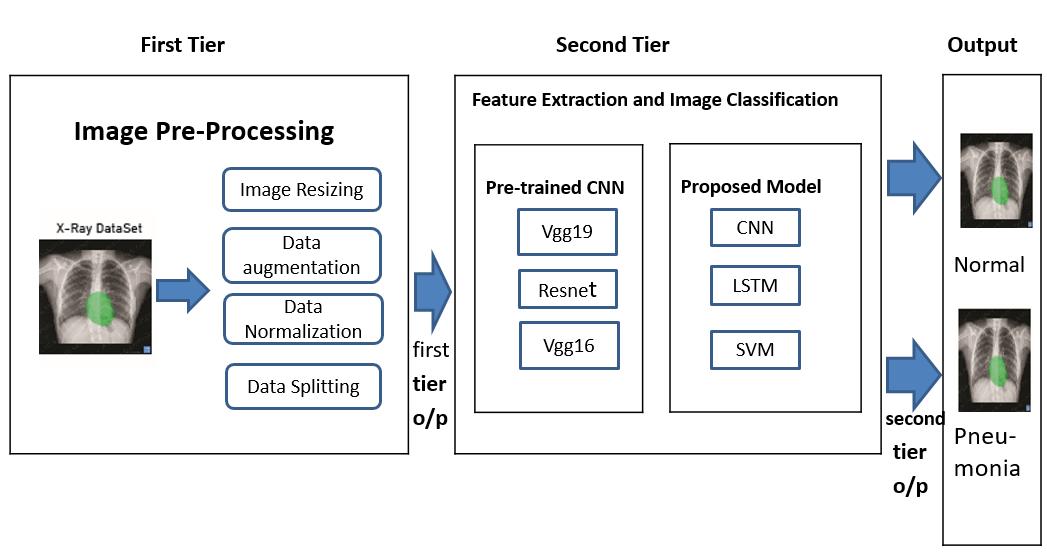
TheCNNarchitectureusedintheresearchconsistsofthreetiers,eachwithitsuniquefunction.Inthefirsttier,theimagepreprocessing is carried out, which includes image resizing, data augmentation, data normalization, and data splitting. The preprocessingstepmakessurethatalltheimagesareuniforminsize,andthetrainingdatasetincludesawiderangeofexamples tohandlereal-worlddifferenceseffectively.TheimageresizingisacrucialstepbeforepassingthemtotheCNNmodel.
Inthesecondtier,twosub-partscarryoutfeatureextractionandimageclassification.Thefirstsub-partusespre-trainedCNN modelssuchasVGG19,Resnet,andVGG16,which haveexcellentresultsinclassification.Thesemodelshavebeentrainedon large datasets such as the ImageNet dataset, which includes more than 14 million images categorized into 1000 different classes. By using pre-trained models, CNN can leverage the learned features to classify the images accurately. In the second sub-part,a proposed model usingCNN,LSTM, and SVM isused for feature extractionand imageclassification. Theproposed model is trained on the pre-processed images, and the features are extracted using the CNN layers. The LSTM layer is employed to understand the order of images in a sequence. Finally, the SVM classifier is applied to categorize the images as eithernormalorpneumonia.
In the final tier, the output section shows whether the input image is normal or pneumonia. The model output is compared with the actual label of the image, and the accuracy is then calculated. The model’s accuracy is an essential metric. If the accuracy is high, it means that the model is performing well and can be used for real-world applications such as pneumonia detectioninmedicalimaging.
Convolutional Neural Networks (CNNs) are a special kind of neural network created to handle grid-like data, like images or audiospectrums.Theyconsistoflayersthatgraduallylearn moreintricatefeatures fromtheinput.Themaincomponentsof CNNsareconvolutionallayers,whichusefilterstoprocesstheinput,producingfeaturemapsthatemphasizedifferentpartsof thedata.
International Research Journal of Engineering and Technology (IRJET) e-ISSN:2395-0056
Volume: 11 Issue: 12 | Dec 2024 www.irjet.net p-ISSN:2395-0072
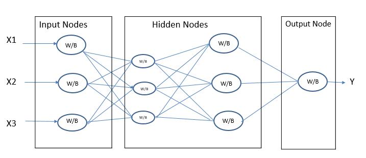
AstandardCNNdesignincludesaninitiallayerforinput,followedbymultipleconvolutionallayersthatincorporateReLU,and poolinglayersthatdownsamplethefeaturemaps.Theoutputoftheconvolutionallayersistypicallyflattenedandfedintoone ormorefullyconnectedlayers,whichlearntoclassifytheinputdataintodifferentcategories. Inadditiontothesecorelayers, modernCNN architecturesoftenincludeadditional layerssuchasbatchnormalization,dropout,andskipconnections,which helptoimprovetrainingstabilityandgeneralizationperformance.
Overall, the architecture of a CNN is carefully designed to learn hierarchical representations of the input data, starting with low-level features such as edges and corners, and building up to more abstract features that capture higher-level concepts such as object categories. This makes CNNs particularly effective for tasks such as image classification, object detection, and segmentation,wherethegoalistoaccuratelyclassifyorlocalizeobjectswithinimages.
3.2.1 VGG architecture
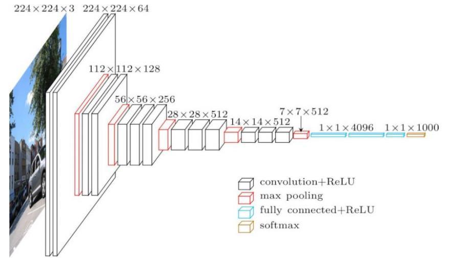
The VGG16 architecture of a convolutional neural network (CNN) includes 16 layers overall, with the initial 13 layers as convolutionallayersandthelastthreeasfullyconnectedlayers.Atthemicrolevel,eachlayerinVGG16hasaspecificpurpose and learns its own set of parameters during training The initial layer is a convolutional layer that processes the raw input image and uses filters to detect basic features like edges and corners. This process is repeated in subsequent layers, where each layer learns to identify increasingly more intricate and detailed features from the previous layer's output. After several layersofconvolutionalandmaxpoolingoperations,thefeaturemapsgeneratedarereshapedintoasinglevectorandthenfed intothedenselayersforclassification.Thesecomponentslearntocombineextractedfeaturesintohigh-levelrepresentations thatareusefulfordiscriminatingbetweendifferentclasses.
Thenetwork'sperformancecanbeoptimizedatthemicrolevelbyadjustingtheparticularparametersforeachlayer,suchas howtoclassifypneumonialungultrasonographyimages. Forinstance,thecomplexityofthelearnedfeaturescanbeadjusted by changing the number of neurons in the dense layers, while the convolutional layers' filters can be modified to capture differentlevelsofdetailintheinputimage.Animageof224x224goestotheinputforthefirstconvolutionallayer(cov1).The image undergoes a series of convolutional layers, where 3x3 filters are applied the smallest size effective for capturing various aspects in the image. Spatial padding in the convolutional layer maintains spatial resolution after processing, with a paddingof1pixelfor3x3convolutionlayers.Theconvolutionstrideremainsfixedat1pixel.Followingtheconvolution,there arefivemax-poolinglayersforspatialpooling.Convolutionallayerstackissucceededbythreefullyconnected(FC)layers.The firsttwohave4096channelseach,whilethethirdhas1000channels(oneforeachclass)for1000-wayILSVRCclassification. TheSoftMaxlayeractsasthefinallayer.
International Research Journal of Engineering and Technology (IRJET) e-ISSN:2395-0056
Volume: 11 Issue: 12 | Dec 2024 www.irjet.net p-ISSN:2395-0072
3.2.2.
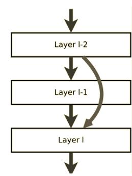
ResidualNetwork,isadeeplearningarchitecturethatwasdesignedtohelpovercomethedegradationprobleminverydeep neural networks. In pneumonia detection using CNN, ResNet plays crucial role in accurately categorizing chest X-ray images showingpneumonia Itskeyjobistoenabletheneuralnetworktolearnfromanyleftoverinformationintheinputimage.This is achieved by introducing skip connections or shortcuts that skip over one or more layers in the neural network. Such shortcutsprovideanalternatepathfortheinformationtopassthroughthenetwork,helpingiteffectivelylearnanyremaining information.Theresidualconnectionsareformedbyaddingtheinputfeaturemapstotheoutputfeaturemapsofoneormore convolutional layers in the network. By doing so, the output feature maps will have additional information from the input feature maps, which helps the network to learn the residual information more efficiently. In other words, these shortcuts enable the network to grasp the distinction between the input and output feature maps, which is crucial for accurately classifyingpneumoniainchestX-rayimages.
Inpneumoniadetection,ResNetiseffectiveinaccuratelyclassifyingthedifferentclinicalstagesofpneumonia.Thisisbecause ResNet can capture and learn from the subtle differences in lung ultrasound images that may be missed by other neural network architectures. Additionally, the use of residual connections in ResNet helps to prevent the degradation problem commonly faced by very deep neural networks, allowing for better accuracy and performance. ResNet is a type of artificial neural network (ANN) inspired by the structure of pyramidal cells in the brain's cortex. ResNet models typically include double-ortriple-layerskipswithnonlinearitieslikeReLUandbatchnormalizationinbetween.Theseskipsallowthenetwork to learn more effectively. HighwayNets are ResNet models where the skip weights can be adjusted using an extra weight matrix.DenseNetsaremodelswithmultipleparallelskips.Incontrast,anon-residualnetworkisconsideredaregularnetwork whencomparedtoaresidualneuralnetwork.Aresidualneuralnetwork'scanonicalformisdepictedinthefigure.Activation fromlayerℓ−2isbypassedinfavoroflayerℓ−1.
International Research Journal of Engineering and Technology (IRJET) e-ISSN:2395-0056
Volume: 11 Issue: 12 | Dec 2024 www.irjet.net p-ISSN:2395-0072
LSTM is a type of Recurrent Neural Network (RNN) that is particularly effective for sequence prediction problems. It is designed to model temporal sequences and their long-range dependencies more accurately than conventional RNNs. LSTMs havememorycellsthatcanstoreinformationinmemoryforextendedperiods.ThisfeaturehelpsLSTMsavoidthevanishing gradient problem that standard RNNs suffer from, allowing them to learn long-term dependencies. LSTMs are composed of layersofLSTMunits,whereeachunitcontainsthememorycellandthethreegates.Theyprocessdatasequencesonestepata time,maintainingthestatethroughthesequence.
SVM is a type of supervised learning method used for tasks like classification and regression. It finds the best hyperplane to dividedataintodifferentclassesinahigh-dimensionalspace.Ahyperplaneislikeadecisionboundarythatseparatesclasses, with the optimal one maximizing the margin between them. Data points closest to the hyperplane affect its position and direction,andonlythesepointsareusedtocreateit.SVMcanhandlenon-linearclassificationusingakerneltrick,whichmaps inputfeaturesintohigher-dimensionalspaces.
ThelibrariesusedinPneumoniaDetectionBasedonConvolutionNeuralNetworkresearchareallimportantforvarioustasks related to the processing and analysis of X-ray images. Keras is a library used for data preprocessing which is necessary for preparingthedatabeforeitcanbefedintotheneuralnetwork.Insuchcase,itisusedtoimporttheX-rayimagesandprovide utilities for working with image data. The sklearn is for how well the machine learning model detects pneumonia in X-ray images It offers several metrics like accuracy, precision, and recall to measure the model's performance TensorFlow is a widely used machine-learning library that is specifically designed for numerical computation. It is used in this research to buildandtraintheconvolutionneuralnetworkmodel,whichisthebackboneofthepneumoniadetectionsystem. Seabornand matplotlibarebothvisualizationlibrariesthatareusedtocreateinformativestatisticalgraphics.Inthiscase,theyare usedto generatevisualrepresentationsofthemodel'sperformance,makingiteasiertointerpretandunderstandtheresults. Finally, NumPyisalibraryusedfornumericalcomputingandprovidesobjectsformulti-dimensionalarrays.Itisusedinthisresearch toperformarithmeticoperationsandhandlecomplexnumberswhileprocessingtheX-rayimages.
In the research on Pneumonia Detection using Convolutional Neural Networks, they assessed the combined CNN-LSTM-SVM model'sperformancewithvariousmetrics.The evaluationhadtwophases:trainingandtesting.Duringtraining,theytracked metricslikeaccuracy,loss,precision,recall,andF1scoresacross50epochstogaugethemodel'sperformance.Theyselected themodelwiththebestperformanceon testingset.Aftertraining,theytestedthemodelonanindependenttestsettocheck how well it generalizes. They used metrics such as accuracy, precision, recall, and F1 score for evaluation. Additionally, they
International Research Journal of Engineering and Technology (IRJET) e-ISSN:2395-0056
Volume: 11 Issue: 12 | Dec 2024 www.irjet.net p-ISSN:2395-0072
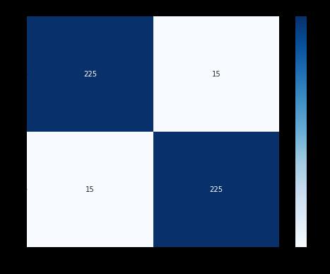
analyzed the model's performance using confusion matrices, which detail true positives, true negatives, false positives, and falsenegativesforeachclasstoassessclassificationaccuracy.
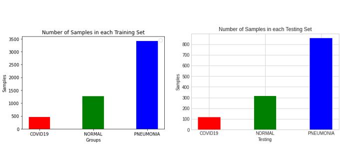
The results of the evaluation and analysis showed that the combined CNN-LSTM-SVM model was able to accurately detect pneumonia in chest X-rays with a high level of precision and recall. The analysis also showed that the model was able to accurately differentiate between normal and abnormal X-rays. In this research, the dataset of X-ray images for pneumonia detectionwasobtainedfromKaggleandthenuploadedtoGoogleDrive.Thenecessarypathwasprovidedtomountthedrive foreasyaccesstothedataset.Thedatasetwasdividedintotwocategories,namelytestandtraining.Eachcategoryfurther had three sub-categories - normal, COVID, and pneumonia datasets. The number of images in each sub-category was calculated usingthepanda’slibrary,whichhelpedinunderstandingthedistributionofdata.Togetabetterunderstandingofthedataset, it was visualized using graphs and charts. The testing and training datasets were plotted into bar charts to visualize the distributionofimages.Themodelarchitecturewasalsovisualizedusingmax-pooling.Thehistoryofthemodelwasvisualized and plotted in a graph to understand the performance of the model during the training phase. This helped in understanding theaccuracyofthemodel anditsabilitytoclassifytheX-rayimagesaccurately.Overall,thesevisualizationsandcalculations werecrucialforassessingthemodel'sperformanceandimplementingimprovementstoattainsuperiorresults.
The findings from the Pneumonia Detection Based on CNN research, aimed at identifying pneumonia in X-ray images, were promising. For the metrics like accuracy, precision, recall, and F1-score were implemented. The proposed model demonstratedanoverallaccuracyof93.75%,whichindicatesthatthemodelisreliableandeffectiveindetectingpneumonia. Furthermore,themodelachievedahighprecisionof93.99%andarecallof93.75%.Thismeansthatthemodelcanaccurately detect pneumonia while minimizing false positives. The F1-score, which reflects the model's accuracy, was calculated at 93.86%,indicatingitsoverallperformanceintermsofprecisionandrecall.
International Research Journal of Engineering and Technology (IRJET) e-ISSN:2395-0056
Volume: 11 Issue: 12 | Dec 2024 www.irjet.net p-ISSN:2395-0072
The table shows the metrics value obtained from our model. Moreover, the comparison of different models was done, and it wasobservedthattheproposedmodel outperformedthemodelssuchasVGG16,VGG19,andResNet50. Itachieveda higher accuracyrateandreducedthenumberoffalsepositivecases.
The confusion matrix was also used to analyze the performance of the model. The confusion matrix showed that out of 480 test images, the model correctly classified 450 images, and misclassified 30 images. These results indicate the model is effectivefordetectingpneumoniainchestX-ray.
5. Conclusion
Inconclusion,thecombinationof CNNmodelslikeResnet, VGG16andVGG19 withLSTMandSVM hasoutstanding resultsin spottingpneumoniainX-ray.Themodelwedevelopedprovided93.75%accuracy,93.99%precision,93.75%recallwithanF1 scoreof93.86%.Theevaluationmetricsshowthatthemodelhasahighlevelofaccuracyindetectingpneumonia,whichcan be utilized in the real world by medical practitioners to treat pneumonia. The study also demonstrates the importance of imagepreprocessing,featureextraction,andclassificationinachievingaccurateresults.Overall,theresearchsuggeststhatthe proposedmodel canbea valuabletoolinassistingmedical practitionersinpneumonia diagnosis andtreatment.WhileCNNs have been effective, future exploration of other network architectures like recurrent neural networks and attention-based modelscouldfurtherenhanceaccuracy.
References
1. D. Varshni, K. Thakral, L. Agarwal, R. Nijhawan and A. Mittal, "Pneumonia Detection Using CNN based Feature Extraction,"2019IEEEInternationalConferenceonElectrical,ComputerandCommunicationTechnologies(ICECCT), Coimbatore,India,2019,pp.1-7,doi:10.1109/ICECCT.2019.8869364.
2. P. Rajpurkar, J. Irvin, K. Zhu, B. Yang, H. Mehta, T. Duan, D. Ding, A. Bagul, C. Langlotz, K. Shpanskaya, M. P. Lungren, and A. Y. Ng, ``CheXNet: Radiologist-level pneumonia detection on chest X-rays with deep learning,'' 2017, arXiv:1711.05225.[Online].Available:https://arxiv.org/abs/1711.05225
International Research Journal of Engineering and Technology (IRJET) e-ISSN:2395-0056
Volume: 11 Issue: 12 | Dec 2024 www.irjet.net p-ISSN:2395-0072
3. D. S. Kermany, M. Goldbaum, W. Cai, C. C. Valentim, H. Liang, S. L. Baxter, A. McKeown, G. Yang, X. Wu, F. Yan, and J. Dong, ``Identifying medical diagnoses and treatable diseases by image-based deep learning,'' Cell, vol. 172, no.5, pp. 1122_1131.e9,Feb.2018.
4. S.R.Islam,S. P. Maity,A. K. Ray,andM.Mandal,``Automaticdetection of pneumonia oncompressedsensingimages usingdeeplearning,''inProc.IEEECan.Conf.Elect.Comput.Eng.(CCECE),May2019,pp.1_4.
5. S.Rajaraman,S.Candemir,G.Thoma,S.Antani,``Visualizingandexplainingdeeplearningpredictionsforpneumonia detectioninpediatricchestradiographs,''Proc.SPIE,vol.10950,Mar.2019,Art.no.109500S.
6. Z.Li,C.Peng,G. Yu, X.Zhang,Y.Deng,and J. Sun, ``DetNet:Design backbone for object detection,''inProc.Eur.Conf. Comput.Vis.(ECCV),2018,pp.334_350.
7. I. Sirazitdinov, M. Kholiavchenko, T. Mustafaev, Y. Yixuan, R. Kuleev, and B. Ibragimov, ``Deep neural network ensemble for pneumonia localization from a large-scale chest X-ray database,'' Comput. Electr. Eng., vol. 78, pp. 388_399,Sep.2019.
8. S. Yao, Y. Chen, X. Tian and R. Jiang, "GeminiNet: Combine Fully Convolution Network With Structure of Receptive FieldsforObjectDetection,"inIEEEAccess,vol.8,pp.60305-60313,2020,doi:10.1109/ACCESS.2020.2982939.
9. L.Račić,T.Popović,S.čakićandS.Šandi,"PneumoniaDetectionUsingDeepLearningBasedonConvolutionalNeural Network," 2021 25th International Conference on Information Technology (IT), Zabljak, Montenegro, 2021, pp. 1-4, doi:10.1109/IT51528.2021.9390137.
10. Lamia A, Fawaz A. Detection of Pneumonia Infection by Using Deep Learning on a Mobile Platform. Comput Intell Neurosci.2022Jul30;2022:7925668.doi:10.1155/2022/7925668.PMID:35942467;PMCID:PMC9356824.
11. Mabrouk A, Díaz Redondo RP, Dahou A, Abd Elaziz M, Kayed M. Pneumonia Detection on Chest X-ray Images Using Ensemble of Deep Convolutional Neural Networks. Applied Sciences. 2022; 12(13):6448. https://doi.org/10.3390/app12136448
12. M. Yaseliani, A. Z. Hamadani, A. I. Maghsoodi and A. Mosavi, "Pneumonia Detection Proposing a Hybrid Deep Convolutional Neural Network Based on Two Parallel Visual Geometry Group Architectures and Machine Learning Classifiers,"inIEEEAccess,vol.10,pp.62110-62128,2022,doi:10.1109/ACCESS.2022.3182498.
13. M.Shaikh,Q.A.Arain,I.F.SiddiquiandH.A.Shaikh,"AutomatedClassificationofPneumoniafromChestX-RayImages usingDeepTransferLearningEfficientNet-B0Model,"202214thInternationalConferenceonMathematics,Actuarial Science, Computer Science and Statistics (MACS), Karachi, Pakistan, 2022, pp. 1-6, doi: 10.1109/MACS56771.2022.10022700.
14. M. Yamaç, M. Ahishali, A. Degerli, S. Kiranyaz, M. E. H. Chowdhury and M. Gabbouj, "Convolutional Sparse Support Estimator-BasedCOVID-19RecognitionFromX-RayImages,"inIEEETransactionsonNeuralNetworksandLearning Systems,vol.32,no.5,pp.1810-1820,May2021,doi:10.1109/TNNLS.2021.3070467.
15. D. Saikrishna et al., "Pneumonia Detection Using Deep Learning Algorithms," 2021 2nd International Conference on Intelligent Engineering and Management (ICIEM), London, United Kingdom, 2021, pp. 282-287, doi: 10.1109/ICIEM51511.2021.9445310.
16. T. Gabruseva, D. Poplavskiy and A. Kalinin, "Deep Learning for Automatic Pneumonia Detection," 2020 IEEE/CVF Conference on Computer Vision and Pattern Recognition Workshops (CVPRW), Seattle, WA, USA, 2020, pp. 14361443,doi:10.1109/CVPRW50498.2020.00183.
17. I.Mrad,R.Hamila,A.Erbad,T.Hamid,R.MazharandN.Al-Emadi,"MachineLearningScreeningofCOVID-19Patients Based on X-ray Images for Imbalanced Classes," 2021 9th European Workshop on Visual Information Processing (EUVIP),Paris,France,2021,pp.1-6,doi:10.1109/EUVIP50544.2021.9484001.
18. N. Vo-Dinh, A. Ha-Bao and H. T. Huynh, "An Approach for COVID-19 Identification from Chest X-ray Images Using High-Resolution Networks," 2022 RIVF International Conference on Computing and Communication Technologies (RIVF),HoChiMinhCity,Vietnam,2022,pp.140-144,doi:10.1109/RIVF55975.2022.10013838.
19. W. Wang and G. Chakraborty, "Evaluation of Malignancy of Lung Nodules from CT Image Using Recurrent Neural Network," 2019 IEEE International Conference on Systems, Man and Cybernetics (SMC), Bari, Italy, 2019, pp. 29922997,doi:10.1109/SMC.2019.8913885.
20. M.Meghana,M.BhargavaramandV.Sannareddy,"CIDC-Net:Chest-XRayImagebasedDiseaseClassificationNetwork usingDeepLearning,"20226thInternationalConferenceonElectronics,CommunicationandAerospaceTechnology, Coimbatore,India,2022,pp.1148-1152,doi:10.1109/ICECA55336.2022.10009383.
21. R. Jullapak and T. Yampaka, "COVID-19 Classification using DCNNs and Exploration Correlation using Canonical Correlation Analysis," 2021 18th International Joint Conference on Computer Science and Software Engineering (JCSSE),Lampang,Thailand,2021,pp.1-6,doi:10.1109/JCSSE53117.2021.9493846.
International Research Journal of Engineering and Technology (IRJET) e-ISSN:2395-0056
Volume: 11 Issue: 12 | Dec 2024 www.irjet.net p-ISSN:2395-0072
22. A.Kundu,C.MishraandS.Bilgaiyan,"COVID-SEGNET:DiagnosisofCovid-19CasesonRadiologicalImagesusingMask R-CNN,"2021SeventhInternationalconferenceonBioSignals,Images,andInstrumentation(ICBSII),Chennai,India, 2021,pp.1-5,doi:10.1109/ICBSII51839.2021.9445190.
23. Q.Wang,B.Yang,W.LiuandG.Chen,"X-rayImagesDetectionofCOVID-19BasedonDeepwiseSeparableDenseNet," 2021 IEEE 6th International Conference on Signal and Image Processing (ICSIP), Nanjing, China, 2021, pp. 294-298, doi:10.1109/ICSIP52628.2021.9688876.
24. K. Duraipandian and S. Somasundaram, "Detection of Corona Virus from Chest X-Rays Using CNN Algorithm," 2022 FourthInternationalConferenceonEmergingResearchinElectronics,ComputerScienceandTechnology(ICERECT), Mandya,India,2022,pp.1-6,doi:10.1109/ICERECT56837.2022.10059923.
25. R. Hazra and S. Majhi, "Detecting Respiratory Diseases from Recorded Lung Sounds by 2D CNN," 2020 5th International Conference on Computing, Communication and Security (ICCCS), Patna, India, 2020, pp. 1-6, doi: 10.1109/ICCCS49678.2020.9277101.
26. F.Li,X.LuandJ.Yuan,"MHA-CoroCapsule:Multi-HeadAttentionRouting-BasedCapsuleNetworkforCOVID-19Chest X-Ray Image Classification," in IEEE Transactions on Medical Imaging, vol. 41, no. 5, pp. 1208-1218, May 2022, doi: 10.1109/TMI.2021.3134270.
2024, IRJET | Impact Factor value: 8.315 |