
SVM based CSR disease detection for OCT and Fundus Imaging
[1]Arpitha C N,
[1] Asst. Professor, CS&E Department,
[1] Adichunchanagiri Institute of Technology
[1] Chikamagaluru, India
[2] Revani Naik,
[2] M.Tech. Scholar, CS&E Department,
[2] Adichunchanagiri Institute of Technology
[2] Chikamagaluru, India
Abstract CSR is a retinal disease that affects vision and causes blindness. The CSR is brought on by an accumulation of watery fluid behind the retina. The early detection and treatment of CSR disease enables the avoidance and cure of eye damage. Retinal diseases include glaucoma, Drusen, and diabetic retinopathy (DR), as well as Central Serous Retinopathy (CSR), Choroidal Neovascularization (CNV), Age-Related Macular Degeneration (AMD), Diabetic Macula Edema (DME), and Age-Related Macular Atrophy (ARMA). Several cutting-edge methods and research assist the automatic identification of CSR. Two imaging techniques were employed to identify CSR disease. The two imaging or scanning (dataset) methods used are optical coherence tomography (OCT) angiography and fundusimaging.Thenovelbasedtechnologyusedinthis work provides automatic CSR detection. A Support Vector Machine (SVM) is used to categorize and identify CSR disease. The findings after implementation are examinedandpresented.
Keywords - Support Vector Machine (SVM), Central Serous Retinopathy (CSR), OCT imaging and Fundus imaging.
1. INTRODUCTION
Retina is a thin layer which is made up of sensitive tissuesthatislocatedbeneaththeeyeball,neartotheoptic nerve. It takes the concentrated light signals which comes from eye lens, transforms to neural impulses, and then transmits those signals to the brain, so that brain can interpret the images. The anatomy of the retina and an overview of the most prevalent retinal diseases, including glaucoma, cardiovascular disease, AMD, CNV, and CSR, DR, andDME,areprovidedinthissection.Thefollowingsection covers the imaging techniques used for classifying and detect retinal diseases. This study is interested in CSR disease.
[3] Archana P,
[3] Asst. Professor, CS&E Department,
[3] Adichunchanagiri Institute of Technology
[3] Chikamagaluru, India
[4] Kavya.T.M
[4] Asst. Professor, CS&E Department,

[4] Adichunchanagiri Institute of Technology
[4] Chikamagaluru, India
1.1 Structure of the Eye
The regions of the human eye that are most easily identifiedarethesclera,cornea,iris,andpupil.Theinternal surface ismadeup ofthe retina,macular,fovea, optic disc, and posterior pole, as depicted in Fig. 1. When we gaze at something,lightentersoureyesthroughthecornea,where itpartiallyconcentratestheimagebeforereachingthepupil and lens. The image is further strengthened by the lens. Afterpassingthroughthevitreous,thepictureisfocusedon the macula, a portion of the central retina [12]. For tasks like reading, writing, and colour discrimination, humans canperceivefinedetailthankstothisspecializedareaofthe retina. The second half of the retina, known as the peripheral retina, regulates side vision. The retina, a layer of tissue intheeye,convertslightthatenters intoa neural signal that is then sent to the brain for additional processing [13]. As a result, the retina serves as a brain extension.A web of bloodarteries supplies the retina with blood. For instance, diabetes has the ability to harm the retina'sbloodvesselsandimpairitsfunctionality

Fig 1:schematicofhumaneye
Using imaging techniques including fundus photography, auto-fluorescence (AF), optical coherence tomography (OCT) and angiography, one may diagnose retinal retina and identify CSR diseases. In more recent Deep Learning (DL) studies, each patient are potential source of priceless diagnostic information that would be utilized to train current Machine Learning (ML) models to obtain improved treatment and diagnosis. The creation of marksandchangesinthelayersarethediseasefeatureson
colouredfundusscanimagearemostcommonfeaturesthat areusedbyAIMLmethodsinretinaldiseaseDetection.The implementation and acceptance of the OCT approach has allowedfortheefficientuseofthesetechniques.Becauseof this,automaticfractioningofretinal pathology,intraretinal fluid, colour epithelial disintegration, drusen and topographical atrophy, may now be carried out with the samestandardasmanuallydonebyhuman.Anotherareaof study is creating a profoundly settled picture of retinal vasculature utilising OCT scans to produce several successively created images. The identification of retinal diseases such “diabetic retinopathy”, “hypertensive retinopathy”, “age-related macular degeneration”, and glaucomahaverecentlybenefitedfromthedevelopmentof various automated techniques. Clinical systems now often use the automated detection and diagnosis of retinal illnesses through the evaluation of retinal pictures. These automated methods give the doctors more precise and optimaloutcomes.
Until recently, detection of retina related diseases required labour-intensive, incorrect, and wasteful manual approaches. In contrast, computer-aided retinal disease identificationsystemsarequick,simple,accurate,andcosteffective. Additionally, they don't rely much on an expert ophthalmologist's capacity to recognise the disease from different scanned pictures.Thisreview researchisfocused on specific retinal disease called “central serous retinopathy” (CSR) or “central serous chorioretinopathy” (CSC). Fig 2 depicts a fundus camera image of a normal retina.
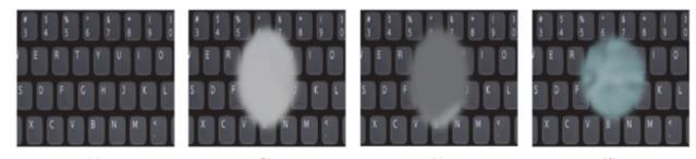
with a serous RPE separation, in certain CSR instances, a sub-retinal fluidaccumulationpersistsforthreemonthsor longer,causinglong-termvisualproblems.Sub-retinalfluid levels fluctuate regularly in situations with CSR like this, majority of the patients will recover early. According to historical statistics, up to half of CSR patients may face relapsesoftheconditionwithinoneyear,necessitatingthe patienttoundertakevarietyoftherapeuticproceduresthat might last up to few months in patients with chronic CSR, first-time CSR and recurrent CSR. Fig. 3 shows the visual differenceforhealthyeyevisionandCSRaffectedvision.
Fig.2:Retinalnormalview
1.2 About CSR disease
CSR is most common disease now a days. this disease causesbyanaccumulationoffluidbeneaththeretina,itcan seriously impair vision owing to the retina's thin tissue layer. As a result, early identification effectively makes it possibletotakethenecessaryprecautionstoregainvision, which results in full recovery. According to studies, CSR often affects single eye, although it is possible that may damage both eyes. In other instances, patients may also heal without any therapy after some period of time. With severe CSR, a patient may have vision obscuration, may result in metamorphopsia, slight hyperopia, contrast sensitivity,andshadingvision.
The focal retina typically has central serous separation, occasionally with dull yellowstores,and rarely
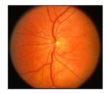
Fig.3:HealthyeyevisionandCSRaffectedvision
The objective of this work is to build appropriate model to detect CSR disease. The accuracy of SVM algorithm is better as compared to other algorithms. This paperisorganizedassectionIdescribestheintroductionof the proposed system, section II describes the literature survey done for this work, section III describes about research methodology used in this work, section IV describes result and discussions, section V describes conclusionoftheproposedwork.
2. LITEREATURE REVIEW
Studies and research on the CSR disease are outlined and developed in the literature review. Based on established criteria for automatic CSR detection, a large number of pertinent and related CSR papers and publications were selected for study and evaluation. Various cutting-edge CSR detection techniques based on AIML and DL were selected for analysis in this research. The author [1] proposed a survey paper on all recent publications and developments based on the classification, detection of CSR disease using AIML and Deep Learning methods, using both the imaging technics. The author recommends that the recent AIML and deep leaning methods gives more prominent results. The author [2] proposedawayforanautomatedsystemforCentralSerous Retinopathy(CSR)diseasediagnosisutilizingDeepSVMfor OCT scan images. Here, the author only worked on OCT imaging, our work also involved a comparison of fundus images. According to author [3], who presented Capsule network-based designs, the created approach outperformed UNet architecture and used less training parameters.Theauthor[4]presented"IndoCyanineGreen Angiography" (ICGA) and acute CSR and different OCT angiography characteristics along with the difference in accuracy. One of the retinal diseases called "Macular Edoema"(ME)wassuggestedbytheauthor[5]inareview paperonOCTandfundusimages.
In the year 2020, author [6] offered a rational solution to CSR detection issues. Three steps make up the process. The initial phase requires the comprehensive reconstruction of the 3-D OCT retinal surface, and the second phase entails the creation of two feature sets, one forcystfluidandtheotherforthicknessprofile.Thetopicof theretinaiscategorizedusingtheSVMclassifieralgorithm. The lodged method used multiple OCT photos for the identification of CSR whereas various researchers experimented with OCT, fundus photographs. Author [7] proposed a decision support system, was published in 2019, and made a clumsy attempt to identify CSR from retinalimagesusinganSVMclassifier.Beforesegmentation is applied, the input image is sparsely de-noised. From the retinal layers created by this segmentation, a profile of retinal thicknessisproduced.SVMclassifierwasemployed in this study. In the work "Multi-Disease Detection in RetinalImagingBasedonAssemblingHeterogeneousDeep Learning Models" published in German Medical Data Sciences 2021, Author [9] suggested that employing heterogeneous deep learning models can produce better outcomes.
3. RESEARCH METHEDOLOGY
3.1 Proposed Work Flow
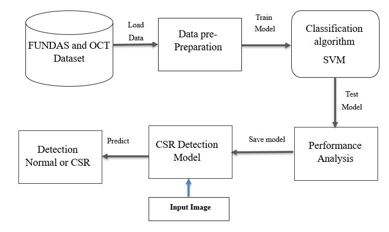
Theworkflowbriefsaboutprogressofprocessandhow the system behaves for the boundary environments. The Workflow diagram and flow of data are depicted in fig 4, the figure describes how process progresses in entire work.
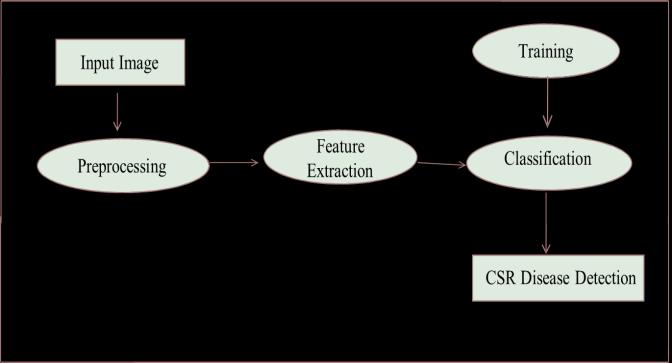
The architecture diagram provides an overview of an entire system, identifying the main components and interfaces that are used for the purpose. Fig 5 demonstrates the architecture diagram for the proposed system. Design explains the architecture components that areusedfordevelopingasoftwareproduct.
Fig 4:Dataflowdiagramofproposedsystem
Thefirststepinthisprocessistoprovideaninputofa retinal picture, which is then preprocessed to identify for any errors or improve visualization for the next steps. Following preprocessing, the eye scan's numerous components and characteristics are retrieved during the featureextractionstep.Thedatasetisthensegmentedinto separate subset for training or testing process. The suitablemodelisbuiltandthealgorithmicmodellearnsto identify any CSR diseases in given input images after analyzinghundredsofimages.
This model takes the input as two type of dataset OCT datasetandFundusdatasetofretinalimages,theseimages are preprocessed using some pre-processing technics for better visualization. After preprocessing the suitable model is built by optimizing several parameters and increasingtheepochsusingSVMalgorithm.Performanceis analyzed and presented in performance section. research work.Webrieflydiscusstwopubliclyaccessibledatasets.
3.2 Overview of Retinal Imaging dataset
Two publicly accessible CSR-related datasets were used and examined in this paper. These datasets are collection ofOCT,fundusphotos.Theseavailabledatasetswereused by numerous researchers and may be easily downloaded using their unique URLs. To accomplish testing objectives and train Machine Learning models, a fraction of the fullonimagesfromthesedatasetsareusedinexperimental
1) Oct Dataset: It is non-invasive imaging method that used to capture 3-D volumetric photographs, and it has emerged as the most preferred method for examining the retinal anatomy. The retina's cross-section picture is recorded using waves. By using OCT scans, the eye expert candiagnoseoftheretina'smanylayersanddeterminethe thicknesseachlayer.
The labelled OCT pictures is found in Zhang's lab and also available in an OCT imaging database on the website of the “University of Waterloo” in Canada, this is opensource repository of OCT dataset. OCT image databases are widely accessible. It has been especially mentioned in relation to using image-based deep learning for medical diagnoses and curable diseases. For the purposeofexperiments,weused340OCTimages.
2) Fundus Dataset: The"fundusphotography"approach of acquiring the retinal red-free image is considered as an alternative for OCT imaging. Fundus imaging is a twodimensional (2-D) depiction of the three-dimensional retinal tissues cast onto the imaging surface. This is accomplishedbytheuseofreflectedlight,withtheamount of light reflected from the retinal tissue directly related to thepictureintensityonthe2-Dprojection.Thistechnology worksbasedoncolorphotographicfilmandthestatistical methodology. Similar to this, digital representation of retina offers a fast, towering-resolution, and consistent image that is instantaneously available and managed for thecreationofanimage.Additionally,fundusphotography is frequently used for clinical examinations and disease records, with the possibility for telemedicine and tolerant training.ThephotosproducedbyFundusmethodscanalso includeordinaryandextendedperspectives.
3.3 Splitting of dataset
Dataset is divided into two parts Train Data, Test Datausingthebuilt-inlibraryknownas“sklearn”.
3) Training Data: A for this work uses 80% of the original dataset and this numbers may vary depending on the needsof the experiment.This data is usedtotrain the model, which tries to learn from the labeled dataset. Both the input and the predicted result are included in the trainingdata.

4) Test Data: The test dataset is 20% of the original dataset and used to evaluate the model. It is used for the model’sevaluationprocessafteritisfullytrained.
3.4 Data Pre-preparation
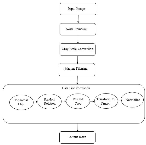
Two publicly accessible CSR-related datasets are examined in this article. These CSR databases are the consists of OCT and fundus images, and a typical dataset constitute of a variety of records. These freely available datasets typically used and accessed by many academics, and may be quickly found using their unique connections. Data pre-preparationiscarriedoutasseeninfig 6, figure shows the image transformation from the original to the transformedimage.
The preprocessing stage in our proposed system consists of four main phases, namely noise removal, grayscale conversion, median filtering, and data transformation.Datatransformationconsists offiveimage transformation steps such as “random horizontal flip”, “random rotation”, “random resizing”, “transforming to tensor” and normalizing the data. Image preprocessing flowisasshowninfig 6.
3.5 Classification
The primary objective of this initial batch of results is to identify any CSR grade. OCT and the Fundus dataset were used for it. With all the characteristics of these images, we trained an SVM classifier. The performance was also enhanced by choosing the SVM parameters and characteristics that were the most pertinent. Thus, a linear kernel function in the SVM technique was used to achieve the best accuracy for our detection. The results section displays the confusion matrix for the best detector. Tables 1 and 2 list the OCT and Fundus Dataset's accuracy, precision, and recall for proposedsystem.
3.6 Performance Measurement
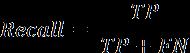
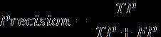
In this proposed system we utilized five different metrics to measure the performance namely accuracy, precision, recall, f1_score which are defined as below equation1,equation2,equation3,equation4.
whereFP, FN, TP, TN stand for false positive, false negative, true positive and true negative, respectively. f denotes the predictor (model). Each group of classes is examinedindependentlyforFP,FN,TPandTN.
The proposed work is implemented using Python language and Streamlit framework which is open source, Streamlithelpsinfastestbuildingandsharingthemachine learningwebapplications,itisanPythonbasedlibrary.We ranthisexperimentinlocalsystemwithaCorei5processor with8GBRAM.
The obtained result of OCT images dataset using SVM classification algorithms against the measurement metrics accuracy, precision, recall, f1_score are as displayed in the table1.
Algorithm is implemented and performance is measured. SVM performance is using performance metrics, It is observedthatthealgorithmreachedaccuracyupto91%.
5. CONCLUSION
In the same way obtained result of Fundus images dataset using SVM classification algorithms against the measurement metrics accuracy, precision, recall, f1_score are as displayed in the table2. We observe that this algorithmperformsgoodforbothinputdataset.
The automated identification of CSR is supported by a number of innovative methods and studies. It was found that ML and DL techniques will enhance CSR analysis and detection.TheOCTdatasetandtheFundusdatasetareused as inputs in this work. The Support Vector Machine (SVM) is applied for classification, this approach excels at identifying CSR diseases. Since it provides results with a 91% accuracy rate, it appears to be the one among most effectivemethodfordetectingCSRdisease.
REFERENCES
[1] Syed Ale Hassan, Shahzad Akbar, Amjad Rehman, ‘‘Recent Developments in Detection of Central Serous Retinopathy through Imaging and Artificial Intelligence Techniques,’’IEEEAccessjournal,2021.
[2] S.A.E.Hassan,S.Akbar,S.Gull,A.Rehman,andH. Alaska.‘‘Deeplearning-basedautomaticdetectionofcentral serous retinopathy on optical coherence tomographic (OCT) images’’ Published in 1st Int. Conf. Artificial IntelligenceDataAnalytics(CAIDA),Apr.2021.
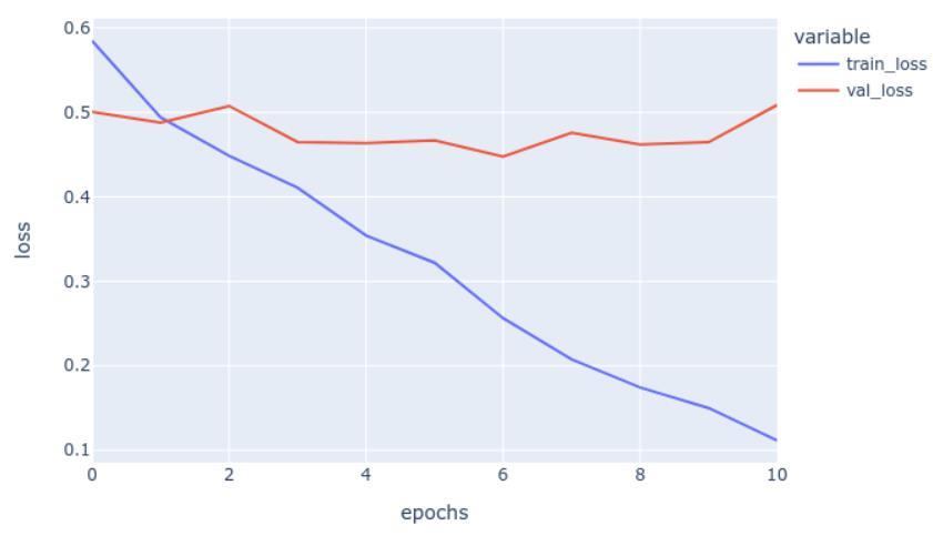
Performance analysis is done for both OCT images datasetandFundusimagedatasetwhileimplementationof this work. The output obtained from performance result is asdisplayedinthebelowgraphinthefig 7,figure depicts the graph against the parameters epochs vs loss, graph implemented to show training loss and validation loss, we observethattraininglosshasbeenreduceddrastically.
[3] S. Prabha, S. SaiRanjith Kumar,G.Gopal Reddy, K. Sakthidasan Sankaran, “Retinal Image Analysis Using Machine Learning’’, 2020 International Conference on Communication and Signal Processing (ICCSP), Date of Conference: 28-30 July 2020, DOI: 10.1109/ICCSP48568.2020.9182227.
[4] M. Vijaya Maheswari; G. Murugeswari, ‘‘A Survey on Computer Algorithms for Retinal image Preprocessing and Vessel Segmentation,’’ 2020 International Conference on Inventive Computation Technologies (ICICT), Date of
Conference: 26-28 February 2020, DOI: 10.1109/ICICT48043.2020.9112470.
[5] Fatma A. Hashim; Nancy M. Salem; Ahmed F. Seddik, ‘‘Preprocessing of color retinal fundus images’’, 2021 Second International Japan-Egypt Conference on Electronics, Communications and Computers (JEC-ECC), DateofConference:17-19December2021.
[6] A. M. Syed, T. Hassan, M. U. Akram, S. Naz, and S. Khalid,‘‘Automateddiagnosisofmacularedemaandcentral serous retinopathy through robust reconstruction of 3D retinal surfaces,’’Comput. Methods Programs Biomed., vol. 137,pp.1–10,Dec.2016,doi:10.1016/j.cmpb.2016.09.004.
[7] B. Ramasubramanian, S. Selvaperumal, A. Nasim, and A. Jameel, ‘‘A comprehensive review on various preprocessing methods in detecting diabetic retinopathy’’ 2019 International Conference on Communication and Signal Processing (ICCSP), Date of Conference: 06-08 April 2019.
[8] Samina Khalid,1,2 M. Usman Akram,3 Taimur Hassan,3,4 Ammara Nasim,4 and Amina Jameel “Fully AutomatedRobustSystemtoDetectRetinalEdema,Central Serous Chorioretinopathy, and Age Related Macular DegenerationfromOpticalCoherenceTomographyImages” Research Article, Hindawi BioMed Research International Volume 2017, Article ID 7148245, 15 pages, https://doi.org/10.1155/2017/7148245.
[9] DominikMULLER,SOTO-REY,andFrankKRAMER, “Multi-Disease Detection in Retinal Imaging Based on Ensembling Heterogeneous Deep Learning Models”, German Medical Data Sciences 2021: Digital Medicine: Recognize - Understand – Heal R. Rohrig et al. (Eds.)., doi:10.3233/SHTI210537.
[10] Osama Ouda, Eman AbdelMaksoud, A. A. Abd ElAzizand Mohammed Elmogy © 2021 The authorsandIOS Press., “Multiple Ocular Disease Diagnosis Using Fundus Images Based on Multi-Label Deep LearningClassification” Electronics Article, lectronics 2022, 11, 1966. https://doi.org/10.3390/electronics11131966.