A Deep Learning Approach for the Detection and Identification of Neovascularization in Fundus Images
Sheik Arshad1 , Rahul Paul2 , Birali Prasanthi3 , K Mahesh Kumar41 Student, Bachelors in CSE, Mahatma Gandhi Institute of Technology, Hyderabad, Telangana, India

2 Student, Bachelors in CSE, Mahatma Gandhi Institute of Technology, Hyderabad, Telangana, India
3Assistant Professor, Dept. of Computer Science and Engineering, Mahatma Gandhi Institute of Technology, Hyderabad, Telangana, India
4Associate Professor, Dept. of Computer Science and Engineering, Mahatma Gandhi Institute of Technology, Hyderabad, Telangana, India
Abstract – Diabetic patients are at ahighriskofdeveloping a retinal disorder called Proliferative Diabetic Retinopathy (PDR). In PDR Neovascularization is considered as one of the major conditions in which there is an abnormal random growth of blood vessels on the retina. Neovascularization can cause severe vision loss and blindness if it is not detected and treated in its early stages. Fundus images include the images of the rear of an eye. Using these fundus images Neovascularization can be detected and classifiedintoseveral stages. Neovascularization has a small size and random abnormal growth pattern. This could be a challenging task to detect with normal Image processing techniques. Deep learning methods can be used to detect Neovascularization because of their ability to perform automatic feature extraction on objects with complex features. The proposed system is implemented based on the performance of popular pre-trained deep neural networks suchasInceptionResNetV2, DenseNet, ResNet50, ResNet18, AlexNet,andVGG19networks. The best Convolutional Neural Network (CNN) model can be used to build and implement the Neovascularizationdetection and classification Model.
Key Words: CNN, Deep Learning, Neovascularization, Fundus Images, Pre-trained Networks.
1. INTRODUCTION
Neovascularization is the formation of abnormal blood vesselsintheretina,whichcanbeacomplicationofvarious retinal diseases, including diabetic retinopathy. The presenceofneovascularizationcanleadtoseverevisionloss and blindness if left untreated. Thus, early detection and timelyinterventionarecrucialforpreventingorminimizing thedamagecausedbythiscondition.TheNeovascularization is further classified into 5 conditions - Healthy Eye, Mild, Moderate,Proliferate,andSevere.
Fundus images correspond to the retinal view of an eye whichrepresentstherearofaneye.Theseimagesaremostly usedfordetectingeye-relateddisorders.Fundusimagesare commonlyusedforthescreeninganddiagnosisofdiabetic retinopathy.However,detectingneovascularizationinthese
images can be challenging due to the presence of various confoundingfactorssuchasimagenoise,variabilityinimage quality, and the presence of other retinal abnormalities. Hence, developing automated and accurate methods for detectingneovascularizationinfundusimagesisessential.
Deeplearningtechniques,particularlyconvolutionalneural networks(CNNs),haveshownremarkablesuccessinvarious image-processing tasks, including medical image analysis. Thesetechniquescanlearncomplexfeaturesfromtheinput imagesanddetectthepresenceofNeovascularizationand alsoclassifythemintodifferentcategories(Mild,Moderate, Healthy,Proliferate,Severe).
Theproposedapproachforneovascularizationdetectionin fundusimagescombinesdeeplearningandimageprocessing techniques to develop a reliable and accurate system. By usingaminimumamountoffundusimagedataset,thedeep learning model can learn to identify and classify the conditionofneovascularizationinfundusimageswithafiner accuracy.Overall,theproposedapproachcanhelpimprove theearlydetectionandmanagementofneovascularization, leadingtobetterpatientoutcomesandreducedhealthcare costs.
1.1 LITERATURE SURVEY
Several research studies have proposed divergent image processingmethodstodetectNeovascularizationinfundus images. But it is still a challenging task to detect Neovascularizationduetoitsabnormalrandomgrowthand small sizepattern. Recently, Deeplearning techniquesare getting immensely popular due to their advancement in ArtificialIntelligenceinBiomedicalImageProcessing.These methodscanbeemployedtotrainaCNNmodeltodetectand classifyNeovascularizationinfundusImages.
1) By the literature survey of the research papers, the existing system methods which include image processing and machine learning algorithms provide preferable accuracyfordetectingNeovascularization,butthisislimited. TheabnormalrandomgrowthpatternofNeovascularization
must be detected and identified so that the condition of Neovascularizationcanbeclassifiedaccurately.

2) The existing image processing techniques are better suited for detecting the Neovascularization based on the training data samples. The growth pattern still remains challenging to detect and also it changes from patient to patient. The growth pattern is abnormal and small. The detectionmechanismmustbeabletolearnthebehaviorof thegrowthpatternofbloodvesselsinordertodetectand classifyitproperly.
3)Theexistingsystemswithimageprocessingtechniques canbeimprovedbyemployingdeeplearningmethodsfor feature extraction and classification. The deep learning modelscanbetrainedtolearnthebehavioroftherandom growth pattern of the blood vessels formation and can be able to detect and classify the condition of Neovascularization by training the model with an ample amountoftrainingdatasetandvalidatingthemodelwitha validationdatasetconsistingofdifferentgrowthpatternsof bloodvessels.Thesemodelscanbeusedtoactasabaseto build the system for detecting and classifying the Neovascularizationfromthefundusimages.
M. C. S. Tang, S. S. Teoh, H. Ibrahim, and Z. Embong, in "A Deep Learning Approach for the Detection of Neovascularization in Fundus Images Using Transfer Learning"[1]proposedadeeplearningneuralnetworkthat is capable of detecting the Neovascularization in fundus images.Theperformanceofthetransferlearningtechnique is evaluated using four pre-trained networks AlexNet, GoogLeNet,ResNet18,andResNet50,andusedtobuildthe proposed system for detecting the presence of Neovascularization in fundus images. The combination of ResNet18 and GoogLeNet pre-trained models is used in buildingtheproposedsystemfordetectingthepresenceof Neovascularizationinfundusimages.
S. C. Munasingha, K. K. Priyankara, R. G. Upasena, and A. Ikedain"ANovelMethodofDetectingNeovascularization Regions in Digital Fundus Photographs" proposed novel methods to detect Neovascularization using image processingtechniques.The proposedsystemincorporates imageprocessingtechniquesandmachinelearningmethods on the fundus images to segment the regions with Neovascularization. This segmentation can help find the regionofabnormalgrowthofbloodvesselsandcanbeable todetectNeovascularization.
H.Huang,X.Wang,andH.Main"AnEfficientDeepLearning NetworkforAutomaticDetectionofNeovascularizationin ColorFundusImages"proposedadeeplearningnetworkto automaticallydetectNeovascularizationinfundusimages. ThemodelisdevelopedusingFeaturePyramidNetworkand Vovnetandthemodelisevaluatedwithfundusimagesfrom the real-time cases. The experimental results with testing
showed that it has less training and test time and high accuracywhencomparedwithMaskR-CNN.
2 METHODOLOGY
The proposed network to detect and classify Neovascularization in fundus images is developed using deep learning methods and transfer learning approaches. The popular pre-trained networks are evaluated on the trainingandvalidationsetsoffundusimagesandthemodel with the best accuracy metrics is considered as the base model for developing the proposed network for the detectionandclassification.Theproposedsystememploys twotasks-DetectionandClassification.
DETECTION:
The detection stage includes training the neural network model to precisely detect Neovascularization in fundus images. The abnormal random growth of blood vessels makesitchallengingtodetect.Themodelshouldbecapable oflearningthebehavioroftherandomgrowthoftheblood vessels and should be able to detect the presence of Neovascularization. The image processing techniques and feature extraction methodologies can be combined with neural networks to accurately detect the presence of Neovascularization in fundus images. This detection stage helps in further classifying the stage and condition of neovascularizationdetectedintheeyesofthepatient.
CLASSIFICATION:
Theclassificationstageoftheproposedsystemenablesthe model to predict the condition of Neovascularization in fundus images. Neovascularization can lead to permanent vision loss if not treated early. The early detection of Neovascularizationcanhelp patients withtheirtreatment andrecovery.Thereare5stagesinDiabeticRetinopathy(Healthy/Normal,Mild,Moderate,Severe,andProliferative). the presence of Neovascularization is detected in the Detectionstageandafterthedetectionstagetheconditionis classifiedbasedonthegrowthoftheabnormalbloodvessels on the retina in the Classification stage. This helps the patientstogettheconventionalandrighttreatmentfortheir conditiondetectedthroughtheirfundusimages.Themodel shouldbeabletolearntherandomgrowthpatternsofthe5 stages and accurately detect the right condition of the detectedNeovascularizationintheretina.
SYSTEM ARCHITECTURE:

System architecture refers to the high-level design or blueprint of the entire conceptual model of the proposed system.Itprovidesastructuraloverviewofhowthesystem isbuiltandorganized.Theproposedsystemisbuiltontopof the pre-trained neural networks using transfer learning approaches.Theunderlyingarchitectureforimplementing theproposedsystemisgivenbelow.
RestNet18,AlexNet,andVGG19networks.Themodelisthen loaded into a flask object to provide an interface for the patient/user to interact with the system and upload their fundusimagetochecktheircondition.
2.1 MODEL TRAINING:
I. IMAGE PROCESSING:
The fundus image dataset is collected according to the 5 stages-(Healthy,Mild,Moderate,Proliferate,andSevere).In theimageprocessingtechniques,thegroundtruthsof the fundus images are obtained for the model training. The datasetissplitintotrainingandvalidationsetstoevaluate themodelperformanceoveritstrainingtime.theproposed network is developed using a transfer learning approach. Thepre-trainednetworksareloadedandtrainedoverthe train and validation datasets and the model with the best performance metrics is employed for developing the proposedsystemtodetectandclassifyNeovascularizationin fundus images. The popular pre-trained networks used include - Inception ResNetV2, DenseNet, ResNet50, RestNet18,AlexNet,andVGG19networks.Themodelisthen loaded into a flask object to provide an interface for the patient/user to interact with the system and upload their fundusimagetochecktheircondition.
Fig -2:SystemArchitecture
WORKING PRINCIPLE:
The fundus image dataset is collected according to the 5 stages-(Healthy,Mild,Moderate,Proliferate,andSevere).In theimageprocessingtechniques,thegroundtruthsof the fundus images are obtained for the model training. The datasetissplitintotrainingandvalidationsetstoevaluate themodelperformanceoveritstrainingtime.theproposed network is developed using a transfer learning approach. Thepre-trainednetworksareloadedandtrainedoverthe train and validation datasets and the model with the best performance metrics is employed for developing the proposedsystemtodetectandclassifyNeovascularizationin fundus images. The popular pre-trained networks used include - Inception ResNetV2, DenseNet, ResNet50,
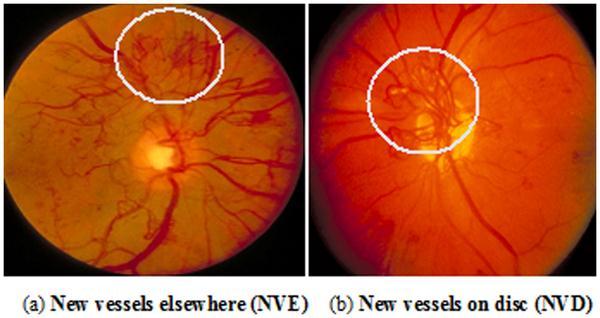
Fig -3:Abnormalbloodvesselsgrowthintheretina
Ground truth images are essential for evaluating the performanceofneovascularizationdetectionalgorithmsor systems. They are used as a reference for comparing the resultsobtainedfromautomatedimageanalysismethods, such as segmentation and classification algorithms, to determine the accuracy, sensitivity, specificity, and other performancemetricsofthealgorithms.
II. FEATURE EXTRACTION & CLASSIFICATION:
Thefeatureextractionmethodcomprisesextractingrelevant features or attributes from the segmented blood vessels. Thisinformationcanbeusedtodiscriminateandunderstand normal bloodvesselsandtheirgrowthwiththeabnormal growthofbloodvesselsintheretina.Thiscanidentifythe severityoftheNeovascularizationdetectedintheeyeofthe
patient. The features extracted could include vessel diameter,vesseltortuosity,vesselbranchingpatterns,and othershape,intensity,andtexturefeatures.
Classificationtechniquesareappliedintheneuralnetwork modelstotrainincategorizingthesegmentedbloodvessels into 5 classes of diabetic retinopathy stages. The Convolutional Neural network is trained on the image datasetsforclassification.Theseneuralnetworksaretrained onlabeleddatasetstolearnthepattern,growth,andfeatures of the healthy and abnormal blood vessels and make predictionsonthenewfundusimages.
III. TRANSFER LEARNING:
Transfer learning is a machine learning technique that leverages knowledge learned from one task or domain to improve the performance of another, related task, or domain. It involves training a pre-trained model, typically trainedonalargedatasetforadifferenttask,onasmaller dataset,orona different task,to benefit fromthelearned representationsandfeatures.Transferlearninghasgained significantattentionandpopularityinrecentyearsduetoits ability to overcome data limitations and improve the performanceandefficiencyofmachinelearningmodelsin various domains, including image recognition, natural languageprocessing,andspeechrecognition,amongothers.
One of the key advantages of transfer learning is that it allows models to leverage the knowledge and representationslearnedfromalargedatasettoperformwell on a smaller dataset with limited labeled data. This is especially beneficial in scenarios where obtaining large, labeleddatasetsfortrainingischallenging,time-consuming, or expensive. By utilizing a pre-trained model, transfer learningcanhelpovercomethelimitationsoflimiteddata, resulting in more accurate and robust models. Transfer learningcanbeimplementedindifferentways,depending onthespecifictaskanddomain
The proposed system is constructed using the transfer learningapproach.Inthisapproach,thepre-trainedmodelis usedasafixedfeatureextractor,wherethelayersofthepretrainedmodelareusedtoextractrelevantfeaturesfromthe input data. These features are then fed into a separate classifierormodelforthetargettask.Thisallowsthemodel to benefit from the learned representations and features fromthepre-trainedmodel,whichmayhavealreadylearned genericpatternsorhigh-levelfeaturesthataretransferable tothetargettask.Thedeeparchitectureoflayersofthepretrainedneuralnetworkmodelshelpsthesystemtolearnthe featuresfromthedatasetsandunderstandthebehaviorof the growth pattern of the blood vessels. However, it is important to carefully select the appropriate pre-trained model,considerthedomainandtasksimilaritybetweenthe sourceandtargettasks,andevaluatetheperformanceofthe transferredmodeltoensureitseffectivenessforthespecific targettask.
2.2 PRE-TRAINED NEURAL NETWORKS: I INCEPTION RESNETV2:
InceptionResNetv2isadeepconvolutionalneuralnetwork (CNN)architecturethatcombinestheInceptionmoduleand theresidualconnectionsfromResNet. InceptionResNetv2is a pre-trained network that has been trained on a large dataset to learn the features from images, making it a powerfultoolforawiderangeofcomputervisiontasks.The Inception module in Inception ResNetv2 is designed to extract features at multiple scales by using convolutional layers with different filter sizes (1x1, 3x3, and 5x5) and poolingoperations,andthenconcatenatingtheoutputs.This allows the network to capture both local and global contextualinformation,makingitmorerobusttovariations inobjectscalesandorientations.Residualconnectionsallow fortheefficienttrainingofverydeepnetworksbyalleviating the vanishing gradient problem, which can occur in deep neuralnetworks.Residualconnectionsalsohelptoimprove theaccuracyandstabilityofthenetworkduringtrainingby allowing the network to learn both the residual and the identitymapping,whichcanfacilitatetheflowofgradients andinformationacrossthelayers.
II. DENSENET:
DenseNet, short for Densely Connected Convolutional Networks, is a deep Convolutional Neural Network (CNN) architecture.Itisknownfor itsuniquedenseconnectivity pattern,whereeachlayerreceivesinputfromallprevious layers, resulting in highly connected and densely packed featuremaps.DenseNetisapre-trainednetwork,meaningit has been trained on a large dataset to learn meaningful featuresfromimages,makingitapowerfultoolforvarious computer vision tasks. The dense connectivity pattern in DenseNetallowsforefficientfeaturereuseandgradientflow

throughout the network, as each layer has access to the featuremapsofallpreviouslayers.Thisdenseconnectivity leadstobetterfeaturepropagation,reducesthenumberof parametersneeded,andenhancesthegradientflowduring training.
III. RESNET50:
ResNet50 is a popular pre-trained Convolutional Neural Network (CNN) architecture. ResNet stands for Residual Network, and 50 in ResNet50 refers to the depth of the network,whichconsistsof50layers.ResNet50isknownfor its residual connections, which allow for the efficient trainingofdeepnetworksandhavebeenshowntoimprove accuracyandstabilityduringtraining.ResNet50isorganized into blocks, where each block contains multiple convolutional layers followed by batch normalization and activation functions, with residual connections across the blocks. The basic building block of ResNet50 is the bottleneck block, which consists of three convolutional layerswithdifferentfiltersizes(1x1,3x3,and1x1)toreduce computationalcomplexity,followedbybatchnormalization andactivationfunctions.
IV. RESNET18:
ResNet stands for Residual Network, and 18 in ResNet18 refers to the depth of the network, which consists of 18 layers. ResNet18 is known for its residual connections, whichallowfortheefficienttrainingofdeepnetworksand havebeenshowntoimproveaccuracyandstabilityduring training.Thebasicbuilding block ofResNet18isthebasic block,whichconsists oftwo convolutional layerswiththe samefiltersize(3x3),followedbybatchnormalizationand activationfunctions.TheresidualconnectionsinResNet18 skiponeormoreconvolutionallayersanddirectlyconnect the input of the block to the output, allowing for efficient featurepropagationandreuse.
V. ALEXNET:
AlexNet is a pioneering pre-trained convolutional neural network(CNN)architecture.AlexNetisknownforitsdeep architecture,useofrectifiedlinearunits(ReLU)asactivation functions, and introduction of dropout regularization to mitigateoverfitting.AlexNet consists offiveconvolutional layers followed by three fully connected layers. The convolutional layers are responsible for extracting hierarchical features fromtheinputimage, while thefully connectedlayersareusedforclassification.Thearchitecture ofAlexNetischaracterizedbyitsdepth,withatotalofeight layers, which was considered deep at the time of its introduction. ReLU introduces non-linearity into the network, allowing it to learn complex patterns and representationsfromtheinputdata.
VI. VGG19:
VGG19isknownforitssimplicityanduniformityindesign, consistingof19layers,with16convolutionallayersand3 fully connected layers. VGG19 is widely used for image recognition tasks, such as image classification and object detection, due to its strong performance and ease of implementation.ThemaincharacteristicofVGG19isitsdeep architecture,withmultipleconvolutionallayersstackedon top of each other, followed by fully connected layers for classification.VGG19usessmallconvolutionalfilters(3x3)in all its convolutional layers, which allows for a deeper networkwithasmallernumberofparameterscomparedto larger filters. This leads to a more efficient and computationally feasible architecture. VGG19 also has a uniformdesign,wherealltheconvolutionallayershavethe samenumberoffilters(64,128,256,or512)andthesame padding (same) to maintain the spatial dimensions of the input.Thisuniformityindesignmakesiteasiertoimplement andfine-tunethenetwork.
3. CHALLENGES
The proposed system to detect and classify Neovascularizationinfundusimagesisdevelopedbasedon deeplearningmethods.Variouschallengesarefacedwhen trainingadeepneuralnetworkonafundusimagedataset. Someofthosechallengesinclude:
I. DATA AVAILABILITY:
Fundus images of patients with neovascularization are relatively rare, making it challenging to obtain a large datasetfortrainingdeeplearningmodels.Thiscanresultin modelswithlimitedgeneralizationcapabilitiesandreduced accuracyandperformance.
II. VARIABILITY IN GROWTH PATTERNS:
Neovascularizationcanmanifestindifferentpatterns,such asfineorcoarsevessels,whichcanvaryinsize,shape,and appearance.Thisvariabilitymakesitchallengingtodevelop a robust deep learning model that can accurately detect neovascularizationacrossdifferentpatternsandstages.The abnormalgrowthpatternsofthebloodvesselsintheretina makes it challenging to develop the model to learn the behaviorofthegrowthpatternandpredictthecondition.
III. NOISE AND ARTIFACTS:
Fundusimagescanbeaffectedbyvariousartifacts,suchas uneven illumination, blur, and noise, which can affect the accuracy of neovascularization detection. Deep learning models need to be robust to these artifacts to achieve reliabledetectionperformance.
IV. INTERPRETABILITY:
Deep learning models are often considered as "black box" models, making it difficult to interpret and explain the rationale behind their predictions. In the context of Neovascularizationdetection,interpretabilityisimportant for gaining trust and acceptance from clinicians and stakeholders.
V MODEL OVERFITTING:
Deeplearningmodelsarepronetooverfitting,wherethey may learn to memorize the training data instead of generalizing from it. This can result in poor model performanceonunseendata,includingfundusimageswith Neovascularization.
VI. COMPUTATIONAL RESOURCES:
Deep learning models typically require significant computationalresources,includingpowerfulhardwareand largeamountsofmemory,fortrainingandinference.Access tosuchresourcesmay belimitedinsomesettings, posing challenges in developing and deploying deep learning models for neovascularization detection in resourceconstrainedenvironments.
4. TESTING AND RESULTS
Thepre-trainednetworksaretrainedonthefundusimage dataset. The image dataset is divided into training and validationdatasetandeachpre-trainednetworkistrained onthosedatasets.Thevalidationdatasetisusedtoevaluate the performance metrics and accuracy of the model in detectingandclassifyingtheNeovascularizationinfundus images.Themodelwiththebestaccuracymetricsisadopted forconstructingthedeepneuralnetworkfordetectingand classifyingthestageofNeovascularization.
The pre trained networks employed for developing the proposednetworkaretrainedover3000+imagesoftraining dataandvalidatedover300+imagesofvalidationdataset. Thenetworkwhichperformsbestunderoptimalconditions and gives a better accuracy for less training time is considered for building the proposed system. The testing stagehereincludestheaccuracytestingofeachpretrained network to find the best model architecture to build the system.
4.1 MODEL COMPARISION:
Table-1:PerformanceofNeuralNetworks
TheInceptionResNetV2Modelhasshownthehighaccuracy of86%withlesstrainingtimeoveralesstrainingdataset compared to other models. This pre trained network InceptionResNetV2isusedforbuildingtheproposedsystem fordetectingandclassifyingtheNeovascularization.
4.2 RESULTS
From the model training and validation, the Inception ResNetV2 model has better accuracy of prediction when compared to other CNN Models. This forms the base for developingtheproposedsystemandtheproposedmodelis loadedintoaPythonFlaskobjecttocreateawebinterface fortheuserstouploadfundusimagesandclassifyitsstage.
5. CONCLUSION
The Neovascularization detection and classification of diabeticretinopathyinfundusimagesusingdeeplearning methods has a great impact in advancing the field of ophthalmology.Throughleveragingpre-trainedmodelsand utilizingdeeplearningtechniques,researchersandclinicians havebeenabletoachieveremarkableresultsinautomating the detection of Neovascularization, a critical feature in various retinal diseases such as diabetic retinopathy. Transferlearninghasbeenfoundtobeaneffectiveapproach inovercomingthelimitationsoflimitedannotateddataand reducing the need for large datasets, which are often challengingtoobtaininmedicalimaging.Byleveragingpretrained models, such as convolutional neural networks (CNNs),thataretrainedonlargedatasetsfromothertasks, transfer learning allows for the extraction of meaningful featuresfromfundusimagesandenablesthedevelopmentof accurate and robust neovascularization detection models withrelativelysmallerdatasets.

The integration of transfer learning with deep learning approaches has further improved the accuracy and robustnessofneovascularizationdetectionmodels.Byfinetuningpre-trainedCNNswithdomain-specificdata,transfer learning allows for the adaptation of the model to the specific characteristics of the fundus images, leading to improvedperformanceindetectingNeovascularization.
The performance of six pre-trained convolutional neural networks, which are Inception ResNetV2, DenseNet, ResNet50,ResNet18,AlexNet,andVGG19,wasinvestigated for building the best CNN model for developing the neovascularization detection model through transfer learning.Evaluationresultsshowthatthetransferlearning approach yields superior performance. The Inception ResNetV2Modelarchitectureisconsideredthebestmodel fordevelopingtheproposedsystemandthemodelistrained andloadedintoaflaskobjecttoclassifythefundusimages into 5 classes of Neovascularization conditions (Healthy, Mild,Moderate,Severe,andProliferate).
6. FUTURE SCOPE
More fundus images can be added to the training and validationdatasetstoincreasetheaccuracyofdetection andclassification.Thefundusimagesofpatientsinrealtime can be served as new validation sets to fit the modeltrainingandimproveitsaccuracyofprediction.
Thedetectionmodelincorporatedonthewebsitegives thepatientsthechancetochecktheirconditionandget immediatetreatment.
Themodeltrainingoverlargefundusimagedatasetsto buildamuchmoreaccuratedetectionmodelcouldbe efficient when the system is connected to a GPU, allowing it to run smoothly and reduce the model trainingtime.
ThedetectionmodelcanalsobeembeddedintoanIOT device for real-time detection and classification purposesintheMedicalIndustry.
Continuedresearchanddevelopmentinthisfield,along with careful validation and standardization, will contributetothefurtheradvancementandintegration of these approaches into clinical practice, ultimately benefitingpatientsandimprovingthemanagementof retinaldiseases.
REFERENCES
[1] M.C.S.Tang,S.S.Teoh,H.Ibrahim,andZ.Embong,"A Deep Learning Approach for the Detection of Neovascularization in Fundus Images Using Transfer Learning," in IEEE Access, vol. 10, pp. 20247-20258, 2022,doi:10.1109/ACCESS.2022.3151644.
[2] S.C.Munasingha,K.K.Priyankara,R.G.Upasena,andA. Ikeda,"ANovelMethodofDetectingNeovascularization RegionsinDigitalFundusPhotographs,"2022IEEE4th Eurasia Conference on Biomedical Engineering, HealthcareandSustainability(ECBIOS),2022,pp.1-4, doi:10.1109/ECBIOS54627.2022.9945014.
[3] H. Huang, X. Wang, and H. Ma, "An Efficient Deep Learning Network for Automatic Detection of NeovascularizationinColorFundusImages,"202143rd Annual International Conference of the IEEE Engineering in Medicine & Biology Society (EMBC), 2021, pp. 3688-3692, doi: 10.1109/EMBC46164.2021.9629572.
[4] M.Z.KhanandY.Lee,"RetinalImageAnalysistoDetect NeovascularizationusingDeepSegmentation,"20214th InternationalConferenceonInformationandComputer Technologies (ICICT), 2021, pp. 110-114, doi: 10.1109/ICICT52872.2021.00026.
[5] K.Firdausy,O.Wahyunggoro,H.A.NugrohoandM.B. Sasongko, "A Study on Recent Developments for Detection of Neovascularization," 2019 11th International Conference on Information Technology andElectricalEngineering(ICITEE),2019,pp.1-6,doi: 10.1109/ICITEED.2019.8929941.
[6] H. Huang, H. Ma, and W. Qian, "Automatic Parallel Detection of Neovascularization from Retinal Images Using Ensemble of Extreme Learning Machine," 2019 41st Annual International Conference of the IEEE Engineering in Medicine and Biology Society (EMBC), 2019, pp. 4712-4716, doi: 10.1109/EMBC.2019.8856403.