
BRAIN TUMOR’S DETECTION USING DEEP LEARNING
Mrs. S.Sridevi. [1], S.Geethalakshmi[2].
Professor [1], Professor, Department of Electronics and communication engineering, K.S.R College of engineering, Tamil Nadu, India

Student [2], Department of Electronics and communication engineering, K.S.R College of engineering, Tamil Nadu, India
ABSTRACT:
Thesecondmostnormalsortofdiseasetoday is cancers. The disease poses a threat to a significant number of patients. For the recognizable proof of developments like frontal cortex malignant growths, the clinical field requires a quick, electronic, reasonable, and dependable technique. The benchmarks and wildernesses used to create the cerebrum cancer dataset. Location is urgent to treatment. Specialists guarantee a patient's security at whatever point a growth can be definitively distinguished. There are many different ways this application handles pictures. This app helps doctors treattumorpatientseffectivelyandsavemanylives.A tumor is nothing more than an excessive number of cellsgrowingunchecked.Braincancercellseventually consume all nutrients intended for healthy cells and tissues as they grow, resulting in mental dissatisfaction. Currently, doctors manually examine MR images of the patient's brain to locate a brain tumor. This takes a long time and could lead to a wrong diagnosis of the tumor. A mass of tissue that grows out of control is called a tumor. We can make use of the Profound Learning designs CNN (Convolution Brain Organization), NN (Brain Organization), and KFOLDS to locate a mind expansion. K FOLDS Underwriting upholds the exactness mean worth from the CNN assessment. K Folds improves outcomes by testing the CNN model using evaluation metrics like accuracy, sensitivity, specificity, precision, recall, and F1-score. The great outcomes created by the proposed framework show that K FOLDS Approval is more powerful than different models. The outcomes demonstrate the way that the proposed strategies can autonomously concentrate on the area important to distinguish mindgrowths.
Keywords: Cancerdataset,MRimages,CNNmodelandK FOLDS Approval, accuracy, sensitivity, specificity, precision,recall,andF1-score.
I.INTRODUCTION
Thecerebrumisthemostfundamentalandfundamental of all the organs in the human body. One of the typical
clarifications behind brokenness of psyche is frontal cortex development. A tumor is nothing more than an uncontrolled proliferation of an excessive number of cells. Because of their development, cerebrum growth cells in the end consume all supplements expected for sound cells and tissues, bringing about mind disappointment. The location and size of a brain tumor arecurrentlydeterminedbydoctorsmanuallyexamining MRimagesofthepatient'sbrain.Thisresultsinmistaken ID of the malignant growth and is thought of as exceptionally dreary. Brain cancer is a deadly disease that affects many people. It is feasible to analyse mind cancers early because of the accessibility of the identificationandcharacterizationframework.Inclinical finding, the most troublesome errand is disease arrangement. Using Convolution Brain Organization Calculation for X-ray images of various patients, this project manages a framework that uses PC-based systemstoidentifycancerbarriersandgroupthetypeof growth. To find mind growths in the X-ray pictures of disease patients, different picture handling techniques like element extraction, improvement, and division are used. Picture Pre-Handling, Picture Division, Component Extraction, and Order are the four phases of the Picture Handling method for identifying mind cancer. Using image processing and neural network techniques, brain tumors in MRI images can be detected and categorized moreaccurately.
The brain is the heart of the nervous system in humans. It is arranged in human head and it is covered by the skull. The capacity of the human mind to exert control over each and every part of the body one kind of organ empowers people to persevere and acknowledge any climate. The capacity to think, feel, and act is made conceivable by the human cerebrum. Brain tumors typically fall into one of two categories: A benign brain tumor is a kind of cell that slowly grows; gliomas are a kind of threatening cerebrum growth. Malignant tumors are secondary brain tumors. It originates from astrocytes, which are non-neuronal brain cells. Essential cancers, in general, are less forceful, however they put a great deal of squeeze on the cerebrum, making it quit working appropriately. The optional cancers spread into other tissues more quickly and with greater force. Discretionary psyche malignant growth begins through other piece of the body. These growths contain a
metastatic disease cell that has spread to the cerebrum, lungs, and different pieces of the body. Discretionary frontal cortex malignant growth is uncommonly risky. The clarification of helper frontal cortex development make is in a general sense due lungs sickness, kidney illness,bladderdangerousdevelopment,etc.
The most appealing image was created by Attractive Reverberation IMAGING (X-ray) in 1969. In 1977, the most exceptional technique and the main X-ray picture for the human body were created. We can see the inner construction of the mind exhaustively on account of Xray,andfromthatpoint,wecanseethedifferentsortsof tissues in the human body. When compared to other clinical imaging techniques like X-beam and PC tomography, X-ray images have a higher quality. An MRI is a useful tool for determining whether a person has a brain tumor. Tumor-induced change can be mapped using T1-weighted, T2-weighted, and FLAIR (Fluid attenuated inversion recovery) MRI images. Application .The essential objective of utilizations is to recognize cancers. The development of this application was primarily motivated by the need to provide prompt treatmentandsafeguardthehumanlifethatisindanger.
This application can be beneficial to both patients and doctors. Manual identification is quicker, more precise, and easier to use. This application is plan, and its objective is to settle those issues. Using it is straightforward.
1.1CONVOLUTIONALNETWORK
The Convolutional brain network is delivered by performing convolution on fake brain organizations (ANNs). A CNN is structured by neurons with loads and predispositions that can be learned. The engineering of convolutional brain networks is comprised of three fundamentallayers:thepoolinglayer,thefullyconnected layer,andtheconvolutional layer.Aconvolutionalneural network, or CNN, gets its name from one or more convolutional layers. Convolutional layers are utilized to recognize specific nearby highlights in the information pictures.Eachhubofaconvolutionallayerhasaspatially associated subset of neurons associated with it. The detectionoflocalforms(orstructures)inthechannelsof theinputimageisaidedbythis.Theconvolutionallayer's nodes share the weights on the connections in order to search for the K Folds Validation local trait in the input channels. Each shared weight set is referred to by the name "kernel" (convolution kernel). The feature map shows the strength of the local features that are to be detected across the input images that are learned by convolutional layer kernels. The pooling layer, which follows the convolution layer in the CNN architecture, is primarily responsible for reducing the representation's size.
1.2 LITERATURE REVIEW
A Robust Approach for Brain Tumour Detection in Magnetic Resonance Images Using Fine-tuned Efficient Net
Author:Hasnainalishah,Faisalsaeed,sangseokyun,junhyunpark
Anumberofstudiesthatusedeeplearningandmachine learningtoidentifyinfectiousbraintumorsandstandard images are examined in this section. Abd-Ellah and othersconducted a thoroughexamination ofa varietyof diagnosticstrategiesforbrainMRIimages.Furthermore, the constraints and execution measurements of regular AI and profound learning strategies were inspected by the creators. The creators of this study introduced various techniques for distinguishing cerebrum malignant growths from MR pictures. For deeper segmentation, their study utilized CNNs, SVMs, and multi-class SVMs with a three-dimensional base. When contrasted with other ML classifiers, the DL strategy created exceptional outcomes and a solid technique for fragmenting and grouping cerebrum growths. To extract features from MR images and provide them to ML classifiers (Naive Bayes, SVMs, and Multilayer perceptron’s) as input, the authors of a different study proposed a deep learning neural model. The Volume 10, 2022, 65427 by H. A. Shah and others proposal: The robust method for detecting brain tumors in magnetic resonance images had a classification accuracy of 96% when using SVMs as classifiers. Hossain and co. For the characterization and division of mind growths, various machines and DL procedures, including SVMs, K-NN, multi-facet perceptron, Guileless Bayes, and irregular woods calculations. With a classification accuracy of 92.4%, traditional SVMs ranked highest of these methods. They also suggested a five-layer custom CNN architecturethatcoulddetectbraintumorsinMRimages with an accuracy of 97.2 percent. The VGG19 CNN architecture and K-means clustering were suggested by Khan and others for using MRI images to classify and segment brain tumors. Before the proposed method slices an input MR modality, intensities are preprocessed using a statistical normalization approach. Theywere94%accurateoverall.
II.EXISTING SYSTEM
Tumor extraction from MR brain images and its simplified representation for everyone's comprehension Using a Convolution Neural Network, this system determineswhetherthegivenMRbrainimagecontainsa tumor or not. The algorithm is used to detect brain tumors, but its accuracy varies over time, and loss occurs. Filtering, erosion, dilation, threshold, and outliningthetumorwithedgedetectionarethemethods used.
2.1DRAWBACKS:
Accuracy varies depending on the image, and lossvaluegraduallyrises.
The threshold values are determined by the variousentropyfunctions,whichinturnhavean effect on the segmented results. The complexity ofthecodeandthedifficultyofvisualizationare alsofactors.
III.PROPOSED FRAMEWORK
Dataset, Pre-dealing with, Split the data, Manufacture CNN model train Significant Cerebrum network for a very long time, and plan. We can take different X-ray pictures from the dataset and utilize one of them as the information picture. During the picture's pre-handling, themarkwasencodedandtheimagewasresized.When wesplitthedata,theimagewasdividedinto20%Testing
Data and 80% Training Data. Create a CNN model and train a deep neural network for epochs after that. K FOLDS approves the precision mean value of the CNN calculation. K Folds improves outcomes by testing the CNN model using evaluation metrics like accuracy, sensitivity, specificity, precision, recall, and F1-score. K FOLDSVALIDATIONisdemonstratedtobemoreefficient thanothermodelsbytheimpressiveoutcomesproduced bytheproposedsystem.
3.1BENEFITS
Sharpening and noise reduction can be accomplishedeffectively.
CNNisappliedtothedataset;accuracyhasbeen preciselyachieved.
K Folds Approval approves the precision productivitybythemeanworth
IV. Block Diagram
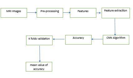
V. SYSTEM IMPLEMENTATION
In the framework order, execution is the cycle that produces the most lower-level framework components. Frameworkcomponentsaremade,purchased,orreused. Production includes all of the steps involved in making, removing, joining, and finishing hardware, coding and testingsoftware,andcreatingoperationalproceduresfor operatorroles.Anassemblysystemthatmakesuseofthe planned specialized and board cycles may be required if executionincludesacreationinteraction.
Asystem element'simplementation process isdesigned, created, or fabricated in accordance with its requirements and/or design properties. The component is constructed with appropriate technologies and industry practices. The combination cycle and frameworkdefinitionprocessesarebothaffectedbythis interaction. When a decision is made to create a component of a framework, the results of the execution interaction may produce requirements for the design of thelargerframework,dependingontheinnovationsand frameworks chosen; as derived system requirements, thoseconstraintsaretypicallyaddedtothesetofsystem requirements for this higher-level system. The architectural design must take these constraints into account.
If a decision is made to purchase or reuse an existing system element, it must be identified as a constraint or system requirement that applies to the architecture of the higher-level system. Then again, the execution cycle could incorporate some change or acclimations to the structure need to be composed into a more huge level systemoraggregate.
Depending on the technologies involved and the circumstance in which the system requirement must be integrated into a higher-level aggregate, the implementation process may also include handling, packaging, and storage. Encouraging the supporting documentationforasystemneed,similartothemanuals for action, upkeep, as well as foundation, is moreover a piece of the execution cycle; these artifacts are utilized during the system's deployment and use phase. The structure part essentials and the connected check and endorsementrulesarecommitmentstothiscooperation; the point-by-point yields of the building configuration processarethesourceofthesedatasources.
4.1BlockDiagram
The purpose of implementing the system can be summarizedasfollows:puttingprogressingbackingand support of the new framework inside the Performing Association and making the framework accessible to a pre-arranged set of clients (the sending). Putting the newly developed system into production, ensuring that all of the data required at the beginning of operations is accurate and readily available, and verifying that the
business functions that interact with the system are working as intended are all part of the process of deploying the system at a finer level of detail. To move responsibilityfornewframeworkfromtheTaskGroupto thePerformingAssociation,themethodofactivityofthe framework support liabilities should move from one of framework advancement to framework backing and upkeep.
5.1DEEPLEARNING
Down to earth Uses of Profound Learning Programmed Discourse Acknowledgment: In all of the major commercial speech recognition systems, recurrent neural networks similar to your smart phone's assistant arecurrentlythedeeplearningmethodthatis utilized the most frequently. PC Vision: Images are used totrainthemachine to see things,andthe machinesare currently showing "godlike" accuracy for picture recognition .Normal Language Handling: Current profound learning strategies have further developed language demonstrating and interpretation. Instead of simply remembering phrase-to-state interpretations, GoogleDecipherinterpretsbasedonthesemanticsofthe entire sentence using deep learning techniques. In light of its encouraging in various applications, including PC vision, standard language taking care of, and support learning,significantlearningiscurrentlyoneofthemost notable and observable subfields of computerized reasoning. Unsupervised, supervised, and reinforcement machine learning are all possible applications of deep learning.Itgoesthroughtheseinavarietyofways.
5.1.2 Contribute to artificial intelligence: In reinforcement machine learning, an agent learns how to makethemostofarewardsignalbymakingchoicesina particular environment. The expert connects with the environment by acting and observing the results. Profoundlearningcanbeutilizedtolearnstrategiesora bunch of activities that expand the complete prize over the long haul. Utilizing profound support learning calculationslikeProfound QorganizationsandProfound Deterministic Arrangement Angle (DDPG), tasks like messing around and mechanical technology are supported
5.2PYTHON
5.1.1Supervised intelligence simulation
:
The brain network determines how to make predictions or group information based on the named datasets in the AI method known as administered AI. Here, both the info highlights and the objective factors are placed. Back propagation is the process by which the brain network learns how to make expectations in light of the cost or mistake caused by the difference between the expected and actual goal. Profound learning calculations like Convolutionalbrainorganizationsandintermittentbrain networksmakeitconceivabletoplayoutmanyregulated errands, including opinion examination, language interpretation, picture characterization and acknowledgment, and feeling grouping. Artificial intelligence without help from anyone else: The simulatedintelligenceprocedureknownassoloartificial intelligence includes the cerebrum network sorting out some way to find models or gathering datasets considering unlabelled datasets. There are no goal factorsforthissituation.Whereasthehiddenpatternsor relationships in the datasets must be discovered by the machineitself.Autoencodersandgenerativemodelsare used in extensive learning calculations for single tasks likebunching,dimensionalityreduction,andirregularity recognition.
OnenotableprogramminglanguageisPython.Guidovan Rossum was the creator of it, and it was released in 1991.server-side web development, science, programming advancement, and framework preplanning. Python can be used on a lot of different operatingsystems,includingWindows,Linux,Raspberry Pi, and Mac. The sentence structure of this language is similar to that of English. Programmers can write programs with fewer lines of code than in other programminglanguagesthankstoPython'ssyntax.Since Pythonrunsonaninterpretercircumstance,codecanbe sought after promptly it is formed. This suggests that prototypingcantakeaveryshortamountoftime.Python can be used in the following three ways: practical, procedural, and object-situated Python was at first developed by Guido van Rossum at the Public Investigation Foundation for Math and Programming in the lastchoice piece of the 1980s as a trade for the ABC language. It is a powerful and adaptable programming language. Since its initial release, Python has gained popularity due to its syntax, which is regarded as clear and expressive and was designed with code readability inmind.Pythonisaveryadvancedlanguage.
This suggests that Python code is generally transmitted inundisputedEnglish,givingthePi instructionsthatare simple to understand and quick to learn. This is completely different from low-level languages like "building agent," which are closer to how computers "think"butarehardforpeopletounderstandunlessthey haveinsight.
Because of its significant level language and direct sentence structure, Python is a valuable instrument for programming novices. The Raspberry Pi Foundation recommendsthisprogramminglanguageforpeoplewho want to move beyond Scratch. Python is a large-scale, article-organized programming language with unique, unifiedsemanticsdesignedprimarilyforapplicationand web development. In terms of rapid application development, it is extremely engaging because it providesoptionsfordynamicrestrictingandwriting.
International Research Journal of Engineering and Technology (IRJET) e-ISSN: 2395-0056
Volume: 10 Issue: 06 | Jun 2023 www.irjet.net p-ISSN: 2395-0072
Because of its remarkable phonetic construction and focus on understand ability, Python is easy to learn as a result of its relative ease. Compared to code written in other programming languages, Python code is much simpler to read and translate. Because it lets groups work together without having to worry about language or experience barriers, it makes it cheaper to support andimproveprograms.
Besides,Pythonkeepsupwiththeutilizationofmodules andgatherings,andthatproposesthatundertakingscan besortedoutinaparticularstyleandcodecanbereused acrossvariousundertakings.Wheneveryouhavemadea fundamentalmoduleorbundlethatcanbescaledforuse in different tasks, it is easy to import or product these modules.
The most encouraging aspect of Python is probably the unrestricted parallel access to the standard library, mediator,andsourcedesigns.SincePythonandallofthe major devices are accessible on all of the major stages, there is also no particularity. As a result, this is a wonderfuloptionforprofessionalswhowouldrathernot have to worry about paying a lot of money for improvements.
We will use Python 3, which is the most recent and significant version of Python, for this instructional task. Nevertheless, Python 2 continues to be extremely wellknown despite the fact that it only receives security updates. Using a text editor, Python code will be written in this tutorial. An IDE like Thonny, Pycharm, Netbeans, or Overshadowing can be used to write Python code. These IDEs are especially useful for managing more extensivecollectionsofPythondocuments.
Applications Python can be used to create web applications on a server. Python can be utilized close by programming to make work processes Python can interface with information base frameworks. Additionally, it can read and alter files. Python can be used to handle a lot of data and do complicated math .Pythoncanbeusedtodevelopsoftwarethatisreadyfor productionorforrapidprototyping.
Assuming that you're making the model program Out of gear as opposed to a plain content manager, you'll see that the message is multi-hued, (where varieties are addressedasvaryingshadesofdimintheprintversion) Syntax highlighting is a feature of integrated development environments (IDEs) and more advanced text editing tools. Punctuation featuring changes the shade of segments of the text as per their capability, to makethe program more clearinitially.It likewise makes it simple to recognize supposed linguistic structure mistakes brought about by neglecting to put an endquote in a print order or neglecting to remark out a comment. For this short model, sentence structure
featuring isn't required however in bigger projects, it tends to be a priceless apparatus for tracking down blunders. Syntax highlighting if you created the helloworld.py program in a text editor, you will need to open a terminal window from the Accessories menu on the desktop You can run your program by typing the followingonceyouareintheappropriatedirectory:This instructs the operating system to run Python and load the helloworld.py file for execution. Python, in contrast toIDLE'sPythonshell,will quitwhenthefilereachesits endandreturnyoutotheterminal.
5.3PIP:
It is the Python software package management system usedforinstallationandmanagement.
5.4ANACONDANAVIGATOR:
Anaconda Navigator is a scientific computing distributionofthePythonandRprogramminglanguages thatisfree and open-source.Itsgoal is to makepackage management and deployment simpler. Bundle adaptations are overseen by the bundle the executive’s framework conda. The Boa constrictor conveyance incorporatesinformationsciencebundlesreasonablefor Windows, Linux, and MacOS. The conda package and virtual environment manager are among the 1,500 packages included in the Anaconda distribution, all of which were selected from PyPI. Anaconda Navigator, a graphical alternative to the command-line interface (CLI),isalsoincluded.
5.5NotebookforJupyter:
Thecondapackageandvirtualenvironmentmanagerare among the 1,500 packages included in the Anaconda distribution, all of which were selected from PyPI. It likewise incorporates a GUI, Boa constrictor Pilot, as a graphical option in contrast to the order line interface (CLI). A Jupyter Notebook document is a versioned schema-based JSON document with an ordered list of input and output cells that can include code, text mathematics, plots, and rich media and typically end with ". ipynb" add-on. Tensor Motion: Tensor flow is a free and open-source software library for a variety of tasks involving dataflow and differentiable programming. It is also utilized in neural networks and other machine learning applications as a symbolic math library.Googleusesitforbothresearchandproduction.
VI. MODULES
6.1 PREPROCESSING:
These X-ray images are taken as part of the necessary step. The fundamental and initial step in altering the nature of the brain X-ray image is the pre-
handling. Decreasing rash commotions and resizing the picturearetwosignificantstagesinpre-handling.During the initial phase, the grayscale version of the brain MRI image is created. The distorted, unwanted noises in the brain picture are removed using the adaptive bilateral filteringtechnique.
6.2 PICTURE OBTAINING FROM DATASET
In picture handling, picture obtaining is completed by recovering a picture for handling from a dataset. This works on the analysis and increases the arrangement precision rate. Because processing cannot occur without an image, it is the first step in the workflow sequence. There have been no alterations made to the final image. Here, we utilize the nearby gadget'srecordwaytohandlethepicture.Channelsarea non-direct method of separating images that is used to remove noise. Picture Upgrade Picture upgrade is a technique that is utilized to further develop the picture quality and detectable quality using PC helped programming.Thisapproachcombinesgoalandabstract enhancements. This method includes points and local operations. The values of the district input pixels are requiredforthelocal operations.Thereare two typesof image enhancement: spatial and change region techniques
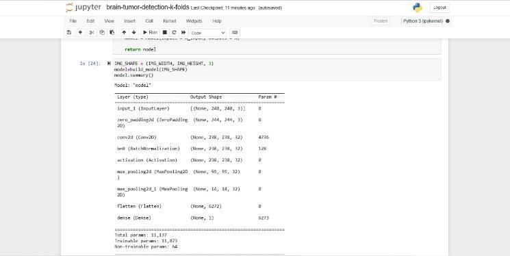
6.3WORKING OF CNN MODEL
Convolution 2D: Convolution 2D focuses on the highlighted portion of the input image. It provided the outcomewithalatticestructure.
MAX Poolig2D: It takes the biggest component from the amendedelementmapinMAXsurveying2D.
Dropout:Randomlyselectedneuronsareignoredduring preparation,whichresultsindropout.
Flatten: Level feed yield into every layer that is associated.Thedataarepresentedasalist.
Dense: a direct activity in which the weight of each information is linked to each result. Following that was thenonlinearactivationfunction.
6.4 K FOLDS ENDORSEMENT
Make a CNN model and train a significant cerebrum network for a very long time after that. The CNN computation's accuracy mean worth is endorsed by K
FOLDS. The K-fold cross-validation technique is used to divide the input dataset into K groups of samples of equal size. Folds are the names of these examples. The expectationcapabilitymakesuseofthek-1foldsforeach learning set, while the other folds are used for the test set. Sinceitis easy tograsp anddelivers resultsthat are lessone-sidedthanthosecreatedbydifferentstrategies, this approach is generally utilized in CVs. By testing the CNN model with assessment metrics like exactness, awareness,explicitness,accuracy,review,andF1-score,K Foldsfurtherdevelopstheresults.Duetotheimpressive outcomes that the proposed system produces, K FOLDS VALIDATIONperformsbetterthanothermodels.
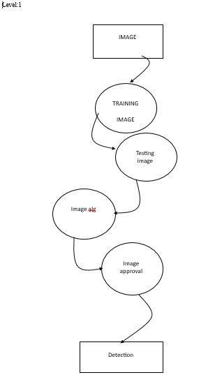
7. DATAFLOW DIAGRAM
Succession outlines in UML shows how item collaborate withoneanotherandtherequestthosecommunications happen. It's vital to take note of that they show the collaborations for a specific situation. Interactions are depicted as arrows, and the processes are shown vertically
VIII CONCLUSION
In conclusion, feature-based image processing investigates methods like image pre-processing, image segmentation, features extraction, and classification. Additionally, find out about CNN's profound learning strategies. We were able to determine the accuracy and loss of the system, as well as whether or not the tumor was present, by plotting the graph. Later on, Folds approval will be utilized to decide the mean worth of precision.BytestingtheCNNmodelutilizingassessment measurements like exactness, awareness, particularity, accuracy, review, and F1-score, K Folds further develops results. Due to the impressive outcomes that the proposed system produces, K FOLDS VALIDATION performsbetterthanothermodels.
IX.FUTURE WORK
The proposed work's dataset challenge must be overcome in the future work. Applying the model to additional publicly available large brain tumor datasets canenhanceperformance.Wecanutilizethisexploration work introduced as the basis for one more examination in the clinical field. In order to construct a trustworthy model based on multiple instances of medical field learning, there is a lot of room for improvement. Framework Execution In the order challenge for identifyingamindgrowthinX-raypictures,theessential key to the characterization cycle is the ID of a right example.Thegrouping modelsfacevariousissuesinthe X-ray pictures, which can prompt miss learning and a decreaseinorderexactness.
X.RESULT& DISCUSSION
Plottingthegraphallowedustoascertainwhetherornot the tumor was present, as well as the accuracy and loss of the system. The mean value of precision will be determined later using Folds approval. By testing the CNN model using evaluation estimations like precision, mindfulness,distinction,exactness,survey,andF1-score, K Overlap further creates results. K FOLDS VALIDATION outperforms other models due to the impressive results producedbytheproposedsystem.
REFERENCES
10.2ImageSegmentation
10.1CNNImplementation
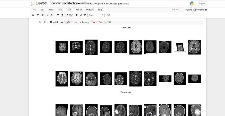
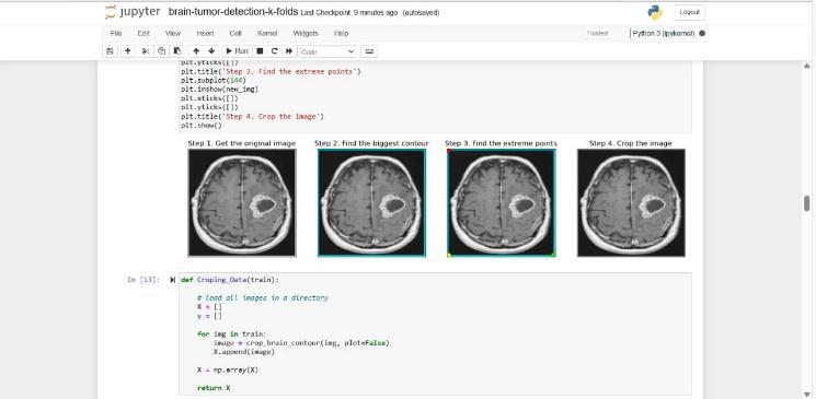
10.3TumorDetection
1] Sartaj. Brain Tumor Classification (MRI) Dataset. Accessed: Jun. 10, 2021. [Online]. Available: https://www.kaggle.com/ sartajbhuvaji/brain-tumorclassification-mri [17]N.Chakrabarty.BrainMRIImages for Brain Tumor Detection Dataset. Accessed: Jun. 10, 2021. [Online]. Available: https://www.kaggle.com/ navoneel/brain-mri-images-for-brain-tumor-detection
[2] D. C. Preston. Magnetic Resonance Imaging (MRI) of theBrainandSpine:Basics.Accessed:Dec.31,2021.
[3] H. Mohsen, E.-S. A. El-Dahshan, E.-S. M. El-Horbaty and A.-B. M. Salem, “Classification using deep learning neural networks for brain tumors,” Future Computing andInformaticsJournal,pp.68-71,2018.
[4] S. Bauer, C. May, D. Dionysiou, G. Stamatakos, P. Buchler and M. Reyes, “Multiscale modeling for Image Analysis of Brain Tumor Studies,” IEEE Transactions on BiomedicalEngineering,vol.59,no.1,pp.25-29,2012.
[5] Islam, S. M. Reza and K. M. Iftekharuddin, “Multifractal texture estimation for detection and segmentation of brain tumors,” IEEE Transactions on BiomedicalEngineering,pp.3204-3215,2013.
[6] M. Huang, W. Yang, Y. Wu, J. Jiang, W. Chen and Q. Feng, “Brain tumor segmentation based on local independent projection-based classification,” IEEE
TransactionsonBiomedicalEngineering,pp.2633-2645, 2014.7]Hamamci,N.Kucuk,K.Karaman,K.EnginandG. Unal, “Tumor-cut: Segmentation of brain tumors on contrast enhanced MR images for radiosurgery applications,”IEEETransactionsonMedicalImaging,pp. 790-804,2012
[8] L. Luo et al., “Altered brain functional network dynamicsinobsessive–compulsivedisorder,”Hum.Brain Mapping,vol.42,no.7,pp.2061–2076,May2021.
[9] J. Calza et al., “Altered cortico–striatal functional connectivity during resting state in obsessive–compulsive disorder,” Frontiers Psychiatry, vol. 10, p. 319,May2019.
[10]M.DeNeelingandM.M.VanHulle,“Single-paradigm and hybrid brain computing interfaces and their use by disabledpatients,”J.NeuralEng.,vol.16,no.6,2019,doi: 10.1088/1741-2552/ab2706.