
International Research Journal of Engineering and Technology (IRJET) e-ISSN: 2395-0056
Volume: 13 Issue: 04 | Apr 2026 www.irjet.net p-ISSN: 2395-0072
International Research Journal of Engineering and Technology (IRJET) e-ISSN: 2395-0056
Volume: 13 Issue: 04 | Apr 2026 www.irjet.net p-ISSN: 2395-0072
Sachin G Lokapure1,2 , Saniya S. Pidde2 , Vasundhara N Patil2 , Pradnya P. Patil2 , Supriya K. Patil2 , Mouh P. Phom2
1 SAGLO Research Equipment’s and SAGLO R & D Division, Plot No. 41, Serve No. 104, MIDC Shah Lulla Mahanagar, Trutiya 3rd, Savali, Tal, Miraj, Maharashtra 416410
2Appasaheb Birnale College of Pharmacy Sangli, South Shivaji Nagar, Dist- Sangli, 416416, Maharashtra, India
Abstract - Textile engineering plays a crucial role in healthcare and public safety, where fiber microstructure directlyinfluencestheperformance of medicaland protective materials. This study presents the design, development, and evaluation of a novel, low-cost textile microscope fabricated using additive manufacturing. The device was constructed through the assembly of 32 precision-designed components using the Anet ET4 Plus 3D printer, with optimized parametersandpolylacticacid(PLA)astheprimarymaterial. TheCADmodel wasprocessedusingUltimakerCuratoachieve high-resolution fabrication without support structures. The performance of the developed textile microscope was comparatively evaluated against a standard Olympus microscope using various textile and healthcare-related samples, including surgical masks, N95 masks, dressing bandages, and cotton fibers. Observations indicate that the proposedmicroscopeprovidescomparableimagingcapability for fiber morphology, pore structure, and sample analysis, whileofferingsignificantadvantagesinportabilityandeaseof handling. The results demonstrate that fiber characteristics such as diameter, orientation, and porosity can be effectively analyzed usingthedeveloped system,supportingapplications in textile quality control, pharmaceutical materials, and protectivefabrics.Furthermore,themicroscopeshowsstrong potentialforindustrial,forensic,andeducationaluseduetoits lowcostandscalabilitythrough3Dprinting.Inconclusion,the proposed textile microscope represents a practical, costeffective alternative to conventional microscopy systems, enabling accessible and real-time fiber analysis for diverse applications in healthcare and textile engineering.
Key Words: Textile Microscopy, 3D Printed Microscope, Fibre Morphology, Additive Manufacturing, Healthcare Textiles,FiltrationEfficiency,PortableMicroscopy,Polylactic Acid(PLA)
Textileengineeringhasincreasinglybecomeavitalfieldin healthcare and public safety, where fiber science and microscopic analysis play a central role. From medical compressionbandagesusedintreatingvenouslegulcersto protectivefacemasksdesignedtoreduceviraltransmission,
the performance of these products is closelylinked to the microstructure of the fibers. Characteristics such as fiber orientation,porosity,andmechanicalbehaviorsignificantly affecttheireffectiveness,durability,andusercomfort[1,2].
Inthecaseofcompression bandages,researchshowsthat thedesignofwovenstructuresandthetreatmentofyarns are crucial in achieving the desired therapeutic pressure. Advancedtechniques,includingspectroscopicanalysis,have beenusedtoevaluatebandageporosityandpredictoptimal tensionlevels[3].Whenexperimentalfindingsarecompared with theoretical predictions based on Laplace’s law, noticeabledifferencesareoftenobserved,highlightingthe complex nature of textile behavior under practical conditions [1]. Materials such as cotton and viscose-lycra exhibitdifferentloadandelongationcharacteristics;cotton tends to generate higher pressure but may lead to fluctuations during movement [2]. Innovations such as double-weft constructions and the incorporation of silver nanoparticle-treated yarns demonstrate how fiber-level modificationscanenhanceantimicrobialperformanceand supportwoundhealing[4].
Similarly,thedevelopmentofprotectivefacemasksduring theCOVID-19pandemichighlightedtheimportanceoftextile fiberstructureinfiltrationefficiencyandsafety.Guidelines fromtheWorldHealthOrganizationemphasizetheroleof proper material selection and multi-layer construction in effectivemaskperformance[5].Microscopicstudiesreveal that fiber morphology, pore size distribution, and surface treatments directly influence a mask’s ability to trap particleswhilemaintainingbreathability[6].
Materials such as electro spun nanofibers, melt-blown polypropylene, and spun bond nonwovens have been extensively studied for their filtration capabilities at the nanoscale [9,12]. While nanofiber-based layers offer high surfaceareaforparticlecapture,theymayfacelimitationsin mechanical strength and durability. Hybrid structures combiningmicro-andnanofibers,particularlyinmodified melt-blownfabrics,haveshownimprovedperformanceby enhancing filtration pathways while maintaining user comfort[10].
Comparative studies of different face masks including surgical masks, cotton masks, FFP2 and FFP3 respirators,
International Research Journal of Engineering and Technology (IRJET) e-ISSN: 2395-0056
Volume: 13 Issue: 04 | Apr 2026 www.irjet.net p-ISSN: 2395-0072
antiviralpolyester,andsilkmasks providedeeperinsights intotheirstructuralandmechanicalproperties.Techniques such as scanning electron microscopy (SEM), pore size analysis,andtensiletestingshowthatmulti-layeredmasks like FFP2 and FFP3 achieve filtration efficiencies of up to ~98% due to dense fiber arrangements and reduced porosity [7,8]. However, this often comes with reduced mechanicalflexibility.Incontrast,cottonandsilkmasksoffer more uniform fiber orientation and better breathability, whileantiviralpolyestermasksincorporatetreatmentssuch assilverchlorideforself-sanitizingproperties[4,6].
Taken together, these studies emphasize a common principle:themicroscopiccharacteristicsoffibersformthe foundation for designing effective medical and protective textiles. Whether used for therapeutic compression or respiratory protection, these materials must balance performance, durability, and comfort. Advances in microscopy fromSEMimagingtospectroscopic porosity analysis enable precise characterization of textile structuresandguideinnovationsinyarnengineering,fabric construction, and multi-layer design [10,11]. Ultimately, textile science plays a transformative role in healthcare, wheremicroscopicfiberbehaviortranslatesintoimproved patientoutcomesandenhancedpublicsafety.
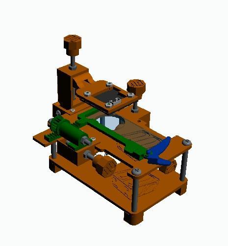
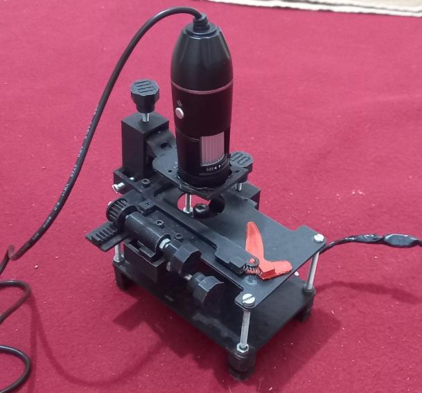
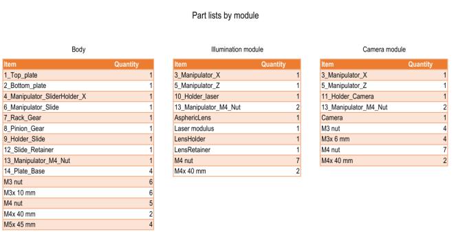
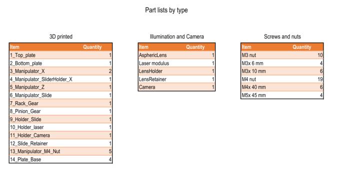
The development of the novel textile microscope was accomplished using advanced additive manufacturing techniques,involvingtheprecisefabricationandassemblyof 32individualcomponents.Theprintingprocesswascarried out using the Anet ET4 Plus (Model 84 Plus 1850, China), selected for its capability to deliver high dimensional accuracyandreliabilityin producingintricate geometries. The complete design was initially conceptualized and validated within a computer-aided design (CAD) environment, where each component was meticulously dimensioned and verified to ensure proper alignment, tolerancecontrol,andfunctionalintegration.
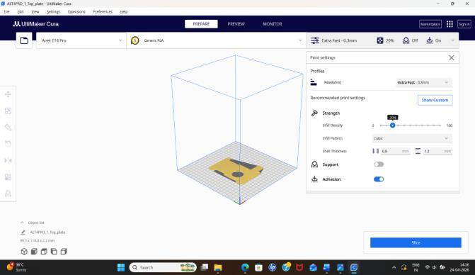
Following validation, the finalized CAD model was exported to Ultimaker Cura (version 5.10.2), which was employed as the slicing platform to generate machinereadable instructions (G-code) for the printing process. Material selection was carefully considered, and PLA (polylacticacid)waschosenduetoitsfavourableproperties, includingbiocompatibility,easeofprocessing,andadequate mechanicalstrengthforstructuralcomponents.
Toachieveoptimalperformance,theprintingparameters weresystematicallyoptimized.Ahighinfilldensityof90% wasimplementedusingahoneycombpattern,providinga balancebetweenstructuralstrengthandmaterialefficiency. Thelayerthicknesswasmaintainedat0.1mmtoensurefine resolution,smoothsurfacefinish,andaccuratereproduction ofmicro-scalefeatures.Buildplateadhesionwasmaintained
at 100% throughout the process to eliminate risks of warpingordetachment.Notably,the designwasexecuted without the need for support structures, reflecting the efficiency of the CAD optimization and print orientation strategy.
The resulting device was a fully functional textile microscope, fabricated with high fidelity to the original designspecifications.Thisworkdemonstratesthepractical feasibility of utilizing 3D printing technology for the development of advanced scientific instrumentation. Furthermore,theapproachhighlightssignificantadvantages over conventional commercially available microscopes, includingreducedmanufacturingcost,enhancedportability, and the ability to customize design features for specific textileanalysisapplications.
International Research Journal of Engineering and Technology (IRJET) e-ISSN: 2395-0056
Volume: 13 Issue: 04 | Apr 2026 www.irjet.net p-ISSN: 2395-0072
1.2. Preparation of slides
Firstly,theslidesarewashedwithalcoholandairdried.The sample is then loaded on the slide, depending upon the nature of the sample paraffin oil is used if necessary. The coverslipismounteduponthesample.
1.3 Observing the prepared slide under the Olympus microscope
Thepreparedslideisthenobservedunderthemicroscope underthesuitablemagnification.Theimageiscapturedwith thehelpofmicroscopecameraandsoftware.
1.4. Observing the Fiber sample under the Textile microscope
When observing under Textile microscope the sample is directlyplacedonthesensorofthemicroscope,ifthesample is solid in nature paraffin oil is used which aids in better resolution.Thesampleisdirectlyobservedonascreenand an image is taken with the help of software for the comparativestudies.
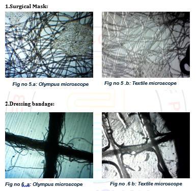
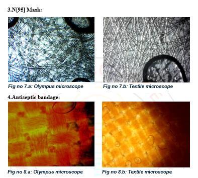
Textilemicroscopyoffersstrongmarketingapplicabilityby transformingtechnicalvalidationintoconsumer-facingvalue propositions. By enabling fiber identification, brands can confidentlymarketauthenticityandpremiumquality;fabric structure analysis provides evidence of innovation and durability that can be showcased in campaigns; blend analysis supports performance-based claims such as comfort,elasticity,orresilience;qualitycontrolanddefect detectionreinforcenarrativesofflawlesscraftsmanshipand reliability; and counterfeit detection strengthens brand protection and consumer trust. The result obtained after observingthedifferentsamplesunderboththemicroscopes suggeststhattheTextilemicroscopeisequallygoodasthe Olympus microscope which is used as a standard for this experiment, and the Textile microscope having handling advantagesalongwithitsportabilitymakesitfairlybetter microscopeforemergencytestingandothermicroscopically applications.Thecomparativestudyisshownintable1.
Table -1: FabricSamplesusedforcomparisonstudy.
Sr.no Samples Type of samples Typical size range Pharmaceutica l studies 1 Surgical mask
~1030μm Usedas protective barrier; prevents microbial contamination inhospitalsand pharmaceutical manufacturing areas.
Usedforwound dressing;study offiber structurehelps in understanding absorbencyand drug-loaded dressing materials.
International Research Journal of Engineering and Technology (IRJET) e-ISSN: 2395-0056
Volume: 13 Issue: 04 | Apr 2026 www.irjet.net p-ISSN: 2395-0072
3 N[95] mask solid ~110μm
High-efficiency filtration(≥95% for0.3µm particles); importantin infection controland sterile pharmaceutical environments.
4 Antiseptic bandage solid ~1525μm Contains antimicrobial agents; microscopic studyhelps evaluatefiber porosityand drugretention capacity
5 Cotton solid ~1225μm Naturalfiber usedin pharmaceutical preparations (absorbent cotton,gauze); analyzedfor purity,fiber morphology, and absorbency.
The output received after observing the samples under both the microscopes:
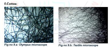
The novel textile microscope enables fiber identification, blendanalysis,anddefectdetection,ensuringrawmaterial authenticityandconsistentproductstandards.Itsportable design makes it suitable for on-site inspections in mills, garmentfactories,andexporthouses.
The microscope is applicable in analyzing surgical masks, N95 masks, dressing bandages, and antiseptic bandages. Theseapplicationsarecriticalwherefibermorphologyand porositydirectlyaffectsafetyandtherapeuticperformance. Italsosupportsthedevelopmentofdrug-loadeddressings andantimicrobialfabricsbyvalidating fiberstructureand retentioncapacity.
Themicroscopeisusefulincounterfeitdetectionofbranded fabricsandmedicaltextiles,protectingbothmanufacturers and consumers. It can be marketed to forensic labs and regulatoryagenciesforauthenticityverification.
The affordable, 3D-printed design makes the microscope accessible for academic institutions, textile R&D labs, and
International Research Journal of Engineering and Technology (IRJET) e-ISSN: 2395-0056
Volume: 13 Issue: 04 | Apr 2026 www.irjet.net p-ISSN: 2395-0072
trainingcenters.Itencouragesinnovationinfiberscience, nanofiberresearch,andprotectivetextiledevelopment.
1. Cost Advantage
Being 3D-printed with PLA material, the novel textile microscope is significantly cheaper than conventional brandedmicroscopeslikeOlympusMagnus.Itsportability andeaseofhandlingaddvalueforindustriesneedingrapid, field-leveltesting.
2. Target Market Segments
The microscope can serve various sectors including the textile and apparel industry, pharmaceutical and medical device industry, educational institutions,and forensicand regulatoryagencies.
3. Commercial Positioning
Itcanbemarketedasacost-effectivealternativetoOlympus Magnusforroutinetextileanalysis.Itsuniqueportabilityand emergency testing capability provide differentiation in competitivemarkets.Thepotentialforscalingproductionvia 3D printing makes it attractive for startups and small laboratories.
3. CONCLUSIONS
The novel textile microscope bridges affordability with functionality, making it highly applicable across textiles, healthcare, education, and forensic industries. Its market value lies in being a portable, low-cost, yet reliable alternativetoestablishedmicroscopes,withstrongpotential for adoption in both developing and advanced industrial settings.
ACKNOWLEDGEMENT
The authors thank R & D team of SAGLO RESEARCH EQUIPMENT Industry for development of this device. And AppasahebBirnalePharamcyCollege,Sangliforsupport.
[1] Partsch H, Clark M, Bassez S, et al. Measurement of lower leg compression in vivo. Dermatol Surg. 2006;32(2):224–233.
[2] ThomasS.Compressionbandagesinvenouslegulcers.J WoundCare.1997;6(6):327–330.
[3] ReddyKM,GuptaR,etal.Textilebandageperformance usingporosityanalysis.TextResJ.2012;82(5):475–484.
[4] Ramakrishnan G, Venkatachalam G. Antimicrobial textiles using silver nanoparticles. J Ind Text. 2011;41(1):45–57.
[5] World Health Organization. Advice on mask use in COVID-19.Geneva:WHO;2020.
[6] Konda A, Prakash A, Moss GA, et al. Aerosol filtration efficiencyoffabrics.ACSNano.2020;14(5):6339–6347.
[7] LeungNHL,ChuDKW,ShiuEYC,etal.Respiratoryvirus sheddingandmasks.NatMed.2020;26:676–680.
[8] DrewnickF,PikmannJ,etal.Aerosolfiltrationefficiency of mask materials. Atmos Chem Phys. 2021;21:1993–2008.
[9] PodgórskiA,BałazyA,GradońL.Nanofibersinfiltration. ChemEngSci.2006;61(20):6804–6815.
[10] Hutten IM. Handbook of Nonwoven Filter Media. Elsevier;2016.
[11] Kellie G. Nonwoven Fabrics: Raw Materials, Manufacture,Applications.WoodheadPublishing;2016.
[12] ZhangS,LiuH,YinX,etal.Electrospunnanofibersfor filtration.Nanomaterials.2019;9(1):1–23.





SachinGLokapure Director SAGLOResearchEquipment’sand SAGLOR&DDivision Assist.Prof@ABCP,Sangli
SaniyaS.Pidde ResearchScholar@ABCP,Sangli
VasundharaNPatil ResearchScholar@ABCP,Sangli
PradnyaP.Patil ResearchScholar@ABCP,Sangli
SupriyaK.Patil ResearchScholar@ABCP,Sangli
International Research Journal of Engineering and Technology (IRJET) e-ISSN: 2395-0056
Volume: 13 Issue: 04 | Apr 2026 www.irjet.net p-ISSN: 2395-0072

P.Phom ResearchScholar@ABCP,Sangli