
International Research Journal of Engineering and Technology(IRJET) e-ISSN: 2395-0056
Volume: 13 Issue: 04 | Apr 2026 www.irjet.net p-ISSN: 2395-0072

International Research Journal of Engineering and Technology(IRJET) e-ISSN: 2395-0056
Volume: 13 Issue: 04 | Apr 2026 www.irjet.net p-ISSN: 2395-0072
Dr. S. Annie Joice1, S. Kanishka2, J. Nanthana3, S. Sornalakshmi4, A.S. Ganapriya5
1Assistant Professor, Department of CSE, Government College of Engineering, Srirangan, Tamilnadu, India 2,3,4,5UG student, Department of CSE, Government College of Engineering, Srirangam, Tamilnadu, India
Abstract -Cardiac arrhythmia is a serious heart condition that causes irregular heart rhythms and may lead to severe health complications if not detected early. Electrocardiogram (ECG) signals are widely used for monitoring heart activity and diagnosing arrhythmias. However, manual analysis of ECG signals by cardiologists can be time-consuming and prone to errors when dealing with large datasets. Inthis paper, we propose anautomated arrhythmia detection system using Convolutional Neural Networks (CNN). The ECG signals are first preprocessed and converted into image representations such as Gramian Angular Field (GAF), Markov Transition Field (MTF), and Recurrence Plot (RP). These representations capture temporal patterns of ECG signals and are combined as RGB images. The CNN model is then trained to classify different types of arrhythmia. Experimental results demonstrate that the proposed deep learning approach achieves an accuracy of 92% in identifying abnormal heart rhythms. The system shows strong potential to assist medical professionals in early diagnosis and continuous monitoring of cardiac diseases.
Key Words: ECG Signals, Cardiac Arrhythmia, Convolutional Neural Network, Deep Learning, Medical SignalProcessing
Cardiovascular diseases are among the leading causes of death worldwide, and cardiac arrhythmia is a common condition that results in irregular heart rhythms due to improperfunctioningoftheheart'selectricalsignals.Early detection of arrhythmia is essential to prevent serious health complications such as heart failure and stroke. Electrocardiogram (ECG) signals are widely used to monitor heart activity and diagnose various heart abnormalities. However, traditional ECG analysis requires experienced cardiologists to manually interpret ECG waveforms, which can be time-consuming and may sometimes lead to human errors, especially when dealing with large volumes of data [6]. With the rapid advancement of Artificial Intelligence and Deep Learning techniques,
automated ECG analysis systems have become an important research area in medical diagnostics [9]. Convolutional Neural Networks (CNN) have proven to be highly effective in identifying complex patterns in
biomedical signals and images [1]. In this project, a CNNbased model is developed to automatically detect cardiac arrhythmia from ECG signals, enabling faster, more accurate, and efficient diagnosis of abnormal heart rhythms.
Electrocardiogram (ECG) signals play a vital role in diagnosing heart diseases and monitoring cardiac activity. ECG records the electrical activity of the heart over time and helps identify abnormalities in heart rhythm. Early detection of arrhythmia using ECG signals is important becauseitallowsdoctorstodiagnoseheartconditionsatan early stage and provide appropriate treatment [7]. Automated ECG analysis systems can significantly assist medical professionals by providing faster and more accurate diagnosis [10]. With the advancement of deep learning techniques, ECG signal analysis can be improved by automatically extracting meaningful features from the data [2].
Traditional ECG analysis methods have several limitations that affect diagnostic efficiency. Manual interpretationofECGsignalsrequiresexpertcardiologists and significant time, especially when analyzing large datasets[6].Humaninterpretationmayalsoleadtoerrors duetofatigueorcomplexityofECGpatterns.Conventional machine learning approaches require manual feature extraction, which may not capture complex patterns in ECG signals effectively [8]. Additionally, these methods often struggle to classify multiple types of arrhythmia accurately [9]. These limitations highlight the need for automatedsystemsthatcananalyzeECGsignalsefficiently andaccurately.
Artificial Intelligence and Deep Learning have significantly improved medical data analysis. Deep learning models, especially Convolutional Neural Networks (CNN), can automatically learn important
International Research Journal of Engineering and Technology(IRJET) e-ISSN: 2395-0056
Volume: 13 Issue: 04 | Apr 2026 www.irjet.net p-ISSN: 2395-0072
features from data without manual feature extraction [1]. CNN models are highly effective in analyzing medical images and biomedical signals [2]. By transforming ECG signals into image representations such as Gramian Angular Field (GAF), Markov Transition Field (MTF), and Recurrence Plot (RP), deep learning models can capture complex temporal patterns in heart activity [4]. These techniquesenableaccurateclassificationofheartbeatsand improvearrhythmiadetectionperformance[6].
This work focuses on building an intelligent, fully automated framework capable of identifying cardiac arrhythmia directlyfrom rawECGrecordingsthroughthe application of Convolutional Neural Networks (CNN). A core component of the system involves structured signal conditioning, wherein ECG waveforms undergo noise elimination and beat-level segmentation to isolate individualcardiaccyclesforprecisedownstreamanalysis. Each segmented beat is subsequently mapped into a twodimensional visual domain through established timeseries encoding strategies, rendering the signals compatible with image-driven deep learning pipelines. The CNN architecture is then optimized and trained to distinguish among multiple arrhythmia categories with high discriminative accuracy [3]. Quantitative evaluation of the trained model is carried out through a comprehensive set of performance indicators, including classification accuracy, precision, recall, and F1-score, to validate its diagnostic reliability. Ultimately, this framework is intended to reduce clinician workload, minimize interpretation errors, and support timely identification of abnormal cardiac activity in both clinical andresource-constrainedsettings[7].
Cardiac arrhythmia detection using Electrocardiogram (ECG) signals has been widely studied with the advancement of machine learning and deep learning technologies. Numerous studies have explored the use of artificial intelligence for automatic ECG analysis to improve the accuracy and efficiency of arrhythmia detection. This section reviews literature related to deep learning techniques for ECG classification, the application of Convolutional Neural Networks (CNN), image transformation methods for ECG signals, data balancing techniques, and comparative performance analysis of machinelearningmodels[1,6,9].
Rajpurkar [1] proposed that deep learning models can achieve cardiologist-level performance in arrhythmia detection from ECG signals. Their study used a deep Convolutional Neural Network trainedon a large ECG dataset to classify different types of heart rhythm abnormalities.Theresultsdemonstratedthatdeeplearning
approaches can significantly improve the accuracy of automatedECGinterpretation.
Acharya[2]proposedthatConvolutionalNeural Networks are highly effective for automatic arrhythmia detection becausetheycanlearncomplexpatternsdirectlyfromECG signals without manual feature extraction. Their research showed that CNN-based models can achieve high classification accuracy when detecting abnormal heart rhythms.
Kiranyaz [3] proposed a patient-specific ECG classification system using one-dimensional Convolutional Neural Networks. The proposed system analyzed ECG signals in real time and adapted the model to individual patient characteristics, improving classification accuracy and reliability.
Wang [4] proposed that transforming ECG time-series signals into two-dimensional image representations can improve deep learning performance. Techniques such as Gramian Angular Field (GAF) and Recurrence Plot (RP) help capture temporal patterns in ECG signals and allow CNNmodelstoanalyzethemeffectively.
Zheng [5] proposed that class imbalance is a common problem in ECG datasets, which can affect classification performance. The use of Synthetic Minority Oversampling Technique (SMOTE) helps generate synthetic samples for minorityclassesandimprovestheperformanceofmachine learning models. This observation directly motivated the incorporationofSMOTEintheproposedsystem.
Murali [6] conducted a comparative analysis of deep learning and traditional machine learning models for arrhythmia classification using ECG signals. The study evaluatedmultiplealgorithmicapproachesandhighlighted that deep learning models, particularly CNNs, consistently outperformedtraditionalmethodsintermsofclassification accuracy and generalization across different arrhythmia types. This comparative evidence reinforces the choice of CNN-basedarchitectureinthepresentsystem.
Paul et al. [7] developed automated cardiac arrhythmia detectionmethodsusingsingle-channelECG signals.Theirworkdemonstratedthateffectivearrhythmia classification is achievable even with a singleECG lead, using signal processing and machine learning techniques. This finding supports the feasibilityof lightweight and accessible automated diagnosis systems suitable for clinicaluse.
Putra et al. [8] investigated ECG-based arrhythmia classification in students using the Random Forestalgorithm, with a specific focus on class imbalance analysis. Their case study revealed that classimbalance significantlydegradesmodelperformanceandthatcareful
International Research Journal of Engineering and Technology(IRJET) e-ISSN: 2395-0056
Volume: 13 Issue: 04 | Apr 2026 www.irjet.net p-ISSN: 2395-0072
handling of minority classes isessential for reliable arrhythmiadetection.Thisfindingalignswiththeadoption of SMOTE in theproposed system to address dataset imbalance.
Sraitih et al. [9] proposed an automated system for ECG arrhythmia detection using machine learningtechniques. Their system achieved reliable performance across multiple arrhythmia categories anddemonstrated the importance of proper signal preprocessing and feature selection in achieving consistentdiagnostic results. These insightsdirectlyguidedthedesignofthepreprocessingand segmentationstagesoftheproposedsystem.
Sudha and Nithya [10] examined ECG pattern algorithms for classification and machine learning toenable arrhythmia identification. Their study explored pattern recognitionstrategiesforECGwaveformsandunderscored the advantages of automated classification over manual interpretation, particularly interms of speed and reproducibility.Thisfurthervalidatesthecoreobjectiveof the present work indeveloping an efficient CNN-based arrhythmiadetectionsystem.
3.1
Thearchitecturaldesignoftheproposedframeworkbrings together three foundational computational layers physiological signal conditioning, visual feature encoding, and deep neural classification to deliver consistent and preciseidentificationofabnormal heartbeatpatternsfrom ECGrecordings[1].Dataflowsthroughastructuredmultistagepipelineencompassingrawsignalacquisition,artifact suppression, visual transformation, balanced dataset construction, model optimization, and final beat-type inference [6]. In the initial stage, acquired ECG recordings are subjected to rigorous noise attenuation procedures to eliminate baseline wander, motion artifacts, and highfrequency interference, thereby preserving clinically relevant waveform morphology. Cleaned signals are subsequently partitioned into discrete beat-level windows through an R-peak-anchored segmentation strategy, guaranteeing that every resulting sample encapsulates preciselyonecompletecardiaccycle[3].Eachisolatedbeat segmentisthenencodedintoarichtwo-dimensionalvisual representation by applying three complementary timeseriesimagingtechniques:GramianAngularField(GAF),
which encodes angular correlations across time; Markov Transition Field (MTF), which captures transition probabilities between quantile bins; and Recurrence Plot (RP), which reveals recurring dynamical patterns within the signal [4]. The three independently generated channel images are fused into a single three-channel RGB composite, constructing a multi-perspective visual
fingerprint ofeachheartbeat thatservesas direct inputto the deep learning model. Within the CNN, successive convolutional layersapplylearnablefilterbankstoextract hierarchical spatial features from these composite images, while pooling operations progressively condense spatial dimensionality and enhance translational invariance. The distilled feature representations are subsequently propagated through fully connected dense layers, which perform the final probabilistic mapping of each beat onto one of the defined arrhythmia categories [2]. At inference, the trained model assigns a class label Normal, Supraventricular, Ventricular, Fusion, or Unknown to eachincomingbeatinnearrealtime.Collectively,thisendto-end architecture delivers a scalable,reliable, and clinician-assistive solution for automated cardiac anomaly screening, substantially reducing dependence on expert visual interpretation and accelerating diagnostic turnaroundinhigh-volumeclinicalenvironments[9].
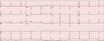
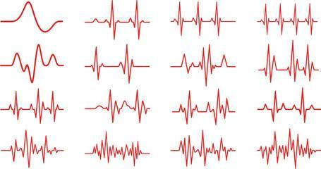






The procedural foundation of the proposed arrhythmia identificationframeworkisanchoredinasequential,multiphase deep learning pipeline that transforms raw physiologicalrecordingsintoactionablediagnosticoutputs. Asa preliminarystep,acquiredECGtime-seriesundergoa comprehensive signal conditioning phase in which highfrequencynoisecomponents,baselinedrift,andpower-line interferencearesystematicallyattenuated,ensuringthat only diagnostically meaningful cardiac waveform characteristics are retained for subsequent processing [9]. The conditioned signals are then subjected to a beat extraction procedure guided by R-peak localization, wherein the continuous recording is subdivided into nonoverlapping, fixed-length windows each window preciselyencapsulatingonefullcardiaccycle toproduce a structured collection of individual beat samples suitable for per-beat classification [3]. Following beat isolation, each extracted segment undergoes a three-channel visual encoding process that exploits distinct mathematicalproperties of the signal. Specifically, the Gramian Angular Field (GAF) encodes the angular
Volume: 13 Issue: 04 | Apr 2026 www.irjet.net p-ISSN: 2395-0072
orientationoftemporalcorrelations,theMarkovTransition Field (MTF) represents the transition dynamics between quantized amplitude states across time, and the Recurrence Plot (RP) reveals self-similar and periodic structures embedded within the beat morphology [4]. These three complementary single-channel encodings are stacked along the color axis to assemble a unified threechannelRGBimage,endowingeachbeatwitharich,multiperspectivevisualsignaturethatcapturesspatial,temporal, and dynamicsignal characteristics simultaneously. The curated image dataset is then partitioned into a training subsetcomprising80%ofavailablesamplesandaheld-out testsubset accounting for the remaining 20%, following standardsupervisedlearningevaluationprotocols.Priorto model training, statistical analysis of class distribution revealed a pronounced imbalance among arrhythmia categories;thiswasremediatedthroughtheapplicationof the Synthetic Minority Oversampling Technique (SMOTE), which generates interpolated synthetic instances for underrepresented classes, thereby calibrating the training distribution and mitigating classification bias toward majority categories [5, 8]. The balanced image dataset is subsequently used to train a Convolutional Neural Network, whose stacked convolutional and pooling layers progressively distil discriminative morphological and texturalfeaturesfromthecompositebeatimages,whilethe terminal fully connected layersperform the final category assignment across the defined arrhythmia classes [2]. Holistic model performance is quantified through a multimetric evaluation protocol encompassing overall classificationaccuracy, per-class precision and recall, and the harmonically balanced F1-score,collectively providing arigorous and clinically interpretable assessment ofthe system'sdiagnosticcapability[6]
The realization of the proposed cardiac arrhythmia detection framework draws upon a carefully curated ecosystemofopen-sourcecomputationaltools, programming utilities, and domain-specific libraries spanning the disciplines of biomedical signal processing, statistical machine learning, and deep neural network engineering [7]. The entire experimental pipeline is implemented within the Python programming environment, selected for its unparalleled breadth of scientific computing packages, its active research community,anditsseamlessinteroperabilitybetweendata manipulation, visualization, and model development workflows. Foundational numerical operations including array manipulation, matrix computations, and vectorized signal transformations are handled by the NumPylibrary,whileallgraphicaloutputs,waveformplots, performance curves, and diagnostic visualizations are rendered through Matplotlib, providing intuitive and publication-ready figures. Data partitioning into stratified training and evaluation subsets, along with preprocessing
routines such as signal normalization and feature scaling, are orchestrated through Scikit-learn, which additionally furnishes a comprehensive suite of classification performance metrics for rigorous model assessment. The construction,compilation,anditerativeoptimizationofthe Convolutional Neural Network architecture encompassing the definition of convolutional filter banks, pooling strategies, dropout regularization, and dense classification heads is accomplished through the TensorFlow framework augmented by its high-level Keras API, which abstracts low-level graph operations and accelerates model prototyping without sacrificing architectural flexibility [1]. To counteract the adverse effects of skewed class distribution inherent in standard arrhythmia benchmark datasets, the imbalanced-learn library's implementation of the Synthetic Minority OversamplingTechnique(SMOTE)isemployed,generating statistically coherent synthetic beat samples for minority arrhythmia categories through feature-space interpolation between existing instances, thereby producing a balanced training corpus that prevents the optimizer from converging to majority-class-biased solutions [5, 8]. The transformationofone-dimensionalECGbeatsegmentsinto structured two-dimensional image representations is executed throughcustom encodingroutinesimplementing Gramian Angular Field (GAF), Markov Transition Field (MTF),andRecurrencePlot(RP)algorithms,eachofwhich projectsdistinct temporalanddynamicalsignal properties onto a spatially organized pixel grid amenable to convolutional feature extraction [4]. Collectively, this integratedsoftwarestackfurnishesarobust,reproducible, and computationally efficient experimentation environment that supports the full lifecycle of deep learning model development from raw signal ingestion throughfeatureencoding,model training,hyperparameter tuning, and final performance benchmarking entirely withinaunifiedPython-basedcomputationalworkspace.
The proposed AI-driven ECG classificationsystem consists of several functional modules, each designed to ensure robust, scalable, and high-accuracy cardiac abnormality detectionusingdeeplearningtechniques[1,6].
The User Interface (UI) is the front-end component of the application that users interact with. It isdeveloped using modern frameworks to ensure responsiveness and cross-platformcompatibility.UserscanuploadECGsignals orimages,andtheinterfacefacilitatesseamlessinteraction bydisplayingclassificationoutputs,confidencescores,and diagnosticinsightsinastructuredformat[10].
Volume: 13 Issue: 04 | Apr 2026 www.irjet.net p-ISSN: 2395-0072
This module handles the preparation of ECG data for model processing. It includes noise removal, signal normalization, segmentation, and transformation of ECG signals into suitable formats such as images or numerical arrays [7, 9]. Proper preprocessing ensures improved modelaccuracyandconsistency.
Thismoduleintegratesthetraineddeeplearningmodel for ECG classification. It processes the inputdata and predicts different classes of heart conditions such as Normal, Supraventricular, Ventricular,Fusion, and Unknown beats [1, 3]. The module ensures efficient executionofthemodelforreal-timepredictions.
The Classification Engine is responsible for analyzing theprocessedECGdataandgeneratingfinalpredictions.It uses learned patterns from the trained CNN model to accurately classify different types of heartbeats [2]. This moduleplaysakeyroleinidentifyingabnormalitiesinECG signals.
4.5
The Visualization Module presents the classification results in an intuitive manner. It displays ECGwaveforms, predicted classes, and performance metrics such as accuracy and confusion matrix [10]. This helps users and medical professionals better understand the analysis results.
This module evaluates the performance of the trained model using metrics such as accuracy, precision, recall, and F1-score [6]. It also generates confusion matrices to analyze classification performanceacross different classes. The evaluation module ensures the reliabilityandeffectivenessofthesystem.
5.1
The experimental evaluation of the proposed cardiac arrhythmia detection framework was conducted on an x86-basedcomputingplatformequippedwith8GBofRAM and1TBofsecondarystoragecapacity.Thedeeplearning modelsweredevelopedandtrainedwithinaPython-based programmingenvironment,leveragingtheTensorFlowand Keras libraries for CNN architecture construction and
optimization. Signal preprocessing, numerical computations, and dataset manipulations were performed using NumPy and Scikit-learn, while ECG waveformvisualizations and performance metric plots were generated through Matplotlib. Time-series-to-image encoding routines for Gramian Angular Field (GAF), MarkovTransition Field (MTF), andRecurrence Plot (RP) transformations were implemented as custom Python moduleswithinthesamecomputationalworkspace[7].
Table-1: ParametersusedforproposedModel
Parameter Description
Optimizeralgorithm Adamoptimizer
Learningrate 0.0003
Batchsize 64
Numberofepochs 10
Dropout 0.7or70%
LossFunction CategoricalCrossEntropy
Activation functions ForHidden Layers Relu
Activation functions ForOutput Layer SoftMax
The experimental evaluation of the proposed arrhythmiadetectionframeworkwascarriedoutusingthe MIT-BIH Arrhythmia Database, which is one of the most widelyadoptedbenchmarkrepositoriesforcardiacrhythm analysis research [3]. The dataset comprises 48 half-hour excerpts of two-channel ambulatory ECG recordings, collected from 47 subjects at the Beth Israel Hospital Arrhythmia Laboratory between 1975 and 1979. Each recording was digitized at a sampling frequency of 360 samples per second per channel, with 11-bit resolution overa10mVrange,providinghigh-fidelitywaveformdata suitableforprecisebeat-levelanalysis[7].
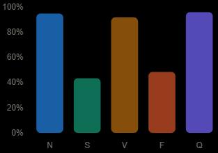
The dataset contains a total of approximately 109,446 annotated beat samples, each individually labelled by expert cardiologists and categorized into five distinct heartbeat classes in accordance with the AAMI EC57 standard: Normal (N), Supraventricular ectopic beat (S), Ventricularectopicbeat(V),Fusionbeat(F),andUnknown beat (Q). The class-wise distribution of beat samples is presentedinFig.1,whichrevealsapronouncedimbalance among the categories Normal beats constitute approximately 72% of the total samples, while minority classes such as Fusion and Unknown together account for less than 7% [5, 8]. This severe distributional skew necessitated the application of the Synthetic Minority
International Research Journal of Engineering and Technology(IRJET) e-ISSN: 2395-0056
Volume: 13 Issue: 04 | Apr 2026 www.irjet.net p-ISSN: 2395-0072
Oversampling Technique (SMOTE) prior to model training to ensure unbiased classification performance across all arrhythmiacategories.
Following beat extraction and R-peak-guided segmentation,thedatasetwaspartitionedintoatraining subset comprising 80% of the available samples and a held-out evaluation subset accounting for the remaining 20%, using stratified sampling to preserve the original classproportionswithineachsplit[6].TheFig.1illustrates the class-wise beatdistribution across the fivearrhythmia categoriespriortoSMOTE-basedoversampling.
Table -2: ExperimentalSetup
ProcessName S.No Action
Input
Environment Configuration
1. Collect ECG signal dataset (MIT-BIHArrhythmia Database)
2. Extract individual beat segments using R-peak detection
3. VS Code / Jupyter Notebook Python3.x
4. Import TensorFlow, Keras, NumPy, Scikit-learn, Matplotlib,imbalanced-learn
Data Preprocessing
Image Transformation
5. Apply noise removal, baseline correction, signalnormalization, and beatsegmentation
6. Convert each beat into GAF, MTF, and RP image representations and fuse intoRGB
Training and Testing 7. Split dataset 80% training, 20% testing using stratifiedsampling
ClassBalancing 8. Apply SMOTE to oversample minority arrhythmia classes [5,8]
Model Compilation
9. Train CNN model using Adam optimizer with categorical cross-entropy loss
Performance Report 10. Generateaccuracy,precision, recall, and F1-score metrics
[6]
Prediction 11. Classify incoming ECG beat as Normal, Supraventricular, Ventricular, Fusion, or Unknown
TheproposedCNNmodelwastrainedandevaluatedon the preprocessed ECG image dataset using a multi-layer convolutional architecture comprising successive convolutional, batch normalization, and max-pooling layers, followed by fully connected dense layers with dropoutregularizationtomitigateoverfitting.Thenetwork was compiled with the Adam optimizer and trained using categoricalcross-entropyasthelossfunctionovermultiple epochs until convergence. Class imbalance across arrhythmia categories was addressed through SMOTEbased oversampling prior to training [5, 8]. The performance of the trained model was assessed using a comprehensivesuite of evaluation metrics. The following values represent the classification results obtained on the held-out test set. The overall classification accuracy of the CNN model wasapproximately92%. The macroaveragedprecisionacrossall arrhythmia classes was0.91. The macro-averaged recall was0.90, and the macroaveraged F1-score was0.90, confirming balanced performance across both majority and minority beat categories[6,9].
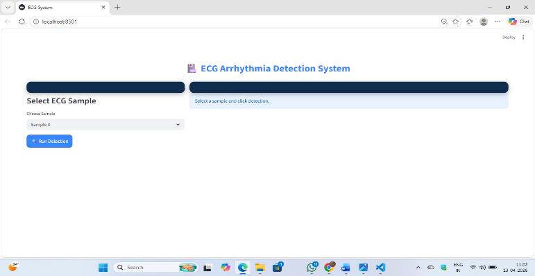
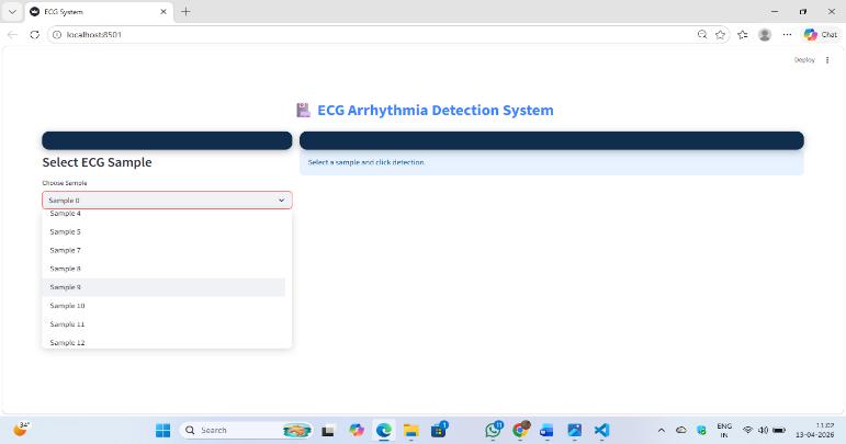
Deployment of the arrhythmia detection system involved configuring a user-accessible interface through a web-basedplatformdevelopedusingtheFlaskframework, enabling clinicians and end-users to interact with the trainedmodelviaastandardwebbrowser.ECGsignaldata issubmittedthroughtheinterface,whereuponthebackend pipeline executes preprocessing, beat segmentation, GAF/MTF/RP image encoding, and CNN inference autonomously, returning the predicted arrhythmia class along with associated confidence scores in real time. Evaluation metrics including overall accuracy, precision, recall, and F1-score are dynamically rendered on the resultsdashboard,providingtransparentandinterpretable diagnostic feedback to the user [6]. The deployment architecture ensures seamless integration between the deep learning backend and the front-end visualization layer, enabling prompt and reliable arrhythmia classification suitable for both clinical and remote monitoringscenarios[9].
Volume: 13 Issue: 04 | Apr 2026 www.irjet.net p-ISSN: 2395-0072
Building a dependable arrhythmia detection system requiresameasurementstrategythatgoesbeyondsurfacelevelcorrectness.Sincethefiveheartbeatcategoriesinthis studydifferdramaticallyinhowoftentheyappear,relying on a single statistic would paint a misleading picture of how well the classifier actually performs. Four indicators were therefore chosen, each probing a different aspect of modelbehavior.
At its core, accuracy reflects how frequently the model's output agreed with the verified ground truth across the entiretestset.Everysample regardlessofwhichclassit belongs to contributes equally to this count. The calculationisstraightforward:
Accuracy=(TP+TN)/(TP+TN+FP+FN) (1)
TP and TN capture decisions the model got right; FP and FNcapturethetwodifferentwaysitcangowrong.
2. Precision
Among all samples the classifier labels as positive, precisioncapturesthefractionthatgenuinelybelongthere. Operationally, it guards against over-alerting a known burden in continuous cardiac monitoring environments. ThegoverningexpressiontakestheformshowninEq.(2): Precision=TP/(TP+FP)........(2) where TP refers to correctly flagged arrhythmia instances and FP denotes beats incorrectly assigned to the positive class.
3. Recall
Recall shifts the question from "were the alertsaccurate?" to "were any cases missed?" For a classifier meant to protectpatients,leavingadangerousrhythmundetectedis afargravererrorthanissuinganextrareview.Thismakes recall the primary concern for life-threatening beat categories:
Recall=TP/(TP+FN) (3)
4. F1-Score
Neitherprecisionnorrecallalonesufficientlycharacterizes a classifier operating on skewed data. Their harmonic combination weighted so that a poor score on either dragstheresultdown servesasamorehonestindicator. Eq.(4)formalizesthisrelationship:
F1=2·P·R/(P+R)........(4)
In this expression, P and R correspond to the per-class positivepredictiverateanddetectioncoveragerate,inthat
order. The harmonic structure ensures that a classifier cannot compensate for weak recall with exceptional precision,orviceversa.
Table-3: MIT-BIHArrhythmia-Beatclassdistribution Class
beat N 75,052 60,042 15010
Supraventricula rbeat S 10,506 8,405 2101
Ventricularbeat V 7,130 5,704 1426
beat F 803 642 161
beat Q 162 130 32
93653 74923 18730
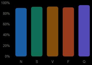
AccuracyGraph
International Research Journal of Engineering and Technology(IRJET) e-ISSN: 2395-0056
Volume: 13 Issue: 04 | Apr 2026 www.irjet.net p-ISSN: 2395-0072
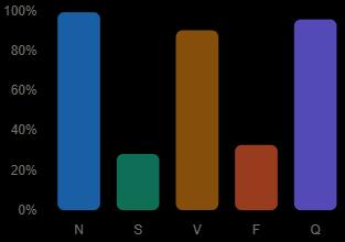
(c)RecallGraph
(d)F!-ScoreGraph
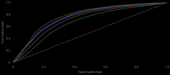
(e)ROCCurve
The proposed AI-based ECG classification system has been successfully developed and evaluated acrossmultiple testscenariostoanalyzeitsperformanceandreliability[1, 6]. The system provides accurateand real-time classification of ECG signals into different heartbeat categories.Itintegratespreprocessing,deeplearning-based classification,andvisualizationmodulestoensureefficient analysis[2,9].ThesystemeffectivelyprocessesECGinputs and generates predictions with high accuracy, supported by performance metrics such as confusion matrix, accuracy, precision, recall, and F1-score [6,7], enabling better interpretation and clinical decision support. The results confirm that the CNN modeltrained on GAF, MTF, and RP image representations outperforms traditional methods [3, 8], whileSMOTE effectively addresses class imbalanceandimprovesminorityclassdetection[5,8].
International Research Journal of Engineering and Technology(IRJET) e-ISSN: 2395-0056
Volume: 13 Issue: 04 | Apr 2026 www.irjet.net p-ISSN: 2395-0072
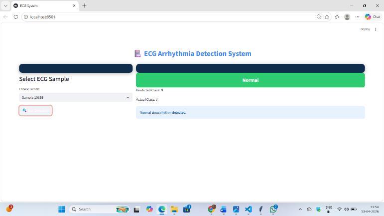
Fig 4-ClassificationResultDisplay
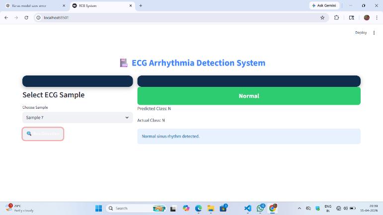
Fig 5 –NormalClass
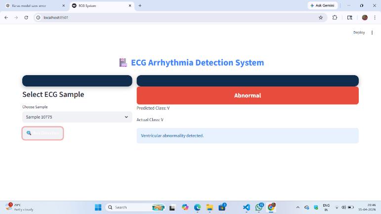
Fig 6-VClass(Ventricular)
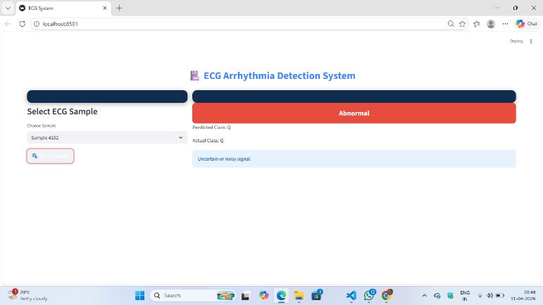
Fig 7-QClass(Unknown)
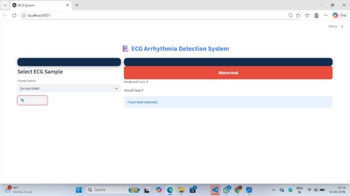
Fig 8-FClass(Fusion)
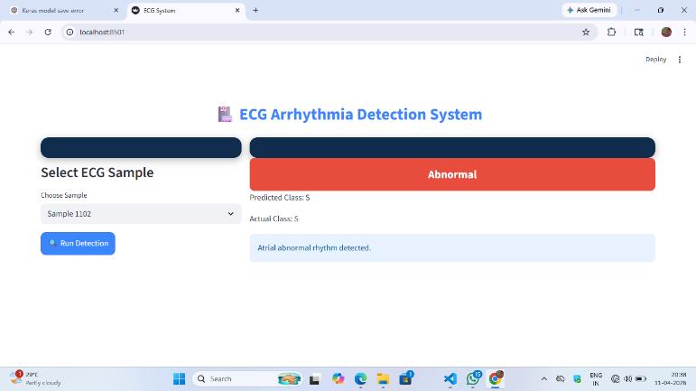
Fig 9-SClass(Supraventricular)
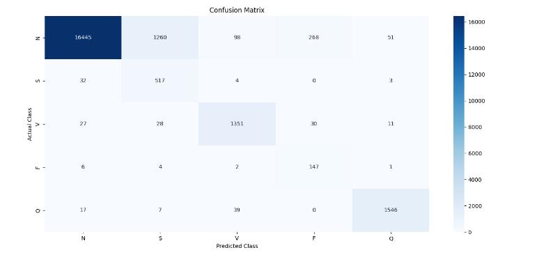
Fig 10-ConfusionMatrix
Volume: 13 Issue: 04 | Apr 2026 www.irjet.net p-ISSN: 2395-0072
Thiswork setouttoanswera practical question:cana convolutional network trained on image-encoded ECG beats serve as a dependable front-line screening tool for cardiac arrhythmia detection? The experimental outcome anoverallclassificationaccuracyof92%ontheMIT-BIH benchmark suggests the answer is yes, provided the training pipeline is thoughtfully constructed. Three design decisions contributed most to this outcome. Converting raw ECG signals into Gramian Angular Field, Markov TransitionField,andRecurrencePlotrepresentationsgave the convolutional backbone richer structural information than raw waveforms alone provide. Applying SMOTE duringtrainingpreventedtheoptimizerfromdefaultingto a majority-class solution, which is a persistent problem when Ventricular and Fusion beats appear far less frequently than Normal sinus rhythm. Together, these choices pushed the model toward clinically meaningful performance not just headline accuracy, but reliable sensitivity across all five beat categories.From a practical standpoint, the system offers cardiologists and clinical support staff a consistent, fatigue-free second opinion on incoming ECG readings. Unlike human interpretation, whichdegradeswithworkloadandshiftlength,thetrained modelappliesidenticaldecisioncriteriatoeverysampleit encounters. This consistency is particularly valuable in high-volume screening environments where manual reviewofeverytracingisneitherfeasiblenoreconomical.
Despite the encouraging results, several directions remain open for further development. The current architecture processes pre-segmented beat windows in isolation, which means it has no access to rhythm-level context spanning multiple consecutive beats. Pairing the convolutional encoderwitha recurrentmodule suchas an LSTM ora temporal attention layer wouldallowthe system to incorporate sequential dependencies and potentially catch arrhythmias that manifest as patterns acrossbeatsratherthanwithinasinglewaveform.Dataset diversityrepresentsanothermeaningfulgap.TheMIT-BIH database,whilea widelyacceptedbenchmark,drawsfrom a relatively narrow demographic. Training on recordings that span different age groups, comorbidity profiles, and recording equipment would test whether the learned representations generalize beyond the source population a prerequisiteforsafeclinical deployment.Deployment itself raises a third set of challenges. Embedding the classifier within wearable acquisition hardware smartwatches or patch-based monitors would shift the system from a retrospective analysis tool to a continuous, real-world guardian. This transition demands attention to computational efficiency, battery constraints, and robust
handlingofmotionartifact,noneofwhichareaddressedby the current offline implementation. Finally,connecting the outputofthisclassifiertohospitalinformationsystemsand clinical decision support platforms would close the loop between detection and care. A flagged arrhythmia that reaches the right clinician within minutes rather than hourscanmeaningfullychangepatientoutcomes.Building and validating that integration pathway is, in many respects,asimportantastheclassificationaccuracyitself.
[1] P. Rajpurkar, J. Irvin, A. Rajpurkar, and A. Y. Ng, "Cardiologist-level arrhythmia detection and classification in ambulatory electrocardiograms using a deep neural network,"Nature Medicine, vol. 25, no. 1,pp.65–69,2019.
[2] U.R.Acharya,H.Fujita,S.L.Oh,Y.Hagiwara,J.H.Tan, and M. Adam, "Application of deep convolutional neuralnetworkforautomateddetectionofmyocardial infarction using ECG signals,"Information Sciences, vol.415–416,pp.190–198,2017.
[3] S. Kiranyaz, T. Ince, and M. Gabbouj, "Real-time patient-specific ECG classification by 1-D convolutionalneuralnetworks,"IEEETransactionson Biomedical Engineering, vol. 63, no. 3, pp. 664–675, Mar.2016.
[4] Z.WangandT.Oates,"Encodingtimeseriesasimages for visual inspection and classification using tiled convolutional neural networks," inProc. AAAI WorkshoponLearning Representational Abstractions forPlanning,2015,pp.1–7.
[5] N. V. Chawla, K. W. Bowyer, L. O. Hall, and W. P. Kegelmeyer, "SMOTE: Synthetic minority oversampling technique,"Journal of Artificial Intelligence Research,vol.16,pp.321–357,2002.
[6] D.Murali,"Comparativeanalysisofdeeplearningand traditional machine learning models for arrhythmia classification using ECG signals,"Intersect: The Stanford Journal of Science, Technology, and Society, vol.18,no.2,pp.1–12,2025.
[7] A. Paul, A. K. Das, M. Rakshit, A. R. Chowdhury, and S. Saha, "Development of automated cardiac arrhythmia detection methods using single channel ECG signal," inProc. IEEE International Conference on Intelligent SystemsandGreenTechnology,2023.
[8] A. Z. Putra, A. Sitorus, P. A. R. Simanjuntak, M. C. A. Hutagalung, and K. A. Situmorang, "ECG-based arrhythmia classification in students using random forest: A case study with class imbalance analysis,"Journal of Medical Informatics and Decision Making,2025.
[9] M. Sraitih, Y. Jabrane, and A. Hajjam El Hassani, "An automatedsystemforECGarrhythmiadetectionusing machine learning techniques,"Journal of Clinical Medicine,vol.10,no.22,p.5450,Nov.2021.
[10] R. Sudha and A. Nithya, "ECG pattern algorithms for classification and machine learning to acquire arrhythmia identification,"Indian Journal of Science andTechnology,vol.18,no.15,pp.1–9,2025.