
International Research Journal of Engineering and Technology (IRJET) e-ISSN: 2395-0056
Volume: 12 Issue: 05 | May 2025 www.irjet.net p-ISSN: 2395-0072
International Research Journal of Engineering and Technology (IRJET) e-ISSN: 2395-0056
Volume: 12 Issue: 05 | May 2025 www.irjet.net p-ISSN: 2395-0072
Prof .M. Kavitha1 , Vishali Lakshmanan2 , S. Vasuki3 , K. Abarna4 , M.Harini⁵
1Assistant Professor, Government College of Engineering Srirangam, Tamil Nadu, India 2,3,4,5UG Student, Department of CSE, Government College of Engineering Srirangam, Tamil Nadu, India
ABSTRACT
Due to their complexity and sensitivity, classifying brain diseases is a very difficult task. Because brain tumors are serious and sometimes fatal, early detection and diagnosis are essential for developing an efficient treatment plan. A vital medical imaging tool, magnetic resonance imaging (MRI) allows for the detailed, non-invasive visualization of the internal structures of the brain. When it comes to diagnosing and treating brain tumors,, magnetic resonance imaging (MRI) plays a critical role. Starting with dataset preprocessing, the method applies to MRI scans and clinical data from people with different brain conditions, including cases of tumors and non-tumors. Training and testing sets make up the dataset. MRI tumor detection requires a number of processes, including feature extraction, classification, and image postprocessing. For classifying and detecting brain tumors, the system makes use of the YOLO (You Only Look Once) algorithm with the CNN model, a pre-trained model utilizing the approach of transfer learning. The proposed framework not only uses the pre-trained model to improve the performance of training a better model but also uses thresholding to refine the dataset for better accuracy and data augmentation for increasing the number of images in the dataset. Preliminary outcomes show that the family of YOLO models performs better than previous architectures because it scales all dimensions of depth, width, and resolution of an image using a compound coefficient with a constant ratio. The results also demonstrated that by scaling the baseline architecture, the model is able to capture complicated features, and thus the overall performance ofthe system is significantly improved.
KEYWORDS: Brain tumor classification, convolutional neural network, medical imaging, deep learning, YOLO model
One of the most vital organs in the human body, the brain aids in decision-making and regulates the operationofallotherorgans.Itisprincipallyinchargeof managing the daily voluntary and involuntary bodily functions and serves as the central nervous system's command center. The tumor is an uncontrolled, proliferatingmassofunwantedtissuegrowthinsideour
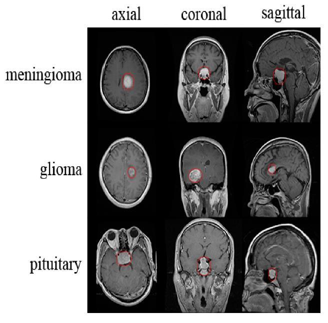
brainthatresemblesafibrousweb.Approximately3,540 children under the age of 15 are diagnosed with brain tumors this year. It is crucial to have a proper understanding of braintumorsand their stagesinorder to prevent and treat the illness. An abnormal mass of tissue in which cells grow and multiply uncontrollably, seemingly unchecked by the mechanisms that control normal cells, is called an intracranial tumor, or brain tumor. Although there are over 150 distinct types of brain tumors known to exist, primary and metastatic brain tumors are the two main categories. Tumors that arisefrom thebrain'stissuesorthe brain'ssurrounding tissuesarereferredtoasprimarybraintumors.Primary tumors can be classified as benign or malignant, glial (made up of glial cells) or non-glial (developed on or in thebrain'sstructures,suchasnerves,bloodvessels,and glands).Tumorsthatoriginateinotherpartsofthebody, like the breast or lungs, and spread to the brain, usually via the bloodstream, are referred to as metastatic brain tumors. Metastatic tumors are malignant and categorizedascancer.Thetypesoftumorsareshownin Fig1.
SaeedMohsenetal.[1]describedusinghyper-parameter optimization to implement ResNext101_32 × 8d and VGG19 models to improve the accuracy of brain tumor
International Research Journal of Engineering and Technology (IRJET) e-ISSN: 2395-0056
Volume: 12 Issue: 05 | May 2025 www.irjet.net p-ISSN: 2395-0072
detection and classification. These two models have the benefit of being architecturally simple, which can lower computational cost (training time), and they are based on a transfer learning process. The Kaggle repository contained a dataset that was used for testing and training the models. The first of six steps involved uploading the dataset of MRI images, which had been divided into images for training and testing. Image normalization, or pre-processingtheMRI scans,wasthe second step. Determining the total number of training epochswasthethirdstep.Thefourthstepinvolvedusing afittingfunctiontotrainthemodelwiththechosenMRI images.Thefifthstepwastotestthepredictioncapacity oftheVGG19modelusingtheMRItestimages.Thefinal stepwastoevaluatetheperformanceofthemodelusing differentmetricsontheMRItestimages.
In the clinical field, precise imaging is essential to producing accurate diagnoses as demonstrated by Solanki, Shubhangi, et al. [2]. The techniques used to acquire artifacts, such as CT, PET, and magnetic resonance imaging, have an impact on the effectiveness of clinical images. Real images from a magnetic resonance scan may include a great deal of unnecessary and undesired detail. A consequence of Rician noise is magnetic resonance imaging. This survey encompasses allthenoteworthycharacteristicsandthelatestresearch conducted so far, along with its limitations and challenges. Gaining a better understanding of how to conductnewresearchinanappropriatemannerwithina reasonable timeframe will be beneficial to the researchers. A generic approach is still needed, despite the significant contributions made by deep learning approaches.Whentrainingandtestingareconductedon comparable achievement features (intensity range and resolution), these methodologies yield better results; additionally, even the smallest deviation between the trainingandtestingimaginingshasa directinfluenceon the robustness of the methodologies. Future research couldbedonetoidentifybraincancersmoreaccurately, usingreal patient data asopposedto mediocremethods of capturing images (scanners). Classification accuracy may be improved by combining deep features with handcraftedcharacteristics.
Shah, et al.,...[3] carried out To diagnose brain tumors, VS-BEAM,auniquecomputer-aideddiagnosisalgorithm, has been introduced. To determine whether tumors are present in MRI images, the ensemble architecture combines several models. The final abnormality is determined by the algorithm through a voting mechanism, which increases diagnosis accuracy and efficiency. For effective feature extraction, we incorporate Squeeze and Excitation (SE) Attention Blocks into the CNN (Convolutional Neural Network) network. We came to the conclusion that SE attention combined with CNN is more logical than CNN networks
alone. Using various classifier algorithms, ensemble learning is employed by the VS-BEAM algorithm. While the second method, also a dense classifier, approaches the multiclass brain tumor classification problem as three binary class classification problems, the first method uses a classical dense network to solve the problem as a multiclass classification. Lastly, a Bayesian classifierthatestimatestheposteriordistributionisused bythethirdclassifiertocategorizethetumor.Duringthe evaluation stage, the suggested method for identifying anddividingupbraintumorsinMRIpicturesmeetshigh performance standards. The end-to-end model showed encouraging outcomes and might be applied in ongoing clinical studies for computer-assisted brain tumor diagnosis.
A teacher-student-based LCDEiT framework for classifying tumors from brain MRIs was presented by Ferdousetal.[4].Theframeworkconsistsofanexternal attention-based image transformer backbone for image classification, and a gated-pooled CNN-based teacher model for knowledge extraction. The need for the large dataset of vision transformers has been offset by the knowledge gained from the teacher model. By adding external attention to the backbone transformer model, which decreases complexity linearly with respect to the number of patches, the quadratic complexity caused by self-attention in the transformer encoder is removed. Accordingtothefindings,thetransformer-basedstudent model at the core of the suggested framework produces the best classification results, with F1-scores of 0.978 and 0.937 for the Figshare and BraTS-21 datasets, respectively. This illustrates how well image transformers with robust learners can be applied in the field of medical imaging, where quick computation is a critical requirement to start treating a patient who is in critical condition. To address the challenges associated with a higher misclassification rate for lower sample classes, the imbalance dataset handling technique, such as class-wise augmentation, may be used in the future. Even though the suggested LCDEiT model performed better on two different Figshare and BraTS-21 datasets, the model's universality could still be enhanced by expandingtheexperimentaldatabase.
Two research issues, including the security issue with the IoMT environment and the classification of brain tumors, were resolved by Ramprasad et al. [5]. The goal ofthisworkistoofferacomprehensivesolutiontothese issues. The study focuses on securing brain tumor images through the use of TIWT in the MIW implementation. The purpose of this is to safeguard patient privacyandconfidentiality when medical data is transmitted over the Internet of medical devices. The goal is to ensure that the source image of the brain tumor is transmitted over a secured environment, thereby preventing attackers from seeing the image.
International Research Journal of Engineering and Technology (IRJET) e-ISSN: 2395-0056
Volume: 12 Issue: 05 | May 2025 www.irjet.net p-ISSN: 2395-0072
Creating a precise system for classifying brain tumors is another goal of the research. It makes use of transfer learning techniques, namely BWO-GA for feature extraction and a transfer learning-based RU-Net model for segmentation. AlexNet, a transfer learning-based network, is then used to classify the segmented tumor region into benign and malignant tumors. The ultimate goal is to increase the accuracy of brain tumor classification, which will allow for early prediction and possibly save lives. The segmentation operation at the IoMT receiver is carried out using the RU-Net model in order to determine the exact location of the tumor. Furthermore, multilevel features are extracted using BWO-GA, and the best features are selected based on attributesfoundinnature.Furthermore,anAlexNetthat is based on transfer learning is trained with the best featuresfortumorclassification.
Utilize machine learning algorithms in the current system to classify brain tumors using algorithms for feature extraction andclassification.Inthe field of brain tumor detection from medical images, machine learning algorithms play a critical role in facilitating the early diagnosisandsuccessfultreatmentofthesedisorders.
Despitebeingrelativelysimple,theK-NearestNeighbors (K-NN) algorithm continues to be a useful method. It is helpfulforfindingsimilarcaseswithinadatasetbecause itgivesaclasslabeltoadatapointbasedonthemajority class of its closest neighbors in the feature space. AdaBoost and Gradient Boosting are two examples of ensemble methods that have proven effective in increasing classification accuracy by combining the predictions of multiple classifiers. These techniques makeuseoftheadvantagesofvariousbaseclassifiersto improvetherobustnessandgeneralizationofthemodel. When it comes to brain tumor detection, these machine learning algorithms are a great resource for medical professionals. They help identify brain tumors quickly andaccurately,whichimprovespatientoutcomes.
Another useful tool is Support Vector Machines (SVM), particularlyforbinaryclassificationtaskslikeidentifying tumor and non-tumor regions in medical images. To create precise classifications, SVMs can make use of a variety of features that have been extracted from the images,suchastexture,intensity,andshapedescriptors.
Decision trees and random forests are also frequently used.Theycanbeappliedtoclassificationtasksandhave theadvantageoffeatureselection.Thesealgorithmshelp distinguishbetweenregionsthataretumorousandthose thatarenot,usingfeaturesandattributesextractedfrom medicalimages.
To determine whether the brain contains tumors or other abnormal growths, brain tumor detection is an essential medical procedure. For successful treatment and good patient outcomes, brain tumors must be identified as soon as possible. The method that is most frequently used makes use of medical imaging techniques, specifically CT and MRI scans. Trained radiologists and physicians can see and locate tumors thanks to these non-invasive techniques that produce detailedimagesofthebrain.Inordertohelpradiologists interpretmedicalimages,machinelearningandartificial intelligence including deep learning models like YOLO (You Only Look Once) have been used in increasing numbers. By automatically identifying and categorizing tumorsfromimages,thesemodelsincreaseaccuracyand efficiency. The process of creating effective treatment plans, which may involve radiation therapy, chemotherapy, surgery, or a combination of these methods, depends on the accurate detection of brain tumors.Toimprovepatientoutcomesandqualityoflife, earlydiagnosisanddetectionareessential."BrainTumor Detection Using Transfer Learning," the name of the proposed system, aims to greatly improve the effectiveness and precision of brain tumor detection in medicalimaging.Usingpre-traineddeeplearningmodels on large datasets and fine-tuning them for the specific task of brain tumor detection is how this system takes advantage of the power of transfer learning. Data collection and preprocessing are crucial steps in the system architecture. An array of MRI scan images, both with and without tumors, is collected. Preprocessing techniques include data augmentation, pixel normalization,andimageresizingtoimprovethequality and diversity of the dataset. The transfer learning approach is the system's central component. Here, the fundamental architecture is a pre-trained deep learning model, such as YOLO. The brain tumor detection task leveragesfeaturesandknowledgeextractedfromalarge datasetinotherdomains.Thepre-trainedmodel'slayers extract features, and the fully connected layers or detection heads are adapted to the intricacies of brain tumor classification and localization. This quickens the development process and enhances the model's efficacious brain tumor detection capabilities. The system's strength is its ability to achieve high accuracy with a significant reduction in the need for a large dataset dedicated to brain tumor images. The dataset is divided into three separate subsets in order to thoroughly assess the system's efficacy and capacity for generalization: training, validation, and testing. The robustness of the model can also be verified and the chance of overfitting reduced by using cross-validation techniques. Another crucial component of fine-tuning is hyperparameter tuning, which entails optimizing variables like learning rates, batch sizes, and the use of
International Research Journal of Engineering and Technology (IRJET) e-ISSN: 2395-0056
Volume: 12 Issue: 05 | May 2025 www.irjet.net p-ISSN: 2395-0072
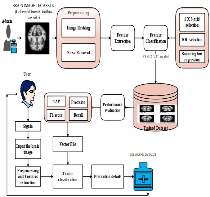
regularization strategies. This fine-tuning procedure is essential to guaranteeing optimal performance and an efficient convergence of the model. The next step involves model evaluation, in which the accuracy, precision,recall,andF1-score amongotherestablished metrics areusedtoevaluatethesystem'sperformance. This assessment is carried out on the specific testing dataset, which enables a precise comprehension of the system's capacity to accurately identify brain tumors. Additionally, this system can expedite the development of brain tumor detection models and make them accessibleforreal-worldmedicalapplications,ultimately improving patient care and outcomes. The proposed modelisshowninfig2.
Fig 2, images are collected from KAGGLE websites related to brain tumors such as Glioma tumor, Meningioma tumor, Pituitary tumor and No tumor. And thenperformpreprocessingstepssuchasnoiseremoval using median filtering algorithm. Finally select the pretrained model such as YOLOV11 model. Based on tumorclassification,calculatetheperformancesystemin termsofaccuracyandconfusionmatrix.Intestingphase, input the brain image and perform classification using best model file. Finally predict the tumors and provide theprecautiondetails
4.1
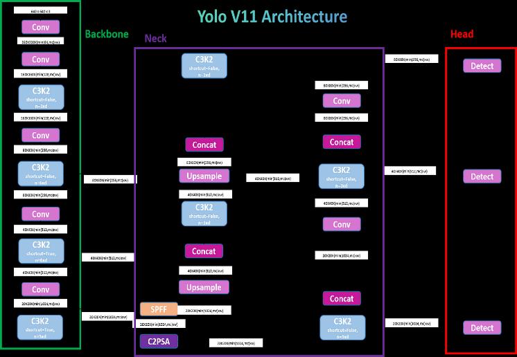
The YOLOv11 model, a highly advanced version of the YOLO(YouOnlyLookOnce)family,isdesignedtodeliver state-of-the-art performance in real-time object detection and classification tasks, including medical imaging applications like brain tumor detection. For brain tumor classification, YOLOv11 uses an enhanced backbone network with deeper feature extraction capabilities, improved attention mechanisms, and dynamicanchor-freedetectionheadsthatbetteradaptto complex medical images. The process begins with the collection and annotation of MRI brain scans, where
tumors are marked with precise bounding boxes. The images undergo preprocessing steps such as resizing, normalization, and data augmentation to ensure robustness and improve generalization. YOLOv11 processes these images through its highly optimized architecture, efficientlyextractingspatial andcontextual features to detect tumors of varying shapes, sizes, and intensities. By integrating advanced techniques like transformer-based modules and self-supervised pretraining, YOLOv11 achieves remarkable accuracy in identifyingandclassifyingtumorsintocategoriessuchas benign or malignant. The model supports multi-scale detection, enabling it to identify tiny tumors that might otherwise be missed by earlier models. Training is accelerated with intelligent optimization strategies, hyperparameter tuning, and automatic label correction mechanisms, ensuring faster convergence and higher reliability. Oncetrained, YOLOv11offers real-time,highprecision detection and classification of brain tumors, significantly assisting radiologists in early diagnosis and enhancing patient outcomes through quicker and more accurate medical decision-making. Fig 3 shows the YOLOV11architecture.
The proposed Yolov11 model, leveraging transfer learning, was evaluated on a neuroimaging dataset consistingofMRIscansofpatientswithmultipletypesof brain tumors. The dataset was pre-processed using contrast enhancement and skull-stripping techniques to improve model accuracy. The model was trained and validated using 80-20 data split, with cross-validation appliedtoensurerobustness.
International Research Journal of Engineering and Technology (IRJET) e-ISSN: 2395-0056
Volume: 12 Issue: 05 | May 2025 www.irjet.net p-ISSN: 2395-0072
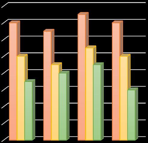
4: Performance chart
The results demonstrate that yolov11 model surpasses other deep learning models in predictive accuracy, showing a substantial improvement in tumor classification.
In conclusion, the utilization of the Efficient Net models for brain tumour detection represents a significant advancement in the field of medical imaging and healthcare. These deep learning models, renowned for theirefficacyinimageanalysis,offervaluabletoolstoaid medicalprofessionalsintheearlyandaccuratediagnosis of brain tumors. YOLOV11 model, with its wellestablished architecture and strong image classification capabilities, provides a solid foundation for this critical task. Its adaptability and versatility make it a reliable choiceforclassifyingbrainMRIand CTscans,enhancing the speed and accuracy of tumour identification. The Efficient net model can be customized and fine-tuned to accommodatespecificdatasetrequirements,allowingfor precisebraintumourdetectionwhileadheringtoethical and regulatory guidelines in healthcare. Its adaptability extends to various medical imaging tasks, including the detectionofabnormalitiesinX-raysandMRIscans.From the model implementation, YOLOV11 model provided improved efficiency in disease prediction. So, user can input the image and classified brain tumour types with diagnosisdetails
Future enhancements of the proposed system may include the integration of multimodal imaging such as CT, PET, and fMRI to enable a more comprehensive
diagnosis. Extending the model to perform tumor segmentation and volume estimation would assist in treatment planning and monitoring. Real-time optimization and deployment in clinical settings, including on edge devices, could further improve its practical applicability. Addressing class imbalance, incorporating explainable AI techniques for greater transparency,analyzinglongitudinal MRIdata fortumor progression,anddeveloping3Ddeeplearningmodelsto capture spatial features more effectively are also promisingdirectionsforfutureresearch.
[1] Mohsen, Saeed, et al. "Brain Tumor Classification Using Hybrid Single Image Super-Resolution Technique with ResNext101_32x8d and VGG19 Pre-Trained Models."IEEEAccess(2023).
[2]Solanki,Shubhangi,etal."BrainTumorDetectionand Classification using Intelligence Techniques: An Overview."IEEEAccess(2023).
[3]Shah,S.MuhammadAhmedHassan,etal."Classifying andLocalizingAbnormalitiesinBrainMRIusingChannel Attention Based Semi-Bayesian Ensemble Voting Mechanism and Convolutional Auto-Encoder."IEEE Access(2023).
[4] Ferdous, Gazi Jannatul, et al. "LCDEiT: A Linear Complexity Data-Efficient Image Transformer for MRI Brain Tumor Classification."IEEE Access11 (2023): 20337-20350.
[5] Ramprasad, M. V. S., Md Zia Ur Rahman, and Masreshaw D. Bayleyegn. "SBTC-Net: Secured Brain Tumor Segmentation and Classification Using Black Widow with Genetic Optimization in IoMT."IEEE Access(2023).
[6] Mohammed, Yahya MA, Said El Garouani, and Ismail Jellouli. "A survey of methods for brain tumor segmentation-based MRI images."Journal of Computational Design and Engineering10.1 (2023): 266-293.
[7] Kazerooni, Anahita Fathi, et al. "The Brain Tumor Segmentation (BraTS) Challenge 2023: Focus on Pediatrics (CBTN-CONNECT-DIPGR-ASNR-MICCAI BraTS-PEDs)."ArXiv(2023).
[8]Chang, Yankang, etal."Dpafnet:A residual dual-path attention-fusion convolutional neural network for multimodal brain tumor segmentation."Biomedical SignalProcessingandControl79(2023):104037.
[9] Moawad, Ahmed W., et al. "The Brain Tumor Segmentation (BraTS-METS) Challenge 2023: Brain
International Research Journal of Engineering and Technology (IRJET) e-ISSN: 2395-0056
Volume: 12 Issue: 05 | May 2025 www.irjet.net p-ISSN: 2395-0072
Metastasis Segmentation on Pre-treatment MRI."ArXiv(2023).
[10] Peng, Yanjun, and Jindong Sun. "The multimodal MRI brain tumor segmentation based on ADNet."Biomedical Signal Processing and Control80 (2023):104336.
© 2025, IRJET | Impact Factor value: 8.315 | ISO 9001:2008 Certified Journal | Page134