
International Research Journal of Engineering and Technology (IRJET) e-ISSN: 2395-0056
Volume: 13 Issue: 04 | Apr 2026 www.irjet.net p-ISSN: 2395-0072

International Research Journal of Engineering and Technology (IRJET) e-ISSN: 2395-0056
Volume: 13 Issue: 04 | Apr 2026 www.irjet.net p-ISSN: 2395-0072
T Shrungare1 , Prof. Ram Madhav Deshmukh2 , Dr. Sushma Telrande3
Abstract - Brain tumor detection plays a vital role in medical diagnosis, as earlyandaccurateidentificationdirectly impacts patient treatment and survival. Traditional manual analysis ofMRI images is time-consuminganddependsheavily on expert radiologists, which may result in inconsistent diagnoses. To overcome these limitations, this paper presents an automated brain tumor detection system based on deep learningtechniques. Theproposedsystemusesaconvolutional neural network–based model to detect and localize tumor regions in MRI images. Image preprocessing methods such as resizing and normalization are applied to improve detection accuracy. The systemprovides real-timepredictionsthrougha user-friendly interface, enabling faster and more reliable analysis. Experimental results show that the proposed approach effectively reduces diagnostic time while maintaining accurate tumor detection.
Keywords: Brain Tumor Detection, Medical Image Processing, Deep Learning, MRI Analysis, Convolutional Neural Network, Computer-Aided Diagnosis, Artificial Intelligence in Healthcare.
Brain tumors are among the most serious neurological disorders,oftenleadingtoseverehealthcomplicationsand, in critical cases, death if not detected at an early stage. A brain tumor occurs due to abnormal and uncontrolled growthofcellswithinthebrain,whichcandisruptnormal brain functions such as memory, vision, and motor coordination.Findingproblemsearlyandgettingtheright diagnosis quickly is very important for making good treatmentplansandhelpingpatientslivelonger.
Magnetic Resonance Imaging, or MRI, is one of the most common ways to see inside the brain and find tumors because it shows soft tissues in great detail.. However, manual interpretation of MRI scans requires extensive expertise and careful observation by radiologists. This processistime-consumingandmaybeaffectedbyhuman fatigue,subjectivity,andthecomplexityoftumorstructures. In many healthcare facilities, especially in rural or underresourced regions, the shortage of skilled medical professionals further increases the risk of delayed or inaccuratediagnosis.
Recentadvancementsinartificialintelligence(AI)anddeep learning have shown significant potential in addressing these challenges. Deep learning models, especially convolutional neural networks (CNNs), can automatically learncomplexpatternsfrommedicalimagesanddoaccurate
classificationanddetectiontasks.Thesemodelslessenthe need for manual analysis and provide consistent results, making them suitable for medical image processing applications.
This paper proposes an automated brain tumor detection system using deep learning techniques to analyze MRI images efficiently. The system aims to detect and localize tumor regions with high accuracy while minimizing diagnostictime.Byintegratingintelligentimageprocessing withauser-friendlyinterface,theproposedapproachassists medicalprofessionalsindecision-makingandsupportsearly diagnosis.Theimplementationhighlightstheeffectivenessof AI-based solutions in enhancing medical imaging analysis andimprovinghealthcareoutcomes.
Theproposedbraintumordetectionsystemisdesignedwith a modular architectureto ensure accuracy, efficiency, and ease of use. It integrates MRI image acquisition, preprocessing, deep learning–based analysis, and result visualization to achieve automated and reliable tumour detection.
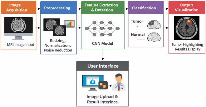
MRI brain images are collected from standard medical datasetsoruploadedbyusersthroughthesysteminterface. ThismoduleensurescompatibilitywithcommonlyusedMRI imageformats.
Theacquiredimagesarepre-processedtoenhancequality andconsistency.Operationssuchasresizing,normalization, and noise reduction are applied to improve model performanceanddetectionaccuracy
International Research Journal of Engineering and Technology (IRJET) e-ISSN: 2395-0056
Volume: 13 Issue: 04 | Apr 2026 www.irjet.net p-ISSN: 2395-0072
Feature Extraction and Detection Module
A convolutional neural network is used to find important featuresfromtheimagesthathavealreadybeenprocessed. Thismoduledetectsandlocalizestumorregionsbylearning discriminativepatternsbetweennormalandabnormalbrain tissues.
Classification Module
Detected tumor regions are classified based on learned features.Thisclassificationprovidesmeaningfuldiagnostic informationthatassistsinpreliminarymedicalassessment.
Output Visualization Module
The detection results are displayed by highlighting tumor regions on MRI images. Real-time visualization enables fasterandclearerinterpretationofresults.
User Interface Layer
simple and user-friendly interface allows users to upload imagesandviewresultseasily,ensuringsmoothinteraction withthesystem.
Overall,thesystemarchitectureenablesefficientintegration ofdeeplearningwithmedicalimageanalysis,providingfast, accurate,andpracticalbraintumourdetectionforhealthcare applications.
Several studies have explored machine learning and deep learningtechniquesforbraintumordetectionusingmedical images. Early methods used traditional image processing techniques like thresholding and segmentation,which providedbasictumoridentificationbutwerehighlysensitive to noise and image variations. These approaches lacked robustness and were unsuitable for complex clinical environments.Withtheadvancementofmachinelearning, classifierslikeSVM,KNN,anddecisiontreeswereusedfor brain tumor classification. Even though these methods improvedaccuracy,theyneededmanualfeatureextraction and showed limited generalization across diverse MRI datasets.Deeplearningmodels,particularlyconvolutional neural networks, later achieved better performance by automaticallylearningfeaturesfromMRIimages.
To overcome this limitation, object detection frameworks like Faster R-CNN, SSD, and YOLO were introduced in medical imaging. These models enabled simultaneous detection and classification with faster inference. Recent advancements in deep learning–based detection methods have further improved accuracy and processing speed. However,thereisstillaneedforefficientanduser-friendly systemssuitableforreal-worldhealthcareuse.Theproposed workaddressesthisgapbydevelopinganautomatedbrain tumordetectionsystemthatenhancesaccuracyandreduces diagnostictime.
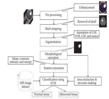
The proposed brain tumor detection system follows a systematic methodology designed to ensure accurate analysis, efficient processing, and practical usability. The implementationisdividedintomultiplestages,startingfrom data acquisition to final result generation. Each stage contributestotheoverallperformanceandreliabilityofthe system.
Dataset Collection
The first step involves collecting MRI brain images from publiclyavailableandauthenticatedmedicaldatasets.The dataset includes both tumor-affected and normal brain scans,allowingthemodeltolearnmeaningfuldistinctions between healthy and abnormal tissues. The diversity of images in terms of resolution, contrast, and tumor appearance improves the generalization capability of the system.
To enhance the quality of input data, preprocessing techniquesareappliedbeforemodeltraining.AllMRIimages are resized to a fixed resolution to maintain consistency. Pixel intensity normalization is performed to standardize imagevalues,reducingtheimpactofilluminationvariations. In addition, noise reduction techniques are applied to eliminateirrelevantartifacts,enablingthemodeltofocuson importanttumor-relatedfeatures.
Aconvolutionalneuralnetwork–baseddeeplearningmodel is employed for tumor detection and localization. The network automatically extracts hierarchical features from the preprocessed MRI images. During training, the model learnstoidentifydiscriminativepatternsthatdifferentiate tumor regions from normal brain tissues. Training is
International Research Journal of Engineering and Technology (IRJET) e-ISSN: 2395-0056
Volume: 13 Issue: 04 | Apr 2026 www.irjet.net p-ISSN: 2395-0072
performed using optimized hyperparameters such as learningrate,batchsize,andnumberofepochstoachieve stableconvergenceandimprovedaccuracy.
Tumor Detection and Classification
Once trained, the model processes unseen MRI images to detect tumor regions. The detection module identifies the presence of abnormal regions, while the classification componentcategorizesthedetectedoutputastumorornontumor.Thiscombinedapproachprovidesbothlocalization and classification, supporting effective preliminary diagnosis.
System Integration and User Interface
The trained model is integrated into a user-friendly applicationinterfacethatallowsuserstouploadMRIimages andviewresults.Thesystemgeneratesreal-timepredictions andvisuallyhighlightsdetectedtumorregions.Thisdesign ensureseaseofuseformedicalprofessionalsandreduces thetimerequiredformanualanalysis.
Performance Evaluation
Thesystemperformanceisevaluatedusingstandardmetrics suchasaccuracy,precision,recall,andF1-score.Testingis conducted on unseen MRI images to assess the generalizationcapabilityofthemodel.Theevaluationresults demonstratetheeffectivenessoftheproposedmethodology inachievingreliableandefficientbraintumordetection.
Theproposedbraintumordetectionsystemwasevaluated using a separate test dataset to assess its performance on unseen MRI images. This evaluation ensured reliable measurementofthemodel’sgeneralizationcapability.The system demonstrated effective tumor detection across differentimagesamples.
Performance Evaluation
The model achieved strong performance in distinguishing tumor-affected images from normal scans. Evaluation metrics such as accuracy, precision, recall, and F1-score indicatereliabledetectionresults.Highrecallvaluesshow thesystem’sabilitytoidentifytumorcasesaccurately,which isessentialformedicaldiagnosis.
Detection and Localization
ThesystemsuccessfullylocalizedtumorregionswithinMRI images by highlighting affected areas. This capability improvesclinicalrelevancebyprovidingclearvisualization of tumor position and size. The model remained robust acrossvariationsintumorshapeandappearance.
Discussion
Compared to traditional manual analysis and classical machine learning methods, the proposed system reduces diagnostic time while maintaining consistent accuracy.By automatically learning features, it minimizes human
dependencyandsupportsradiologistsindecision-making. Overall,theresultsdemonstratethatthesystemiseffective and suitable for practical healthcare applications, particularlyinresource-limitedenvironments.
Comparison with Traditional Methods
Compared to traditional manual analysis and classical machine learning techniques, the proposed system significantly reduces diagnostic time while maintaining reliableaccuracy.Unlikeconventionalmethodsthatrelyon handcraftedfeatures,thedeeplearningmodelautomatically learnsrelevantfeatures,resultinginimprovedconsistency and reduced human dependency. This makes the system suitable for assisting radiologists in routine diagnostic workflows.
Thispaperpresentedanautomatedbraintumordetection system based on deep learning techniques for efficient analysisofMRIimages.Theproposedapproachsuccessfully detectsandlocalizestumorregionswhilereducingreliance on manual interpretation. By integrating image preprocessing and a convolutional neural network–based model, the system provides accurate and consistent diagnosticsupport.Experimentalresultsdemonstratethat thesystemeffectivelyreducesdiagnostictimeandsupports early identification of brain tumors. The user-friendly implementation makes the proposed solution suitable for assisting medical professionals, especially in healthcare environmentswithlimitedaccesstospecializedexpertise.
Although the proposed system shows promising results, severalenhancementscanbeexploredinfuturework.The performance of the model can be further improved by trainingonlargerandmorediverseMRIdatasetstoenhance generalization. Future extensions may include multi-class tumorclassification,segmentationoftumorboundaries,and integration with clinical decision support systems. Additionally, deploying the system on cloud or mobile platforms can increase accessibility and real-time usage. Incorporating explainable AI techniques may also help improve transparency and trust in automated medical diagnosis.
[1]J.Redmon,S.Divvala,R.Girshick,andA.Farhadiwrotea papercalled"YouOnlyLookOnce:Unified,real-timeobject detection"whichwaspublishedintheIEEEConferenceon ComputerVisionandPatternRecognition(CVPR)in2016, pages779to788.
[2]J.RedmonandA.Farhadipublishedanotherpapertitled "YOLOv3:Anincrementalimprovement"asanarXivpreprint in2018,withtheidentifierarXiv:1804.02767.
International Research Journal of Engineering and Technology (IRJET) e-ISSN: 2395-0056
Volume: 13 Issue: 04 | Apr 2026 www.irjet.net p-ISSN: 2395-0072
[3] A. Bochkovskiy, C. Y. Wang, and H. Y. M. Liao wrote "YOLOv4:Optimalspeedandaccuracyofobjectdetection" whichwasalsoanarXivpreprintin2020,withtheidentifier arXiv:2004.10934.
[4]U.R.Acharyaandotherswrote"Automateddiagnosisof braintumorusingMRIimages:Adeeplearningapproach" which was published in IEEE Access, volume 8, pages 177547to177559in2020.
[5]S.Pereira,A.Pinto,V.Alves,andC.A.Silvawrote"Brain tumorsegmentationusingconvolutionalneuralnetworksin MRI images" which appeared in the IEEE Transactions on MedicalImaging,volume35,issue5,pages1240to1251in May2016.
[6]G.Litjensandotherswroteasurveytitled"Asurveyon deep learning in medical image analysis" which was publishedinMedicalImageAnalysis,volume42,pages60to 88in2017.
[7]K.Suzukiwrote"Overviewofdeeplearninginmedical imaging"whichwaspublishedinRadiologicalPhysicsand Technology,volume10,issue3,pages257to273in2017.
[8]M.Havaeiandotherswrote"Braintumorsegmentation withdeepneuralnetworks"whichwaspublishedinMedical ImageAnalysis,volume35,pages18to31in2017.
[9] T. Jo, K. Nho, and A. J. Saykin wrote "Deep learning in Alzheimer’sdisease:Diagnosticclassificationandprognostic prediction using neuroimaging data" which appeared in FrontiersinAgingNeuroscience,volume11,pages1to10in 2019.
[10]O.Ronneberger,P.Fischer,andT.Broxwrote"U-Net: Convolutionalnetworksforbiomedicalimagesegmentation" whichwaspublishedintheProceedingsoftheInternational Conference on Medical Image Computing and ComputerAssisted Intervention (MICCAI), pages 234 to 241 in 2015.image segmentation,” Proc. Int. Conf. Medical Image ComputingandComputer-AssistedIntervention(MICCAI),pp. 234–241,2015.
[11]A.Estevaetal.,“Aguidetodeeplearninginhealthcare,” Nature Medicine,vol.25,no.1,pp.24–29,2019.
[12]Kaggle,“BrainMRIImagesforBrainTumorDetection,” [Online]. Available: https://www.kaggle.com. Accessed: 2025.