
International Research Journal of Engineering and Technology (IRJET) e-ISSN:2395-0056
Volume:13Issue:04|Apr2026 www.irjet.net p-ISSN:2395-0072

International Research Journal of Engineering and Technology (IRJET) e-ISSN:2395-0056
Volume:13Issue:04|Apr2026 www.irjet.net p-ISSN:2395-0072
Rambabu Pemula1,Ellari Tejaswini Polisetti2 , Kavya Gatla3 , Shohana Javaji4 , Sathwika Katta5
12345 Department of Information Technology, Vidya Jyothi Institute of Technology, Telangana, India
Abstract - In the medical research field, medical imaging stands as a very important base because the precision of diagnostic pictures is highly necessary for the quick treatment of sick people. MRI data is often affected by noise interference during acquisition and transmission, which may impact image quality. Misinterpretation in the manual examination by a medical professional can be caused by this noise interference. Because MRI is necessary for health, medicalimagingmustremaincleartotheeyes.
This research presents an automated diagnostic framework which was built to connect difficult deep learning structures and medical work Computer-aided systems assist medical professionals in diagnosis. A comparative study was implemented using three most modern Convolutional Neural Networks (CNNs): VGG16, ResNet50, and DenseNet121. The automated diagnostic framework uses a Flask-based inner working part to look at MRI scans. Extraction of details and sorting into Glioma, Meningioma, Pituitary, and No Tumor categories are performed by the automateddiagnostic framework.
Data from the tests show that while VGG16 and ResNet50 give steady starting numbers, DenseNet121 shows better strength. It has been observed that DenseNet121 works best for these tasks. High success with a peakaccuracyof95.13% and fast working time was achieved by DenseNet121. To make the medical worker feel sure, the automated diagnostic framework includes a safe data control system and live picture charts using Chart.js. Such a proposed solution provides a cheap and fast second checking tool. Mistake by people is reduced while the automated diagnostic framework speeds up the choice-making work in thestudyofbraintumors.
Key Words: Brain Tumor Classification, Deep Learning, Densenet121, Flask Web Framework, Medical ImageAnalysis,sMRIDiagnostics.
Thehumanbrainpossessesthehighestcomplexityamong allbiologicalstructureswithinthecentralnervoussystem becauseanysicknesssuchasabraintumoroftenresultsin heavy neurological damage or death. Early detection of brain tumors is critical for effective treatment..Magnetic Resonance Imaging (MRI) is considered the best method for neuro-imaging because this technology offers high soft-tissue contrast without entering the body. Even thoughthistoolishelpful,lookingatMRIslicesbymedical professionalsisaverydetailedandslowtask.Accordingto
reports by Patil and Bhosale, pictures are frequently damaged by electronic noise while the data is being gathered,whichmakestheworkformedicalprofessionals muchharder.
Within medical centers that see many patients, the huge amountofwork formedicalprofessionalsmakesthelevel oftirednessgrow.Ithasbeenobservedthatthistiredness creates a high chance for errors during the identification of sickness. The human brain requires a lot of focus from the medical professional, but when many patients arrive, thefocusonthehumanbrainmightdecrease.Becausethe brain tumor is hard to see, mistakes happen when the medical professional is tired. A diagnostic error is often caused by heavy exhaustion because the human brain images are very difficult to read. Since the human brain controls the entire body, any small mistake during the reading of images might lead to a bad outcome for the patient. Solving these difficult problems requires Computer-Aided Diagnosis (CAD) systems because Computer-Aided Diagnosis (CAD) systems offer a dependable second thought for medical professionals. Experts claim that the rise of Deep Learning and Convolutional Neural Networks (CNNs) has made pulling out details from medical images very fast. Since Deep Learning has appeared, pulling out details from medical images is done with high efficiency. Subtle patterns in tumor textures are spotted by these computer programs eventhoughhumaneyescannotseethosepatternsclearly.
Constructing a strong web-based framework for brain tumorclassificationisthemaingoalofthisresearchpaper. While some researchers use just one design, this study tests three famous designs which are VGG16, ResNet50, andDenseNet121.ThesethreedesignsareputintoaFlask web interface so that a useful tool is made. A useful tool classifies medical pictures into four categories which are Glioma, Meningioma, Pituitary, and No Tumor. Real-time visualdataisshownbythesystemwhentheuseruploads avisualfile.Theprimarygoalofthisworkisshowingthat DenseNet121 gives the most steady accuracy of 95.13% because DenseNet121 has thick connections between its layers. Many people believe that DenseNet121 performs better than other designs for brain tumor tasks. DenseNet121 is a good choice for early clinical checking because DenseNet121 is very reliable. This framework is developed so that working positions in hospitals become moreefficient.
International Research Journal of Engineering and Technology (IRJET) e-ISSN:2395-0056
Volume:13Issue:04|Apr2026 www.irjet.net p-ISSN:2395-0072
Evidenceofpreviousscientificinvestigationsinthefieldof brain tumor detection is shown in this section. Reviewing existing methodologies helps in understanding current challenges.Brain tumor detection academic examinations from ten key studies are highlighted here. The limitations of automated attempts were analyzed because the history ofthefieldmatters.
Srinivasan et al. [1] are the scientific investigators who suggested a hybrid deep CNN model for multi-class classification. High correctness level of 99.53% was achieved because they used grid-search hyperparameter tuning. While the correctness level was very high, the scientific investigation noted that a high risk of overfitting existsonsmallerdatasets.
Mohamed Musthafa M. and his team[2] used a ResNet50 model with Grad-CAM to build an AI system. This made things easier for doctors, because they could clearly see how the model was thinking. It showed heatmaps that showedtheimportantpartsoftheimage,sodoctorscould understandwhythemodelgaveacertainresult.Thismade the results feel more clear. The method only worked for simple cases like detecting two types of things. The ResNet50 backbone needed a lot of computer power. However, the ResNet50-based approach requires high computational resources, making it less suitable for realtimeapplications.
AbdulRahmanetal.[3]arethescientificinvestigatorswho created an explainable CNN architecture. Identifying relevant key features was the focus because the scientific investigators wanted to reduce model working difficulty. Clinical trust was successfully built through transparent decision-making.
Research of Deepak S. and Ameer P. M. [4], pre-trained CNN models were used by these investigators to get featuresfromMRIthroughtransferlearning.Expertsclaim thattheoperationalresultsofthismethodreliedheavilyon the excellence levels of weights that came from ImageNet, whichisnotamedicaldataset.Becausethemodelwasnot trained specially for medical images, the CNN models did notgivethesameresultseverytime.
Sultan H. H. and his team[5]made a CNN model that can identifydifferent types oftumorsandgives more accurate results.AmajorissuewiththisCNNmodelwasthatittook a long time to make predictions in real-time using regular computers.Thisdelayhappenedbecausethemodelneeded to do a lot of calculations that regular computers couldn't handle quickly. The CNN architecture had to process a lot ofdatatosorttumortypesandthisputastrainoncommon computingtools.Asaresultthepredictionspeedwasslow which wasa problem for real-time applications. The work of Sultan H. H. Et al. Showed that a multi-class CNN architecture could achieve precision levels but it required more efficient computing tools to process the heavy calculations.
Creation of the detection system is achieved through a deep learning pipeline which provides diagnostic precision. A structured pipeline improves model performanceandreliability.
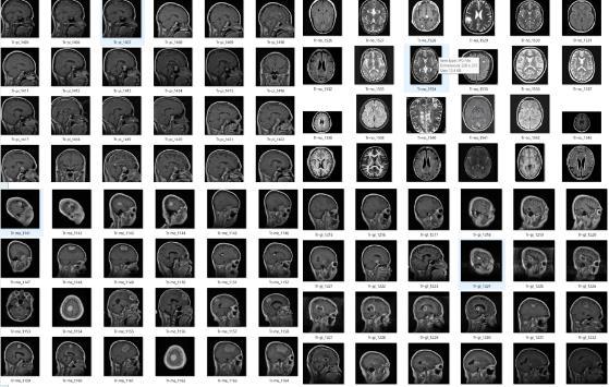
1.Data Collection: Information collection began when 3,264 high-resolution, T1-weighted contrast-enhanced MRI images were used. The information gathering is organized into four grouping categories: Glioma (926), Meningioma (937), Pituitary (901), and No Tumor (500). detection system relies on this information collection to distinguishbetweenvarioustypesofbraintumor.
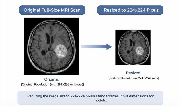
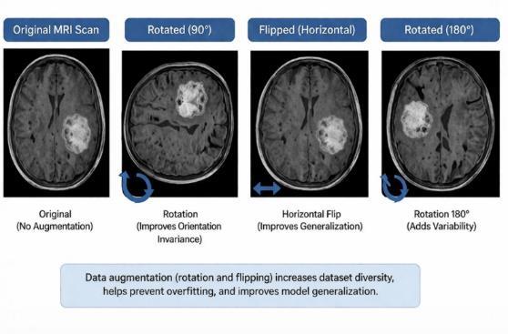
2. Image Preprocessing: Preparation of the medical pictures involves adjusting every slice in dimension to 224x224 because the architecture requires specific agreement. Observers have noted that Bicubic Interpolation helps the medical pictures fit the detection systemrequirements.Contrastenhancement(CLAHE)was applied while the tumor margins needed clarity for the medical professional. Pixel values were rescaled when stabletrainingwasneeded.
3. Data Splitting: Division of the information gathering occurs by creating three separate parts: Training (80%), Validation (10%), and Testing (10%). Experts claim that partitioning the dataset prevents the detection system fromseeingthesamemedicalpicturestwice.TheTraining
International Research Journal of Engineering and Technology (IRJET) e-ISSN:2395-0056
Volume:13Issue:04|Apr2026 www.irjet.net p-ISSN:2395-0072
section was used while the detection system learned the tumorfeatures.
4. Model Building (Transfer Learning): Construction of the model uses existing structures from ImageNet so the detection system benefits from previous knowledge. Researchers have stated that altering VGG16, ResNet50, and DenseNet121 is effective for medical classification. Thesepre-trainedmodelswereusedbecausetheyalready understand shapes and colors in medical pictures. Using transfer learning means that the detection system does notstartfromzero.
VGG16 (Visual Geometry Group) :This model framework functions as an ordered structure of 13 convolutionaland3fullyconnectedlayersthatusethe same3X3filters.Expertsclaimthatthesortingsection in this scientific investigation was replaced with a Global Average Pooling layer and a 512-unit dense layer so that high-level area characteristics are collected.Asteadyfoundationforextractinglow-level texture details and finding the large shapes of brain massesisprovidedbyVGG16.
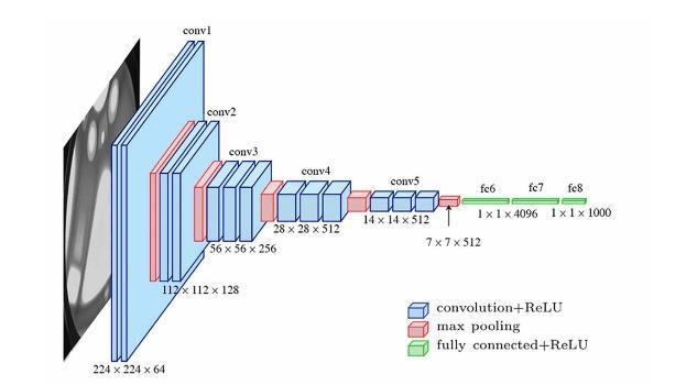
Fig4: ArchitecturalDiagramoftheVGG16
ResNet50 (Residual Network) :Identity Mappings, whicharealsocalledSkipConnections,areintroduced to this model to lessen the problem of gradients vanishing in deep networks. Signal integrity is kept
across the 50 layers when ResNet50 learns residual functions (F(x) + x). It has been observed that a "Bottleneck" designwith1X1and3X3convolutionsis utilized by this model because this maintains high working speed while capturing the hard meaningful dataneededforseeingtumorgrowth.
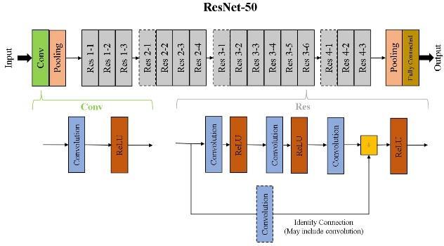
Fig5: ArchitecturalDiagramoftheResNet50
DenseNet121 (Densely Connected Network): Gradient flow is made stronger in this model by a forward pattern where every layer connects to every subsequent layer. Extensive Feature Reuse is promoted by this connection, which makes sure that basicedgesandsmalltexturedetailsfromearlylayers stay available for the final Softmax classifier. Many researchers believe that this model needs fewer parameters than ResNet50 even though this model has higher responsiveness to the small pixel changes neededforfindingtumorsearly
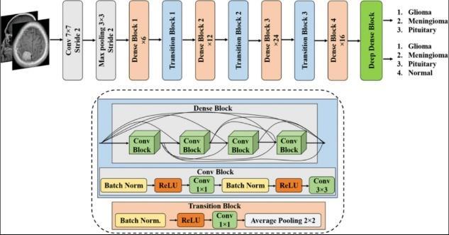
5. Model Training & Testing: The model training and testing process was performed using a two-stage approach, consisting of initial training with frozen base layers followed by fine-tuning to improve performance.The Adam optimizer was utilized alongside the Categorical Cross-Entropy loss function for a duration of 50 epochs. Because the model training & testing required stability, this specific process was followed by thedevelopers.
1. Accuracy: It describes the total rightness of the classification system when the calculation of the number of right samples happens among the total group. It has
International Research Journal of Engineering and Technology (IRJET) e-ISSN:2395-0056
Volume:13Issue:04|Apr2026 www.irjet.net p-ISSN:2395-0072
beenobservedthataccuracyprovidesaclearviewofhow well the system performs. The division of correct results bythetotalnumberofitemsishowaccuracyiscalculated becauseitrepresentsthewholetruthofthedata.
Accuracy =
2. Precision: Precision measures the proportion of correctly predicted positive cases. Precision is important because precision stops the wrong identification of healthy cases as sick cases. High results for precision are achieved when the predictions become more dependable. Reliable predictions are created by precision so that false positivesstayatalowlevel.
Precision =
3. Recall: The capacity of the model to find real tumor cases is measured by recall. Medical professionals state thatthisisnecessarysothatnotumorsaremissedduring thecheckup. Sensitivityisimproved when recall numbers increase.Recallensuresthatactualsicknessisnotignored becauserecallfocusesonfindingeverypositivecase.
Recall =
4. F1-Score:AbalancedassessmentisprovidedbytheF1score which is the harmonic average of precision and recall. It has been observed that this becomes helpful when the reduction of both types of errors is required. Better performance of the whole system is shown by a high F1-score. F1-score remains a key value for researchers because F1-score combines two different measuresintoone.
F1-Score =2x
4. RESULTS AND DISCUSSION
Evaluation of the system performance is conducted through a comparative analysis of VGG16, ResNet50, and DenseNet121 when using unseen MRI data.All models demonstrated effective classification performance on the dataset..Becausethesystemperformanceistestedonnew data, the results show that the logic of the machine is correct.
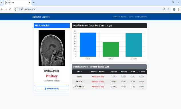
Regarding the outcomes, a highest accuracy of 95.13% is achieved by DenseNet121, which indicates better feature extraction and classification performance. While VGG16 provides stable baseline results, the performance is improvedbyResNet50becauseResNet50capturesdeeper features. It has been observed that the correctness of the modelsvariesbasedonthearchitecture.
Dense connectivity causes the great performance of DenseNet121 because dense connectivity makes feature reuse better and helps gradient flow. Fine-grained variationsinMRIimagesarecapturedbythemodel more effectively when the connections are dense. Scientists claim that the internal structure of DenseNet121 is very strongforthistask.
System reliability is increased by the multi-model approach because the multi-model approach chooses the best-performing model instead of using only one prediction. It is widely thought that the chances of misclassification are reduced when multiple models are compared. Confidence in the results is grown when the multi-modelapproachisapplied.
Accurate, consistent, and efficient tumor detection is provided by the proposed system, which makes the proposedsystemgoodforreal-timehealthcareassistance. Ithasbeennotedthattheproposedsystemhelpsmedical professionalsduringtheirworkingpositions.
[1]S.Srinivasanetal.,“AhybriddeepCNNmodelforbrain tumour image multi-classification,” BMC Medical Imaging, 2024.
[2]M.MohamedMusthafaetal.,“Enhancingbraintumour detection in MRI images through explainable AI using Grad-CAMwithResNet50,” BMCMedicalImaging,2024.
[3] S. Iftikhar et al., “Explainable CNN for brain tumor detection and classification through XAI based key featuresidentification,” BrainInformatics,2025.
[4] V. Verma and A. Aggarwal, “Deep learning: A revolutionizing approach to brain tumor classification using MRI,” South Eastern European Journal of Public Health,2025.
[5] N. Noreen et al., “A study of ensemble learning for brain tumor classification using deep learning,” IEEE Access,2020.