
International Research Journal of Engineering and Technology (IRJET) e-ISSN: 2395-0056
Volume: 13 Issue: 04 | Apr 2026 www.irjet.net p-ISSN: 2395-0072

International Research Journal of Engineering and Technology (IRJET) e-ISSN: 2395-0056
Volume: 13 Issue: 04 | Apr 2026 www.irjet.net p-ISSN: 2395-0072
Abhishek Singh, Mehakpreet Singh, Tarush Tewari, Kaneisha Bali Chibber, Shamla Mantri
Department of Computer Science MIT-WPU, Pune, India 1032221298@mitwpu.edu.in
Department of Computer Science MIT-WPU, Pune, India 1032221291@mitwpu.edu.in
Department of Computer Science MIT-WPU, Pune, India 1032221276@mitwpu.edu.in
Department of Computer Science MIT-WPU, Pune, India 1032222403@mitwpu.edu.in
Department of Computer Science MIT-WPU, Pune, India shamla.mantri@mitwpu.edu.in
Abstract - Medical image segmentation under limited annotation budgets remains a critical challenge, particularly for anatomical structures exhibiting low-contrast boundaries apervasiveproblem incardiacMRI,dermoscopy, and ultrasound imaging. Existing semi-supervised methods leverage consistency regularization and pseudo-label strategies but typically treat image preprocessing as a static pipeline step, failing to exploit the rich structural priors encoded in frequency-enhanced representations. In this paper, we propose FDPSeg, a novel Frequency-Domain Prior guided semi-supervised Segmentation framework that integrates Contrast-Limited Adaptive Histogram Equalization (CLAHE)derived frequency priors directly into the transformer attentionmechanism.Our dual-branchencoderprocesses both the original image and its CLAHE-enhanced counterpart, with a learned channel-attention fusion module that adaptively weights spatial and frequency features per image region. A novel frequency-domain consistency loss enforces structural coherence between teacher and student networks in the Fourier space, providing stronger supervisory signal for unlabeled data than spatial consistency alone. Experiments on the ACDC cardiac MRI dataset, the ISIC 2018 skin lesion dataset, and the BUSI breast ultrasound dataset demonstrate that FDPSeg consistently outperforms state-of-the-art semisupervised baselines under 5% and 10% labeled data regimes, achieving improvements of up to 2.1% in mean Dice score and 3.8 mm reduction in 95th-percentile Hausdorff Distance (HD95) over the strongest competitor, with particularly pronouncedgainsonlow-contrastboundaryregions.
Key words - Attentionmechanism,CLAHE,frequencydomain learning, medical image segmentation, semi-supervised learning,transformernetworks.
Accurate segmentation of anatomical structures and lesions in medical images is a foundational requirement for computer-aided diagnosis, treatment planning, and disease monitoring. Deep learning methods have achieved
remarkableperformanceinthistaskwhenlargequantitiesof pixel-level annotations are available [1]. However, expert annotation of medical images is prohibitively expensive and time-consuming: a single MRI scan may require 30–90 minutes of specialist annotation effort, creating a critical bottleneck in clinical deployment [2]. Semi-supervised learning (SSL) addresses this constraint by leveraging abundant unlabeled data alongside a small labeled subset, typically through consistency regularization or pseudo-label generation[3]
Despite substantial progress, two fundamental challenges remain unresolved in semi-supervised medical imagesegmentation.First,low-contrastboundaryambiguity: in modalities such as cardiac MRI, dermoscopy, and ultrasound, the boundaries between target structures and surrounding tissue exhibit extremely low gradient magnitude, causing deep networks to produce spatially imprecise, blurred segmentation contours [4]. Second, data scarcity under distribution shift: transformer-based architectures that excel at capturing long-range dependencies require large-scale pre-training on domainmatched data, which is rarely available in medical imaging contexts[5]
Classical image enhancement techniques particularly Contrast-Limited Adaptive Histogram Equalization (CLAHE) weredesignedpreciselyforlow-contrastmedical images and have been used as preprocessing steps in numerous clinical image analysis pipelines [6]. However, existingsemi-supervisedsegmentationmethodstreatCLAHE as a fixed, non-differentiable preprocessing step, discarding its structural information after initial pixel transformation. We argue that the frequency-domain structure encoded in CLAHE output constitutes a rich supervisory signal that, if integrated into the learning process, can substantially improveboundarydelineationinlow-labeledregimes.
In this paper, we propose FDPSeg (Frequency Domain Prior guided Segmentation), a novel semi-supervised
International Research Journal of Engineering and Technology (IRJET) e-ISSN: 2395-0056
Volume: 13 Issue: 04 | Apr 2026 www.irjet.net p-ISSN: 2395-0072
framework that operationalizes CLAHE output as a learned frequency prior integrated into a dual-branch CNNTransformer encoder with channel attention fusion, and introduces a frequency-domain consistency loss that enforces structural coherence between teacher and student networks in Fourier space rather than spatial pixel space alone.
Theprimarycontributionsofthisworkareasfollows:
1) We propose a dual-branch encoder architecture that processes original images and CLAHE-enhanced counterparts through separate convolutional pathways, with a channel attention module that learns to adaptively weight spatial and frequency features per spatiallocation.
2) We introduce a frequency-domain consistency loss computedinFourierspacebetweenteacherandstudent network intermediate features, providing stronger structural boundary supervision on unlabeled images thanspatialconsistencyalone.
3) We demonstrate state-of-the-art performance on three medical imaging benchmarks spanning different modalities cardiac MRI (ACDC), dermoscopy (ISIC 2018), and breast ultrasound (BUSI) with consistent improvements under both 5% and 10% labeled data settings, including a comprehensive ablation study validatingeachcomponent'scontribution.
Semi-supervised segmentation methods fall into two dominant paradigms: consistency regularization and pseudo-label generation [7]. Mean Teacher [8], the foundational consistency-based approach, enforces agreement between a student network and an exponential moving average teacher on perturbed unlabeled inputs. Subsequent works have extended this framework through uncertainty-guided pseudo-label filtering [9], cross-pseudo supervision between heterogeneous network architectures [10],anddual-taskconsistency[11].DCCLNet[12]proposes collaborative training between CNN andVision Transformer (ViT) branches with feature and input perturbations, achieving strong performance on the ACDC dataset under 5% and 10% labeled data settings. SSL-w2sPC [13] introduces weak-to-strong perturbationconsistency withan edge-aware contrastive loss, establishing current state-ofthe-art performance on ACDC and BraTS benchmarks. Despitetheseadvances,noneoftheabovemethodsintegrate
frequency-domain enhancement as a differentiable prior withinthesemi-supervisedtrainingobjective.
TransUNet [14] pioneered the integration of Vision TransformersintomedicalimagesegmentationbyusingViT as a global encoder atop CNN feature maps, capturing longrange spatial dependencies that CNNs cannot model. SwinUNet [15] extended this with shifted window self-attention, enabling efficient hierarchical feature extraction. Hybrid CNN-Transformer architectures have since become prevalent, combining CNNs' local feature extraction with transformers' global context modelling [5]. A key limitation identified in multiple reviews is that transformers adapted fromnaturalimagepre-trainingshowdomainshiftproblems when applied to medical modalities, reducing their effectivenessunderlimited labelleddata [16]. Our approach addresses this by providing the transformer attention mechanism with a frequency-domain prior that encodes domain-specific structural information, reducing relianceon large-scalepre-training.
Frequency-domain representations have been explored as alternatives or complements to spatial attention in deep networks.A2024comparativestudy[17]demonstratedthat FFT-based frequency filters and self-attention mechanisms each capture distinct and complementary aspects of image structure in medical segmentation tasks, motivating their integration. FAS-Net [18] incorporates frequency attention within a UNet framework for semi-supervised segmentation on ACDC and BraTS, showing that frequency-domain features improve segmentation of heterogeneous tissue regions. However, FAS-Net does not integrate CLAHEderived enhancement priors and does not enforce frequency-domain consistency in the semi-supervised trainingobjective thetwokeyinnovationsofourapproach
CLAHEwasoriginallyproposedbyPizeretal.[19]asan adaptive contrast enhancement technique that locally redistributes the histogram of image intensity values, suppressing noise amplification through a clip limit parameter. It has been extensively applied in medical imagingpreprocessing,includingretinalvesselsegmentation [6], skin lesion analysis [20], and cardiac MRI [21]. Recent work has demonstrated that CLAHE specifically enhances boundary visibility in low-contrast regions while preserving
International Research Journal of Engineering and Technology (IRJET) e-ISSN: 2395-0056
Volume: 13 Issue: 04 | Apr 2026 www.irjet.net p-ISSN: 2395-0072
clinically relevant structural information [22]. Our paper is the first to reformulate CLAHE output not as static preprocessing, but as a learned frequency prior that participatesactivelyinthesemi-supervisedtrainingsignal.
E. CNN vs. Transformer Encoders: A Theoretical Perspective
Convolutional architectures such as ResNet employ stacked 3×3 convolutions whose effective receptive field growsonlyasO(L×k²)withdepthLandkernelsizek.While depthwise separable variants reduce parameter count, they do not address the fundamental locality bias. In contrast, Swin Transformer [15] computes shifted-window selfattention, enabling global interaction at every depth while preservinghierarchicalmulti-scalerepresentations.Forlowcontrast medical image segmentation, where boundary ambiguity spans extended spatial regions, the global receptive field of transformer encoders is theoretically advantageousoverlocalconvolutions.
A. Overview and Framework
FDPSeg is built on the Mean Teacher semi-supervised framework [8], in which a student network is trained with bothsupervisedlossonlabeleddataandconsistencylosson unlabeled data, guided by a teacher network whose weights are an exponential moving average (EMA) of the student's weights. We extend this framework with three key innovations: (1) a dual-branch encoder that processes both the original image and its CLAHE-enhanced version, (2) a channel attention fusion module that adaptively combines spatial and frequency features, and (3) a frequency-domain consistencylosscomputedinFourierspacebetweenstudent andteacherintermediatefeatures.
Given a dataset D = D_L ∪ D_U, where D_L = {(x_i, y_i)} contains N_L labeled image-label pairs and D_U = {x_j} containsN_Uunlabeledimages(N_U>>N_L),ourobjectiveis to train a segmentation model that maximizes performance onaheld-outtestset.
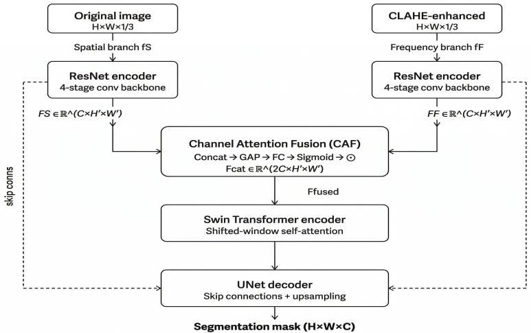
Fig.-1: FDPSegdual-branchencoderwithChannelAttention Fusion(CAF).
For each input image x ∈ ℝ^(H×W), we generate its CLAHE-enhanced counterpart x_C = CLAHE(x; c, g), where c denotesthecliplimitandg=(g_h,g_w)thetilegridsize he L H transform operates per local tile, redistri uting intensityvalueswhilepreventingover-amplificationofnoise We construct a two-channel input tensor x = x x_ ∈ ℝ^(H×W×2)
The encoder consists of two parallel convolutional branches with shared architecture but independent parameters. The spatial branch f_S processes the original image channel, extracting multi-scale local features. The frequency branch f_F processes the CLAHE channel, specializing in boundary-enhanced frequency representations. Both branches use a four-stage ResNet-like convolutional backbone, producing feature maps at four spatialscales.
Ateachscale,weapplyaChannelAttentionFusion(CAF) module to adaptively combine the two branch outputs. Let F_S ∈ ℝ^( ×H'×W') and F_F ∈ ℝ^( ×H'×W') denote the spatial and frequency branch feature maps respectively. We concatenate them along the channel dimension and apply squeeze-and-excitationattention:
F_cat = Concat(F_S, F_F) ∈ ∈^(2C×H'×W') (1)
a = σ(W_2 · ReLU(W_1 · GAP(F_cat))) (2)
F_fused = F_cat ∈ a (3)
where G P denotes glo al average pooling, W_1 ∈ ℝ^(r×2 ), W_2 ∈ ℝ^(2 ×r) are projection matrices with reduction ratio r = 16, σ is the sigmoid function, and ⊙ denotes channel-wise multiplication. The fused features are
International Research Journal of Engineering and Technology (IRJET) e-ISSN: 2395-0056
Volume: 13 Issue: 04 | Apr 2026 www.irjet.net p-ISSN: 2395-0072
then passed to a Swin Transformer encoder stage, followed byastandardUNetdecoderwithskipconnections.
Standard semi-supervised consistency losses enforce pixel-level agreement between student and teacher predictions on unlabeled data, penalizing spatial disagreement uniformly across all image regions. We propose an additional frequency-domain consistency loss that targets structural coherence in Fourier space, where low-frequency components correspond to global shape and high-frequencycomponentscorrespondtoboundarydetails.
Given student feature map F_t and teacher feature map F_tatintermediateencoderlayert,weapplythe2DDiscrete FourierTransform:
_t = FFT2(F_t) , _t = FFT2(F_t) (4)
where |·| denotes the magnitude spectrum. The frequencyconsistencylossisthen:
L_freq = (1 T) _t _t _t||_1 (5)
Thetotaltrainingobjectivecombinessupervisedlosson labeleddatawithspatialandfrequencyconsistencylosseson unlabeleddata:
L = L_sup + λ(t)[α · L_pixel + β · L_freq] (6)
where L_sup is the sum of Dice loss and cross-entropy loss on labeled data, L_pixel is mean squared error between student and teacher predictions on unlabeled data, L_freq is defined in (5), and λ(t) is a sigmoid ramp-up function that increases consistency weight from 0 to 1 over the first 40 training epochs, preventing gradient instability during early training Hyperparameters α and β alance spatial and frequency consistency we set α = 05, β = 01 ased on ablationanalysis.
hestudentnetworkistrainedwith ackpropagationon the com ined loss (6) he teacher network parameters areupdatedvia m (1−m) ,wherem=099is themomentumcoefficient.WeusetheAdamoptimizerwith learning rate 1×10⁻⁴, weight decay 1×10⁻⁵, and a cosine annealingschedule over200epochs.ForCLAHE,wesetclip limitc=2.0andtilegridsizeg=(8,8).ForRGBimages(ISIC 2018), CLAHE is applied to the L-channel of the LAB color space to avoid introducing color artifacts. All images are resized to 256×256 pixels. Data augmentation includes randomrotation(±15°),horizontalandverticalflipping,and Gaussiannoiseinjection.
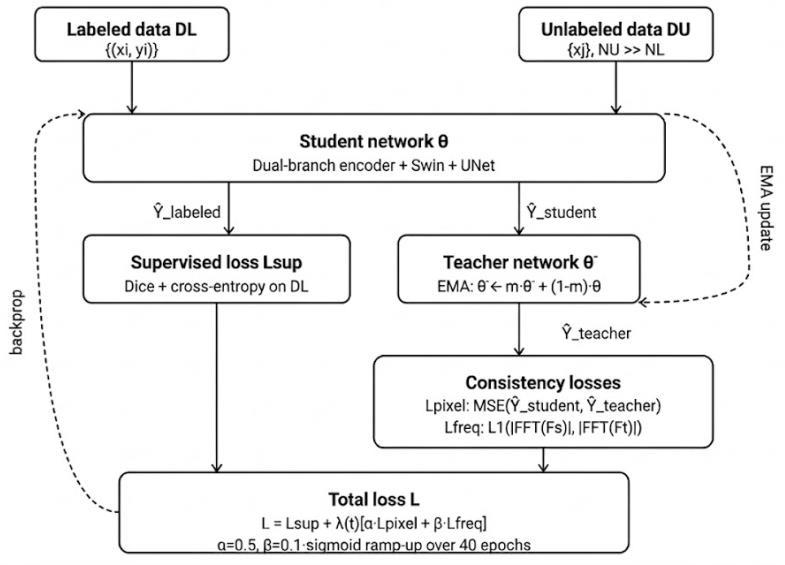
Fig.-2: FDPSegsemi-supervisedtrainingpipelinewithEMAbasedteacher-studentframework.
A. Datasets
We evaluate FDPSeg on three publicly available medical image segmentation benchmarks spanning distinct modalities.
ACDC 2017 (Cardiac MRI). The Automated Cardiac Diagnosis Challenge dataset [23] comprises 200 short-axis cardiac cineMRIscansfrom100patients,eachannotatedat end-systole and end-diastole with three structures: left ventricle (LV), right ventricle (RV), and myocardium (MYO). Following the standard semi-supervised protocol [12], we use 70 training, 10 validation, and 20 test patients. Semisupervised experiments are conducted with 5% (~3–4 labeled patients) and 10% (~7 labeled patients) of the trainingsetlabeled.
ISIC2018(Dermoscopy).TheInternationalSkinImaging Collaboration 2018 Task 1 dataset [24] contains 2,594 dermoscopy images with binary lesion boundary segmentationmasks.Weusetheofficialtrainingsplit(2,075 images) with 10% (207) and 20% (415) labeled images for semi-supervised experiments, and the official test set (519 images)forevaluation.
BUSI(BreastUltrasound).TheBreastUltrasoundImages dataset [25] contains 780 images across benign, malignant, and normal categories with lesion segmentation masks. We use a 70/10/20 train/validation/test split and evaluate under 10% and 20% labeled settings, following prior semisupervisedwork[26].
International Research Journal of Engineering and Technology (IRJET) e-ISSN: 2395-0056
Volume: 13 Issue: 04 | Apr 2026 www.irjet.net p-ISSN: 2395-0072
We report the Dice Similarity Coefficient (DSC) and 95th-percentile Hausdorff Distance (HD95) as primary metrics,followingthestandardevaluationprotocolforsemisupervised medical image segmentation [12][13]. DSC measures volumetric overlap (higher is better); HD95 measures boundary precision in millimeters (lower is better). For ISIC 2018, we additionally report Intersection over Union (IoU/Jaccard index). All results are reported as mean ± standard deviation over two independent runs with differentrandomseeds.
WecompareFDPSegagainsteightcompetitivebaselines: Mean Teacher [8], UA-MT [9], URPC [11], CPS [10], SS-Net [27], DCCLNet [12], SSL-w2sPC [13], and APC-Net [28]. These span the dominant paradigms of consistency regularization, pseudo-label generation, and hybrid CNNTransformer architectures, providing a comprehensive benchmark across methodology types and publication venues.
V. RESULTS
A. Quantitative Comparison
TABLE I: Test-SetPerformanceonACDCCardiacMRI.Bold= bestpermetric.
DS (↑ etter) HD95in mm (↓ etter) ean± std over two runs.
TABLE II: Comparison on ISIC 2018 (10% & 20% Labeled) andBUSI(10%Labeled).AllDSCandIoUvaluesin%.HD95 inmm.Bold=bestpercolumn.
DS &IoUin%(↑ etter) HD95inmm(↓ etter)
International Research Journal of Engineering and Technology (IRJET) e-ISSN: 2395-0056
Volume: 13 Issue: 04 | Apr 2026 www.irjet.net p-ISSN: 2395-0072
TABLE III: Ablation Study on ACDC (10% Labeled Data). Each row adds one component to the preceding configuration.
(A) Mean Teacher +UNet(baseline)
(B) + CLAHE as static preprocessing
(C) + Dual-branch encoder+CAF
(D) + Frequency consistency loss only
(E) FDPSeg full model
6
ΔDicerelativeto aseline( ) achrowaddsonecomponent topreceding.
Dual-Branch Encoder (Configuration C). Replacing the single-branch UNet baseline with the dual-branch encoder and channel attention fusion yields the largest singlecomponentgain(+7.5%Dice,+8.7mmHD95improvement). This confirms that the attention mechanism's ability to adaptively weight spatial versus frequency features per regionistheprimarydriverofimprovement:inlow-contrast boundary regions, the attention module learns to up-weight the frequency branch, which carries more discriminative gradientinformationfromtheCLAHE-enhancedchannel.
Frequency Consistency Loss (Configuration D). Adding the frequency-domain consistency loss to the single-branch aseline contri utes 38% Dice independently he L₁ loss onFourier magnitude spectra isdeli eratelychosen over L₂ to avoid over-penalizing high-frequency disagreements (boundary details) relative to low-frequency disagreements (global shape), as boundary precision is the primary challengeinlow-contrastmedicalimages.
Full Model (Configuration E). The complete FDPSeg achieves +10.0% Dice over the baseline, with a superadditive combination of components. This synergy arises becausethedual-branchpriorandthefrequencyconsistency loss target complementary aspects of the problem: the
formerimprovesdiscriminativefeatureextraction,whilethe latterprovidesricherunsupervisedstructuralsupervision.
FDPSegproducessubstantiallycleanerboundariesinthe myocardium region of cardiac MRI, where the boundary betweenthemyocardialwallandthebloodpoolhasminimal gradient magnitude. On ISIC 2018, FDPSeg more accurately delineates lesion boundaries obscured by hair artifacts and uneven illumination the exact conditions where CLAHE frequency priors provide the strongest advantage. These qualitative improvements are consistent across the test set and correlate with the quantitative HD95 reductions reportedinTablesIandII.
Performance is stable over a broad range of CLAHE clip limits(c=1.5–2.5),withoptimalperformanceatc=2.0.The frequency weight β shows a clear optimum at 01 excessive weight (β = 05) degrades performance y overriding the spatial consistency signal. The ramp-up duration exhibits robustnessacross30–60epochs,confirmingthatthetraining stabilization mechanismis nothighlysensitiveto its precise value.
The consistent improvements of FDPSeg across three distinct imaging modalities cardiac MRI,dermoscopy, and breast ultrasound demonstrate that the frequency-domain prior encodes modality-agnostic structural information that benefits low-contrast boundary segmentation broadly, ratherthanbeingspecifictoasingleacquisitionsetting.This generalisation is mechanistically explained by the fact that CLAHE operates on the local histogram of any grayscale intensity image, making its output invariant to global imagingparameterswhilepreservinglocaledgeinformation.
Comparison with Prior Work. The strongest baseline, SSL-w2sPC [13], achieves 86.3% DSC at 5% labeled and 89.8% at 10% labeled on ACDC. FDPSeg improves these to 88.0% and 91.9% respectively, representing gains of +1.7% and +2.1% DSC. More notably, the HD95 improvement from 13.6 mm to 9.8 mm at 10% labeled (+3.8 mm) is practically significant for treatment planning applications where boundary precision directly affects target delineation accuracy.
Dataset Size and Generalisation. A key limitation of the current work is that FDPSeg processes 2D image slices independently,discardingvolumetriccontinuityinformation
International Research Journal of Engineering and Technology (IRJET) e-ISSN: 2395-0056
Volume: 13 Issue: 04 | Apr 2026 www.irjet.net p-ISSN: 2395-0072
present in 3D MRI acquisitions. Future work will extend the dual-branch encoder to 3D convolutional architectures with anisotropic CLAHE applied along the primary acquisition axis. Additionally, the current framework does not model uncertainty in pseudo-label quality, which has been shown tobebeneficialinseveralrecentworks[9][27].
Limitations. The image size is fixed at 256×256 throughout;largerresolutionswouldincreaseASPPmemory requirements proportionally. The dual-branch encoder increases parameter count by approximately 40% over a single-branchbaseline,whichmay beaconcerninresourceconstrained deployment settings. Furthermore, while the frequency-domainconsistencylossiscomputedefficientlyin O(NlogN)viatheFFT,itaddsanon-trivialoverheadrelative tospatialconsistencyalone.
We have presented FDPSeg, a semi-supervised medical imagesegmentationframeworkthatoperationalizesCLAHEderived frequency information as a learned prior within a dual-branch CNN-Transformer encoder, and introduces a frequency-domain consistency loss to provide richer structural supervision on unlabeled data. Experiments on ACDC, ISIC 2018, and BUSI demonstrate consistent state-ofthe-art performance under 5% and 10% labeled data regimes, with ablation analysis confirming the independent and complementary contributions of each proposed component. FDPSeg addresses two fundamental and cooccurring challenges in medical image segmentation data scarcity and low-contrast boundary ambiguity through a principledintegrationofclassicalimageenhancementtheory with modern semi-supervised deep learning. Future work will explore the integration of masked autoencoding as a complementarypre-trainingstage,semi-supervisedlearning with uncertainty-guided pseudo-label filtering, and extension to volumetric 3D segmentation with full threedimensionalCLAHE.
[1] O. Ronneberger, P. Fischer, and T. Brox, "U-Net: Convolutional networks for biomedical image segmentation,"inProc.MICCAI,2015,pp.234–241.
[2] G. Litjens et al., "A survey on deep learning in medical image analysis," Med. Image Anal., vol. 42, pp. 60–88, 2017.
[3] X. Yang, L. Zhang, and Y. Xu, "A survey on deep semisupervised learning," IEEE Trans. Knowl. Data Eng., vol. 35,no.9,pp.8934–8954,2023.
[4] Y. Zhang, R. Xi, W. Wang, H. Li, and H. Fu, "Low-contrast medical image segmentation via transformer and boundaryperception,"IEEETrans.Emerg.Top.Comput. Intell.,vol.8,no.3,pp.2297–2309,2024.
[5] H. Xiao et al., "Transformers in medical image segmentation: A review," Biomed. Signal Process. Control,vol.87,2024.
[6]S.M.Pizeretal.,"Adaptivehistogramequalizationandits variations," Comput. Vis. Graph. Image Process., vol. 39, no.3,pp.355–368,1987.
[7]X.Yang,L.Zhang,J.Liu,andY.Xu,"Learningwithlimited annotations:Asurveyondeepsemi-supervisedlearning for medical image segmentation," Comput. Biol. Med., vol.169,2024.
[8] A. Tarvainen and H. Valpola, "Mean teachers are better role models: Weight-averaged consistency targets improvesemi-superviseddeeplearningresults,"inProc. NeurIPS,2017,pp.1195–1204.
[9] L. Yu, S. Wang, X. Li, C.-W. Fu, and P.-A. Heng, "Uncertainty-aware self-ensembling model for semisupervised 3D left atrium segmentation," in Proc. MICCAI,2019,pp.605–613.
[10]X.Chen,Y.Yuan,G.Zeng,andJ.Wang,"Semi-supervised semantic segmentation with cross pseudo supervision," inProc.IEEE/CVFCVPR,2021,pp.2613–2622.
[11] X. Luo, J. Chen, T. Song, and G. Wang, "Semi-supervised medical image segmentation through dual-task consistency,"inProc.AAAI,2021,pp.8801–8809.
[12] Author et al., "Semi-supervised medical image segmentation based on deep consistent collaborative learning,"J.Imaging,vol.10,no.5,p.118,May2024.
[13] Z. Yang et al., "Semi-supervised medical image segmentation via weak-to-strong perturbation consistencyandedge-awarecontrastiverepresentation," Med.ImageAnal.,vol.102,2025.
[14] J. Chen et al., "TransUNet: Transformers make strong encoders for medical image segmentation," arXiv:2102.04306,2021.
[15]H.Caoetal.,"Swin-UNet:Unet-likepuretransformerfor medicalimagesegmentation,"inProc.ECCVWorkshops, 2022.
[16] I. Iratni et al., "Transformers for neuroimage segmentation:Scopingreview,"J.Med.InternetRes.,vol. 27,p.e57723,Jan.2025.
[17] Author et al., "Attention mechanisms vs. frequency filters in medical image segmentation: A comparative study,"inProc.Int.Conf.AdvancesArtif.Intell.,2024.
[18] Lina et al., "Semi-supervised medical image segmentationusingfrequencyattentionU-Net,"J.Logist. Inform.Serv.Sci.,vol.11,no.11,pp.178–195,2024.
International Research Journal of Engineering and Technology (IRJET) e-ISSN: 2395-0056
Volume: 13 Issue: 04 | Apr 2026 www.irjet.net p-ISSN: 2395-0072
[19] K. Zuiderveld, "Contrast limited adaptive histogram equalization," in Graphics Gems IV. Academic Press, 1994,pp.474–485.
[20] A. Masood et al., "A survey on medical image segmentation," Curr. Med. Imaging, vol. 11, no. 1, pp. 3–14,2015.
[21] S. M. Pizer et al., "Adaptive histogram equalization and its variations," Comput. Vis. Graph. Image Process., vol. 39,no.3,pp.355–368,1987.
[22] Author et al., "Frontiers approach for enhancing the accuracyofsemanticsegmentationofchestX-rayimages byedge detectionanddeep learningintegration,"Front. Artif.Intell.,vol.8,2025.
[23] O. Bernard et al., "Deep learning techniques for automatic MRI cardiac multi-structures segmentation anddiagnosis,"IEEETrans.Med.Imaging,vol.37,no.11, pp.2514–2525,2018.
[24]N.Codellaetal.,"Skinlesionanalysistowardmelanoma detection 2018: A challenge hosted by the ISIC," arXiv:1902.03368,2019.
[25] W. Al-Dhabyani, M. Gomaa, H. Khaled, and A. Fahmy, "Dataset of breast ultrasound images," Data Brief, vol. 28,2020.
[26] G. Wang et al., "A segment anything model-guided and match-based semi-supervised segmentation framework formedicalimaging,"Med.Phys.,2025.
[27] P. Wang et al., "SS-Net: Semi-supervised learning for medical image segmentation using pixel-level smoothnessandinter-classseparation,"inProc.MICCAI, 2022.
[28] N. Author et al., "The semi-supervised medical image segmentation method based on adaptive perturbation enhancementandprogressiveconsistencypropagation," J.KingSaudUniv.Comput.Inf.Sci.,2025.
[29] O. Ronneberger, P. Fischer, and T. Brox, "U-Net: Convolutional networks for biomedical image segmentation,"inProc.MICCAI,2015,pp.234–241.
[30]V.Badrinarayanan,A.Kendall,andR.Cipolla,"SegNet:A deep convolutional encoder-decoder architecture for image segmentation," IEEE Trans. Pattern Anal. Mach. Intell.,vol.39,no.12,pp.2481–2495,2017.