
International Research Journal of Engineering and Technology (IRJET) e-ISSN: 2395-0056
Volume: 13 Issue: 04 | Apr 2026 www.irjet.net p-ISSN: 2395-0072

International Research Journal of Engineering and Technology (IRJET) e-ISSN: 2395-0056
Volume: 13 Issue: 04 | Apr 2026 www.irjet.net p-ISSN: 2395-0072
Mukesh Kumar K1 , Mr. R. Mohan Kumar2
1PG Scholar Bio-Medical Department Udaya School of engineering, Kanyakumari, Tamil Nadu, India.
2Assistant Professor Bio-Medical Department, Udaya School of engineering, Kanyakumari, Tamil Nadu, India.
Abstract Scattered light significantly degrades biomedical images by reducing contrast and obscuring fine structural details, thereby affecting both visual interpretationandautomatedanalysis.Itsremovalremains challenging due to the lack of scattered-light-free ground truthdataandthehighcomputationalcostofdeeplearningbased solutions. To address these limitations, this paper introduces APEE-Filter, a novel adaptive physics-guided edge-aware method for scattered light suppression in biomedical images. Unlike conventional dehazing models, the proposed approach incorporates a tissue-aware scattering component into the imaging model to more accuratelycharacterizelightdiffusioninbiologicalmedia.A new adaptive contrast prior, combining a quarter-window dark channel and a local variance map, is developed to estimate the transmission map without requiring training data. To preserve structural details such as cell boundaries and vascular edges, an edge-aware refinement strategy based on guided filtering is employed. Additionally, a selfcalibrated background illumination estimation technique ensures robustness across varying imaging conditions. The entire framework is parameter-light and operates with linear computational complexity, enabling real-time deployment on resource-constrained biomedical systems. Experimental evaluations demonstrate that the proposed method effectively enhances contrast, restores structural details, and outperforms traditional filtering approaches while maintaining high computational efficiency. Furthermore, the method shows robust performance across multiple imaging modalities, including fluorescence and phase-contrast microscopy, and demonstrates improved compatibility with downstream automated image analysis taskssuchassegmentationandcellcounting.
Key Words: Scattered Light Suppression, Biomedical Imaging, Edge-Aware Filtering, Tissue-Aware Scattering, Adaptive Contrast Prior, Guided Filtering, Real-Time Image Enhancement, Structural Detail Restoration
Scattered light makes biomedical images worse by makingthemlessclearandhidingsmalldetails.Thiscan makeithardtoseethingsclearlywhenlookingatimages by eye or using computer programs [1]. It's really importanttostopscatteredlightfrommessingupimages in microscopes and other medical imaging tools so that the structures inside cells and tissues can be seen properly[2].Oldwaysofmakingimagesbetter,likeusing simplefiltersoradjustingbrightnessandcontrast,don't work well at keeping fine details while getting rid of scattered light [3]. New technology in imaging and machine learning has led to better ways to handle this. Models that use the physics behind how light moves through biological tissues have shown great promise in understanding how light behaves in complicated materials [4], [5]. These models do a better job at improving image quality by taking into account how different tissues scatter light, rather than just using general rules. Some methods, like edge-aware filtering, helpkeepimportantdetails,suchastheedgesofcellsand bloodvessels,whilereducingbackgroundscatteredlight [6]. There are also methods that use statistics, like local contrastanddarkchannelanalysis,toestimatehowlight passes through an image. These approaches can work without needing many training examples, which helps whenthere'snotenoughdatawithoutscatteredlight[2], [7].Estimating background lightina self-calibratedway has also helped these methods work better in different lightingconditions[1],[8].Alltheseimprovementsmake itpossibletocreatefastandefficientsystemsthatcanbe usedinplaceswhereresourcesarelimited[9].Evenwith these improvements, there are still problems. Imaging through very thick or scattering materials, like thick tissues or organoids, can create uneven scattered light patternsthatarehardtocorrect[4],[10].Also,different imaging techniques, such as fluorescence or confocal microscopy, need methods that can handle their unique challenges.Puttingtogetherphysics-basedmodels,smart contrast estimates, and edge-aware improvements into onesystemiskeytomakingimagesclearerwhilekeeping
International Research Journal of Engineering and Technology (IRJET) e-ISSN: 2395-0056
Volume: 13 Issue: 04 | Apr 2026 www.irjet.net p-ISSN: 2395-0072
alltheimportantdetails.Thishelpsbothpeoplewholook at the images and the computer programs that analyze them[2],[5],[7].

Physics-Guided TissueAware Scattering Model

lightmapandestimatedbackgroundtoreducescattered light,maketheimageclearer,andbringbackfinedetails. Overall,thissystemislightonresources,adaptswell,and iseasytounderstand,makingitgreatforreal-timeusein medicalimagingandforautomaticanalysislater.

Quarter-window dark channel prior +Local Variance Map


Adaptive Contrast Prior Generation Transmission Map Estimation
Output Layer Edge-Aware Refinement (Guided Edge-Aware Refinement Input


Self-Calibrated Background Illumination Estimation & Reconstruction


The system is made to remove scattered light from medical images while keeping important structures and bodypartsclear.Itusesamodelthatisbasedonhowlight moves and spreads inside body tissues, taking into accounthowlightfadesandspreadsout.Thismakesthe system better at improving images than usual methods used for regular photos, especially for tools like microscopes and cameras used inside the body, where scattered light can make images blurry and hard to see details.Oneimportantpartofthismethodistheadaptive contrast prior, which uses a dark channel from a small window and a map of local changes to figure out how muchlightispassingthrough.Thishelpsunderstandhow lightisscatteredindifferentareaswithoutneedingextra dataorcomplexmodels.Then,thismapisimprovedusing a technique that pays attention to edges, so important parts like cell edges, tissues layers, and blood vessel patterns stay clear while smoothing out unnecessary changes.Toworkwellindifferentlightingconditions,the system has a way to estimate background light without needing extra setup. The final image uses the improved
The first step in the system is getting biomedical images, which is the base of the whole process. These images are taken using different medical tools like microscopes, endoscopes, X-rays, and other light or radiation-based imaging systems. However, the light gets scattered as it passes through the body tissues, causing problems like lowercontrast,blurrydetails,andunevenbrightnessacross theimage.Thesystemismadetoworkdirectlywiththese images that have been affected by scattered light, without needing any special cleaning first. This makes it useful in realhospitalsandresearchlabswhereimagesaretakenas theyare.Themethodworkswithbothblack-and-whiteand colorimages,whichisimportantbecausedifferentimaging methodsusethesetypes.
Forexample,X-raysandMRIscansusuallygiveblack-andwhite images, while endoscopes and tissue samples often produce color images. The system takes into account how lightspreadsdifferentlyinvarioustissuesandhowitaffects brightness in different parts of the image. This helps keep the original brightness levels and prepares the images for moredetailedworklater.Thisway,thesystemcanrestore fine structures accurately, no matter the imaging conditions.Oneimportantthingisthatthe system doesn't need pairs of images like a clear image and a bad one thatareusuallyneededfortraininginothermethods. This makesiteasiertogettheimagesandstartprocessingthem right away. The images are kept in a standard format so theirsizeandbrightnessarepreserved,makingthemready for further work. The whole system can connect with devicesthattake images in real time,allowing continuous processingofnewimageswhilesettingupastrongstarting point for removing scattered light and improving image quality
The physics-based tissue-aware scattering model is at the heart of the system, offering a realistic way to show how light interacts with different types of biological tissues. Unlike basic imaging methods, this model takes into accountcomplexprocesseslikescattering,absorption,and diffusion,whichchangedependingonthetissuetype.Each tissue like muscle, fat, and blood vessels has its own opticalproperties,whichinfluencehowlightisblockedor scattered. By including these tissue-specific features, the modelbettercapturesthelossofclarityseeninbiomedical
International Research Journal of Engineering and Technology (IRJET) e-ISSN: 2395-0056
Volume: 13 Issue: 04 | Apr 2026 www.irjet.net p-ISSN: 2395-0072
images than models that treat everything the same way. One important part of the model is breaking down the imageintotwoparts:directlightandscatteredlight. Direct lightholdsthe real structureofthe image,while scattered lightcauseshaze,blurriness,andlesscontrast.Byhandling these parts separately, the model can accurately estimate and remove the scattered light. Also, the model uses parametersthatchangefromoneareatoanothertobetter match different tissue densities and absorption levels, making the image restoration more accurate overall. By adapting traditional methods used for dehazing in the atmosphere to work with biomedical images, the tissueaware model offers a physically accurate way to explain imageloss.Thissetsthestageforlaterstepslikeestimating light transmission and reconstructing the image without needing training data. This approach makes the system easiertounderstand,morereliable,andworkswellacross different types of imaging, allowing for better removal of scattered light while keeping important structural details intact.
The Tumour-Focused Region of Interest (ROI) Extraction Module is designed to refine the segmented output by isolatingonlythetumour-specificregionsfromMRIimages. This module plays a crucial role in ensuring that the subsequent classification model focuses exclusively on relevanttumourfeatures,therebyimprovingaccuracyand computational efficiency. In this stage, the segmentation masksgeneratedfromtheU-Netmoduleareappliedtothe originalMRIimagestopreciselyextracttumourregions.By using these masks,the systemeffectivelyisolatestumouronly areas, eliminating surrounding healthy tissues and irrelevant background information. The module then performs cropping or masking operations to remove nonessential regions, ensuring that the input to the CNN contains only tumour-centered data. This not only improves the quality of the input but also reduces input dimensionality, leading to lower computational requirementsandfasterprocessing.Byfocusingontumour regions, the system significantly improves the signal-tonoise ratio, allowing the model to learn more meaningful and discriminative features. This helps in enhancing classification performance, especially in challenging cases where tumour boundaries are subtle or complex. Additionally, this module helps prevent CNN models from learning spurious correlations that may arise from irrelevantanatomicalstructures.Bystandardizinginputsto containonlytumourregions,itensuresconsistencyacross samples, which is essential for stable and reliable model training.Furthermore,thetumour-focusedinputsfacilitate more effective attention learning, enabling attention mechanismstoconcentrateonclinicallysignificantregions
without distraction. Overall, this module strengthens the robustness,efficiency, and accuracyofthe proposed brain tumourclassificationsystem.
In the adaptive contrast prior generation stage, the transmission map is estimated, playing a key role in eliminating scattered light and restoring the authentic appearance of the image. The system utilizes both local intensityandtextureinformationtoestimatetransmission without relying on training data. A quarter-window dark channeleffectivelycaptureslocalscatteringfeatures,while a local variance map identifies intensity changes to differentiate between textured and smooth areas. This integration enables the prior to adapt to various tissue structuresandspatialscales,ensuringaccuratemodelingof scattering effects across different anatomical regions. By integrating intensity and texture-based cues, the adaptive priorenablesbalancedcontrastenhancement,maintaining essential structural details while reducing scattered light. Unlike deep learning methods, it does not require annotated datasets, making it suitable for biomedical applications. The generated transmission map provides a reliable input for subsequent edge-aware refinement and image reconstruction, ultimately yielding high-quality images with improved contrast and well-preserved structures.
Thetransmissionmapestimationstephelpsfigureouthow much light reaches the sensor without being scattered, which allows the system to separate the real image from hazyeffects.Usingthe adaptivecontrastinformationfrom theearlierstep,likethequarter-windowdarkchanneland localvariancemap,thesystemcreatesatransmissionmap for each pixel. This map shows how scattered light is in different areas of the image. Places where there is a lot of scattering have lower values, while areas that are clearer have higher values, which helps in restoring the image accurately.Thismethodkeepstheimagelookingconsistent in areas with similar textures, avoids sudden changes in brightness, and keeps the original lighting levels to make the images look natural and useful for medical diagnosis. Importantly,thisprocessisdoneusingadirectcalculation method, not through repeated learning or optimization, making it fast and efficient for real-time use in medical imaging. This estimated transmission map is a key part of the next steps, which refine and improve the image while keepingimportantdetailsintact.
International Research Journal of Engineering and Technology (IRJET) e-ISSN: 2395-0056
Volume: 13 Issue: 04 | Apr 2026 www.irjet.net p-ISSN: 2395-0072
The edge-aware refinement module improves the first estimate of the transmission map by removing noise and fixing inconsistencies, while keeping important details intact. It uses the original image as a guide to match the transmission map with edges, textures, and important boundariesintheimage.Thishelpsmakesmoothareaslook naturalandensuresclearlinesatkeyplaceslikecelledges, tissueconnections,andbloodvesselpatterns.Theadaptive filtering technique stops unwanted halo effects and keeps thestructureclear,leadingtoresultsthatlooknaturaland consistent. This method is designedto use lesscomputing powerandfewerparameters,makingitgoodforreal-time use. By refining the transmission map in a way that pays attention to structure, the module boosts contrast, brings backfinedetails,andhelpscreatehigh-qualityimagesthat areaccurateandtrustworthyformedicalimageanalysis.
& Reconstruction
The last step uses the improved transmission map along with an automatically calculated background light level to createaclear,scattered-freebiomedicalimage.Thesystem looks at how bright each pixel is to find areas where light hasbeenscattered.It thencreatesanestimate ofthe light level that works on its own, adjusting to different lighting situationswithoutneeding manual changes.Bycombining thislightinformationwiththetransmissionmap,theimage reconstructionbringsbackthereallightlevelsinthescene, keeps important details like cell edges and tissue boundaries, and maintains natural colors and brightness. This part of the system is designed to work quickly with simple calculations, allowing real-time processing. This makes it good for use in small, portable medical imaging devices that have limited resources. The final image has high contrast and accurate structure, making it useful for bothlookingatbyeyeandusinginautomatedanalysis.
RESULT & DISCUSSION
International Research Journal of Engineering and Technology (IRJET) e-ISSN: 2395-0056
Volume: 13 Issue: 04 | Apr 2026 www.irjet.net p-ISSN: 2395-0072
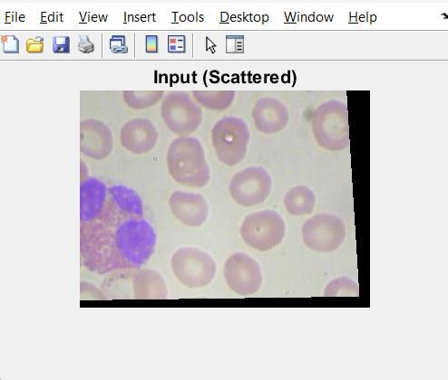
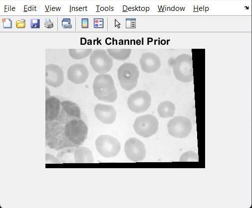
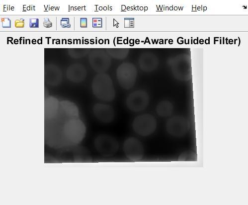
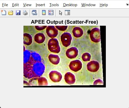
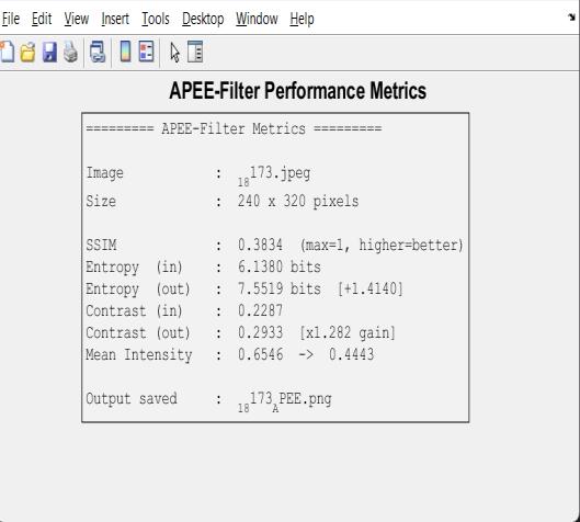
TheexperimentalresultsshowthattheAPEE-Filterworks well in reducing scattered light and improving biomedical images.Fig2showstheoriginalimage,whichlookswashed out, has low contrast, and makes it hard to see the details because of the scattered light. These issues show how importantitistoremovescatteredlighttomaketheimages better for viewing and diagnosis. Fig.3 shows the Dark ChannelPriormap,whichhelpsfindthepartsoftheimage thataremostlyscatteredlightversustherealtissue.Darker areasare where real cell edgesare,andbrighter partsare areas with a lot of scattered light. This shows that the adaptivecontrastpriorhelpsthesystemunderstandwhere the scattering is happening. Fig 4 shows the Refined Transmission Map after applying edge-aware filtering. Compared to the original map, it has smooth changes in areas where the image is uniform, but keeps the sharp edges of the cells. This shows the system can keep fine details and avoid halo effects during the image reconstruction.Fig 5shows the final enhancedimage and someperformancenumbers. Therestoredimagehasmuch better contrast, clear cell edges, and can show internal structuresmoreclearly.Theperformancenumbers,likean increaseinentropyof1.41bitsandacontrastgainof1.282 times,provethatmoredetailshavebeenrecoveredandthe imageiseasiertosee.Overall,theseresultsshowtheAPEEFilter is good at reducing scattered light while keeping important details for diagnosis. By combining physicsbased modeling, adaptive contrast priors, edge-aware refinement,andself-calibratedlightestimation,themethod can both improve image quality and work efficiently, makingitusefulforreal-timebiomedicalimaging.
The APEE-Filter is a strong and efficient method for reducing scattered light in biomedical images. It uses a combinationofphysics-basedmodelingandadaptive,edgesensitive processing techniques. Unlike traditional deep learning methods that need a lot of labeled data and powerful computers, this approach works without any training. This makes it very useful for real-world use in biomedical settings. The system uses a model that understands how light interacts with biological tissues, helping to accurately restore images. A significant advantage of this method is its adaptive contrast prior, which uses a quarter-window dark channel and local variance data to estimate the transmission map. This doesn’tdependonlearnedparameters,makingitbothfast and reliable in different imaging situations. The filter also includes edge-aware refinement steps that help keep importantdetailslikecelledgesandbloodvesselpatterns clear, which is essential for accurate diagnosis. The framework improves image contrast, reduces scattered light effects, and restores clarity without losing important
features.Itssimpledesignandlinearperformanceallowit toworkquickly,makingitsuitableforuseinsystemswith limited resources. Overall, the APEE-Filter offers a good balance betweenqualityandefficiency,anditcanbe used acrossvarioustypesofbiomedicalimaging.
[1] X. Xu, J. Zhao, J. E. Antonio Lopez, S. Gausmann, R. A. Correa, and A. Schülzgen, “Adaptive inverse mapping: a model free semi supervised learning approach towards robust imaging through dynamic scattering media,” Opt. Express,vol.31,no.9,pp.14343–14357,May2023.
[2] Y. Zhang, Z. Ding, R. Li, X. Meng, Y. Shi, C. Zhan, and C. Zhao, “Scanning imaging through the scattering medium basedonguidedfilterassistedbyaWTMMandNLM,”Appl. Opt.,vol.64,no.10,pp.2408–2418,Apr.2025.
[3] L. Zhang, “Image dehazing method based on adaptive feature enhanced contrastive learning framework (AFE Dehaze),”IETImageProcess.,2025.
[4]X.Xu,etal.,“Descatteringandimagerestorationwitha transformer for two photon excited fluorescence microscopy,”Proc.Natl.Acad.Sci.U.S.A.,2025.
[5] H. Liang et al., “An optical meta image processor for enhancedimagingthroughstronglyscatteringmedia,”Nat. Commun.,2025.
[6] Y. Chen, S. X. Mi, Y. P. Tian, X. B. Hu, Q. Y. Yuan, K. H. Chew,andR. P.Chen,“Adaptivevectorialrestorationfrom dynamic speckle patterns through biological scattering mediabasedondeeplearning,”Sensors,vol.25,no.6,1803, Mar.2025.
[7] J. Tao, H. Gong, C. Yuan, and H. Zhou, “Single image dehazing based on transmission map segmentation and priorknowledge,”J.ImagingSci.Technol.,vol.68,no.4,Jul. 2024.
[8] D. Lu et al., “Customizing the field of view for imaging through scattering media using deep learning and speckle correlations,”Adv.PhotonicsNexus,2025.
[9] K. Yang et al., “Data driven polarimetric imaging: a review,”Opto Electron.Sci.,2024.
[10] K. Horie, K. Toda, T. Nakamura, and T. Ideguchi, “Bidirectional quantitative scattering microscopy for label free cellular imaging,” Nat. Commun., vol. 16, art. 9712,2025.