
International Research Journal of Engineering and Technology (IRJET) e-ISSN: 2395-0056
Volume: 13 Issue: 04 | Apr 2026 www.irjet.net p-ISSN: 2395-0072
International Research Journal of Engineering and Technology (IRJET) e-ISSN: 2395-0056
Volume: 13 Issue: 04 | Apr 2026 www.irjet.net p-ISSN: 2395-0072
Geetika Kamaraj1 , Deivanai K2 , Afrin Banu A3 , Ms. R. Santhana lakshmi4
123UG Student, Department of Computer Science and Engineering, Dhanalakshmi Srinivasan University, Tamilnadu, India
4Assistant Professor, Department of Computer Science and Engineering, Dhanalakshmi Srinivasan University, Tamilnadu, India
Abstract - Brain tumor detection and classification are critical tasks in medical diagnostics that require high accuracy,reliability,andinterpretability.Traditionalmethods rely on manualanalysisofMagneticResonanceImaging(MRI) scans, which can be time-consuming and prone to human error. To address these challenges, this paper proposes an Explainable Artificial Intelligence (AI)-based framework for automated brain tumor detection, classification, and clinical support.Theproposed system integratesaYouOnlyLookOnce (YOLO) model for precise tumor localization, followed by region-of-interest (ROI) extraction to enhance classification performance. A Convolutional Neural Network (CNN) is then used to classify tumor types and generate confidence scores. To improve interpretability, Gradient-weighted Class Activation Mapping (Grad-CAM) is employed to visualize the regions influencing the model’s decision. In addition, radiomic feature extraction is performed to obtain intensity, texture, shape, andspatialcharacteristicsofthetumor.Anuncertainty estimationmodule isincorporatedtoevaluatethereliabilityof predictions, enabling risk-aware decision support. Furthermore, the system is deployed through a user-friendly web interface thatsupports multilingualinteraction,allowing patients and healthcare professionals to access diagnostic results, doctor-oriented insights, and detailed reports in their preferred language. The final output includes tumor classification, explainable insights, uncertainty assessment, andpersonalized reports, making the system suitableforrealworld clinical applications. Overall, the proposed approach enhances transparency,accuracy,reliability,andaccessibility in AI-driven healthcare solutions.
Key Words: BrainTumorDetection,ConvolutionalNeural Network(CNN),YOLO,ExplainableAI,Grad-CAM,Radiomics, UncertaintyEstimation,MedicalImageAnalysis
Brain tumors are among the most critical neurological disorders, requiring early and accurate diagnosis for effectivetreatmentplanning.MagneticResonanceImaging (MRI)iswidelyusedfordetectingbrainabnormalitiesdueto itshigh-resolutionimagingcapability.However,traditional diagnosisreliesheavilyonmanualanalysisbyradiologists,
which can be time-consuming, subjective, and prone to humanerror.
Inrecentyears,ArtificialIntelligence(AI)anddeeplearning techniques have shown significant potential in medical imageanalysis.ConvolutionalNeuralNetworks(CNNs)are widelyusedforimageclassificationtasksduetotheirability toautomaticallyextracthierarchicalfeaturesfromimages [3].Similarly,objectdetectionmodelssuchasYouOnlyLook Once (YOLO) enable efficient and real-time localization of regionsofinterestwithinimages[1].Theseadvancements havepavedthewayforautomatedandaccuratebraintumor detectionsystems.
Despitethesedevelopments,manyexistingapproachesfocus primarily on detection and classification while lacking interpretabilityandclinicalinsight.The“black-box”nature of deep learning models makes it difficult for healthcare professionalstotrustthepredictions.Toaddressthisissue, ExplainableArtificialIntelligence(XAI)techniquessuchas Gradient-weighted Class Activation Mapping (Grad-CAM) have been introduced to visualize the regions influencing model decisions [5]. Additionally, radiomics enables the extraction of quantitative features from medical images, providing deeper insights into tumor characteristics and supportingclinicaldecision-making[7].
ThispaperproposesanExplainableAI-basedframeworkfor braintumordetectionandclassificationusingMRIimages. ThesystemintegratesYOLOfortumorlocalization,followed by region-of-interest (ROI) extraction to improve classificationperformance.ACNNmodelisusedtoclassify tumor types, while Grad-CAM is employed to enhance interpretability. Furthermore, radiomic feature extraction and uncertainty estimation are incorporated to provide clinicallyrelevantinsightsandassesspredictionreliability. The system is deployed through a user-friendly web interfacewithmultilingualsupport,enablingaccessibleand efficient interaction for both patients and healthcare professionals.
The proposed approach aims to improve accuracy, transparency,andusabilityinAI-drivenmedicaldiagnostics, thereby contributing to more reliable and accessible healthcaresolutions.
International Research Journal of Engineering and Technology (IRJET) e-ISSN: 2395-0056
Volume: 13 Issue: 04 | Apr 2026 www.irjet.net p-ISSN: 2395-0072
Severalresearchworkshavebeencarriedoutinthefieldof brain tumor detection and medical image analysis using deep learning techniques. Joseph Redmon et al. (2016) introduced the YOLO framework for real-time object detection,enablingfastandefficientlocalizationofobjects withinimages[1].Later,Redmonetal.(2018)improvedthis model with YOLOv3, enhancing detection accuracy and performanceforobjectsofvaryingsizes[2].AlexKrizhevsky etal.(2012)demonstratedtheeffectivenessofConvolutional Neural Networks (CNNs) for image classification tasks, establishing a foundation for modern deep learning approaches in medical imaging [3]. Similarly, Karen SimonyanandAndrewZisserman(2014)proposedadeeper CNNarchitecturethatsignificantlyimprovedclassification accuracy[4].
Ramprasaath R. Selvaraju et al. (2017) introduced GradCAM, an explainable AI technique that provides visual interpretationsofdeeplearningmodeldecisions,improving transparencyincriticalapplicationssuchashealthcare[5]. GeertLitjensetal.(2017)presentedacomprehensivesurvey ondeeplearninginmedicalimageanalysis,highlightingthe effectivenessofCNN-basedmodelsindiseasedetection[6]. Philippe Lambin et al. (2012) introduced the concept of radiomics, whichinvolvesextracting quantitative features frommedicalimagestosupportclinicaldecision-making[7].
Sergio Pereira et al. (2016) applied CNNs for brain tumor segmentation in MRI images, demonstrating improved accuracycomparedtotraditionalapproaches[8].
Bjoern Menze et al. (2015) developed the BRATS dataset, whichservesasabenchmarkforbraintumorsegmentation and analysis [9]. Dinggang Shen et al. (2017) provided an overview of deep learning techniques in medical imaging, emphasizingtheirroleinimprovingdiagnosticperformance [10]. Although these approaches have shown promising results,mostexistingmethodsfocusprimarilyondetection andclassification,withlimitedemphasisonexplainability, uncertaintyestimation,anduseraccessibility.Theproposed system addresses these limitations by integrating YOLObased detection, CNN classification, explainable AI techniques,radiomicfeatureextraction,andamultilingual web-basedinterface.
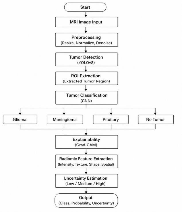
Theproposedsystempresentsanintegratedframeworkfor automatedbraintumordetection,classification,andclinical analysis using MRI images. The methodology combines object detection, deep learning, explainable AI, radiomic feature extraction, and a user-friendly web interface to provide accurate and interpretable results. The overall workflow of the system consists of multiple stages, as describedbelow.
Theoverallarchitectureoftheproposedsystemconsistsof thefollowingstages:
1.InputAcquisition:
• MRI brain image is uploaded by the user through thewebinterface.
2.Preprocessing:
• Imageresizingandnormalizationareperformedto preparetheinputforthemodel.
3.TumorDetection(YOLO):
• YOLO model detects tumor regions in the MRI image.[1]
• Bounding boxes are generated around suspected tumorareas.
4.RegionofInterest(ROI)Extraction:
• The detected tumor region is cropped using boundingboxcoordinates.
• Removesirrelevantbackgroundinformation.
5.TumorClassification(CNN):
• CroppedROIispassedintoaCNNmodel.
• The model classifies tumor type (e.g., glioma, meningioma,pituitary).
• Confidencescoresaregenerated.
6.Explainability(Grad-CAM):
• Grad-CAM generates heatmaps highlighting importantregions.
• Helpsinunderstandingmodeldecisions.
7.RadiomicFeatureExtraction
• Intensityfeatures:mean,standarddeviation
• Texturefeatures:GLCM-basedfeatures
• Shapefeatures:area,perimeter,circularity
• Spatialfeatures:tumorareapercentage,bounding boxsize,tumorlocation
8.UncertaintyEstimation
• Evaluatesreliabilityofmodelpredictions.
• Identifieslow-confidenceoruncertaincases.
9.ReportGeneration
• Combines classification results, features, and explanations.
• Generatesaclinical-stylereportforusers.
10.WebInterfacewithMultilingualSupport
International Research Journal of Engineering and Technology (IRJET) e-ISSN: 2395-0056
Volume: 13 Issue: 04 | Apr 2026 www.irjet.net p-ISSN: 2395-0072
• Displays results including images, heatmaps, and reports.
• Supportsmultiplelanguagesforbetteraccessibility.
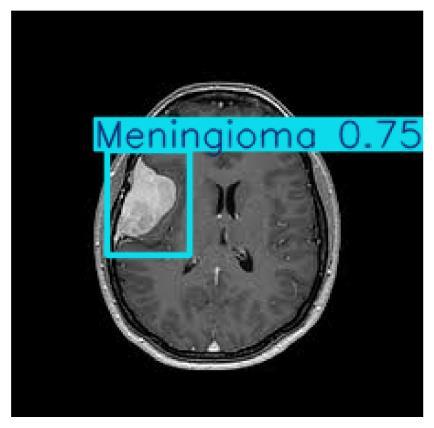
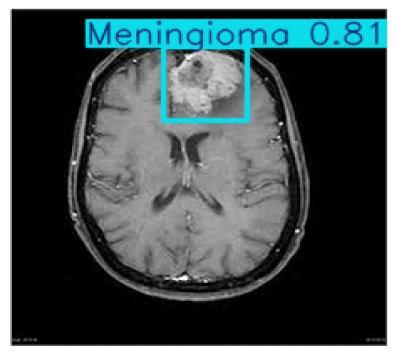
Intheproposedsystem,tumordetectionisperformedusing theYouOnlyLookOnce(YOLO)model,whichenablesrealtime object detection in MRI images [1]. The model processes the entire image in a single forward pass and predicts bounding boxes along with confidence scores for potentialtumorregions.Thisapproachallowsefficientand accurate localization of tumors, even in complex medical images.
3.3 Region of Interest (ROI) Extraction
After tumor detection, the identified region is extracted usingtheboundingboxcoordinatesprovidedbytheYOLO model. This region of interest (ROI) focuses only on the tumorarea,eliminatingirrelevantbackgroundinformation. Thisimprovesthesystemimprovesclassificationaccuracy.
After Cropping Image
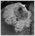
The extracted ROI is provided as input to a Convolutional Neural Network (CNN) for tumor classification. The CNN model learns hierarchical features from the image and classifies it into different tumor types such as glioma, meningioma, and pituitary tumor [3]. The model also generates confidence scores indicating the probability of eachpredictedclass.
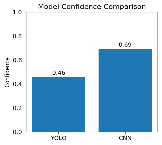
Fig -4: CNNClassifiedImage
International Research Journal of Engineering and Technology (IRJET) e-ISSN: 2395-0056
Volume: 13 Issue: 04 | Apr 2026 www.irjet.net p-ISSN: 2395-0072
To enhance interpretability, Gradient-weighted Class ActivationMapping(Grad-CAM)isappliedtovisualizethe regions influencing the model’s prediction [5]. Grad-CAM generates heatmaps over the input image, highlighting importantareasthatcontributedtotheclassification.This improves transparency and helps in understanding the decision-makingprocessofthemodel.


Radiomic feature extraction is performed on the ROI to obtain clinically relevant information about the tumor. Intensityfeaturessuchasmeanandstandarddeviationare computedtoanalyzepixeldistribution.Texturefeaturesare extracted using the Gray-Level Co-occurrence Matrix (GLCM),whileshapefeaturessuchasarea,perimeter,and circularity describe tumor geometry. Additionally, spatial featuresincludingtumorareapercentage,boundingboxsize, and tumor location are calculated to provide insights into tumorsizeandposition.
An uncertainty estimation module is incorporated to evaluate the reliability of model predictions. The system analyzes confidence scores and prediction behavior to identifycaseswithlowcertainty.Thishelpsindistinguishing betweenreliableanduncertainpredictions,enabling riskawaredecision-makinginclinicalapplications.
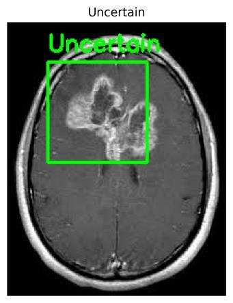
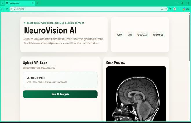
The proposed system is deployed through a user-friendly webinterfacethatallowsuserstouploadMRIimagesand view results. The interface displays detection outputs, classification results, Grad-CAM visualizations, extracted features, and detailed reports. Additionally, multilingual support is integrated to provide outputs and medical insights in different languages, improving accessibility for bothpatientsandhealthcareprofessionals.
The proposedsystemisimplementedusingdeeplearning andwebdevelopmentframeworkstoperformbraintumor detection,classification,andanalysis.Theimplementation detailsaresummarizedasfollows:
Python is used as the primary programming language (version3.10).Deep learningmodelsare developedusing TensorFlow and Keras, while OpenCV is used for image processing. The web application is built using Flask, with HTMLandCSSfortheuserinterface.3
Table -1: ToolsUsedinProject
Category Tools / Technologies
Language
Python3.10version
Detection YOLO(Ultralytics)
Classification TensorFlow,Keras
Computer Vision OpenCV
Feature Extraction NumPy,scikit-image
Machine Learning scikit-learn,joblib
Explainable AI Grad-CAM,SHAP
Backend Flask
Frontend HTML,CSS,JavaScript
Translation GoogleTranslateAPI
Visualization Matplotlib
Report Generation FPDF
International Research Journal of Engineering and Technology (IRJET) e-ISSN: 2395-0056
Volume: 13 Issue: 04 | Apr 2026 www.irjet.net p-ISSN: 2395-0072
4.2.
ThedatasetissourcedfromRoboflowUniverse[11],which providesannotatedMRIimagesforobjectdetectiontasks. Suchdatasetstypicallyincludemultipletumorclassessuch as glioma, meningioma, and pituitary tumors, and are commonlyusedfortrainingdeeplearningmodelsinmedical imagingapplications.
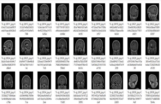
4.3 Training Details:
The YOLO model is used for tumor detection and is finetunedforMRIimages.TheCNNmodelistrainedfortumor classificationusinglabeledimagedata.Thetrainingprocess includes multiple epochs, optimization techniques, and validationtoimproveaccuracyandreduceoverfitting.
4.4
Before training, MRI images are preprocessed to ensure consistency and improve model performance. The preprocessingstepsincludeimageresizingtoafixedinput size,normalizationofpixel values,anddataaugmentation techniques such as rotation, flipping, and zooming. These techniqueshelpinincreasingdatasetdiversityandreducing overfitting,therebyimprovingthegeneralizationcapability ofthemodel.
4.5 Feature Extraction
In addition to deep learning-based predictions, radiomic feature extraction is performed on the detected tumor regions. Intensity features such as mean and standard deviationdescribethedistributionofpixelvalues.Texture featuresbasedonGrayLevelCo-occurrenceMatrix(GLCM) capturespatialrelationshipsbetweenpixels.Shapefeatures suchasarea,perimeter,andcircularityprovideinformation abouttumorgeometry.Spatialfeaturessuchastumorarea percentage,boundingboxsize,andtumorlocationarealso extractedtoprovideclinicallyrelevantinsights.
The trainedmodelsare integratedintoa Flask-basedweb applicationthatallowsuserstouploadMRIimagesandview results. The system provides outputs including tumor classification, confidence scores, Grad-CAM visualizations, extractedfeatures,andmultilingualdiagnosticreports.
5. Model Performance:
5.1 YOLO Metrics –
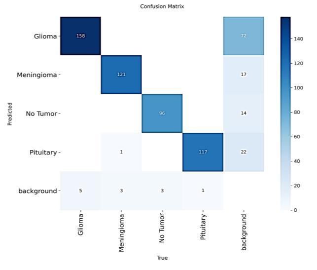
The YOLO model demonstrates strong detection performance with a high mAP@0.5 of 95.63%, indicating accurate localization of tumor regions. The precision and recall values further confirm the model’s effectiveness in identifyingtumorsacrossdifferentclasses.
Table -2: Yolometrics
International Research Journal of Engineering and Technology (IRJET) e-ISSN: 2395-0056
Volume: 13 Issue: 04 | Apr 2026 www.irjet.net p-ISSN: 2395-0072
5.2 CNN Metrics
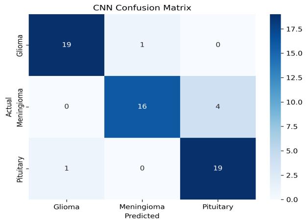
The CNN model achieved an overall accuracy of 90% in classifying brain tumor types. The model shows strong performanceforgliomaandpituitarytumors,whileslightly lowerrecallisobservedformeningiomacases.
Table -3: CNNMetrics
• YOLOTraining:4–5hrs
• Tumor detection (YOLO): 1.0 – 1.5 seconds per image
• CNNTraining:10min
• Classification(CNN):2–3seconds
• Totalpredictiontime:3–5secondsperimage
• Hardwaretested:IntelCorei5Processor,4GBRAM
Severalresearchworkshavebeencarriedoutinthefieldof brain tumor detection using deep learning techniques. YOLO-based models have been widely used for real-time object detection due to their speed and accuracy [1][2]. ConvolutionalNeuralNetworks(CNNs)havedemonstrated strongperformanceinimageclassificationtasks,particularly in medical imaging applications [3][4]. Explainable AI techniques such as Grad-CAM have been introduced to improve transparency by visualizing important regions influencingmodelpredictions[5].Additionally,radiomicsbasedapproachesextractquantitativefeaturesfrommedical imagestosupportclinicaldecision-making[7].Benchmark datasets such as BRATS have played a significant role in evaluatingmodelperformanceinbraintumoranalysis[9].
In this paper, an Explainable Artificial Intelligence (XAI)based framework for brain tumor detection and classification using MRI images has been successfully developed. The proposed system integrates YOLO for accurate tumor localization and a Convolutional Neural Network(CNN)fortumorclassification,achievingreliable performanceinmedicalimageanalysis.
The incorporation of region-of-interest (ROI) extraction enhances classification accuracy by focusing on relevant tumorregions.Additionally,ExplainableAItechniquessuch asGrad-CAMimprovemodeltransparencybyvisualizingthe regions influencing predictions, while radiomic feature extractionprovidesclinicallymeaningfulinsightsintotumor characteristics.
An uncertainty estimation module is included to evaluate predictionreliability,enablingrisk-awaredecision-making. Furthermore,thedeploymentofthesystemthroughawebbased interface with multilingual support enhances accessibilityforbothpatientsandhealthcareprofessionals. Overall, the proposed system offers an accurate, interpretable, and user-friendly solution for brain tumor diagnosis,contributingtoimprovedclinicaldecisionsupport andadvancingAI-basedhealthcareapplications.
International Research Journal of Engineering and Technology (IRJET) e-ISSN: 2395-0056
Volume: 13 Issue: 04 | Apr 2026 www.irjet.net p-ISSN: 2395-0072
Theproposedsystemcanbeusedinhospitalsanddiagnostic centers to assist radiologists in early detection and classificationofbraintumors.Itcanalsobeintegratedinto telemedicine platforms to enable remote diagnosis and consultation. Additionally, the system can serve as an educationaltoolformedicalstudentsandresearchersinthe fieldofmedicalimaging.
The proposed system can be further enhanced by incorporatinglargerandmorediversedatasetstoimprove model generalization and robustness. Future work can explore advanced deep learning architectures such as transformer-basedmodelsforimprovedfeatureextraction andclassificationaccuracy.Integrationof3DMRIanalysis canprovidemorecomprehensivespatialinformationabout tumors,leadingtobetterdiagnosticinsights.
Additionally, the system can be extended for real-time clinical deployment by integrating it with hospital management systems. Mobile and cloud-based implementations can further improve accessibility for remote healthcare services. Enhancing uncertainty estimationtechniquesandincorporatingpatienthistorydata canalsoimprovethereliabilityandclinicalusefulnessofthe system.
[1] J. Redmon et al., “You Only Look Once: Unified, RealTimeObjectDetection,”inProc.IEEECVPR,2016.
[2] J. Redmon et al., “YOLOv3: An Incremental Improvement,”2018.
[3] A.Krizhevskyetal.,“ImageNetClassificationwithDeep ConvolutionalNeuralNetworks,”2012.
[4] K.Simonyanetal.,“VeryDeepConvolutionalNetworks forLarge-ScaleImageRecognition,”2014.
[5] R. R. Selvaraju et al., “Grad-CAM: Visual Explanations fromDeepNetworksviaGradient-BasedLocalization,” 2017.
[6] G.Litjensetal.,“ASurveyonDeepLearninginMedical ImageAnalysis,”MedicalImageAnalysis,2017.
[7] P. Lambin et al., “Radiomics: Extracting More Information from Medical Images Using Advanced FeatureAnalysis,”EuropeanJournalofCancer,2012.
[8] S. Pereira et al., “Brain Tumor Segmentation Using Convolutional Neural Networks in MRI Images,” IEEE Trans.MedicalImaging,2016.
[9] B.H.Menzeetal.,“TheMultimodalBrainTumorImage Segmentation Benchmark (BRATS),” IEEE Trans. MedicalImaging,2015.
[10] D. Shen et al., “Deep Learning in Medical Image Analysis,” Annual Review of Biomedical Engineering, 2017.
[11] Roboflow Universe, “Brain Tumor Detection Dataset,” Available:
https://universe.roboflow.com/aabbcceeffgg/brain-tumordetection-69d9s
[12] OlafRonnebergeretal.,“U-Net:ConvolutionalNetworks for Biomedical Image Segmentation,” Proc. MICCAI, 2015.
[13] Kaiming He et al., “Deep Residual Learning for Image Recognition,”Proc.CVPR,2016.
[14] MingxingTanandQuocV.Le,“EfficientNet:Rethinking ModelScalingforConvolutionalNeuralNetworks,”Proc. ICML,2019.
[15] Tsung-Yi Lin et al., “Feature Pyramid Networks for ObjectDetection,”Proc.CVPR,2017.