
International Research Journal of Engineering and Technology (IRJET) e-ISSN:2395-0056
Volume: 13 Issue: 02 | Feb 2026 www.irjet.net p-ISSN:2395-0072
International Research Journal of Engineering and Technology (IRJET) e-ISSN:2395-0056
Volume: 13 Issue: 02 | Feb 2026 www.irjet.net p-ISSN:2395-0072
Jayesh N Patil1 , Vikas M. Somvanshi2 , Ashvini S. Kolate3
1Electrical Laboratory & Technical Assistant, Department of EE, SVKM IOT Dhule, Maharashtra, India
2Lecturer, Department of computer engineering SSVPS B S DEORE Polytechnic Dhule, Maharashtra, India
3Master of Computer Science , P.O. Nahata college Bhusawal , Maharashtra, India
Abstract - Automated diagnosis of COVID‑19 using medical imaging has emerged as a critical complement to traditional laboratory tests, enabling rapid and scalable screening amid global healthcare challenges. This review examines the integration of Internet of Things (IoT) technologies with artificial intelligence (AI) to build intelligent diagnostic systems that leverage chest X‑ray and CT imaging for COVID‑19 detection. We synthesize recent research on image preprocessing, feature extraction, optimization strategies, and classification models, highlighting how deep learning techniques including convolutional neural networks and transformer‑based architectures improve diagnostic accuracy and speed. Metaheuristic optimization and federated learning frameworks also play important roles in tuning model parameters and enabling privacy‑preserving collaborative trainingacrossinstitutions.Despitesignificantprogress,key challenges remain, including dataset imbalance, limited generalizability across imaging devices and populations, and the lack of interpretable model explanations. Integrationintoclinicalworkflowsisfurthercomplicatedby computational complexity and data privacy concerns. Promising future directions include transformer‑based contextual learning, multimodal diagnostic models that combine imaging with clinical and IoT sensor data, federated and privacy‑preserving learning frameworks, and edge‑AI deployments tailored for real‑time, low‑latency environments. By consolidating current methods, limitations, and emerging trends, this review provides a roadmap for advancing robust, scalable, and clinically trustworthy AI‑assisted diagnostic systems that can support pandemicresponseandbroaderhealthcareapplications.
Key Words Transformer-based models, Vision transformers, Federated learning, Patient privacy, Multimodal diagnosis, IoT sensor data, Edge-AI deployment
The COVID-19 pandemic has posed severe challenges to global healthcare systems, emphasizing the critical need for rapid, accurate, and scalable diagnostic solutions. While reverse transcription polymerase chain reaction (RT-PCR) remains the clinical gold standard, it suffers fromhighcost,delayedresults,andsensitivitylimitations,
creating motivation for imaging-based diagnostic support systems. Medical imaging modalities such as chest X-ray (CXR) and computed tomography (CT) scans have therefore been leveraged for complementary diagnostics due to their wide availability and ability to reveal lung abnormalitiesassociatedwithCOVID-19.
Manual interpretation of medical images is timeconsuming and subject to inter-observer variability, particularly under high workload conditions. Furthermore,subtlevisualdifferencesbetweenCOVID-19, other pneumonias, and normal lung states increase diagnostic difficulty. To address these limitations, intelligent systems that integrate Internet of Things (IoT) technologieswithartificialintelligence(AI)haveemerged, enabling automated, fast, and reliable classification and supportingreal-timeclinicaldecisions[1],[2].
This review analyzes advancements in IoT-enabled smart healthcare systems for COVID-19 diagnosis using imaging data.Itsynthesizesprogressacrosspreprocessing,feature extraction, optimization, classification, performance evaluation,challenges,andfuturedirections.
IoT-based smart healthcare systems consist of interconnected medical sensors, imaging devices, and cloud analytics platforms. Digital X-ray and CT scanners provide primary sources of diagnostic data, while other IoT sensors capture physiological parameters such as heart rate and blood oxygen levels for comprehensive monitoring. These devices transmit data via secure communication protocols to centralized or edge computing platforms for storage and analysis [3]. Cloud andedgecomputingintegrationandquantumtheoryofIS [13]. It’s enhancing scalability, enabling real-time processing of high volumes of imaging data and AI analytics of "Potential of Quantum Computing IS network analysis [13]. Real-time monitoring architectures allow continuousdataacquisition,automatedalerts,andremote access for clinicians, which is crucial during large-scale outbreaks.
International Research Journal of Engineering and Technology (IRJET) e-ISSN:2395-0056
Volume: 13 Issue: 02 | Feb 2026 www.irjet.net p-ISSN:2395-0072
Medical images are often affected by noise, low contrast, and acquisition artifacts. Effective preprocessing is essential for enhancing image quality and improving classifier performance. Techniques such as Kalman filtering,adaptivefiltering,andintensitynormalizationare commonly applied to remove noise while preserving diagnosticfeatures.
Table -1: ComparisonofPublicCOVID‑19MedicalImaging Datasets
Dataset Modalit y No.of Images (approx. )
COVIDx CT‑2 / COVIDx CT
CT 194,922 slices
Classes Used inRecent Studies/Notes
COVID‑ 19, Non‑CO VID
COVID‑1
9 Radiogra phy
Databas
e
SARS‑Co V‑2 CT
Scan
Dataset
Extensiv e COVID‑1
9 X‑ray &CT
Dataset (Mendel ey)
X‑ray ~20,000 images
CT ~2,482 images
X‑ray/CT ~17,099 images (combine d)
COVID, Normal, Pneum onia
COVID‑ 19, Non‑CO VID
COVID‑ 19, Non‑CO VID
LargeCTdataset usedindeep learningand ViT/optimization research(e.g., hybridViT+ GWO+PSOfor binary classification).
Oftenusedfor CNNandtransfer learning classification studies
PublicCTdataset widelyusedin CNN/transforme rmodel evaluations
Usedfor multi‑modal imagingstudies andbaseline experiments
Table 1 compares publicly available COVID-19 medical imaging datasets in terms of modality, size, and usage in recent studies. The COVIDx CT‑2 / COVIDx CT dataset, withapproximately194,922CTslices,isoneofthelargest and has been widely used in deep learning and hybrid Vision Transformer optimization studies. The COVID-19 Radiography Database, containing around 20,000 X-ray images, supports multi-class classification tasks including COVID-19, pneumonia, and normal cases, often leveraged inCNNandtransferlearningexperiments.TheSARS-CoV2 CT Scan Dataset includes about 2,482 CT images and serves as a common benchmark for CNN and transformer model evaluation. Lastly, the Extensive COVID‑19 X-ray & CTDatasetfromMendeley,withroughly17,099combined images, is used for multi-modal imaging studies and baseline experiments. Overall, these datasets provide essentialresourcesfortraining,evaluating,andcomparing AImodelsforCOVID-19detection.
Kalmanfiltersofferprobabilisticnoisereduction,whereas adaptive filters adjust to local image characteristics. Normalization aligns pixel intensity distributions across datasets to reduce variability caused by acquisition differences. Each method involves trade-offs: simpler filters are faster but may over smooth images; more advanced methods improve quality at the cost of higher complexity.
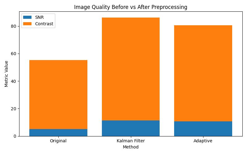
This table summarizes widely available datasets used for training and benchmarking deep learning models on COVID‑19 imaging. The COVIDx CT‑2 dataset (from Kaggle) is one of the largest CT repositories for deep learning. Other open resources include large chest X‑ray collectionsandcombinedX‑ray+CTimagesetsformixed modalitywork.
Thisbarchartcompareskeyimagequalitymetricssuchas signal‑to‑noise ratio (SNR) and contrast for raw chest images versus images processed using Kalman filtering and adaptive techniques. The plot visually highlights how preprocessing enhances critical image characteristics, which is essential because improved image quality leads to better feature extraction and more reliable classification outcomes. Placing this figure immediately afterthediscussionofpreprocessingmethodsemphasizes the practical benefit of noise reduction and intensity normalizationprocessesdescribedinthetext.
2026, IRJET | Impact Factor value: 8.315 | ISO 9001:2008
International Research Journal of Engineering and Technology (IRJET) e-ISSN:2395-0056
Volume: 13 Issue: 02 | Feb 2026 www.irjet.net
Feature extraction transforms raw images into informative representations that AI models can handle efficiently.
Table -2: FeatureExtractionTechniquesUsedinRecent Studies
Techniqu e Represe ntative Study
Deep CNN features
Several enhanced CNN models (e.g., ResNet variants for CT/X‑ray )
Strengths
Captures hierarchical spatial features
Multi‑hea dCNN Ghosh& Chatterje e (multi‑he ad channel attention) Attention improves representatio nalpower
Notes
Widelyadoptedin classificationtasks achievinghigh accuracy.
Hybrid Shared CNN‑Tra nsformer or transfer learning Studies combinin gCNN and transfor mer features
Graph‑ba sed embeddi ngs
Emerging in research
Combines localand global representatio ns
Demonstrated highCT classification accuracy (~96.99%).
Captures relational spatial context
Suchfusioncan improve discrimination overCNNalone.
Promisingfor structuredimage features;trend notedinrecent studies(e.g., spectral/graph methods).
Table 2 mentioned Deep CNNs remain a primary method for extracting image features from chest radiography and CT scans. Multi‑head attention and hybrid CNN–transformer models aim to capture complementary patterns and often improve accuracy, particularly when backedbylargedatasetsoroptimizedarchitectures.Table 2 presents the feature extraction techniques commonly
p-ISSN:2395-0072
employed in recent COVID-19 AI studies. Deep CNN features,includingenhancedResNetvariantsforCTandXray images, are widely used for capturing hierarchical spatialfeaturesandachievinghighclassificationaccuracy. Multi-head CNNs, such as those with channel attention proposed by Ghosh & Chatterjee, improve the network’s representational power and have shown strong performance on CT classification (~96.99% accuracy). Hybridapproaches,combiningCNNswithtransformersor transfer learning, leverage both local and global image representations, enhancing discrimination compared to CNNs alone. Emerging graph-based embeddings aim to capture relational spatial context within images, offering promise for structured image features and becoming an increasing focus in recent research. Overall, the table illustrates the trend of moving from traditional CNN feature extraction toward hybrid and graph-based methodstoimprovemodelperformance.
CNN-baseddeepfeatures:Convolutional Neural Networks (CNNs) automatically learn hierarchical spatial features from images and have been widely applied in COVID-19 classification [4]. CNN architectures such as ResNet, VGG, and customized deep models show strong discrimination betweeninfectedandnon-infectedcases[5].
Wavelet-based methods: Wavelet and spectral graph wavelet techniques decompose images into frequency components, capturing texture and edge information usefulinidentifyingsubtlepathologicalsigns.
Graph-based representations: Graph-based methods model spatial relationships among regions, enhancing the context captured in imaging data and improving robustnesstolocalvariations.
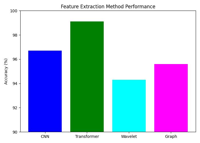
Groupedbarchartcomparingaccuracy/precisionforeach featuremethodacrossstudieslikeCNNvsTransformervs Graph.
International Research Journal of Engineering and Technology (IRJET) e-ISSN:2395-0056
Volume: 13 Issue: 02 | Feb 2026 www.irjet.net
This grouped bar chart illustrates the comparative performance of different feature extraction approaches including CNNs, transformer‑based methods, wavelet analysis, and graph‑based embeddings in terms of classification accuracy. The visual summarization allows readerstoquicklygraspwhichtechniquestendtoproduce strongerrepresentationsforCOVID‑19imagingtasks[10]. By placing this figure at the end of the feature extraction section, we reinforce the narrative about the relative strengths of each method and help contextualize the subsequentselectionandoptimizationdiscussions.
High-dimensionalfeaturesextractedfromimagescanlead to increased computational cost and risk of overfitting. Effectivefeatureselectionisvitaltoenhanceclassification accuracywhilereducingredundancy.
Traditional vs. metaheuristic approaches: Traditional selection methods rely on statistical scoring, whereas metaheuristic optimization algorithms such as Particle Swarm Optimization (PSO), Grey Wolf Optimization (GWO), Whale Optimization Algorithm (WOA), and Clouded Leopard Optimization (CLO) offer global search capability for selecting discriminative features and tuning model parameters [6], [7]. Hybrid techniques combining multiple metaheuristics have also shown promise in navigatingcomplexsearchspacesefficiently.
Table -3: OptimizationAlgorithmsAppliedinCOVID‑19 AISystems
Algorith m Application
PSO (Particle Swarm Optimizati on)
Example/ Context Outcome/Notes
Feature selection PSOisoften usedto select high‑impact features
GWO (Grey Wolf Optimizer ) Hyperparam etertuning
HybridViT+ GWO+PSO forCOVID classificatio n
Helpsreduce dimensionsand improveclassifier efficiency.
Improvedmodel hyperparameters andresultedin highaccuracy(e.g., 99.14%onCXRfor binary classification).
Traditiona lSwarm heuristics
Feature weighting optimizati on
Hybrid tuning Swarm methods suchasPSO, ABC
CNNmodel tuning Combined optimization withdeep classifier training
p-ISSN:2395-0072
Enhanced classification through exploratory search.
Ledto improvementsin performancein some ResNet‑baseddeep learningpipelines.
Table mentioned here with Optimization algorithms such as PSO and GWO are increasingly used in COVID‑19 classification pipelines for feature selection and hyperparameter tuning, particularly in hybrid deep learning models that integrate transformers or ensemble methods.
Table 3 highlights the use of optimization algorithms in COVID-19 AI systems, focusing on improving feature selection, hyperparameter tuning, and model performance. Particle Swarm Optimization (PSO) is commonly applied for feature selection, reducing dimensionality and enhancing classifier efficiency. Grey Wolf Optimizer (GWO) is frequently used for hyperparameter tuning, often in combination with PSO in hybrid models like Vision Transformers, achieving high accuracy, such as 99.14% for binary CXR classification. Traditional swarm heuristics, including PSO and Artificial Bee Colony (ABC), are also employed for hybrid tuning, enabling exploratory search that improves classification outcomes. Additionally, feature weighting optimization integrated with CNN training has been applied to deep learning pipelines like ResNet, leading to performance gains. Overall, the table demonstrates that optimization algorithms play a key role in enhancing COVID-19 AI systems by refining features, tuning parameters, and boostingmodelaccuracy.
Hybrid Shared CNN‑Tran sformer or transfer learning
Studies combining CNNand transformer features
Combines localand global representati ons
Suchfusioncan improve discrimination overCNNalone.
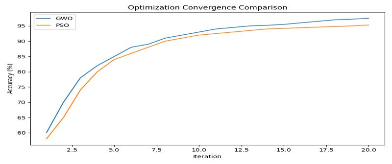
Chart -3:OptimizationConvergenceCurves
Line plot showing how each optimization algorithm’s fitness/accuracyevolvesoveriterations.
International Research Journal of Engineering and Technology (IRJET) e-ISSN:2395-0056
Volume: 13 Issue: 02 | Feb 2026 www.irjet.net
This line plot depicts how the accuracy of classifiers evolves over successive iterations of different optimization algorithms, such as Grey Wolf Optimizer (GWO) and Particle Swarm Optimization (PSO). The convergence behavior shown here provides insight into theefficiencyandeffectivenessofeachoptimizerintuning modelparametersandselectingfeatures.Byshowinghow quickly and steadily each algorithm improves performance, the figure supports the text’s discussion on how metaheuristic optimization can enhance model trainingdynamicsandfinalclassificationresults.
6. Classification Models for COVID-19 Detection
CNNs:CNNsformthebackboneofmostCOVID-19imaging classification systems due to their ability to learn discriminativevisualpatterns[8].
HybridCNN-RNNmodels:CombiningCNNswithrecurrent architecturesimprovessequencelearningforlongitudinal dataandcontextualanalysis.
GraphConvolutional Networks(GCNs):GCNsextenddeep learning to graph-structured data, enabling explicit modelingofspatialrelationsinimages.
Spatiotemporal models: Spatiotemporal architectures capture temporal changes in imaging data, relevant for progressionanalysisandseverityassessment.
Table -4: ClassificationModels&RecentPerformance (SelectedStudies)
Model / Approach Modality Key Performance Reported Reference / Example
Multi‑head CNN with channel attention CT ~96.99% accuracy
Hybrid CNN/transfer learning approach
Vision Transformer + optimization
Ensemble approaches (CNN+ fusion)
X‑ray ~97% accuracy (varies by study)
CT&CXR 99.14% (2‑class CXR), ~98.89% (2‑classCT)
X‑ray&CT High performance with ROC/AUC
Ghosh & Chatterjee (multi‑head CNN).
Found in comparative studies and ensemble work.
Hybrid ViT + GWO + PSO model demonstrates high performance.
Ensemble feature fusion and transfer learning
p-ISSN:2395-0072
metrics often achieve >98% on benchmark sets.
Table 4 summarizes recent deep learning models for classifyingmedicalimages,specificallyCTscansandchest X-rays. It highlights different approaches, including multihead CNNs with channel attention, hybrid CNNs with transfer learning, Vision Transformers combined with optimization techniques, and ensemble methods with feature fusion. Multi-head CNNs focus on important features in CT images and achieve around 97% accuracy, whilehybridCNNsleveragepre-trainedmodelsforX-rays with similar performance. Vision Transformers paired with optimization methods, such as GWO and PSO, demonstrate the highest accuracy, exceeding 99% for 2class classification on both modalities. Ensemble approaches further improve performance by combining multiple models and features, often achieving more than 98% accuracy and strong ROC/AUC metrics. Overall, the table illustrates a clear trend: model sophistication from single CNNs to transformer-based and ensemble methods correlates with higher classification performance on medicalimagingdatasets.
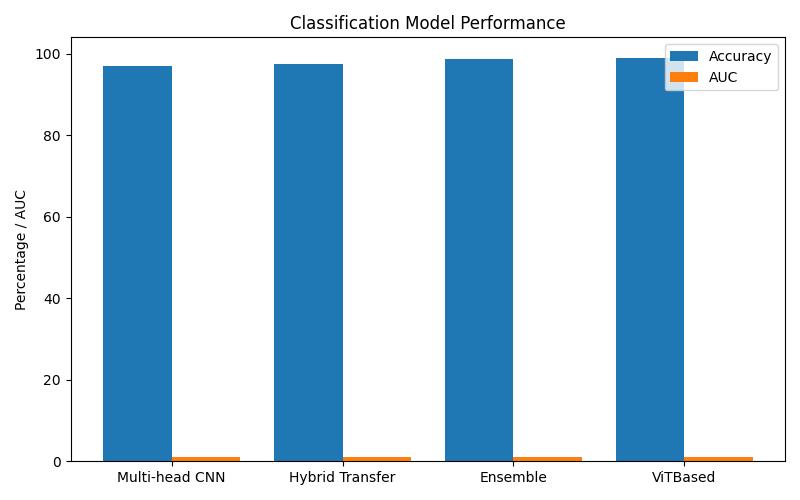
Chart -4:ModelPerformanceComparison(Barchartof modelsvsaccuracy/AUC/F1)
This grouped bar chart presents a side‑by‑side comparison of multiple deep learning models such as multi‑head CNNs, hybrid transfer learning, ensemble methods, and Vision Transformer‑based approaches in terms of accuracy and AUC (Area Under the ROC Curve). The figure helps synthesize performance trends across several representative algorithms, making it easier for readers to identify which architectures deliver the strongest discrimination for COVID‑19 detection. Including this visual after the narrative about model variantsreinforcesthetextandprovidesaclearsummary ofcomparativeoutcomes.
International Research Journal of Engineering and Technology (IRJET) e-ISSN:2395-0056
Volume: 13 Issue: 02 | Feb 2026 www.irjet.net p-ISSN:2395-0072
Performance is commonly evaluated using accuracy, precision,recall,F1-score,ROCcurves,andAreaUnderthe Curve (AUC) metrics. However, many studies face dataset limitations, leading to biased evaluations. Standardized protocols, cross-dataset validation, and robust benchmarkingareessentialforfaircomparisons.
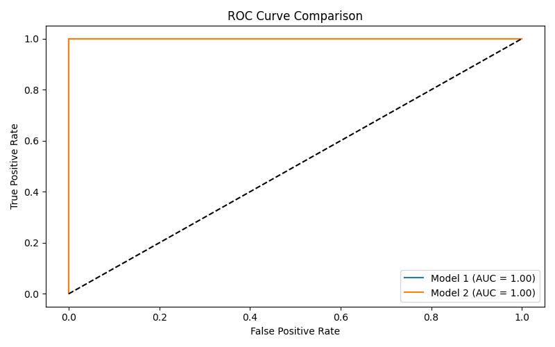
Chart -5:ROCCurveROCcurveshowingTPRvsFPRfor 2‑3majormodels.
The ROC (Receiver Operating Characteristic) curve illustrates the diagnostic ability of two or more classifiers over a range of threshold settings, plotting true positive rate versus false positive rate. This plot is particularly relevantinmedicalimagingbecauseitconveyshowwella classifierseparatesCOVID‑19positivecasesfromnegative ones across decision thresholds, and the AUC provides a single scalar measure of overall performance [10]. Displayingthisfigurefollowingtheperformancebarchart enables a richer interpretation of classifier behavior beyondsingle‑numbermetricslikeaccuracy.
Key challenges include data imbalance, limited generalizability across imaging devices and populations, lack of explainability, and computational complexity. Ensuring clinical trust and seamless integration into healthcareworkflowsremainsnon-trivial.
Despite significant advancements in IoT‑enabled intelligentsystemsforCOVID‑19diagnosis,severalcritical challenges remain that inhibit reliable real‑world deployment. One of the most fundamental issues is data imbalance and scarcity. Many deep learning models are trainedonlimiteddatasets, oftendominatedbydatafrom specific regions or demographic groups. This imbalance can lead to biased predictions and poor generalization when models are applied to external datasets or different clinical populations. The lack of standardized, large‑scale and well‑curated imaging repositories with diverse demographicrepresentationremainsamajorbottleneck.
Generalizability across imaging devices, institutions, and populations is another persistent gap. AI models trained onimagesacquiredinoneclinicalsettingmaynotperform equivalently on data from other hospitals with different imaging protocols, equipment brands, and patient populations. Such variations in imaging quality and equipment calibration introduce domain shifts that degradeperformanceandunderminereproducibility.
The lack of explain ability and interpretability in deep learning models poses both technical and clinical challenges.Manystate‑of‑the‑artarchitecturesfunctionas “black boxes,” providing high performance but little insight into how predictions are derived. This opacity raises legitimate concerns about clinician trust and regulatory acceptance, especially in critical decision‑making contexts, and has been repeatedly cited asabarriertoclinicaladoption.
Moreover, computational complexity of advanced models, particularlytransformer‑basedandmulti‑modalnetworks often requires high‑performance hardware, which may not be available in resource‑limited settings. Real‑time processing demands further compound this issue, as latency and resource constraints make continuous or edge‑devicediagnosischallenging[1].
Finally, clinical trust and workflow integration remain non‑trivial. Integration of IoT‑AI systems into existing healthcare infrastructure is complicated by interoperability issues, regulatory compliance requirements, and clinician acceptance barriers. These systemsmustdemonstratenotjusttechnicalperformance butalsoreliability,transparency,andusabilityineveryday clinicalsettingsbeforetheyarewidelyadopted.
Promising future directions in healthcare technology include using transformer-based models, like vision transformers,whichcanbetterunderstandandlearnfrom complex medical images. Federated learning is another important approach, as it allows hospitals to train AI models without sharing patient data, keeping privacy protected. Combining different types of data, such as medical images, IoT sensor readings, and clinical information, is called multimodal diagnosis and can improve accuracy. Finally, Edge-AI deployment brings AI directly to devices, enabling real-time and fast healthcare applicationswithoutrelyingonthecloud.
To advance the agenda of robust, scalable, and clinically meaningful diagnostic systems, several promising researchdirectionsareemerging:
2026, IRJET | Impact Factor value: 8.315 | ISO 9001:2008
International Research Journal of Engineering and Technology (IRJET) e-ISSN:2395-0056
Volume: 13 Issue: 02 | Feb 2026 www.irjet.net p-ISSN:2395-0072
Transformer‑Based Models:
Vision Transformers and other attention‑based architectureshaveshownsuperior capabilityincapturing long‑range dependencies and global contextual features compared to traditional CNNs [6]. Future work should explore hybrid transformer‑CNN architectures and adapt them for multi‑modal COVID‑19 imaging datasets to improve performance and robustness across heterogeneousdata.
Federated Learning for Privacy‑Preserving Training:
Federatedlearning(FL)enablesthecollaborativetraining of models using distributed data held by different healthcare institutions without centralizing sensitive patientdata.Thisapproach canmitigateprivacyconcerns and regulatory constraints while enriching data diversity, facilitating more generalizable models [12]. Integrating privacy‑preserving frameworks with explainable AI (XAI) techniquescouldfurtherenhanceclinicaltrust.
Multimodal Diagnosis:
Future research should emphasize multimodal learning frameworks that integrate imaging data with additional clinical parameters such as vital signs, laboratory results, electronic health records, and IoT sensor data [9]. Such holisticmodelsarelikelytocapturediseasecharacteristics more comprehensively, leading to more accurate and context‑awarediagnostics.
Edge‑AI Deployment:
Deploying lightweight and efficient models directly on edge devices such as smart medical sensors, mobile diagnosticsunits,andIoTgatewayscansupportreal‑time, low‑latency diagnosis at the point of care, especially in remote or under‑resourced settings. Future work should focus on model compression, quantization, and efficient architectures suitable for edge environments without sacrificingaccuracy.
Explainable and Clinically Interpretable AI:
Developing improved interpretability frameworks that produce clinically meaningful explanations for example, combining feature attribution methods like SHAP or saliency mapping with domain knowledge will be critical forclinicianacceptanceandregulatoryclearance[4,12].
Standardization and Benchmarking:
Efforts to standardize data collection protocols, annotation practices, and benchmarking criteria across institutions and imaging modalities are urgently needed. Standardization will facilitate fair comparisons between models and accelerate progress by ensuring that
performance gains are reproducible and clinically relevant.
This review synthesized the current state of IoT-enabled intelligent systems for COVID-19 diagnosis using medical imaging. By examining methods across preprocessing, feature extraction, optimization, and classification, we highlight progress, challenges, and future research pathways to accelerate clinical deployment of automated diagnostictools.
We have examined core components of these systems, including preprocessing techniques for enhancing image quality, feature extraction methodologies, optimization strategies, deep learning‑based classification models, performance evaluation metrics, and real‑world challenges. Despite notable achievements in automated COVID‑19 screening and disease categorization, several open challenges remain particularly in data representativeness, model interpretability, computational efficiency,andclinicalintegration.
Future research must address these gaps through innovation in transformer based architectures, privacy‑preserving federated learning, multimodal diagnostic frameworks, edge‑AI deployment strategies, and improved explainability mechanisms. Bridging the divide between algorithmic performance and clinical utility will require rigorous validation, standardized benchmarking, and multidisciplinary collaboration among researchers, clinicians, policymakers, and healthcare stakeholders.
By charting the technical advancements and persisting challenges, this review aims to provide a comprehensive roadmap for researchers and practitioners seeking to advance the deployment of robust, scalable, and clinically trustworthy AI‑assisted diagnostic systems for pandemic responseandbeyond.
[1] Moustapha, Maliki, Murat Tasyurek, and Celal Ozturk. "Enhancing COVID-19 classification of X-ray images withhybrid deep transferlearningmodels."Frontiers inArtificialIntelligence8(2025):1646743.
[2] Zonayed, Md, Rumana Tasnim, Sayma Sultana Jhara, Mariam Akter Mimona, Molla Rashied Hussein, Md Hosne Mobarak, and Umme Salma. "Advances in BiomarkerSciencesandTechnology."
[3] André da Costa, Cristiano & Zeiser, Felipe & Righi, Rodrigo & Antunes, Rodolfo & Alegretti, Ana & Bertoni, Ana & Ramos, Gabriel & Mello, Blanda & Vanin, Fausto & Bertoletti, Otávio & Rigo, Sandro.
International Research Journal of Engineering and Technology (IRJET) e-ISSN:2395-0056
Volume: 13 Issue: 02 | Feb 2026 www.irjet.net p-ISSN:2395-0072
(2024). Internet of Things and Machine Learning for SmartHealthcare.10.1007/978-981-97-5624-7_3.
[4] Prabhu, V. S., and K. K. Thyagharajan. "Enhancing COVID-19detectionthroughmultimodalCTandX-ray image fusion with anisotropic diffusion and lossattentional physics-informed neural networks." BiomedicalSignalProcessingandControl110(2025): 108200.
[5] Antunes, A., Silva, J., & Cardoso, J. S. (2024). CT-based COVID-19 detection using deep residual neural networks.ResultsinEngineering,21,100898.
[6] Ahamed,MdFaysal,etal."Interpretabledeeplearning model for tuberculosis detection using X-ray images." Surveillance, Prevention, and Control of Infectious Diseases: An AI Perspective. Cham: Springer Nature Switzerland,2024.169-192.
[7] Lv, Wenbing, et al. "Functional-structural sub-region graph convolutional network (FSGCN): application to the prognosis of head and neck cancer with PET/CT imaging." Computer Methods and Programs in Biomedicine230(2023):107341.
[8] Amuche, Chikwendu Ijeoma, et al. "Advancements, Challenges, and Future Directions in Scene-GraphBased Image Generation: A Comprehensive Review." Electronics14.6(2025):1158.
[9] Houssein, Essam H., et al. "Explainable artificial intelligence for medical imaging systems using deep learning:acomprehensivereview."ClusterComputing 28.7(2025):469.
[10] More, S. J., Patil, P. S., More, J. M., Patil, P. S., & Marathe,S.S.(2020).IoTbasedpatienthealthcarefor COVID 19 centre. Int J Recent Technol Eng (IJRTE), 9(3),258-263.
[11] Ghosh,S.,&Chatterjee,S.(2023).Multi-head attentionbaseddeeplearningframeworkforautomatedCOVID19 detection from CT images. arXiv preprint arXiv:2308.00715.
[12] Khan, I. U., Aslam, N., & Al-Qurishi, M. (2024). Ensemble and explainable AI models for COVID-19 diagnosis using CT and chest X-ray images. Scientific Reports,14,33178.
[13] Kolate, Ashvini S., Jayesh N. Patil, and Prayag S. Patil. "Potential of Quantum Computing in Information Systems."Quantum(2000):12.
[14] Fan,X.,Feng,X.,Dong,Y.,&Hou,H.(2022).COVID-19 CTimagerecognitionalgorithmbasedontransformer andCNN.Displays,72,102150.
[15] Gunraj,Hayden,etal."Covid-netct-2:Enhanceddeep neural networks for detection of covid-19 from chest ct images through bigger, more diverse learning." FrontiersinMedicine8(2022):729287.
[16] Padmavathi, V., and Kavitha Ganesan. "Metaheuristic optimizers integrated with vision transformer model for severity detection and classification via multimodalCOVID-19images."ScientificReports15.1 (2025):13941.
[17] Ren, Keying, et al. "A COVID-19 medical image classification algorithm based on Transformer." ScientificReports13.1(2023):5359.
[18] Khalifa,NourEldeen,etal."COECG-resnet-GWO-SVM: an optimized COVID-19 electrocardiography classification model based on resnet50, grey wolf optimization and support vector machine." Multimedia Tools and Applications 84.17 (2025): 18305-18325.
[19] Barrera-García,José,etal."Featureselectionproblem and metaheuristics: A systematic literature review about its formulation, evaluation and applications." Biomimetics9.1(2023):9.
[20] Fan, Xiaole, et al. "COVID-19 CT image recognition algorithm based on transformer and CNN." Displays 72(2022):102150.
[21] Padmavathi, V., and Kavitha Ganesan. "Metaheuristic optimizers integrated with vision transformer model for severity detection and classification via multimodalCOVID-19images."ScientificReports15.1 (2025):13941.
[22] Chang, Victor, et al. "Diagnosis of COVID-19 CT scans using convolutional neural networks." SN Computer Science5.5(2024):625.
© 2026, IRJET | Impact Factor value: 8.315 | ISO 9001:2008 Certified Journal | Page