

Welcome to the 2025 Houston Methodist Academic Institute Annual Report.
Moving a discovery from research to patient care is transformation in action — uniting scientific rigor with real-world testing to accelerate the journey from idea to impact. Our researchers and clinicians work side by side to translate ideas from the laboratory into real-world solutions that improve lives. This year’s report celebrates that journey by spotlighting the people, partnerships and breakthroughs driving change across medicine.
In cancer research, our scientists are challenging long-held assumptions about how tumors respond to treatment. “Rewriting the Rules of Tumor Resistance” explores how our researchers have discovered a critical mechanism, chemotherapyinduced pyroptotic inflammatory cell death that can inadvertently reprogram the tumor microenvironment, making it more hospitable to chemo-resistant cancer stem cells. In “A New Spark in Cancer Therapy,” a platinum-based nanodrug offers a promising alternative to traditional chemotherapy by potentially delivering treatment more precisely while reducing harm to healthy tissue.
The escalating battle against antibiotic-resistant bacteria has a promising new weapon, thanks to a Houston Methodist research team: an antimicrobial peptide, optimized via machine learning, that demonstrates activity against the most stubborn pathogens in medicine.
Dementia is one of our most urgent health challenges. In the U.S., health care costs related to Alzheimer’s disease and other dementias exceed $360 billion annually, with Texas ranking third in the number of people living with or caring for someone with Alzheimer’s and related dementias. In “Driving Dementia Innovation When It Matters Most,” our researchers outline a comprehensive approach focused on identifying and addressing disease drivers early —
long before irreversible decline occurs. As the number of Texans affected by Alzheimer’s grows, Houston Methodist scientists and physicians are building a seamless pipeline to transform how we understand, diagnose and treat memory disorders.
This year’s report also highlights the growth and evolution of our medical education training programs. We congratulate the Texas A&M University School of Engineering Medicine (EnMed) Class of 2025 — the largest class since the program’s inception in 2019. These “physicianeers,” as we call them, are trained in both engineering and medicine, equipping them with a unique perspective on patient care and problem-solving. This partnership is vital to advancing health care, and we look forward to seeing this next generation of “physicianeers” design innovative solutions to address complex medical challenges.
That same commitment is reflected in the expansion of our Graduate Medical Education programs. In 2025, we commemorated 20 years as an independent medical education training institution. What began with 12 Accreditation Council for Graduate Medical Education (ACGME)-accredited programs in 2005 has grown to more than 80 programs in 2025, including 59 ACGME-accredited residencies and fellowships and 29 GME-sponsored fellowships.
Every day, our researchers, faculty, fellows and trainees advance transformational work that changes the landscape of patient care. This annual report reflects their dedication and the profound impact they make on our patients and families. It is an honor to witness and support their contribution to better health.
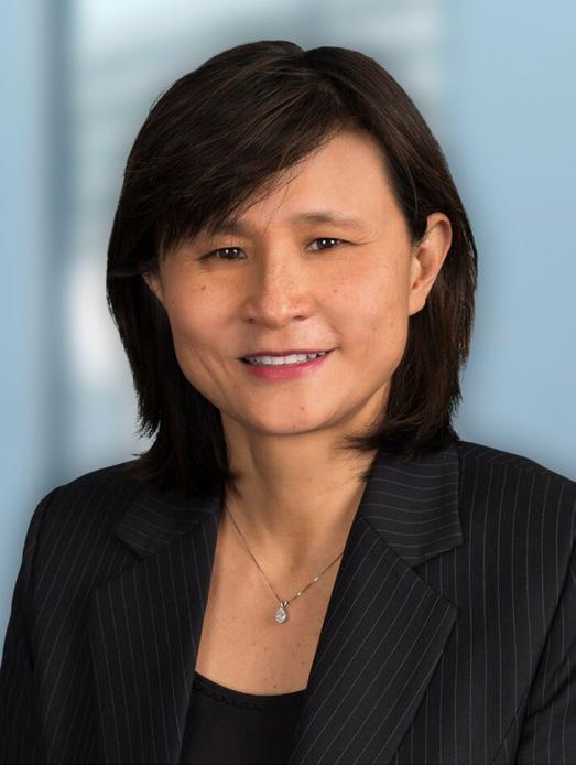
Jenny C. Chang, MB.ChirB., MD, MCHM
Ernest Cockrell, Jr. Presidential Distinguished Chair
Executive Vice President and Chief Academic Officer, Houston Methodist President and CEO, Houston Methodist Academic Institute

At the Houston Methodist Academic Institute, transformation is not aspirational — it is operational. Our research enterprise is purpose-built to move discovery rapidly and responsibly into patient care, fueled by close integration with clinical teams and a deep understanding of regulatory, translational and implementation pathways. By uniting scientific rigor with real-world testing inside a leading health system, we accelerate the journey from idea to impact — delivering innovations that are not only groundbreaking, but immediately impactful for patients, physicians and the future of medicine.

Transformational discovery does not advance on science alone — it requires belief, partnership and sustained investment. Many of the most promising medical innovations falter in the critical space between discovery and clinical adoption.
Through visionary philanthropy, Houston Methodist accelerates the translation of breakthrough ideas into life-changing care, providing the resources needed to progress high-impact research when and where it matters most. Philanthropic gifts serve as catalysts for real-world transformation, ensuring innovation reaches patients.
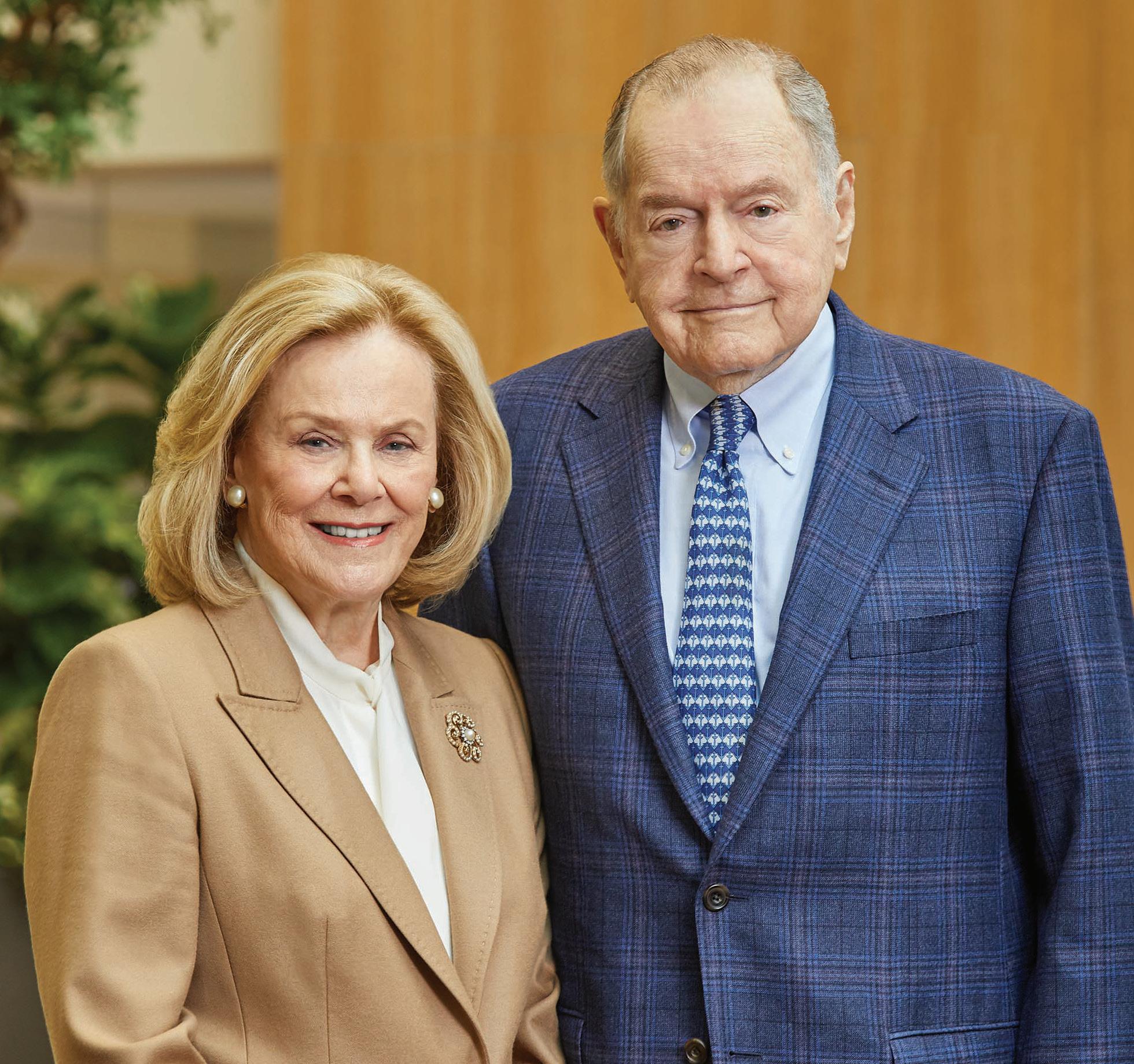
The Duncan family, longtime supporters of Houston Methodist, has made a transformational $25 million commitment from The Duncan Fund to support the Department of Medicine. In recognition of their gift, Houston Methodist renamed the largest department in its system the Houston Methodist Charles W. Duncan Jr. Department of Medicine.
“Our vision is to redefine how care is delivered in our country by creating a national model for true continuity — one that follows the patient across every transition, from home to hospital and back again,” said Eleftherios Mylonakis, MD, PhD, FIDSA, Charles and Anne Duncan Presidential Distinguished Chair. “We are also advancing a deeper understanding of health span, shifting from reactive treatment to a proactive lifelong strategy that maximizes not just how long we live but how well we live.”
The Duncan family has demonstrated an unwavering commitment to advancing health care and supporting others in our community. The late Charles W. Duncan Jr. was a lifetime member of the Houston Methodist board and had a profound impact on all aspects of the organization. Houston Methodist is deeply grateful for this gift and for what it means to those who work every day to improve our patients’ lives.
Ten Houston Methodist leaders were selected to fill endowed chairs, representing exceptional expertise and innovation in their respective fields. Through the generous foresight of the benefactors who established these endowed positions, chairholders draw from an anchored, long-term financial resource to support their leading-edge work.

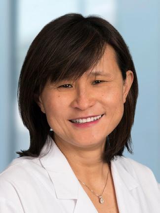
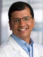
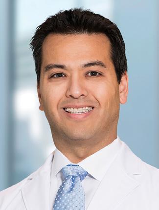
Arica A. Brandford, PhD, JD
Dorothy and Mickey Ables Community Outreach and Engagement Distinguished New Century Chair
Dr. Mary and Ron Neal Cancer Center
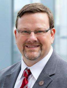
Keith Syson Chan, PhD
Neal Cancer Center Distinguished Chair
Dr. Mary and Ron Neal Cancer Center
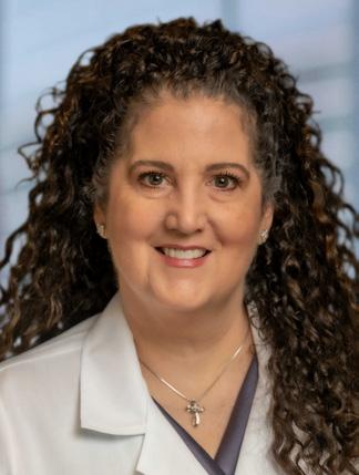
Jenny C. Chang, MB.ChirB., MD, MCHM
Ernest Cockrell, Jr. Presidential
Distinguished Chair
Executive Vice President and Chief Academic Officer
President and CEO
Academic Institute
Sunil Dacha, MD
Nina and Michael Zilkha Centennial Chair in Gastrointestinal Health in Honor of Dr. Eamonn Quigley
Underwood Center for Digestive Health
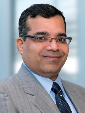
Anthony Echo, MD
Henrietta and Terence Hall
New Century Chair in Orthopedics Department of Orthopedic Surgery
Jay E. Maddock, PhD, FAAHB
The Coneway Family Centennial Endowed Chair in the Center for Health & Nature
Cindy M. Martin, MD
Lois E. and Carl A. Davis Chair in Heart Failure
DeBakey Heart & Vascular Center
Kumar Pichumani, PhD
Dagmar Dunn Pickens Gipe Distinguished Professorship in Brain Tumor Research Department of Neurosurgery
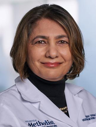
Zeenat Safdar, MD, MS, FACP, FCCP, ATSF
Ashley Lain Barras Distinguished Chair in Pulmonology
Charles W. Duncan Jr. Department of Medicine
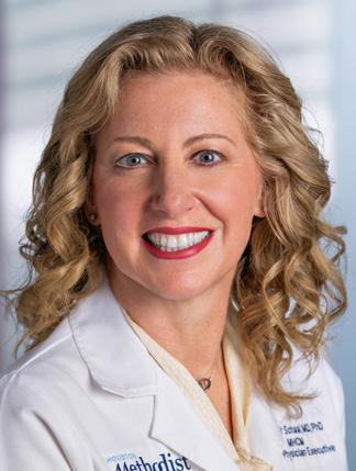
Shlomit Schaal, MD, PhD, MHCM
Lenny C. Katz Chair in Health Outcomes and Quality in Honor of Stuart M. Dobbs, MD
Executive Vice President Chief Physician Executive
The Houston Methodist Academic Institute recognizes the distinctive and critical role that physician-scientists play in advancing its core missions as an academic medical center. The Clinical Scholars program meets that need by helping to grow our core of rising physician-scientists.

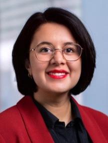
Akshjot Puri, MD
Assistant Professor of Medicine
Charles W. Duncan Jr. Department of Medicine
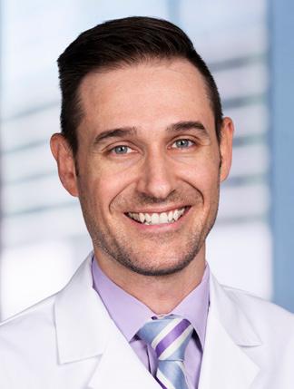
Namrata Vasquez, PhD
Assistant Professor of Psychology in Clinical Psychiatry and Behavioral Health Department of Psychiatry and Behavioral Health
Paul M. Schroder, MD, PhD
Assistant Professor of Surgery Department of Surgery
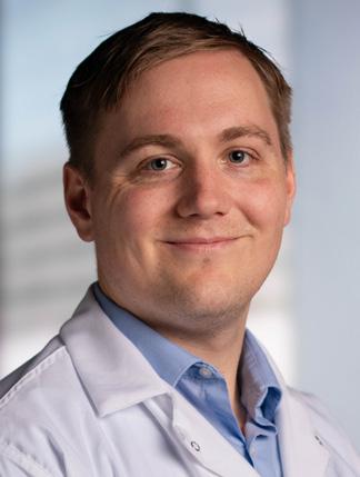
Ewan K.S. McRae, PhD
Assistant Professor of Cardiovascular Sciences
Ewan K.S. McRae, PhD, is Houston Methodist’s first Pew Scholar. His research will focus on determining approaches for designing stable, controllable therapeutic RNAs.
The Pew Scholars Program in the Biomedical Sciences provides funding to young investigators of outstanding promise in science relevant to the advancement of human health. The program makes grants to selected academic institutions to support the independent research of outstanding individuals who are in their first few years of their appointment at the assistant professor level. Members of the Office of Faculty and Research Development team manage the limited submission and mentor the selected candidate with grantsmanship coaching and writing support.

Scientific discoveries at Houston Methodist begin with urgent clinical questions and move forward with purpose. Across interdisciplinary expertise, education and collaboration, our teams are making breakthroughs designed for maximum impact. By integrating scientific insight, cutting-edge technology and clinical experience, innovations are rapidly refined, validated and positioned for transformation — accelerating the path from discovery to patient care. Research reflects a commitment to delivering smarter, more precise and more accessible solutions that improve outcomes and shape the future of medicine.


Houston Methodist researchers are moving beyond incremental gains in cancer therapies toward fundamentally smarter treatment strategies. By harnessing catalytic nanomedicine and uncovering how the tumor microenvironment fuels treatment resistance, these studies reveal new ways to overcome therapeutic limitations. Together, they open the door to faster-acting, more selective treatments and dual-targeted approaches that improve outcomes for patients with drug-resistant cancers.

Houston Methodist researchers have uncovered a critical mechanism driving chemoresistance in aggressive cancers — one that could reshape how we approach tumor microenvironment-targeted therapies. In a study published in Science Advances, the team led by co-corresponding authors Hongbo Beth Gao, PhD, and Keith Syson Chan, PhD, Neal Cancer Center Distinguished Chair, revealed that chemotherapyinduced pyroptotic inflammatory cell death can inadvertently reprogram the tumor microenvironment, making it more hospitable to chemoresistant cancer stem cells (CSCs). The key culprits are a subset of fibroblasts known as αSMA+ cancer-associated fibroblasts (CAFs), which are transformed into collagen-producing inflammatory CAFs (iCAFs).


Our findings surprisingly revealed that not all forms of cell deaths are helpful in fighting cancer. Combination treatment with belnacasan, to stop this type of ‘fiery’ cell death, can improve chemotherapy response.
Using advanced single-cell transcriptomics and spatial profiling, researchers mapped the tumor microenvironment in chemo-resistant cancers and identified iCAFs as key players. These pyroptosisactivated fibroblasts secrete pro-inflammatory cytokines — notably IL-6 and CXCL12 — that initiate STAT3 signaling in neighboring CSCs. This activation creates a fibrotic niche that shelters CSCs and enhances their resistance to chemotherapy.
“In a sense, the ‘fiery’ or inflammatory death of these cancer cells from the chemotherapy actually made things worse, not better,” added Chan.
CSCs are notorious for their role in relapse and metastasis. By showing that iCAFs are not just passive bystanders but active enablers of CSC-driven resistance, this study shifts the paradigm. It suggests that targeting the iCAF-CSC interaction could be a potent strategy to overcome treatment failure in solid tumors.
The team found that belnacasan, a caspase-1 inhibitor, can block this inflammatory cascade. In preclinical mouse models, combining belnacasan with chemotherapy significantly improved treatment response by preventing the conversion of CAFs into collagen-rich iCAFs.
This dual-targeted approach — attacking both the cancer cells and their supportive microenvironment — could represent a major shift in how we treat not only bladder cancer but potentially other solid tumors.
These findings open the door to dual-targeted therapies — those that disrupt both the CSCs and their supportive stromal niche. In preclinical models, blocking IL-6 signaling sensitized tumors to chemotherapy offered a promising translational pathway. While further studies are needed to explore the broader applicability of this strategy, early signs are encouraging. The same inflammatory mechanisms may be at play in breast cancer, suggesting a wider therapeutic window for belnacasan and similar agents.
As researchers continue searching for smarter, safer ways to target cancer, one emerging strategy stands out.
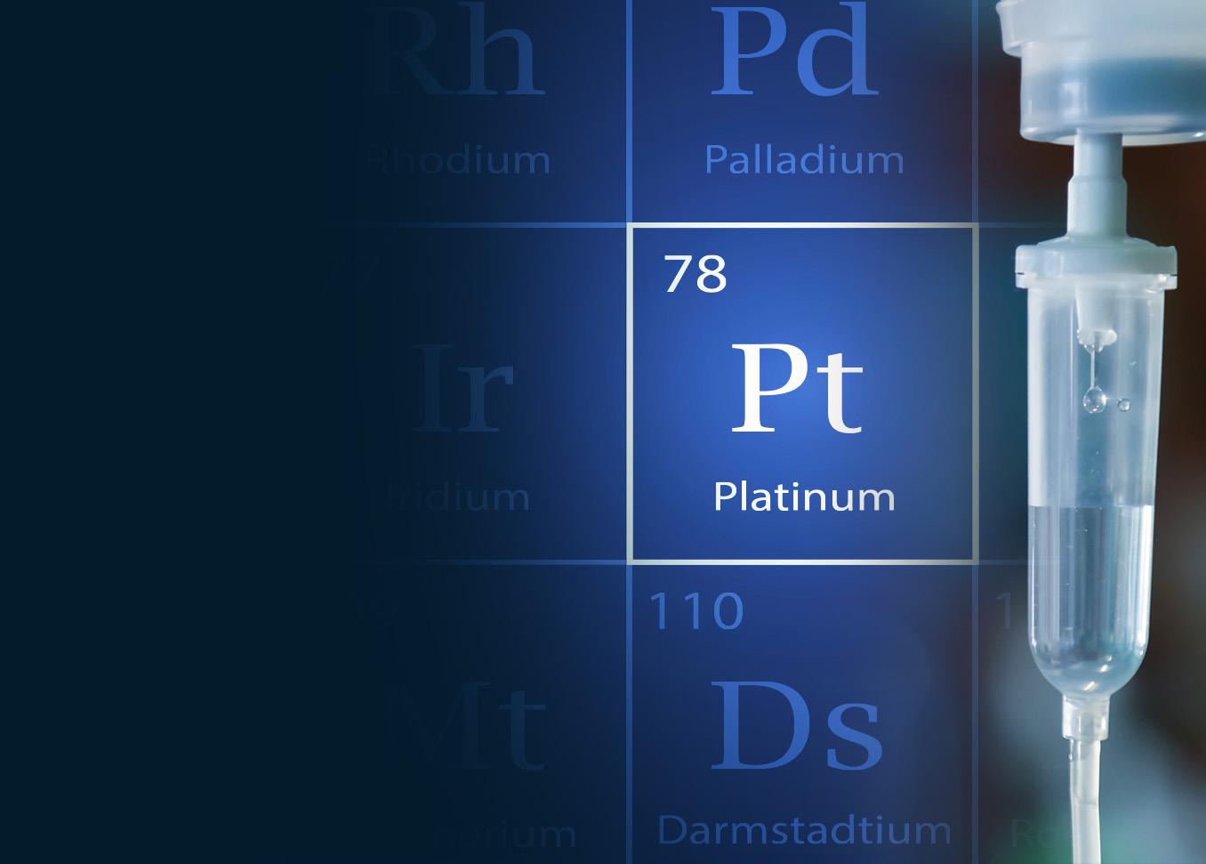
At the heart of this innovation is platinum. For decades, platinum-based drugs like cisplatin and oxaliplatin have been used to treat a variety of cancers. These drugs bind to DNA and block cell division, ultimately triggering apoptosis. But traditional platinum compounds are slow-acting, often toxic to healthy cells, and increasingly resistant to the very tumors they’re meant to destroy.
Now, a team of researchers led by Junhua Mai, PhD, has uncovered a new platinum formulation that harnesses reactive oxygen species (ROS) to kill cancer cells. The team conducted a thorough investigation into how different metals influence ROS generation and found that several platinum compounds produced the highest levels of intracellular ROS among the tested metals. In a recent study published in Biomaterials, the team unveiled a new platinum-based nanomaterial called “carrier-platin,” which uses ROS — particularly hydroxyl radicals, among the most damaging molecules in biology — to trigger cancer cell death with remarkable speed and specificity.
Unlike conventional chemotherapies, which take hours or days to take effect, carrier-platin acts within minutes.
“At the molecular level, it’s a nano-engineered complex of platinum nanoparticles embedded in a biodegradable polymer carrier made from poly (L-glutamic acid/L-aspartic acid). This carrier doesn’t just provide structure — it fine-tunes the chemical environment around the platinum, dramatically enhancing its ability to catalyze the breakdown of hydrogen peroxide (H₂O₂), which is naturally abundant in tumor cells,” said Yongbin Liu, PhD, first author on the study.
This catalytic reaction produces hydroxyl radicals in bursts so intense, they overwhelm cancer cells’ already fragile redox balance. The result is a rapid, non-apoptotic form of necrotic cell death that is strikingly selective for malignant cells.
When tested in colorectal, breast, ovarian, lung and kidney cancer cell lines, including those resistant to conventional chemotherapy, carrier-platin delivered a consistent and lethal blow. Meanwhile, non-cancerous cells were largely spared. The explanation? Healthy cells contain higher levels of glutathione (GSH) and lower basal H₂O₂, buffering them against the ROS storm.
“ ”
Tumor cells repeatedly exposed to carrier-platin over weeks did not develop resistance. In contrast, the same cell lines quickly grew resistant to oxaliplatin. Furthermore, carrier-platin retained full potency in cells that had already become resistant to cisplatin, taxanes and other drugs.
– Junhua Mai, PhD
But the innovation doesn’t stop there. One of the most vexing problems in oncology is drug resistance. Carrier-platin seems to break that pattern. “Tumor cells repeatedly exposed to carrier-platin over weeks did not develop resistance,” said Mai. “In contrast, the same cell lines quickly grew resistant to oxaliplatin. Furthermore, carrier-platin retained full potency in cells that had already become resistant to cisplatin, taxanes and other drugs.”
Studies in murine models added another layer of promise. In aggressive tumors — including those resistant to platinum drugs — carrier-platin halted tumor growth and, in many cases, eliminated it altogether. Even at high doses, the formulation showed minimal toxicity. This safety profile was attributed in part to the polymeric carrier, which stabilizes the drug and helps restrict its activity to the acidic, ROS-rich tumor microenvironment.
The mode of death triggered by carrier-platin is distinct from that of traditional chemotherapies. Instead of inducing apoptosis, the programmed cell death that involves cellular shrinkage and DNA fragmentation, carrier-platin initiates necrosis via l ysosomal membrane permeabilization and ER stress — hallmarks of overwhelming oxidative injury. Though it shares some features with ferroptosis, another ROS-dependent cell death pathway, carrier-platin’s mechanism is unique. Its effects are iron-independent and too rapid to align with ferroptosis’s slower, lipid-peroxidationdriven trajectory.
“This research could lead to new therapies, providing hope for patients with drug-resistant tumors, and for clinicians running out of options,” said Mai.
Transplant is redefining how malignancies are treated by integrating transplantation into cancer care — a concept unheard of a few decades ago. Today, transplantation offers curative potential for select patients with liver, bile duct, colorectal and neuroendocrine cancers. As an emerging discipline that unites the expertise of cancer and transplant specialists, transplant oncology exemplifies the kind of interdisciplinary innovation in which Houston Methodist is a national leader.

Elsevier recently published the first and only book on transplant oncology, “Transplant Oncology: A Frontier in Multidisciplinary Cancer Care,” edited by Maen Abdelrahim, MD, PhD, PharmD. This groundbreaking volume examines how transplant oncology is redefining treatment for malignancies once considered untreatable by transplantation.
Coined as a term in 2015, transplant oncology integrates cancer and transplant center expertise into an emerging discipline with curative potential. What seemed impossible decades ago is now reality: certain cancers — including liver, bile duct, colon, and neuroendocrine tumors that metastasize to the liver — can be treated through transplantation. Vincenzo Mazzaferro, MD, PhD, widely regarded as the field’s godfather, pioneered cancer treatment by transplantation in 1996 and contributed to the book’s preamble and opening chapter.
The 26-chapter book, authored by global pioneers, spans six sections covering transplant oncology’s
evolution, oncological transplantation indications, immunotherapy applications, precision medicine integration, solid organ transplant considerations, and surgical aspects. Topics include circular tumor DNA applications, immunotherapy in special populations, surgical innovations and recent clinical trial successes.
In his foreword, Houston Methodist President and CEO Marc Boom, MD, noted the book, “speaks to the incredible progress so many of our contemporaries have made in the fight against cancer and is also a call to action ... to keep thinking ahead and moving forward.”

Nobel laureate James P. Allison, PhD, emphasized in the second foreword that transplant oncology’s integration with organ transplantation represents a comprehensive approach to finding cancer cures, particularly through understanding the interplay between cancer and transplant immunology.
The book addresses critical questions: Can immunotherapy treat patients at higher risk of secondary cancers — those receiving post-transplant immunosuppressive therapies? Can it shrink tumors while patients await donor organs during typical wait times ranging from days to years?
Abdelrahim, who continues pushing boundaries through pioneering research collaborations, notes: “This book increases our visibility as an institution. Our state-ofthe-art cancer center and highly ranked transplant center together have given birth to this book. This is an exciting time since now we can treat stage IV colon cancer by transplantation.”
The field expands through active global collaboration. Abdelrahim chaired the first U.S. transplant oncology conference at Houston Methodist in 2022 and serves as editor-in-chief of the recently launched journal section, Transplant Oncology
This is a global book with contributions from researchers from all seven continents. It will certainly change the field by sharing and increasing knowledge of what has already been accomplished, opening doors and expanding transplant oncology practice globally. “ ”
– Maen Abdelrahim, MD, PhD, PharmD

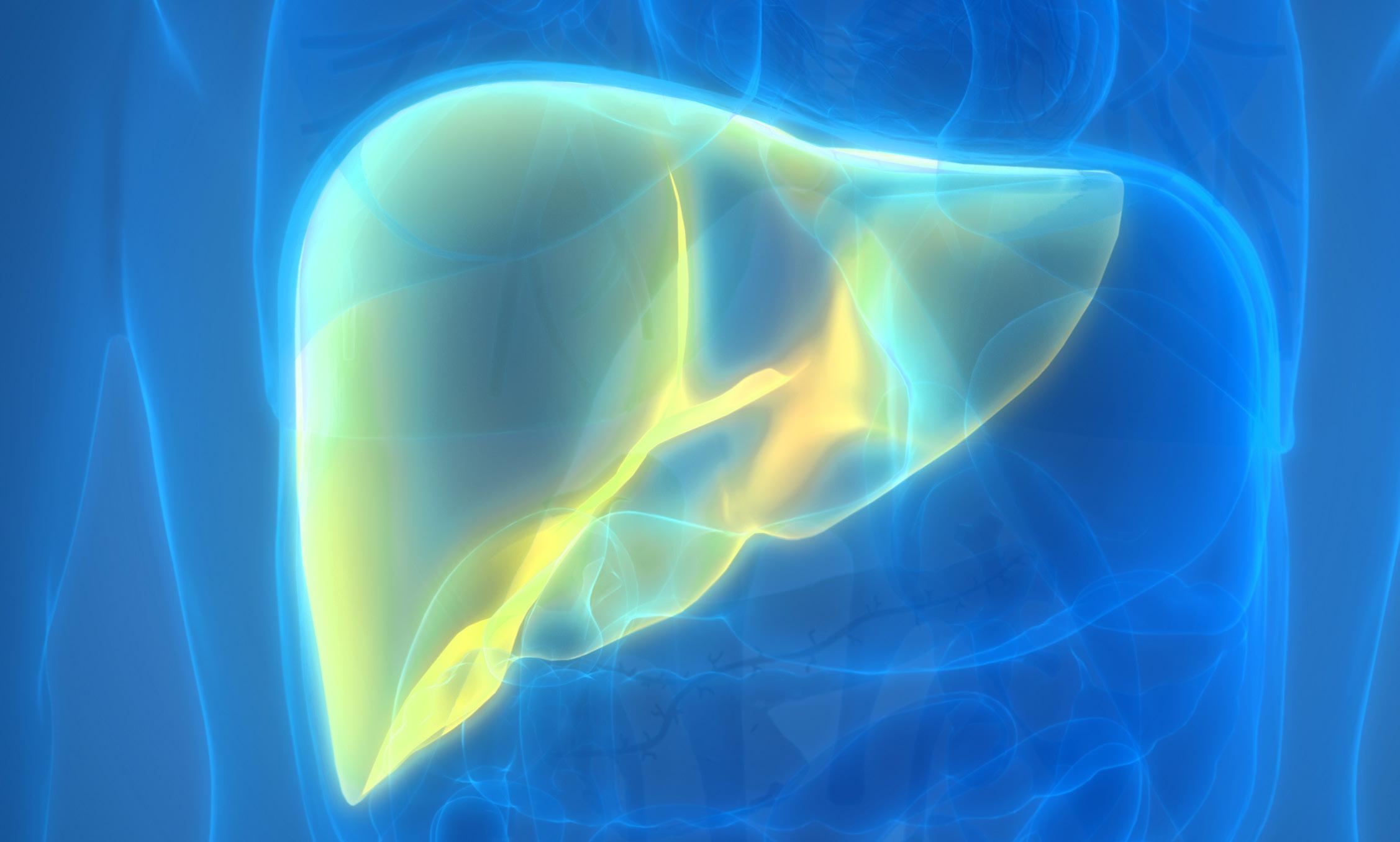
Despite the liver’s prowess at self-renewal, certain cancers can infiltrate the liver enough to prohibit surgical resection. In these cases, transplant oncology offers patients a path toward better disease management, improved health outcomes and a potential cure.
In a review published in the journal Cancers, Maen Abdelrahim, MD, PhD, PharmD, and his collaborators have presented an overview of the evolution of transplant oncology as a treatment for certain liver cancers that cannot be surgically removed. The authors have discussed the feasibility of neoadjuvant treatments before transplantation and suggest post-transplantation disease surveillance methods to optimize patient care.
According to the American Institute for Cancer Research, liver cancer is the sixth most common form of cancer worldwide with a survival rate of around 20%. The most frequently encountered form of liver cancer is hepatocellular carcinoma, which is often observed in people with cirrhosis or chronic liver disease. However, cancers inside and outside the liver are also associated with cholangiocarcinomas and the metastatic progression of neuroendocrine and colorectal cancers.
Patients with liver cancer can be on long-term chemotherapy, which profoundly compromises their quality of life. When possible, cancerous tissue is surgically removed to mitigate the dependence on drug therapies. But in some instances, the liver is heavily scarred or has widespread cancer, making surgical resection extremely challenging. In these circumstances, liver transplantation is a line of treatment that holds promise to dramatically improve patient survival.
The criteria required to screen hepatocellular carcinoma patients eligible for liver transplants have been dubbed the Milan criteria. This selection protocol requires the tumor diameter for single lesions to be less than or equal to 5 cm, and less than or equal to 3 cm for multiple but within three lesions. In addition, the cancer must have no vascular invasion or metastasis. Liver transplants on these patients, the authors reported, boost the five-year survival rate to 80-85%.
Over the years, the Milan criteria have been expanded to include bigger liver cancers and other unresectable liver malignancies resulting from bile duct and metastatic cancers. In some cases, patients may receive bridge treatments, including chemo and immunotherapy, to downsize the liver cancer to meet the Milan criteria for transplantation.
TRANSPLANT & HEPATOBILARY SURGEON
GASTROENTEROLOGIST
As proof of concept for neoadjuvant bridging therapy, a collaborative effort between the Houston Methodist J.C. Walter Jr. Transplant Center and Houston Methodist Dr. Mary and Ron Neal Cancer Center were the first to report a prospective case series of intrahepatic cholangiocarcinoma patients receiving chemotherapy followed by liver transplantation. These patients had a five-year survival rate comparable to those with hepatocellular carcinoma who received liver transplants.
LIVER ONCOLOGIST
The authors also discussed the feasibility of minimally invasive postoperative disease surveillance methods, particularly liquid biopsies to monitor the circulating tumor DNA in the bloodstream. This technique could facilitate detecting small traces of residual disease after transplantation and the propensity of disease recurrence.
INTERVENTION RADIOLOGIST
T
CURE
IMMUNOLOGIST
INTERVENTION RADIOLOGIST
PATHOLOGIST
RADIATION THERAPIST
CHEMOTHERAPIST
Although a promising treatment for certain unresectable liver tumors, transplant oncology is best suited for hospitals that both specialize in cancer treatment and perform transplantation surgeries at high enough volumes. Furthermore, the field is highly interdisciplinary, requiring the integration of physicians and scientists from diverse fields—such as gastroenterology, immunology, and radiology—under one umbrella organization. Abdelrahim noted that Houston Methodist is one of the few uniquely poised institutions to be a transplant oncology center of excellence.
Most people believe that liver transplant oncology is restricted to a certain small number of patients with small tumors, but that isn’t the case anymore. Combined with tailored protocols and therapies, transplantation has resulted in survival rates comparable across varying tumor sizes, with five-year survival reaching 60% for complex cases, significantly higher than the current 20% survival rate for resection and chemotherapy alone.
”
– R. Mark Ghobrial, MD, PhD J.C. Walter Jr. Presidential Distinguished Chair
Houston Methodist is reshaping infectious disease research by tackling two of its greatest challenges: identifying elusive pathogens and defeating drug-resistant bacteria. Through innovations in genome amplification and AI-guided antimicrobial peptide design, researchers are speeding diagnostics and unlocking new pathways for treatment development. This work reflects a cutting-edge strategy for precision infectious disease care and outpacing emerging global threats.

In the escalating battle against antibiotic-resistant bacteria, researchers have unveiled a promising new weapon: an antimicrobial peptide, optimized via machine learning, that demonstrates activity against the most stubborn pathogens in medicine. Published in The Journal of Clinical Investigation, the study details the development of CIT-8, a short, 13-amino-acid peptide engineered to target methicillin-resistant Staphylococcus aureus (MRSA) and Vancomycin-resistant S. aureus (VRSA). These superbugs are notorious for evading conventional antibiotics, often leading to prolonged hospital stays, increased mortality and soaring health care costs.

The innovation lies not just in the peptide itself, but in how it was designed. The team, led by Eleftherios Mylonakis, MD, PhD, FIDSA, Charles and Anne Duncan Presidential Distinguished Chair, began with citropin 1.1, a naturally occurring antimicrobial peptide with modest activity. Using a hybrid approach that combined traditional peptide design techniques — like truncation and substitution — with machine learning algorithms trained on over 14,000 known antimicrobial peptides, they created CIT-8. This new peptide eradicated 100 million drug-resistant bacteria within 30 minutes in lab tests, including MRSA and VRSA, and significantly reduced bacterial load in mouse models of skin infection.
“Machine learning allowed us to explore a vast sequence space that would be impossible to navigate manually,” said Mylonakis. “By integrating computational modeling with biological insight, we’ve accelerated the path from concept to candidate in a molecule that’s both potent and precise.”
The CIT-8 peptide works by depolarizing and permeating bacterial membranes, leading to cell death. This mechanism is particularly promising because it reduces the likelihood of resistance development. The CIT-8 peptide also demonstrated
efficacy in reducing biofilm viability by up to 10,000-fold, a critical feature given the resilience of biofilms in chronic infections. When applied topically in a murine model, it slashed bacterial burden by over 99%. Importantly, CIT-8 showed minimal toxicity to human cells, suggesting a favorable safety profile for future clinical applications.
This breakthrough is more than a scientific milestone — it’s a blueprint for future drug development. The team’s methodology, which combines machine learning with structure-guided design, was also successfully applied to three other peptides, demonstrating its versatility and scalability.
Mylonakis emphasized the broader implications: “Antibiotic resistance is one of the defining challenges to global health today. With CIT-8, we’re not just responding — we’re anticipating. Our approach not only delivers a potent new therapeutic; it also opens the door to designing many more. It’s a paradigm shift in how we think about antimicrobial discovery.”
As academic medical centers continue to lead the charge against emerging health threats, this work exemplifies the power of interdisciplinary collaboration — where data science meets microbiology to solve real-world problems.

In the evolving world of genomic science, a new frontier is being shaped in the careful amplification of life’s tiniest traces.
At the forefront of this movement is Rodrigo de Paula Baptista, PhD, a leading innovator in the use of multiple displacement amplification (MDA) to enable genome sequencing of low-input and hard-to-culture pathogens.
While much of modern sequencing depends on having ample, high-quality DNA, many organisms that pose serious health threats simply won’t cooperate. They grow poorly in lab cultures, yield scant DNA or come from degraded clinical samples. This is where de Paula Baptista’s research shines.
In a recent publication, Molecular Ecology Resources, the team demonstrated how MDA — combined with Oxford Nanopore’s long-read sequencing — enables complete genome assembly from DNA inputs far below traditional thresholds. Starting with less than a single cell’s worth of DNA, the team was able to generate contiguous or chromosomal-level assemblies of bacterial and protozoan genomes.
To solve the persistent challenge of chimeric artifacts generated during MDA, the team also developed the Concatemer Detection Tool (CADECT) — a custom bioinformatics pipeline that automatically detects and removes problematic reads, thereby improving genome contiguity and fidelity. This innovation salvages compromised samples and opens new possibilities for studying rare pathogens, complex microbiomes or archived samples with limited material.
This study is just one part of a much larger effort led by de Paula Baptista, whose lab is integrating MDA into a diverse suite of global health, clinical and computational research programs.
MDA has become a game-changer in clinical scenarios, where timely genome sequencing can mean the difference between a targeted treatment and a missed diagnosis. In particular, the team has used MDA to rapidly amplify DNA directly from patient samples, speeding up antimicrobial resistance profiling and improving clinical outcomes.
This method is being adapted for single-cell genomics, enabling researchers to study intra-host diversity, mixed infections and even strain switching — factors critical for understanding disease progression, immune evasion and treatment resistance.
de Paula Baptista’s program is tuned to the needs of low-resource settings, where diagnostic tools are often constrained by sample quality and availability. For instance, Candidozyma auris, a multidrug-resistant fungus causing global outbreaks, presents significant challenges for genomic surveillance due to its tough cell wall and low DNA yield.
MDA now allows the team to sequence its genome from minimal input, expediting workflows without compromising quality.
The team is building a comprehensive genomic ecosystem that spans from amplification to analysis. Alongside CADECT, the team is developing tools
for phenotype classification, high-resolution strain typing and detection of mixed infections. These computational frameworks help researchers extract meaningful biological insights from complex, noisy sequencing data.
The team is also contributing to federal collaborations with the NIH and USDA to expand the genomic representation of poorly characterized or novel microbes, some of which are foodborne or agents of mucosal infections. Many of the organisms they study are overlooked, but matter, especially in places where public health infrastructures are under strain.
The lab’s future is aimed at tackling hard genomic problems involving low-abundance pathogens, complex infection environments and underserved populations. The integration of MDA with real-time nanopore sequencing, coupled with automated data refinement tools, sets the stage for next-generation precision diagnostics, from the ICU to remote field labs.

MDA allows us to work with precious, degraded or miniscule samples that were once considered unusable for high-resolution sequencing. This is especially impactful in urgent clinical scenarios or in global health settings where pathogens remain underrepresented in public databases.
Dementia represents one of the most urgent health challenges, with health care costs exceeding $360 billion nationwide and Texas ranking third in the U.S. for the number of people living with or caring for someone with Alzheimer’s disease and related dementias. Researchers are advancing a comprehensive dementia strategy — spanning molecular discovery, immune modulation, regenerative therapies and precision imaging — to address disease drivers long before irreversible decline.

Neuroinflammation is linked to a host of detrimental brain disorders, including Alzheimer’s and Parkinson’s disease. A collaborative research team from Houston Methodist and Rice University has developed a way to fight back with a tiny, bioengineered system to deliver anti-inflammatory proteins to specific targets in the brain.
The study, published in Biomaterials, led by Robert Krencik, PhD., reveals the creation of AstroCapsules, a revolutionary union of bioengineering and neuroscience that encloses human astrocytes (star-shaped, resident brain cells that are crucial to healthy function of the central nervous system) within small biocompatible hydrogel capsules.
Researchers found that encapsulating astrocytes engineered to secrete the anti-inflammatory protein interleukin-1 receptor antagonist (IL-1Ra) significantly reduced neuroinflammation, as measured by inflammatory biomarkers. Importantly, 300-micrometer AstroCapsules—roughly the size of large grains of sand—were confirmed to function when implanted into the brain, demonstrating they can withstand the body’s immune response. The research was conducted using lab-grown human brain tissue samples, known as organoids, and mouse models.
“This system solves several current problems with cell-based therapeutics to the nervous system,” Krencik said. “Capsules will form a physical barrier between the implanted astrocytes and brain tissue cells are expected to locally secrete anti-inflammatory proteins while avoiding immune rejection and unwanted migration throughout the brain. This will increase stability of the treatment while reducing side effects.” This represents a new way to treat devastating neurodegenerative diseases and showcases how cell therapy can actively diminish damaging inflammation in the brain.
Our hope is that this work will help move cell therapies closer to becoming real treatment options for patients with neurodegenerative disease.
”
– Robert Krencik, PhD

Obesity has long been acknowledged as a risk factor for a wide range of diseases, but a more precise link between obesity and Alzheimer’s disease has remained a mystery, until now.
A first-of-its-kind study discovered that adipose-derived extracellular vesicles — tiny cell-to-cell messengers in the body — can signal the buildup of amyloid-β plaque in obese individuals. These plaques are a key feature of Alzheimer’s disease.
The study, “Decoding Adipose–Brain Crosstalk: Distinct Lipid Cargo in Human Adipose-Derived Extracellular Vesicles Modulates Amyloid Aggregation in Alzheimer’s Disease,” published in Alzheimer’s & Dementia: The Journal of the Alzheimer’s Association, explores the link between obesity, which affects about 40% of the U.S. population, and this dreaded neurodegenerative disease that affects more than 7 million people in the U.S.
As recent studies have underscored, obesity is now recognized as the top modifiable risk factor for dementia in the United States.
– Stephen T. Wong, PhD
John S. Dunn Presidential Distinguished Chair in Biomedical Engineering
Researchers found that the lipid cargo of these cell messengers differs between people with obesity and lean individuals; specifically, the levels of these lipids affected how quickly amyloid-β clumped together in laboratory models. Using mouse models and patient body fat samples, researchers examined the vesicles, which are tiny membrane-bound particles that travel throughout the body and act as messengers involved in cell-to-cell communication. These minuscule communicators are also capable of crossing the blood-brain barrier.
Targeting these tiny cell messengers and disrupting their communication, which leads to plaque formation, may help reduce the risk of Alzheimer’s disease in people with obesity. Researchers recommend that future studies focus on drug therapies that can halt or slow the accumulation of Alzheimer’s-related toxic proteins, such as amyloid-β, in at-risk individuals.
Houston Methodist is leading the next era of dementia research — linking molecular discovery, advanced imaging and regenerative medicine to change how we understand and treat neurodegenerative disease. From mapping the brain’s waste-clearance pathways to developing next-generation cell therapies, our scientists and physicians are building a seamless pipeline from discovery to patient care.

As the number of Texans affected by Alzheimer’s disease and related dementias continues to grow, our researchers are taking bold steps to understand, prevent and one day reverse the underlying causes of neurodegeneration. By uniting discovery, technology and patient care, our scientists and clinicians are transforming how these diseases are studied and treated.
Jun Li, MD, PhD, John M. O’Quinn Foundation Presidential Distinguished Chair in Neurology, and his team are mapping the cellular and molecular mechanisms that drive dementia and related neurodegenerative disorders. Anchored by the
John M. O’Quinn Foundation Neurodegenerative Disorders Laboratory, their work integrates advanced imaging, ultrastructural analysis and single-cell transcriptomics to reveal how axonal degeneration, myelin loss and neuroinflammation contribute to cognitive decline.
A new 3D Ultrastructure Laboratory now allows researchers to visualize neurons and glial networks in remarkable detail using serial block-face electron microscopy and quantitative MRI. These insights, combined with genetic tools that precisely manipulate neural circuits, are uncovering potential therapeutic targets that could halt or even reverse disease progression.
Complementary studies by Kyuson Yun, PhD, Chair in Neurodegenerative Disease Research, Jon B. Toledo, MD, PhD, Ann and Billy Harrison Centennial Chair in Alzheimer’s Research, and Alireza Faridar, MD, Stanley H. Appel Chair in Translational Neuroscience, expand this foundation — pioneering single-cell biomarker discovery, early-detection transcriptomics and investigations into neuroinflammatory pathways.
The next wave of medicine will be built from living cells — and Houston Methodist is prepared. Drawing on extensive experience in Current Good Manufacturing Practices production and infusion networks for oncology, the institution is extending this infrastructure to neurodegenerative disease research.
At the Ann Kimball & John W. Johnson Center for Cellular Therapeutics, researchers are developing and testing new classes of therapies that modulate immune and regenerative processes within the brain. These include regulatory T-cell (Treg)-modulating and exosome-based treatments that address neuroinflammation and neuronal loss at their roots.
At the Center for Neural Systems Restoration (CNSR) — a collaboration between Houston Methodist and Rice University — Gavin W. Britz, MBBCh, MPH, MBA, FAANS, and his team are pioneering research into the brain’s waste-clearance
system, known as the glymphatic pathway. This network, closely linked to sleep, removes toxic proteins and metabolic byproducts from the brain.
When disrupted, it may contribute to the onset and progression of Alzheimer’s disease and other forms of dementia.
Using multimodal imaging, physiologic monitoring and the 7-Tesla MRI, CNSR investigators visualize cerebrospinal-fluid flow in real time and study how to enhance it through noninvasive electrical stimulation. By integrating neuroengineering, precision imaging and physiologic monitoring, the team is developing innovative strategies to restore healthy brain clearance and potentially slow or prevent neurodegeneration.
The Houston Methodist Research Institute’s laboratories work with clinical programs such as the Nantz National Alzheimer Center. Advanced imaging, on-site radiopharmacy capabilities and a system-wide infusion network make the Institute uniquely equipped to deliver next-generation therapies.
Partnerships with Rice University and other Texas collaborators further extend this pipeline into device and technology innovation — such as neural-monitoring interfaces and physiologic-sensor prototypes — turning scientific insight into tangible advances for patients.
This integrated approach represents more than research readiness; it’s a new model for dementia innovation and translating discovery into hope for patients and families facing the challenges of neurodegenerative disease.



In the ever-evolving landscape of Alzheimer’s disease (AD) research, a new study offers a spark of hope — one rooted in recalibrating the immune system itself.
Published in Alzheimer’s Research & Therapy, the phase 2a clinical trial led by Alireza Faridar, MD, Stanley H. Appel Chair in Translational Neuroscience; Stanley Appel, MD, the Peggy and Gary Edwards Distinguished Chair in ALS Research; and their colleagues explored the use of low-dose interleukin-2 (IL-2) to expand regulatory T cells (Tregs) in patients with mild to moderate AD. Preliminary results suggest that immune modulation may offer a viable strategy to slow disease progression and reduce neuroinflammation.
Alzheimer’s disease has long been associated with the accumulation of amyloid-beta and tau proteins. Recent research has illuminated another key player: inflammation. In patients with this form of neurodegeneration, the immune system can become hyperactive, releasing inflammatory mediators that exacerbate neuronal damage. Tregs are a specialized subset of CD4+ T cells that act as the immune system’s peacekeepers. Their primary role is to maintain immune homeostasis by suppressing excessive immune responses that could damage healthy tissue. In healthy individuals, Tregs help prevent autoimmune reactions and keep inflammation in check. But in Alzheimer’s patients, studies have shown that Treg numbers and function are often diminished. This imbalance can lead to unchecked inflammation in the brain, contributing to neurodegeneration. The rationale for IL-2 therapy stems from its ability, at low doses, to selectively expand Tregs without activating effector T cells or other pro-inflammatory pathways.
This randomized, double-blind, placebo-controlled trial enrolled 38 participants aged 50 to 86 with biomarkerconfirmed AD. Participants were randomized to receive subcutaneous low-dose IL-2 or placebo every four weeks or every two weeks over a 21-week treatment period, followed by a nine-week observation phase. The primary endpoints focused on the safety and modulation of Treg frequency and function. Secondary endpoints assessed inflammatory biomarkers, cerebrospinal fluid analytes, and cognitive performance via the Alzheimer’s Disease Assessment Scale-Cognitive Subscale.
This clinical trial marks an important step in translating immunomodulatory strategies into clinical practice. We’re not claiming a cure, but we are seeing biological changes that suggest we’re on the right path. The immune system is not just a bystander in Alzheimer’s — it’s a participant. And now, perhaps, a therapeutic partner.
”

– Alireza Faridar, MD Stanley H. Appel Chair in Translational Neuroscience
IL-2, while traditionally administered in high doses for cancer immunotherapy, exhibits a unique immunomodulatory profile at low doses by preferentially expanding Tregs without activating effector T cells or promoting systemic inflammation. This positions IL-2 as a candidate for restoring immune balance in neurodegenerative conditions characterized by chronic inflammation.
All participants completed the study, and no serious adverse events or deaths were reported, underscoring the favorable safety profile of low-dose IL-2 in this population. Both IL-2 dosing regimens increased Treg counts and their suppressive function; however, the every-four-weeks schedule demonstrated superior immunologic outcomes.
Evidence suggests that Tregs may modulate microglial activation states, promote neuroprotective phenotypes, and support blood-brain barrier integrity. These functions position Tregs as central regulators of neuroimmune interactions in AD. The observed biomarker shifts suggest that IL-2-induced Treg expansion may attenuate neuroinflammation and facilitate endogenous repair mechanisms.
The implications of this study reinforce the concept that immune dysfunction may be a driver of neurodegeneration. By restoring immune balance, researchers hope to slow disease progression and potentially alter its trajectory.
Houston Methodist is leading the future of heart health through research that reaches patients where they are — and intervenes before disease turns deadly. From telehealth-enabled hypertension management to arrhythmia risk prediction, these studies redefine prevention at every level.
Nearly half of American adults have hypertension, and just one in four has their blood pressure under control. A healthy diet, exercise and medication can help patients control hypertension, but unaddressed social needs can sometimes deter progress.
Enter the Pressure Check study, a $19-million project funded by the Patient-Centered Outcomes Research Institute. This multi-center, national study aims to reduce hypertension management disparities by addressing social needs in underserved patient populations via a novel telehealth, health advocate and community health worker model.
Led by Yale School of Medicine, the project involves close partnerships with Houston Methodist, Massachusetts General Hospital, and Sentara Healthcare. The study comprises three phases to determine the most effective care model for blood pressure control. Phase one educates participants on blood pressure health and provides them with a remote blood pressure monitor and a referral to a primary care provider. Phase two
adds telehealth visits to the resources provided in phase one, and phase three adds assistance from a community health worker to address participants’ outstanding social needs.
Khurram Nasir, MD, MPH, William A. Zoghbi, MD, Centennial Chair in Cardiovascular Health, and Zulqarnain Javed, MD, PhD, MBBS, MPH, are leading Houston Methodist’s efforts in the multidisciplinary five-year project, which launched in 2022.
“With each health system recruiting 300 to 350 patients, the Pressure Check study is one of the largest national hypertension trials to date,” explained Nasir. “Of the four health system partners, Houston Methodist is proud to be a leader in patient recruitment thus far.”
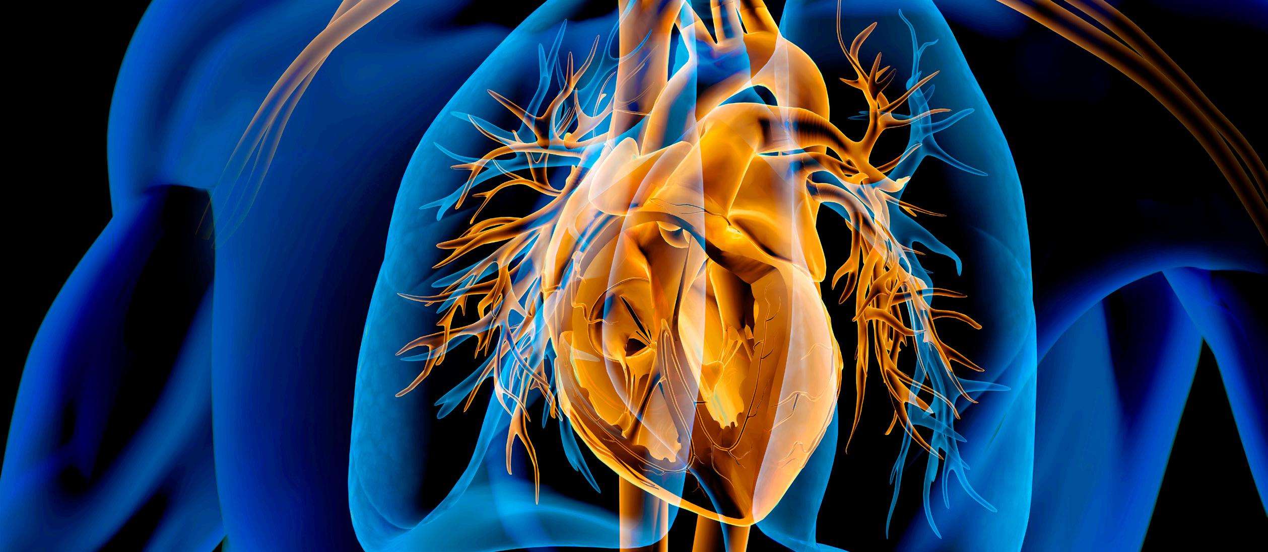
Nasir attributes this patient recruitment success to Houston Methodist’s close collaboration with community partners, including Jometra HawkinsSneed, a health advocate at the African American Male Wellness Agency. Sneed’s expertise has been instrumental in establishing and sustaining partnerships with 10 community-based organizations (CBOs) in and around Houston where patient recruitment takes place.
“We go to these CBOs at least once a month to screen potential study participants,” Hawkins-Sneed said. “We also promote the study within the CBOs. Each CBO has a goal of enrolling at least two people for the study each month, which equals to 20 people per month.”
Recruitment is focused on populations that are more susceptible to having high blood pressure due to unaddressed social needs. These social needs stem from inequitable conditions that impact health outcomes, including economic instability, lack of access to affordable health care and food insecurity. These non-clinical factors account for approximately 80% of health outcomes and are known broadly as the social determinants of health.
Social needs disproportionately impact communities of color, contributing to the hypertension disparities researchers hope to better understand and address through the Pressure Check study. Phase two of the study will address these social needs in early 2025. Study participants will be screened for more than 40 social needs that contribute to hypertension using a social determinant of health screening tool informed by validated social risk assessment indices, including the polysocial risk score tool pioneered by Javed. Hawkins-Sneed’s team of community health workers will also address these social needs by navigating patients to community resources, including Harris Health, Unite Us, Harris County Public Health, Texas 211, and others.
Upon study completion, the four health systems will share experiences and data to determine the most effective care model for hypertension, as well as the effectiveness of community-based interventions across study sites.
Javed explained, “This insight into community engagement and the study results can open more doors to further address disparities in hypertension and other conditions, such as obesity and diabetes, through community-based partnerships.”

The National Institutes of Health has awarded the Houston Methodist DeBakey Heart & Vascular Center an $8.7 million grant to advance research into life-threatening arrhythmias associated with mitral valve prolapse (MVP).
The research will address gaps in understanding the pathogenesis of MVP and developing better risk assessment tools for potentially life-threatening complications of the condition, which affects 2%-3% of the population — roughly 200 million people globally.
The NIH grant will fund a multi-year study on sudden cardiac death (SCD) risk assessment and mechanistic insights in arrhythmic MVP using cardiac MRI and circulating proteomic biomarkers. Additional research by Dipan J. Shah, MD, FACC — Beverly B. and Daniel C. Arnold Distinguished Centennial Chair — and his team includes a prospective SCD risk stratification study using cardiac MRI and echocardiography machine learning. Their work also involves analyzing circulating proteomics to phenotype myocardial remodeling in aortic stenosis and gaining mechanistic insights into arrhythmic mitral valve prolapse (MVP) through proteomic biomarkers.
Researchers are trying to determine what factors lead to mitral valve prolapse patients developing arrhythmias, including identifying early markers of risk through imaging, cardiac monitoring and blood biomarker analysis.
“The goal is to eventually have the ability to look at an imaging scan and predict which patients are at highest risk, allowing for earlier interventions,” explained Shah.
Typically benign, MVP causes some patients to develop ventricular arrhythmias that can lead to SCD.
Although MVP has long been associated with risk of SCD in a subset of patients, tools for identifying that subset and assessing an individual’s risk remain limited. Our researchers aim to change that by establishing new guidelines and developing a validated risk prediction model for MVP-associated SCD.
Specifically, Houston Methodist’s ongoing research efforts will include:
• Deep Phenotyping of MVP Patients
Research team will conduct comprehensive analyses of MVP patients to identify clinical, imaging and mechanistic factors associated with ventricular arrhythmias.
• Biomarker Development
The team will investigate a panel of blood biomarkers, including FDA-approved and novel proteomic markers, to develop a cost-effective screening strategy for identifying MVP patients with myocardial fibrosis — a key risk factor for SCD.
• Risk Prediction Model
Researchers will assemble a multicenter cohort of over 2,000 MVP patients with contrast-enhanced cardiac magnetic resonance imaging and perform longitudinal follow-up to create and validate a novel risk prediction model for SCD and life-threatening arrhythmias.
Houston Methodist’s ongoing research efforts underscore the growing recognition of MVP’s potential for severe outcomes, particularly among otherwise healthy young women.
Recently, researchers presented findings at the American Heart Association’s Annual Scientific Sessions showing that women had a threefold higher risk of arrhythmias compared to men. Myocardial fibrosis was another significant marker, with affected patients also experiencing a threefold increased risk.
Current treatment options for high-risk MVP patients include beta blockers, catheter ablation, and implantable defibrillators. However, the team is also investigating whether mitral valve surgery could reduce arrhythmic risk.
“A key part of this study is determining which patients would benefit from proactive treatment,” noted Shah.
The study is enrolling patients scheduled for cardiac MRI scans at Houston Methodist. Eligible patients are invited to participate in longitudinal monitoring, which includes Holter monitoring, additional echocardiography and blood sample collection.
“As one of the key groups in the country studying this, we received one of the largest grants of its kind from the NIH,” Shah said. “Our hope is that this study will provide the insights and targeted interventions needed to prevent tragic, unexpected deaths like the one that inspired this grant.”
This project has the potential to revolutionize the way we identify and manage mitral valve prolapse patients at risk for sudden cardiac death.
By combining advanced imaging techniques with biomarker analysis, we aim to provide clinicians with reliable tools to intervene early and improve patient outcomes.
”
–
Dipan J. Shah, MD, FACC Beverly B. and Daniel C. Arnold Distinguished Centennial Chair
Houston Methodist orthopedic research is advancing care for patients whose professions place extraordinary demands on the musculoskeletal system. From uncovering hidden spinal risks in elite ballet dancers to refining surgical strategies that restore knee function in professional athletes, these studies exemplify how precision research translates into performance-preserving care.

The knees are well-suited for most of what we demand from them, but no one is harder on knee articular cartilage than a professional athlete. It’s no surprise then that the incidence of articular cartilage knee injuries is generally higher in athletes than nonathletes.
Injuries to articular cartilage are especially problematic for patients with high demands on their knees, causing pain and swelling that limit participation in high-level athletics. Articular cartilage is a thin, white, living tissue covering the ends of bones where they meet to form joints such as in the knee. It allows the bones to glide over one another without friction, protects the bones and enables smooth movement. Considering the inherent limits to healing within joints, managing damage to articular cartilage is particularly challenging. If severe, surgery is usually the only option.
Osteochondral allograft transplant — where an allograft osteochondral plug is fit in a congruent fashion to the patient’s articular surface — is an elegant surgical option to treat osteochondral lesions with a surface area of at least 2 cm2 This procedure provides a viable choice for injuries with extensive subchondral edema, even with prior marrow stimulation, it carries less risk of donor site morbidity. Current literature offers reassuring outcomes of osteochondral allograft transplant in general athletes, but minimal data exists on the outcomes in high-level, professional athletes.
Understanding the success or failure of osteochondral allograft transplants in professional athletes is critical for performance expectations and career longevity.


“ ”
Focusing on a unique, high-demand patient population, this study demonstrates that osteochondral allograft transplantation in professional athletes yields a high rate of return to play at a similar or higher level, even when performed with concomitant procedures such as meniscal allograft transplantation.
Present literature relies on studies with enough independent variables (beyond the treatment itself) influencing an athlete’s return to preinjury status; however, caution must be used in interpreting the results. To investigate osteochondral allograft outcomes in professional athletes without as many confounding factors, Sachin Allahabadi, MD, worked with a team at Rush University Medical Center in Chicago on a study evaluating the experience of his mentor and senior surgeon, Brian J. Cole, MD, MBA, in treating professional athletes with osteochondral allograft transplant, including analyzing clinical outcomes and return to sport.
With Allahabadi as first author, the study included a retrospective review of professional athletes who underwent primary osteochondral allograft to the knee between January 1, 2001, and January 1, 2021, by a single surgeon. All procedures were performed by the senior author (Cole), a fellowship-trained orthopedic surgeon with a high-volume referral-based practice in cartilage restoration. To be included in the
study, athletes had to play at the professional level and have a minimum of two years of follow-up. Fifteen professional athletes representing a variety of sports participated. The majority had undergone prior surgeries to the operative knee.
Patients demonstrated statistically significant postoperative improvements at the most recent time point for all PROs (P < .01). Eleven returned to sport at a mean of 1.22 years, and of the eight undergoing isolated osteochondral allograft, seven returned at 1.28 years. Ten athletes returned to sport at the same level or higher as before surgery. Significant improvements were observed in all assessed patient-reported outcome scores. Two of the three patients who underwent concomitant meniscal allograft transplant were able to return to sport at the same level or higher than pre-surgery. Three underwent second-look arthroscopy, one of whom underwent cartilage debridement of the osteochondral allograft.
High-level athletes can expect significant postoperative improvement in clinical outcomes with this procedure.
Female ballet dancers disproportionately have idiopathic scoliosis, according to a new study that concludes there is a need for more screening and evidence-based interventions early in ballerinas’ careers.

The study, conducted in the Department of Orthopedics and Sports Medicine, found that more than 1 in 5 female participants exhibited spinal curvatures — more than triple the rate of male participants and more than double the rate of the general female population. Study participants were drawn from the Houston Ballet.
“Our research uncovers a critical link between the intense physical demands placed on young ballerinas and the structural development of their spines,” said Comron Saifi, MD, C. James and Carol Walter Looke Chair in Orthopedic Spine Surgery and the study’s principal investigator. “The unique combination of early-age training, low body fat and biomechanical stressors appears to significantly elevate the risk of scoliosis.”
Saifi said the ballet could benefit from safeguards similar to pitch limits implemented in baseball to prevent injury. He also noted that these safeguards might significantly reduce the long-term impact on female dancers’ spinal health.
Idiopathic scoliosis, defined as a spinal curvature greater than 10 degrees, most commonly emerges during adolescence in girls. In ballet dancers, it can present performance challenges and increase risks of injury as well as lead to post-career pain, reduced flexibility and musculoskeletal issues.
The study, published The Journal of Bone and Joint Surgery, involved a retrospective analysis of data collected from 98 Houston Ballet dancers —
49 women and 49 men — during routine physical exams between 2017 and 2022. The mean ages were 25 for the men and 27 for the women.
Saifi’s team utilized advanced imaging techniques — including dual-energy X-ray absorptiometry (DXA) scans — to comprehensively evaluate spinal curvature, bone density and body composition.
The study found that while 20.4% of the women had idiopathic scoliosis, only 6% of the men had the condition, consistent with such norms in the male population.
The research leverages Houston Methodist’s close partnership with the Houston Ballet and represents a deep commitment to understanding and addressing musculoskeletal disorders in high-performance athletes. Saifi noted in the study that idiopathic scoliosis in professional ballet is understudied.
Saifi’s team identified several distinct physiological markers among female subjects with scoliosis that were not observed in dancers without scoliosis. These markers include lower fat mass and distinct skeletal dimensions, such as longer trunks and wider shoulders
and hips. These findings suggest that anatomical and physiological traits common in professional dancers might predispose them to spinal curvature.
Despite limitations inherent in supine imaging — typically underestimating curvature — the study still identified a high prevalence, suggesting even greater risks in standard standing evaluations.
“With DXA scans, the subjects are lying flat on the machine, making it harder to detect scoliosis due to the lack of gravity on the spine,” Saifi said.
“It is likely that within our study, the percentage of ballerinas with scoliosis is probably even higher than 21% and the magnitude of their curve is probably a lot more than the DXA scan captured.”
Notably, the research sets an initial benchmark in spinal health assessment for ballet performers that could be instrumental in future studies.
“The study highlights the limited data on adolescent idiopathic scoliosis prevalence and the need to identify specific risk factors. It suggests further research into the impact of starting intense ballet training at an early age.”
Ballet is an extraordinary art form. Our responsibility as physicians is to preserve dancers’ musculoskeletal health, so their careers can be both distinguished and sustainable. “ ”
–
C.

MD
and Carole
Breakthrough discoveries are accelerating the pace of progress in digestive health.
Houston Methodist research shows compelling new evidence that advanced liver disease caused by MASH may be reversible — signaling a transformative shift in how physicians understand and treat this growing public health challenge.

Two new studies led by hepatologist Mazen Noureddin, MD, MHSc, provide the strongest evidence to date that cirrhosis caused by metabolic dysfunction-associated steatohepatitis (MASH) may be reversible — a milestone once considered unattainable in hepatology.
The phase 2 trials, published in The New England Journal of Medicine (NEJM) and The Lancet, found the investigational drug efruxifermin — an engineered fibroblast growth factor 21 analogue — had robust antifibrotic effects in patients with advanced metabolic liver disease.
Together, the studies position efruxifermin as one of the most promising therapeutic candidates yet for MASH. The drug showed benefits across both compensated cirrhosis and pre-cirrhotic disease.
The NEJM study focused on patients with biopsyconfirmed compensated cirrhosis due to MASH, representing the most advanced stage of fibrotic
liver disease short of decompensation. In this randomized, placebo-controlled phase 2b trial, patients received either a once-weekly subcutaneous efruxifermin at two dose levels or a placebo, and underwent serial liver biopsies at baseline, 36 weeks and 96 weeks.
While the trial narrowly missed its primary endpoint at 36 weeks, longer-term follow-up revealed statistically significant fibrosis regression at 96 weeks, particularly at the higher dose. Nearly a quarter of the patients receiving the 50-mg dose achieved at least a one-stage improvement in fibrosis without worsening of steatohepatitis — compared to 11% in the placebo group. Importantly, some patients improved from stage F4 to F3 fibrosis, effectively moving out of the cirrhosis category.
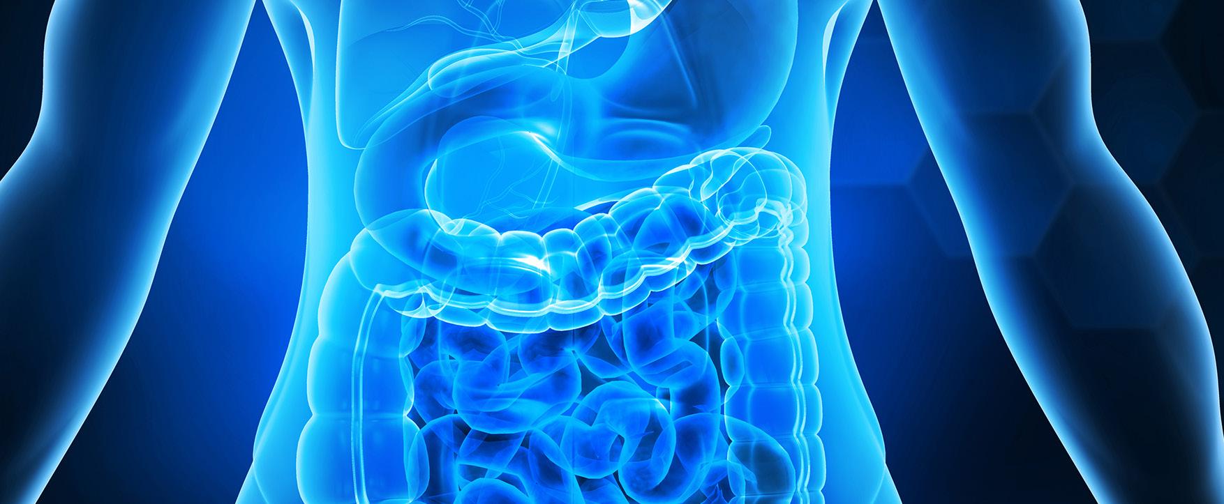
“The transition from F4 to F3 is what makes this study so revolutionary,” Noureddin said. “It means these patients are no longer cirrhotic and may no longer face the need for liver transplantation down the line.”
The drug was generally well-tolerated, with mostly mild to moderate gastrointestinal side effects such as nausea, vomiting and diarrhea. There was no signal of drug-induced liver injury.
The second study, published in The Lancet, examined efruxifermin in patients with moderate to severe fibrosis (F2 to F3), a population at high risk for progression to cirrhosis. Results from the 96-week trial showed striking efficacy, particularly at the higher dose.
In paired biopsy analyses, up to 75% of patients receiving 50 mg of efruxifermin achieved at least a one-stage improvement in fibrosis without worsening of MASH — one of the highest response rates ever reported in this disease population. Significant improvements were also seen in steatohepatitis resolution and metabolic markers.
“These are some of the most robust antifibrotic responses we’ve ever seen,” Noureddin said.
“And they were achieved without meaningful weight loss, showing this is not simply a metabolic or weight-loss effect.”
MASH affects an estimated 6% to 7% of U.S. adults and is now a leading cause of liver transplantation, particularly among women. While recent FDA approvals have expanded treatment options for earlier-stage disease, no therapies are currently approved for MASH-related cirrhosis.
Noureddin emphasized that efruxifermin’s ability to inhibit fibrogenesis while promoting fibrolysis — the breakdown of scar tissue — represents a mechanistic advance that may change the treatment paradigm.
“We are entering what I would call the pre-golden age,” he said. “The true golden age will be when we can reliably reverse or prevent cirrhosis — and this data suggest we are very close.”
Phase 3 trials of efruxifermin are now underway and investigators are hopeful the findings will soon translate into clinical practice.
“After decades of having nothing for these patients, this is real hope,” Noureddin said. “And it’s only the beginning.”

Houston Methodist and Rice University teamed up to form the Digital Health Institute (DHI), a collaborative effort designed to harness the power of artificial intelligence (AI), biomedical engineering and integrated data systems to tackle urgent health care needs.
Together, the teams built sophisticated algorithms that draw on multimodal data to uncover patterns in disease progression, predict individual health risks and deliver personalized prevention and treatment strategies. This holistic approach is shaping the future of precision medicine, improving hospital efficiency and expanding access to high-quality care in underresourced communities.
DHI is also a training ground for the next generation of digital health leaders, translating research into real-world impact across diagnostics, therapeutics, remote monitoring and health equity.

The aim is to accelerate the digital transformation of health care by uniting Rice University’s world-class leadership in engineering, artificial intelligence and data science with Houston Methodist’s excellence in clinical and academic medical research. This partnership is committed to translating research into tangible solutions and equitably delivering novel digital health solutions that enhance patient care, advance medical discovery, improve operational efficiency and expand health care access.
The aspiration is to deliver personalized, predictive and proactive health care through cutting-edge research and universally scalable solutions that redefine the health care experience.
We can now have a future that we can predict, we can personalize and balance health care as we move forward. We have to remember that it’s not all about technology. At the heart of all this is people. We serve patients … with dignity, empathy, intelligence and integrity. “ ”
The Digital Health Institute (DHI), a joint initiative of Rice University and Houston Methodist, held its inaugural summit last fall at Rice, drawing about 350 registered guests for a daylong look at how artificial intelligence, data science and engineering are transforming patient care.

The DHI, established in 2024, connects researchers and clinicians working at the intersection of technology and medicine. Its focus areas span artificial intelligence and machine learning, digital diagnostics, wearable and implantable sensors, robotics and computational biology. Through sustained collaborations, the institute aims to advance precision medicine, reduce clinician workload and improve patient outcomes.
Jenny Chang, MB.ChirB., MD, MHCM, Ernest Cockrell, Jr. Presidential Distinguished Chair, and Rice’s Amy Dittmar, Howard R. Hughes Provost, opened the summit with remarks on the growing collaboration between the two institutions.
Chang said the partnership brings together “clinical excellence in cardiovascular engineering, data science and innovation” to shape a more personalized future of health care.
Dittmar described the institute as an example of Rice’s strategic focus on health innovation and partnership
with the Texas Medical Center. The institute, she said, “provides a unique opportunity to design, develop and measure the efficacy of products and systems from beginning to end, all with people at the center of it.”
She highlighted projects ranging from wearable and implantable sensors to AI tools for medical imaging, and simulations that improve clinical decision-making.
“Together, our institutions are expanding access to quality health care through the responsible use of artificial intelligence,” Dittmar noted.
Khurram Nasir, MD, MPH, William A. Zoghbi Centennial Chair in Cardiovascular Health, and DHI co-director, said the institute was founded to overcome the fragmentation that often stalls digital health innovation — the gap “between data and discovery, between development and deployment.” He also emphasized that Houston Methodist’s integrated data infrastructure, combined with Rice’s engineering capabilities, could help raise Houston’s profile as a leading hub in digital health translation.

Rice’s Ashutosh Sabharwal, PhD, Ernest Dell Butcher Professor of Engineering and DHI co-director, spoke about the rapid growth of research and talent around digital health.
Sabharwal noted the partnership is not only producing research, but also streamlining how ideas move from concept to clinical testing.
Pothik Chatterjee, DHI executive director, detailed how the institute is building an ecosystem to accelerate translation — from algorithm design and validation to regulatory approval and commercialization — positioning Houston as a national model for AI-driven health innovation.
Luay Nakhleh, PhD, dean of Rice’s George R. Brown School of Engineering and Computing, noted the university’s long history of health-related engineering
partnerships. He also underscored Rice’s collaboration with the Texas Medical Center dating to early projects such as the artificial heart in the 1960s and remains central to the school’s mission today.
“We bring engineering and computing expertise to the table, and Houston Methodist brings the complementary clinical expertise that is needed for an area like digital health,” Nakhleh said.
The summit’s structure reflected the DHI’s overarching mission: to unite clinicians, engineers and entrepreneurs in developing and deploying technologies that improve patient outcomes.
By linking Rice’s engineering and AI expertise with Houston Methodist’s clinical innovation ecosystem, the DHI is piloting a unique model for how academic and medical partnerships can accelerate patient-centric care.


Education is central to transformation at Houston Methodist, where the next generation of physicians, researchers and health care leaders are being prepared to advance medicine with purpose and precision. Through collaborative, innovation-driven learning and hands-on training, learners are equipped to transform knowledge into meaningful clinical impact and better outcomes for the communities they serve.
Houston Methodist marked two decades of advancing medical education through its Graduate Medical Education (GME) Programs.
In 2004, Houston Methodist marked a transformative chapter in its history, separating from a longtime academic partner and establishing an independent academic medical center of distinction.
Faced with the sudden loss of medical trainees and research programs, Houston Methodist responded with vision and determination. A year later, the first 12 Accreditation Council for Graduate Medical Education (ACGME)-accredited programs were launched with an inaugural class of residents. From zero residency programs and zero fellows, the program is now a thriving education enterprise that complements a world-class research institute.
Today, Houston Methodist offers more than 80 programs, including 59 ACGME-accredited residencies and fellowships and 29 GME-sponsored fellowships. In 2025, 447 residents and fellows were trained, each contributing to the mission of excellence in education, research and patient care.
In the same year, the GME programs witnessed the graduation of 150 physicians, whose dedication strengthens a commitment to leading medicine and improving lives.

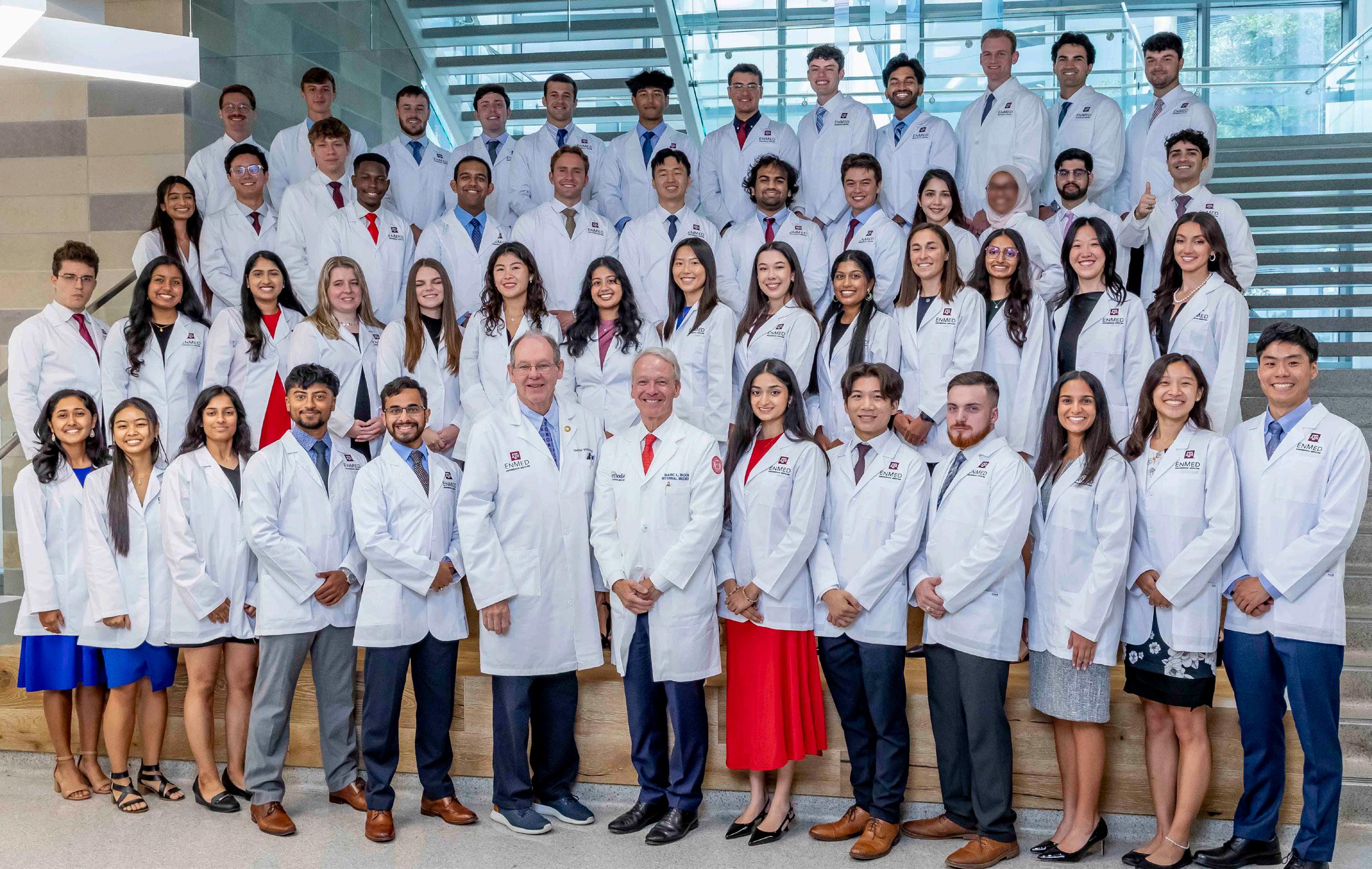
A collaboration between Houston Methodist and the Texas A&M School of Engineering Medicine (EnMed) celebrated Match Day for the third consecutive year.
The EnMed Class of 2025 experienced a 100% match rate on Match Day, which is held annually at the TMC3 Helix. Forty-seven EnMed students were matched to residency programs across the country through the National Resident Matching Program. The residents learn where they will train for residency and their specialty. The top specialties were internal medicine, radiology diagnostic, family medicine, surgery, and anesthesiology.
EnMed operates in close collaboration with Houston Methodist Hospital and the Texas A&M University Colleges of Medicine and Engineering, with a goal of transforming health care through translational interdisciplinary research, the development of transformative medical technologies, and the formation and training of “physicianeers.”
The level of curiosity our EnMed students have is above the average for a medical student. Curiosity about problem-solving and innovation is exactly the sort of engineering aspect that we’re trying to incorporate into medicine. These EnMed students have a fundamentally different way of thinking about a medical problem — thinking as problem-solvers, they see medical issues through the lens of an engineer in a very methodological way.
”
– Timothy B. Boone, MD, PhD
Craig C. Brown and Suzanne H. Smith Centennial Chair in Medical Education
Timothy B. Boone, MD, PhD, Craig C. Brown and Suzanne H. Smith Centennial Chair in Medical Education, added, “These students are driven by community service. They are always finding places to do community service and help the underserved.”
“After four years of a blended engineering and medicine curriculum throughout their pre-clerkship, clinical rotations during clerkship, and finding time to innovate and perform engineering immersions, our EnMed students reached their pinnacle,” said Amy Wright, EdD, MBA, Associate Dean of Student Affairs and Admissions and former Director of Educational Partnerships. “I couldn’t be prouder. Great job, Class of 2025!”
The Texas A&M University School of Engineering Medicine (EnMed) hosted its largest annual White Coat Ceremony for the Class of 2025.
Alan B. Lumsden, MD, Walter W. Fondren III Presidential Distinguished Chair, DeBakey Heart & Vascular Center, and Chair, Department of Cardiovascular Surgery, gave the keynote address.
This notable event featured EnMed students receiving their first white coat — a rite of passage signifying their transition into the medical community. Founded in 2019, the EnMed program is a collaboration between Texas A&M’s School of Engineering Medicine and Houston Methodist to transform health care through the development and training of “physicianeers,” the creation of medical technologies and translational research.
The EnMed physicianeer is an innovative, problem-solving physician uniquely qualified to address some of health care’s greatest challenges. These graduates receive a Medical Doctorate and Master of Engineering degree focused on the design and implementation of impactful medical technologies through a revolutionary curriculum.
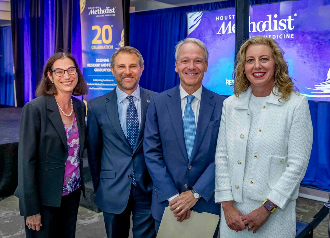
The Constance M. and Byron F. Dyer Fellowship Award at Houston Methodist supports early-career professionals who show innovative work. The 2025 winners are:
Laura Kim, MD
Ngoc-Anh A. Nguyen, MD
Raksha Raghunathan, PhD
Chandra Bautista, PhD

Konrad Harms, MD
Ashish Jain, MD
Adel Khan, MBBS
Nicole Stephens, MSN, BSN
Rice University BioScience Research Collaborative, in partnership with the Smalley-Curl Institute and COMSOL Inc., hosted the COMSOL Inc. Multiphysics Workshop & Poster Session.
The poster session served as a networking and competitive opportunity to showcase and discuss innovative research based on numerical simulations. Elisa Serafini, Graduate Research Fellow, was named second place poster winner.
The event included expert-led presentations and live demonstrations featuring COMSOL Multiphysics® — a modelbuilding software that allows engineers and scientists to simulate designs, devices and processes in fields of engineering, manufacturing and scientific research.
The Houston Methodist Academic Institute oversees the Education Institute and Research Institute, including 876 faculty and 45,300 learners. The Academic Institute aligns our research and education initiatives in service to the clinical mission, providing solutions that answer the call for new technologies and skills our clinicians need for patient care.
The Houston Methodist Education Institute coordinates our primary academic affiliation with Weill Cornell Medicine and other joint programs, including the Engineering Medicine Program at Texas A&M University School of Engineering Medicine. The Education Institute also oversees continuing medical education and graduate medical education, and supports more than 917 students and postgraduate trainees for medical, nursing, allied health and research education programs.
The Houston Methodist Research Institute supports research programs and infrastructure that enable faculty across the system to bring new scientific discoveries to patients as rapidly as possible through the full Cycle of Translation from conceptual bench research to prototyping and development to clinical trials and FDA approval. The Research Institute supports more than 1,940 clinical studies and trials and $120 million in extramurally funded translational research programs.
Judge Ewing Werlein, Jr., Senior Chair
W. Benjamin Moreland, Chair
Edward R. Allen, III
Steven Birdwell
John F. Bookout, III
Marc L. Boom, MD
P. Embry Canterbury
Jenny C. Chang, MD
David Chao
Martin Craighead
Leslie Doggett
Elaine Finger
Antonio Gotto, MD, D.Phil
Robert A. Harrington, MD
Edward A. Jones
Evan H. Katz
Sippi K. Khurana, MD
Steven Looke
Ransom Lummis
David A. Modesett
James Muschalik
Gregory Nelson
Joe Bob Perkins
Ward Sheffield
Jeffrey F. Simmons
Suzanne (Sue) Smith
Christopher G. Stavros
Steven D. Stephens
Spencer A. Tillman
David M. Underwood, Jr.
Amy Waer, MD
Martha S. Walton
Donna Sims Wilson