11:21
Página 1
ENDOCRINOLOGIA MOLECULAR
21/11/2007
TREB. SOC. CAT. BIOL., VOL. 56, 2005
SobreCub. 56 Endocrinologia
ENDOCRINOLOGIA MOLECULAR JAUME REVENTÓS EDITOR
Il·lustració de la sobrecoberta
TREBALLS DE LA SOCIETAT CATALANA DE BIOLOGIA VOLUM 56, 2005
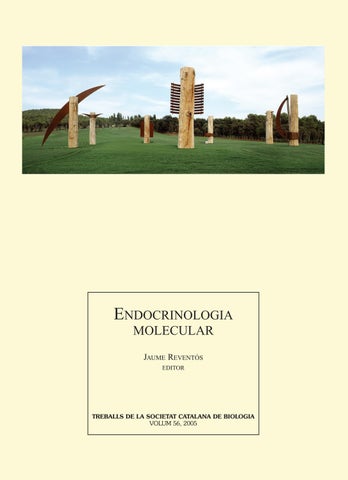
Tità Cromlech, d’Enric Pladevall 2000-2001. Fusta i ferro. 40 m de diàmetre Col·lecció privada, Girona
11:21
Página 1
ENDOCRINOLOGIA MOLECULAR
21/11/2007
TREB. SOC. CAT. BIOL., VOL. 56, 2005
SobreCub. 56 Endocrinologia
ENDOCRINOLOGIA MOLECULAR JAUME REVENTÓS EDITOR
Il·lustració de la sobrecoberta
TREBALLS DE LA SOCIETAT CATALANA DE BIOLOGIA VOLUM 56, 2005
Tità Cromlech, d’Enric Pladevall 2000-2001. Fusta i ferro. 40 m de diàmetre Col·lecció privada, Girona
11:24
Página 1
TREBALLS DE LA SOCIETAT CATALANA DE BIOLOGIA
VOLUM 56, 2005
ÍNDEX
ENDOCRINOLOGIA MOLECULAR Endocrinologia i amics. En homenatge i memòria de José Sáez (1934-2003) . . . . . . . . . . . . . . . . . . . . . . . . . . J. REVENTÓS Remodelatge de la cromatina durant la inducció per progesterona del promotor de l’MMTV . . . . . . . . . . G. P. VICENT, A. S. NACHT, J. CLAUSELL, T. BECHTOLD, C. BALLARÉ I M. BEATO Fetal testis and estrogenic endocrine disrupters . . . . . . . . . . . . . . . . . . . . . . . . . . . . . . . . . . . . . . . . . . . . . . . . . . R. HABERT, C. LEVACHER, C. PAIRAULT I V. ROUILLER-FABRE Paracrine regulation of spermatogenesis: the virtue of dialogue . . . . . . . . . . . . . . . . . . . . . . . . . . . . . . . . . . . . B. JÉGOU I C. PINEAU Protamines i infertilitat en l’home . . . . . . . . . . . . . . . . . . . . . . . . . . . . . . . . . . . . . . . . . . . . . . . . . . . . . . . . . . . . . . R. OLIVA An overview of the proacrosin/acrosin system in human spermatozoa . . . . . . . . . . . . . . . . . . . . . . . . . . . . . M. H. VAZQUEZ-LEVIN, L. I. FURLONG, C. M. VEAUTE I P. D. GHIRINGHELLI Acció dels andrògens en el testicle: un paper per a la meiosi . . . . . . . . . . . . . . . . . . . . . . . . . . . . . . . . . . . . . . . C. A. SUAREZ-QUIAN, J. REGADERA, M. NISTAL, P. GONZÁLEZ-PERAMATO, Á. SERRANO, O. M. TIRADO, D. M. SELVA, N. TORAN, F. MUNELL I J. REVENTÓS Receptors d’IGF-1 i marcadors funcionals de les cèl·lules de Sertoli . . . . . . . . . . . . . . . . . . . . . . . . . . . . . . . . . L. BASSAS I M. ANTICH Ultrastructural immunocytochemistry of a Leydig cell enzime and a sperm tail protein using antigen retrieval by microwave irradiation . . . . . . . . . . . . . . . . . . . . . . . . . . . . . . . . . . . . . . . . . . . . . . . . . . . . . . . . . . . . . M. P. MUSSE I H. E. CHEMES L’expressió al testicle de la proteïna transportadora d’esteroides sexuals humana (SHBG) té lloc a les cèl·lules germinals i no a les cèl·lules de Sertoli . . . . . . . . . . . . . . . . . . . . . . . . . . . . . . . . . . . . . . . . . . . . . . . . . . D. M. SELVA I G. L. HAMMOND Sex hormone-binding globulin (SHBG) in the ovary . . . . . . . . . . . . . . . . . . . . . . . . . . . . . . . . . . . . . . . . . . . . . . T. FORGES, A. GÉRARD, P. MONNIER-BARBARINO I H. GÉRARD Diferències sexuals i d’edat: una comparació de la leptina sèrica, els components de l’insulin-like growth factor-I i les concentracions de la globulina d’unió a hormones sexuals en una població sana . . . . . . . . . . . J. M. GÓMEZ, F. J. MARAVALL I J. SOLER Biosynthesis and regulation of dehydroepiandrosterone (DHEA) in brain . . . . . . . . . . . . . . . . . . . . . . . . . . . Y. LIU I V. PAPADOPOULOS Human prepubertal aromatase deficiency: physiological and pathophysiological lessons learned from this experiment of nature . . . . . . . . . . . . . . . . . . . . . . . . . . . . . . . . . . . . . . . . . . . . . . . . . . . . . . . . . . . . . . . . . . . . . A. BELGOROSKY I M. A. RIVAROLA The auto/paracrine regulation of endocrine functions: a history of TGF-β and the adrenal cortex . . . . . . . J.-J. FEIGE I E. M. CHAMBAZ Cortisol: funcions i importància del receptor glucocorticoide. Una visió comparada . . . . . . . . . . . . . . . . . . L. ACERETE, S. MACKENZIE I L. TORT L’hormona adrenocorticòtropa plasmàtica en el període postoperatori immediat com a índex predictor de la curació de la malaltia de Cushing . . . . . . . . . . . . . . . . . . . . . . . . . . . . . . . . . . . . . . . . . . . . . . . . . . . . . . . . . C. VILLABONA Insight into the association between phospholipase C-γ 1 and the insulin receptor . . . . . . . . . . . . . . . . . . . . H.-J. JANG, Y.-K. KWON, S. KOLE I M. BERNIER Factors angiogènics en la retinopatia diabètica proliferativa . . . . . . . . . . . . . . . . . . . . . . . . . . . . . . . . . . . . . . . M. GARCIA-RAMÍREZ, C. HERNÁNDEZ, E. CARRASCO I R. SIMÓ The human placenta: an atypical endocrine organ . . . . . . . . . . . . . . . . . . . . . . . . . . . . . . . . . . . . . . . . . . . . . . . . D. EVAIN-BRION I A. MALASSINÉ Growth hormone and aging . . . . . . . . . . . . . . . . . . . . . . . . . . . . . . . . . . . . . . . . . . . . . . . . . . . . . . . . . . . . . . . . . . . C. ARIZNAVARRETA, A. P. GARCIA, V. SALAZAR, L. M. GARCIA SEGURA, I. AZCOITIA, J. CHOWEN, E. VARA I J. A. F. TRESGUERRES «Hormone-refractory» prostate cancer. A putative new mechanism: the upside-down response to androgens . . . . . . . . . . . . . . . . . . . . . . . . . . . . . . . . . . . . . . . . . . . . . . . . . . . . . . . . . . . . . . . . . . . . . . . . . . . . . . . . . . M.-O. JOLY-PHARABOZ, J.-J. KALACH, J. CHANTEPIE, B. NICOLAS, A. RUFFION I J. ANDRÉ ETV5 i RUNX1, nous factors de transcripció implicats en la invasió miometrial del carcinoma endometrial . . . . . . . . . . . . . . . . . . . . . . . . . . . . . . . . . . . . . . . . . . . . . . . . . . . . . . . . . . . . . . . . . . . . . . . . . . . . . . . . . M. ABAL, J. PLANAGUMÀ, A. GIL-MORENO, A. DOLL, M. MONGE, M. GONZÁLEZ, T. BARÓ, Á. GARCÍA, J. CASTELLVÍ, S. RAMÓN Y CAJAL, J. XERCAVINS, F. ALAMEDA I J. REVENTÓS
9 13 25 33 47 59
ENDOCRINOLOGIA MOLECULAR
21/11/2007
TREBALLS DE LA SOCIETAT CATALANA DE BIOLOGIA ENDOCRINOLOGIA MOLECULAR VOLUM 56, 2005
75 91 101 107 117 127 135 147 159 165 175 183 195 211 223
235
243
FILIAL DE L’INSTITUT D’ESTUDIS CATALANS
TREB. SOC. CAT. BIOL., VOL. 56, 2005
Cub. Volum 56.qxd
BARCELONA 2007
Interior Cub. Volum 56
21/11/2007
11:20
Página 1
NORMES DE PUBLICACIÓ DE TREBALLS DE LA SOCIETAT CATALANA DE BIOLOGIA Abast. Treballs de la Societat Catalana de Biologia (Treb. Soc. Cat. Biol.) publica: a) monografies i revisions sobre temes de ciències de la vida encarregades per la Societat, i b) comunicacions presentades a les sessions ordinàries i extraordinàries organitzades per la Societat o les seves seccions especialitzades. Els temes poden ser proposats pels membres del Consell Directiu o per socis que desitgin ocupar-se de la preparació d’un volum de la revista fent d’editors. El Consell Directiu decideix si un tema és apropiat per dedicar-hi un volum de Treb. Soc. Cat. Biol. Idioma. Els originals seran redactats en català, excepte en casos justificats per als quals el Consell Directiu pugui acceptar una altra llengua. Lliurament dels originals. La submissió implicarà que el material és original i que no ha estat enviat, acceptat o publicat en cap altre lloc. Els originals els lliurarà l’editor del volum per correu electrònic (scb@iecat.net), adreçat al president del Consell de Redacció. Cal enviar els següents arxius, preferentment empaquetats amb ZIP: — Un arxiu únic en format RTF o MS Word anomenat Text_TSCB_[cognom de l’editor], amb tot el text del volum, incloent-hi, en aquest ordre: coberta, índex, presentació, els capítols en l’ordre desitjat i les seves taules, si n’hi ha. Les pàgines hauran d’anar numerades consecutivament començant per la núm. 1, que serà la de la coberta. Tot el text serà escrit íntegrament amb Times New Roman de cos 12 amb interlineat d’1,5. L’arxiu amb el text no podrà contenir figures ni objectes incrustats. — Un arxiu per cada figura anomenat Figura_[número de figura]_[cognom del primer autor]. Les figures, si no contenen text (a part del peu), es poden remetre en qualsevol dels formats gràfics habituals (PNG, JPEG, TIFF); si contenen text, s’han d’enviar en un format que permeti editar-lo (EPS, PDF, Powerpoint). — Cal enviar també una còpia impresa electrònica en format PDF de tot el volum, anomenada TSCB_[cognom de l’editor], amb tot el contingut, incloent-hi les figures. Aquest arxiu es farà servir com a text imprès de referència durant el procés de tractament del text. Per aquest motiu, els autors i l’editor han de tenir cura personalment que en aquesta còpia hi hagi tot el contingut imprès correctament, incloent-hi sobretot els símbols o caràcters especials, equacions, etc. Cada article o, en un volum monogràfic, cada capítol, incloent-hi la bibliografia, taules i figures, hauria de tenir una extensió entre quinze i vint fulls DIN A4, escrits amb el tipus, mida i interlineat esmentats abans. Només es podrà ultrapassar aquest límit amb l’acord previ del Consell de Redacció de Treballs de la SCB. Els volums monogràfics haurien de contenir un mínim d’uns deu capítols. El Consell de Redacció consultarà especialistes, els quals recomanaran si un article pot publicar-se immediatament, necessita revisió, o bé és rebutjat. Els autors rebran notificació de la decisió. Si el treball requereix revisió, es facilitaran als autors els comentaris escrits dels especialistes que l’hagin revisat. Estructura dels originals. Els originals que no s’ajustin a aquestes instruccions seran retornats als autors perquè els modifiquin abans de la seva tramitació. Dintre de cada capítol o article se seguirà el següent ordre pel que fa als diferents components:
— Títol, escrit en majúscules i negreta. — Nom i primer cognom de l’autor. Si n’hi ha més d’un, els seus noms aniran separats amb una coma. L’autor per a la correspondència s’identificarà amb un asterisc posat al final del seu cognom. — Adreça postal completa de tots els autors. Telèfon, fax i correu electrònic de l’autor per a la correspondència — Resum en català, d’un màxim de dues-centes paraules. — Paraules clau en català, màxim de cinc. — Resum en anglès (que, òbviament, haurà d’ésser traducció fidel del resum en català i haurà d’anar precedit de la traducció del títol del manuscrit). — Paraules clau en anglès, màxim de cinc. — Text, que, si és una revisió, podrà estar dividit fins a dos nivells en els apartats (no numerats) que es creguin més oportuns. Si es tracta d’un article de recerca, el manuscrit haurà d’incloure Introducció, Materials i mètodes, Resultats i Discussió. No es podran fer servir notes a peu de pàgina ni text subratllat. — Agraïments, si escau. — Referències, segons el format indicat més endavant. — Taules. Portaran un títol descriptiu breu i aniran numerades correlativament amb números àrabs. Una taula per full. — Text de peus de figures. Il·lustracions. Les il·lustracions cal que siguin de bona qualitat i contrast, tenint en compte la possible reducció, ja que, a causa del format de la revista, un cop publicades poden ésser, com a màxim, de 14 cm d’ample per 19 cm d’alt. Totes les il·lustracions seran en blanc i negre, excepte en cas que el color sigui essencial per a comprendre’n el significat. Atès que la publicació de fotografies en color encareix molt el procés editorial, els autors que desitgin incloure alguna fotografia en color hauran de pagar les despeses extra. En cas que les taules o figures hagin estat publicades anteriorment, caldrà indicar-ne la procedència i, si escau, el copyright. Referències. Totes les referències, tant publicades com en premsa, citades al text, hauran d’ésser incloses a la llista de referències, ordenades alfabèticament. En el text s’indicaran els noms dels autors i l’any de publicació per ordre cronològic; en el cas que una referència tingui més de dos autors, se citarà el primer seguit d’et al. A continuació hi ha exemples de l’estil que s’ha de seguir per a citar articles, capítols o monografies. CODINA, C.; VILADOMAT, F.; BASTIDA, J.; LLABRÉS, J. M. (1989). «Potencial biotecnològic del cultiu de cèl·lules vegetals per a l’obtenció de productes farmacèutics». Treb. Soc. Cat. Biol., 40: 47-70. MELLADO, R. P. (1987). «Vectores utilizados para la manipulación y expresión de genes». A: VICENTE, M.; RENART, J. Ingeniería genética. Madrid: CSIC, 21-30. WOLFFE, A. (1995). Chromatin structure and function. Londres: Academic Press. Proves d’impremta i extrets. De tots els originals acceptats, l’autor de correspondència en rebrà unes proves d’impremta per a corregir-les, i aquestes proves hauran d’ésser retornades, per correu urgent, a: Secretaria de la Societat Catalana de Biologia, c/ Maria Aurèlia Capmany, 14-16, 08001 Barcelona (tel. 933 248 584; fax 932 701 180; a/e: scb@iec.cat) dins el termini màxim d’una setmana. En les proves no podran ésser introduïdes correccions d’estil, i només hi seran admeses les de caràcter ortogràfic i tècnic. Un cop publicat l’article, es lliuraran a l’autor vint extrets sense càrrec.
TREBALLS DE LA SOCIETAT CATALANA DE BIOLOGIA INSTRUCTIONS FOR AUTHORS Scope. Treballs de la Societat Catalana de Biologia (Treb. Soc. Cat. Biol.) publishes: a) monographs and reviews on life sciences topics commissioned by the Society; and b) lectures presented at both ordinary and extraordinary sessions organized by the Society or their specialized sections. Submission of originals. Submission implies that the material is original and that has not been submitted, accepted or published elsewhere. Originals will be submitted by the editor of the volume to the following e-mail address scb@iecat.net addressed to the President of the Publications Board. The following files will be provided, preferably ZIP-compressed: — A single file in format RTS or MS Word named Text_TSCB_ [editor’s last name], containing all the text of the volume, including, in this order, the following parts: cover page, table of contents, presentation, the chapters in the order that they should appear and their corresponding tables. Pages should be consecutively numbered, staring with page 1, which will be the cover page. All text should be written in Times New Roman, pt 12 with 1.5 spaces between lines. The text file cannot contain figures and incrustated objects. — One file for each figure named Figure_[figure number]_ [last name of the first author]. Figures that do not contain text characters can be submitted in standard graphic format (PNG, JPEG, and TIFF); figures that do contain text have to be submitted in an editable format (EPS, PDF, Powerpoint). — A single PDF file named TSCB_[editor’s last name] containing all text plus all figures. Authors and the editor are responsible that in this file, all contents appear correctly printed, in particular special symbols and equations. Each article or, in a monograph, each chapter, including text plus references, tables and figures, should be between fifteen and twenty DIN A4 sheets long, written with the font type and spaces between lines specified above. The maximum length can be surpassed only with prior approval from the Treballs de la SCB Publications Board. Monographs should have a minimum of 10 chapters. The Publications Board will contact specialists who will recommend immediate publication, acceptance after modifications or rejection of the submitted originals. Authors will be notified in due course. If revision is required, authors will receive the corresponding written comments for modifications. Structure of manuscripts. Manuscripts that do not conform to these instructions will be returned to the authors for modifications before their processing. Each chapter or manuscript should have the following structure: — Title, written in capitals and bold case. — Name and last name of author. If there is more than one author, their names will be separated with commas. The corresponding author will be identified with an asterisk placed after their last name.
— Full postal address of all authors. Phone, fax and e-mail address of the corresponding author. — Abstract, maximum two hundred words. — Keywords, maximum five — Text of manuscript. In the case of review articles, a maximum of two (not numbered) subheadings will be accepted. In the case of research articles, the manuscript will include the following sections: Introduction, Materials and Methods, Results, Discussion. Footnotes or underlined text will not be accepted. — Acknowledgments, if appropriate — References (see format below) — Tables. They will be preceded with a brief, descriptive title and will be numbered with Arabian numerals. One table per page — Figure legends Figure. Figures should be of good quality and contrast, taking into account that they can be reduced in size, and that once published they can be at most 14 cm width x 19 cm height. All figures will be in black and white, unless the use of color is essential to understand their meaning. The authors will pay extra costs of publishing color figures. If tables or figures have been previously published elsewhere, the original source must be indicated and it will be the responsibility of authors to deal with possible copyrights. References. All references published and in press cited within the text will have to be included in the reference list arranged in alphabetical order by the last name of the first author. In the text, the name of the authors and the publication year should be used in chronological order. If there are more than two authors, only the name of the first author followed by «et al.» will be used. The following examples represent the appropriate manner in which to cite articles, book chapters and books, respectively. CODINA, C.; VILADOMAT, F.; BASTIDA, J.; LLABRÉS, J. M. (1989). «Potencial biotecnològic del cultiu de cèl·lules vegetals per a l’obtenció de productes farmacèutics». Treb. Soc. Cat. Biol., 40: 47-70. MELLADO, R. P. (1987). «Vectores utilizados para la manipulación y expresión de genes». A: VICENTE, M.; RENART, J. Ingeniería genética. Madrid: CSIC, 21-30. WOLFFE, A. (1995). Chromatin structure and function. Londres: Academic Press. Galleyproofs and reprints. Once a manuscript has been accepted, the corresponding author will receive the galleyproofs. Galleyproofs will be returned by courier to: Secretaria de la Societat Catalana de Biologia, c/Maria Aurèlia Capmany, 1416, 08001 Barcelona (tel. +34-933 248 584; fax +34-932 701 180; e-mail: scb@iec.cat) within one week. Style corrections cannot be accepted in galleyproofs. Only typographic and technical corrections will be accepted. Once published, authors will receive 20 reprints free of charge.
TREBALLS DE LA SOCIETAT CATALANA DE BIOLOGIA FILIAL DE L’INSTITUT D’ESTUDIS CATALANS
VOLUM 56, 2005
BARCELONA 2007
CONSELL DE REDACCIÓ FRANCESC PIFERRER, president, Institut de Ciències del Mar, CSIC, Barcelona RICARD ROCA, secretari, vocal de lexicografia, SCB MERCÈ PIQUERAS, vocal, presidenta de l’ACCC JOAN JOFRE, Vocal, Universitat de Barcelona RAMON BARTRONS, Vocal, Universitat de Barcelona LLUÍS SERRA, Vocal, Universitat de Barcelona CÈLIA MARRASÉ, Vocal, Institut de Ciències del Mar
© dels autors dels articles Editat per la Societat Catalana de Biologia (filial de l’Institut d’Estudis Catalans) Carrer del Carme, 47. 08001 Barcelona Compost per Ricard Roca ricardroca@gmail.com Imprès a Limpergraf, SL Polígon industrial Can Salvatella, carrer de Mogoda, 29-31 Barberà del Vallès ISSN: 0212-3037 Dipòsit Legal: B. 12164-1963
Endocrinologia molecular
Jaume Reventós editor
TREBALLS DE LA SOCIETAT CATALANA DE BIOLOGIA VOLUM 56, 2005
TREBALLS DE LA SOCIETAT CATALANA DE BIOLOGIA VOLUM 56, 2005
ÍNDEX Endocrinologia molecular Endocrinologia i amics. En homenatge i memòria de José Sáez (1934-2003) . . . . . . . . . . . . . J. Reventós Remodelatge de la cromatina durant la inducció per progesterona del promotor de l’MMTV . . . . . . . . . . . . . . . . . . . . . . . . . . . . . . . . . . . . . . . . . . . . . . . . . . . . . . . . . . . . . . . . . . . . . . G. P. Vicent, A. S. Nacht, J. Clausell, T. Bechtold, C. Ballaré i M. Beato
9
13
Fetal testis and estrogenic endocrine disrupters . . . . . . . . . . . . . . . . . . . . . . . . . . . . . . . . . . . . R. Habert, C. Levacher, C. Pairault i V. Rouiller-Fabre
25
Paracrine regulation of spermatogenesis: the virtue of dialogue . . . . . . . . . . . . . . . . . . . . . . B. Jégou i C. Pineau
33
Protamines i infertilitat en l’home . . . . . . . . . . . . . . . . . . . . . . . . . . . . . . . . . . . . . . . . . . . . . . . . R. Oliva
47
An overview of the proacrosin/acrosin system in human spermatozoa . . . . . . . . . . . . . . . M. H. Vazquez-Levin, L. I. Furlong, C. M. Veaute i P. D. Ghiringhelli
59
Acció dels andrògens en el testicle: un paper per a la meiosi . . . . . . . . . . . . . . . . . . . . . . . . . C. A. Suarez-Quian, J. Regadera, M. Nistal, P. González-Peramato, Á. Serrano, O. M. Tirado, D. M. Selva, N. Toran, F. Munell i J. Reventós
75
Receptors d’IGF-1 i marcadors funcionals de les cèll. ules de Sertoli L. Bassas i M. Antich
...................
91
Ultrastructural immunocytochemistry of a Leydig cell enzime and a sperm tail protein using antigen retrieval by microwave irradiation . . . . . . . . . . . . . . . . . . . . . . . . . . . M. P. Musse i H. E. Chemes
101
L’expressió al testicle de la proteïna transportadora d’esteroides sexuals humana (SHBG) té lloc a les cèll. ules germinals i no a les cèll. ules de Sertoli . . . . . . . . . . . . . . . . . . . D. M. Selva i G. L. Hammond
107
Sex hormone-binding globulin (SHBG) in the ovary . . . . . . . . . . . . . . . . . . . . . . . . . . . . . . . . 117 T. Forges, A. Gérard, P. Monnier-Barbarino i H. Gérard
Diferències sexuals i d’edat: una comparació de la leptina sèrica, els components de l’insulin-like growth factor-i i les concentracions de la globulina d’unió a hormones sexuals en una població sana . . . . . . . . . . . . . . . . . . . . . . . . . . . . . . . . . . . . . . . . . . J. M. Gómez, F. J. Maravall i J. Soler Biosynthesis and regulation of dehydroepiandrosterone (DHEA) in brain . . . . . . . . . . . . Y. Liu i V. Papadopoulos
127
135
Human prepubertal aromatase deficiency: physiological and pathophysiological lessons learned from this experiment of nature . . . . . . . . . . . . . . . . . . . . . . . . . . . . . . . . . . . A. Belgorosky i M. A. Rivarola
147
The auto/paracrine regulation of endocrine functions: a history of TGF-β and the adrenal cortex . . . . . . . . . . . . . . . . . . . . . . . . . . . . . . . . . . . . . . . . . . . . . . . . . . . . . . . . . . . . . . . . J.-J. Feige i E. M. Chambaz
159
Cortisol: funcions i importància del receptor glucocorticoide. Una visió comparada L. Acerete, S. MacKenzie i L. Tort
...
165
L’hormona adrenocorticòtropa plasmàtica en el període postoperatori immediat com a índex predictor de la curació de la malaltia de Cushing . . . . . . . . . . . . . . . . . . . . . . C. Villabona
175
Insight into the association between phospholipase C-γ1 and the insulin receptor . . . . . H.-J. Jang, Y.-K. Kwon, S. Kole i M. Bernier
183
Factors angiogènics en la retinopatia diabètica proliferativa . . . . . . . . . . . . . . . . . . . . . . . . M. Garcia-Ramírez, C. Hernández, E. Carrasco i R. Simó
195
The human placenta: an atypical endocrine organ . . . . . . . . . . . . . . . . . . . . . . . . . . . . . . . . . . 211 D. Evain-Brion i A. Malassiné Growth hormone and aging . . . . . . . . . . . . . . . . . . . . . . . . . . . . . . . . . . . . . . . . . . . . . . . . . . . . C. Ariznavarreta, A. P. Garcia, V. Salazar, L. M. Garcia Segura, I. Azcoitia, J. Chowen, E. Vara i J. A. F. Tresguerres “Hormone-refractory” prostate cancer. A putative new mechanism: the upside-down response to androgens . . . . . . . . . . . . . . . . . . . . . . . . . . . . . . . . . . . . . . . . . . . . M.-O. Joly-Pharaboz, J.-J. Kalach, J. Chantepie, B. Nicolas, A. Ruffion i J. André ETV5 i RUNX1, nous factors de transcripció implicats en la invasió miometrial del carcinoma endometrial . . . . . . . . . . . . . . . . . . . . . . . . . . . . . . . . . . . . . . . . . . . . . . . . . . . . . M. Abal, J. Planagumà, A. Gil-Moreno, A. Doll, M. Monge, M. González, T. Baró, Á. García, J. Castellví, S. Ramón y Cajal, J. Xercavins, F. Alameda i J. Reventós
223
235
243
José Sáez, 1934-2003
DOI: 10.2436/20.1501.02.1
Endocrinologia molecular (Jaume Reventós, ed.) Treballs de la SCB. Vol. 56 (2005) 9-12
ENDOCRINOLOGIA I AMICS. EN HOMENATGE I MEMÒRIA DE JOSÉ SÁEZ (1934-2003) Jaume Reventós Unitat de Recerca Biomèdica, Institut de Recerca de l’Hospital Universitari Vall d’Hebron. Adreça per a la correspondència: Jaume Reventós. Unitat de Recerca Biomèdica, Institut de Recerca de l’Hospital Universitari Vall d’Hebron. Pg. de la Vall d’Hebron, 119-129. 08035 Barcelona. Adreça electrònica: jreventos@ir.vhebron.net.
Quan vaig encetar professionalment el que era la meva vocació de recerca, vaig entrar en contacte amb l’endocrinologia. Aquesta és una especialitat mèdica encara força general, ja que es basa en una àmplia vinculació funcional entre tots els teixits, òrgans i sistemes de l’organisme i les hormones a causa de la ubiqüitat de les funcions d’aquestes. Després de la meva formació de metge resident en bioquímica a l’Hospital Infantil Vall d’Hebron, vaig anar a Lió, França, al laboratori del José Sáez, per fer la meva tesi doctoral en l’àmbit de la biologia cell. ular. En concret, i utilitzant un model de cocultiu de cèll. ules testiculars porcines, vàrem posar en evidència les interaccions entre les cèll. ules de Leydig i les de Sertoli. De fet, aquest era un dels múltiples projectes de recerca bàsica que el José duia a terme en el seu laboratori en les àrees de la regulació i diferenciació de les cèll. ules esteroidogèniques. Havia conegut el José uns mesos abans, en l’ocasió d’una de les seves sovintejades visites a Barcelona. A Lió vaig trobar-me, i posteriorment conèixer millor, un castellà originari de la província de Burgos, d’aparença adusta i sòbria i que feia, com a primera impressió, poques concessions a la broma i a la distensió.
Format com a metge i posteriorment especialitzat en pediatria i endocrinologia a Madrid, el José s’havia desplaçat a Lió, uns quants anys abans, per completar la seva formació en endocrinologia pediàtrica i també, ja en aquell moment, en recerca bàsica dins del mateix àmbit. En pocs anys més, i després d’una estada postdoctoral als Estats Units, fou contractat com investigador per l’INSERM, i arribà successivament fins a la màxima categoria entre els directors de recerca («directeur de classe exceptionelle») de l’esmentat institut de recerca francès. La seva ment brillant, unida a una intensa dedicació a la recerca i capacitat de treball, varen resultar en contribucions de gran importància en el camp de l’endocrinologia, la reproducció i la diferenciació cell. ular. Nogensmenys, i basant-se sempre en aquesta recerca fonamental, el José no perdia mai de vista la perspectiva i sentit que totes les seves troballes poguessin tenir en la pràctica clínica, tota vegada que la seva formació inicial ho fou en medicina. En l’ocasió d’haver de contractar un director de recerca per a l’Institut de Recerca de l’Hospital Universitari Vall d’Hebron, i a instàncies del director que hi havia en aquell moment a l’hospital, vaig contactar el José per
10
J. REVENTÓS
conèixer quin interès podria tenir en ocupar aquell càrrec. El vaig trobar un diumenge, aïllat a la seva casa del Beaujolais, treballant en un capítol de revisió sobre la cèll. ula de Leydig. Li vaig anar explicant quin era el perfil de la persona que buscàvem per ocupar un càrrec d’aquelles característiques i vaig anar percebent com progressivament s’interessava més i més pel què li deia. Ell, com a metge, investigador i professional dedicat des de molts anys a la recerca, va veure ràpidament el repte que aquell càrrec representava i això li atreia molt. Lluny de pensar que, essent a prop del final de la seva carrera professional, el millor era no emprendre reptes complicats d’aquella dimensió, el José en feu la lectura oposada i la magnitud del repte es convertí en un potent atractiu. Les accions preses pel José com a director de recerca varen significar la definitiva consolidació del nou Institut de Recerca en un veritable centre professional de recerca biomèdica en un context hospitalari. L’Hospital Universitari Vall d’Hebron va emprendre un camí clarament en potent progressió, i va aconseguir molts recursos que varen permetre la remodelació dels edificis de recerca, la incorporació d’un nombre important de nous investigadors, el reconeixement per part dels organismes de recerca nacionals, estatals i europeus i, el que és més important, la considerable millora de la seva recerca tant en l’àmbit més bàsic com en el translacional. L’èxit d’aquells anys es degué sobre tot a la gran capacitat de fer recerca de qualitat de tots els investigadors (bàsics i clínics) de l’Hospital de la Vall d’Hebron. Aquesta, nogensmenys, fou ben canalitzada pel director, atesa la seva sòlida formació com a científic, investigador i director de recerca adquirida durant molt anys. No voldria estendre’m més, ja que queda prou clar el que vull dir. Sí que és important esmentar, però, la gran feina feta des de la seva intensa dedicació però sempre amb autocrítica i humilitat. Un dels darrers grans científics de tots els temps deia que no es podia ser un
bon científic si no s’era humil. Deia quelcom així: «nosaltres, com a científics, només podrem fer una petita o mínima contribució a la ciència. En el millor dels casos, serem capaços de comprendre’n una altre petita porció. Però el que queda, continua essent immens». La ciència és massa extraordinària, gran, complexa, delicada, bonica, difícil, i no pot ser contemplada des de l’arrogància ni la prepotència. El José també ens va donar un exemple d’aquesta humilitat necessària per a ser un bon científic. La seva estada a Catalunya, encara que a temps parcial, li va permetre conèixer bé el nostre país, i, amb tota seguretat, canviar algunes de les idees que tenia sobre els catalans. Recordo encara com de diferents eren les nostres discussions a principis dels vuitanta, quan ens vàrem conèixer, i vivia allunyat d’aquí. A mitjan 2003, en l’ocasió de la visita a Vall d’Hebron del doctor Pierre Corvol, director científic de l’INSERM, i davant de la pregunta que el doctor Corvol em va fer a propòsit de les ambicions dels catalans en relació als nostres desigs de més autonomia i autogovern, el José prengué la paraula i esmentà amb precisió i claredat totes les reivindicacions que els catalans tenim pendents. Podria dir que aquest castellà/francès havia entès molt bé Catalunya, quines eren les legítimes ambicions que tenim com a Nació, combregat amb el tarannà català que recull serietat, ambició, respecte, intimitat, seny, treball, identitat, etc. En un dissabte de tardor de l’any 2003, en concret el dia 29 de novembre, l’equip directiu de l’Hospital Vall d’Hebron mantenia una reunió de treball en el Parador d’Aiguablava. Poc abans de començar les sessions, un malaurat accident aturava la vida del José Sáez. En ocasió de l’acte d’acomiadament pel José que vam celebrar al nostre hospital, li vaig dedicar un poema de Miquel Martí i Pol, titulat «Solstici», i que vull incloure també aquí perquè reflecteix en gran mesura la seva forma d’actuar i d’entendre la professió, la recerca, les relacions humanes, la vida, etc. Diu així:
ENDOCRINOLOGIA I AMICS
Reconduïm-la a poc a poc, la vida, a poc a poc i amb molta confiança no pas pels vells topants ni per dreceres grandiloqüents, sinó pel discretíssim camí del fer i desfer de cada dia. Reconduïm-la amb dubtes i projectes, i amb turpituds, anhels i defallences; humanament, entre brogit i angoixes, pel gorg dels anys que ens correspon de viure. En solitud, però no solitaris, reconduïm la vida, amb la certesa que cap esforç no cau en terra eixorca. Dia vindrà que algú beurà a mans plenes l’aigua de llum que brolli de les pedres d’aquest temps nou que ara esculpim nosaltres.*
L’endocrinologia és una especialitat mèdica, tant en clínica com en investigació, realment apassionant. És també l’especialitat que ha desenvolupat més aviat una recerca bàsica d’alt nivell vinculant la bioquímica, la biologia cell. ular i la molecular en projectes, en molts casos, de caire translacional o amb una orientació envers l’aplicació clínica. També és una passió compartir la nostra vida amb els amics i éssers estimats. L’una m’ha portat als altres, i, a tots els altres ens ha vinculat i unit, i ens seguirà unint arreu del món, la recerca en aquesta especialitat biomèdica. Aquest llibre vol reunir ambdues passions, i també testimoniar al nostre desaparegut amic José l’admiració, estima, confiança i amistat que els participants li hem professat. La Societat Catalana de Biologia (SCB), seguint la seva política de publicacions dels darrers anys, ha escollit l’àrea temàtica de l’endocrinologia per al seu proper volum de Treballs, i m’ha confiat la seva edició. És aquest un motiu de satisfacció per a mi, per la qual cosa vull agrair a la Societat, però a la vegada també un reconeixement envers l’endocrinologia i la seva importància en la biologia moderna. Aquesta especialitat ha evolucionat espectacularment i, com deia al principi, el nombre de noves molècules implicades en aquest diàleg entre cèll. ules no deixa de créixer dia rere dia. Arribar a comprendre tot això, el que passa a nivell molecular, i com es tradueix en
11
la patologia humana, és quelcom apassionant per als que tenim el privilegi de viure-ho de prop. La regulació de l’expressió gènica de les hormones, les funcions dels receptors de les hormones esteroïdals, els càncers hormonodependents, els nous reptes plantejats en la diabetis, les noves funcions de les hormones en endocrinologia comparada, etc., són aspectes, entre d’altres, tractats en aquest volum. És cert que és impossible arribar al fons de totes les qüestions però sí que el present volum pot servir per obrir portes en els diferents àmbits als desitjosos de saber-ne més de cada tema concret. Com deia, l’endocrinologia, o disciplina biomèdica que estudia les funcions de les hormones i dels factors de creixement en els organismes, es fonamenta en els senyals generats per les interaccions entre les hormones i llurs receptors. Aquestes són d’una gran precisió o especificitat, ja que cada hormona té el seu receptor que només s’uneix a aquesta i no a les altres. Aquest és un dels fonaments que permet que tantes hormones i factors de creixement circulin en promiscuïtat en els fluids biològics, principalment a la sang, i tots puguin fer les seves funcions, trobar els seus receptors i no molestar-se entre si, és a dir, fer les seves tasques encomanades tot deixant que els altres facin les que els corresponen. L’any 1985, tot començant la meva formació postdoctoral a la Universitat Rockefeller de Nova York, vaig conèixer l’escultor Enric Pladevall, que era en aquell moment, com jo, becari Fulbright en aquella ciutat. L’Enric va estar sempre molt interessat en el que jo feia i em preguntava moltes coses de biologia que, segons deia ell mateix, l’inspiraven per a pensar les seves escultures. L’Enric Pladevall és avui un escultor ben conegut que, recentment, ha inaugurat a l’Empordà la seva casa-museu, l’Olivar, on reuneix ja unes quantes de les seves escultures. Algunes d’aquestes ill. ustren amb precisió el que podrien ser les esmentades interaccions específiques entre dues molècules que es busquen entre moltes d’altres,
12
J. REVENTÓS
es troben perquè s’identifiquen, i, finalment, s’uneixen amb força perquè l’una està feta per l’altra i a l’inrevés. La ill. ustració de la sobrecoberta, que es diu Tità Cromlech, vol recordar aquesta unió específica entre les hormones i llurs receptors. Finalment, vull agrair a totes les persones que han fet possible l’aparició d’aquest volum. En primer lloc, a tots els autors que han contribuït amb els seus articles a la compilació d’aquest treball; al vocal de publicacions de la SCB, en Francesc Piferrer, per la seva abnegada entrega a la tasca encomanada tot anant-me al darrera per fer-me complir els terminis de lliurament, així com haver fet que les nostres publicacions hagin anat adquirint un rigor i nivell envejables. També a tots els membres de la comissió de publicacions de la SCB; al Ricard Roca, vocal de lexicografia de la SCB, per la seva feina de composició del volum; a la Lisa Piccione, coll. aboradora fidel de l’altre costat de l’Atlàntic, per les seves correccions dels manuscrits en anglès; a la Laura Casado per la seva dedicació a les tasques diverses de preparació del llibre; a les estimades secretàries administratives de la SCB, la Mariàngels Gallego i la Maria Teresa Sánchez, per aguantar-me i per fer tot el que fan i molt més
i, també, a tots els amics del José que volien contribuir amb un capítol en aquest llibre d’homenatge però que, per qüestions d’espai o d’agenda, no ho han pogut fer. També als meus amics Miquel Martí i Pol, que ens deixà pocs dies abans que el José, per ensenyar-nos tantes coses amb les seves poesies, i a l’Enric Pladevall per haver-me cedit la fotografia de la sobrecoberta. Finalment, a la Simone Sáez, per la seva ajuda en contactar i reunir a molts dels amics del José, per les estones que hem compartit i seguirem compartint tot parlant de la vida i del que ens uneix, o senzillament, per la seva amistat. Aquell dissabte de novembre, a Aiguablava, va perdre la vida una persona jove, amb unes immenses ganes de viure, que era prop de fer setanta anys. Científic íntegre, apassionat de la recerca endocrinològica, amb una ment brillant, aguda, que serà sempre recordat per la seva generositat, disponibilitat, rigor intell. ectual, i per la seva gran estimació a la vida, a la ciència i la medicina i per ser amic dels seus amics.
*«Solstici», del llibre L’àmbit de tots els àmbits, de Miquel Martí i Pol, Barcelona, Edicions del Mall, 1982.
DOI: 10.2436/20.1501.02.2
Endocrinologia molecular (Jaume Reventós, ed.) Treballs de la SCB. Vol. 56 (2005) 13-24
REMODELATGE DE LA CROMATINA DURANT LA INDUCCIÓ PER PROGESTERONA DEL PROMOTOR DE L’MMTV Guillermo P. Vicent, A. Silvina Nacht, Jaume Clausell, Thomas Bechtold, Cecilia Ballaré i Miguel Beato Centre de Regulació Genòmica (CRG), Universitat Pompeu Fabra (UPF), Parc de Recerca Biomèdica de Barcelona (PRBB). Adreça per a la correspondència: Miguel Beato. Centre de Regulació Genòmica (CRG), Universitat Pompeu Fabra (UPF), Parc de Recerca Biomèdica de Barcelona (PRBB). Passeig Marítim, 37-49. 08005 Barcelona. Adreça electrònica: miguel.beato@crg.es.
RESUM Per a comprendre els mecanismes que governen l’expressió dels gens inclosos en els genomes eucariòtics és necessari prendre en consideració la manera en què les regions reguladores d’aquests gens estan organitzades en la cromatina. Les diferències en l’organització de la cromatina i en les modificacions químiques dels components de la cromatina expliquen els diferents patrons de l’expressió gènica en diversos tipus de cèll. ules i la seva resposta específica a senyals externs. Basant-nos en els nostres estudis sobre la inducció hormonal del promotor del mouse mammary tumour virus (MMTV), podem concloure que la seqüència nucleotídica primària determina no solament la manera en què la doble hèlix de DNA envolta l’octàmer d’histona, i així l’accessibilitat de punts d’unió per als factors de transcripció, sinó també la manera en què aquests factors estableixen sinergismes i la naturalesa del remodelatge de la cromatina dependent d’ATP. A més, la senyalització via crosstalk amb cascades de cinases citoplasmàtiques canvia l’estructura de la cromatina en els gens diana i és fonamental per a la correcta regulació a través de receptors d’hormones esteroidees. Paraules clau: MMTV, cromatina, receptor de progesterona, remodelatge de la cromatina, senyalització Erk.
SUMMARY Understanding the mechanisms governing the expression of the genes encompassed in the eukaryotic genomes requires a careful consideration of the way regulatory regions of these genes are packaged in chromatin. Differences in the chromatin organization and in the chemical modifications of chromatin components account for the different patterns of gene expression in various cell types and for their specific response to external signals. Based on our studies
14
G. P. VICENT, A. S. NACHT, J. CLAUSELL, T. BECHTOLD, C. BALLARÉ I M. BEATO
on the hormonal induction of mouse mammary tumour virus (MMTV) promoter we conclude that the primary nucleotide sequence determines not only the way the DNA double helix wraps around the histone octamer, and so the accessibility of binding sites for transcription factors, but also the way these factors synergize and the nature of the ATP-dependent chromatin remodelling. Moreover, signalling via crosstalk with cytoplasmic kinase cascades changes the chromatin structure of target genes and is essential for proper regulation by steroid hormone receptors. Keywords: MMTV, chromatin, progesterone receptor, chromatin remodelling, Erk signalling.
La disponibilitat de la seqüència completa del genoma humà és només el punt de partida per a la gran comesa d’entendre el funcionament de les instruccions genòmiques. Això vol dir desxifrar quins tipus d’informació abasta el genoma, com és codificada i, el més important, com són implementades aquestes instruccions d’una manera interactiva durant el desenvolupament embrionari i postnatal per a donar lloc a un organisme en funcionament. Aquest darrer aspecte, que ara atrau l’atenció d’un gran nombre de científics, constitueix el que coneixem com a epigenòmica, i inclou moltes de les àrees de recerca més punyents, com són ara cèll. ules mare, reprogramació cell. ular i diferenciació, desenvolupament, impressió all. èlica, senescència cell. ular i regulació gènica. Tots aquests processos són finalment controlats, encara que no exclusivament, al nivell d’organització de la cromatina, per exemple, mitjançant modificacions en la manera com el genoma és empaquetat amb histones i altres proteïnes cromosòmiques dins el nucli cell. ular. L’epigenètica ens recorda que el que heretem de cèll. ula a cèll. ula durant la divisió cell. ular no és solament DNA, sinó també cromatina amb totes les modificacions químiques sofertes durant el desenvolupament embrionari i la diferenciació cell. ular. La transmissió d’aquestes marques epigenètiques, que determinen la diferència entre una cèll. ula del fetge, del múscul o de la pell, assegura la continuïtat de la identitat cell. ular durant el creixement de l’òrgan o la regeneració del teixit. Sota determinades condicions fisiològiques aquestes marques epigenètiques són establertes progressivament durant el procés
de diferenciació cell. ular. Les alteracions en l’establiment o la transmissió de les marques epigenètiques menen a canvis patològics en la identitat cell. ular, com són ara la pèrdua del fenotip diferenciat o un creixement cell. ular descontrolat, tal com observem en el càncer. Només la comprensió dels mecanismes involucrats en l’establiment, descodificació i transmissió de la informació epigenètica ens permetrà obtenir bon profit en biologia i medicina d’haver desxifrat la seqüència completa del genoma humà.
INDUCCIÓ DE LA REGULACIÓ HORMONAL DEL PROMOTOR DE L’MMTV L’organització dels promotors eucariòtics i de les regions estimuladores en cromatina té un paper important en tots els processos que requereixen interaccions de proteïnes amb la informació reguladora codificada al DNA. La participació de la dinàmica de la cromatina en la regulació gènica ha estat estudiada en gran detall. Un dels sistemes model més ben caracteritzats és la regulació hormonal de l’expressió del mouse mammary tumour virus (MMTV). Durant la inducció hormonal del promotor de l’MMTV hi ha canvis ràpids de l’estructura de la cromatina, tal com ho prova l’aparició d’un lloc hipersensitiu de DNAasa I en una regió que conté els elements de resposta a l’hormona (hormone responsive elements, HRE) (Zaret i Yamamoto, 1984). Aquests HRE foren identificats per primer cop en experiments amb el receptor de glucocorticoides
REMODELAMENT DE LA CROMATINA DURANT LA INDUCCIÓ PER PROGESTERONA DEL PROMOTOR DE L’MMTV Figure 1. Binding of factors to the MMTV promoter as free DNA
Excés d’HR
NF1 HR
HR
1
5
HR HR HR HR 2
3
4 NF1
TATA
HRE Excés d’NF1 HR HR HR
HR
HR
HR NF1
1
5
2
3
4 NF1
TATA
HRE
Figura 1. Unió de receptors hormonals (HR) i el factor nuclear 1 (NF1) al DNA promotor de l’MMTV. En presència d’un excés d’HR, NF1 no pot accedir al seu lloc d’unió (plafó de dalt), mentre que, en presència de concentracions equimolars o superiors de NF1, els HR no es poden unir eficientment als elements Figurede2.resposta In vivo all the cis-regulatory elements are occupied on the a l’hormona (HRE) en el promotor (plafó de sota) surface of a nucleosome-like particle (Brüggemeier et al., 1990).
HR HRHR HRHR HR HR HR HR HR NF1 NF1 1
5
2
3
4 NF1
TATA
HRE Figura 2. En cèll. ules en cultiu que contenen una sola còpia cromosòmica del promotor de l’MMTV tots els elements cisreguladors són ocupats en la superfície de la partícula nucleosòmica. El rectangle mostra la posició translacional del nucleosoma deduïda a partir dels patrons de digestió de la nucleasa en cèll. ules intactes (Truss et al., 1995). Els símbols són equivalents als de la Figure1.3. In reconstituted nucleosomes HRs can bind to the figura HRE1 and 4 while NF1 cannot bind
HR HR HR HR
HR HR
1
5
2
3
4 NF1
NF1
TATA
HRE Figura 3. La unió de factors al promotor de l’MMTV acoblat en forma de nucleosomes in vitro. Les posicions dels marcs translacionals de les dues classes principals de nucleosomes s’indiquen mitjançant els rectangles. Els HR es poden unir a l’HRE1 i a l’HRE4, però no als HRE centrals 2, 3 i 5. NF1 no pot accedir al seu lloc d’unió (Piña et al., 1990). Els símbols són equivalents als de la figura 1.
15
(GR) (Chandler et al., 1983; Payvar et al., 1983; Scheidereit et al., 1985), però després es veié que també uneixen el receptor de progesterona (PR) amb gran afinitat. (Chalepakis et al., 1988; Ahe et al., 1985) i mitjancen l’activació de la transcripció per progesterona. (Cato et al., 1987). El promotor de l’MMTV s’organitza en nucleosomes posicionats, amb un nucleosoma que cobreix els HRE i el lloc d’unió de NF1 (Richard-Foy i Hager, 1987). L’activació hormonal completa del promotor requereix no solament els HRE, sinó també el lloc d’unió de NF1, cosa que indica que els dos factors sinergitzen in vivo (Brüggemeier et al., 1990; Chalepakis et al., 1988). Tanmateix, en experiments de transcripció in vitro amb el promotor de l’MMTV, els receptors de l’hormona activen la transcripció (Kalff et al., 1990), però no es detecta sinergisme amb l’NF1 (vegeu la figura 1). En lloc d’això l’NF1 competeix amb els receptors hormonals per la unió i transactivació en DNA nu (Brüggemeier et al., 1990). No obstant això, en cèll. ules intactes tant els receptors hormonals com l’NF1 ocupen els seus llocs d’unió simultàniament després de la inducció hormonal a la superfície d’una partícula nucleosòmica (vegeu la figura 2) (Truss et al., 1995). Aquests resultats suggereixen un paper important de l’organització nucleosòmica del promotor per a una inducció eficient. Quan el DNA del promotor de l’MMTV és acoblat en nucleosomes in vitro, adopta una orientació rotacional precisa en la superfície de l’octàmer d’histones que exposa els HRE externs 1 i 4 però deixa inaccessibles els HRE centrals 2, 3 i 5, que són essencials per a la inducció hormonal (vegeu la figura 3) (Piña et al., 1990). Tanmateix, l’NF1 no es pot lligar a seqüències promotores de l’MMTV acoblades en nucleosomes regulars, perquè envolta completament la doble hèlix de DNA (Einsfeld et al., 1997; Piña et al., 1990). Per tant, vam concloure que el nucleosoma ha d’experimentar canvis durant la inducció que permetin la
Figure 4. Synergism between PR and NF1 depends on dynamic chromatin 16
G. P. VICENT, A. S. NACHT, J. CLAUSELL, T. BECHTOLD, C. BALLARÉ I M. BEATO
Minicromosomes
DNA PRB NF1
- - - + - - - + + + - + - - - + + +
wt ΔHRE
1
2
3
4
5
6
Sinergia:
7
1
8
1
2
3
4
5
6
7
20 4 1
8
4
Figura 4. Transcripció in vitro a partir del promotor de l’MMTV com a DNA nu o com a minicromosomes acoblats en extractes embrionaris de Drosophila. El plasmidi MMTV-CAT (bé com a DNA nu o després d’acoblar-se en forma de minicromosomes en extracte d’embrions preblastodèrmics de Drosophila) fou usat com a font per a la transcripció amb extractes nuclears de cèll. ules HeLa, en presència dels factors de transcripció indicats (Di Croce et al., 1999). Com a control es va fer servir la mateixa construcció amb els HRE eliminats (∆HRE). Encara que PR B activa de manera eficient la transcripció a partir de DNA lliure, NF1 no és gaire actiu i no pot sinergitzar amb PR B en DNA nu (carrils 1 a 8 negres). En minicromosomes el PR B sol no és gaire efectiu, però sinergitza de manera efectiva amb NF1 (carrils 1 a 8 grisos). L’extensió de la sinergia s’indica per sota de l’autoradiograma.
unió simultània dels receptors i l’NF1 i el sinergisme funcional. Pocs minuts després del tractament amb progesterona de cèll. ules de càncer de mama que contenen una còpia del promotor de l’MMTV integrada en els seus cromosomes, un lloc hipersensitiu a DNAasa I característic i definit apareix a prop de l’eix de simetria del nucleosoma que abasta els HRE (Truss et al., 1995). El mateix lloc hipersensitiu es pot induir mitjançant tractament a concentracions moderades d’inhibidors de desacetilases d’histones (HDAC), com són ara el butirat de sodi o la tricostatina A (Bartsch et al., 1996), fet que suggereix que reflecteix una «obertura» de la cromatina a causa de l’augment en l’acetilació de les histones. Durant els darrers deu anys hem dedicat la nostra atenció a entendre la naturalesa del canvi induït per aquesta hormona en l’estructura de la cromatina i com es produeix.
LES LLIÇONS APRESES DELS MINICROMOSOMES ACOBLATS IN VITRO Per a estudiar la bioquímica de la interacció entre els receptors hormonals i el promotor de l’MMTV organitzat en cromatina organitzada hem fet servir sistemes d’acoblament de cromatina que generen cadenes de nucleosomes que mimetitzen el comportament de la cromatina natural. Els millors resultats els vam obtenir amb extractes d’embrions preblastodèrmics de Drosophila, que contenen abundants histones nucleosòmiques (histones core) i el mecanisme necessari per a un acoblament eficient de la cromatina (Venditti et al., 1996). Els minicromosomes acoblats en aquests extractes mostren el mateix posicionament translacional i rotacional de nucleosomes sobre el promotor de l’MMTV que el que ha estat detectat en la cromatina de cèll. ules de càncer de mama intactes, amb un nucleosoma que ocupa els HRE i els llocs d’unió de NF1. En absència dels factors de transcripció específics de seqüència, aquests minicromosomes de MMTV són silenciats transcripcionalment.
REMODELAMENT DE LA CROMATINA DURANT LA INDUCCIÓ PER PROGESTERONA DEL PROMOTOR DE L’MMTV
Figura 5. Unió de factors al promotor de l’MMTV com a DNA nu o acoblat en forma de minicromosomes. Els experiments de protecció a la DNAasa I foren utilitzats per a detectar l’unió de PR B i NF1 al promotor de l’MMTV com a DNA lliure (plafó esquerre) o després de l’acoblament en forma de minicromosomes, a concentracions altes de PR B (plafó central) i a concentracions baixes de PR B (plafó dret) (Di Croce et al., 1999). S’indiquen les posicions dels HRE i dels llocs d’unió a NF1 i es mostra a l’esquerra un esquema del promotor.
L’addició d’aquests factors de manera individual produeix una estimulació feble per part del PR, i molt poca o gens d’activació per part de NF1. Tanmateix, l’addició dels dos factors junts produeix una forta activació transcripcional sinèrgica (vegeu la figura 4), que és dependent de la preincubació dels minicromosomes amb PR en presència d’ATP, cosa que suggereix que cal un procés remodelador de la cromatina dependent d’ATP (Di Croce et al., 1999). Els experiments preliminars indiquen que el complex responsable d’aquest episodi de remodelatge en extractes embrionaris és NURF (Tsukiyama i Wu, 1995), el qual és reclutat cap als minicromosomes per part de PR (Di Croce et al., 1999). En experiments de protecció i empremta de DNA a altes concentracions de PR es detecta la unió dependent d’ATP dels receptors als HRE, a la vegada que NF1 és incapaç d’unir-se
17
al promotor de MMTV en minicromosomes. Tanmateix, la preincubació dels minicromosomes amb PR i ATP facilita la unió de NF1 al promotor i genera una empremta contínua sobre els HRE i el lloc NF1 (vegeu la figura 5, al mig del plafó) (Di Croce et al., 1999), tal com s’ha vist in vivo (Truss et al., 1995). Així, en extractes que acoblen cromatina dinàmica es pot reproduir el comportament fisiològic del promotor de l’MMTV. A concentracions més baixes i fisiològiques de PR no s’observa empremta en el DNA, fins i tot en presència d’ATP, però els nivells baixos de receptor són suficients per a sinergitzar amb l’NF1 i generar una empremta contínua sobre els HRE i l’NF1 (vegeu el plafó de la dreta de la figura 5). Aquests resultats indiquen que sota condicions fisiològiques no solament el PR ajuda l’NF1 a unir-se, sinó que també es necessita NF1 per a una unió òptima (Di Croce et al., 1999). És interessant veure com no es necessita el domini de transactivació de NF1 per a aquesta sinergia recíproca amb PR, cosa que suggereix que l’única funció de NF1 és estabilitzar la conformació «oberta» del nucleosoma i, d’aquesta manera, facilitar l’accés de PR als HRE ocults.
SORTIDA DELS DÍMERS H2A/H2B IN VIVO I IN VITRO Per a identificar l’activitat remodeladora dependent d’ATP involucrada en l’obertura de la cromativa de l’MMTV i definir la naturalesa de la conformació nucleosòmica «oberta», s’han realitzat experiments d’immunoprecipitació de cromatina en cèll. ules T47D de càncer de mama que portaven una sola còpia del promotor de l’MMTV (Truss et al., 1995). Trenta minuts després del tractament amb l’anàleg sintètic de la progesterona R5020, s’ha pogut detectar PR unit al promotor de l’MMTV, juntament amb el coactivador Src-1 i els complexos remodeladors de cromatina Brg1 i SNF2h (vegeu la figura 6) (Vicent et al., 2004). D’a-
18
G. P. VICENT, A. S. NACHT, J. CLAUSELL, T. BECHTOLD, C. BALLARÉ I M. BEATO
Figure 6. Recruitment of ATP-dependent chromatin remodeling complexes in T47DML cells treated with hormone for 30´
Experiment ChIP:
EtOH: R5020:
-
+ -
- + + -
+
M
Entrada 1 % α-PR
+ -
- + + -
+
Promotor MMTV
EtOH: R5020:
α -Brg1
α -Src1
+ + – – – – + + cicles
Promotor MMTV α -hSnf2h
Figura 6. Experiments d’immunoprecipitació de cromatina (ChIP) en cèll. ules T47D-ML: segrest de PR, coactivadors i complexos remodeladors de cromatina dependents d’ATP. Les cèll. ules MMTV-ML foren tractades amb el progestàgen sintètic R5020 o amb el vehicle (etanol, EtOH) durant 30 min i sotmeses a experiments ChIP amb la utilització d’anticossos contra PR, el coactivador Src-1, l’ATPasa Brg1 (plafó central) o l’ATPasa hSnf2h (plafó de sota). El control de DNA introduït (1 %) i el precipitat foren amplificats mitjançant PCR amb la utilització d’oligos específics per al nucleosoma B del promotor de l’MMTV (Vicent et al., 2004).
questa manera, aquestes dues ATPases remodeladores podrien formar part del complex responsable dels canvis dependents d’ATP en la sensibilitat de la cromatina a nucleases detectades trenta minuts abans de l’exposició a les hormones (Truss et al., 1995). Al mateix temps, hi ha una pèrdua selectiva d’histones H2A i H2B des del nucleosoma B del promotor, però no des dels nucleosomes adjacents C o D (vegeu la figura 7) (Vicent et al., 2004). Per a provar si els complexos remodeladors dependents d’ATP poden desplaçar les histones H2A i H2B dels nucleosomes de l’MMTV s’han realitzat experiments in vitro amb nucleosomes acoblats i el complex ySwi/Snf aïllat de Saccharomyces cerevisiae. Sorprenentment, es va trobar que, en presència d’ATP, ySwi/Snf podia desplaçar H2A i H2B dels nucleosomes del promotor de l’MMTV, però no dels nucleosomes acoblats del promotor ribosòmic del ratolí en fragments de DNA de la mateixa llargària (vegeu la figura 8) (Vicent et al., 2004). Com que les proteïnes que es van
fer servir en aquest experiment eren altament purificades o recombinants, hem de concloure que la seqüència nucleotídica determina el resultat del procés de remodelatge. La distinció entre nucleosomes també s’observa en una cadena de nucleosomes. Quan un xip linear de quatre nucleosomes de l’MMTV s’incuba amb ySwi/Snf i ATP només el nucleosoma B perd H2A i H2B. El nucleosoma C adjacent és remodelat, tal com es demostra amb l’alta accessibilitat a l’enzim de restricció RsaI, però conserva els dímers H2A/H2B (vegeu la figura 9) (Vicent et al., 2004).
ELS RECEPTORS HORMONALS I L’NF1 PODEN UNIR-SE AL PROMOTOR DE L’MMTV EN TETRÀMERS H3/H4 POSICIONATS El desplaçament de tots dos dímers H2A/ H2B és un model raonable per a explicar la
Figure 7. Chromatin immunoprecipitation (ChIP) in cells treated with hormoneDURANT for 30´ LA INDUCCIÓ PER PROGESTERONA DEL PROMOTOR DE L’MMTV REMODELAMENT DE LA CROMATINA
19
sap que un tetràmer d’histones H3 i H4 es posiciona en la seqüència del promotor de -32 nuc B l’MMTV d’una manera semblant a l’octàmer -210 d’histones, i que l’NF1 es pot unir a una parnuc C tícula d’un tetràmer H3/H4 amb una afinitat -420 nuc D relativament alta (Spangerberg et al., 1998). -610 1 2 3 4 5 6 7 8 9 10 11 12 La qüestió és si PR pot accedir als HRE centrals en la seqüència de l’MMTV organitzada Figura 7. Experiments de ChIP en cèll. ules T47D-ML: sortial voltant d’un tetràmer H3/H4. Per responda de H2A i H2B del promotor de l’MMTV a conseqüència de dre aquesta pregunta, vam realitzar experila inducció hormonal. Les cèll. ules MMTV-ML foren tractades ments de retardament en gel amb DNA lliure amb el progestàgen sintètic R5020 o amb el vehicle dissolvent i amb DNA reconstituït al voltant d’un ocdurant 30 min i sotmeses als experiments de ChIP amb la utilitzatàmer d’histones o d’un tetràmer d’H3/H4. ció d’anticossos contra la histona H2B (plafó esquerre), el domini globular de la histona H2A (plafó central) o l’extrem carboxiterEls resultats mostren que, mentre que el PR minal de la H2A (plafó dret). El control de DNA incorporat i el només pot unir-se als HRE exposats en la parDNA precipitat foren amplificats amb encebadors específics del tícula de l’octàmer, també pot accedir als HRE nucleosoma B de l’MMTV (fila de dalt), del C (fila del mig) o Figure 8. ChIP experiments with mononucleosomes and centrals en la partícula del tetràmer (vegeu del D (fila de sota). H2A i H2B només foren desplaçades del nuantibodies to core cleosoma B però no dels adjacents C ihistones D (Vicent et al., 2004). la figura 10). Per tant, un tetràmer d’histones H3 i H4 representa un model plausible per a l’estructura de la conformació del nucleosoma Ab α H2A α H4 α H2B «obert» detectat en la inducció hormonal. SWI/SNF SWI/SNF SWI/SNF R-5020
input α-H2B
input α- H2A
- + - +
- + - + - + - +
input
-
+
-
+
input α-ct H2A
input
-
+ MMTV
1
2
3
4
5
6
7
LA HISTONA H1 PARTICIPA EN L’OPTIMITZACIÓ DE LA INDUCCIÓ HORMONAL
8
SWI/SNF
-
+
-
+
cicles rDNA
Ab
α H2A
α H4
Figura 8. Experiments de ChIP amb nucleosomes acoblats in vitro: desplaçament de H2A i H2B de l’MMTV però no de nucleosomes rDNA. Els octàmers recombinants d’histona foren usats per a acoblar nucleosomes mitjançant diàlisi salina en fragments de DNA de la mateixa llargària (210 pb), derivats del promotor de l’MMTV o del promotor de l’rDNA del ratolí. Les dues seqüències posicionen el nucleosoma en dos marcs principals de translació. Després de la incubació amb ySwi/Snf purificat en presència d’ATP, H2A i H2B es perden dels nucleosomes de l’MMTV (plafó central), però no dels nucleosomes rDNA (no es mostren les dades) (Vicent et al., 2004).
unió de PR i NF1 a la cromatina del promotor de l’MMTV en la inducció hormonal? Se
Els experiments descrits fins ara no tenen en compte les histones internucleosòmiques (histona H1), un component estructural molt important de la cromatina dels metazous. Les histones internucleosòmiques constitueixen una família àmplia de proteïnes que comparteixen un domini globular comú i mostren extensions variables N i C-terminals, amb residus bàsics i llocs de fosforilació per a diverses cinases. El domini globular s’uneix al DNA en el lloc d’entrada i a la pseudodíade, mentre que el domini C-terminal es posa en contacte amb el DNA al lloc de sortida i imposa un canvi en la seva direcció (vegeu la figura 11). L’estructura de l’extensió N-terminal unida no ha estat esbrinada. Un cop vistes aquestes interaccions, es considera que les histones internucleosòmiques segellen el DNA nucleosòmic i, per tant, limiten la di-
20
G. P. VICENT, A. S. NACHT, J. 9. CLAUSELL, BECHTOLD, BALLARÉ I M. from BEATOnucleosome B Figure HistonesT.H2A and H2BC.are displaced
but not from nucleosome C, athough both nucleosomes are remodeled D
C Rsa I
bp
B
Sac I
A
Rsa I
MW
- + - + SWI/SNF
1
2
Sac I MNasa
800 700 500 400 300 200
input SWI/SNF
-
+
3
4
α-H2A
α-H2B
α-H4
-
-
-
+
+
5
+ nuc B
nuc C
1
2
3
4
5
6
7
8
Figura 9. Remodelament in vitro d’una cadena de nucleosomes mitjançant ySwi/Snf. La meitat 30 -terminal de l’MMTV-LTR fou usada per a generar una cadena de quatre nucleosomes amb la utilització d’histones recombinants i diàlisi salina (plafó esquerre de dalt). La incubació amb ySwi/Snf purificat i ATP va portar al remodelatge dels nucleosomes C i B, tal com es demostra mitjançant una digestió major amb SacI i RsaI, respectivament (plafó dret a dalt). Els experiments de ChIP mostraren que H2A i H2B es perden del nucleosoma B remodelat, però no del C (plafó de sota) (Vicent et al., 2004).
nàmica del nucleosoma. De fet, s’ha vist que en presència de la histona H1 unida, el complex ySwi/Snf no pot remodelar nucleosomes (Horn et al., 2002). A més a més, s’ha investigat el paper de la histona H1 en la inducció hormonal del promotor de l’MMTV. Fent servir mononucleosomes acoblats mitjançant diàlisi salina, s’ha trobat que la histona H1 s’uneix de manera asimètrica als nucleosomes de l’MMTV, amb una preferència clara per l’extrem distal 50 del DNA nucleosòmic (Vicent et al., 2002). D’acord amb el model acceptat, es va trobar que la incorporació de H1 als minicromosomes de l’MMTV augmenta l’espaiament dels nucleosomes i redueix l’accés dels factors generals de transcripció al promotor, tot inhibint d’aquesta manera la transcripció basal (Koop et al, 2003). Tanmateix, els valors absoluts de transcripció i inducció mitjançant una combinació de PR i NF1 foren augmentats en minicromosomes que contenien H1 (Koop et al., 2003).
Aquest efecte inesperat fou degut a un millor posicionament dels nucleosomes en presència de H1 (Vicent et al., 2002) i, com a conseqüència, a una millor unió del PR (Koop et al., 2003; Vicent et al., 2002) core i a una proporció més alta de promotors que participaven en la transcripció (Koop et al., 2003). A més, en presència de PR unit, H1 fou fosforilada i, conseqüentment, eliminada del promotor a l’inici de la transcripció (Koop et al., 2003). Llavors, un component estructural de la cromatina que funciona com un repressor general de la transcripció contribueix a una millor regulació d’un promotor induïble mitjançant la reducció de la transcripció basal i la millora de la transcripció induïda.
CROSSTALKS DE SENYALITZACIÓ AMB CASCADES DE CINASES A part dels efectes nuclears en la trans-
Figure 10. Full occupancy of HREs in H3/H4-tetramers REMODELAMENT DE LA CROMATINA DURANT LA octamer INDUCCIÓ PERparticles PROGESTERONA DEL PROMOTOR DE L’MMTV but not in core
DNA 0,5 -3,0
OCT μl
0,5 -3,0
TET μl
0,5 -3,0
μl
DNA-PR 5
TET-PR 5
OCT-PR 2
TET-PR 2
OCT
21
TET
DNA Figura 10. El receptor de la progesterona s’uneix a tots els HRE en la superfície del tetràmer de les histones H3 i H4. Els experiments de retardament amb gel amb PR B i un fragment de DNA corresponen al nucleosoma B del promotor de l’MMTV com a DNA nu (plafó esquerre), com a un octàmer d’histones nucleosòmiques (plafó central) o com a tetràmer d’histones H3 i H4 (plafó dret) (Spangenbeg et al., 1998). En DNA lliure es forma una banda migratòria molt lenta ja a baixes concentracions, que correspon a l’ocupació simultània de tots els HRE (DNAPR 5). En la partícula de l’octàmer es troba molt menys d’aquesta banda migratòria lenta, i es detecta una altra banda de migració ràpida (OCT-PR 2), que correspon a l’ocupació de HRE1 i HRE4; la unió a l’octàmer és de baixa afinitat, tal com es demostra amb la gran quantitat d’octàmer no ocupat (OCT). En el tetràmer H3/H4 la banda de migració més ràpida apareix a concentracions més baixes de PR (TET-PR 2), i s’hi troba una quantitat més elevada de la banda migradora lenta (TET-PR 5); la unió al tetràmer H3/H4 té afinitat més alta que la unió a l’octàmer, tal com es veu amb la gairebé completa desaparició del tetràmer lliure (TET). L’esquema de dalt ofereix una representació gràfica de l’ocupació dels HRE.
cripció, les hormones esteroidees també tenen influència en les rutes de senyalització citoplasmàtiques, particularment en les cascades de cinases. Per exemple, els estrògens que actuen mitjançant el seu receptor nuclear normal, Erα, activen de manera ràpida i transitòria les rutes Src/Ras/Erk (Migliaccio et al., 1996) i PI3K/AKT (Castoria et al., 2001), i aquestes activacions són essencials per a la resposta proliferativa de les cèll. ules de càncer de mama. S’ha vist que la progesterona pot provocar una resposta proliferativa en les cèll. ules de càncer de mama mitjançant un crosstalk semblant, però aquest efecte no solament requereix el receptor de progesterona PR B, sinó que és mitjançat per una interacció amb l’ERα lliure (Migliaccio et al., 1998). L’efecte dels progestàgens en les cascades de
senyalització és inhibit per antiprogestàgens i també per antiestrògens, que són essencials per a la inducció de la proliferació cell. ular (Castoria et al., 1999). Dues regions a la meitat N-terminal del PR B, l’ERID-I i l’ERID-II, interactuen amb el domini d’unió de lligand d’ERα, i són necessàries per a l’activació de la ruta Src/Ras/Erk (vegeu la figura 12) (Ballare et al., 2003). El PR B també pot interactuar directament amb el c-Src mitjançant una regió rica en prolina localitzada entre l’ERIDI i l’ERID-II (vegeu la figura 12), però aquesta interacció no és necessària per a l’activació de la cascada Src/Ras/Erk en les cèll. ules de càncer de mama que disposen d’ERα (Ballare et al., 2003). Recentment hem vist que una interacció semblant entre PR B i Erβ és necessària per a la resposta proliferativa a progestàgens
22
G. P. VICENT, A. S. NACHT, J. CLAUSELL, T. BECHTOLD, C. BALLARÉ I M. BEATO
Figure 11. An H3/H4-tetramer (right) allows better access to DNA than the octamer core particle (left) díade
díade H3
H3
H3
H3
H4
H4 H2A H2A H2B Figura 11. Model de la partícula de l’octàmer (plafó esquerre) i del tetràmer H3/H4 (plafó dret), amb el DNA corresponent. La major accessibilitat de la doble hèlix de DNA del tetràmer H3/H4 és evident. A més, la manca de dímers H2A/H2B pot portar a l’estirament dels extrems del DNA nucleosòmic, tal com s’indica amb les fletxes.
en cèll. ules de l’estroma d’endometri, les quals contenen concentracions molt baixes de receptors d’hormona esteroidea, insuficients per a la regulació transcripcional de l’expressió dels gens nuclears (Vallejo et al., 2005). En experiments pendents de publicar s’ha vist que blocar l’activació per progestàgens de la cascada Src/Ras/Erk no solament inhibeix la proliferació cell. ular, sinó que també té una influència destacada en l’activació dels promotors dels gens reporters clàssics que contenen PRE. Així, el crosstalk entre receptors nuclears i rutes de senyalització de cinases té un paper essencial en el control de la resposta cell. ular a les hormones esteroidees gràcies a la connexió de les rutes de senyalització controlades per altres hormones o els factors de creixement que afecten la membrana cell. ular. Resta per esbrinar la influència final de l’activació d’aquestes cascades de senyalització en l’expressió dels gens sensibles a les hormones, però un mecanisme plausible podria implicar la modificació posttraduccional dels elements estructurals de la cromatina.
CONCLUSIONS PRINCIPALS A partir dels experiments esmentats en aquesta revisió podem concloure que la seqüència nucleotídica del DNA en el promotor de l’MMTV conté informació que determina: — La posició translacional i rotacional del nucleosoma. — La resposta del nucleosoma al remodelatge dependent d’ATP mitjançant Swi/Snf. — La unió asimètrica de la histona H1 al DNA internucleosòmic 50 . Aquesta influència estructural de la seqüència nucleotídica en l’organització de la cromatina té efecte en la sinergia funcional entre els factors de transcripció específics de seqüència i, així, en la resposta transcripcional del promotor a l’administració de progesterona.
AGRAÏMENTS El treball experimental esmentat a dalt ha estat finançat mitjançant beques de la Generalitat de Catalunya, el Ministeri d’Educació i Ciència i la Fundación Científica de l’Asociación Española Contra el Cáncer. GPV és un
REMODELAMENT DE LA CROMATINA DURANT LA INDUCCIÓ PER PROGESTERONA DEL PROMOTOR DE L’MMTV
23
BIBLIOGRAFIA Ahe, D. von der; Janich, S.; Scheidereit, C.; Renkawitz, R.; Schütz, G.; Beato, M. (1985). «Glucocorticoid and progesterone receptors bind to the same sites in two hormonally regulated promoters». Nature, 313: 706-709. Figure 12. C.;Globular domain T.; Sancho, E.; Di Ballare, Uhrig, M.; Bechtold, Domenico, M.; Migliaccio, A.; Auricchio, F.; Beato, (red) and C-terminal domain (2003). «Two domains of the progesterone recep(cyan) M. of H1 bound to tor interact with the estrogen receptor and are required nucleosomal DNA (according toc-Src/Erk pathway in for progesterone activation of the GD cells». Mol. Cell. Biol., 23: 1994-2008. Bharathmammalian et al. 2003) Bartsch, J.; Truss, M.; Bode, J.; Beato, M. (1996). «Moderate increase in histone acetylation activates the mouse mammary tumor virus promoter and remodels its nucleosome structure». Proc. Natl. Acad. Sci. USA, 93: 1074110746. Bharath, M. M.; Chandra, N. R.; Rao, M. R. (2003). «Molecular modeling of the chromatosome particle». Nucleic Acids Res., 31: 4264-4274. Brüggemeier, U.; Rogge, L.; Winnacker, E. L.; Beato, M. (1990). «Nuclear factor I acts as a transcription factor on the MMTV promoter but competes with steroid hormone receptors for DNA binding». EMBO J., 9: 22332239. Figura 12. Localització de la histona H1 en Castoria, G.; Barone, M. V.; Di Domenico, M.; Bilanel DNA nucleosòmic. El domini globular de H1 cio, A.; Ametrano, D.; Migliaccio, A.; Auricchio, (a sota) i el domini C-terminal (a dalt) contacten F. (1999). «Non-transcriptional action of oestradiol and amb el DNA d’entrada, l’eix de simetria i el DNA progestin triggers Dna synthesis». EMBO J., 18: 2500Figure 13. Interactions between, ERα, PRB and c-Src de sortida, respectivament (Bharath et al., 2003). 2510. Castoria, G.; Migliaccio, A.; Bilancio, A.; Di DomeniAF2 co, M.; De Falco, A.; Lombardi, M.; Fiorentino, R.; hERα LBD DBD Varricchio, L.; Barone, M. V.; Auricchio, F. (2001). 156 315 545 595 «PI3-kinase in concert with Src promotes the S-phase entry of oestradiol- stimulated MCF-7 cells». EMBO J., 20: AF2 IF AF3 AF1 6050-6059. ERID-I ERID-II DBD LBD hPRB Cato, A. C. B.; Henderson, D.; Ponta, H. (1987). «The 933 687 456 556 156 hormone response element of the mouse mammary tumour virus DNA mediates the progestin and androgen induction of transcription in the proviral long terminal Kin-N Kin-C Src SH3 SH2 repeat region». EMBO J., 6: 363-368. Chalepakis, G.; Arnemann, J.; Slater, E. P.; Brüller, YP415 H.; Gross, B.; Beato, M. (1988). «Differential gene acFigura 13. Interaccions entre ERα, PR B i c-Src. Repretivation by glucocorticoids and progestins through the sentació esquemàtica de l’estructura del domini d’ERα (dalt), hormone regulatory element of mouse mammary tumor PR B (mig) i c-Src (sota). Les interaccions entre ERα i PR B es virus». Cell, 53: 371-382. mostren amb fletxes vermelles, i les interaccions d’ambdós reChandler, V. L.; Maler, B. A.; Yamamoto, K. R. (1983). ceptors amb c-Src es mostren amb fletxes blaves (Ballare et al., «DNA sequences bound specifically by glucocorticoid 2003). receptor in vitro render a heterologous promoter hormone responsive in vivo». Cell, 33: 489-499. membre postdoctoral del Programa Ramón y Di Croce, L.; Koop, R.; Venditti, P.; Westphal, H. M.; Cajal. Nightingale, K.; Becker, P.; Beato, M. (1999). «Twosteps synegism between progesterone receptor and the DNA binding domain of NF1 on MMTV minichromosomes». Mol. Cell, 4: 45-54. Eisfeld, K.; Candau, R.; Truss, M.; Beato, M. (1997).
CTD
24
G. P. VICENT, A. S. NACHT, J. CLAUSELL, T. BECHTOLD, C. BALLARÉ I M. BEATO
«Binding of NF1 to the MMTV promoter in nucleosomes: Influence of rotational phasing, translational positioning and histone H1». Nucleic Acids Res., 25: 37333742. Horn, P. J.; Carruthers, L. M.; Logie, C.; Hill, D. A.; Solomon, M. J.; Wade, P. A.; Imbalzano, A. N.; Hansen, J. C.; Peterson, C. L. (2002). «Phosphorylation of linker histones regulates ATP-dependent chromatin remodeling enzymes». Nat. Struct. Biol., 9: 263-267. Kalff, M.; Gross, B.; Beato, M. (1990). «Progesterone receptor stimulates transcription of mouse mammary tumour virus in a cell-free system». Nature, 344: 360-362. Koop, R.; Di Croce, L.; Beato, M. (2003). «Histone H1 enhances synergistic activation of the MMTV promoter in chromatin». EMBO J., 22: 588-599. Migliaccio, A.; Di, D. M.; Castoria, G.; De, F. A.; Bontempo, P.; Nola, E.; Auricchio, F. (1996). «Tyrosine kinase/p21ras/MAP-kinase pathway activation by estradiol- receptor complex in MCF-7 cells». EMBO J., 15: 1292-1300. Migliaccio, A.; Piccolo, D.; Castoria, G.; Di Domenico, M.; Bilancio, A.; Lombardi, M.; Gong, W.; Beato, M.; Auricchio, F. (1998). «Activation of the src/p21 ras/ erk pathway by progesterone receptor via a crosstalk with estrogen receptor». EMBO J., 17: 2008-2018. Payvar, F.; Defranco, D.; Firestone, G. L.; Edgar, B.; Wrange, Ö.; Okret, S.; Gustafsson, J. A.; Yamamoto, K. R. (1983). «Sequence-specific binding of glucocorticoid receptor to MTV DNA at sites within and upstream of the transcribed region». Cell, 35: 381-392. Piña, B.; Brüggemeier, U.; Beato, M. (1990). «Nucleosome positioning modulates accessibility of regulatory proteins to the mouse mammary tumor virus promoter». Cell, 60: 719-731. Richard-Foy, H.; Hager, G. L. (1987). «Sequence-specific positioning of nucleosomes over the steroid-inducible MMTV promoter». EMBO J., 6: 2321-2328. Scheidereit, C.; Geisse, S.; Westphal, H. M.; Beato, M. (1983). «The glucocorticoid receptor binds to defined
nucleotide sequences near the promoter of mouse mammary tumour virus». Nature, 304: 749-752. Spangenberg, C.; Eisfeld, K.; Stünkel, W.; Luger, K.; Flaus, A.; Richmonds, T. J.; Truss, M.; Beato, M. (1998). «The MMTV promoter positioned on a tetramer of histones H3 and H4 binds nuclear factor 1 and OTF1». J. Mol. Biol., 278: 725-739. Truss, M.; Bartsch, J.; Schelbert, A.; Haché, R. J. G.; Beato, M. (1995). «Hormone induces binding of receptors and transcription factors to a rearranged nucleosome on the MMTV promoter in vivo». EMBO J., 14: 1737-1751. Tsukiyama, T.; Wu, C. (1995). «Purification and properties of an ATP-dependent nucleosome remodelling factor». Cell, 83: 1011-1020. Vallejo, G.; Ballaré, C.; Barañao, L.; Beato, M.; Saragüeta, P. (2005). «Progestin activation of nongenomic pathways via crosstalk of PR with ER induces proliferation of endometrial stromal cells». Mol. Endocrinol., publicat el 14 de juny de 2005 com a doi:10.1210/me.2005-0016. Venditti, P.; Di Croce, L.; Kauer, M.; Blank, T.; Becker, P. B.; Beato, M. (1998). «Assembly of the MMTV promoter in minichromosomes with positioned nucleosomes precludes NF1 binding but not restriction enzyme cleavage». Nucleic Acids Res., 26: 3657-3666. Vicent, G. P.; Meliá, M. J.; Beato, M. (2002). «Asymmetric binding of histone H1 stabilizes MMTV nucleosomes and the interaction of progesterone receptor with the exposed HRE». J. Mol. Biol., 324: 501-517. Vicent, G. P.; Nacht, A. S.; Smith, C. L.; Peterson, C. L.; Dimitrov, S.; Beato, M. (2004). «DNA instructed displacement of H2A and H2B at an inducible promoter». Mol. Cell, 16: 439-452. Zaret, K. S.; Yamamoto, K. R. (1984). «Reversible and persistent changes in chromatin structure accompanying activation of a glucocorticoid-dependent enhancer element». Cell, 38: 29-38.
DOI: 10.2436/20.1501.02.3
Endocrinologia molecular (Jaume Reventós, ed.) Treballs de la SCB. Vol. 56 (2005) 25-32
FETAL TESTIS AND ESTROGENIC ENDOCRINE DISRUPTERS René Habert, Christine Levacher, Catherine Pairault and Virginie Rouiller-Fabre Laboratory of Differentiation and Radiobiology of the Gonads, Unit for Research on Gametogenesis and Genotoxicity, INSERM U566, CEA, University Paris 7. Corresponding author: René Habert, Professor in University Paris 7, Denis Diderot, Director of the Unit for Research on Gametogenesis and Genotoxicity, INSERM U566, CEA, University Paris 7, Laboratory of Differentiation and Radiobiology of the Gonads, CEA/DSV/DRR/SEGG/LDRG. BP 6, Route du Panorama, 92265 Fontenay Aux Roses, France. E-mail: rene.habert@cea.fr.
RESUM Des de fa ja molt temps, se sap que els estrògens tenen un paper molt important en la reproducció femenina, i hi ha ja també una forta evidència que poden també estar implicats en la regulació reproductiva masculina. En els humans, en diversos països i durant els darrers cinquanta anys, s’ha observat una disminució considerable en el recompte d’espermatozoides i un augment en la incidència de càncers testiculars, criptorquídies i hipospàdies. En les espècies salvatges, també s’han observat canvis semblants en la vida reproductiva dels mascles. Aquests desordres reproductius han estat atribuïts als xenobiòtics i, particularment, als xenoestrògens que, darrerament, han augmentat progressivament en diversitat i concentració en l’entorn i en el menjar. Estudis epidemiològics, clínics i experimentals han suggerit que l’exposició excessiva als estrògens i als xenoestrògens durant la vida fetal i neonatal podrien conduir a desordres reproductius en la vida adulta. En aquesta revisió presentem, en un model in vitro, que els estrògens afecten directament el desenvolupament del testicle fetal. També demostrem clarament que els testicles fetals i neonatals són molt sensibles als estrògens, ja que el silenciament del receptor d’estrògens alfa comporta un augment de l’esteroïdogènesi, i el silenciament del receptor d’estrògens beta facilita el desenvolupament de la línia cell. ular germinal en el mascle. Paraules clau: estrògens, entorn, testicle, desenvolupament, fetus.
SUMMARY Estrogens play a major role in female reproduction, but there is now compelling evidence that they may also be involved in the regulation of the male reproductive function. In humans, a decrease in sperm count and an increase in the incidences of testicular cancer, cryptorchidism and hypospadias, have been observed in many countries in the last fifty years. Changes in the male reproductive function have also been observed in wildlife. These male reproductive disorders
26
R. HABERT, C. LEVACHER, C. PAIRAULT AND V. ROUILLER-FABRE
have been attributed to xenobiotics, and particularly to xenoestrogens, which have steadily increased in diversity and concentration in the environment and in food. Epidemiological, clinical and experimental studies have suggested that excessive exposure to estrogens and xenoestrogen during fetal/neonatal life can lead to reproductive disorders in adulthood. We showed, in an in vitro model, that estrogens directly affected the development of the fetal testis. We also clearly demonstrated that fetal and neonatal testes were very sensitive to estrogens, as the invalidation of the estrogen receptor alpha led to an increase in steroidogenesis, and the invalidation of the estrogen receptor beta enhanced development of the germ cell lineage in the male. Keywords: estrogens, environment, testis, development, fetus.
Concerns about environmental changes and their possible effects on reproduction in humans and animals have increased considerably over the last few decades.
CHANGES IN THE MALE REPRODUCTIVE FUNCTION In the last few decades, we have observed an increase in the various abnormalities affecting the reproductive capacities of certain aquatic species and a qualitative and quantitative decrease in human sperm production. Furthermore, the incidence of testicular cancer (the most frequent cancer affecting young men) has increased steadily over the last twenty years, in all of the countries where studies have been made. The incidence of several other disorders affecting the male genital tract also seems to be increasing. This is the case with cryptorchidism (in which the testes do not descend into the scrotum), which is observed in 2 to 4% of neonates, and hypospadias (the ectopic opening of the urethra), which affects 0.3 to 0.7% of boys at birth (see figure 1). Various lines of evidence suggest that these abnormalities could be linked. For example, a comparative study in various European countries showed that the incidence of each of these four types of abnormality (changes in sperm quality, testicular cancer, cryptorchidism and hypospadias) is highest in Denmark and lowest in Finland. It has
also been clearly demonstrated that cryptorchidism is a risk factor for the other three abnormalities and that hypospadias and oligospermia are risk factors for testicular cancer. Finally, men who go on to develop testicular cancer tend to be less fertile. Thus, these four abnormalities may be seen as different symptoms of the same syndrome: testicular dysgenesis syndrome (TDS).
ARE THESE CHANGES DUE TO THE DISRUPTION OF TESTICULAR DEVELOPMENT IN THE FETUS? The testes begin to carry out their two major functions (gametogenesis and steroidogenesis) during fetal development. The Sertoli cells are the first cells to differentiate. They surround the germ cells, also known as gonocytes, to form the seminiferous tubules 12 days post-conception (dpc) in mice, 13.5 dpc in rats, and 42 to 45 dpc in humans. The Sertoli cells continue to actively multiply until puberty, but no further Sertoli cell proliferation is observed during the rest of a man’s life. The germ cells are formed in the epiblast and migrate across the extra-embryonic and embryonic territories to the genital crests. The germ cells actively proliferate during this migration and after reaching the gonad. They then differentiate into A spermatogonia (also known as spermatogonial stem cells) after birth. The fetal Leydig cells differentiate shortly after the
FETAL TESTIS AND ESTROGENIC ENDOCRINE DISRUPTERS
27
North America Europe Number of spermatozoa
6,0
100 50 0
5,0 4,5 4,0 3,5 0
1920
1940
1960
1980
1973 1977 1981 1985 1989 1993 1997
2000
Year 40
Year
Hypospadias
40
Cryptorchidism
35
Number / 1000 birth
35
Number / 10000 birth
Cancer of the tesis
5,5
150
Incidence /10000
Number (×106) / ml sperm
200
30 25 20 0 1970 1973 1976 1979 1982 1985 1988 1991
Year of birth
30 25 20 0 1970 1973 1976 1979 1982 1985 1988 1991
Year of birth
Figure 1. The evolution of the number of spermatozoa and of the abnormalities in the male reproFigure 1: Evolution the(Toppari number of 2001; spermatozoa of 2004.) abnormalities of male ductive system in recent of years. et al., Sharpe andand Irvine,
reproductive system throughout the last years. D’après Toppari et al., 2001 (référence 3) et Sharpe et Irvine 2004 (référence 2)
Sertoli cells and secrete two hormones, essential for the masculinization of the fetus: testosterone and insulin-like factor 3 (Insl3). It is currently believed that TDS is probably caused by changes in the development of the fetal testis (see figure 2) for the following reasons: — Hypospadias results from a defect in the production or activity of androgens during fetal development. — Cryptorchidism results from abnormalities in the production or activity of Insl3 or the androgens regulating the transabdominal and transinguinal descent, respectively, of the testes. — Although the cellular origin of testicular cancer has not been demonstrated, there is evidence to suggest that the tumor cells originate from gonocytes that fail to differentiate nor-
mally into spermatogonia. Indeed, tumor cells in the early stages of this cancer (carcinoma in situ, CIS) have the same morphological characteristics and express the same immunohistochemical markers as gonocytes (e.g., alkaline phosphatase, c-kit, etc.). Furthermore, CIS has been reported in male infants, only a few months old, consistent with the idea of a fetal origin for the cancerous cells. — Finally, decreases in sperm production in male adults may result from a wide variety of dysfunctions. Little is known about the control of spermatogenesis, but this control seems to involve extraordinarily complex intratesticular and intragerminal endocrine regulation. We know that the stock of gonocytes will go on to become spermatogonial stem cells in the adult forms during fetal testicular development. Experimentally decreasing the number
28
R. HABERT, C. LEVACHER, C. PAIRAULT AND V. ROUILLER-FABRE
Environmental factors (xeno-estrogens)
Genetic mutations
TESTICULAR DYSGENESIS SYNDROME
Changes of Sertoli cells functions
Changes of fetal germ cell development
Changes of Leydig cells functions
Deficiency in androgens
Decrease in sperm quality
Carcinoma in situ
Hypospadias
Deficiency in Insl3
Cryptorchidism
Figure 2. A schematic representation of the fetal origin of alterations in male reproductive func2 : Schematic representation of the fetal origin of the alterations of male tions. Figure (Skakkebaek et al., 2001.) reproductive functions. From Skakkebaeck et al., 2001 (référence 5)
a
b
the capacity of gonocytes to differentiate into spermatogonial stem cells.
THE DELETERIOUS EFFECTS OF XENOESTROGENS Figure 3. Aaspect histological aspecttestis ofobserved the mouse neonatal testis Figure 3 : Histological of the mouse neonatal 2 days after birth. A: Seminiferous cord after coloration by Tushmann’s blue. Arrows indicate the gonocytes which are observed 2 days after birth. a) A seminiferous cord after colidentified by their large, spherical, lightly stained nuclei containing fine chromatin granules and two or more globular nucleoliTushmann’s and by a clearly visible cytoplasmic membrane. Arrowheads the Sertoli oration using blue. Arrows indicate theindicate gonocytes, cells characterized by their irregular densely stained nucleus located in basal position in the seminiferous cord and byare no clear cytoplasmic membrane.. Leydig cells, located in intertitial compartment stained by which identified by theirB:large, spherical, lightly-stained nuimmunohistochemical labeling of 3ß-hydroxysteroid dehydrogenase (3ßHSD) are often grouped in clusters clei, containing fine chromatin granules and two or more globular at this developmental stage. nucleoli and by a clearly visible cytoplasmic membrane. Arrowheads indicate the Sertoli cells, characterized by their irregular and densely-stained nucleus, located at the basal position in the seminiferous cord and by no clear cytoplasmic membrane. b) Leydig cells, located in the intertitial compartment, stained by immunohistochemical labeling of 3β-hydroxysteroid dehydrogenase (3βHSD), are often grouped in clusters at this developmental stage.
of gonocytes during fetal development leads to a decrease in sperm production in the adult. Similar decreases in adult sperm production result from the experimental reduction of the number of Sertoli cells during the perinatal period. Thus, adult sperm production depends partly on fetal gametogenesis and on
Sharpe and Skakkebaek suggested in 1993 that the reported problems in the domain of the male reproductive function could be due to the deleterious effects on the developing fetus of chemical pollutants, which have steadily increased in diversity and concentration in the environment. Many observations and experiments, over the course of the last twenty years, are consistent with this hypothesis. The principal chemical pollutants implicated in these effects are endocrine disrupters with estrogenic (xenoestrogens) or anti-androgenic activity. These compounds include pesticides, plastifying agents, surfactants and phytoestrogens, present in food and, to some extent, the atmosphere. Studies of wildlife have provided evidence for the involvement of endocrine disrupters in the deterioration of the male reproductive function. A number of publications have re-
FETAL TESTIS AND ESTROGENIC ENDOCRINE DISRUPTERS
ported the effects of exposure to high concentrations of chemical pollutants with estrogenic properties on animals in their natural environment. The observed effects vary from subtle changes to permanent changes in the reproductive function, such as feminization or changes in sexual behavior. A key, clinical argument concerns the abnormalities observed in the sons of women treated with diethylstilbestrol (DES), a strong estrogen agonist, during pregnancy in the 1950s, 60s and 70s. Some studies have shown that those men have a high incidence of genital malformations, cryptorchidism and testicular cancers, together with changes in sperm quality, whereas others have reported no change in fertility. These discrepancies in the results may depend on the phase of the pregnancy in which the treatment was given, suggesting that there could be particular periods of time, when the developing testis is more sensitive to xenoestrogens. Another clinical argument concerns the recent demonstration of the negative effects of phthalates (plastifiers found in cosmetics, almost all PVC products, and many paints) on the development of the male reproductive apparatus. There is a correlation between the level of exposure (estimated by measuring the phthalates concentration in the urine of pregnant women) and the lower levels of masculinization in young children (a decrease in the distance between the anus and the base of the penis and an increase in the percentage of boys showing incomplete descent of the testes). There is also a considerable body of experimental evidence. First, estrogen receptors (ERα and ERβ) are present in the testis from the earliest stages of differentiation. ERα has been detected in the undifferentiated gonads in mice from 10 dpc onwards. This receptor is present in Leydig cells in rodent testes but is not present in human testes. ERβ has been detected in the testes of mice, from 14 dpc onwards. It is found primarily in the gono-
29
cytes, but also in the Leydig cells in mice and in the Sertoli cells of all of the species studied. Many studies have shown that, in rodents, males exposed to exogenous estrogens (e.g., DES, ethynylestradiol, bisphenol A), in utero or during neonatal development, develop hypospadias, display impaired testicular descent, or show a decrease in sperm production. Similarly, exposure to high doses of estrogens in utero has been shown to lead to testicular cancer in mice, rats, hamsters and dogs in adulthood.
ENDOGENOUS ESTROGENS AND FETAL/NEONATAL TESTIS DEVELOPMENT Although exogenous estrogens were suspected of having a deleterious effect on testicular development, in the recent past little was known about the effects of endogenous estrogens. Studies on male mice with invalidated receptors for estrogens (ERαKO or ERβKO) or aromatase (ArKO), an analysis of patients lacking aromatase, and another study on an individual with an ERα mutation have shown that the absence of estrogens or of certain estrogen signaling pathways may also impair the reproductive capacity. Adult ERαKO and ArKO males are sterile, due to deficiencies in fluid reabsorption in the epididymis in ERαKO mice and abnormal spermatogenesis in ArKO mice. It has, therefore, been clearly shown that estrogens are required for male reproductive functions in the adult. However, none of these studies provided information about the role of estrogens on fetal development. We recently studied the fetal and neonatal development of the testis (see figure 3) in ERαKO and ERβKO mice. We showed that the invalidation of ERβ led to a 50% increase in the number of gonocytes two days post- partum (dpp) (see figure 4) and which was still observed at 6 dpp. This re-
ERβ +/+ ERβ –/–
20 15
***
10 5 0
Proliferation
Quiescence
13.5
17.5
15.5
Proliferation 19.5 0
dpc 2 dpp
Number of gonocytes per testis (×103)
R. HABERT, C. LEVACHER, C. PAIRAULT AND V. ROUILLER-FABRE
Number of gonocytes per testis (×103)
30
ERα+/+ ERα -/15 10 5 0 2 dpp
Age Figure 44.: Evolution The evolution of the numberofofgonocytes gonocytes in in the the mouse fetal andand neonaFigure of the number mouse testis testisduring during fetal neonatal tal stages afterERß ERβor or ERα ER gene Proliferation and and quiescent periods of the of gonocytes, stages after genedeactivation. inactivation. Proliferation quiescient periods the gonocytes throughout the period, are indicated. Gonocytes on histological testis sec- testis throughout theconsidered considerddevelopmental developmental period are indicated. Gonocytes on histological tions fromfrom 13.5 13.5 dpc, 15.5 dpc17.5 and dpc 2 dppand animals counted. Values are means ± SEM of sections dpc,dpc, 15.517.5 dpc, 2 dppwere animals were counted. Values are means ± 4 to 13ofanimals. P < 0.001 wild-type t test’s Student. (Delbes et al.,From 2004.)Delbes et al 2004 SEM 4 to 13***: animals. *** :versus P<0.001 versusinwild type in t test’s Student. (reference 20).
sulted from an increase in the mitotic activity of these cells and a decrease in their apoptotic activity, with no change in the number of Sertoli cells. Conversely, the invalidation of ERα did not affect the number of germ or Sertoli cells, but it did increase testosterone production (see figure 5). This increase in testosterone production was observed from 13 dpc onwards. Thus, it began less than two days after the first Leydig cells differentiated and resulted from direct estrogenic action on the testis. There were no changes in the luteinizing hormone (LH) secretion, whereas changes in LH concentration seemed to play a key role in the case of the ERαKO adults. The activity of each fetal Leydig cell increased, as shown by the hypertrophy of these cells and the increase in the mRNA levels for three proteins involved in testosterone synthesis (StAR, P450scc and P450c17). In contrast, the invalidation of ERβ had no effect on fetal testicular steroidogenesis (see figure 5). We have thus shown that, in contrast to what is observed in adults, endogenous estrogens have a negative effect on the functions and development of the testis during
fetal and neonatal life. Each of the estrogen receptors plays a distinct role in testicular development, with ERβ being involved in the regulation of germ cell development and ERα being involved in the regulation of Leydig cell development and function. Over the last few years, we have developed an organotypic culture system for rat fetal and neonatal testes, which can reproduce the development in vitro of the various types of testis cells observed in vivo. Using this model in a collaborative study, we have shown that estrogens have a direct, negative effect on the development of the three principal cell types. Studies on the ontogenesis of estrogen effects in vitro during fetal and neonatal development are currently under investigation in our lab, and the first results have demonstrated that there are periods in which the testes are particularly sensitive to estrogens.
UNANSWERED QUESTIONS The available experimental data suggest that the irreversible deleterious effects of es-
Ex vivo testosterone production (pg/testis/hour
FETAL TESTIS AND ESTROGENIC ENDOCRINE DISRUPTERS
250
13.5 dpc
1200
31
2 dpp **
***
ERα +/+
1000 200
ERα –/– 800
150
ERβ +/+ 600
100 50 0
ERβ –/–
400 *** basal
200 + LH
0
basal
+ LH
Figure 5. The production of testosterone by the fetal and neonatal mouse testis after ERβ or ER Figure 5 : Production of testosterone by the fetal and neonatal mouse testis after ERß or gene Testes were collected day 13.5 gestation on post-natal ERαdeactivation. gene inactivation. Testes were on collected on(13.5 day dpc) 13.5of(13.5 dpc) and of gestation and day on 2postnatal (2 dpp) and oncultured floating Millipore filters for 3 hfilters in the for presence or the absence daywere 2 (2cultured dpp) and on floating Millipore 3 h in(+LH) the présence (+LH) (basal) 100 ng/ml ovineof LH. The testosterone secreted the mediumsecreted was measured RIA. Values or theofabsence (basal) 100ng/ml ovine LH. The in testosterone in thebymedium was are means ±by SEM of 7Values to 19 fetuses. **: p±< SEM 0.01 and p <fetuses. 0.001 versus wild-type in t*** test’s Student. measured RIA. are means of 7***: to 19 ** : p<0,01 and : p<0.001 (Delbes et al.,type 2005.) versus wild in t test’s Student. From Delbes et al 2005 (reference 21).
trogens on testicular development and the masculinization of the genital tract occur during fetal and neonatal development. We have shown that endogenous estrogens regulate the physiological development of the testis during fetal and neonatal life, by controlling the two principal functions of the fetal and neonatal testis: gametogenesis and steroidogenesis. We have seen that dysfunctions in the establishment of the germ cell lineage are likely to induce testicular cancers and to lead to changes in sperm production. Estrogens also control the secretion of testosterone and, therefore, play an important role in the masculinization of the genital tract and the descent of the testicles. However, the link between the effects observed at birth and their consequences for adult fertility remains unclear, particularly as many of the mechanisms regulating spermatogenesis do not become operational until puberty. Thus, deficiencies in estrogen pathways have positive effects on the fetus and neonate, but they have negative effects on adults. It would be advisable to develop mice strains with conditional gene knockouts, functioning only during fetal development; this would be helpful in demonstrating that the
abnormalities induced by the absence of estrogens or estrogens signaling, restricted to fetal and neonatal life, have consequences on the adult. There seem to be periods during fetal and neonatal development, when the testes are particularly sensitive to estrogens, and this estrogen sensitivity depends on the species and the strain considered. It is, therefore, particularly important to study the sensitivity of the human fetal testes, as, unlike rodent testes, human testes do not contain ERα but do contain multiple variants of ERβ. Finally, all of the available experimental data demonstrate the deleterious effects of high doses of xenoestrogens, and some show that exposure to low doses has no irreversible deleterious effects. It, therefore, remains unclear whether increases in the xenoestrogen levels in the environment have a direct effect on the human male reproductive function, and some studies have suggested that the concentrations of endocrine disrupters reached in the body are too low for xenohormonal effects to be observed. However, the lipophilic nature of these compounds may lead to their accumulation, and their effects may be amplified by the combined consequences of diverse
32
R. HABERT, C. LEVACHER, C. PAIRAULT AND V. ROUILLER-FABRE
xenoestrogens. The growing body of epidemiological and experimental evidence, linking the actions of estrogens with the male reproductive function, may justify medical and environmental preventive measures. Lastly, a very recent study has highlighted the importance of this problem by demonstrating that rats descended from a greatgrandfather, exposed to high levels of endocrine disrupters during fetal development, have low levels of sperm production. Thus, changes in fetal testis development are observed, not only in the contaminated individual during adulthood, but they are also transmitted to subsequent generations through an as yet undetermined mechanism.
FOOTNOTE BY RENÉ HABERT This paper is dedicated to my mentor, José Maria Sáez, who became a very close friend. The members of my laboratory, who also collaborated with him and wanted to honor his memory, have signed their names to this work. The choice of a topic for this review, in relation to the work of José M. Sáez, was an
easy task, so intense were his scientific ideas and production. This paper, which deals with the actions of estrogens on the fetal testis, is in direct connection with the fact that José M. Sáez assumed very early on that estrogens played an important and direct role in testicular regulation. As early as 1978, he wrote, “it is apparent from the present study that oestradiol, at the doses used, produces a lowering effect on testosterone levels via a direct action at the testicular level, rather than an indirect action on the hypothalamic-pituitary axis”; Acta endocrinologica, 89: 379-392.
REFERENCES Anway, M. D.; Cupp, A. S.; Uzumcu, M.; Skinner, M. K. (2005). «Epigenetic transgenerational actions of endocrine disruptors and male fertility». Science, 308: 14661469. Atanassova, N.; McKinnell, C.; Turner, K. J.; Walker, M.; Fisher, J. S.; Morley, M.; Millar, M. R.; Groome, N. P.; Sharpe, R. M. (2000). «Comparative effects of neonatal exposure of male rats to potent and weak (environmental) estrogens on spermatogenesis at puberty and the relationship to adult testis size and fertility: evidence for stimulatory effects of low estrogen levels». Endocrinology, 141: 3898-3908.
DOI: 10.2436/20.1501.02.4
Endocrinologia molecular (Jaume Reventós, ed.) Treballs de la SCB. Vol. 56 (2005) 33-46
. PARACRINE REGULATION OF SPERMATOGENESIS. THE VIRTUE OF DIALOGUE Bernard Jégou and Charles Pineau Inserm, U625, GERHM. Corresponding author: Bernard Jégou. Inserm, U625, GERHM. IFR 140, Campus de Beaulieu, Rennes, F-35042 France. E-mail: Bernard.jegou@rennes.inserm.fr.
RESUM Molt aviat en la història de la humanitat, la gent ja sabia que hi havia una relació entre l’existència dels testicles i algunes funcions importants. L’accessibilitat d’aquest òrgan tenia, indubtablement, quelcom a veure amb això. Des del període neolític, la castració ha estat utilitzada per a domesticar o engreixar alguns animals i per a castigar els homes que cometien adulteri. La idea de relacionar el testicle amb una funció endocrina i l’experimentació en aquesta àrea tingué lloc el 1849, amb els treballs de Berthold, que varen consistir a trasplantar els testicles d’un pollet normal a un pollet castrat, i va mostrar a continuació que la cresta i els caràcters sexuals secundaris del darrer animal es mantenien després d’aquest transplantament. El 1889, a París, els experiments de Brown-Séquard, que consistien en l’autoinjecció d’extractes testiculars, varen ser igualment crucials per a establir el concepte de hormona, a pesar de la ridiculització a la qual sovint, fins encara avui en dia, són sotmesos. Émile Zola, per escriure el seu llibre titulat Le docteur Pascal, de les sèries Rougon-Macquart, es va inspirar en aquest treball. En aquest llibre, el doctor s’injecta a si mateix extractes de testicle de conill i proclama el recobrament de la vigoria, incloent-hi la sexual. Nogensmenys, la paradoxa referent al testicle persisteix, mentre que se’n parla molt sense saber realment encara avui com funciona. Paraules clau: espermatogènesi, cèll. ula de Sertoli, cèll. ula germinal, paracrinologia.
SUMMARY Very early in the history of humanity, people discovered that there was a relationship between the existence of testicles and certain important functions. Undoubtedly, the accessibility of this organ had something to do with this. Since the Neolithic Period, castration has been used as a means of domesticating and fattening certain animals and of punishing men for adultery. The idea of an endocrinological function and of experimentation in this area first arose in 1849 with the work of Berthold, who transplanted testes from a normal cockerel into a castrated cockerel and showed that the cockscomb and secondary sexual characteristics of the animal
34
B. JÉGOU AND C. PINEAU
were maintained after this transplantation. In 1889, in Paris, the experiments of Brown-Séquard, involving the self-injection of testicular extracts, were also crucial in establishing the concept of a hormone, despite the derision with which such experiments would be regarded today. Emile Zola’s book in the Rougon-Macquart series, Le docteur Pascal, was inspired by that study. In the book, the doctor injected himself with extracts of rabbit testis and claimed to have recovered all his vitality, including his sexual vigor. However, the paradox concerning the testis remains, as we regularly continue to discuss it without really understanding how it functions. Keywords: testis, spermatogenesis, Sertoli cell, germ cells, paracrinology.
MALE STERILITY, AN ABNORMALITY OF POORLY UNDERSTOOD ORIGIN The large degree of ignorance on how the testis functions has diverse consequences, some of which may be damaging, since the cause of male sterility is frequently unknown. In half of all cases, we cannot establish the origin of this sterility or sub-fertility. This situation has resulted in a lack of available treatments and has presented major difficulties in the development of male contraception. Another consequence of this ignorance is that we are currently reduced to more or less futile speculation about the reasons for the deterioration in sperm quality reported at various sites around the world and in the Parisian region, in particular (Jégou et al., 1999). Furthermore, although everyone is aware of the serious problems posed by sexually transmitted diseases at the levels of the individual, the society and the planet, we still know nothing about how the viruses gets into the testes. There are many possible reasons for this extremely large gulf between knowledge about the testis and knowledge about other physiopathological situations. There are certainly cultural blocks. For instance: why are we more interested in female fertility than in male fertility? And, why does social pressure more often lead to the woman being identified as the one responsible for infertility in the absence of formal investigations? Considerable progress in the control of fertility in other mammals has been made in the zootechnical domain, where initial ad-
vances have been made in work with female domesticated animals. These findings have been transferred to clinical practice and have been applied to women, though obviously, they could not be applied to men. Another aspect that may account for the lack of progress in the field of male fertility is the complexity of the testis. It is much easier to control ovulation in women than to control the daily production of 100 to 200 million spermatozoa in men.
AN INTIMATE “CONVERSATION” BETWEEN CELLS We will now take a journey to the centre of the testis to unravel a secret: the intimacy of the “conversation” between cells in this organ. On a testis section (see figure 1) seen under a scanning electron microscope, we can easily recognize the seminiferous tubules, in the center of which the spermatozoa are released. We can also see the material between these tubules, known as the “interstitial tissue,” that contains blood vessels and Leydig cells, which produce androgens, and macrophages. The testis can certainly be divided into two compartments—the seminiferous tubules and the interstitial tissue—but these compartments are not independent. In particular, we cannot say, as was once asserted in works on this subject, that the seminiferous tubules are responsible for testicular exocrine function and that the interstitial tissue is responsible for endocrine function. Indeed, it
. PARACRINE REGULATION OF SPERMATOGENESIS. THE VIRTUE OF DIALOGUE
Figure 1.
Testicular architecture in the rat.
has been clear for at least fifteen years that the Sertoli cells in the seminiferous tubules produce hormones such as inhibin and activin. The seminiferous tubules, therefore, also possess extremely important endocrine functions. The two key elements are spermatozoa and testosterone. There is certainly no need to explain why spermatozoa are so important, and without testosterone there would be no spermatozoa. Testosterone is also responsible for the maintenance of secondary sexual characteristics, control over the skeletal muscle mass and sexual behavior in many species.
NUMEROUS COMMUNICATION FACTORS The following is a brief explanation of the role of hormonal control in testicular function. The pituitary gland produces two hormones: luteinising hormone (LH) and folliclestimulating hormone (FSH). LH induces the production of testosterone and androgens in Leydig cells, whereas FSH acts upon Sertoli
35
cells and regulates certain functions essential to spermatogenesis. In return, testosterone exerts a negative control over the hypothalamus and pituitary gland. The Sertoli cells produce certain hormones, including inhibin, which have a peripheral effect. However, we do not know how the 500 to 1000 million testicular cells co-ordinate their activities to produce 100 to 200 million spermatozoa every day in men. We understand the broad framework of sperm production regulation, but we still know little about the details of the fine regulation of this process within the organ itself. Our journey to the center of this organ moves us towards the infinitely small, in terms of scale and in terms of the distance between the cells producing a factor and those responding to it. This corresponds to the transition from endocrinology to paracrinology that was described by José Saez and his collaborators (Lejeune et al., 1992; see figure 4). Endocrinology deals with the concept of a “hormone” in the traditional sense of the word. This involves the production by an organ of a molecule, which is then transported elsewhere in the bloodstream and, in most cases, acts from a distance upon another organ. In paracrinology, the distances involved are much smaller, with one type of cell in a given organ producing a factor that acts upon a neighboring cell of a different type. In most cases, this recipient cell is also capable of regulating intercellular dialogue. From paracrinology, we can move on to notions of an even greater intimacy between cells: the notions of autocrinology and intracrinology. Autocrinology corresponds to the communication between two cells of the same type, such as two Leydig cells, for example. These two cells produce a factor (e.g., testosterone), which acts upon cells of the same type. In intracrinology, the distance is even shorter because the factor is not even secreted; rather, it acts upon the cell that produced it (see figure 2). For the sake of simplic-
FIGURE 2
36
B. JÉGOU AND C. PINEAU
From ENDOCRINOLOGY to PARACRINOLOGY to AUTOCRINOLOGY to JUXTACRINOLOGY to INTRACRINOLOGY
Figure 2.
From endocrinology to intracrinology.
Figure 3.
A “double image” of Salvador Dali.
ity, we will use the single term “paracrinology” to refer to paracrinology, autocrinology and intracrinology. Within the scientific community, there are those who would contest the notion that the intimate regulation within the organ itself has any real importance when compared to genetic programming, the veritable “biological clock” of cells of different types. To those who express skepticism about the crucial importance of cell to cell interaction in the control of testicular function, I would like to answer them with a metaphor, as represented in a painting by S. Dalí, in which the observer can
discern the image of a face looking forwards and also see a dog looking to the right (see figure 3). Those who cannot see the point of paracrine regulation are like those who can only see the face in S. Dalí’s painting, without being able to see the image of the dog. In 1938, S. Dalí said that seeing the double image (the dog and the face) was going beyond simple appearances: once the observer sees it, it provides the key to a third image, and then a fourth, and so on. He said that the only intellectual limitation is the “paranoid” limitation, otherwise known as imagination. It would seem that for a scientist, this metaphor has great significance. Testosterone was the first paracrine factor to be identified. Between the seminiferous tubules and the interstitial tissue, which contains the Leydig cells, there is a space from which it is possible to sample lymph and blood. This ability allowed the identification of testosterone as the first paracrine factor. This distance is not found in the dialogue between the cells composing the seminiferous tubules. Testosterone does not seem to act directly upon germ cells. Instead, it acts upon the peritubular and Sertoli cells, which in turn, emit certain signals that regulate spermatogenetic functions (Jégou et al., 1999; Sharpe, 1994). Testosterone may act directly on Leydig cells and, of course, it may be exported from the testicle via nearby blood vessels. The seminiferous tubules, in exchange, send a certain number of signals to the interstitial cells. Some of these signals have been previously identified and reviewed in the last fifteen years (Lejeune et al., 1992, 1996; Jégou, 1992, 1993) (see figure 4). Here, we will only deal with inhibin and activin from this list. However, the existence of this list demonstrates the existence of a dialogue between the tubules and the interstitial tissue. We will now look at the dialogue that takes place within the seminiferous tubules, as this is the central interest of my research team.
. PARACRINE REGULATION OF SPERMATOGENESIS. THE VIRTUE OF DIALOGUE
Figure 4.
37
Endocrine and paracrine regulation of Leydig cell function.
Let’s consider a single tubule (see figure 1). The seminiferous epithelium lining the tubule and defining the limits of the tubule lumen is extremely unusual, and it is unique in its complexity. Don Fawcett, the greatest anatomist of the first half of the 20 th century, was among those who introduced the techniques of electron microscopy into cell biology. He said that this epithelium was the most complex to be found anywhere in the body (Fawcett, 1975). It remodels itself continually, displays cell division and differentiation, and is the only site of meiosis in men. There is only one problem in this structure: the distance between the neighboring cells is minimal or even nonexistent. Cell contact is, therefore, maximal (“juxtacrinology”; see figure 2) and as intimate as it can be, to the extent that it was only with the advent of electron microscopy that the debate (which had lasted a whole century) about the existence or absence of separation between Sertoli cells finally came to an end (Jégou et al., 1992). In 1865, E. Sertoli identified the Sertoli cell as a self-contained entity (Sertoli, 1865). This conclusion was immediately contradicted by others who claimed that the Sertoli cell formed a syncytium with the germ cells. The intercellular boundaries were definitively delimited thanks to electron microscopy, which showed
Sertoli and his supporters to have been correct (Jégou et al., 1992). The obvious problem is how to study interactions when there is no distance between cells. How can we understand any interactions without disrupting them? Here, we diverge from the views of Claude Bernard, who established the criteria for biological experimentation in his Introduction à l’étude de la médecine experimentale in 1865 and for whom it was essential to prove the existence of a phenomenon. When we address a problem, such as the communication between cells within a seminiferous tubule, it is often impossible to determine whether what we observe in vitro corresponds to the true situation in vivo. Experimentation is becoming increasingly automated, but, paradoxically, the consequence of this is that the experimenter is becoming increasingly important. This is due to the fact that the interpretation of the complex network of cell-cell interactions requires the integration of data obtained from different approaches: in vivo, in vitro, in situ, ex vivo, etc.
38
B. JÉGOU AND C. PINEAU
Figure 5. Schematic representation of a section of a seminiferous tubule (adapted by Jégou, Med/Sciences. La cellule de Sertoli, Actualité du Médecine Sciences. Original figure by Russell et al., 1990).
THE SOCIETY OF TESTICULAR . CELLS. PARTICIPANTS IN CLOSELY COORDINATED ACTIONS Before going any further, certain familiar notions concerning the participants in this small society of gonad cells need to be briefly recalled. They are represented in figure 5. Andropause (an analogy of menopause and defined as gamete exhaustion) does not really exist, due to the continuous self-renewal of spermatogenetic stem cells in the testis. The spermatogonia differentiate by means of a series of mitoses and then, at a given moment, they generate the primary spermocytes. These
cells then enter meiotic prophase, undergoing the two successive divisions of meiosis to generate the haploid cells: the spermatids. Each spermatid is then transformed into a spermatozoon. Spermatogenesis can be broken down into three steps—the mitotic phase, the meiotic phase, and the metamorphic phase known as spermiogenesis. Is there a difference between spermatids and spermatozoa? In the recent past, some practitioners of the assisted reproductive technologies have claimed that spermatids and spermatozoa are more or less the same thing, although this has not yet been demonstrated. We disagree, and, given our current lack of knowledge on this point, it seems to me very risky to use spermatids for microinjection into ovocytes in medically assisted procreation. We often warn doctors to pay attention, because the gulf between science and technoscience is widening, and this may generate serious problems in the future (e.g., cloning). As pointed out in the introduction, research into this poorly understood domain can guarantee that the transfer of technology into clinical practice still remains an adventure, in the best sense of the word, without becoming adventurism (Jégou, 1995). Each of the vertical columns in figure 6 corresponds to an association between cells that may be encountered in testis sections at a particular location in the tubule (Clermont, 1972). As the tubules are wound up within the testis, one transverse section may differ considerably from another. The reason for these associations, which are fixed for a given species, is that the stem cells involved in spermatogenesis do not multiply continually at a given site in the tube. Instead, in the rat they multiply every twelve days. The inter-generation time, therefore, corresponds to exactly twelve days: the interval between two stem cell divisions. Extraordinarily, spermatogenesis is coordinated both transversely and longitudinally within the tubule (associ-
. PARACRINE REGULATION OF SPERMATOGENESIS. THE VIRTUE OF DIALOGUE
39
Figure 6. “Cycle et onde de l’épithélium séminifère chez le rat” (Sharpe, R. M. (1994). In: Knobil, E.; Neil, J. D. [ed.]. The Physiology of Reproduction. New York: Raven Press, p. 1363-1434.)
ation 1 preceding association 2, which itself precedes association 3, and so on). How can we explain this coordination? We know nothing about the nature of the factors that generate a transverse signal initiating the division of a stem cell in spermatogenesis and then generating a signal that stops the division. One possible explanation is that all the germ cells—from the spermatogonia to the most mature spermatids—are connected by intracytoplasmic bridges (Russell, 1980), resulting in clonal germinal development. Theoretically, sixteen spermatogonia are capable of generating more than 4,000 mature spermatids (Huckins, 1965). However, in reality there is considerable wastage. Humans have the lowest spermatogenetic yields among mammals because, during the various mitoses and meioses, “quality control” mechanisms eliminate 70% of germ cells by means of apoptosis. This longitudinal interconnection between cells in the tubules thus facilitates the passage of developmental signals, but it also provides instructions for cellular suicide.
Here again, it seems that regulation of the spermatogenetic process and of the germinal genome involves more than just the biological clock or cellular genetics. . THE SERTOLI CELL. ORCHESTRATOR OF THE TESTICULAR MICROENVIRONMENT There is another possible explanation for germinal coordination: along the entire length of the tubule, we know that there are open junctions between Sertoli cells. These cells can, therefore, communicate with each other along the entire length of the tubule. If we place a microelectrode at a certain point in the tubule, we can pass a current through the tubule and rapidly measure the distance covered by this signal, clearly demonstrating that communication does indeed occur between Sertoli cells. There is also another type of coordination based on a dialogue between Sertoli cells and germ cells. Each type of association between cells shown in this diagram imprints
7 40 FIGURE B. JÉGOU AND C. PINEAU
43 °C
15 min
Figure 7. Schematic representation of the exposure of rat testes to a moderate temperature.
a certain physical and biological message on the Sertoli cells. The Sertoli cells at a given site in the tubule are not exactly in phase with the neighboring Sertoli cells. They are synchronized, but they do not function in exactly the same way. This enables the Sertoli cells to orchestrate tubule functions. Sertoli cells carry out essential functions, including protecting the testis against infection and harmful molecules via the blood-testis barrier. This barrier is often compared to the blood-brain barrier because, at the start of the last century, the first histologists found that dyes injected into animals penetrated most organs but did not enter the brain or the interior of the seminiferous tubules. This lack of penetration to the brain is accounted for by the existence of junctions between the endothelial cells of the blood vessels. Similar junctions exist between Sertoli cells in the testis, which perform the same function (Ploen and Setchell, 1992). These tight junctions prevent the passage of dyes and other experimental markers. The Sertoli cells create an absolutely unique microenvironment in the organism, allowing
meiosis and post-meiotic maturation to occur. From the earthworm to humans, these tight junctions between Sertoli cells isolate the meiotic cells. Thus, meiosis seems to need such security, and the Sertoli cells seem to produce all the substances required for the development of meiotic germ cells. Sertoli cells are thought to produce several hundred thousand proteins, a minority of which have already been identified. These proteins include proliferation factors, differentiation factors, binding proteins, transport proteins, proteases, protease inhibitors, matrix components, antioxidant agents protecting germ cells, junctional constituents and other membrane components (Jégou, 1992, 1993; Jégou et al., 2002; Griswold, 1993).
THE BASIS OF CELLULAR COORDINATION We have carried out a series of experiments over the years that will now be discussed. Our investigations into the synchronization of the seminiferous epithelium cycle have been carried out over a period of fifteen years and will be briefly summarized here. What do we mean by the synchronization of the seminiferous epithelium cycle? The transverse and longitudinal synchronization within the tubules has already been mentioned. Researchers have long been intrigued by the fact that in rats, and in many other species, the spermatozoa detach from the seminiferous tubules (in a process known as spermiation) at the moment when the stem cells for spermatogenesis enter the first mitotic division. We need to understand how this system is synchronized so that the youngest cells start to divide at the precise moment that the oldest cells leave. This story began in Melbourne in the 80s in the laboratory of DM de Kretser and has since continued in Rennes. Developments occurred in New York with W. Bardin and in Finland
. PARACRINE REGULATION OF SPERMATOGENESIS. THE VIRTUE OF DIALOGUE
Figure 8. < 0.001.
41
Effect of heating on ABP production. NS: Normal production; *: P < 0.05; **: P
with M. Parvinen and, in its latest developments, in the framework of a collaboration between our laboratory and José Saez in Lyon (Cudicini et al., 1997). The distances between cells are minute or non-existent in the tubules, and currently, we only have rudimentary tools to study the cellular interactions in the testis. We can isolate testicular cells and culture them, but by doing so, we destroy the delicate interactions between these cells. So, to reconstitute cellular intersections, we need to reconstitute the testis—a real paradox. Therefore, we need to develop different, but highly complementary approaches to tackle this difficult question. We and other international groups have developed systems for the culture of Sertoli cells in vitro. We have also developed another system for the isolation of germ cells. However, as soon as they have been isolated, these cells
die. This clearly demonstrates that the influence of Sertoli cells is essential for the development of the germ cell lineage. We have also managed, in a limited manner, to co-culture Sertoli and germ cells. We carry out immunocytochemical staining in situ, with markers for tubules. In collaboration with our colleagues led by M. Parvinen in Finland, we have also developed a system for the short-term culture of seminiferous tubules in the presence of tritiated thymidine, with and without various factors thought to affect germ cell proliferation (Dorval-Coiffec et al., 2005; Syed et al., 1995). I will now present an example of a subtractive approach: moderate heating of the testes in anaesthetized rats (see figure 7). The testes were heated to 43 °C for 15 minutes. Previous studies showed that this treatment specifically destroyed the primary spermacytes present (Steinberger and Dixon, 1959). The various
42
B. JÉGOU AND C. PINEAU
Table 1. Maturation-depletion of the germ cell line following exposure of the rat testis to 43 °C for 15 minutes, from 7 to 56 days post-exposure. N: Normal appearance; ↓: decreased; ↓↓: moderate decrease; ↓↓↓: very important decrease Stages of germ cell development
Control
7 days
14 days
26 days
56 days
Spermatogonia
N
N
N
N
N
Leptotene-zygotene spermatocytes
N
N
N
N
N
Pachytene spermatocytes
N
↓↓
↓↓
N
N
Early spermatids
N
↓↓
↓↓
N
N
Late spermatids
N
↓
↓
↓↓↓
N
categories of germ cells, from spermatogonia to mature spermatids—the oldest and most differentiated cells—are shown in Table 1. The “N” indicates normality in the controls. Seven days after the exposure of the testes to moderate heating, we saw a “window” in the creation of primary spermatocytes and young spermatids, despite the pool of mature spermatids being only slightly reduced in size. Fourteen days later, this window was displaced with the cells repopulating the seminiferous tubules. It should be noted that 26 days after this very short treatment, the only population of germ cells lacking in the seminiferous tubules was that of the mature spermatids. Everything returned to normal within 56 days of the heat treatment. This demonstrated an absence of intrinsic suffering as a result of this treatment. In parallel, we followed Sertoli cell activity. We did this by determining the levels of a very specific marker for Sertoli cells: androgen-binding protein (ABP; see figure 8). This figure shows the specific production of ABP in normal seminiferous tubules in a normal testis, throughout the duration of the experiment. In animals exposed to heat, there was a break in the curve corresponding to the absence of ABP at day 26. ABP levels then gradually began to recover, reaching normal levels when the germ cell complement of the seminiferous tubules was completely restored. Thus, at 26 days, when the tubule lacked only mature spermatids, Sertoli cell function was completely disrupted. It was easy to draw a conclusion from this manipu-
lation: in vivo, the elongated spermatids positively controlled the normal physiology of the testis and the functioning of Sertoli cells, in particular (Jégou, 1991; Jégou et al., 1984).
MECHANISMS OF INTERCELLULAR COORDINATION It is always rewarding for a researcher to be the first to describe a phenomenon. However, the reward is even greater for researchers demonstrating the mechanisms underlying the observed biological effect. This raises questions about the mechanisms by which mature spermatids exert their controlling effects on Sertoli cells. We have formulated a number of hypotheses. Mature spermatids may produce factors secreted into the intercellular space (that is several microns wide), which interact with Sertoli cells. This seems unlikely, because, as already mentioned, the chromatin of a spermatozoon or of a mature spermatid is so compact that transcription and translation rates are likely to be low. The membrane contacts may be involved since the number of contacts with Sertoli cells decreases in mature spermatids. However, this cannot be studied experimentally as we cannot isolate mature spermatids without destroying them. The effects of mature spermatids on Sertoli cells may also be due to physical constraints. During spermiogenesis (the process generating spermatozoa from spermatids), the cytoplasm of the Sertoli cell remains connected to that of the
. PARACRINE REGULATION OF SPERMATOGENESIS. THE VIRTUE OF DIALOGUE
Figure 9. Diagrammatic representation of successive stages in sperm release (Fawcett, D. V. (1973). In: Segal, J. S. [et al.] [ed.]. The Regulation of Mammalian Reproduction. Springfield: III, Charles C. Thomas.)
germ cells throughout the extraordinary deformation of these cells. Physical constraints leading to changes in the cytoskeletal architecture of the Sertoli cells have consequences for transcription activity, because the cytoskeleton is anchored to the nuclear envelope. Unfortunately, this third hypothesis is also impossible to test experimentally. These experimental impossibilities have led us to focus on a fourth possibility that can be tested. Researchers are imaginative, it is true, but nature often imposes constraints on their choices. It is possible that material is transferred from the spermatids to the Sertoli cells. In figure 9, we can see a spermatid at a stage when it is strongly anchored in the Sertoli cell cytoplasm. The spermatid gradually disengages from the Sertoli cell during the last stages of its maturation. In the final stage, in which it becomes a spermatozoon, 90% of the spermatid’s cytoplasm is jettisoned, becoming a “residual body” that is rapidly taken up by phagocytosis and digested by the Sertoli cell. We can isolate both Sertoli cells and these residual bodies. We are also interested in this aspect, and it is with this, at the start of the last century, that morphologists, who pushed concepts so far forward, defined in their work all the concepts on which the current notion of paracrinology is based. Perhaps the most
43
important of these morphologists was a German, Roosen-Runge, who fled anti-Semitic persecution in Hamburg to seek refuge in the United States, where he carried out remarkable scientific work. In 1952, he published an article in which he suggested that since the only extraordinary event in Sertoli cells, following the departure of the spermatozoa, was the phagocytosis of residual bodies, this process might be involved in the entrance of stem cells into the process of spermatogenesis, as these two phenomena occured at the same time (Roosen-Runge, 1952, 1962). Our favorite experimental model for culture and co-culture is the rat. However, we sometimes use human material, when available. We have used Sertoli cells from rats of different ages, which we cultured with or without peritubular cells (the cells bordering the tubules). We then added (or did not add) residual bodies. We collected the culture media and determined various Sertoli cell markers, including interleukin 1. We have observed that none of the previously discussed Sertoli cell parameters (e.g., ABP and inhibin) were affected by residual bodies. In contrast, major changes were observed in interleukin 1 (IL-1) levels. Why should we be interested in IL-1? A few years ago, a foreign scientist came to our laboratory, Dr. V. Syed, who had just discovered that the testes produced large amounts of IL-1, a cytokine implicated in inflammatory phenomena. At first, we could not fit this discovery into a specific physiological context.We showed that it was the Sertoli cells that produced this cytokine. In the immune system, IL-1 is produced primarily by monocytes or macrophages. (Monocytes are the circulating form of this particular cell type of the haemoatopoietic lineage.) The macrophage displays low levels of IL-1 gene transcription and translation and is said to be quiescent. The function of the macrophage is phagocytosis, leading to the destruction of bacteria and serving as the first line of defense in infections. This analogy between the phagocytic activ-
44
B. JÉGOU AND C. PINEAU
Figure 10. “Les théories ne sont que des vérités partielles et provisoires qui nous sont nécessaires, comme des degrés sur lesquels nous nous reposons, pour avancer dans l’investigation.” Claude Bernard, Introduction à l’étude de la médecine expérimentale, 1865.
ity of macrophages and the phagocytosis of residual bodies by Sertoli cells, as well as the observation of IL-1 production by both Sertoli cells and macrophages led us to investigate whether this molecule was activated in Sertoli cells following in vitro exposure to various molecules.
THE ACTIVATORS OF SERTOLI CELLS First, we investigated classical macrophage activators: silica granules, latex beads, zymosan (yeast extract), and lipopolysaccharides (constituents of the gram-negative bacterial cell wall). All these activators of macrophages proved to be powerful activators of IL-1 production in Sertoli cells (Gérard et al., 1992).
We then wanted to identify the physiological activators of Sertoli cells. We considered the residual bodies, because these bodies are taken up by phagocytosis in Sertoli cells, just as bacteria are taken up by macrophages. Our results showed that the residual bodies did indeed activate IL-1 production in Sertoli cells. However, all these experiments were carried out in vitro, so it is difficult to affirm that they reflect reality. A study that had recently been carried out in Sweden showed that the production of IL-1 in situ increased following the release of spermatozoa (Soder et al., 2000). We are, therefore, taking this possibility very seriously. The next question to be tackled was the role played by this IL-1. A group had just shown that IL-1 stimulated the incorporation of tritiated thymidine into the DNA of mitotic and meiotic germ cells (Soder et al., 2000). This suggests that when spermatozoa detach from the tubule, phagocytosis of the residual bodies induces the production of IL-1, which is mitogenic and which may trigger the replication of spermatogenic stem cells and the entry into meiosis of spermatocytes. Nonetheless, we know that in the immune system and other systems, IL-1 often acts by stimulating the production of many other cytokines, including interleukin 6 – IL-6). This inflammatory molecule is also produced by macrophages. We investigated whether Sertoli cells produced IL-6, and we showed that this was the case (Syed et al., 1993).
REALITIES AND COINCIDENCES IN THE ESTABLISHMENT OF A THEORY If we follow IL-1 production by Sertoli cells in co-culture after the addition of residual bodies and then the production of IL-6, there is a time lag of about ten hours in IL-6 production. This time lag suggests that IL-1 production probably induces IL-6 production in the testis. We checked this coin-
. PARACRINE REGULATION OF SPERMATOGENESIS. THE VIRTUE OF DIALOGUE
45
CONCLUSION
Figure 11. “The story I wanted to tell you has ended, but this is another story.” Fyodor Dostoevsky.
cidence (according to Claude Bernard, the greatest danger in biology is coincidence!) by adding antibodies that specifically neutralized Il-1 to the medium. We found that that abolishing IL-1 activity blocked Il-6 production. We have shown in situ that little IL-6 is produced in the presence of very small quantities of IL-1. We have also shown that IL-1 production peaks at the time of spermatozoon release, with IL-6 production occurring only once this peak has been reached (Syed et al., 1995). We have also demonstrated that IL-6 inhibits meiotic DNA replication. This suggests that the release of residual bodies during spermiation triggers the production of IL-1, which triggers the production of IL-6 via an autocrine mechanism. According to this model, IL-1 triggers the entry of cells into meiosis or mitosis, with IL-6 having the opposite effect, to limit the action time (Syed et al., 1995).
This long history of research demonstrates the importance of using different methodological approaches. However, we should not forget the teachings of Claude Bernard who said that theories are only partial and temporary truths, which enable us to advance in our investigations (see figure 10). In this context, Claude Bernard referred to “philosophical doubt.” If this message has been understood, then the last figure in this review in homage to José Saez will make sense. It is inspired by a contemporary of Claude Bernard, Fyodor Dostoevsky, who wrote, “The story I wanted to tell you has ended, but this is another story.” (See figure 11.) Thank you so much, José, for your friendship and the trust you have expressed in our work. This article deals with a domain in which José Saez was a pioneer. It was written in homage to this outstanding researcher whose intellectual rigor and scientific advice have been a source of inspiration and support to us.
REFERENCES Berthold, A. A. (1849). «Transplantation der Hoden». Arch. Anat. Physiol. Med., 16: 42-46. Brown-Séquard. (1889). «The effects produced in man by subcutaneous injections of liquid obtained from the testicles of animals». Lancet, 2: 105. Clermont, Y. (1972). «Kinetics of spermatogenesis in mammals: seminiferous epithelium cycle and spermatogonial renewa». Physiol. Rev., 52: 198-236. Cudicini, C.; Lejeune, H.; Gomez, E.; Bosmans, E.; Ballet, F.; Saez, J. M.; Jégou, B. (1997). «Human Leydig cells and sertoli cells are producers of interleukin-1 and 6». J. Clin. Endocrinol. Metab., 82: 1426-1433. Dorval-Coiffec, I.; Delcros, J. G.; Hakovirta, H.; Toppari, J.; Jégou, B.; Piquet-Pellorce, C. (2005). «Identification of the leukemia inhibitory factor cell targets within the rat testis». Biol. Reprod., 72(3): 602-611. Fawcett, D. W. (1975). «Ultrastructure and function of the
46
B. JÉGOU AND C. PINEAU
Sertoli cell». In: Hamilton, D. W.; Greep, R. O. [ed.]. Handbook of physiology. Section 7: Endocrinology. Male Reproductive System. Baltimore: Williams and Wilkins, p. 21-55. Gérard, N.; Syed, V.; Jégou, B. (1992). «Lipopolysaccharide, latex beads and residual bodies are potent activators of Sertoli cell interleukin-1α production». Biochem. Biophys. Res. Commun., 185: 154-161. Griswold, M. D. (1993). «Unique aspects of the biochemistry and metabolism of Sertoli cells». In: Russell, L. D.; Griswold, M. D. [ed.]. The Sertoli cell. Clearwater, Florida: Cache River Press, p. 485-492. Huckins, C. (1965). «Duration of spermatogenesis in preand post puberal Wistar rat». Anat Rec., 1: 364. [Abstract] Jégou, B. (1991). «Spermatids are regulators of Sertoli cell function». In: Robaire, B. [ed.]. The male germ cell spermatogonium to fertilization. Ann. N. Y. Acad. Sci. Vol. 637, p. 340-353. — (1992). «The Sertoli Cell». In: de Kretser, D. M. [ed.].The Testis. Vol. 6, núm. 2. Londres: Baillière’s Clinical Endocrinology and Metabolism, p. 273-311. — (1993). «The Sertoli-germ cell communication network». Int. Rev. Cytol., 147: 25-96. — (1995). «Tout ce que vous avez toujours voulu savoir sur le testicule sans jamais oser le demander». Editorial Medecine/Sciences, 11: 517-518. Jégou, B.; Auger, J.; Multigner, L.; Pineau, C.; Thonneau, P.; Spira, A.; Jouannet, P. (1999). «The saga of the sperm count decrease in humans wild and farm animals». In: Gagnon, C. [ed.]. The male gamete: from basic science to clinical applications. Vienna, IL, USA: Cache River Press, cap. 41, p. 445-454. Jégou, B.; Laws, A. O.; De Kretser, D. M. (1984). «Changes in testicular function induced by short-term exposure of the rat testis to heat: further evidence for interactiion of germ cells, Sertoli cells and Leydig cells». Int. J. Androl., 7: 244-257. Jégou, B.; Pineau, C.; Dupaix, A. (1999). «Paracrine control of testis function». In: Wang, C. [ed.]. Male Reproductive Function. Endocrine Updates Series. Kluwer Academic Publishers, cap. 3, p. 41-64. Jégou, B.; Pineau, C.; Toppari, J. (2002). «Spermatogenesis in vitro in mammals». In: de Jonge, C. J.; Barratt, C. L. R. [ed.]. Assisted reproductive technology. Accomplishments and new horizons. Part I. The gametes: present and future. Cambridge: Cambridge University Press, p. 3-25. Jégou, B.; Syed, V.; Sourdaine, P.; Byers, S.; Gérard, N.; Velez de la Calle, J. F.; Pineau, C.; Garnier,
D. H.; Bauché, F. (1992). «The dialogue between late spermatids and Sertoli cells in vertebrates: a century of research». In: Nieshlag, E.; Habenicht, U. F. [ed.]. Spermatogenesis fertilization-contraception. Molecular, cellular and endocrine events in male reproduction. Shering Foundation Series Springer-Verlag, p. 57-95. Lejeune, H.; Jégou, B.; Carreau, S.; Saez, J. M. (1996). «La régulation paracrine et autocrine des fonctions testiculaires par les cellules germinales». In: Drosdowsky, M. [ed.]. Endocrinologie Sexuelle de l’Homme, cap. 7, p. 77103. Lejeune, H.; Skalli, M.; Chatelain, P. G.; Avallet, O.; Saez, J. M. (1992). «The paracrine role of Sertoli cells on Leydig cell function». Cell Biology and Toxicology, 8: 73-83. Ploen, L.; Setchell, B. P. (1992). «Blood-testis barriers revisited: A homage to Lennart Nicander». Int. J. Androl., 15: 1-4. Roosen-Runge, E. C. (1952). «Kinetics of spermatogenesis in mammal». Ann. N. Y. Acad. Sci., 55: 574-584. — (1962). «The process of spermatogenesis in mammals». Biol. Rev., 37: 343-377. Russell, L. D. (1980). «Sertoli-germ cell interrelations: A review». Gamete Research, 3: 179-202. Russell, L. D.; Ettlin, R. A.; SinhaHikim, A. P.; Clegg, E. D. (1990). Histological and histopathological. Evaluation of the testis. Cache River Press. Sertoli, E. (1865). «Dell’esistenza di particulari cellule ramificate nei canalicoli seminiferi dell’ testiculo umano». Morgagni, 7: 31-39. Sharpe, R. M. (1994). «Regulations of spermatogenesis». In: Knobil, E.; Neil, J. D. [ed.]. The Physiology of Reproduction. Nova York: Raven Press, p. 1363-1434. Soder, O.; Sultana, T.; Jonsson, C.; Wahlgren, A.; Petersen, C.; Holst, M. (2000). «The interleukin-1 system in the testis». Andrologia, 32(1): 52-55. Steinberger, E.; Dixon, W. J. (1959). «Some observation on the effect of heat on the testicular germinal epithelium». Fertil. Steril., 10: 578-595. Syed, V.; Gérard, N.; Kaipia, K.; Bardin, C. W.; Parvinen, M.; Jégou, B. (1993). «Identification, ontogeny, and regulation of an interleukin-6-like factor in the rat seminiferous tubule». Endocrinology, 132: 293-299. Syed, V.; Stéphan, J. P.; Gérard, N.; Legrand, A.; Parvinen, M.; Bardin, C. W.; Jégou, B. (1995). «Residual bodies activate Sertoli cell IL-1 release which triggers IL6 production by an autocrine mechanism, through the lipoxygenase pathway». Endocrinology, 136: 3070-3078.
DOI: 10.2436/20.1501.02.5
Endocrinologia molecular (Jaume Reventós, ed.) Treballs de la SCB. Vol. 56 (2005) 47-58
PROTAMINES I INFERTILITAT EN L’HOME Rafael Oliva Laboratori de Genètica Humana, Unitat de Genètica, Departament de Ciències Fisiològiques I, Facultat de Medicina, Universitat de Barcelona i Hospital Clínic, Institut d’Investigacions Biomèdiques August Pi i Sunyer (IDIBAPS). Adreça per a la correspondència: Rafael Oliva. Laboratori de Genètica Humana, Unitat de Genètica, Departament de Ciències Fisiològiques I, Facultat de Medicina, Universitat de Barcelona. Casanova, 143. 08036 Barcelona. Adreça electrònica: roliva@ub.edu.
RESUM Les protamines són les proteïnes majoritàries del nucli de l’espermatozoide en moltes espècies i actuen empaquetant el genoma patern. En l’home s’han detectat alteracions en els nivells de les protamines P1 i P2 associats a infertilitat. Ratolins transgènics defectius en l’expressió de les protamines també presenten alteracions en l’estructura del nucli de l’espermatozoide i un grau variable d’infertilitat. Existeix també evidència que defectes en els nivells de protamines poden associar-se a un increment de lesió en el genoma de l’espermatozoide. En el present treball es revisen els principals articles disponibles sobre la relació entre protamines i infertilitat en l’home. Paraules clau: Protamina, infertilitat, espermatozoide, cromatina, mutació, ICSI, genoma.
SUMMARY Protamines are the major proteins present in the nuclei of the spermatozoa and they act packing the paternal genome. Changes in the expression of P1 and P2 protamines have been found to be associated with infertility in man. Transgenic mice defective in the expression of protamines also present several structural defects in the sperm nucleus and have variable degrees of infertility. There is also evidence that altered levels of protamines can be associated with an increase of injury in the genome of the spermatozoa. In the present work the main available papers on the relationship between protamines and infertility in man are revised. Keywords: Protamine, infertility, spermatozoa, chromatin, mutation, ICSI, genome.
INTRODUCCIÓ Les protamines són les proteïnes més abundants del nucli de l’espermatozoide i actuen
empaquetant el genoma patern (Kossel, 1928; Bloch, 1969; Mezquita i Teng, 1977; Subirana, 1982; Mezquita, 1985; Oliva i Dixon, 1991; Lewis et al., 2003; Aoki i Carell, 2003). Es
48
R. OLIVA
tracta de proteïnes amb un alt contingut d’aminoàcids amb càrrega positiva i, en especial, d’arginina (un 48 % en les protamines humanes; vegeu la taula 1). En vertebrats es coneixen dos tipus de protamines, les P1, que estan presents en totes les espècies de vertebrats estudiades, i la família de la protamina P2 (vegeu la taula 1). La protamina P2 està formada pels components P2, P3 i P4, i es troba només en algunes espècies de mamífer, entre les quals es troben l’home i el ratolí. Els components de la família P2 difereixen només per l’extensió N-terminal d’un a quatre residus, encara que el component P2 és el majoritari de la família (vegeu la taula 1 i la figura 1; Gusse et al., 1986; Oliva i Dixon, 1991; Bianchi et al., 1992; Arkhis et al., 1991). La protamina P1 és sintetitzada com una proteïna madura, mentre que els components de la família de la protamina P2 són generats per proteòlisi a partir d’un precursor codificat per un únic gen (Queralt et al., 1995). El contingut de protamina P1 en el nucli de l’espermatozoide humà és semblant al contingut de protamina P2 (relació P1/P2 aproximada d’1) (Balhorn et al., 1988; Yebra et al., 1993). Des del punt de vista evolutiu es tracta de proteïnes que han augmentat el nombre de residus amb càrrega positiva, cosa que permet formar un complex condensat amb el DNA del genoma patern que té una forta càrrega negativa. A més, les protamines de diverses espècies incorporen cisteïnes en la seva seqüència, fet que permet la creació de ponts disulfur entre les molècules de protamines adjacents que estabilitzen fortament el complex nucleoprotamina. Existeix ja una clara evidència que, almenys les protamines de bivalbes, poden haver evolucionat a partir de la histona H1 (Lewis et al., 2004). Un altre aspecte que caracteritza les protamines és que es troben entre les proteïnes amb un dels nivells més elevats de variació evolutiva (Oliva i Dixon, 1991; Queralt et al., 1995; Oliva, 1995; Lewis et al., 2003). S’ha proposat que la causa d’aquesta ràpida evolució es trobaria en una
selecció darwiniana positiva (Wyckoff et al., 2000; Clark i Civetta, 2000). Aquesta proposta estaria recolzada per l’observació, en comparar la seqüència de protamines de diferents espècies, que la relació entre substitucions no sinònimes (els canvis de nucleòtid que canvien l’aminoàcid) per residu i les substitucions sinònimes per residu és superior a 1 (Wyckoff et al., 2000). Existeixen diverses funcions proposades per a les protamines. La més òbvia seria la de condensar el genoma patern en un nucli com més compacte i hidrodinàmic millor. Els espermatozoides amb el nucli més condensat i hidrodinàmic tindrien avantatge sobre els altres i aconseguirien fecundar primer l’oòcit i, per tant, transmetrien el canvi genètic avantatjós a través d’una marcada selecció darwiniana a les futures generacions. Un altra funció proposada per a les protamines seria la de protecció del missatge genètic patern fentlo inaccessible a possibles nucleases o mutàgens del medi intern o extern. Aquesta hipòtesi podria tenir sentit si es considera que la presència d’un genoma lesionat en l’espermatozoide és compatible amb la fecundació de l’oòcit però no amb el seu desenvolupament. Però les dades actuals més aviat indiquen que la presència de protamines pot actuar potenciant l’efecte de certs tòxics o metalls pesants a l’espermatozoide. Un altra possible funció per a les protamines seria que actuessin competint i desplaçant factors de transcripció i d’altres proteïnes per tal de deixar el missatge genètic patern desproveït d’informació epigenètica, de manera que en permetria la reprogramació per l’oòcit.
ALTERACIONS DE LES PROTAMINES EN L’ESPERMATOZOIDE EN PACIENTS INFÈRTILS La primera evidència d’alteracions en el contingut de protamines es descriu en un treball en el qual els autors no detecten protami-
PROTAMINES I INFERTILITAT EN L’HOME
Taula 1.
49
Seqüència de les protamines humanes P1 i P2
Protamina
Seqüència d’aminoàcids
P1
ARYRCCRSQSRSRYYRQRQRSRRRRRRSCQTRRRAMRCCRPRYRPRCRRH
Família P2: P2
RTHGQSHYRRRHCSRRRLHRIHRRQHRSCRRRKRRSCRHRRRHRRGCRTRKRTCRRH
P3
GQSHYRRRHCSRRRLHRIHRRQHRSCRRRKRRSCRHRRRHRRGCRTRKRTCRRH
P4
ERTHGQSHYRRRHCSRRRLHRIHRRQHRSCRRRKRRSCRHRRRHRRGCRTRKRTCRRH
nes en els espermatozoides de diversos pacients infèrtils sinó només histones (Silvestroni et al., 1976). Subsegüentment es descriuen per a un grup independent diversos pacients amb un patró proteic anòmal caracteritzat per la presència de proteïnes addicionals en l’espermatozoide humà, tot i que en aquest treball no es fa referència a alteracions en els nivells de protamines (Chevalier et al., 1987). Però el primer treball que analitza les protamines en una sèrie àmplia de controls fèrtils (n = 17) i compara les dades amb les de pacients (n = 7), detecta un quocient P1/P2 augmentat en sis dels set pacients estudiats (Balhorn et al., 1988). Seguidament es descriu que el percentatge de protamines d’homes fèrtils no es diferencia de manera significativa en comparació amb els pacients infèrtils amb paràmetres seminals normals, però sí que varia en els pacients amb paràmetres seminals anòmals (Bach et al., 1990). Un altre grup independent descriu que els pacients amb anomalies morfològiques a l’espermatozoide caracteritzades per la presència d’un cap rodó contenen menys protamines i més histones que els espermatozoides normals (Blanchard et al., 1990). L’observació d’una disminució de protamina P2 i d’una relació P1/P2 augmentada es confirma uns anys més tard (Belokopytova et al., 1993). Però no és fins a la descripció de la primera sèrie àmplia de pacients (n = 116) que es determina que un 3,4 % dels pacients (n = 4) presenten una marcada reducció de protamina P2 (Yebra et al., 1993; Yebra i Oliva, 1993), mentre que la resta de pacients presenten un quocient P1/P2 normal (22,4 %) o bé lleugerament alterat (74,1 %).
Totes aquestes observacions plantejaren la qüestió de l’origen de la reducció del nivells de protamina P2 relatius als nivells de protamina P1 en alguns dels pacients. La detecció d’un increment de precursors de protamina P2 en els pacients amb un increment del quocient P1/P2 va acotar el possible origen en un defecte o aturada del processament del precursor de la protamina P2 (Yebra et al., 1998). Aquestes dades són coherents amb els resultats de l’anàlisi dels continguts de fòsfor i sofre d’espermatozoides individuals analitzats per la tècnica de PIXE (Particle Induced X-ray Emission; Bench et al., 1998). En aquest mateix estudi es detecta addicionalment que les relacions de protamines P1/P2 varien en mostres diferents dels mateixos pacients obtingudes separades en el temps (Bench et al., 1998). Tots aquests treballs inicials es varen fer analitzant les mostres de semen sense fraccionar. És un fet conegut que en una ejaculació humana normal es troben tant poblacions d’espermatozoides morfològicament normals com espermatozoides aberrants. Per tant, es va plantejar si les alteracions detectades en la relació P1/P2 afectaven la totalitat de les cèllules de la mostra dels pacients o bé eren el resultat d’una barreja de poblacions. Es coneixia ja que la centrifugació d’espermatozoides en gradient de Percoll en permetia el fraccionament segons la seva morfologia i mobilitat, i que les fraccions més denses estaven enriquides en menys proteïnes intermèdies i més protamina P2 madura (Colleu et al., 1996). Però la separació mitjançant gradient de Percoll de les cèll. ules de les ejaculacions individuals de pacients infèrtils i de controls
50
R. OLIVA
1
2
3
Protamina P1
s’ha trobat alterada concomitantment a l’estrès tèrmic en el testicle de ratolí (Iuchi et al., 2003).
Família protamina P2
Figura 1. Electroforesi en gel de poliacrilamida de les protamines extretes d’espermatozoides de diversos pacients infèrtils (1-3) i tenyides amb Coomassie Blue.
ALTERACIONS DE LES PROTAMINES I CAPACITAT DE FERTILITZACIÓ IN VITRO DELS ESPERMATOZOIDES
i l’anàlisi subsegüent de la relació P1/P2 en cadascuna de les fraccions va demostrar que no existien diferències significatives en la relació P1/P2 entre les diverses fraccions que diferien en la morfologia i mobilitat (Mengual et al., 2003). En canvi, sí que es varen detectar diferències significatives en la relació P1/P2 quan es comparaven el grup de pacients astenozoospèrmics amb el grup de pacients normozoospèrmics o amb els controls (Mengual et al., 2003). Si bé en els treballs previs es va descriure un increment de la relació P1/P2, en un article recent en què s’analitzen 272 pacients infèrtils i 87 donants es detecta un nou grup de pacients caracteritzat per una disminució de la relació P1/P2 (Aoki et al., 2005). A part dels estudis en pacients infèrtils, l’expressió de protamines en l’espermatozoide també s’ha determinat en relació a l’estrès tèrmic en testicles normals (Love i Kenney, 1999; Evenson et al., 2000). L’estrès tèrmic en el testicle de cavall s’associa a una disminució de la formació de ponts disulfur en les protamines (Love i Kenney, 1999). Aquest aspecte també s’ha estudiat en l’espècie humana mesurant els nivells d’expressió de les protamines en un pacient que acabava de passar un episodi d’hipertèrmia induïda per la grip (Evenson et al., 2000). En concret, es va trobar que apareixia un precursor de la protamina P2 entre els dies 33 i 39 posthipertèrmia. Aquest autors també varen detectar que la relació P1/P2 es mantenia en el rang normal, mentre la relació entre histones i protamines pujava lleugerament els dies 33 i 39 posthipertèrmia. L’expressió del gen corresponent a la protamina P2 també
La primera evidència que una alteració de les protamines podria estar en relació amb la capacitat de fecundació in vitro dels espermatozoides es va basar en la comparació de la relació P1/P2 en dos grups de pacients infèrtils classificats d’acord al fet si havien assolit un índex de fertilització superior o inferior al 50 % (Khara et al., 1997). En concret, aquests autors trobaren una relació P1/P2 entre 0,55 i 1,29 en el grup amb índex de fertilització igual o superior al 50 %, mentre que tres dels pacients infèrtils que havien assolit un índex de fertilització inferior al 50 % presentaven una relació fora d’aquest rang (Khara et al., 1997). No obstant això, aquests mateixos autors no recolzaren la idea que la relació alterada P1/P2 fos la causa primària de la reducció en l’índex de fertilització. Uns anys mes tard es descriu, en una sèrie més àmplia de pacients, que existeix una reducció significativa en l’assaig de penetració en dotze dels tretze pacients sense protamina P2 en comparació amb els pacients amb protamina P2 (Carell i Liu, 2001). Però cal tenir en compte que en aquest treball es detecta una proporció molt incrementada de pacients sotmesos a fertilització in vitro sense P2 detec1 table (17 %, 13 de 75), fet que contrasta amb altres articles publicats per altres grups (Yebra et al., 1993; Mengual et al., 2003) o en treballs més recents publicats pel mateix grup (Aoki et al., 2005). En aquest treball també descriuen que la detecció de bandes de precursors de protamines s’associa a una reducció en la capacitat de penetració (Carell i Liu, 2001). Aquest fet lligaria amb les observacions prèvies que els pacients amb un increment de
PROTAMINES I INFERTILITAT EN L’HOME
la relació P1/P2 també tenen nivells augmentats de precursors de P2 (Yebra et al., 1998). No obstant això, aquests autors no trobaren cap diferència significativa en els resultats del tractament per ICSI comparant el grup de pacients sense P2 detectable en comparació amb els pacients amb P2 (Carell i Liu, 2001). Més recentment es descriu que els espermatozoides positius per a la tinció amb cromomicina A3, que indirectament indica una possible deficiència en protamines, presenten un percentatge de fertilització in vitro del 36,8 %, xifra molt per sota de l’índex assolit (64,6 %) amb els espermatozoides negatius per a la tinció amb aquest colorant (Nasr-Esfahani et al., 2004). En un altre estudi s’intenta l’expressió dels gens de protamines P1 i P2 en espermàtides testiculars de pacients azoospèrmics biopsiats en tractament per ICSI (Mitchell et al., 2005). Aquests autors troben una expressió més baixa de l’mRNA corresponent al gen de la protamina P1 en les parelles que no han aconseguit un embaràs en comparació amb les que sí que han aconseguit un embaràs (Mitchell et al., 2005). En el treball més recent i complet publicat en el moment de tancar la present revisió, troben que la reducció de la relació P1/P2 resulta en una dràstica reducció dels índexs de fertilització in vitro en comparació amb els pacients amb una relació P1/P2 normal o augmentada (Aoki et al., 2005). Part de l’explicació de la relació entre fertilització in vitro i deficiència de protamines podria venir d’una sèrie d’experiments de fertilització in vitro amb espermatozoides lesionats experimentalment tractats amb ditiotritol (DTT), com a procediment per trencar els ponts disulfur que estabilitzen l’estructura nucleoprotamina (Ahmadi i Ng, 1999a). Aquests autors troben que en els espermatozoides lesionats amb DTT la capacitat de fertilització in vitro és normal, però que, en canvi, presenten una disminució del desenvolupament postimplantació (Ahmadi i Ng,
51
1999a). En un altre article publicat pels mateixos autors descriuen que els espermatozoides tractats amb DTT presenten una dràstica reducció en la capacitat d’unió i de penetració de l’oòcit en el test de penetració en hàmster. Però, no obstant això, si es fa servir ICSI, els espermatozoides lesionats amb DTT assoleixen fins i tot nivells més alts de formació de pronuclis i descondensació del cap de l’espermatozoide en comparació amb els controls, tot i que en aquest últim treball els autors no estudien el desenvolupament ulterior dels embrions (Ahmadi i Ng, 1999b).
VARIACIONS EN ELS NIVELLS DE TRANSCRITS DE PROTAMINES I INFERTILITAT EN L’HOME En un dels primers treballs d’anàlisi de l’expressió de protamines en cèll. ules testiculars aïllades per citometria de flux es va descriure una absència completa de l’expressió del gen P2 en les espermàtides rodones (Ziyyat et al., 1999). Fent servir biòpsies de testicle i hibridació in situ es va poder demostrar que les espermàtides rodones de pacients infèrtils presentaven nivells disminuïts dels missatgers de protamina 1 i de protamina 2 (Steger et al., 2000, 2001). Addicionalment, en aquest treball es va observar que la relació entre nivells de mRNA corresponents al gen de la protamina 1 i els corresponents al gen de la protamina 2 mantenien una correlació amb la fertilitat dels pacients. El mateix grup va assolir resultats similars fent servir PCR a temps real (Steger et al., 2003). Un altre grup independent també ha identificat alteracions en l’expressió dels gens de protamina en biòpsies de pacients azoospèrmics (Friel et al., 2002). Però la presència de mRNA corresponents als gens de protamines no solament pot ser detectada en testicle sinó també en l’espermatozoide madur, tant per tècniques de microxips (Miller et al., 1999; Miller, 2000) com per la tècnica de RT-PCR (Lambard et al., 2004). Un
52
R. OLIVA
fet interessant és que en aquest últim treball es varen detectar diferències en l’expressió del gen P1 en fraccions d’espermatozoides que diferien en la mobilitat i densitat procedents de donants normozoospèrmics (Lambard et al., 2004). Aquest fet és coherent amb les dades prèvies que indiquen que fins i tot dins d’una ejaculació normal existeixen diferències en els nivells d’expressió de protamines entre les diferents cèll. ules.
POLIMORFISMES I MUTACIONS EN ELS GENS DE PROTAMINES Tan aviat com es va observar la presència d’alteracions en els nivell de protamines en l’espermatozoide d’alguns pacients infèrtils es va proposar la possible presència de mutacions en els gens corresponents (Yebra et al., 1993). Aquesta idea venia recolzada addicionalment pel coneixement que la manca de protamina P2 en el nucli de l’espermatozoide d’algunes espècies de mamífer tals com el porc o el toro era deguda a la presència de mutacions en els gens corresponents (Maier et al., 1990). No obstant això, l’anàlisi mutacional preliminar del gen de la protamina no va mostrar la presència de mutacions patogèniques en cap dels quatre pacients amb nivells més alterats de relació P1/P2 (Yebra et al., 1993). Però seguint amb aquest tipus d’estudis mutacionals es varen detectar diversos polimorfismes en els gens de protamines (Queralt et al., 1993; Schnulle et al., 1994). Anàlisis subsegüents molt més completes en trenta-sis pacients infèrtils que presentaven evidència d’alteracions de la cromatina de l’espermatozoide no varen detectar cap mutació en els gens de la protamina P1, P2 o en la proteïna de transició 1 (Schlicker et al., 1994). En un altre tipus d’anàlisi que incloïa també l’estudi d’àmplies regions flanquejants dels gens es va trobar un canvi en dos de cinc individus afectes que afectava potencialment la regió de
contacte a la matriu nuclear (Kramer et al., 1997). Molt més recentment un grup japonès ha començat l’estudi mutacional dels gens de la protamina P1 i P2 en 226 pacients estèrils i en 270 barons de fertilitat provada (Tanaka et al., 2003). En aquest cas es trobaren quatre polimorfismes (SNP) en la regió codificant del gen de P1 que no resulten en canvi d’aminoàcid, i un SNP (c248t) en el gen P2 que sí que causa un codó de terminació a la protamina. Els autors proposen que la terminació prematura de la traducció del gen de la protamina P2 en el pacient amb el canvi c248t seria la causa de la seva infertilitat (Tanaka et al., 2003). En aquest mateix treball també es descriu la identificació d’un SNP a la regió 30 del gen P1 i de dos SNP en la intro del gen P2. Malgrat que els estudis mutacionals previs (Yebra et al., 1993; Schlicker et al., 1994; Tanaka et al., 2003) suggerien que les mutacions de protamines devien ser causes molt rares d’infertilitat a l’home, en una comunicació al congrés de la Societat Americana d’Andrologia de 2005 es va presentar evidència de la presència de mutacions patogèniques en els gens de protamina en alguns pacients addicionals (Iguchi et al., 2005). En aquest treball es varen estudiar trenta-quatre pacients infèrtils que presentaven un fenotip a l’espermatozoide semblant a l’assolit per la genoanull. ació dels gens de la protamina P1 o P2 al ratolí (Cho et al., 2001, 2003). En concret, en aquest treball s’identifica una mutació en heterozigosi que produeix un canvi d’arginina a serina a la seqüència d’aminoàcids en dos pacients infèrtils independents. Aquest canvi crearia una nova diana de fosforilació en la protamina 1. A més, aquests autors identifiquen diversos canvis en la regió promotora que mereixen estudi addicional (Iguchi et al., 2005).
PROTAMINES I INFERTILITAT EN L’HOME
LLIÇONS APRESES . DELS MODELS EXPERIMENTALS. RESULTATS DERIVATS DELS MODELS TRANSGÈNICS . ACIONS I GENOANULL DE PROTAMINES I PROTEÏNES DE TRANSICIÓ El primer model de ratolí transgènic per a una protamina va correspondre al del gen complet de la protamina 1 de ratolí marcat (Peschon et al., 1987). Els ratolins resultants expressaven correctament la construcció introduïda en les espermàtides rodones, cosa que indicava el bon reconeixement dels elements reguladors en el transgènic (Peschon et al., 1987; Zambrowics et al., 1993). A més, els ratolins eren fèrtils, cosa que indicava que petites variacions en el nivell d’expressió de la P1 eren compatibles amb una funció aparentment normal de l’espermatozoide (Peschon et al., 1987). Línies de transgènics generats amb el promotor de la protamina 2 de ratolí acoblat a gens marcadors també recolzaren la idea que els elements reguladors de les construccions de protamines eren correctament reconeguts pels factors endògens i resultaven en una correcta expressió en l’estadi d’espermàtides rodones (Stewart et al., 1999). Models subsegüents dissenyats per sobreexpressar prematurament o en excés el gen de la protamina 1 en ratolí indicaren que es produïa una condensació prematura de la cromatina nuclear, anomalies de la morfologia del cap i processament incomplet de la protamina P2 (Peschon et al., 1989; Lee et al., 1995). Però el primer transgènic d’expressió heteròloga d’una protamina va correspondre a la sobreexpressió de la protamina de gallina en ratolins, i resultava en una disrupció de la cromatina de l’espermatozoide (Rhim et al., 1995). No obstant això, encara que no era previsible, aquest tipus de ratolins varen resultar ser fèrtils, fet que va permetre postular que un empaquetament molt precís del DNA de les cèll. ules germinals no seria essencial per a
53
la descondensació i formació del pronucli en els oòcits fecundats (Rhim et al., 1995). Estudis subsegüents varen caracteritzar en detall les anomalies en l’espermatozoide d’aquests ratolins i varen confirmar la seva fertilitat relativa (Maleszewski et al., 1998). La sobreexpressió del cluster de gens de protamina humans en ratolí va demostrar una conservació del patró temporal d’expressió, cosa que indicava que els elements reguladors eren reconeguts pels factors de transcripció de ratolí (Stewart et al., 1999). Però un treball subsegüent en el qual es varen generar genoanull. acions del gen de la P1 o del gen de la P2 va demostrar que els ratolins genoanull. ats per a només un dels allels de la P1 o de la P2 eren infèrtils (Cho et al., 2001). Atès que les protamines s’expressen en les fases haploides (Oliva et al., 1988) es podria pensar que la disrupció d’un allel no hauria d’afectar l’expressió del gen de protamina en l’altra meitat de cèll. ules que tenen el gen normal. Però és conegut que la citocinesi és incompleta en les cèll. ules espermatogèniques, que queden connectades per ponts citoplasmàtics que poden permetre a les espermàtides compartir mRNA (Braun et al., 1989). Uns anys més tard aquests mateixos autors varen detectar la presència de lesió en el DNA dels espermatozoides d’aquests ratolins infèrtils (Cho et al., 2003). De rellevància també, varen observar que si es feia servir la tècnica d’ICSI s’aconseguien activar els oòcits de ratolí, però pocs podien desenvolupar-se fins a l’estadi de blastocist (Cho et al., 2003). Cal esmentar que un fenomen similar s’ha descrit en molts pacients infèrtils amb el DNA lesionat sotmesos a tractament per ICSI. Un altre tipus de model extensament estudiat correspon als ratolins genoanull. ats per als gens de les proteïnes de transició 1 o 2 (Adham et al., 2001; Meistrich et al., 2003; Zhao et al., 2004; Suganuma et al., 2005). En els genoanullats dobles per a les dues proteïnes de transició el remodelament de la morfologia nuclear, la repressió de la transcripció, la desaparició de
54
R. OLIVA
les histones i la deposició de protamines resulta relativament normal. No obstant això, es va observar que la condensació de la cromatina resultava irregular, que la protamina P2 no resultava processada i que moltes de les espermàtides allargades presentaven trencaments en el DNA (Zhao et al., 2004).
INTERACCIÓ DE LES PROTAMINES AMB METALLS I FUNCIÓ REPRODUCTIVA A causa de la seva naturalesa, les protamines no solament formen interaccions electrostàtiques amb el DNA, sinó que també tenen el potencial d’unir-se amb metalls o altres agents, tant formant part de la fisiologia normal com resultant en alteracions potencials de la cromatina. Una de les primeres observacions que va estimular l’estudi de la possible associació entre protamines i un metall va ser l’observació que el zinc és molt abundant en el nucli de l’espermatozoide (Morisawa i Mori, 1972). Diversos estudis subsegüents corroboraren aquestes observacions inicials i varen proposar que el zinc a l’espermatozoide podria estabilitzar la cromatina a través de la seva unió a grups tiol que no participessin en la formació de ponts disulfur. L’observació que les protamines P2 podien ser proteïnes del tipus zinc finger amb un Cys2/His2 va obrir noves perspectives per avançar en la comprensió de la seva funció (Bianchi et al., 1992, 1994; Bal et al., 2001). La quantificació per PIXIE dels nivells de zinc en espermatozoides de diverses espècies va demostrar que el contingut de zinc és proporcional a la quantitat de protamina P2, cosa que indica que aquest metall s’uniria de forma estequiomètrica a aquesta protamina (Bench et al., 2000) en una relació 1:1. La interacció entre el zinc i la protamina P2 tindria, doncs, un paper important en la funció normal de l’espermatozoide, i tant la seva deficiència com el seu excés podria causar alteracions (El-Tawil,
2003; Piao et al., 2003; Matsuda i Watanabe, 2003). Però a part de la presència fisiològica de zinc a l’espermatozoide, existeix una clara evidència de la presència tòxica de metalls pesants tals com el plom, el coure o el níquel, i la seva toxicitat podria ser tant directa com a través de la seva interacció amb la protamina P2 (Johansson i Pellicciari, 1988; QuintanillaVega et al., 2000a, 2000b; Hernández-Ochoa et al., 2005; Bal et al., 1997a, 1997b, 2000; Liang et al., 1999). L’associació entre aquests metalls i la infertilitat a l’home és clara i els mecanismes estan essent investigats molt activament. A part dels metalls pesants, altres contaminants, tal com els pesticides, també tenen el potencial d’alterar l’estructura de la cromatina de l’espermatozoide. En el cas del Diazinon s’ha trobat que el mecanisme tòxic podria estar mitjançat a través de l’alteració de la fosforilació de les protamines (Piña-Guzman et al., 2005).
PERSPECTIVES DE FUTUR La recerca en el camp de la relació entre les protamines i la infertilitat es troba ara en un moment candent, però queden moltes qüestions per resoldre. Fonamentalment queda encara per resoldre quin és el mecanisme del reemplaçament nucleohistona-nucleoprotamina, i quines altres proteïnes, a part de les protamines, queden en el nucli de l’espermatozoide i per què. Manquen també dades que ajudin a comprendre millor la funció de les protamines. En l’àmbit més aplicat caldrà aprofundir més si les alteracions de les protamines de l’espermatozoide són, per si soles, causa directa de la infertilitat independentment del seu origen etiològic, o bé si les alteracions de les protamines són conseqüència de la causa etiològica però per si soles no afectarien la fertilitat. Formant part de l’escomesa d’aquesta qüestió caldrà també acabar d’explicar quina és la relació entre presència de trenca-
PROTAMINES I INFERTILITAT EN L’HOME
ments en el DNA, alteracions en els nivell de protamines en l’espermatozoide, canvis epigenètics i infertilitat. A més, aquest aspecte pot tenir transcendència per al potencial de transmissió de lesions del genoma a futures generacions. També falta per aclarir el possible paper de les mutacions i dels polimorfismes dels gens de protamines trobats en alguns pacients, i com aquests últims poden afectar la relació de protamines en els espermatozoides. Serà també interessant determinar fins a quin punt la presència de mutacions genètiques o factors de risc genètic en altres gens associats a infertilitat altera l’expressió de les protamines. En aquesta revisió també s’ha posat de manifest que factors ambientals o exògens tals com la presència de contaminants o l’estrès tèrmic poden afectar l’estructura de la cromatina de l’espermatozoide en un procés on també participen les protamines. Per tant, possiblement també s’acabi estudiant la interacció entre factors genètics i ambientals en la determinació de la maduresa molecular i la normalitat del nucli de l’espermatozoide. És previsible que les eines actuals de la genòmica, transcriptòmica i proteòmica contribueixin a detectar proteïnes i factors implicats en la remodelació normal del nucli de l’espermatozoide i en la identificació dels mecanismes patogènics que resulten en infertilitat.
AGRAÏMENTS Els treballs recents del nostre grup citats en aquesta revisió han estat finançats per projectes del Ministeri de Ciència i Tecnologia (BMC2003-03937), fons FEDER, Ministeri de Sanitat i Consum V-2003-REDC07A-O i per l’ajut de la Generalitat de Catalunya (2001SGR00382) als grups consolidats. S’agraeix la revisió crítica del treball feta pel professor Cristóbal Mezquita.
55
BIBLIOGRAFIA Adham, I. M.; Nayernia, K.; Burkhardt-Gottges, E.; Topaloglu, O.; Dixkens, C.; Holstein, A. F.; Engel, W. (2001). «Teratozoospermia in mice lacking the transition protein 2 (Tnp2)». Mol. Hum. Reprod., 7: 513-520. Ahmadi, A.; Ng, S. C. (1999a). «Developmental capacity of damaged spermatozoa». Hum. Reprod., 14: 2279-2285. — (1999b). «Destruction of protamine in human sperm inhibits sperm binding and penetration in the zona-free hamster penetration test but increases sperm head decondensation and male pronuclear formation in the hamster-ICSI assay». J. Assist. Reprod. Genet., 16: 128-132. Aoki, V. W.; Carrell, D. T. (2003). «Human protamines and the developing spermatid: their structure, function, expression and relationship with male infertility». Asian J. Androl., 5: 315-324. Aoki, V. W.; Liu, L.; Carrell, D. T. (2005). «Identification and evaluation of a novel sperm protamine abnormality in a population of infertile males». Hum. Reprod., 20: 1298-1306. Arkhis, A.; Martinage, A.; Sautiere, P.; Chevaillier, P. (1991). «Molecular structure of human protamine P4 (HP4), a minor basic protein of human sperm nuclei». Eur. J. Biochem., 200: 387-392. Bach, O.; Glander, H. J.; Scholz, G.; Schwarz, J. (1990). «Electrophoretic patterns of spermatozoal nucleoproteins (NP) in fertile men and infertility patients and comparison with NP of somatic cells». Andrologia, 22: 217-224. Bal, W.; Dyba, M.; Szewczuk, Z.; Jezowska-Bojczuk, M.; Lukszo, J.; Ramakrishna, G.; Kasprzak, K. S. (2001). «Differential zinc and DNA binding by partial peptides of human protamine HP2». Mol. Cell. Biochem., 222: 97-106. Bal, W.; Jezowska-Bojczuk, M.; Kasprzak, K. S. (1997). «Binding of nickel(II) and copper(II) to the N-terminal sequence of human protamine HP2». Chem. Res. Toxicol., 10: 906-914. Bal, W.; Lukszo, J.; Kasprzak, K. S. (1997). «Mediation of oxidative DNA damage by nickel(II) and copper(II) complexes with the N-terminal sequence of human protamine HP2». Chem. Res. Toxicol., 10: 915-921. Bal, W.; Wojcik, J.; Maciejczyk, M.; Grochowski, P.; Kasprzak, K. S. (2000). «Induction of a secondary structure in the N-terminal pentadecapeptide of human protamine HP2 through Ni(II) coordination. An NMR study». Chem. Res. Toxicol., 13: 823-830. Balhorn, R.; Reed, S.; Tanphaichitr, N. (1988). «Aberrant protamine 1/protamine 2 ratios in sperm of infertile human males». Experientia, 44: 52-55. Belokopytova, I. A.; Kostyleva, E. I.; Tomilin, A. N.; Vorobev, V. I. (1993). «Human male infertility may be due to a decrease of the protamine P2 content in sperm chromatin». Mol. Reprod. Dev., 34: 53-57. Bench, G.; Corzett, M. H.; Yebra, L.; Oliva, R.; Bal-
56
R. OLIVA
horn, R. (1998). «Protein and DNA contents in sperm from an infertile human male possessing protamine defects that vary over time». Mol. Reprod. Dev., 50: 345-353. Bench, G.; Corzett, M. H.; Kramer, C. E.; Grant, P. G.; Balhorn, R. (2000). «Zinc is sufficiently abundant within mammalian sperm nuclei to bind stoichiometrically with protamine 2». Mol. Reprod. Dev., 56: 512-519. Bench, G. S.; Friz, A. M.; Corzett, M. H.; Morse, D. H.; Balhorn, R. (1996). «DNA and total protamine masses in individual sperm from fertile mammalian subjects». Cytometry, 23: 263-271. Bianchi, F.; Rousseaux-Prevost, R.; Bailly, C.; Rousseaux, J. (1994). «Interaction of human P1 and P2 protamines with DNA». Biochem. Biophys. Res. Commun., 201: 1197-1204. Bianchi, F.; Rousseaux-Prevost, R.; Hublau, P.; Rousseaux, J. (1994). «Interaction of mammalian sperm nuclear protamines and peptides derived thereof with immobilized zinc». Int. J. Pept. Protein Res., 43: 410-416. Bianchi, F.; Rousseaux-Prevost, R.; Sautiere, P.; Rousseaux, J. (1992). «P2 protamines from human sperm are zinc -finger proteins with one CYS2/HIS2 motif». Biochem. Biophys. Res. Commun., 182: 540-547. Blanchard, Y.; Lescoat, D.; Le Lannou, D. (1969). «Anomalous distribution of nuclear basic proteins in round-headed human spermatozoa». Andrologia, 22: 549555. Bloch, D. P. (1969). «A catalog of sperm histones». Genetics., 61 (supl.): 93-111. Braun, R. E.; Behringer, R. R.; Peschon, J. J.; Brinster, R. L.; Palmiter, R. D. (1989). «Genetically haploid spermatids are phenotypically diploid». Nature, 26(337): 373376. Carrell, D. T.; Liu, L. (2001). «Altered protamine 2 expression is uncommon in donors of known fertility, but common among men with poor fertilizing capacity, and may reflect other abnormalities of spermiogenesis». J. Androl., 22: 604-610. Chevaillier, P.; Mauro, N.; Feneux, D.; Jouannet, P.; David, G. (1987). «Anomalous protein complement of sperm nuclei in some infertile men». Lancet., 2: 806-807. Cho, C.; Jung-Ha, H.; Willis, W. D.; Goulding, E. H.; Stein, P.; Xu, Z.; Schultz, R. M.; Hecht, N. B.; Eddy, E. M. (2003). «Protamine 2 deficiency leads to sperm DNA damage and embryo death in mice». Biol. Reprod., 69: 211-217. Cho, C.; Willis, W. D.; Goulding, E. H.; Jung-Ha, H.; Choi, Y. C.; Hecht, N. B.; Eddy, E. M. (2001). «Haploinsufficiency of protamine-1 or -2 causes infertility in mice». Nat. Genet., 28: 82-86. Clark, A. G.; Civetta, A. (2000). «Evolutionary biology. Protamine wars». Nature, 403: 261-263. Colleu, D.; Lescoat, D.; Gouranton, J. (1996). «Nuclear maturity of human spermatozoa selected by swim-up or by Percoll gradient centrifugation procedures». Fertil. Steril., 65: 160-164. El-Tawi, A. M. (2003). «Zinc deficiency in men with
Crohn’s disease may contribute to poor sperm function and male infertility». Andrologia, 35: 337. Evenson, D. P.; Jost, L. K.; Corzett, M.; Balhorn, R. (2000). «Characteristics of human chromatin structure following an episode of influenza and high fever: a case study». J. Androl., 21: 739-746. Friel, A.; Houghton, J. A.; Glennon, M.; Lavery, R.; Smith, T.; Nolan, A.; Maher, M. (2002). «A preliminary report on the implication of RT-PCR detection of DAZ, RBMY1, USP9Y and Protamine-2 mRNA in testicular biopsy samples from azoospermic men». Int. J. Androl., 25: 59-64. Gusse, M.; Sautiere, P.; Belaiche, D.; Martinage, A.; Roux, C.; Dadoune, J. P.; Chevaillier, P. (1986). «Purification and characterization of nuclear basic proteins of human sperm». Biochim. Biophys. Acta, 884: 124-134. Hernandez-Ochoa, I.; Garcia-Vargas, G.; Lopez-Carrillo, L.; Rubio-Andrade, M.; Moran-Martinez, J.; Cebrian, M. E.; Quintanilla-Vega, B. (2005). «Low lead environmental exposure alters semen quality and sperm chromatin condensation in northern Mexico». Reprod. Toxicol., 20: 221-228. Iuchi, Y.; Kaneko, T.; Matsui, S.; Sasagawa, I., Fijii, J. (2003). «Concerted changes in the YB2/RYB-a Protein and Protamine 2 messenger RNA in the mouse testis under heat stress». Biol. Reprod., 68: 129-135. Iguchi, N.; Yang, S.; Lamb, D.; Hecht, N. B. (2005). «A mutation in the protamine 1 gene of two infertile men creates a putative new site for phosphorylation». ASA 30th Annual Meeting «Androgens and Their Target Organs», Seattle, Washington, EUA. Johansson, L.; Pellicciari, C. E. (1988). «Lead-induced changes in the stabilization of the mouse sperm chromatin». Toxicology, 51: 11-24. Khara, K. K.; Vlad, M.; Griffiths, M.; Kennedy, C. R. (1997). «Human protamines and male infertility». J. Assist. Reprod. Genet., 14: 282-290. Kossel, A. (1928). The protamines and histones. Londres: Longmans Green. Kramer, J. A.; Zhang, S.; Yaron, Y.; Zhao, Y.; Krawetz, S. A. (1997). «Genetic testing for male infertility: a postulated role for mutations in sperm nuclear matrix attachment regions». Genet. Test., 1: 125-129. Lambard, S.; Galeraud-Denis, I.; Martin, G.; Levy, R.; Chocat, A.; Carreau, S. (2004). «Analysis and significance of mRNA in human ejaculated sperm from normozoospermic donors: relationship to sperm motility and capacitation». Mol. Hum. Reprod., 10: 535-541. Lee, K.; Haugen, H. S.; Clegg, C. H.; Braun, R. E. (1995). «Premature translation of protamine 1 mRNA causes precocious nuclear condensation and arrests spermatid differentiation in mice». Proc. Natl. Acad. Sci. USA, 92: 12451-12455. Lewis, J. D.; Saperas, N.; Son, Y.; Zamora, M. J.; Chiva, M.; Ausio, J. (2004). «Histone H1 and the origin of protamines». Proc. Natl. Acad. Sci. USA, 101: 4148-4152. Lewis, J. D.; Song, Y.; de Jong, M. E.; Bagha, S. M.;
PROTAMINES I INFERTILITAT EN L’HOME
Ausio, J. (2003). «A walk though vertebrate and invertebrate protamines». Chromosoma, 111: 473-482. Liang, R.; Senturker, S.; Shi, X.; Bal, W.; Dizdaroglu, M.; Kasprzak, K. S. (1999). «Effects of Ni(II) and Cu(II) on DNA interaction with the N-terminal sequence of human protamine P2: enhancement of binding and mediation of oxidative DNA strand scission and base damage». Carcinogenesis, 20: 893-898. Love, C. C.; Kenney, R. M. (1999). «Scrotal stress induces altered sperm chromatin structure associated with a decrease in protamine disulfide bonding in the stallion». Biol. Reprod., 60: 615-620. Maier, W. M.; Nussbaum, G.; Domenjoud, L.; Klemm, U.; Engel, W. (1990). «The lack of protamine 2 (P2) in boar and bull spermatozoa is due to mutations within the P2 gene». Nucleic Acids Res., 18: 1249-1254. Maleszewski, M.; Kuretake, S.; Evenson, D.; Yanagimachi, H.; Bjordahl, J.; Yanagimachi, R. (1998). «Behavior of transgenic mouse spermatozoa with galline protamine». Biol. Reprod., 58: 8-14. Matsuda, Y.; Watanabe, T. (2003). «Effects of oyster extract on the reproductive function of zinc-deficient mice: bioavailability of zinc contained in oyster extract». Congenit. Anom. (Kyoto), 43: 271-279. Meistrich, M. L.; Mohapatra, B.; Shirley, C. R.; Zhao, M. (2003). «Roles of transition nuclear proteins in spermiogenesis». Chromosoma, 111: 483-488. Mengual, L.; Ballesca, J. L.; Ascaso, C.; Oliva, R. (2003). «Marked differences in protamine content and P1/P2 ratios in sperm cells from percoll fractions between patients and controls». J. Androl., 24: 438-447. Mezquita, C. (1985). «Chromatin composition, structure and function in spermatogenesis». Revis. Biol. Celular., 5(5-14): 1-124. Mezquita, C.; Teng, C. S. (1977). «Studies on sex-organ development. Changes in nuclear and chromatin composition and genomic activity during spermatogenesis in the maturing rooster testis». Biochem. J., 164: 99-111. Miller, D. (2000). «Analysis and significance of messenger RNA in human ejaculated spermatozoa». Mol. Reprod. Dev., 56: 259-264. Miller, D.; Briggs, D.; Snowden, H.; Hamlington, J.; Rollinson, F.; Lilford, R.; Krawetz, S. A. (1999). «A complex population of RNAs exists in human ejaculated spermatozoa: implications for understanding molecular aspects of spermatogenesis». Gene, 237: 385-392. Mitchell, V.; Steger, K.; Marchetti, C.; Herbaut, J. C.; Devos, P.; Rigot, J. M. (2005). «Cellular expression of protamine 1 and 2 transcripts in testicular spermatids from azoospermic men submitted to TESE-ICSI». Mol. Hum. Reprod., 11: 373-379. Morisawa, M.; Mori, H. (1972). «Heavy metals and spermatozoan motility. I. Distribution of iron, zinc and copper in sea urchin spermatozoa». Exp. Cell. Res., 70: 311316. Nasr-Esfahani, M. H.; Salehi, M.; Razavi, S.; Mardani, M.; Bahramian, H.; Steger, K.; Oreizi, F.
57
(2004). «Effect of protamine-2 deficiency on ICSI outcome». Reprod. Biomed. Online, 9: 652-658. Oliva, R. (1995). «Sequence, evolution and transcriptional regulation of mammalian P1 type protamines». A: Jamieson, B. G. M.; Ausió, J.; Justine, J. L. [ed.] Advances in spermatozoal phylogeny and taxonomy. Mem. Mus. Natn. Hist. Nat., 166: 537-548. Oliva, R.; Dixon, G. H. (1991). «Vertebrate protamine genes and the histone-to-protamine replacement reaction». Prog. Nucleic Acid. Res. Mol. Biol., 40: 25-94. Oliva, R.; Mezquita, J.; Mezquita, C.; Dixon, G. H. (1988). «Haploid expression of the rooster protamine mRNA in the postmeiotic stages of spermatogenesis». Dev. Biol., 125: 332-340. Peschon, J. J.; Behringer, R. R.; Brinster, R. L.; Palmiter, R. D. (1987). «Spermatid-specific expression of protamine 1 in transgenic mice». Proc. Natl. Acad. Sci. USA, 84: 5316-5319. Peschon, J. J.; Behringer, R. R.; Palmiter, R. D.; Brinster, R. L. (1989). «Expression of mouse protamine 1 genes in transgenic mice». Ann. N. Y. Acad. Sci., 564: 186197. Piao, F.; Yokoyama, K.; Ma, N.; Yamauchi, T. (2003). «Subacute toxic effects of zinc on various tissues and organs of rats». Toxicol. Lett., 145: 28-35. Pina-Guzman, B.; Solis-Heredia, M. J.; QuintanillaVega, B. (2005). «Diazinon alters sperm chromatin structure in mice by phosphorylating nuclear protamines». Toxicol. Appl. Pharmacol., 202: 189-198. Queralt, R.; Adroer, R.; Oliva, R.; Winkfein, R. J.; Retief, J. D.; Dixon, G. H. (1995). «Evolution of protamine P1 genes in mammals». J. Mol. Evol., 40: 601-607. Queralt, R.; Fabregues-Boixar, O. de; Adroer, R.; Gene, M.; Gomez-Catalan, J.; Huguet, E.; Oliva, R. (1993). «Direct sequencing of the human protamine P1 gene and application in forensic medicine». J. Forensic Sci., 38: 1491-1501. Quintanilla-Vega, B.; Hoover, D.; Bal, W.; Silbergeld, E. K.; Waalkes, M. P.; Anderson, L. D. (2000a). «Lead effects on protamine-DNA binding». Am. J. Ind. Med., 38: 324-329. — (2000b). «Lead interaction with human protamine (HP2) as a mechanism of male reproductive toxicity». Chem. Res. Toxicol., 13: 594-600. Rhim, J. A.; Connor, W.; Dixon, G. H.; Harendza, C. J.; Evenson, D. P.; Palmiter, R. D.; Brinster, R. L. (1995). «Expression of an avian protamine in transgenic mice disrupts chromatin structure in spermatozoa». Biol. Reprod., 52: 20-32. Schnulle, V.; Schlicker, M.; Engel, W. (1994). «A (GA)n repeat polymorphism in the human protamine 2 (PRM 2) gene». Hum. Mol. Genet., 3: 1445. Schlicker, M.; Schnulle, V.; Schneppel, L.; Vorob’ev, V. I.; Engel, W. (1994). «Disturbances of nuclear condensation in human spermatozoa: search for mutations in the genes for protamine 1: protamine 2 and transition protein 1». Hum. Reprod., 9: 2313-2317.
58
R. OLIVA
Silvestroni, L.; Frajese, G.; Fabrizio, M. (1976). «Histones instead of protamines in terminal germ cells of infertile: oligospermic men». Fertil. Steril., 27: 1428-1437. Steger, K.; Failing, K.; Klonisch, T.; Behre, H. M.; Manning, M.; Weidner, W.; Hertle, L.; Bergmann, M.; Kliesch, S. (2001). «Round spermatids from infertile men exhibit decreased protamine-1 and -2 mRNA». Hum. Reprod., 16: 709-716. Steger, K.; Fink, L.; Failing, K.; Bohle, R. M.; Kliesch, S.; Weidner, W.; Bergmann, M. (2003). «Decreased protamine-1 transcript levels in testes from infertile men». Mol. Hum. Reprod., 9: 331-336. Steger, K.; Fink, L.; Klonisch, T.; Bohle, R. M.; Bergmann, M. (2002). «Protamine-1 and -2 mRNA in round spermatids is associated with RNA-binding proteins». Histochem. Cell. Biol., 117: 227-234. Steger, K.; Pauls, K.; Klonisch, T.; Franke, F. E.; Bergmann, M. (2000). «Expression of protamine-1 and 2 mRNA during human spermiogenesis». Mol. Hum. Reprod., 6: 219-225. Stewart, K. S.; Kramer, J. A.; Evans, M. I.; Krawetz, S. A. (1999). «Temporal expression of the transgenic human protamine gene cluster». Fertil. Steril., 71: 739-745. Subirana, J. A. (1982). A: Andre, J. [ed.] Proceedings of the forth international symposium of spermatogly. La Haia: Nijhoff, p. 197-213. Suganuma, R.; Yanagimachi, R.; Meistrich, M. L. (2005). «Decline in fertility of mouse sperm with abnormal chromatin during epididymal passage as revealed by ICSI». Hum. Reprod. [En premsa] Tanaka, H.; Miyagawa, Y.; Tsujimura, A.; Matsumiya, K.; Okuyama, A.; Nishimune, Y. (2003). «Single nu-
cleotide polymorphisms in the protamine-1 and -2 genes of fertile and infertile human male populations». Mol. Hum. Reprod., 9: 69-73. Wyckoff, G. J.; Wang, W.; Wu, C. I. (2000). «Rapid evolution of male reproductive genes in the descent of man». Nature, 403: 304-309. Yebra, L. de; Ballesca, J. L.; Vanrell, J. A.; Bassas, L.; Oliva, R. (1993). «Complete selective absence of protamine P2 in humans». J. Biol. Chem., 268: 10553-10557. Yebra, L. de; Ballesca, J. L.; Vanrell, J. A.; Corzett, M.; Balhorn, R.; Oliva, R. (1998). «Detection of P2 precursors in the sperm cells of infertile patients who have reduced protamine P2 levels». Fertil. Steril., 69: 755-759. Yebra, L. de; Oliva, R. (1993). «Rapid analysis of mammalian sperm nuclear proteins». Anal. Biochem., 209: 201203. Zambrowicz, B. P.; Harendza, C . J.; Zimmermann, J. W.; Brinster, R. L.; Palmiter, R. D. (1993). «Analysis of the mouse protamine 1 promoter in transgenic mice». Proc. Natl. Acad. Sci. USA, 90: 5071-5075. Zhao, M.; Shirley, C. R.; Hayashi, S.; Marcon, L.; Mohapatra, B.; Suganuma, R.; Behringer, R. R.; Boissonneault, G.; Yanagimachi, R.; Meistrich, M. L. (2004). «Transition nuclear proteins are required for normal chromatin condensation and functional sperm development». Genesis, 38: 200-213. Ziyyat, A.; Lasalle, B.; Testard, J.; Briot, P.; Amar, E.; Finaz, C.; Lefevre, A. (1999). «Flow citometry isolation and reverse-transcriptase chain reaction characterization of human round spermatidscin infertile patients». Hum. Reprod., 14: 379-387.
DOI: 10.2436/20.1501.02.6
Endocrinologia molecular (Jaume Reventós, ed.) Treballs de la SCB. Vol. 56 (2005) 59-74
AN OVERVIEW OF THE PROACROSIN/ACROSIN SYSTEM IN HUMAN SPERMATOZOA Mónica H. Vazquez-Levin, 1 Laura I. Furlong, 1,3 Carolina M. Veaute 1,4 and P. Daniel Ghiringhelli 2 1 Instituto de Biología y Medicina Experimental. Consejo Nacional de Investigaciones Científicas y Técnicas (CONICET). Universidad de Buenos Aires. 2 Laboratorio de Ingeniería Genética y Biología Celular y Molecular (LIGBCM), Departamento de Ciencia y Tecnología, Universidad Nacional de Quilmes. 3 Current address: Research Unit on Biomedical Informatics (GRIB) IMIM/UPF. 4 Current address: Cátedra de Inmunología Básica, Universidad Nacional del Litoral.
Corresponding author: Mónica H. Vazquez-Levin. Instituto de Biología y Medicina Experimental. Consejo Nacional de Investigaciones Científicas y Técnicas (CONICET). Universidad de Buenos Aires. Vuelta de Obligado, 2490. 1428 Buenos Aires, Argentina. E-mail: mhvaz@dna.uba.ar.
RESUM Entre totes les proteïnases espermàtiques semblants a la tripsina estudiades, l’acrosina (EC 3.4.21.10) ha estat identificada en totes les espècies estudiades, i ha estat associada amb el potencial fertilitzador de l’esperma. En aquesta breu revisió, volem presentar els principals aspectes cell. ulars, moleculars i bioquímics del sistema proacrosina/acrosina definits pel nostre grup de recerca i per molts altres equips de recerca del camp de la biologia de la reproducció. Com a resultat de tots aquests estudis, presentem el model putatiu de la interacció del sistema proacrosina/acrosina amb les glicoproteïnes de la zona pell. úcida i la demostració de l’activació del proenzim i la seva activitat com a proteïnasa. Paraules clau: humans, fertilització, esperma, acrosina, zona pell. úcida.
SUMMARY Among all of the sperm acrosomal trypsin-like proteinases studied, acrosin (EC 3.4.21.10) has been identified in all of the species evaluated and has been associated with human sperm fertility potential. In this report, a brief overview of cellular, biochemical, and molecular aspects related to the human proacrosin/acrosin system, has been compiled from studies performed by our research team, as well as contributions by numerous groups from the clinical and basic field of reproductive biology. As a result of these studies, a putative model for the interaction
60
M. H. VAZQUEZ-LEVIN, L. I. FURLONG, C. M. VEAUTE AND P. D. GHIRINGHELLI
between the proacrosin/acrosin system and the zona pellucida glycoproteins, in association with proenzyme activation and proteinase activity, is presented. Keywords: human, fertilization, sperm, acrosin, zona pellucida.
Abbreviations: Zona pellucida (ZP); human preproacrosin (h-preproacrosin); human proacrosin (h-proacrosin); bp (base pair); mRNA (messenger ribonucleic acid); 50 UTR (50 untranslated region); Domain I (DI); Domain II (DII); Domain III (DIII); amino terminus (N-terminus), carboxy terminus (C-terminus), in vitro fertilization and early embryo transfer (IVF-ET), enzyme-linked immunoassay (ELISA). Sperm-egg interaction is a specialized cell adhesion process that leads to fertilization and the activation of development. Once spermatozoa reach the vicinity of the egg, they interact with the glycoproteins of the egg’s extracellular coat, called the zona pellucida (ZP). Sperm-binding to the ZP triggers the exocytosis of the acrosomal granule (acrosomal exocytosis), wherein fusion of the sperm plasma membrane and the outer acrosomal membrane occurs, followed by the release of the acrosomal content; acrosome-reacted sperm penetrate the ZP and finally bind and fuse to the egg’s plasma membrane (Yanagimachi, 1994; Wassarman et al., 2001). Evidence of the participation of sperm’s trypsin-like proteinases in the acrosomal exocytosis and ZP penetration first came from experiments showing a blockade on sperm penetration by the addition of trypsin inhibitors (Liu and Baker, 1993; Llanos et al., 1993). Among all of the acrosomal trypsin-like proteinases studied acrosin (EC 3.4.21.10) has been associated with human sperm fertility potential (see below). In this report, a brief overview of cellular, biochemical, and molecular aspects related to the h-proacrosin/acrosin system, will be presented. Data has been compiled from studies performed by our research team, as well as contributions made by numerous groups
from the clinical and basic field of reproductive biology.
MOLECULAR CLONING OF THE SEQUENCE ENCODING H-PROACROSIN The cDNA encoding h-proacrosin (Baba et al., 1989a; Adham et al., 1990), as well as the human genomic sequence (Keime et al., 1990; Vazquez-Levin et al., 1992), have been previously reported. The h-proacrosin structural gene pattern (exon/intron) is very similar to that characterized in other members of the serine proteinases family: it is organized into five exons (exon 1: 77 bp from the ATG starting codon, exon 2: 204 bp, exon 3: 284 bp, exon 4: 146 bp, and exon 5: 555 bp), which are arranged in two clusters, separated by a long intron (Keime et al., 1990). The amino acids of the acrosin active site are localized on exon 2 (His69), 3 (Asp123) and 5 (Ser221), and the substrate recognition site, Asp215, is located on exon 4 (Keime et al., 1990; Vazquez-Levin et al., 1992) (The nucleotide sequence reported for human proacrosin at the EMBL/GenBank can be found under the following accesion numbers: Y00970, Baba et al., 1989; X17349, Keime et al., 1990; M77378-M77381, VazquezLevin et al., 1992.) In addition, the amino acids involved in proenzyme processing towards the activation to a mature enzyme (see below) are localized in exon 2 and 5. Moreover, exon 5 contains a Pro rich region, which is absent in other serine proteinases. The transcription initiation site was localized to the C residue at position –74, upstream from the translation initiation codon ATG (Keime et al., 1990); sequence analysis of over 1 kbp nucleotides from the 50 UTR showed an absence
AN OVERVIEW OF THE PROACROSIN/ACROSIN SYSTEM IN HUMAN SPERMATOZOA
of an identifiable TATA and CCAAT boxes and the presence of a highly GC-rich region, as well as several regulatory elements identified in other testis specific genes (Keime et al., 1990; Vazquez-Levin et al., 1992; VazquezLevin et al., 1996; Schulten et al., 2001). Although the h-pre-proacrosin gene was initially described as a single copy gene mapped to chromosome 22 (q13-qter) (Adham et al., 1989), a report by Fan et al. (2002), identified the truncated copy of exons 4 and 5 of the h-proacrosin gene in chromosome 2, in a region called the 2qFUS region, which resulted from the fusion of duplications in the sub-telomeric regions of chromosomes 9p and 22q, now located in the 2q13-2q14.1 region.
EXPRESSION OF H-PROACROSIN DURING SPERMATOGENESIS Expression of the pre-proacrosin gene is specific to male germ cells, finding an mRNA form of approximately 1.6 kbp in all of the species studied; however, evidence regarding the cell stage where transcription and translation occur, is still controversial. The human protein has been detected in pachytene spermatocytes (Escalier et al., 1991), although regulation of mRNA transcription and translation in humans has not been reported. The expression of mouse acrosin mRNA and its functional association with polysomes was first detected in pachytene spermatocytes, and increased transcription and translation was found throughout spermiogenesis (Kashiwabara et al., 1990). In the rat, transcription of the acrosin gene was found at day 19 of spermatogenesis, which does not contain haploid cells (Nayernia et al., 1994), but proacrosin biosynthesis was first identified in early spermatids (Phi-van et al., 1983); similarly, transgenic mice, carrying a construct with 2.3 kbp of the proacrosin 50 flanking sequence and the CAT reporter, showed transcription of the CAT gene in pachytene spermatocytes, al-
61
though the enzyme was first found in round spermatids (Nayernia et al., 1992). Partial control of acrosin gene translation may result from the association of the DNA/RNA binding protein MSY2 to stored or translationally delayed acrosin transcripts (Yang et al., 2005).
STRUCTURAL FEATURES OF H-PROACROSIN The primary structure of h-proacrosin has been deduced from its cDNA sequence (Baba et al., 1989a; Adham et al., 1990). The proenzyme N-terminus is preceded by a highly hydrophobic, cleavable signal peptide of 19 residues. The protein sequence of proacrosin can be divided into three major domains: DI, which encodes the amino acids 1-23 of the light chain, DII or catalytic domain, which contains the residues of the heavy chain for the mature enzyme, and DIII or tail domain, which comprises the C-terminal region of the heavy chain (see figure 1a). The amino acid sequence of h-proacrosin shows an overall high degree of similarity to those reported in other mammals: DI is highly conserved within all of the species (91-97% similarity), and the same is observed in DII (75-92%). In contrast, DIII shows a broad range of sequence conservation across the species (27% ascidian/mouse and 83% boar/human) and is not present in other members of the serine proteases superfamily. In most mammals, except the mouse and rat, this region is highly hydrophilic and unique because of its high Pro content; on the other hand, the ascidian proacrosin C-terminus is not Pro rich and has two CUB domains (Kodama et al., 2001). Different functions have been proposed for the proenzyme DIII (Klemm et al., 1991; Mori et al., 1995; Kodama et al., 2001; see below). The molecular weight of h-proacrosin deduced from its amino acidic sequence is 43,860 Daltons, although a higher Mr (55-60 KDa)
62
M. H. VAZQUEZ-LEVIN, L. I. FURLONG, C. M. VEAUTE AND P. D. GHIRINGHELLI
Figure 1. a) A schematic representation of the h-pre-proacrosin primary structure: signal peptide (19 amino acids), DI (light chain, residues 1-23), DI (heavy chain, residues 24-322) and DIII (heavy chain, residues 323-402). Cys residues involved in the inter-chain disulfide bonds are indicated. b) Inset: the super-imposed backbones of boar and human beta-acrosin structures. The 3D structure of h-acrosin (amino acid 24-282) was computationally predicted, using an automated protein modeling service (SWISS-MODEL 36.0002 and Swiss-Pdb Viewer v3.6b3 Expasy Proteomics Server, Swiss Institute of Bioinformatics, Switzerland) and by using the previously determined 3D structure of boar beta-acrosin as a model (Tranter et al., 2000). The N- and C- ends are represented by black-filled and white-filled circles, respectively. Yellow lines represent intra-chain disulfide bridges. Main: A 3D-model of an h-acrosin heavy chain (amino acids 24-284), obtained as indicated in the Inset. Representation of the polypeptide chain (gray ribbons), where beta sheet (arrows) and alpha helix structures are shown. Red shaded Cα indicate protease active site residues: 64LTAAH 69, 118TEGND 123 and 217QGDS 221; blue shades Cα correspond to amino acids that may be involved in interaction with the ZP components: 47NSHR 50, 72VGKNN 76 , and 249RAKR 251.
has been estimated by SDS-PAGE under reducing conditions; the discrepancy between both values has been attributed, at least in
part, to the presence of sugar residues (see below). In this regard, the h-proacrosin sequence contains two N-linked glycosylation sites
AN OVERVIEW OF THE PROACROSIN/ACROSIN SYSTEM IN HUMAN SPERMATOZOA
(Asn-Xaa-Thr) in the light and heavy chains. In addition, several putative O-glycosylation sites are present, some of which are partially conserved in other species (data not shown). The ability of h-proacrosin to bind to Concanavalin A-Sepharose suggests its glycosylation (Schleuning et al., 1976), although additional studies are needed to fully characterize its glycosylated status. The light and heavy chains of proacrosin are connected through inter-chain disulphide bonds (see figure 1a), in addition to the presence of several intra-chain bonds. Biochemical identification of these residues was carried out on boar proacrosin (Töpfer-Petersen et al., 1990); the arrangement of the disulfide bonds in h-proacrosin is probably the same as in boar, taking into account that the 12 Cys residues are highly conserved within the species (data not shown). The 3D structure of h-proacrosin has not been determined yet; however, the crystal structures of mature beta-acrosin from both ram and boar have been solved in complex with p-aminobenzamidine (Tranter et al., 2000). Using these 3D structures as templates (a boar-human sequence homology of DII: 74% and ram-human: 79%), our group obtained the predicted 3D structure of hproacrosin DII (residues Ile 24-Thr282) (see figure 1b). In close correspondence to the findings obtained in other species (see inset), a high number of beta sheets and the alpha helix structures were found; moreover, the amino acids from the catalytic triad were localized in two beta sheet subdomains. The 3D-structure of DIII remains to be determined, and since no similarity to protein sequences deposited in the Proteins Data Bank (PDB) has yet been found, a model of the complete proacrosin structure cannot be obtained.
63
ACTIVATION OF H-PROACROSIN TO THE MATURE ACTIVE ENZYME BETA-ACROSIN Acrosin is synthesized and stored mainly in its zymogen form, proacrosin (Siegel et al., 1986; Hardy et al., 1991), and is activated to the mature enzyme and released during acrosomal exocytosis (Tesarik et al., 1990; Moos et al., 1993). By immunocytochemical analysis, human sperm have shown the localization of proacrosin/acrosin to the acrosomal cap of ejaculated and capacitated cells, while the proteinase system has been associated with the equatorial segment or is absent after the sperm have undergone the acrosome reaction (Zahn et al., 2002). Studies done on guinea pig sperm have found that proacrosin/acrosin is located in the acrosomal matrix (Noland et al., 1989), as well as on the outer and inner acrosomal membrane (Urch, 1991); with regard to the human proenzyme system, even though it behaves as a membrane-associated protein, sequence analysis has not revealed any motifs that would indicate its association with sperm membranes (Zahn et al., 2002). The activation of h-proacrosin (55-60 KDa) to the mature active enzyme, beta-acrosin (34 KDa), can be achieved by raising the pH and involves processing of both the N- and Cterminal protein regions, with the generation of several enzymatically active intermediates (52, 43, 22-24, 16 KDa). Protein activation patterns obtained from purified proacrosin (Siegel et al., 1986) or from whole human sperm extracts (Zahn et al., 2002) have been reported, showing similarities to those described for the boar enzyme (Baba et al., 1989b). Proacrosin activation can be modulated by synthetic compounds (Zahler and Polakoski, 1977), including sulphated polymers and phospholipids (Parrish et al., 1978), DNA (Eberspaecher et al., 1991), the acrosomal protein sp32 (Baba et al., 1994a), and by natural inhibitors present in seminal plasma (Lee and Wei, 1994; Elisen et al., 1998); with
64
M. H. VAZQUEZ-LEVIN, L. I. FURLONG, C. M. VEAUTE AND P. D. GHIRINGHELLI
regard to the latter, our group recently completed a study that described the inhibitory effect of caltrin (calcium trypsin inhibitor), a proteinase inhibitor purified from rat seminal vesicles, upon h-proacrosin activation in whole sperm extracts (Biancotti et al., manuscript under consideration). In addition to the compounds mentioned above, ZP glycoproteins have been found to accelerate boar proacrosin activation (TöpferPetersen and Cechova, 1990), but they had no effect upon the activation of trypsinogen (Eberspaecher et al., 1991). It is possible that, since proacrosin and other zymogens from the trypsin family mainly differ on their protein C-termini (DIII in proacrosin), this specific domain could be the target of ZP regulation of proacrosin activation. In this regard, the studies conducted on boar proacrosin first suggested that ZP binding sites would be present in the C-terminal region of proacrosin and would be lost during its activation (Mori et al., 1995); in agreement with these findings, our group demonstrated the ability of hZPA to bind to this protein region in h-proacrosin (see below). The regulation of proacrosin activation may also involve phosphorylation of Ser/Thr residues, a post-translational modification that has been shown to alter protein stability to intracellular proteolysis and/or to affect enzyme catalytic efficiency (Reddy et al., 1996). A molecular analysis done on h-proacrosin by our group revealed the presence of several putative PKC phosphorylation sites (Ser48, Thr287, Thr356). In agreement with these findings, the enhancement of the ZP-induced acrosome reaction of human sperm involves activation of PKC (Liu and Baker, 1997); and, the detection of PKC alpha and beta II isoforms was reported in the sperm equatorial segment (Rotem et al., 1992). Protein Tyr sulfation may also regulate proacrosin activation/activity; this post-translational modification occurs in secretory, lysosomal, and plasma membrane proteins, as well as in pro-
teins of the trans Golgi network (Hille and Huttner, 1990), and alters protein sensitivity to proteolytic cleavage (i.e., in vitro chymotryptic cleavage does not occur at the C-terminus of sulphated tyrosine residues; Huttner, 1987). In h-proacrosin, a putative Tyr388 sulfation site was identified in our analysis and located within a region that participate in the initial steps of proenzyme processing (Zahn et al., 2002). In addition, this protein region may participate in the interaction with sp32; biochemical studies will help to confirm these in silico findings.
H-PROACROSIN/ACROSIN FUNCTION(S) DURING MAMMALIAN FERTILIZATION Trypsin-like proteinase activity Biochemical studies have characterized acrosin as a serine proteinase (Urch, 1991). A BLAST analysis carried out on the whole sequence confirmed acrosin homology with other proteases from the same family, i.e., chymotrypsin, trypsin, plasminogen, kallikrein, and thrombin, as well as testicular TESP1 and TESP-2 proteases. The catalytic triad (His69, Asp123 and Ser221) was found in all species at conserved positions; moreover, the Asp215 residue was conserved within the species and appeared to act as a recognition residue for specific hydrolysis of the Arg/Lys bonds in peptides. As acrosin is a trypsinlike serine proteinase, several methods have been described to assess its enzymatic activity, including spectrophotometry and gelatinolysis; among the spectrophotometric methods, a simple clinical assay for h-acrosin enzymatic activity was initially described by Kennedy and collaborators (1989) and has been widely used by many groups (see below). For a long time, acrosin was thought to aid in sperm penetration by the limited proteolysis of ZP glycoproteins, facilitating the
AN OVERVIEW OF THE PROACROSIN/ACROSIN SYSTEM IN HUMAN SPERMATOZOA
passage of motile spermatozoa through the egg’s extracellular matrix (Urch, 1991). However, its role as ZP proteinase has been challenged by studies using an homologous recombination model of male mice, carrying a disruptive mutation in the proacrosin gene leading to the deletion of a portion of exon 2 and the entire exon 3 (Baba et al., 1994b; Adham et al., 1997). Acrosin-deficient mice were found to be fertile, although acrosin –/– sperm exhibited delayed penetration of the ZP and fertilization at the early stages of insemination, when compared to Acr +/+ and Acr +/– (Baba et al., 1994b); moreover, the incubation of oocytes with equal quantities of wild-type and acrosin-deficient sperm rendered only fertilized eggs carrying the Acr +/+ sperm (Adham et al., 1997). Further studies have suggested that these findings could result from a delay in the release of the acrosomal components (Yamagata et al., 1998), indicating that acrosin must play a role in their coordinated release. In a more recent study, the incubation of Acr –/– sperm along with eggs, treated to harden the ZP, rendered a significantly lower number of penetrated eggs, as compared to wild-type sperm, suggesting that the cells lacking acrosin were at a disadvantage in their ability to penetrate the ZP (Nayernia et al., 2002). These results could imply the potential contribution of alterations in proacrosin/acrosin functions in the sperm fertilizing ability, in cases of multi-factorial infertility. In agreement with this hypothesis, there have been several reports that described an association between a decrease in sperm acrosin amidase activity and the abnormal fertilization of human oocytes in vitro (see below). Thus, further work is needed to clarify the “ZP lysin” role of acrosin in species other than the mouse.
65
Alterations in H-acrosin activity in infertile patients, and its impact upon male fertility As mentioned before, total sperm acrosin enzymatic activity can be determined in ejaculated sperm by means of a spectrophotometric assay using an exogenous substrate (Kennedy et al., 1989). Utilizing this methodology, an association between acrosin enzymatic levels and male fertility has been reported, finding lower acrosin levels in infertile men than in fertile controls (Gerhard et al., 1989; Koukoulis et al., 1989; Francavilla et al., 1992; El-Segini et al., 2002). Treatment of male infertility by standard in vitro fertilization and early embryonic transfer (IVF-ET) has allowed the identification of an association between abnormal levels of acrosin enzymatic activity and the reduced ability of sperm to fertilize human oocytes (Tummon et al., 1991; Sharma et al., 1993; De Jonge et al., 1993). A biochemical and molecular evaluation of the proacrosin/acrosin system was performed by our group on a subset of male patients with no apparent cause of infertility, undergoing treatment with standard IVF-ET (Marí et al., 2003). Out of over 200 cases, a total of 27 patients were included in the study, finding an average acrosin activity within normal values (53 µIU/million spermatozoa). However, 5 of the 27 cases (19%) had an abnormal acrosin activity, and four of them (80%) had an FR lower than 50%; similar observations have been recently reported (Chaundhury et al., 2005). Additional evaluations made in our study revealed the lack of sperm in the egg cytoplasm, in cases with abnormal acrosin activity. In addition, there were no decondensed paternal DNA in the ooplasm from all of the cases studied but one, indicating that a diminished acrosin enzymatic activity was associated with a sperm penetration failure in most of the cases. When testing whether the decreased enzymatic activity was the result of an abnormal expression of the protein in the male gamete,
66
M. H. VAZQUEZ-LEVIN, L. I. FURLONG, C. M. VEAUTE AND P. D. GHIRINGHELLI
no major differences in the signal were observed by immunocytochemical analysis of the sperm proacrosin/acrosin system, suggesting that alterations in the proenzyme activation and/or in the enzyme function, rather than a lack of enzyme, was responsible for the diminished activity. Western immunoblotting of protein sperm extracts, incubated to undergo proacrosin activation, revealed a significant decrease in the conversion to the mature enzymatic active form, the 34 KDa beta acrosin, which could explain the reduced activity. Altogether, these studies first described the association between an abnormal acrosin enzymatic activity and a diminished in vitro fertilization rate in patients with unexplained infertility, and they found abnormalities in the proenzyme activation in these cases, which could justify the abnormal enzymatic activity (Marí et al., 2003). A previous study (Shimizu et al., 1997) reported a lack of reactivity in the human enzyme towards a boar anti-acrosin antibody and a change in the enzyme’s peptidic map in protein samples from patients with an abnormal acrosin activity, suggesting that alterations in the activity could have resulted from changes in the proteinase amino acid sequence. To further investigate the molecular basis of the alterations identified in our study, an evaluation of the genomic sequence encoding proacrosin was carried out using Single Strand Conformation Polymorphism (SSCP) and nucleotide sequence analyses of PCR products, amplified from the genomic DNA sequence around the acrosin catalytic triad. No changes were found either in the nucleotide sequence encoding the active site residues or in the sequence around the light/heavy chain cleavage site (Marí et al., 2003), although in some cases, two polymorphic points (A777 → G; Tyr → Cys; A902 → G; Met → Val) were identified in exon 5 (Falcinelli and Vazquez-Levin, unpublished data). Further studies will help in understanding the molecular basis of the abnormalities
in the acrosin protease activity of infertile men.
Binding to the ZP glycoproteins activity Acrosome-reacted spermatozoa remain associated with the egg’s extracellular matrix in a process called secondary binding. The molecular basis of this interaction is not fully known, at least in part, due to the difficulty in assessing such events; however, the identification of the sperm and ZP proteins involved in the early steps of gamete interaction, is of great relevance, especially considering the evidence which shows that a large proportion of the recorded fertilization failures is associated with sperm’s inability to bind and penetrate the ZP (Liu and Baker, 2000). The mammalian ZP is composed of three glycoproteins, initially characterized in the mouse and named ZP1, ZP2, and ZP3. The ZP glycoproteins result from the expression of ZPA, ZPB and ZPC genes (Rankin and Dean, 2000); in humans, each gene is translated to a glycoprotein with an estimated molecular mass of 90-110 (ZPA), 64-78 (ZPB) and 57-73 kDa (ZPC) (Bauskin et al., 1999). By sequence analysis, it has been determined that human ZPA is the homologue of mouse ZP2 (mZP2, 57% amino acid similarity) and human ZPC of mouse ZP3 (mZP3, 67% similarity). The similarity between human ZPB and mouse ZP1 (mZP3) is only 33% (Spargo and Hope, 2003); in this regard, Hughes and Barratt (1999) first found a human genomic sequence, orthologous to the mouse ZP1 (67%) and paralogous to the human ZPB gene (Spargo and Hope, 2003). In addition, the human ZP1 protein was recently identified using tandem mass spectrometry, indicating that ZP is composed of at least four glycoproteins (Lefievre et al., 2004). On the other hand, contrasting with the extensive information suggesting that oligosaccharides from mZP3 could participate in its lig-
AN OVERVIEW OF THE PROACROSIN/ACROSIN SYSTEM IN HUMAN SPERMATOZOA
and activity during primary sperm binding and in its ability to induce the acrosome reaction (Wassarman et al., 2001), much less is known about the function of the other ZP components. Several reports have suggested that mZP2 could be involved in secondary sperm binding to the ZP (Bleil and Wassarman, 1986; Beil et al., 1988; Keer et al., 2002), while mZP1 could maintain the integrity of the ZP structure (Bleil and Wassarman, 1986). However, recent models have also proposed that mZP2 could regulate the supra-molecular structure of the ZP, required to support sperm binding (Rankin et al., 2003). Similar to the mouse, several pieces of evidence indicate that human ZP sugar moiety plays a major role in receptor sperm binding (Mori et al., 1993; Benoff, 1997; Ozgur et al., 1998), particularly fucose (Tesarik et al., 1993; Lucas et al., 1994) and mannose (Mori et al., 1989; Benoff et al., 1993; Chen et al., 1995), as well as sulphated glycans (Oehninger et al., 1990). Biochemical studies with native human ZP glycoproteins have been hampered by the scarcity of this biological material, although to overcome this limitation, several groups developed in vitro systems to produce recombinant human ZP proteins in mammalian cells (Van Duin et al., 1994; Whitmarsh et al., 1996; Harris et al., 1999; Dong et al., 2001). Further studies also demonstrated that recombinant ZPC was effective in inducing acrosomal exocytosis of human sperm (Van Duin et al., 1994; Whitmarsh et al., 1996; Dong et al., 2001; Bray et al., 2002). With regard to the identification of sperm receptors for secondary binding to the ZP, studies conducted on animal models suggested the participation of proacrosin/acrosin. In those studies, it was found that specific antibodies for acrosin interfered with the sperms ability to interact with the ZP (Peknicova et al., 2001), and biochemical studies showed the ability of animal proacrosin/acrosin to bind to homologous native ZP glycoproteins (Jansen et al., 1995; Richardson
67
et al., 1996; Howes et al., 2001; Kodama et al., 2001). Finally, spermatozoa from acrosindeficient mice were found to have decreased binding activity to mZP2, when compared to wild-type cells (Howes et al., 2001). Our group first demonstrated the ability of hproacrosin to recognize native human egg ZP glycoproteins (Furlong et al., 2000). These studies were performed with whole solubilized ZP; however, the scarce material precluded the assessment of the participation of each ZP component in the interaction. In a later study, recombinant ZP glycoproteins were used with the aim of evaluating the interaction between each ZP component and h-proacrosin/acrosin; the results of those studies showed that the ZPA had the highest affinity to bind to h-proacrosin/acrosin. In addition, significantly lower affinity binding was obtained to ZPB and ZPC proteins (Furlong et al., 2005a These results indicated that all of the ZP glycoproteins tested could be involved in the interaction of proacrosin/acrosin with the ZP, as had been suggested in other studies done on human (Koyama et al., 1991; Rath et al., 2002), porcine (Yurewicz et al., 1998) and bovine models (Yonezawa et al., 2001). In the study by Furlong et al. (2005a), competitive experiments indicated that h-acrosin binding to the ZPA could be mediated by ZP oligosaccharides; the ligand recognized by proacrosin/acrosin would have mannosyl and fucosyl residues and sulphated glycans, probably as part of a complex structure. Complementary experiments performed with BSA-mannose as a surrogate for the ZP glycoproteins, supported the evidence alleging the participation of mannose residues in the interaction (Furlong et al., 2005b). In agreement with these findings, it has been reported that the mannose content of the human ZP is greater than in other mammals (Maymon et al., 1994) and that mannose residues have been involved in the mechanism of secondary sperm binding/penetration of the human ZP (Chen et
68
M. H. VAZQUEZ-LEVIN, L. I. FURLONG, C. M. VEAUTE AND P. D. GHIRINGHELLI
al., 1995; Mori et al., 1989). H-proacrosin binding sites were localized at both the N- and C-terminal regions of the protein and were mapped to residues 56-160 and 300-402; nevertheless, the binding of full-length proacrosin to ZP proteins was significantly higher than that observed in the N-terminal fragments. These data highlighted the relevance of the sequence between positions 300-402 (DIII) for interaction with the ZP components, since studies from the animal models tested truncated recombinant proteins (Furlong et al., 2005a). The sequence KRLQQLIE, proposed to be involved in the binding of boar native proacrosin to the ZP glycoproteins (Urch and Patel, 1991; Mori et al., 1995), was localized in this protein region (residues 367-374) and was highly conserved across the species (Zahn et al., 2002); consequently, this motif could mediate, at least in part, h-proacrosin binding to the ZP glycoproteins through the interaction of its basic residues (arginine, lysine) with the ZP oligosaccharides. In summary, our studies suggested that hproacrosin/acrosin binding to the ZP was mainly a sugar-based interaction, residing primarily on the ZPA; the ligand recognized by h-proacrosin/acrosin had mannosyl and fucosyl residues and sulphated glycans. In addition, proacrosin/acrosin interaction with the ZP glycoproteins could be mediated by contact sites, present both at the proenzyme Nand C-protein ends.
Antibodies for H-proacrosin/acrosin: the incidence and impact upon protein functions and fertility. Clinical findings and development of an experimental model The presence of anti-sperm antibodies (ASA) has been associated with 10-25% of the reported cases of infertility (Bronson, 1999). However, the pathophysiology of ASArelated infertility has not yet been established. In women consulting for infertility, ASA have
been detected in serum, follicular fluid, and also in vaginal cervical secretions (Mazumdar and Levine, 1998). Some adverse effects have been attributed to sperm immobilization in the female tract, sperm agglutination and cytolysis, the impairment of sperm-egg interaction by interfering with the dispersion of the cumulus mass, in addition to sperm binding and ZP penetration, sperm fusion, and early embryonic development (Bronson et al., 1982; Kamada et al., 1985; Mahony et al., 1991; Vazquez-Levin et al., 1992, 1997; Bohring et al., 2002; Taneichi et al., 2002). However, the identity of the sperm antigens, recognized by these antibodies in most of the cases, is still unknown; in particular, the presence and incidence of anti-acrosin antibodies and their effect upon fertilization has not been reported. Several methods are routinely used in the assessment of ASA in cells and fluids; however, considering that the proacrosin/acrosin system is trapped in the acrosome until the acrosome reaction, the methods used worldwide (i.e., Immunobead Binding Test, IBT; MARTest) do not allow the identification of antiacrosin antibodies, because they only detect surface anti-sperm antibodies. A study designed by our group determined the incidence of anti-acrosin antibodies in sera from women consulting for infertility, and it was also able to determine the protein region(s) recognized by the antibodies and the effect of those antibodies on the proacrosin/acrosin function(s). Using an ELISA with recombinant proacrosin (Rec-40) and N-terminal fragments (Rec-30, Rec-20, Rec-10) as antigen (Furlong et al., 2000), 6 of 34 sera (18%) were found to recognize proacrosin/acrosin proteins, but only a few of them were considered to be immunopositive towards the sperm head by the clinical IBT. In all sera carrying anti-acrosin antibodies, it was noted that the antibodies only recognized the proacrosin C-terminus (a region found to participate in proacrosin binding to the ZP glycoproteins) and inter-
AN OVERVIEW OF THE PROACROSIN/ACROSIN SYSTEM IN HUMAN SPERMATOZOA
69
Figure 2. A putative model of h-proacrosin/acrosin interaction with the ZP glycoproteins, in association with proenzyme activation and proteinase activity: proacrosin would initiate contact with the ZP glycoproteins (mainly ZPA) during the early stages of acrosomal exocytosis through binding sites on domains DII and DIII of the proenzyme; the ZP-proacrosin interaction would accelerate proenzyme activation towards betaacrosin (enzymatically active), and the contact sites present in DIII would be lost. Beta-acrosin could remain associated with the ZPA through contact points located on DII (weaker binding than that displayed by the proenzyme). Beta-acrosin would aid in the coordinated release of the acrosomal contents and the hydrolysis of the matrix. Proteinase inhibitors (i.e., caltrin, sp32, etc.), and in pathological conditions, anti-acrosin antibodies, would modulate proacrosin activation, affecting sperm detachment from the ZP, the process towards the mature enzyme, and consequently, sperm penetration.
ferred with h-proacrosin ability to interact with the recZPA and, in some cases, with its ability to undergo activation to the enzymatically active form. In a similar way, the monoclonal antibody AcrC5F10, that recognized the proacrosin C-terminus (Furlong, et al, 2000), inhibited h-proacrosin interaction with the ZPA and sperm ability to undergo the ZPacrosome reaction. Altogether, the studies reported the incidence of anti-acrosin antibodies in sera from female patients undergoing infertility treatment, as well as its negative effect upon the proacrosin/acrosin functions and sperm performance. In general, these studies have proposed a potentially deleterious effect upon sperm-egg interaction (Veaute et al., 2003a). In agreement with these observations, female mice carrying circulating anti-
proacrosin/acrosin antibodies, generated in our group by genetic immunization with the sequence encoding h-proacrosin, showed significant levels of anti-acrosin antibodies and affected both the proacrosin/acrosin functions and the IVF rate, suggesting an inhibitory effect of the anti-acrosin antibodies upon sperm’s fertilizing ability (Veaute et al., 2001, 2003b).
A model for H-proacrosin/acrosin binding to the ZP, and proenzyme activation to mature active beta-acrosin Based on the experimental evidence presented here, a simple, putative model of hproacrosin/acrosin interaction with the ZP
70
M. H. VAZQUEZ-LEVIN, L. I. FURLONG, C. M. VEAUTE AND P. D. GHIRINGHELLI
glycoproteins, in association with proenzyme activation and proteinase activity, can be envisaged (see figure 2): proacrosin could initiate contact with the ZP glycoproteins during the early stages of acrosomal exocytosis, when acrosomal contents are exposed to the sperm surface, as also suggested for mouse sp56 (Kim and Gerton, 2003). Proacrosin interaction with the ZP components, mainly ZPA, would take place through binding sites, located on both domains DII and DIII of the proenzyme. ZP-proacrosin interaction would accelerate proenzyme activation towards the mature form, beta-acrosin (Töpfer-Petersen and Cechova, 1990), and the contact sites present in DIII would be lost. Beta-acrosin could remain associated with the ZPA through contact points located on DII, but this binding would be weaker than that displayed by the proenzyme. Enzymatically, active beta-acrosin would aid in the coordinated release of the acrosomal contents and in sperm progression through the ZP matrix, driven by sperm motility and the hydrolysis of the matrix. Several proteins, such as caltrin proteins and other proteinase inhibitors, acrosomal sp32 and, in pathological conditions, anti-acrosin antibodies, would modulate proacrosin activation, thus affecting sperm detachment from the ZP, the process towards the mature enzyme, and consequently, ZP sperm penetration. Future studies will help us to understand the mechanisms that regulate h-proacrosin binding to the ZP glycoproteins, its activation during sperm acrosomal exocytosis, and its functions during ZP sperm penetration at a molecular level, and thus, we will be able to identify the alterations that negatively impact male fertility potential.
GRANT SUPPORT Preparation of this article was supported by a grant from the Agencia Nacional de Promo-
ción de la Ciencia y Tecnología (PICT 2000) to MHVL.
REFERENCES Adham, I.; Klemm, U.; Maier, W.; Engel, W. (1990). «Molecular cloning of human preproacrosin cDNA». Human Genet., 84: 125-128. Adham, I. M.; Grzeschik, K. H.; Geurts Van Kessel, A. H., Engel, W. (1989). «The gene encoding the human preproacrosin (Acr) maps to the q13-qter region on chromosome 22». Human Genet., 84: 59-62. Adham, I. M.; Nayernia, K.; Engel, W. (1997). «Spermatozoa lacking acrosin protein show delayed fertilization». Mol. Reprod. Dev., 46: 370-376. Baba, T.; Azuma, S.; Kashiwabara, S.; Toyoda, Y. (1994a). «Sperm from mice carrying a targeted mutation of the acrosin gene can penetrate the oocyte zona pellucida and effect fertilization». J. Biol. Chem., 269: 31845-31849. Baba, T.; Michikawa, Y.; Kawakura, K.; Arai, Y. (1989a). «Activation of boar proacrosin is effected by processing at both N- and C- terminal portions of the zymogen molecule». FEBS Lett., 244: 132-136. Baba, T.; Niida, Y.; Michikawa, Y.; Kashiwabara, S.; Kodaira, K., Takenaka, M.; Kohno, N.; Gerton, G. L.; Arai, Y. (1994b). «An acrosomal protein, sp32, in mammalian sperm is a binding protein specific for two proacrosins and an acrosin intermediate». J. Biol. Chem., 269: 10133-10140. Baba, T.; Watanabe, K.; Kashiwabara, S.; Arai, Y. (1989b) «Primary structure of human proacrosin deduced from its cDNA sequence». FEBS Lett., 244: 296-300. Bauskin, A. R.; Franken, D.; Eberspaecher, U.; Donner, P. (1999). «Characterization of human zona pellucida glycoproteins». Mol. Human Reprod., 5: 534-540. Benoff, S. (1997). «Carbohydrates and fertilization: an overview». Mol. Human. Reprod., 3: 599-637. Benoff, S.; Cooper, G. W.; Hurley, I.; Napolitano, B.; Rosenfeld, D. L.; Scholl, G. M.; Rosenfeld, D. L. (1993). «Human sperm fertilizing potential in vitro is correlated with differential expression of a head-specific mannose- ligand receptor». Fertil. Steril., 59: 854-862. Bleil, J. D.; Greve, J. M.; Wassarman, P. M. (1988). «Identification of a secondary sperm receptor in the mouse egg zona pellucida: role in maintenance of binding of acrosome-reacted sperm to eggs». Dev. Biol., 128: 376-386. Bleil, J. D.; Wassarman, P. M. (1986). «Autoradiographic visualization of the mouse egg’s sperm receptor bound to sperm». J. Cell Biol., 102: 1363-1371. Bohring, C.; Skrzypek, J.; Krause, W. (2001). «Influence of antisperm antibodies on the acrosome reaction as determined by flow cytometry». Fertil. Steril., 76: 275-80. Bray, C.; Son, J. H.; Kumar, P.; Harris, J. D.; Meizel, S. (2002). «A role for the human sperm glycine receptor/Cl -
AN OVERVIEW OF THE PROACROSIN/ACROSIN SYSTEM IN HUMAN SPERMATOZOA
channel in the acrosome reaction initiated by recombinant ZP3». Biol. Reprod., 66: 91-97. Bronson, R. A. (1999). «Antisperm antibodies: a critical evaluation and clinical guidelines». J. Reprod. Immunol., 45: 159-183. Bronson, R. A.; Cooper, G. W.; Rosenfeld, D. L. (1982). «Sperm-specific iso- and auto-antibodies inhibit binding of human sperm to the human zona pellucida». Fertil. Steril., 38: 724-729. Chaundhury, K.; Das, T.; Chakravarty, B.; Bhattacharyya, A. K. (2005). «Acrosin activity as a potential marker for sperm membrane characteristics in unexplained male infertility». Fertil. Steril., 83: 104-109. Chen, J-S.; Doncel, G. F.; Alvarez, C.; Acosta, A. A. (1995). «Expression of mannose-binding sites on human spermatozoa and their role in sperm-zona pellucida binding». J. Androl., 16: 55-63. De Jonge, C.; Tarchala, S. M.; Rawlins, R. G.; Binor, Z.; Radwanska, E. (1993). «Acrosin activity in human spermatozoa in relation to semen quality and in vitro fertilization». Human Reprod., 8: 253-257. Dong, K. W.; Chi, T. F.; Juan, Y. W.; Chen, C. W.; Lin, Z.; Xiang, X. Q.; Mahony, M.; Gibbons, W. E.; Oehninger, S. (2001). «Characterization of the biologic activities of a recombinant human zona pellucida protein 3 expressed in human ovarian teratocarcinoma (PA-1) cells». Am. J. Obstet. Gynecol., 184: 835-843. Duin, M. van; Polman, J. E. M.; Breet, I. T. M. de; Ginneken, K. van; Bunschoten, H.; Grootenhuis, A.; Brindle, J; Aitken, R. J. (1994). «Recombinant human zona pellucida protein ZP3 produced by Chinese hamster ovary cells induces the human sperm acrosome reaction and promotes sperm-egg fusion». Biol. Reprod., 51: 607-617. Eberspaecher, U.; Gerwien, J.; Habenicht, U.-F.; Schleuning, W.-D.; Donner, P. (1991). «Activation and subsequent degradation of proacrosin is mediated by zona pellucida glycoproteins, negatively charged polysaccharides, and DNA». Mol. Reprod. Dev., 30: 164170. Elisen, M. G.; Van Kooij, R. J.; Nolte, M. A.; Marquart, J. A.; Lock, T. M.; Bouma, B. N.; Meijers, J. C. (1998). «Protein C inhibitor may modulate human sperm-oocyte interactions». Biol. Reprod., 58: 670-677. El-Segini, Y.; Schill, W. B.; Kohn, F. M.; Zeid, S. A.; Kamshushy, A. A.; Marzouk, S. (2002). «Assessment of sperm functions in infertile patiens with varicoceles». Andrologia, 34: 291-295. Escalier, D.; Gallo, J. M.; Albert, M.; Meduri, G.; Bermudez, D.; David, G.; Schrevel, J. (1991). «Human acrosome biogenesis: inmunodetection of proacrosin in primary spermatocytes and of its partitioning pattern during meiosis». Development, 113: 779-788. Fan, Y.; Newman, T.; Linaedopoulou, E.; Trask, B. J. (2002). «Gene content and function of the ancestral chromosome fusion site in human chromosome 2q13-2q14.1 and paralogous regions». Genome Res., 12: 1663-1672.
71
Francavilla, F.; Palermo, G.; Gabriele, A.; Cordeschi, G., Poccia, G. (1992). «Sperm acrosin activity and fluorescence microscopic assessment of proacrosin/acrosin in ejaculates of infertile and fertile men». Fertil. Steril., 57: 1311-1316. Furlong, L. I.; Harris, J. D.; Vazquez-Levin, M. H. (2005a). «Binding of recombinant human proacrosin/ acrosin to zona pellucida (ZP) glycoproteins. I. Studies with recombinant human ZPA, ZPB, and ZPC». Fertil. Steril., 83: 1780-1790. Furlong, L. I.; Hellman, U.; Krimer, A.; Tezón, J. G.; Charreau, E. H.; Vazquez-Levin, M. H. (2000). «Expression of human proacrosin in Escherichia coli and binding to zona pellucida». Biol. Reprod., 62: 606-615. Furlong, L. I.; Veaute, C.; Vazquez-Levin, M. H. (2005b). «Binding of recombinant human proacrosin/acrosin to zona pellucida (ZP) glycoproteins. II. Participation of mannose residues in the interaction». Fertil. Steril., 83: 1791-1796. Gerhard, I.; Fröhlich, E.; Eggert-Kruse, W.; Klinga, K.; Runnebaum, B. (1989). «Relationship of sperm acrosin activity to semen and clinical parametres in infertile patients». Andrologia, 21: 146-154. Hardy, D. M.; Oda, M. N.; Friend, D. S.; Huang, T. T. F. (1991). «A mechanism for differential release of acrosomal enzymes during the acrosome reaction». Biochem. J., 275: 759-766. Harris, J. D.; Seid, C. A.; Fontenot, G. K.; Liu, H. F. (1999) «Expression and purification of recombinant human zona pellucida proteins». Protein Expr. Purif., 16: 298-307. Hille, A.; Huttner, W. B. (1990). «Occurrence of tyrosine sulfate in protein—a balance sheet. 2. Membrane proteins». Eur. J. Biochem., 188: 587-596. Howes, E.; Pascall, J. C., Engel, W.; Jones, R. (2001). «Interactions between mouse ZP2 glycoprotein and proacrosin; a mechanism for secondary binding of sperm to the zona pellucida during fertilization». J. Cell Sci., 114: 4127-4136. Hughes, D. C.; Barratt, C. L. R. (1999). «Identification of the true human orthologue of the mouse ZP1 gene: evidence for a greater complexity in the mammalian zona pellucida?». Biochim. Biophys. Acta, 1447: 303-306. Huttner, W. B. (1987). «Protein tyrosine sulfation». TIBS, 12: 361-363. Jansen, S.; Quigley, M.; Reik, W.; Jones, R. (1995). «Analysis of polysulfate-binding domains in porcine proacrosin, a putative zona adhesion protein from mammalian spermatozoa». Int. J. Dev. Biol., 39: 501-510. Kamada, M.; Daitoh, T.; Hasebe, H.; Irahara, M.; Yamano, S.; Mori, T. (1985). «Blocking of human fertilization in vitro by sera with sperm-immobilizing antibodies». Am. J. Obstet. Gynecol., 153: 328-31. Kashiwabara, S.; Arai, Y.; Kodaira, K.; Baba, T. (1990). «Acrosin biosynthesis in meiotic and postmeiotic spermatogenic cell». Biochem. Biophys. Res. Comm., 173: 240245.
72
M. H. VAZQUEZ-LEVIN, L. I. FURLONG, C. M. VEAUTE AND P. D. GHIRINGHELLI
Keime, S.; Adham, I.; Engel, W. (1990). «Nucleotide sequence and exon-intron organization of the human proacrosin gene». Eur. J. Biochem., 190: 195-200. Kennedy, W. P.; Kamiski, J. M.; Ven, H. H. van der; Jeyendran, R. S.; Reid, D. S.; Blackwell, J.; Bielfield, P.; Zaneveld, L. J. D. (1989). «A simple, clinical assay to evaluate the acrosin activity of human spermatozoa». J. Androl., 10: 221-231. Kerr, C. L.; Hanna, W. F.; Shaper, J. H.; Wright, W. W. (2002). «Characterization of zona pellucida glycoprotein 3 (ZP3) and ZP2 binding sites on acrosome-intact mouse sperm». Biol. Reprod., 66: 1585-1595. Kim, K. S.; Gerton, G. L. (2003). «Differential release of soluble and matrix components: evidence for intermediate states of secretion during spontaneous acrosomal exocytosis in mouse sperm». Dev. Biol., 264: 141-152. Klemm, U.; Muller-Esterl, W.; Engel, W. (1991). «Acrosin, the peculiar sperm-specific serine protease». Hum. Genet., 87: 635-641. Kodama, E.; Baba, T.; Yokosawa, H.; Sawada, H. (2001). «cDNA cloning and functional analysis of ascidian sperm acrosin». J. Biol. Chem., 276: 24594-600. Koukoulis, G. N.; Vantman, D.; Dennison, L.; Banks, S. M.; Sherins, R. J. (1989). «Low acrosin activity in a subgroup of men with idiopathic infertility does not correlate with sperm density, percent motility, curvilinear velocity, or linearity». Fertil. Steril., 52: 120-127. Koyama, K.; Hasegawa, A.; Inoue, M.; Isojima, S. (1991). «Blocking of human sperm-zona interaction by monoclonal antibodies to a glycoprotein family (ZP4) of porcine zona pellucida». Biol. Reprod., 45: 727-735. Lee, S. L.; Wei, Y. H. (1994). «The involvement of extracellular proteinases and proteinase inhibitors in mammalian fertilization». Biotechnol. Appl. Biochem., 19: 31-40. Lefievre, L.; Conner, S. J.; Salpekar, A.; Olufowobi, O.; Ashton, P.; Pavlovic, B.; Lenton, W.; Afnan, M.; Brewis, I. A.; Monk, M.; Hughes, D. C.; Barratt, C. L. (2004). «Four zona pellucida glycoproteins are expressed in the human». Human Reprod., 19: 1580-1586. Liu, D. Y.; Baker, H. W. G. (1993). «Inhibition of acrosin activity with a trypsin inhibitor blocks human sperm penetration of the zona pellucida». Biol. Reprod., 48: 340348. — (1997). «Protein kinase C plays an important role in the human zona pellucida-induced acrosome reaction». Mol. Human Reprod., 3: 1037-1043. — (2000). «Defective sperm-zona pellucida interaction: a major cause of failure of fertilization in clinical in-vitro fertilization». Human Reprod., 15: 702-708. Llanos, M.; Vigil, P.; Salgado, A. M.; Morales, P. (1993). «Inhibition of the acrosome reaction by trypsin inhibitors and prevention of penetration of spermatozoa through the human zona pellucida». J. Reprod. Fertil., 97: 173-178. Lucas, H.; Bercegeay, S.; Le Pendu, J.; Jean, M.; Mirallie, S.; Barriere, P. (1994). «A fucose-containing epitope potentially involved in gamete interaction on
the human zona pellucida». Human Reprod., 9: 15321538. Mahony, M. C.; Blackmore, P. F.; Alexander, N. J.; Bronson, R. A. (1991). «Inhibition of human sperm zona pellucida tight binding in the presence of antisperm antibody positive polyclonal patient sera». J. Reprod. Immunol., 19: 287-290. Marí, S. I.; Rawe, V.; Biancotti, J. C.; Charreau, E.; Dain, L.; Vazquez-Levin, M. H. (2003). «Biochemical and molecular studies of the proacrosin/acrosin system in patiens with unexplained infertility». Fertil. Steril., 79: 1676-1678. Maymon, B. B. S.; Maymon, R.; Ben-Nun, I.; Ghetler, Y.; Shalgi, R.; Skutelsky, E. (1994). «Distribution of carbohydrates in the zona pellucida of human oocytes». J. Reprod. Fertil., 102: 81-86. Mazumdar, S.; Levine, A. S. (1998). «Antisperm antibodies: etiology, pathogenesis, diagnosis and treatment». Fertil. Steril., 70: 799-810. Moos, J.; Peknicova, J.; Tesarik, J. (1993). «Protein-protein interactions controlling acrosin release and solubilization during the boar sperm acrosome reaction». Biol. Reprod. 49: 408-415. Mori, E.; Kashiwabara, S.; Baba, T.; Inagaki, Y.; Mori, T. (1995). «Amino acid sequences of porcine Sp38 and proacrosin required for binding to the zona pellucida». Dev. Biol., 168: 575-583. Mori, K.; Daitoh, T., Irahara, M.; Kamada, M.; Aono, T. (1989). «Significance of D-mannose as a sperm receptor site on the zona pellucida in human fertilization». Am. J. Obstet. Gynecol., 161: 207-211. Mori, K.; Daitoh, T.; Kamada, M.; Maeda, N.; Maegawa, M.; Hirano, K.; Irahara, M.; Aono, T. (1993). «Blocking of human fertilization by carbohydrates». Hum. Reprod., 8: 1729-1732. Nayernia, K.; Adham, I.; Shamsadin, R.; Muller, C.; Sancken, U.; Engel, W. (2002). «Proacrosin-deficient mice and zona pellucida modifications in an experimental model of multifactorial infertility». Molec. Hum. Reprod., 8: 434-440. Nayernia, K.; Burkhardt, E.; Beimesche, S.; Keime, S.; Engel, W. (1992). «Germ cell-specific expression of a proacrosin-Cat fusion gene in transgenic mouse testis». Mol. Reprod. Dev., 31: 241-248. Nayernia, K.; Reim, K.; Oberwinkler, H.; Engel, W. (1994). «Diploid expression and translational regulation of rat acrosin gene». Biochem. Biophys. Res. Comm., 202: 88-93. Noland, T. D.; Davis, L. S.; Olson, G. (1989). «Regulation of proacrosin conversion in isolated guinea pig sperm acrosomal apical segments». J. Biol. Chem., 264: 1358613590. Oehninger, S.; Acosta, A.; Hodgen, G. D. (1990). «Antagonistic and agonistic properties of saccharide moieties in the hemizona assay». Fertil. Steril., 53: 143-149. Ozgur, K.; Patankar, M. S.; Oehninger, S.; Clark, G. F. (1998). «Direct evidence for the involvement of car-
AN OVERVIEW OF THE PROACROSIN/ACROSIN SYSTEM IN HUMAN SPERMATOZOA
bohydrate sequences in human sperm-zona pellucida binding». Mol. Human Reprod., 4: 318-324. Parrish, R. F.; Straus, J. W.; Polakoski, K. L.; Dombrose, F. A. (1978). «Phospholipid vesicle stimulation of proacrosin activation». Proc. Nat. Acad. Sci. USA, 75: 149152. Peknicova, J.; Capkova, J.; Geussova, G.; Ivanova, M.; Mollova, M. (2001). «Monoclonal antibodies to intraacrosomal proteins inhibit gamete binding in vitro». Theriogenology, 56: 211-223. Phi-Van, L.; Muller-Esterl, W.; Florke, S.; Schmid, M.; Engel, W. (1983). «Proacrosin and the differentiation of the spermatozoa». Biol. Reprod., 29: 479-486. Rankin, T.; Dean J. (2000) «The zona pellucida: using molecular genetics to study the mammalian egg coat». Reviews of Reprod., 3: 114-121. Rankin, T. L.; Coleman, J. S.; Epifano, O.; Hoodbhoy, T.; Turner, S. G.; Castle, P. E.; Lee, E.; GoreLangton, R.; Dean, J. (2003). «Fertility and taxonspecific sperm binding persist after replacement of mouse sperm receptors with human homologs». Dev. Cell, 5: 33-43. Rath, A.; Choudhury, S.; Hasegawa, A.; Koyama, K.; Gupta, S. K. (2002). «Antibodies generated in response to plasmid DNA encoding zona pellucida glycoproteinB inhibit in vitro human sperm-egg binding». Mol. Reprod. Dev., 62: 525-533. Reddy, S. G.; McIlheran, S. M.; Cochran, B. J.; Worth, L. L.; Bishop, L. A.; Brown, P. J.; Knutson, V. P.; Haddox, M. K. (1996). «Multisite phosphorylation of ornithine decarboxylase in transformed macrophages results in increased intracellular enzyme stability and catalytic efficiency». J. Biol. Chem. 271: 24945-24953. Richardson, R. T.; O’Rand, M. G. (1996). «Site-directed mutagenesis of rabbit proacrosin. Identification of residues involved in zona pellucida binding». J. Biol. Chem., 271: 24069-24074. Rotem, R.; Paz, G. F.; Homonnai, Z. T.; Kalina, M.; Lax, J.; Breibart, H.; Naor, Z. (1992). «Ca 2+-independent induction of acrosome reaction by protein kinase C in human sperm». Endocrinology, 131: 2235-2243. Schleuning, W. D.; Hell, R.; Fritz, H. (1976). «Multiple forms of human acrosin: Isolation and properties». Hoppe Seyler’s. Physiol. Chem., 357: 855-865. Schulten, H. J.; Nayernia, K.; Reim, K.; Engel, W.; Burfeind, P. (2001). «Assessment of promoter elements of the germ cell-specific proacrosin gene». J. Cell Biochem., 83: 155-162. Sharma, R.; Hogg, J.; Bromham, D. R. (1993). «Is spermatozoan acrosin a predictor of fertilization and embryo quality in the human?» Fertil. Steril., 60: 881-887. Shimizu, Y.; Kodama, H.; Fukuda, J.; Tanaka, T. (1997). «Evidence of proacrosin molecule abnormality as a possible cause of low acrosin activity and unexplained failure of fertilization in vitro». J. Androl., 18: 281-288. Siegel, M. S.; Bechtold, D. S.; Kopta, C. I.; Polakoski, K. L. (1986). «The rapid purification and partial char-
73
acterization of human sperm proacrosin using an automated fast protein liquid chromatography (FPLC) system». Biochim. Biophys. Acta, 883: 567-573. Spargo, S. C.; Hope, R. M. (2003). «Evolution and nomenclature of the zona pellucida gene family». Biol. Reprod., 68: 358-362. Taneichi, A.; Shibahara, H.; Hirano, Y.; Suzuki, T.; Obara, H.; Fujiwara, H.; Takamizawa, S.; Sato, I. (2002). «Sperm immobilizing antibodies in the sera of infertile women cause low fertilization rates and poor embryo quality in vitro». Am. J. Reprod. Immunol., 47: 4651. Tesarik, J.; Drahorad, J.; Testart, J.; Mendoza, C. (1990). «Acrosin activation follows its surface exposure and precedes membrane fusion in human sperm acrosome reaction». Development, 110: 391-400. Tesarik, J.; Mendoza, C.; Ramirez, J. P.; Moos, J. (1993). «Solubilized human zona pellucida competes with a fucosylated neoglycoprotein for binding sites on the human sperm surface». Fertil. Steril., 60: 344-350. Töpfer-Petersen, E.; Calvete, J.; Schafer, W.; Henschen, A. (1990). «Complete localization of the disulfide bridges and glycosylation sites in boar sperm acrosin». FEBS Lett., 275: 139-142. Töpfer-Petersen, E.; Cechova, D. (1990). «Zona pellucida induces conversion of proacrosin to acrosin». Intern. J. Androl., 13: 190-196. Tranter, R.; Read, J. A.; Jones, R.; Brady, R. L. (2000). «Effector sites in the three-dimensional structure of mammalian sperm beta-acrosin». Structure Fold Des., 8: 1179-1188. Tummon, I. S.; Yuszpe, A. A.; Daniel, S. A.; Deutsch, A. (1991) «Total acrosin activity correlates with fertility potential after fertilization in vitro». Fertil. Steril., 56: 933938. Urch, U. A. (1991). «Biochemistry and function of acrosin». In: Wassarman, P. Elements of mammalian fertilization. Boca Raton, Florida, US: CRC Press, p. 233-248. Urch, U. A.; Patel, H. (1991). «The interaction of boar sperm proacrosin with its natural substrate, the zona pellucida, and with polysulfated polysaccharides». Development, 111: 1165-1172. Vazquez-Levin, M. H.; Ghiringhelli, P. D.; Zahn, A.; Rivarola, V.; Charreau, E. H. (1996). «Identification and localization of molecular sequences associated with regulation of human acrosin expression during spermatogenenis». 52 nd Annual Meeting ASRM, Boston, EUA. Vazquez-Levin, M. H.; Guzman I.; Kaplan, P.; Grunfeld, L.; Garrisi, G.; Navot, D. (1991). «The effect of female antisperm antibodies upon fertilization, early embryonic development and pregnancy outcome». Fertil. Steril., 56: 84-88. Vazquez-Levin, M. H.; Notrica, J. A.; Polak De Fried, E. (1997). «Male immunological infertility: sperm performance on in vitro fertilization». Fertil. Steril., 68: 675-681. Vazquez-Levin, M. H.; Reventos, J.; Gordon, J. (1992).
74
M. H. VAZQUEZ-LEVIN, L. I. FURLONG, C. M. VEAUTE AND P. D. GHIRINGHELLI
«Molecular cloning, sequencing and restriction mapping of the genomic sequence encoding human proacrosin». Eur. J. Biochem., 207: 23-26. Veaute, C.; Fontana, V.; Choren M.; Vazquez-Levin, M.; Cameo, M. (2003b). «Inmunización génica con proacrosina: efectos sobre fertilidad y desarrollo embrionario temprano». [Abstract no. 116] XVIII Reunión Bienal de la Asociación Latinoamericana de Investigadores en Reproducción Humana (ALIRH) (Varadero, Cuba). Rev. Cubana de Salud Pública, 29 (suppl. 1). Veaute, C.; Furlong, L. I.; Biancotti, J. C.; VazquezLevin, M. H. (2001) «Development of antibodies towards human acrosin by gene immunization». [Abstract no. 097] VII International Congress of Andrology (Montreal, Canada). J. Androl., 22 (suppl.): 142. Veaute, C.; Furlong, L. I.; Bronson, R.; Vazquez-Levin, M. H. (2003a). «Antibodies towards proacrosin/acrosin in infertile women». [Abstract no. O24]. 59 th Annual Meeting ASRM. (San Antonio, TX, US). Fertil. Steril., 80 (suppl. 3): S10. Wassarman, P. M.; Jovine, L.; Litscher, E. S. (2001). «A profile of fertilization in mammals». Nat. Cell Biol., 3: 5964. Whitmarsh, A. J.; Woolnough, M. J.; Moore, H. D. M.; Hornby, D. P.; Barratt, C. L. R. (1996). «Biological activity of recombinant human ZP3 produced in vitro: potential for a sperm function test». Mol. Human Reprod., 2: 911-919. Yamagata, K.; Murayama, K.; Okabe, M.; Toshimori, K.; Nakanishi, T.; Kashiwabara, S.; Baba, T. «Acrosin
accelerates the dispersal of sperm acrosomal proteins during acrosome reaction». J. Biol. Chem., 273: 1047010474. Yanagimachi, R. (1994). «Mammalian fertilization». In: Knobil, E.; Neill, J. D. The physiology of Reproduction. New York: Raven Press, p. 189-317. Yang, J.; Medvedev, S.; Reddi, P. P.; Schultz, R. M.; Hecht, N. B. (2005). «The DNA/RNA-binding protein MSY2 marks specific transcripts for cytoplasmic storage in mouse male germ cells». Proc. Natl. Acad. Sci. USA, 102: 1513-1518. Yonezawa, N.; Fukui, Y.; Kuno, M.; Shinohara, H.; Golan, A.; Mitsui, S.; Nakano, M. (2001). «Molecular cloning of bovine zona pellucida glycoproteins ZPA and ZPB and analysis for sperm-binding component of the zona». Eur. J. Biochem., 268: 3587-3594. Yurewicz, E. C.; Sacco, A. G.; Gupta, S. K.; Xu, N.; Gage, D. A. (1998) «Hetero-oligomerization-dependent binding of pig oocyte zona pellucida glycoproteins ZPB and ZPC to boar sperm membrane vesicles». J. Biol. Chem., 273: 7488-7494. Zahler, W. L.; Polakoski, K. L. (1977). «Benzamidine as an inhibitor of proacrosin activation in bull sperm». Biochim. Biophys. Acta, 480: 461-468. Zahn, A.; Furlong, L. I.; Biancotti, J. C.; Ghiringhelli, P. D.; Marin-Briggiler, C. I.; Vazquez-Levin, M. H. (2002). «Evaluation of the proacrosin/acrosin system and its mechanism of activation in human sperm extracts». J. Reprod. Immunol., 54: 43-63.
DOI: 10.2436/20.1501.02.7
Endocrinologia molecular (Jaume Reventós, ed.) Treballs de la SCB. Vol. 56 (2005) 75-89
. ACCIÓ DELS ANDRÒGENS EN EL TESTICLE. UN PAPER PER A LA MEIOSI Carlos A. Suarez-Quian, 1 Javier Regadera, 2 Manuel Nistal, 2,3 Pilar González-Peramato, 2 Álvaro Serrano, 4 Oscar M. Tirado, 5 David M. Selva, 5 Núria Toran, 6 Francina Munell 5 i Jaume Reventós 5 1 Department de Biologia Cell. ular, Georgetown University Medical Center. 2 Departament de Morfologia, Universitat Autònoma de Madrid. 3 Departament d’Urologia, Hospital de Guadalajara. 4 Departament de Patologia, Hospital La Paz. 5 Unitat de Recerca Biomèdica, Institut de Recerca de l’Hospital Universitari Vall d’Hebron. 6 Departament de Patologia, Hospital Universitari Vall d’Hebron.
Adreça per a la correspondència: Carlos A. Suarez-Quian. Department of Cell Biology, Georgetown University Medical Center. 3900 Reservoir Road NW, Washington DC, 20057, EUA. Adreça electrònica: suarez@georgetown.edu.
RESUM La funció que duen a terme els andrògens en l’espermatogènesi és, encara en certa mesura, enigmàtica: mentre que llur implicació és absolutament vital en la iniciació i en el manteniment del procés espermatogènic normal, la seva funció específica encara no està definida de manera precisa. Els andrògens, com les altres hormones esteroïdals, actuen a través del seu corresponent receptor anomenat receptor d’andrògens (AR). Fins avui, no hi ha gaire evidència que recolzi l’existència de diverses isoformes de l’AR com en el cas del sistema estrògensreceptor d’estrògens. Per tant, la pregunta de com els andrògens duen a terme la seva acció en l’espermatogènesi s’ha d’abordar definint dos processos: en primer lloc, s’han d’identificar amb total certesa els tipus cell. ulars testiculars capaços de respondre directament a l’estimulació androgènica. De manera específica, la qüestió per resoldre és quins són els tipus cellulars que expressen l’AR en el testicle. En segon lloc, sabent també que el complex del lligand unit a l’AR actua com a factor de transcripció, caldrà determinar quins són els gens que estaran activats o reprimits en les cèll. ules que tenen AR en resposta a l’estimulació androgènica. Fins que aquestes dues preguntes no estiguin contestades amb tota certesa, el mecanisme pel qual els andrògens regulen l’espermatogènesi serà, en el millor dels casos, especulatiu. En aquesta revisió presentem evidència que els andrògens actuen únicament a les cèll. ules somàtiques del testicle, com són les cèll. ules de Sertoli, les de Leydig, les mioides peritubulars i les cèll. ules del múscul llis que envolten els vasos sanguinis. A més a més, també discutim la possibilitat que els andrògens siguin indispensables per a l’inici de la meiosi, encara
76
C. A. SUAREZ-QUIAN et al.
que continua essent desconegut el mecanisme pel qual els andrògens actuen en aquest procés. Paraules clau: receptor d’andrògens, testicle, cèll. ula de Sertoli, cèll. ula de Leydig, cèll. ula germinal, meiosi.
SUMMARY The role that androgens play in spermatogenesis still remains enigmatic: whereas their involvement is absolutely vital to the initiation and maintenance of the normal spermatogenic process, their specific role is yet to be defined. Androgens, like other steroid hormones, act via their corresponding receptor termed the androgen receptor (AR). To date, there is little evidence to support the notion that there are multiple forms of AR as is the case for the estrogen-estrogen receptor system. Thus, the question of how androgens manifest their action on spermatogenesis becomes one of defining two processes: First, the cell types within the testis that are capable of responding directly to androgen stimulation must be identified with absolute certainty. Specifically, this question can be stated as what cell types in the testis express AR. Second, given that the ligand-bound AR serves as a transcription factor, the question then becomes what are the genes turned on or off in AR positive cells in response to androgen stimulation? Until these two questions are unequivocally answered, the mechanism of how androgens regulate spermatogenesis will remain speculative at best. In this review we present evidence that androgens act solely at the level of the somatic cells of the testis, including Sertoli cells, Leydig cells, peritubular myoid cells and smooth muscle cells surrounding blood vessels. In addition, we discuss the likely possibility that androgens are indispensable for the onset of meiosis, albeit how they accomplish this remains a mystery. Keywords: androgen receptor, testicle, Sertoli cell, Leydig cell, germinal cell, meiosis.
ANDRÒGENS I ESPERMATOGÈNESI L’objectiu del procés espermatogènic és convertir espermatogònies arrodonides en espermatozoides aerodinàmics capaços de fecundar òvuls. Atesa la complexitat del procés espermatogènic, de seguida vénen al cap dues preguntes: primer, com es regula aquest extraordinari procés?; segon, quines són les rutes indispensables que ajuden a mantenir el funcionament normal d’aquest procés? Pel que fa a la primera qüestió, durant la darrera cinquantena d’anys s’han publicat milers d’articles, i se’n continuen publicant, que descriuen les rutes particulars de les hormones i els factors de creixement implicats en la regulació de l’espermatogènesi. Les evidències són més convincents per a uns que per
a altres, però atesa aquesta abundància d’informació, resta clar que encara no coneixem gaire el nombre de mecanismes que regulen l’espermatogènesi. Tanmateix, i pel que fa a la segona qüestió, hi ha proves experimentals convincents que mostren que una ruta de senyalització és realment indispensable per al procés espermatogènic, i aquesta és la ruta androgènica. Es coneix de fa temps que l’espermatogènesi normal en mamífers suposa un balanç delicat entre la proliferació cell. ular i la mort cell. ular programada (Print i Loveland, 2000), un fenomen governat pels andrògens de les cèll. ules de Leydig sota la regulació de la LH de la pituïtària (vegeu la figura 1) (Zirkin, 1998). La testosterona de les cèll. ules de Leydig activa al seu torn els receptors d’andrògens,
. ACCIÓ DELS ANDRÒGENS EN EL TESTICLE. UN PAPER PER A LA MEIOSI
Hipotàlem
Fecunditat compromesa TFM/AIS Flutamida
GnRH
T-ABP Pituïtària LH
ABP T AR cel. Leydig
EDS
T
AR AR
Figura 1. Esquema simplificat del control hormonal de l’espermatogènesi mitjançant andrògens. La inducció de testosterona a la cèll. ula de Leydig (T) és regulada per la LH de la pituïtària que, al seu torn, és controlada per la GnRH de l’hipotàlem. La testosterona s’uneix al receptor d’andrògens (AR), localitzat en les cèll. ules de Leydig, Sertoli i les mioides peritubulars, i l’activa. La testosterona de les cèll. ules de Leydig també s’uneix a la proteïna d’unió a andrògens (ABP), que regula la concentració lliure de T a través de la unió (T-ABP). El tractament de les rates adultes amb EDS destrueix les cèll. ules de Leydig i provoca la disminució de la T intratesticular i una disminució de la fertilitat. Quan es suprimeix l’activitat de l’AR, ja sigui per insensibilitat androgènica o per tractament amb flutamida, la fertilitat es veu seriosament malmesa.
que actuen com un factor de transcripció per a iniciar una certa quantitat de programes cell. ulars (Wilson et al., 2002). La regulació androgènica de l’espermatogènesi no es limita al control dels nivells d’andrògens en circulació, sinó que també suposa una homeòstasi androgènica definida i complexa que necessita andrògens, la proteïna d’unió als andrògens (ABP) i el receptor d’andrògens (AR). Cal tenir en compte també que la concentració adequada de testosterona per a mitjançar i mantenir els efectes androgènics no depèn de la testosterona total, sinó de la testosterona lliure (probablement en funció dels nivells d’ABP) disponible dins l’epiteli seminífer (Munell et al., 2002). Qualsevol pertorbació d’aquesta homeòstasi, tant si és deguda a la disminució de
77
la producció de testosterona de les cèll. ules de Leydig —per exemple, amb el tractament amb EDS que les elimina (Henriksen et al., 1995; Nandi et al., 1999; Woolveridge et al., 1999)—, com a l’increment dels nivells d’ABP —com en el ratolí transgènic per a ABP que té nivells disminuïts de testosterona lliure (Selva et al., 2000, 2004)—, o a la disminució de l’activitat de l’AR —com en la síndrome d’insensibilitat parcial als andrògens (Lamb, 1995; Lamb et al., 2001; Mifsud et al., 2001)—, o a l’exposició a agents que disminueixen experimentalment l’activació de l’AR —per exemple, flutamida (Meachem et al., 1997; O’Donnell et al., 1999; Kassim et al., 1997; Omezzine et al., 2003)—, posa en perill la fertilitat masculina. Així, una conclusió que es pot extreure del gran nombre d’evidències acumulades durant els darrers trenta anys és que una espermatogènesi normal suposa un procés que només podrà avançar si es manté una homeòstasi androgènica normal. Tanmateix, tot i que hi ha consens general en el fet de que els andrògens tenen un paper essencial, si no el principal, en l’inici i el manteniment de l’espermatogènesi quantitativa, el mecanisme mitjançant el qual això es duu a terme encara és bastant desconegut (Zirkin, 1998). Els andrògens, com qualsevol altra hormona esteroïdal, actuen sobre les cèll. ules quan s’uneixen al seu receptor respectiu, moment en el qual el complex hormona-receptor migra al nucli, on actua com a factor de transcripció (Beato et al., 1995). Llavors, suposadament, un mecanisme mitjançant el qual els andrògens controlen l’espermatogènesi és la regulació de la síntesi de novo de proteïnes en les cèll. ules que responen als andrògens. Atesa la necessitat de mantenir l’homeòstasi androgènica perquè l’espermatogènesi avanci, i coneixent com funcionen els andrògens, per tal d’entendre com regulen aquest procés cal respondre dues preguntes: primera, quins són els gens concrets del testicle regulats pels andrògens?, i segona, quins són els tipus cell. ulars del testicle
78
C. A. SUAREZ-QUIAN et al.
Lumen Cèl·lules de Sertoli
Cèl·lules de Leydig
SMC
Mioide peritubular
Figura 2. Localització immunohistoquímica del receptor d’andrògens (AR) en el testicle. L’AR és present al nucli de les cèll. ules de Sertoli, les cèll. ules mioides peritubulars i les cèll. ules de Leydig. Les espermàtides allargades de l’estadi 11 (a dins) també mostren tinció específica. (Figura basada en Vornberger et al., 1994.)
capaços de respondre directament als andrògens? La resposta a la primera pregunta és força simple. Tot i que ha estat el tema de recerca de diversos grups des de fa anys, els gens regulats pels andrògens no s’han començat a conèixer fins fa poc (Zhou et al., 2005). Tanmateix, encara no coneixem bé com encaixen aquests gens en la regulació androgènica de l’espermatogènesi. Així, la resposta més simple per a aquesta qüestió és que totes les proves menen a considerar que l’homeòstasi androgènica és imprescindible per a l’espermatogènesi normal, però encara no sabem com es regula tot el procés. En contrast amb això, ja fa uns quants anys, ens vam proposar d’investigar la resposta més accessible a la segona pregunta cercant com s’expressava el receptor d’andrògens en el testicle. En aquesta revisió proporcionem els nostres resultats, que confirmen que l’acció androgènica en l’espermatogènesi és mitjançada a través del component somàtic testicular, i que és poc probable que vinculi directament la població de cèll. ules germinals.
LLOCS D’ACCIÓ ANDROGÈNICA La història d’on exerceixen els andrògens l’efecte directe en el testicle és llarga i controvertida. Basada sobretot en consideracions teòriques (dutes a terme per la genetista Mary Lyons, en el treball de la qual dos ratolins quimèrics de tipus tfm/salvatge resultaven fèrtils i capaços de passar el gen tfm a la descendència, suposadament perquè les cèll. ules de Sertoli de tipus salvatge podien compensar la manca d’AR funcional de l’esperma tfm), Fritz va suggerir que els andrògens manifestaven els seus efectes únicament a les cèll. ules somàtiques (Fritz, 1978). Els estudis immunohistoquímics (IHC) inicials portats a terme per Wilson, French i coll. aboradors semblaven corroborar les destacades hipòtesis de Fritz (Sar et al., 1990). Els estudis següents, tant en el nostre laboratori com en el d’altres, però, tornaren a plantejar la controvèrsia sobre si les cèll. ules germinals mostraven immunopositivitat per a AR (Vornberger et al., 1994). Vam utilitzar un anticòs específic per a un pèptid d’AR, purificat per afinitat, generat per Gail Prins (1991) i, mentre que les cèll. ules somàtiques testiculars mostraven tinció nuclear, vam demostrar també que hi havia immunotinció específica en el nucli de l’estadi 11è de
. ACCIÓ DELS ANDRÒGENS EN EL TESTICLE. UN PAPER PER A LA MEIOSI
Estadis 2-3
Estadi 5
Estadi 8
Figura 3. Immunolocalització d’AR específica d’estadi en l’epiteli seminífer de rata. La intensitat de la immunotinció d’AR en els nuclis de cèll. ules de Sertoli dels estadis 2-3 al 8. Els estadis 9-14 no mostren tinció d’AR en les cèll. ules de Sertoli. (Figura basada en Vornberger et al., 1994.)
les espermàtides allargades (cèll. ules que han experimentat l’elongació nuclear però encara han de completar la condensació; vegeu la figura 2). Així, en teoria, les cèll. ules germinals que mostraven tinció AR positiva podien ser encara actives transcripcionalment. A més, la tinció AR es va detectar també en el citoplasma de les espermàtides allargades dels estadis 12-19 (vegeu la figura 2). De manera significativa, una sèrie extensiva d’estudis control, incloent-hi anàlisis per Western blot, va demostrar que els resultats de l’IHC eren específics (Vornberger et al., 1994). Altres investigadors, utilitzant diferents antisèrums i protocols d’IHC, no van obtenir resultats semblants (Bremner et al., 1994; Zhou et al., 2002), encara que cal destacar que el nostre grup no va ser l’únic que va informar que les cèll. ules
79
germinals eren immunoreactives per a AR: Kimura et al. (1993) i Zhou et al. (1996), fent servir anticossos diferents dels nostres, van detectar immunopositivitat per a AR en cèllules germinals, encara que en estadis menys diferenciats. El que calia per a deixar resolt aquest assumpte era una hibridació in situ d’àcids nucleics completa, però, tot i que diversos investigadors ho van provar (Shan et al., 1995), la interpretació sense ambigüitats dels resultats d’aquesta aproximació experimental no es va aconseguir. De fet, avui dia encara està per fer la hibridació in situ de l’àcid nucleic de l’mRNA de l’AR al testicle. Probablement, l’observació més fascinant sobre la tinció nuclear de les cèll. ules de Sertoli fou que la intensitat de la tinció variava en funció del cicle de l’epiteli seminífer (vegeu la figura 3). Per exemple, en estadis primerencs del cicle, la intensitat de la immunotinció de l’AR era feble, però esdevenia més forta en estadis més tardans, i finalment culminava amb el senyal més fort en l’estadi 8è, moment en què tenia lloc l’espermiació. És interessant veure com la intensitat de tinció també coincidia amb els estadis que semblaven més susceptibles a la pertorbació de l’homeòstasi androgènica. És a dir, si destruïm les cèll. ules de Leydig amb EDS, el primer estadi que demostraria la mort de les cèll. ules germinals per apoptosi seria el 8è. El desenvolupament d’un protocol d’IHC (basat en l’amplificació amb biotina) que augmentava la sensibilitat del senyal de tinció i minimitzava el soroll de fons potencial, ens va portar a reconsiderar si la tinció d’AR de les cèll. ules germinals podia ser un artefacte (Suarez-Quian et al., 1999). En aquest protocol l’anticòs primari es dilueix fins a un nivell en què es conserva el senyal intens i específic, a costa de perdre el senyal no específic. Els resultats de la utilització d’aquest mètode ens van portar a concloure que la tinció citoplasmàtica de les cèll. ules germinals de què s’havia informat prèviament era probablement el resultat d’un senyal no específic (vegeu la figura 4).
80
C. A. SUAREZ-QUIAN et al.
Figura 4. Immunolocalització d’AR en l’epiteli seminífer de la rata amb la utilització d’un mètode d’IHC millorat. La distribució d’AR en el túbul seminífer fou reavaluada utilitzant un mètode d’amplificació amb biotina (Suarez-Quian et al., 1999). Els plafons a-e representen la immunotinció d’AR amb l’antisèrum cada cop més diluït. Noteu que en els plafons a i b el senyal d’immunotinció es pot detectar al lumen, que es correspon amb la localització de les cèll. ules germinals, juntament amb la tinció positiva per a AR en els nuclis de les cèll. ules de Sertoli i de les cèll. ules peritubulars. Quan la concentració d’antisèrum es redueix, només les cèll. ules de Sertoli i les peritubulars conserven la tinció positiva per a l’AR.
L’alta sensibilitat del protocol IHC d’amplificació amb biotina ens va portar a investigar si la intensitat de la remarcable immunotinció d’AR observada en rates que variava en funció del cicle de l’epiteli seminífer també era present en el testicle humà. De fet, s’ha treballat molt sobre el fet que l’espermatogènesi en rosegadors està altament organitzada d’acord amb un sistema d’associacions cell. ulars anomenades estadis, i aquests s’organitzen alhora en el temps i en l’espai. Tal com es mostra a la
figura 3 per a l’AR, i en molts altres estudis, altres gens i proteïnes s’expressen de manera semblant amb especificitat d’estadi en el testicle dels rosegadors, i aquestes observacions són d’importància inqüestionable (Parvinen, 1982). No és clar, però, que aquest fenomen tingui lloc en totes les espècies, ja que no s’ha observat en els testicles de primats. El testicle humà està organitzat, semblantment, en associacions específiques de cèll. ules, i tret de la secció transversal en un túbul
. ACCIÓ DELS ANDRÒGENS EN EL TESTICLE. UN PAPER PER A LA MEIOSI
81
Figura 5. Localització immunohistoquímica de l’AR en el testicle humà. Utilitzant una alta concentració d’antisèrum, tots els nuclis de les cèll. ules de Sertoli es tenyeixen positivament per a AR (plafó a). A concentracions antisèriques limitants, la tinció AR del nucli de les cèll. ules de Sertoli apareix en funció del cicle de l’epiteli seminífer. (Figura basada en Suarez-Quian et al., 1999.)
únic, es detecten diferents associacions cellulars. Mitjançant una aproximació immunohistoquímica, vam investigar si l’expressió de l’AR en testicles adults depenia del cicle de l’epiteli seminífer. Quan s’utilitzaven concentracions saturants d’anticòs per a detectar l’AR, tal com es veu a la figura 4a, cada nucli de les cèll. ules de Sertoli que es trobava en una secció particular del teixit esdevenia positiu per a AR, cosa que suggeria que, al contrari que en els rosegadors, no hi havia especificitat d’estadi en l’expressió de l’AR. En contrast amb això, quan s’utilitzava el mètode IHC d’amplificació amb biotina, estenent les concentracions funcionals de l’antisèrum, es podia determinar que en la mateixa secció, fins i tot dins els mateixos túbuls, la tinció per l’AR era diferencial en les cèll. ules de Sertoli humanes, en parall. elisme estret amb la intensitat de tinció d’AR observada en rosegadors. Per a coneixença nostra, aquesta era la primera vegada que s’informava de l’existència d’especificitat d’estadi per a una proteïna coneguda en l’epiteli seminífer humà. Així, el nostre estudi confirma que la ruta de senyalització indispensable que es necessita per a una espermatogènesi normal s’expressa de manera semblant, específica d’estadi, en el testicle humà, tal com té lloc en tots els testicles de rosegadors estudiats fins al moment (SuarezQuian et al., 1999).
AR EN EL TESTICLE HUMÀ CRIPTÒRQUID La incidència del criptorquidisme en humans és un fenomen progressivament creixent. La seva etiologia encara és molt desconeguda, encara que s’ha associat a la interrupció de les rutes androgèniques normals durant els estadis fetals provocades per tòxics ambientals (Skakkebaek et al., 2001). En contrast amb això, en rosegadors, un informe recent proporciona evidències que la disrupció del gen Insl3 condueix al criptorquidisme bilateral (Nef i Parada, 1999). Utilitzant una coll. ecció única d’arxiu de testicles criptòrquids solts de trenta-set pacients, vam examinar la distribució de l’AR en cèll. ules de Sertoli humanes (Regadera et al., 2001). Els resultats eren clars pel fet que la intensitat de la tinció per a AR en les cèll. ules de Sertoli era funció de la intensitat de la disgènesi de les cèll. ules de Sertoli (vegeu la figura 6). Era poc probable que les cèll. ules de Sertoli que mostraven més disgènesi fossin positives per a AR (vegeu la figura 6d). Més interessant encara era el fet que la immunotinció d’AR per a les cèll. ules de Sertoli coincidia amb la maduració de les cèll. ules germinals en els túbuls positius per a AR. És a dir, en general, els espermatòcits en fase de paquitè es detectaven clarament en els túbuls que mostraven immunotinció per a AR, però eren absents en
82
C. A. SUAREZ-QUIAN et al.
Figura 6. Localització immunohistoquímica en testicles humans criptòrquids. Es van recollir diversos testicles criptòrquids solts i es van examinar mitjançant immunotinció per a l’AR. Dins el mateix testicle es podien distingir àrees focals amb gran disgenèsia de les cèll. ules de Sertoli (plafons a i b). La tinció nuclear d’AR en les cèll. ules de Sertoli en els testicles criptòrquids era una funció del desenvolupament de la cèll. ula; els túbuls que mostraven cèll. ules de Sertoli positives a l’AR contenien cèll. ules germinals, algunes tan madures com els espermatòcits en fase de paquitè (caps de fletxa). En contrast amb això, l’absència de tinció d’AR coincidia significativament amb l’absència de cèll. ules germinals (plafó d). (Figura adaptada de Regadera et al., 2001.)
els túbuls negatius per a AR. Encara que el procés espermatogènic no s’assolia completament en els testicles criptòrquids, la presència d’AR podia permetre el desenvolupament almenys fins a l’estadi de paquitè. Aquest va ser el nostre primer indici d’un paper potencial per a una homeòstasi androgènica apropiada en el testicle; és a dir, era necessari perquè les cèll. ules germinals poguessin assolir la primera divisió meiòtica. Aquests resultats ens van fer concloure que el criptorquidisme humà, almenys en el cas dels testicles criptòrquids únics, no és probable que sigui ocasionat per la pèrdua d’un sol gen, tal com ocorre en el ratolí genoanullat per al gen Insl3. A més, aquests resultats
suggereixen que el criptorquidisme humà és probablement un esdeveniment multifactorial i que, des d’un punt de vista pràctic, aquestes observacions poden justificar l’existència d’una infecunditat major en el testicle descendent de l’individu criptòrquid per a un testicle (Skakkebaek et al., 2001). Tanmateix, aquest requisit multifactorial per a l’espermatogènesi normal també pot destacar un altre punt rellevant pel que fa a la ruta androgènica en l’espermatogènesi. És a dir, que la ruta androgènica normal també pot estar deteriorada i, fins i tot si es duu a terme l’orquidopèxia, l’espermatogènesi normal no s’esdevindrà.
. ACCIÓ DELS ANDRÒGENS EN EL TESTICLE. UN PAPER PER A LA MEIOSI
Figura 7. Assaig de TUNEL en el testicle del ratolí transgènic per a l’ABP. Les cèll. ules germinals que moren per apoptosi s’evidencien amb la tècnica de TUNEL en el testicle del ratolí control (a) i del transgènic (b). L’increment de l’apoptosi és evident en el plafó b. La identitat de les cèll. ules que moren per apoptosi es correspon a espermatòcits primaris en fase de paquitè en els plafons d i e. (Figura adaptada de Selva et al., 2000.)
LA PROTEÏNA D’UNIÓ ALS ANDRÒGENS Tal com es va comentar a la figura 1, la marca distintiva de l’espermatogènesi normal és el fet que el procés només pot avançar si es manté l’adequada homeòstasi androgènica. Aquest procés comporta el funcionament apropiat de la testosterona de les cèll. ules de Leydig amb l’AR de les cèll. ules de Sertoli. La concentració de testosterona local, tanmateix, és regulada probablement en part per l’ABP de les cèll. ules de Sertoli. Qualsevol pertorbació en l’homeòstasi androgènica posa en perill la fertilitat. Amb aquesta finalitat, es va desenvolupar un model animal de ratolí en què el gen ABP de la rata s’incorporava específicament dins les cèll. ules de Sertoli, i es va utilitzar per a avaluar-ne els efectes en cèll. ules espermatogèniques (Selva et al., 2000, 2004). En aquest model, el transgèn ABP de la rata fa que el nivell de testosterona lliure disminu-
83
eixi. El més aparent en aquest ratolí transgènic era que la terminació de la primera divisió meiòtica es veia significativament perjudicada, tal com es demostrava amb l’increment de la tinció per l’assaig de TUNEL en el testicle del ratolí transgènic (vegeu la figura 7). A més augment, es veia clarament que la tinció per TUNEL marcava preferentment espermatòcits que no havien pogut completar la primera divisió meiòtica. Això ens va fer especular que la sobreexpressió del gen ABP en els testicles de ratolí comportava canvis en la concentració local de testosterona lliure i s’associava amb una terminació defectuosa de la primera divisió meiòtica dels espermatòcits en fase de paquitè (Selva et al., 2000). Hi ha proves addicionals que demostren que l’homeòstasi androgènica té un paper important en l’assoliment de la primera divisió meiòtica, com és la distribució de la immunotinció d’AR en cèll. ules de Sertoli postnatals (vegeu la figura 8). Tal com s’ill. ustra en aquesta figura, l’AR no s’expressa en les cèll. ules de Sertoli fins aproximadament els dies 15 a 20 postnatals, precisament el temps en què la barrera hematotesticular permet l’escomesa meiòtica. Més recentment, i de manera consistent amb aquesta hipòtesi, dos grups han mostrat, fent servir un model de ratolí genoanull. at condicional per l’AR en les cèll. ules de Sertoli, que l’espermatogènesi es bloqueja a l’acabament de la primera divisió meiòtica (De Gendt et al., 2004; Holdcraft i Braun, 2004). Tanmateix, agafades conjuntament, aquestes quatre línies d’evidència —a) la tinció positiva d’AR en testicles criptòrquids s’associa a la maduració de les cèll. ules germinals fins a la primera divisió meiòtica, b) els animals transgènics per a l’ABP mostren una apoptosi incrementada en cèll. ules germinals i també un acabament malmès de la primera divisió meiòtica, c) els estudis d’immunolocalització de l’AR en testicle postnatal demostren que no apareix en cèll. ules de Sertoli fins que no comença la meiosi i d) el ratolí genoanullat condicional de l’AR en cèll. ules de Sertoli
84
C. A. SUAREZ-QUIAN et al.
Figura 8. Immunotinció d’AR en testicles postnatals. Es mostra la distribució d’AR durant el desenvolupament dels túbuls seminífers en els dies 5, 10, 15 i 20 postnatals. Noteu que l’AR és present en les cèll. ules peritubulars en totes les edats, però no apareix en les cèll. ules de Sertoli fins aproximadament el dia 15, i no és prominent fins el dia 20. En aquesta darrera edat, la barrera hematotesticular és present i es pot completar la meiosi.
mostra una incapacitat per a completar la primera divisió meiòtica— porten a la conclusió que una de les accions de la ruta androgènica normal és ajudar a l’acabament de la primera divisió meiòtica. Concloure que l’AR de les cèll. ules de Sertoli és necessari per a l’acabament de la meiosi, tanmateix, no ens ha de portar a extrapolar que la presència d’AR en altres cèll. ules somàtiques del testicle no és igual d’important. De fet, es pot especular que la presència d’AR en aquests altres tipus cell. ulars, en particular les cèll. ules mioides peritubulars, pot ser necessari per a la seva formació i diferenciació. L’AR, per exemple, és present en cèll. ules mioides peritubulars des del moment en què es formen a l’úter, i els nivells sembla que es mantenen constants durant la vida adulta (vegeu la figura 8). A més, sembla que no hi ha cap variació en els nivells d’AR en funció del cicle semi-
nífer (compareu les figures 3, 4 i 8). De fet, és clar que fins i tot si arribéssim a entendre completament com funciona l’AR en les cèll. ules de Sertoli, la comprensió completa de com regula l’AR l’espermatogènesi hauria d’esperar fins que l’activitat d’altres cèll. ules intratesticulars que responen a l’AR fos més coneguda. En relació a aquest punt, hem començat a utilitzar recentment models animals per tractar de desxifrar els papers addicionals de l’homeòstasi androgènica en el testicle.
DISRUPCIÓ DE L’ACTIVACIÓ DE L’AR AMB ÀCID METOXIACÈTIC L’àcid metoxiacètic és el metabòlit tòxic principal del metoxietanol (ME), un èter glicol molt usat en pintures, tints tèxtils, tintes d’impressió, líquid de frens i, més recentment, en la
. ACCIÓ DELS ANDRÒGENS EN EL TESTICLE. UN PAPER PER A LA MEIOSI
85
L’MAA altera l’expressió de l’mRNA d’AR i ABP de manera específica segons l’estadi
1,4
1,4
1,2
1,2
1,0
1,0
0,8
* 12 h MAA
0,6 0,4
0,2
Temps (h)
0 12 AR
Absorbància ABP/L19
Absorbància AR/L19
Control
0,8 0,6 0,4
0,2
Temps (h) Estadis 7-8
*
0
12 ABP
Figura 9. Disrupció de l’expressió normal d’AR i ABP a causa d’un tòxic ambiental. Es van aïllar túbuls seminífers en estadi 8 mitjançant microdissecció i captura amb làser; els nivells de mRNA d’AR i ABP es van analitzar en animals control i tractats amb MAA. La disminució de l’mRNA de l’AR és parall. ela a la de la proteïna analitzada per immunohistoquímica. L’mRNA d’ABP augmenta en resposta a l’MAA, mentre que el de l’AR disminueix. (Figura adaptada de Tirado et al., 2002.)
fabricació de xips per a ordinadors personals. Els estudis en animals mostren una toxicitat reproductiva considerable per a l’ME i l’MEE en mascles, que comporta atròfia testicular i alteració de la fertilitat. A causa del seu efecte tan profund sobre la fertilitat masculina, i de la creença que els tòxics que afecten tan marcadament la fertilitat masculina han de perjudicar la ruta androgènica d’alguna manera, ens vam centrar en l’anàlisi de la possible alteració de l’expressió d’AR en rates adultes mascles tractades de manera aguda amb MAA. En els testicles adults, una dosi única de MAA provoca la mort per apoptosi dels espermatòcits en fase paquitè en determinats estadis del cicle seminífer, un resultat que es pot interpretar com una indicació que l’MAA és tòxic per als paquitens en certs estadis del cicle. Per identificar altres mecanismes potencials d’acció de l’MAA en l’espermatogènesi vam començar a treballar amb el mètode re-
lativament nou de microdissecció per captura amb làser (Suarez-Quian et al., 2000, 2002), dissecant túbuls seminífers en diferents estadis del cicle, per tal d’analitzar l’expressió de l’mRNA de l’AR i l’ABP. Es van recollir cinquanta seccions transversals de túbuls dels estadis més representatius i es va examinar els efectes de l’MAA en els nivells de mRNA d’AR i d’ABP en cada estadi i en funció del temps transcorregut després de l’administració del tòxic. Els resultats van indicar que en estadis control caracteritzats per nivells alts de mRNA d’AR i baixos d’ABP, l’MAA disminuïa els nivells de mRNA d’AR però augmentava els d’ABP, respectivament (vegeu la figura 9). El nivell de proteïna d’AR, analitzat per immunohistoquímica, va mostrar canvis parall. els als de mRNA d’AR. (No es poden fer experiments semblants per a l’ABP perquè no hi ha cap anticòs disponible per a examinar si els nivells de proteïna ABP canvien de
86
C. A. SUAREZ-QUIAN et al.
L’MAA altera l’expressió de l’mRNA d’AR i ABP de manera específica segons l’estadi
1,4
1,2
1,4
Control *
1,2 1,0
Absorbància ABP/L19
Absorbància AR/L19
1,0
0,8
12 h MAA
0,6 0,4
0,2
Temps (h)
0 12 AR
0,8 0,6 0,4
0,2
Temps (h) Estadis 11-12
*
0 12 ABP
Figura 10. Disrupció de l’expressió normal d’AR i ABP mitjançant un tòxic ambiental. Es van aïllar túbuls seminífers corresponents als estadis 11 i 12 mitjançant microdissecció i captura amb làser i es van analitzar els nivells de mRNA d’AR i d’ABP en animals control i tractats amb MAA. L’augment en l’mRNA de l’AR era parall. el al de proteïna analitzada per immunohistoquímica. En teixits control, en els estadis 11 i 12, les cèll. ules de Sertoli no eren mai positives per a AR. L’mRNA d’ABP disminuïa en resposta a MAA, mentre que el d’AR augmentava. (Figura adaptada de Tirado et al., 2002.)
manera parall. ela amb els de mRNA). Inversament, en els estadis control caracteritzats per un nivell baix de mRNA d’AR i un nivell alt d’ABP, l’MAA va incrementar els nivells de mRNA d’AR i va disminuir els d’ABP. De manera semblant, el nivell de proteïna d’AR detectat en nuclis de cèll. ules de Sertoli canviava de manera parall. ela a l’mRNA (vegeu la figura 10). El que és clau en aquesta informació és que l’MAA va mostrar la capacitat d’actuar a les cèll. ules de Sertoli i alterar dràsticament, de manera específica per a cada estadi, el balanç homeostàtic de les dues proteïnes, AR i ABP, el nivell normal d’expressió de les quals és imprescindible per a l’espermatogènesi. Així, en l’adult, es demostra que un mecanisme d’acció de l’MAA és malmetre l’homeòstasi del receptor d’andrògens i això proporciona una base per a entendre com l’exposició a MAA afecta la fertilitat. De nou, el trencament de
l’homeòstasi per a l’AR és incompatible amb la viabilitat dels espermatòcits paquitens. Quins altres efectes té l’MAA? De fet, volíem veure si l’MAA actuava com un antiandrogen o com un estrogen (Tirado et al., 2003, 2004). La raó per a això era clara: qualsevol compost que altera l’activitat androgènica altera l’homeòstasi i porta a una fertilitat malmesa. Per assolir aquest objectiu, vam preparar un assaig de transcripció en el qual una construcció que contenia el promotor del receptor d’andrògens o estrògens juntament a la seqüència reportadora luciferasa es transfectava establement a una línia cell. ular. Vam observar que l’MAA no tenia activitat androgènica per si mateix, però que tenia activitat tant sobre ERα com sobre ERβ. Potser el més interessant de tot era el fet que l’MMA mostrava la capacitat de potenciar l’activació de l’AR per DHT i també potenciava la d’ERα i ERβ. És a dir, aquest compost, introduït dins la ruta
. ACCIÓ DELS ANDRÒGENS EN EL TESTICLE. UN PAPER PER A LA MEIOSI
87
Cèl·lula Nucli
L’MAA potencia la inducció de DHT de MAA Effects on DHT Induction of Probasin-Luciferase in L929 Cells la probasina-luciferasa 30000
*
20000 10000
**
*
M A A
M A A M A A /C A 1 nM DH T D D H H T/ T/ 0. C 1 A m M D H M T/ A 1 A m M D H M T/ A D 5 A H m T/ M 5 M m A M A M A A /C A m M
800 700 600
– MAA Activitat de la
Luciferasa
+ MAA
300
* *
*
*
200 100 0 nh
E2 (-8)
A 5mM
B 5mM
C 5mM
Luciferasa (baixa) Activitat de la Luciferasa (alta)
Activació i potenciació d’ERβ ERalpha activation by MAA + MAA mitjançant MAA
500 400
5
m M 5
m M
m M
0.1
1
M A A
0 N H
Activitat luciferasa
40000
ERbeta activationd’ERα by MAA Activació i potenciació mitjançant MAA 900
Pro/ERE
E2(-8)/ E2(-8)/ A 5mM B 5mM
Luciferasa normalitzada
T/E AR/ER
AR/ER
Luciferasa normalitzada
T/E
5000 4500 4000 3500 3000 2500 2000 1500 1000 500 0
E2(-8)/ C 5mMCC
Concentració MAA
* * * nh
E2 (-8)
* A 5mM
B 5mM
C 5mM E2(-8)/ E2(-8)/ E2(-8)/ A 5mM B 5mM C 5mMConcentració
concentració MAA
Figura 11. Interrupció de l’activitat transcripcional d’AR mitjançant un tòxic ambiental. Es va analitzar la capacitat de l’MAA de modular l’activitat transcripcional d’AR, ERα i ERβ en cèll. ules transfectades amb construccions que contenien l’inici de la transcripció d’AR, ERα i ERβ unides a molècules reportadores (luciferasa), en presència dels corresponents lligands o de MAA. L’MAA no mostrà efectes androgènics, però va potenciar significativament la inducció per DHT. L’MAA tampoc no va mostrar activitat ERα específica, però sí activitat de transcripció per a ERβ i capacitat de potenciar significativament les activitats tant d’ERα com d’ERβ. (Figura adaptada de Tirado et al., 2002, 2004).
androgènica, malmetia de manera significativa l’homeòstasi androgènica. Estudis posteriors han demostrat que el mecanisme d’acció de l’MAA sembla ser la inhibició de l’activitat desacetilasa responsable d’inhibir la transcripció dels receptors d’esteroides (Jansen et al., 2004). Aquestes molècules tan simples poden representar una classe de compostos que afecten de manera molt profunda la reproducció masculina mitjançant l’alteració de la ruta homeostàtica androgènica durant el desenvolupament.
EUA, en les nombroses trobades a què acudíem. Sempre estava disposat a oferir ajuda als investigadors joves i era molt obert discutint la seva recerca pròpia amb ells, encara que quan les àrees d’investigació coincidisssin la informació que generosament oferia hagués pogut ser utilitzada per als seus avenços propis. José fou de la generació en què donar la mà o una paraula significaven quelcom entre amics: el trobarem a faltar.
BIBLIOGRAFIA AGRAÏMENTS M’afalaga que m’hagin demanat d’escriure un article de revisió per honorar José Sáez d’ençà que la seva prematura mort privà la comunitat científica d’un mentor tan inspirador, coll. ega i amic. Quan encara era a l’institut, vaig conèixer José en un Testis Workshop a Baltimore (EUA), i en els anys següents sempre provava de retrobar-lo, tant a Europa com als
Beato, M.; Herrlich, P.; Schutz, G. (1995). «Steroid receptors: many actors in search of a plot». Cell, 83: 851857. Bremner, W. J.; Millar, M. R.; Sharpe, R. M.; Saunders, P. T. (1994). «Immunohistochemical localization of androgen receptors in the rat testis: evidence for stagedependent expression and regulation by androgens». Endocrinology, 135(3): 1227-1234. De Gendt, K.; Swinnene, J. V.; Saunders, P. T.; Schoonjans, L.; Dewerchin, M.; Devos, A.; Tan, K.; Atanassova, N.; Claessens, F.; Lecureuil, C.; Heyns, W.; Carmeliet, P.; Guillou, F.; Sharpe, R. M.; Verhoeven, G, (2004). «A Sertoli cell-selective knockout
88
C. A. SUAREZ-QUIAN et al.
of the androgen receptor causes spermatogenic arrest in meiosis». Proc. Natl. Acad. Sci. USA, 101: 13271332. Fritz, I. B. (1978). «Sites of action of androgens and follicle stimulating hormone on cells of the seminiferous tubule». Biochem. Actions Horm. 5: 249-281. Henriksen, K.; Hakovirta, H.; Parvinen, M. (1995). «Testosterone inhibits and induces apoptosis in rat seminiferous tubules in a stage-specific manner: in situ quantification in squash preparations after administration of ethane dimethane sulfonate». Endocrinology, 136(8): 3285-3291. Holdcraft, R. W.; Braun, R. E. (2004). «Androgen receptor function is required in Sertoli cells for the terminal differentiation of haploid spermatids». Development, 131: 459-416. Jansen, M. S.; Nagel, S. C.; Miranda, P. J.; Lobenhofer, E. K.; Afshari, C. A.; McDonnell, D. P. (2004). «Short-chain fatty acids enhance nuclear receptor activity through mitogen-activated protein kinase activation and histone deacetylase inhibition». Proc. Natl. Acad. Sci. USA., 101(18): 7199-7204. Kassim, N. M.; McDonald, S. W.; Reid, O.; Bennett, N. K.; Gilmore, D. P. Payne, A. P. (1997). «The effects of pre- and postnatal exposure to the nonsteroidal antiandrogen flutamide on testis descent and morphology in theAlbino Swiss rat». J. Anat., 190(4): 577-588. Kimura, N.; Mizokami, A.; Oonuma, T.; Sasano, H.; Nagura, H. (1993). «Immunocytochemical localization of androgen receptor with polyclonal antibody in paraffin-embedded human tissues». J. Histochem. Cytochem., 41: 671-678. Lamb, D. J. (1995). «Genes involved in testicular development and function». World J. Urol., 13(5): 277-284. Lamb, D. J.; Weigel, N. L.; Marcelli, M. (2001). «Androgen receptors and their biology». Vitam. Horm., 62: 199230. Meachem, S. J.; Wreford, N. G.; Robertson, D. M.; McLachlan, R. I. (1997). «Androgen action on the restoration of spermatogenesis in adult rats: effects of human chorionic gonadotrophin, testosterone and flutamide administration on germ cell number». Int. J. Androl., 20(2): 70-79. Mifsud, A.; Sim, C. K.; Boettger-Tong, H.; Moreira, S.; Lamb, D. J.; Lipshultz, L. I.; Yong, E. L. (2001). «Trinucleotide (CAG) repeat polymorphisms in the androgen receptor gene: molecular markers of risk for male infertility». Fertil. Steril., 75(2): 275-281. Munell, F.; Suarez-Quian, C. A.; Selva, D. M.; Tirado, O. M.; Reventos, J. (2002). «Androgen-binding protein and reproduction: where do we stand?». J. Androl., 23(5): 598-609. Nandi, S.; Banerjee, P. P.; Zirkin, B. R. (1999). «Germ cell apoptosis in the testes of Sprague Dawley rats following testosterone withdrawal by ethane 1,2-dimethanesulfonate administration: relationship to Fas?». Biol. Reprod., 61(1): 70-75.
Nef, S.; Parada, L. F. (1999). «Cryptorchidism in mice mutant for Insl3». Nat. Genet., 22(3): 295-299. O’Donnell, L.; Pratis, K.; Stanton, P. G.; Robertson, D. M.; McLachlan, R. I. (1999). «Testosterone-dependent restoration of spermatogenesis in adult rats is impaired by a 5-alpha-reductase inhibitor». J. Androl., 20(1): 109-117. Parvinen, M. (1982). «Regulation of the seminiferous epithelium». Endocr. Rev., 3(4): 404-417. Prins, G. S.; Birch, L.; Greene, G. L. (1991). «Androgen receptor localization in different cell types of the adult rat prostate». Endocrinology, 129: 3187-3199. Print, C. G.; Loveland, K. L. (2000). «Germ cell suicide: new insights into apoptosis during spermatogenesis». Bioessays, 22(5): 423-430. Regadera, J.; Martinez-Garcia, F.; Gonzalez-Permato, P.; Serrano, A.; Nistal, M.; Suarez-Quian, C. A. (2001). «Androgen receptor expression in Sertoli cells as a function of seminiferous tubule maturation in the human testis». J. Clin. Endocrinol. Metabol., 86: 413-421. Selva, D. M.; Tirado, O. M.; Toran, N.; Suarez-Quian, C. A.; Reventos, J.; Munell, F. (2000). «Meiotic arrest and germ cell apoptosis in androgen-binding protein transgenic mice». Endocrinology, 141(3): 1168-1177. — (2004). «Increased estrogen receptor-α expression in spermatocytes undergoingapoptosis in mice over -expressing a rat androgen-binding protein transgene». Biol. Reprod., 71: 1461-1468. Shan, L. X.; Zhu, L. J.; Bardin, C. W.; Hardy, M. P. (1995). «Quantitative analysis of androgen receptor messenger ribonucleic acid in developing Leydig cells and Sertoli cells by in situ hybridization». Endocrinology, 136: 856862. Sharpe, R. M.; Millar, M.; McKinnell, C. (1993). «Relative roles of testosterone and the germ cell complement in determining stage-dependent changes in protein secretion by isolated rat seminiferous tubules». Int. J. Androl., 16(1): 71-81. Skakkebaek, N. E.; Rajpert-De Meyts, E.; Main, K. M. (2001). «Testicular dysgenesis syndrome: an increasingly common developmental disorder with environmental aspects». Hum. Reprod., 16: 972-978. Suarez-Quian, C. A.; Goldstein, S.; Bonner, R. F. (2000). «Laser capture microdissection (LCM): a new tool for reproductive biology research». J. Androl., 21: 601-608. Suarez-Quian, C. A.; Martinez-Garcia, F.; Nistal, M.; Regadera, J. (1999). «Androgen receptor distribution in adult human testis». J. Clin. Endocrinol. Metab., 84(1): 350358. Suarez-Quian, C. A.; Tirado, O. M.; Munell, F.; Reventós, J. (2002). «Laser capture microdissection to assess development». A: Conn, P. M. [ed.] Methods in enzymology. Vol. 356. San Diego: Academic Press, p. 145-156. Tirado, O. M.; Martínez, E.; Rodríguez, O. C.; Danielsen, M.; Selva, D. M.; Reventós, J.; Munell, F.; Suarez-Quian, C. A. (2003). «Short-term effects of
. ACCIÓ DELS ANDRÒGENS EN EL TESTICLE. UN PAPER PER A LA MEIOSI
methoxyacetic acid on androgen receptor and androgenbinding protein expression in adult rat Sertoli cells». Biol. Reprod., 68: 1437-1446. Tirado, O. M.; Selva, D. M.; Toran, N.; Suarez-Quian, C. A.; Jansen, M.; McDonnell, D. P.; Reventos, J.; Munell, F. (2004). «Increased expression of estrogen receptor-β in pachytene spermatocytes after short-term methoxyacetic acid (MAA) administration». J. Androl., 25: 84-94. Turner, K. J.; Morley, M.; MacPherson, S.; Millar, M. R.; Wilson, J. A.; Sharpe, R. M. Saunders, P. T. (2001). «Modulation of gene expression by androgen and oestrogens in the testis and prostate of the adult rat following androgen withdrawal». Mol. Cell. Endocrinol., 178(1-2): 73-87. Vornberger, W.; Prins, G.; Musto, N. A.; SuarezQuian, C. A. (1994). «Androgen receptor distribution in rat testis: new implications for androgen regulation of spermatogenesis». Endocrinology, 134(5): 2307-2316. Wilson, J. D.; Leihy, M. W.; Shaw, G.; Renfree, M. B. (2002). «Androgen physiology: unsolved problems at the millennium». Mol. Cell. Endo., 198: 1-5.
89
Woolveridge, I.; De Boer-Brouwer, M.; Taylor, M. F.; Teerds, K. J.; Wu, F. C.; Morris, I. D. (1999). «Apoptosis in the rat spermatogenic epithelium following androgen withdrawal: changes in apoptosis-related genes». Biol. Reprod., 60(2): 461-470. Zirkin, B. R. (1998). «Spermatogenesis: its regulation by testosterone and FSH». Semin. Cell. Dev. Biol., 9(4): 417421. Zhou, X.; Kudo, A.; Kawakami, H.; Hirano, H. (1996). «Immunohistochemical localization of androgen receptor in mouse testicular germ cells during fetal and postnatal development». Anat. Rec., 245(3): 509-518. Zhou, Q.; Nie, R.; Prins, G. S.; Saunders, P. T.; Katzenellenbogen, B. S.; Hess, R. A. (2002). «Localization of androgen and estrogen receptors in adult male mouse reproductive tract». J. Androl., 23: 870-881. Zhou, Q.; Shima, J. E.; Nie, R.; Friel, P. J.; Griswold, M. D. (2005). «Androgen-regulated transcripts in the neonatal mouse testis as determined through microarray analysis». Biol. Reprod., 72: 1010-1019.
DOI: 10.2436/20.1501.02.8
Endocrinologia molecular (Jaume Reventós, ed.) Treballs de la SCB. Vol. 56 (2005) 91-100
RECEPTORS D’IGF-1 I MARCADORS FUNCIONALS . ULES DE SERTOLI DE LES CÈLL Lluís Bassas 1 i Marta Antich 2 1 Servei d’Andrologia, Fundació Puigvert. 2 Fertilab.
Adreça per a la correspondència: Lluís Bassas. Servei d’Andrologia, Fundació Puigvert. Cartagena, 340. 08025 Barcelona. Adreça electrònica: lbassas@fundacio-puigvert.es.
RESUM Les cèll. ules de Sertoli (CS) participen en el desenvolupament del testicle fetal i en el manteniment físic i metabòlic de l’espermatogènesi a partir de la pubertat. Les CS fetals maduren a CS adultes i s’han descrit diversos marcadors que són útils per identificar-ne l’estat maduratiu. La diferenciació de les CS està regulada per la FSH, els andrògens, les hormones tiroïdals i diversos pèptids reguladors i de creixement. El factor de creixement insulínic de tipus 1 (IGF-1) afavoreix la proliferació de les CS immadures i contribueix a la diferenciació envers un fenotip adult. Les lesions testiculars induïdes per criptorquídia experimental en rata desencadenen una forta regulació positiva dels receptors d’IGF-1 en les CS a partir de la pubertat, mentre que en testicles normals s’expressen poc. En homes adults, els receptors d’IGF-1 també mostren sobreexpressió en testicles criptorquídics, i en altres lesions testiculars d’origen divers. El nombre de receptors a les CS es correlaciona amb el nombre d’espermatogònies i d’espermàtides madures (r = –0,515, p < 0,001). En contrast, els testicles amb bloqueig maduratiu tenen nivells similars als normals. Proposem que els receptors d’IGF-1 podrien ser considerats marcadors de maduració de les CS, sensibles a alteracions tubulars parcials amb pèrdua de l’epiteli germinal i fenòmens inicials de desdiferenciació. Paraules clau: testicle, cèll. ula de Sertoli, receptors IGF-I, espermatogènesi, infertilitat masculina.
SUMMARY Sertoli cells (SC) participate in the development of the fetal testis and —after puberty— in the physical and metabolic support of spermatogenesis. Fetal SC mature into adult SC, and a number of biological markers have been described to identify their maturation status. Differentiation of SC is regulated by FSH, androgens, thyroid hormones, and different regulatory and growth peptides. Insulin like growth factor 1 (IGF-1) promotes the proliferation of immature
92
L. BASSAS I M. ANTICH
SC and contributes to their differentiation to an adult phenotype. Testicular damage induced by experimental cryptorchidism in rat triggers a strong positive regulation of IGF-1 receptors in the CS after puberty, while in normal testes IGF-1 receptors remain low. In adult men, IGF1 receptors also show up-regulation in cryptorchid testes, as well as in other testicular injuries of diverse origin. The number of IGF-1 receptors in SC is correlated with the number of spermatogoniae and elongated spermatids (r = –0,515, p < 0.001). In contrast, testes with maturation arrest display IGF-1 receptor levels similar to normal testes. We propose that IGF-1 receptors could be considered as maturation markers of the SC, sensitive enough to detect partial tubular derangements, showing loss of germinal epithelium and minor de-differentiation. Keywords: testicle, Sertoli cell, IGF-1 receptor, spermatogenesis, masculine infecundity.
INTRODUCCIÓ L’inici i el manteniment de la producció normal de les cèll. ules reproductores masculines està regulada principalment per les gonadotrofines i els andrògens. L’acció primària d’aquestes hormones està dirigida a les cèll. ules somàtiques del testicle. Però la maduració de les cèll. ules germinals està controlada també per moltes altres substàncies reguladores produïdes per les cèll. ules de Sertoli, cèll. ules de Leydig i d’altres, que conformen la unitat tubulointersticial, i que participen en un constant diàleg bioquímic amb les cèll. ules germinals en formació (Skinner, 1991; Spiteri-Grech i Nieschlag, 1993; de Kretser et al., 1998). Les cèll. ules de Sertoli tenen un paper essencial en el desenvolupament estructural del testicle durant l’etapa fetal. Posteriorment, i a partir de la pubertat, la principal funció de les cèll. ules de Sertoli serà el manteniment físic i metabòlic de les diferents fases de l’espermatogènesi (Sharpe, 1994). Aquestes accions clarament separades per la seva finalitat i pel temps se sustenten gràcies al fet que les cèllules de Sertoli fetals tenen propietats morfològiques i funcionals diferents de les adultes. El procés de maduració o «diferenciació», que té lloc abans de la pubertat en l’home, està induït i regulat per hormones i per diversos factors i substàncies que considerem a continuació (Sharpe et al., 2003). La comprensió dels mecanismes implicats en la maduració funcional de les cèll. ules de Sertoli és impor-
tant perquè pot ill. uminar les causes —o almenys les conseqüències— de les alteracions testiculars que podem observar en la clínica i en l’experimentació animal.
. ULES DE SERTOLI CÈLL FETALS I ADULTES
Durant l’última part de la vida fetal i els primers mesos després del naixement, les cèllules de Sertoli proliferen activament en l’home (Cortés et al., 1987) i d’altres mamífers, i determinen en bona part el nombre que es trobarà en arribar a l’etapa adulta. Cada cèllula de Sertoli té la capacitat de sustentar el desenvolupament d’un nombre màxim de cèll. ules germinals. Per tant, la població de cèllules de Sertoli determinarà la quantitat de cèll. ules germinals que produirà el testicle. A més a més de la capacitat proliferativa, les cèll. ules de Sertoli immadures tenen altres característiques funcionals específiques (vegeu la taula 1). La producció d’hormona antimülleriana (AMH) assegura la regressió dels conductes de Müller fetals, i l’activitat aromatasa (P450) podria estar relacionada amb accions dels estrògens en el testicle immadur. Altres productes, com la citoqueratina 18 o l’antigen M2A, tenen un paper poc conegut, però, en tot cas, constitueixen marcadors útils de les cèllules de Sertoli immadures (Steger et al., 1996; Franke et al., 2004). La proliferació de les cèll. ules de Sertoli im-
. ULES DE SERTOLI RECEPTORS D’IGF-1 I MARCADORS FUNCIONALS DE LES CÈLL
madures està controlada per les gonadotrofines (especialment FSH) i per factors de creixement que actuen localment (Gnessi et al., 1997). L’hipotiroïdisme també està associat a macroòrquia per augment del nombre i el volum de les cèll. ules de Sertoli (Jannini et al., 1995). Al començament de la pubertat les cèll. ules de Sertoli experimenten canvis morfològics i funcionals que marcaran la transformació a l’estat adult o madur. Perden la capacitat proliferativa, el nucli es fa més gran i multilobulat i el nuclèol és més evident. El volum del citoplasma augmenta i perd la morfologia columnar, i les cèll. ules adjacents formen unions intercell. ulars fortes, que determinen la creació de compartiments separats, necessaris per a la regulació de les diferents fases de l’espermatogènesi. La creació d’una barrera física afavoreix la formació de la llum tubular on s’aboquen els fluids secretats (vegeu la taula 1). Les cèll. ules germinals queden aleshores sotmeses a les condicions metabòliques d’aquests compartiments, segellats de la resta del medi, i comencen la proliferació i diferenciació de les cèll. ules reproductores.
REGULACIÓ DE LA MADURACIÓ . ULES FUNCIONAL DE LES CÈLL DE SERTOLI En condicions normals, la maduració de les cèll. ules de Sertoli sembla produir-se per l’acció combinada dels andrògens i la FSH (Tan et al., 2005; Allan et al., 2004) i per l’augment de receptors d’andrògens en les cèll. ules de Sertoli (Regadera et al., 2001). És possible que les hormones tiroïdals (i en particular la T 3) contribueixin a potenciar els efectes dels andrògens augmentant l’expressió de receptors d’andrògens (Arambepola et al., 1998). També s’ha proposat que la influència de les cèll. ules germinals que han començat a diferenciar-se pot contribuir a la maduració de les cèll. ules de Sertoli. De fet, diverses alteracions experi-
93
mentals i clíniques en què s’observa pèrdua parcial o total de les cèll. ules germinals s’acompanyen de canvis en les propietats secretores de les cèll. ules de Sertoli i de fenòmens compatibles amb una regressió o desdiferenciació morfològica o biològica (Guitton et al., 2000). Es planteja aleshores la qüestió si els fenòmens d’aparent regressió Sertoliana representen una conseqüència de les alteracions que sofreixen les cèll. ules germinals o, ben al contrari, són els defectes que presenten les cèllules de Sertoli els responsables de la pèrdua o degeneració de l’epiteli germinal. Gairebé una vintena de factors de creixement i pèptids tenen efectes sobre les cèll. ules de Sertoli i altres tipus cell. ulars del testicle (vegeu Gnessi et al., 1997, per a una revisió exhaustiva). En molts casos són produïts localment, i actuen sobre receptors específics situats en les mateixes cèll. ules productores o les seves veïnes (Hoeben et al., 1994). En particular, ens han interessat el factor de creixement insulínic de tipus 1 (IGF-1).
EL SISTEMA IGF-1 El factor de creixement insulínic de tipus 1 (Insulin-like growth factor 1 o IGF-1) és un clar candidat a participar en la regulació local de l’espermatogènesi dels mamífers (SpiteriGrech i Nieschlag, 1992). El testicle disposa de tots els elements que formen el sistema IGF: pèptids, receptors i proteïnes d’unió. Els estudis immunohistoquímics realitzats en homes i en diversos models animals amb espermatogènesi normal evidencien que l’IGF-1 es troba preferentment a les cèll. ules de Sertoli, però també als espermatòcits primaris. D’altra banda, els receptors d’IGF-1 estan presents als espermatòcits secundaris i espermàtides rodones i, en menor quantitat, a les cèll. ules de Sertoli i algunes cèll. ules de Leydig (Vannelli et al., 1988; Forti et al., 1989). S’ha constatat l’expressió de mRNA corresponent als receptors d’IGF-1 per mitjà d’hibridació in situ en
Figura 1 94
L. BASSAS I M. ANTICH
control Unió específica d’IGF-1 marcat amb [125I] (%)
criptorquídia
Edat (dies) Figura 1. Efecte de l’edat i la criptorquídia en els receptors d’IGF-1 al testicle de rata Wistar. Els valors representen les mitjanes ± DS de la unió específica d’IGF-1 marcat amb [ 125I] en preparacions crues de membranes (1 mg/ml de proteïna) de testicles (n) en un mateix experiment i per triplicat. Les diferències entre testicles criptorquídics i control són significatives als seixanta dies (*p < 0,01) i als noranta dies (**p < 0,005). La criptorquídia unilateral es va induir practicant una secció distal del gubernacle el dia 4. En condicions normals els animals inicien el descens testicular i la pubertat poc després dels quinze dies de vida, i completen el desenvolupament cap als trenta-cinc dies. Els animals de noranta dies ja són adults. A aquesta edat els testicles criptorquídics eren un 60 % més petits que els òrgans escrotals control, i la histologia confirmava la marcada disminució del diàmetre tubular i l’absència pràcticament total de l’epiteli germinal.
cèll. ules de Sertoli i espermatòcits de testicle humà. També hi ha mRNA de diferents proteïnes d’unió a les cèll. ules de Sertoli, i cèll. ules de Leydig, en cèll. ules peritubulars i a l’endoteli dels vasos sanguinis testiculars (Zhou i Bondy, 1993). L’IGF-1 exerceix accions biològiques re-
lacionades amb el metabolisme, creixement i diferenciació sobre les cèll. ules de Sertoli, i també sobre les cèll. ules de Leydig (Moore i Morris, 1993) i les germinals (Tajima et al., 1995). Per acció de l’IGF-1, les cèll. ules de Sertoli desenvolupen unions intercell. ulars abundants i incrementen la densitat del reticle endoplasmàtic llis (Pretzer et al., 1994). S’ha suggerit que el dèficit d’IGF-1 que s’observa en certes malalties sistèmiques podria alterar la permeabilitat de la barrera hematotesticular (Castilla-Cortázar et al., 2004). La producció de lactat (principal metabòlit energètic de les cèll. ules germinals) i el transport de glucosa són estimulats per l’IGF-1 (Oonk et al., 1989), i també la secreció d’ABP i transferrina per les cèll. ules de Sertoli (Perez-Infante et al., 1986). És probable que l’efecte promotor de la divisió de les espermatogònies que mostra l’IGF-1 s’exerceixi a través de les cèll. ules de Sertoli (Söder et al., 1992; Nakayama et al., 1999). En la mateixa direcció, s’ha demostrat un efecte antiapoptòtic de l’IGF-1 sobre la línia germinal en situacions de lesió testicular induïda in vivo (Ozkurkcugil et al., 2004). Considerades en perspectiva, les accions de l’IGF-1 sobre les cèll. ules de Sertoli afavoreixen el manteniment i proliferació de les cèllules de Sertoli immadures i, a partir de la pubertat, contribueixen a la diferenciació envers un fenotip adult capaç de sustentar l’espermatogènesi. Les concentracions d’IGF-1 i del respectiu receptor són elevades en testicle de rata prepuberal, i disminueixen quan es produeix el desenvolupament gonadal i la maduració sexual (Oonk i Grooteoed, 1988; Hanson et al., 1989). En aquest sentit, els receptors d’IGF-1 poden considerar-se com un marcador no clàssic de l’estat de maduració sertoliana
LESIONS TESTICULARS I RECEPTORS . ULES DE SERTOLI D’IGF-1 A LES CÈLL Alguns estudis in vivo realitzats en rates in-
Fracció lligat:lliure (%)
. ULES DE SERTOLI RECEPTORS D’IGF-1 I MARCADORS FUNCIONALS DE LES CÈLL
IGF-1 lligat (pM) Figura 2. Representació Scatchard de la unió d’IGF-1 marcat amb [ 125I] en preparacions de membranes de teixit testicular humà amb espermatogènesi normal (*), hipoespermatogènesi ( ), hipogonadisme induït per antagonistes de GnRH (◦) i criptorquídia (•). La unió específica s’expressa com a fracció d’IGF1 lligat: lliure (en percentatge) i representat gràficament enfront de la capacitat total d’unió. Cada punt és la mitjana de duplicats en dos experiments. El pendent va ser semblant en totes les mostres, i els coeficients de correlació de cada línia de regressió va ser > 0,95 en tots els casos. La concentració d’IGF-1 no marcat capaç de competir pel 50 % de la unió de [ 125I]IGF-1 (CI 50) va ser 5 ng/ml. La insulina té una afinitat mil vegades menor, mentre que un anticòs monoclonal antireceptor d’IGF-1 (αIR-3) mostrà una IC 50 de 40 ng/ml, i a concentracions de 15 µg/ml va desplaçar completament l’IGF-1 dels llocs d’unió.
diquen que el pèptid IGF-1 augmenta en el testicle quan s’indueixen lesions experimentals del túbul seminífer i bloqueig de l’espermatogènesi (Bartlet et al., 1990; Spiteri-Grech et al., 1991). Nosaltres vàrem estudiar la regulació dels receptors d’IGF-1 en el testicle de rata en una situació particular de lesió testicular in vivo: la criptorquídia. Es va utilitzar un model experimental in vivo perquè així es mantenien les condicions originals de la unitat tubulointersticial, i s’evitaven possibles interferències derivades del cultiu in vitro de poblacions cell. ulars aïllades. La criptorquídia és una alteració congènita que en la clínica es presenta freqüentment, i que cons-
95
titueix un factor causal d’infertilitat masculina ben reconegut. La secció microquirúrgica distal del gubernacle de la rata just després del naixement ocasiona criptorquídia unilateral quan l’animal es desenvolupa, i és un model reproduïble —per bé que imperfecte— de lesió testicular primària (Bergh i Helander, 1978). En testicles normals, la tinció immunohistoquímica per mitjà d’un anticòs monoclonal antireceptor d’IGF-1 (α-IR3) va confirmar la presència d’immunoreactivitat moderada en espermatòcits primaris, i de més dèbil en espermatogònies i cèll. ules de Sertoli. Les espermàtides madures i els espermatozoides no mostraren receptors IGF-1. A l’interstici, la majoria de cèll. ules de Leydig eren clarament positives, mentre que les cèll. ules peritubulars i les cèll. ules endotelials dels vasos eren negatives (Antich et al., 1995a). En els testicles criptorquídics, el canvi més important en comparació amb els testicles contralaterals (escrotals) va ser un increment marcat de receptors IGF-1 immunoreactius de les cèll. ules de Sertoli, però no es detectaren canvis en el nivell d’immunoreactivitat a les cèll. ules de l’interstici. Per tal de quantificar amb més detall l’expressió de receptors IGF1 funcionals, es prepararen membranes procedents de testicles normals i criptorquídics en diferents moments del desenvolupament testicular. Els experiments d’unió competitiva amb IGF-1 marcat amb radioiode 125 i diversos anàlegs (IGF-1 fred, insulina, anticòs antireceptor IGF-1 α-IR3) mostraren la presència de llocs d’unió per a IGF-1 amb les característiques d’especificitat i afinitat corresponents a receptors específics d’IGF-1. L’anàlisi gràfica de Scatchard mostrà que els canvis d’unió específica eren deguts a canvis de la capacitat total d’unió per mg de teixit testicular i no a modificacions en l’afinitat dels llocs d’unió. El seguiment longitudinal indicava que els receptors d’IGF-1 disminuïen en els testicles normals durant el desenvolupament, amb valors mínims a partir dels seixanta dies després del naixement (animal adult), que eren un 40-
96
L. BASSAS I M. ANTICH
50 % dels assolits el dia 15 (abans de l’inici de la pubertat). En els testicles criptorquídics, el nombre de receptors d’IGF-1 va disminuir inicialment, en parall. el al que s’observà en testicles normals (dies 15 i 30). Però el dies 60 i 90 es va incrementar de nou, amb diferències significatives en relació amb els testicles control (vegeu la figura 1). De fet, la unió específica d’IGF-1 el dia 90 va superar la que mostraven els testicles immadurs (dia 15). Els estudis autoradiogràfics realitzats en seccions testiculars indicaren que l’augment de la unió específica per a IGF-1 corresponia a l’àrea ocupada per les cèll. ules de Sertoli. Cal destacar que els canvis descrits varen afectar únicament el testicle, ja que l’expressió de receptors a l’epidídim de rata (abundants a l’extrem apical de les cèll. ules principals) no es va modificar en les gònades criptorquídiques en comparació amb les normals (Antich et al., 1995a). És possible que el patró de regulació positiva dels receptors IGF-1 en una situació de lesió induïda experimentalment formi part d’una resposta adaptativa desencadenada per les alteracions patològiques dels túbuls seminífers produïdes per la criptorquídia. Per tal de verificar si aquests canvis es reproduïen en l’home, vàrem investigar l’expressió dels receptors d’IGF-1 en espècimens de testicles obtinguts de pacients amb criptorquídia espontània congènita, i també procedents d’homes amb espermatogènesi normal i de pacients infèrtils que presentaven alteracions testiculars idiopàtiques de causa diversa. Utilitzant una metodologia similar (experiments d’unió competitiva amb [ 125I]-IGF-1, autoradiografia de seccions criostàtiques i immunohistoquímica) s’observà que l’IGF-1 es lligava específicament a les membranes obtingudes de preparacions testiculars totals, i que les característiques dels llocs d’unió corresponien a receptors específics d’IGF-1. Per mitjà de mètodes indirectes, es descartà que les preparacions continguessin quantitats significatives de proteïnes d’unió. L’expressió de receptors IGF-1 va ser més
alta en membranes testiculars de pacients amb alteracions tubulars que en els que tenien espermatogènesi normal, i la capacitat total d’unió (R 0) va augmentar proporcionalment a la gravetat de les lesions tubulars. Les diferencies varen ser significatives (p < 0,005) en els grups d’hipoespermatogènesi i aplàsia germinal (només cèll. ules de Sertoli). Els testicles criptorquídics i els corresponents a individus tractats amb antagonistes de GnRH mostraren la major densitat de receptors IGF-1, però el baix nombre de casos no va permetre l’anàlisi estadística. En contrast, els testicles que presentaven parada madurativa completa tenien uns nivells d’unió similar als normals (vegeu la figura 2). El nombre de receptors testiculars es va correlacionar significativament amb el nombre d’espermatogònies per túbul (r = –0,515) i la mitjana d’espermàtides madures (r = –0,515). La puntuació de Jonhsen també es va correlacionar negativament amb la densitat de receptors IGF-1 de testicle. Per mitjà d’autoradiografies es localitzaren receptors d’IGF-1 en els túbuls seminífers de seccions testiculars amb espermatogènesi normal amb una densitat de 29,7 ± 5 (mitjana ± DS) de grans de plata per 100 µm 2. Les zones de l’interstici ocupades per grups de cèll. ules de Leydig mostraren un menor senyal de receptors IGF-1 (17,8 ± 1,8 grans/100 µm 2). Les seccions corresponents a bloqueig maduratiu a nivell d’espermatòcit primari tingueren una distribució i quantitat de receptors semblant a les normals. La unió inespecífica va ser uniforme en tots els casos (9,9 ± 1,5 grans/100 µm 2). En canvi, a les seccions amb només cèll. ules de Sertoli es detectà augment d’unió d’IGF-1 en l’àrea corresponent a les cèllules de Sertoli (50,8 ± 12,5 grans/100 µm 2), mentre que a l’espai intersticial els nivells varen ser de 16,9 ± 8,1 grans/100 µm 2, igual que en testicles normals (vegeu la figura 3). Les tincions immunohistoquímiques amb anticòs antireceptor IGF-1 varen confirmar la ubicació dels receptors en els túbuls seminífers: membranes i citoplasma de cèll. ules de
. ULES DE SERTOLI RECEPTORS D’IGF-1 I MARCADORS FUNCIONALS DE LES CÈLL
a
b
c
d
97
tològics també expressaren alts nivells d’immunoreactivitat. En contrast, les cèll. ules de Leydig d’aquests espècimens varen mantenir nivells de receptors IGF-1 immunoreactius semblants als dels òrgans normals.
MADURACIÓ DEFICIENT . ULES O INVOLUCIÓ DE LES CÈLL DE SERTOLI Figura 3. Autoradiografies generades amb [ 125I]IGF-1 unit a seccions criostàtiques de testicle amb espermatogènesi normal (a, c) i aplàsia germinal (b, d). Les imatges superiors, en camp fosc, corresponen a les mateixes àrees que les imatges en camp clar, a la part inferior. En el testicle normal l’IGF-1 es va unir predominantment a la paret del túbul seminífer. Algunes àrees ocupades per grups de cèll. ules de Leydig mostren un senyal dèbil (a). Els túbuls amb aplàsia germinal, compostos exclusivament per cèllules de Sertoli (b), presenten un senyal fort d’unió, mentre que les zones de l’interstici tenen una densitat de traçador similar a la del testicle normal. Les barres horitzontals representen 50 µm. Es varen incubar seccions criostàtiques (10 µm) muntades en portaobjectes de vidre amb [ 125I]IGF-1 en presència o absència de mateix lligand no marcat. Després de la incubació, rentat i fixació, els portaobjectes es cobriren amb emulsió fotogràfica (NTB-2) i es varen revelar després de deu dies d’exposició, i posteriorment es varen tenyir amb hematoxilina-eosina. Totes les seccions es processaren en un mateix experiment, i les condicions d’exposició i revelatge varen ser idèntiques. Les autoradiografies corresponents a la unió inespecífica (que no es mostren aquí) es varen obtenir incubant 250 ng/ml d’IGF-1 no marcat, i mostraren un patró uniforme, amb una densitat de grans similar al nivell de fons.
Sertoli, algunes espermatogònies, espermatòcits primaris i espermàtides rodones. Els espermatozoides madurs i les cèll. ules peritubulars varen ser negatius. Algunes cèll. ules de Leydig presentaven immunoreactivitat forta, però d’altres tenien una tinció dèbil. En comparació amb els testicles normals, no es veieren diferències dels receptors IGF-1 en les seccions amb bloqueig a l’espermatòcit. Les seccions amb un patró histològic d’aplàsia germinal i les corresponents a pacients tractats amb anàlegs de GnRH mostraren una forta tinció de l’anticòs antireceptor IGF-1 a tota la superfície de les cèll. ules de Sertoli. Les cèll. ules germinals presents en els túbuls pa-
Per identificar quan l’estat maduratiu de les cèll. ules de Sertoli significa una reacció adaptativa enfront d’anomalies intrínseques de les cèll. ules germinals, i quan és el resultat d’un defecte somàtic que s’instaura ja en l’etapa fetal de la vida, resulta especialment important disposar de marcadors biològics. Els nostres resultats confirmen i amplien les dades d’altres autors, en el sentit que els receptors d’IGF-1 presents a les cèll. ules de Sertoli experimenten una regulació positiva en presència de disfuncions testiculars (del túbuls seminífers) de causes diverses i en diferents espècies. Aquesta sobreexpressió podria ser considerada un signe —un marcador no clàssic— d’una certa involució sertoliana. Independentment del mecanisme causal, el nombre de receptors és directament proporcional al grau de deteriorament de l’epiteli germinal i del fracàs de l’espermatogènesi. El fet que no hi hagi augment dels receptors IGF-1 quan la quantitat d’espermatogònies i espermatòcits primaris és normal (com en el cas del bloqueig maduratiu) indicaria que la presència de certs tipus específics de cèll. ules germinals és important per mantenir —a través d’una intercomunicació local amb les cèll. ules de Sertoli— el seu estat de diferenciació funcional adulta. És possible que d’altres factors de creixement i regulació experimentin formes de regulació similars. Per exemple, els receptors del factor de creixement epidèrmic (EGF) augmenten en testicles d’homes amb lesió tubular destructiva (Foresta i Varotto, 1994; Antich et al., 1995b), i la depleció de cèll. ules germinals
98
L. BASSAS I M. ANTICH
Taula 1.
Característiques morfològiques i funcionals de les cèll. ules de Sertoli (CS) fetals i adultes en condicions normals CS fetals (immadures)
CS adultes (madures)
Nucli petit, únic
Nucli gran, multilobulat
Nuclèol poc evident
Nuclèol desenvolupat
Poques unions intercell. ulars
Abundants unions intercell. ulars
Citoplasma escàs, allargat
Citoplasma abundant, irregular
Sense de llum tubular
Formació de llum tubular
Capacitat de proliferació
present
absent
Hormona antimulleriana
present
absent
Receptor d’andrògens
absent
present (variable segons el
Glicoproteïna sulfat 2 (SGP-2)
absent
present
Aromatasa (P450)
present
absent
Citoqueratina 18
present
dèbil
GATA-1
absent
present
p27 Kip1
absent
present
Antigen M2A
present
absent
Morfologia
cicle espermatogènic)
per diferents tècniques provoca canvis secundaris en les cèll. ules de Sertoli que recorden les cèll. ules immadures (Boujard et al., 1995). Així i tot, en alguns casos —com la resistència als andrògens o a les hormones tiroïdals, o l’hipogonadisme hipogonadotròfic— resulta evident que les cèll. ules de Sertoli es mantenen en un estat indiferenciat primari, que les fa incompetents per donar suport a la producció espermàtica (Raipjert-de Meyts et al., 1999; Jannini et al., 2000; Joung et al., 1999). De totes maneres, en la deprivació postpuberal d’andrògens s’observa una reaparició parcial de marcadors propis d’immaduresa en les cèll. ules de Sertoli, que és reversible amb el tractament substitutiu apropiat (Young et al., 1999). En el cas de la criptorquídia, la interpretació resulta més difícil, perquè la ubicació ectòpica del testicle podria lesionar directament les cèll. ules de Sertoli, i impedir-ne la maduració. Alternativament però, la pèrdua progressiva de la línia germinal, sensible a les condicions en què es troba el testicle criptorquídic, pot iniciar els fenòmens reactius de desdiferenciació secundaria descrits anteriorment. Els marcadors de maduració sertoliana es-
mentats abans poden ajudar a definir nivells diferents de desdiferenciació en diverses malalties. L’antigen M2A només persisteix quan, per manca d’acció androgènica o disgenèsia greu, les cèll. ules de Sertoli resten en un estat indiferenciat fetal (Steger et al., 1999; Skakkebaek et al., 2001). També, l’expressió de CK18 i d’AMH està augmentada en magnitud variable (però menor que en cèll. ules fetals) en lesions d’atròfia mixta, aplàsia germinal i lesions produïdes per la criptorquídia (Maymon et al., 2000; Maymon et al., 2002). Els receptors d’alguns factors de creixement com l’IGF-1 podrien ser considerats marcadors més sensibles, perquè serien els primers a mostrar augment d’expressió, fins i tot quan es produeixen alteracions tubulars parcials on les cèll. ules de Sertoli presenten un mínim grau de desdiferenciació. El coneixement de les cèll. ules de Sertoli immadures quan persisteixen en el testicle d’individus adults és important per entendre com es produeixen els errors del desenvolupament que desemboquen en el fracàs funcional del testicle. Tot i que no poden donar suport a una espermatogènesi normal, les cèll. ules de
. ULES DE SERTOLI RECEPTORS D’IGF-1 I MARCADORS FUNCIONALS DE LES CÈLL
Sertoli immadures tenen capacitat de nodrir cèll. ules germinals disgenètiques i desenvolupar tumors testiculars.
BIBLIOGRAFIA Allan, C. M.; Garcia, A.; Spaliviero, J., Zhang, F. P.; Jimenez, M.; Huhtaniemi, I.; Handelsman, D. J. (2004). «Complete Sertoli cell proliferation induced by follicle-stimulating hormone (FSH) independently of luteinizing hormone activity: evidence from genetic models of isolated FSH action». Endocrinology, 145: 15871593. Antich, M.; Fabian, E.; Sarquella, J.; Bassas, L. (1995a). «Effect of testicular damage induced by cryptorchidism on insulin-like growth factor-I receptors in rat Sertoli cells». J. Reprod. Fertil., 104: 267-275. Antich, M.; Farre, R. M.; Fabian, E.; Sarquella, J.; Bassas, L. (1995b). «Localización inmunohistoquímica del IGF-I y su receptor y del receptor de EGF en testículo y epidídimo de rata normal y criptorquídica». Endocrinología, 42: 20-21. Arambepola, N. K.; Bunick, D.; Cooke, P. S. (1998). «Thyroid hormone effects on androgen receptor messenger RNA expression in rat Sertoli and peritubular cells». J. Endocrinol., 156: 43-50. Bartlett, J. M. S.; Spiteri-Grech, J.; Nieschlag, E. (1990). «Regulation of insulin-like growth factor I and stage-specific levels of epidermal growth factor in stage synchronized rat testes». Endocrinology, 127: 747-758. Bergh, A.; Helander, H. F. (1978). «Testicular development in the unilaterally cryptorchid rat». Int. J. Androl., 1: 440-458. Boujrad, N.; Hocherau-De Reviers, M. T.; Carreau, S. (1995). «Evidence for germ cell control of Sertoli cell function in three models of germ cell depletion in the adult rat». Biol. Reprod., 53: 1345-1352. Castilla-Cortázar, I.; Diez, N.; Garcia-Fernandez, M.; Puche, J. E.; Diez-Caballero, F.; Quiroga, J.; Diaz-Sanchez, M.; Castilla, A.; Casares, A. D.; Varela-Nieto, I.; Prieto, J.; Gonzalez-Baron, S. (2004). «Hematotesticular barrier is altered from early stages of liver cirrhosis: effect of insulin-like growth factor 1». World. J. Gastroenterol., 10: 2529-2534. Cortes, D.; Muller, J.; Skakkebaek, N. E. (1987). «Proliferation of Sertoli cells during development of the human testis assessed by stereological methods». Int. J. Androl., 10: 589-596. De Kretser, D. M.; Loveland, K. L.; Meinhardt, A.; Simorangkir, D.; Wreford, N. (1998). «Spermatogenesis». Hum. Reprod., 13: 1-8. Franke, F. E.; Pauls, K.; Rey, R.; Marks, A.; Bergmann, M.; Steger, K. (2004). «Differentiation markers of Sertoli
99
cells and germ cells in fetal and early postnatal human testis». Anat. Embryol., 209: 169-177. Gnessi, L.; Fabbri, A.; Spera, G. (1997). «Gonadal peptides as mediators of development and functional control of the testis: an integrated system with hormones and local environment». End. Rev., 18: 541-609. Hansson, H. A.; Billig, H.; Isgaard, J. (1989). «Insulinlike growth factor I in the developing and mature rat testis: immunohistochemical aspects». Biol. Reprod., 40: 1321-1328. Hoeben, E.; Deboel, L.; Rombauts, L.; Heyns, W.; Verhoeven, G. (1994). «Different cells and cell lines produce factors that modulate Sertoli cell function». Mol. Cell. Endocrinol., 101: 263-275. Jannini, E. A.; Crescenzi, A.; Rucci, N.; Screponi, E.; Carosa, E.; De Matteis, A.; Macchia, E.; D’amati, G.; D’armiento, M. (2000). «Ontogenetic pattern of thyroid hormone receptor expression in the human testis». J. Clin. Endocrinol. Metab., 85: 3453-3457. Jannini, E. A.; Ulisse, S.; D’armiento, M. (1995). «Thyroid hormone and male gonadal function». End. Rev., 16: 443-459. Maymon, B. B.-S.; Paz, G.; Elliott, D. J.; Hammel, I.; Kleiman, S. E.; Yogev, L.; Hauser, R.; Botchan, A.; Yavetz, H. (2000). «Maturation phenotype of Sertoli cells in testicular biopsies of azoospermic men». Hum. Reprod., 15: 1537-1542. Maymon, B. B.-S.; Yogev, L.; Paz, G.; Kleiman, S. E.; Schriber, L.; Botchan, A.; Hauser, R.; Yavetz, H. (2002). «Sertoli cell maturation in men with azoospermia of different etiologies». Fertil. Steril., 77: 904-909. Moore, A.; Morris, I. D. (1993). «The involvement of insulin-like growth factor-I in local control of steroidogenesis and DNA synthesis of Leydig and non-Leydig cells in the rat testicular interstitium». J. Endocrinol., 138: 107-114. Nakayama, Y.; Yamamoto, T.; Abe, S. I. (1999). «IGF-I, IGF-II and insulin promote differentiation of spermatogonia to primary spermatocytes in organ culture of newt testes». Int. J. Dev. Biol., 43: 343- 347. Oonk, R. B.; Grootegoed, J. A. (1988). «Insulin-like growth factor I (IGF-I) receptors on Sertoli cells from immature rats and age-dependent testicular binding of IGF-I and insulin». Mol. Cell. Endocrinol., 55: 33-43. Oonk, R. B.; Jansen, R.; Grootegoed, J. A. (1989). «Differential effects of follicle-stimulating hormone, insulin and insulin-like growth factor I on hexose untake and lactate production by rat Sertoli cells». J. Cell. Physiol., 139: 210-218. Ozkurkcugil, C.; Yardimoglu, M.; Dalcik, H.; Erdogan, S.; Gokalp, A. (2004). «Effect of insulin-like growth factor-1 on apoptosis of rat testicular germ cells induced by testicular torsion». Brit. J. Urol., 93: 10941097. Perez-Infante, V.; Bardin, C. W.; Gunsalus, G. L.; Musto, N. A.; Rich, K. A.; Mather, J. P. (1986).
100
L. BASSAS I M. ANTICH
«Differential regulation of testicular transferrin and androgen-binding protein secretion in primary cultures of rat Sertoli cells». Endocrinology, 118: 383-392. Pretzer, D.; Ghaida, J. A.; Rune, G. M. (1994). «Growth factors (EGF, IGF-I) modulate the morphological differentiation of adult marmoser (Callithrix jacchus) Sertoli cells in vitro». J. Androl., 15: 398-409. Rajpert-De Meyts, E.; Jorgensen, N.; Graem, N.; Muller, J.; Cate, R. L.; Skakkebaek, N. E. (1999). «Expression of anti-Mullerian hormone during normal and pathological gonadal development: association with differentiation of Sertoli and granulosa cells». J. Clin. Endocrinol. Metab., 84: 3836-3844. Regadera, J.; Martinez-Garcia, F.; Gonzalez-Peramatao, P.; Serrano, A.; Nistal, M.; Suarez-Quian, C. (2001). «Androgen receptor expression in Sertoli cells as a function of seminiferous tubule maturation in the human cryptorchid testis». J. Clin. Endocrinol. Metab., 86: 413-421. Sharpe, R. M. (1994). «Regulation of spermatogenesis». A: Knobil, E.; Neill, J. D. [ed.] The Physiology of Reproduction. 2a ed. Nova York: Raven Press, p. 1363-1434. Sharpe, R. M.; Mckinnell, C.; Kivlin, C.; Fisher, J. S. (2003). «Proliferation and functional maturation of Sertoli cells, and their relevance to disorders of testis function in adulthood». Reproduction, 125: 769-84. Skakkebaek, N. E.; Rajpert-De Meyts, E.; Main, K. M. (2001). «Testicular dysgenesis syndrome: an increasingly common developmental disorder with environmental aspects». Hum. Reprod., 16: 972-978. Skinner, M. K. (1991). «Cell-cell interactions in the testis». End. Rev., 12: 45-63. Söder, O.; Baurg, P.; Wahab, A.; Parvinen, M. (1992). «Insulin-like growth factors selectively stimulate spermatogonial, but not meiotic, deoxyribonucleic acid synthesis during rat spermatogenesis». Endocrinology, 131: 2344-2350.
Spiteri-Grech, J.; Nieschlag, E. (1992). «The role of growth hormone and insulin-like growth factor I in the regulation of male reproductive function». Horm. Res., 38: 22-27. — (1993). «Paracrine factors relevant to the regulation of spermatogenesis - a review». J. Reprod. Fertil., 98: 1-14. Spiteri-Grech, J.; Bartlett, J. M. S.; Nieschlag, E. (1991). «Hormonal regulation of epidermal growth factor and insulin-like growth factor-I in adult male hypophysectomized rats treated with ethane dimethane sulphonate». J. Endocrinol., 129: 109-117. Steger, K.; Rey, R.; Kliesch, S.; Louis, F.; Schleicher, G.; Bergmann, M. (1996). «Immunohistochemical detection of immature Sertoli cell markers in testicular tissue of infertile adult men: a preliminary study». Int. J. Androl., 19: 122-128. Steger, K.; Rey, R.; Louis, F.; Kliesch, S.; Behre, H. M.; Nieschlag, E.; Hoepffner, W.; Bailey, D.; Marks, A.; Bergmann, M. (1999). «Reversion of the differentiated phenotype and maturation block in Sertoli cells in pathological human testis». Hum. Reprod., 14: 136-143. Tajima, Y.; Watanabe, D.; Koshimizu, U.; Matsuzawa, T.; Nishimune, Y. (1995). «Insulin-like growth factor-I and transforming growth factor-alfa stimulate differentiation of Type A spermatogonia in organ culture of adult mouse cryptorchid testes». Int. J. Androl., 18: 8-12. Tan, K. A.; De Gendt, K.; Atanassova, N.; Walker, M.; Sharpe, R. M.; Saunders, P. T.; Denolet, E.; Verhoeven, G. (2005). «The role of androgens in Sertoli cell proliferation and functional maturation: studies in mice with total or Sertoli cell-selective ablation of the androgen receptor». Endocrinology, 146: 2674-2683. Young, J.; Rey, R.; Couzinet, B.; Chanson, P.; Josso, N.; Schaison, G. (1999). «Antimullerian hormone in patients with hypogonadotropic hypogonadism». J. Clin. Endocrinol. Metab., 84: 2696-2699.
DOI: 10.2436/20.1501.02.9
Endocrinologia molecular (Jaume Reventós, ed.) Treballs de la SCB. Vol. 56 (2005) 101-106
ULTRASTRUCTURAL IMMUNOCYTOCHEMISTRY OF A LEYDIG CELL ENZIME AND A SPERM TAIL PROTEIN USING ANTIGEN RETRIEVAL BY MICROWAVE IRRADIATION Mariana P. Musse and Héctor E. Chemes Laboratory of Testicular Physiology and Pathology, Center for Research in Endocrinology, CONICET, Endocrinology Division. Buenos Aires Children’s Hospital. Corresponding author: Héctor E. Chemes. División de Endocrinología, Hospital de Niños de Buenos Aires. Gallo, 1330. 1425 Buenos Aires, Argentina. E-mail: hchemes@cedie.org.ar.
RESUM Presentem aquí un protocol simple i ràpid per a la localització de diversos antígens en mostres biològiques incloses en resines acríliques per electromicroscòpia i immunoprecipitació amb or. S’han utilitzat cèll. ules de Leydig de rates adultes separades per gradients de densitat i espermatozoides humans per estudiar la localització subcell. ular d’antígens específics i analitzar la influència de l’exposició a la irradiació per microones sobre la immunoreactivitat valorada per marcatge d’or immunoprecipitat sobre material fixat per formaldehid i inclòs en LR White. Es varen utilitzar seccions ultrafines muntades sobre suports de níquel i immerses en tampó citrat, exposant-les a la irradiació per microones (a màxima potència: 800 W) durant breus períodes de temps. La microscòpia immunoelectrònica es va fer utilitzant anticossos primaris contra l’enzim esteroidogènic (3-beta-hidroxiesteroid-deshidrogenasa, 3-beta-HSD) del reticle endoplasmàtic llis i una proteïna del citosquelet de l’embolcall fibrós de l’espermatozoide (AKAP4). L’anticòs secundari utilitzat fou una immunoglobulina anticonill conjugada amb or (partícules de 15 nm). Les seccions control es varen incubar en sèrum no immune. Les seccions varen ser breument exposades a tetraòxid d’osmi i a acetat d’uranil o foren analitzades, senzillament, sense cap tipus de tinció. El marcatge es va expressar per densitat de partícules d’or per µm 2. El soroll de fons fou negligible. La localització de la 3-beta-HSD fou específica en el reticle endoplasmàtic llis de les cèll. ules de Leydig, mentre que els mitocondris i les gotes lipídiques foren negatives. La proteïna AKAP4, principal component de l’embolcall fibrós de la cua de l’espermatozoide i clàssicament localitzada en les fibres d’aquest embolcall fibrós, i en altres components de la cua de l’espermatozoide, varen ser negatius. Després de l’exposició de les seccions a les microones, la densitat de partícules d’or en el reticle endoplasmàtic llis de les cèll. ules de Leydig i en l’embolcall fibrós dels espermatozoides havia augmentat tres vegades en relació amb els mateixos teixits no exposats a les microones, sense comprometre la localització específica de la immunotinció. Aquest mètode permet un augment significatiu de la sensibilitat i de la densitat del marcatge sense modificar les concentracions d’anticòs, l’especificitat o la
102
M. P. MUSSE AND H. E. CHEMES
tinció no específica o de soroll de fons. Paraules clau: immunomicroscòpia electrònica, tinció amb or, cèll. ula de Leydig, espermatozoides, irradiació amb microones.
SUMMARY We report here a simple and fast protocol for enhanced immunogold electronmicroscopic localization of various antigens in biological materials embedded in acrylic resins. Adult rat Leydig cells separated by density gradients and washed human spermatozoa were used to study the subcellular localization of specific antigens and to test the influence of exposure to microwave irradiation on the immunoreactivity assessed by immunogold labeling on formaldehyde fixed, LR White embedded material. Ultrathin sections mounted on nickel grids and immersed in citrate buffer were exposed for brief periods to microwave irradiation at full power (800 W). Immunoelectron microscopy was achieved using primary antibodies raised against a smooth endoplasmic reticulum steroidogenic enzyme (3β Hidroxysteroid Dehydrogenase, 3βHSD) and a cytoskeletal protein of the sperm fibrous sheath (AKAP4). The secondary antibody was a gold conjugated (15 nm particles) anti-rabbit immunoglobulin. Control sections were incubated in non immune serum. Sections were briefly exposed to osmium tetraoxide and uranyl acetate or were observed without any further staining. Labeling was expressed as gold particle density per µm 2. Background labeling was negligible. 3β-HSD specifically localized to the smooth endoplasmic reticulum of Leydig cells, while mitochondria and lipid droplets were negative. AKAP4, the main protein component of the fibrous sheath of the sperm tail typically localized to the fibers of the fibrous sheath, while other components of the sperm tail were negative. After on section exposure to microwaves, gold density over the Smooth Endoplasmic Reticulum of Leydig cells and the fibrous sheath of spermatozoa increased by three fold over the same tissues not pretreated by microwaves without compromising the specific localization of the immunolabeling. This method allows for a significant increase of sensitivity and labeling density without modifying antibody concentrations, specificity or background staining. Keywords: immunoelectronmicroscopy, gold labeling, Leydig cell, spermatozoa, microwave irradiation.
INTRODUCTION Antigen retrieval techniques by microwaves have been extensively used to enhance the immunoreactivity of a wide range of paraffin embedded normal and pathologic tissues (Cuevas et al., 1994). This technique has allowed the use of higher primary antibody dilutions leading to an increased labeling specificity, and has been applied to the immunolocalization of antigens previously demonstrable only on unfixed sections. Increased antigen immunoreactivity has also
been achieved with microwave heating of freshly frozen cryostat sections or fixed vibratome tissue slices in pre-embeddment protocols (DeHart et al., 1996; Stone et al., 1999). More recently, these techniques have been extended to on section immunoelectron microscopy of tissues embedded in Epoxy resins or metacrylates (Xiao et al., 1996; Brorson et al., 1998; Leong et al., 1998). We report here a simple and fast protocol for enhanced immunogold electronmicroscopic localization of various antigens in biological materials embedded in acrylic resins. We have applied this
ULTRASTRUCTURAL IMMUNOCYTOCHEMISTRY OF A LEYDIG CELL ENZIME AND A SPERM TAIL PROTEIN
protocol to the study of the subcellular localization of 3β-HSD, a steroidogenic enzyme of Leydig cells and of AKAP4 the main protein component of the fibrous sheath of human spermatozoa.
MATERIALS AND METHODS The materials used for immunolocalization were pellets of spermatozoa from infertile patients suffering from a hyperplastic disorder of the fibrous sheath (Chemes et al., 1998), and rat testicular Leydig cells. Spermatozoa were pelleted in a table centrifuge after dilution of semen (1:5) in 0.1 M phosphate buffer, pH 7.4. Leydig cells were collected from testes of normal adult rats by sequential digestion with collagenase, filtration and purification in discontinuous Percoll density gradients. Pellets of spermatozoa or Leydig cells were fixed for 1 hour at 4° C in 5 % phosphate buffered formaldehyde (0.1 M, pH 7.4), rinsed in buffer, dehydrated in an increasing series of ethanol, infiltrated in LR-White Resin, medium grade (London Resin Co, Ltd, Reading, England) and polymerized at 60° C for 24 hours. Thin sections displaying pale gold to silver interference colors were mounted on 300 mesh nickel grids and dried at room temperature. On section ultrastructural immunocytochemical localization was performed with or without pre treatment with microwaves. The grids were hydrated by flotation on distilled water drops. Antigen retrieval was attempted by immersing the grids in 10 mM Na citrate buffer pH 6.0 and subjecting them to 1 minute of microwave irradiation at 800 watts. The grids were subsequently washed and incubated for 30 minutes at room temperature in Blocking Buffer (tris buffered saline: TBS 225 nM, pH 7.5 + 10% normal goat serum) and then floated on drops of primary antibody on a humid chamber overnight at 4° C. Primary antibodies were a rabbit antibody against AKAP4 (the main protein constituent of the sperm fibrous sheath,
103
Carrera et al., 1994) at a 1:100 dilution and a rabbit antibody against human placental 3β-Hydroxysteroid Dehydrogenase (3β-HSD, a smooth endoplasmic reticulum steroidogenic enzyme. Oxygene, Dallas, 1:100). After three washes in TBS the grids were incubated for 1 hour at 4° C with Blocking Buffer containing 15 nm colloidal gold labeled antirabbit IgG (Pelco International, Redding, Ca) at 1:25 or 1:50 dilutions, and rinsed three times in TBS. Grids were subsequently counterstained with 1% Osmium Tetraoxide followed by 1:1 aqueous Uranyl acetate: absolute acetone or were left without any further staining. Specimens were studied and photographed in a Zeiss EM109 transmission Electron Microscope (EM, Zeiss, Oberkochen, Germany). Negative controls were processed identically replacing the primary antibody by equivalent dilutions of non-immune rabbit serum. For quantification of gold particle density, EM pictures of similar magnification were used. Gold particles were counted in square areas and the number of particles per area was divided by the corresponding surface. Density was expressed as number of particles/µm 2.
RESULTS Two kinds of biological materials were used to test the influence of exposure to microwave irradiation on the immunoreactivity assessed by immunogold labeling. Primary antibodies raised against 3β-HSD and a cytoskeletal protein of the sperm fibrous sheath (AKAP4) were used in these experiments. 3β-HSD localization was attempted in purified rat Leydig cells and AKAP4 localization in pelleted human spermatozoa. In both cases, ultrastructural immunocytochemical localization was first tried in tissues not subjected to microwaves. Figure 1A depicts the ultrastructure of a normal adult rat Leydig cell obtained af-
104
M. P. MUSSE AND H. E. CHEMES
Figure 1. A: normal rat Leydig cell, fixed in glutaraldehydeosmium, embedded in Epon Araldite and stained with lead citrate-uranyl acetate. Note normal ultrastructural characteristics with abundant smooth endoplasmic reticulum (SER) and steroidogenic type mitochondria (M). B: negative control subjected to microwave irradiation in citrate buffer and normal rabbit serum instead of primary antibody. The smooth endoplasmic reticulum is seen in the form of spherical vesicles (SER) and mitochondria (M) as dense round structures. Note absence of gold particles.
ter enzyme digestion and density gradient separation. The main characteristics of this cell are the abundance of smooth endoplasmic reticulum (SER) and steroidogenic type mitochondria in the cytoplasm. The very good preservation observed was achieved after glutaraldehyde-osmium fixation and embeddment in Epon-Araldite resins for routine electronmicroscopic evaluation. Figure 1B depicts a negative control immunocytochemistry (non immune serum) of a Leydig cell similarly isolated. In this case fixation and embeddment were performed as indicated in material and methods to facilitate ultraestructural immunocytochemistry with coloidal gold labeling. Due to brief fixation and staining, cytological preservation and ultrastructural detail are poorer that that observed in figure 1A, particularly at the level of the SER that shows dilatation of the flat cisternae. This negative control shows extremely low gold particle density. When anti 3β-HSD antibodies were used on normal rat Leydig cells there was an intense labeling over the SER. The intensity of label varied from cell to
Figure 2. A: No antigen retrieval, anti 3β-HSD antibody (1:100 dilution). Coloidal gold particle density up to 22 particles/µm 2 over the smooth endoplasmic reticulum (SER). Mitochondria (M) and lipid droplets (L) are negative for gold labeling. B: Antigen retrieval (microwaves and citrate buffer). Same dilution of 3β-HSD antibody. Note very significant increase in labeling density over A (up to 127 particles/µm 2). C, D: tails of human spermatozoa treated with anti AKAP4 at 1:100 dilution. The gold label is over the hyperplastic fibers of the fibrous sheath. The axoneme (Axo) is negative and the background labeling very low. C: No antigen retrieval, D microwave irradiation in citrate buffer. The labeling density is markedly increased in D over C. Coloidal gold (15 nm particles, 1:25 dilution) was used as secondary antibody in all cases.
cell. When microwave irradiation was omitted the maximal intensity of labeling ranged between 23 and 36 gold particles/µm 2 (mean 29.5 ± 9.1, see figure 2A). A mean three-fold increment was observed after microwave treatment, with maximal values ranging between 72 and 127 gold particles/µm 2 (mean 99.5 ± 38.9, see figure 2B). The gold label specifically localized over the SER and not over mitochondria or lipid droplets and this specific pattern was not modified by microwave treatment. When human spermatozoa from infertile
ULTRASTRUCTURAL IMMUNOCYTOCHEMISTRY OF A LEYDIG CELL ENZIME AND A SPERM TAIL PROTEIN
patients suffering from Dysplasia of the Fibrous Sheath (Chemes et al., 1998) were immunolabelled with anti AKAP4 antibodies the intensity of label was also variable. Maximal labeling intensity in basal conditions (no microwave irradiation) ranged between 10 and 17 gold particles/µm 2 over the disorganized fibrous sheaths of sperm tails (mean 13.5 ± 4.9, see figure 2C). This density increased to values between 24 and 56 gold particles/µm 2 when the samples were subjected to microwave irradiation under citrate buffer (mean 40 ± 22.6, see figure 2D). This also represented a mean three fold increment over the basal condition. The gold label was specifically localized over the fibers of the Fibrous Sheath, but not over the axoneme. Background labeling was neligible as was that observed in control sections (normal rabbit serum instead of primary antibody, not shown).
DISCUSSION Microwave irradiation has been successfully introduced to accelerate tissue fixation and increase immunoreactivity of numerous tissue antigens on unfixed tissue, vibratome sections of fixed material and paraffin blocks (Cuevas et al., 1994; DeHart et al., 1996; Leong and Sormunen 1998; Stone et al., 1999). More recently, the same techniques have been applied to epoxy and metacrylate embedded materials where the use of postembedding on-section immunoelectron microscopy has demonstrated good potential (Brorson and Strom, 1998). Similar results were achieved by the use of other methods of tissue heating such as autoclaving or pressurized boiling (Xiao et al., 1996) The introduction of colloidal gold particles of sharp definition and varying sizes in ultrastructural immunocytochemistry has made possible the precise subcellular localization of tissue antigens without compromising subcellular detail. Provided that the primary anti-
105
body is of good quality, the concentration and accessibility of antigens in the tissue under study is probably the primary determinant of labeling density in immunogold electron microscopy. In addition to these factors, various modifications in the protocol utilized can lead to stronger tissue labeling. Such a result can be achieved by increasing the concentration of the primary antibody or the gold labeled secondary antibody, but beyond certain concentrations this approach may result in loss of specificity and/or unwanted background. The method here presented allows for a significant increase of labeling density without modifying antibody concentrations. This is due to enhanced immunoreactivity by microwave irradiation that “unmasks” tissue antigens concealed by aldehyde fixationinduced protein cross-linking, making them more accessible to the primary antibody. A short 1-minute exposure in a family type microwave oven resulted in a dramatic threefold increment of particle density with no detectable changes in specificity and very low levels of background labeling. Similar results were obtained by Brorson and Nguyen (2001) in autoclaved epoxy sections heated to 135˚ C which yielded very important increases in labeling density. This technique is particularly important in cases of low or negative labeling. The same group (Brorson et al., 1999) has indicated that the increase of labeling due to heating LR White embedded tissues only operates for paraformaldehyde fixed material. This can be extended to formalin-fixed tissues, as is the case for human spermatozoa and rat Leydig cells in the present report. The subcellular localization observed in the present study confirms the specificity of the label, over the cisternae of the SER in the cytoplasm of Leydig cells in the case of 3βHSD and overlying the fibers of the fibrous sheath with anti AKAP4 antibodies. The protocol utilized by us was successful in two different biological materials (of rat and human origin) treated with antibod-
106
M. P. MUSSE AND H. E. CHEMES
ies raised against human or mouse antigens, indicating that it can be utilized in different situations. Moreover, this postembedding onsection procedure is extremely simple and can be successfully used for the localization of antigens adversely affected by fixation and or embedding.
ACKNOWLEDGMENTS The authors acknowledge Dr Stuart Moss for the generous gift of AKAP 4 antibodies and to Mrs Yolanda Castaño de Mansilla for her technical help.
REFERENCES Brorson, S. H. (1999). «Fixative-dependent increase in immunogold labeling following antigen retrieval on acrylic and epoxy sections». Biotech. Histochem., 74: 248-260. Brorson, S. H.; Nguyen, G. H. (2001). «Increased level of immunogold labeling of epoxi sections by rising the temperature significantly beyond 100 degrees C in the antigen retrieval medium». Micron., 32: 591-597. Brorson, S. H.; Strom, E. H. (1998). «A new inmunoelectron microscopy approach for the detection of immunoglobulin and complement deposits in epoxy-
embedded renal biopsies». Ultrastruct. Pathol., 22: 449457. Carrera, A.; Gerton, G. L.; Moss, S. B. (1994). «The major fibrous sheath polypeptide of mouse sperm: structural and functional similarities to the A-kinase anchoring proteins». Dev. Biol., 165: 272-284. Chemes, H. E.; Brugo Olmedo, S.; Carrere, C.; Oses, R.; Carizza, C.; Leisner, M.; Blaquier, J. (1998). «Ultrastructural Pathology of the sperm flagellum. Association between flagellar pathology and fertility prognosis in severely asthenozoospermic men». Human Reproduction, 13: 2521-2526. Cuevas, E. C.; Bateman, A. C.; Wilkins, B. S.; Johnson, P. A.; Williams, J. H.; Lee, A. H.; Jones, D. B.; Wright, D. H. (1994). «Microwave antigen retrieval in immunocytochemistry: a study of 80 antibodies». J. Clin. Pathol., 47: 448-452. Dehart, B. W.; Kan, R. K.; Day, J. R. (1996). «Microwave superheating enhances immunocytochemistry in the freshly frozen rat brain». Neuroreport, 4: 2691-2694. Leong, A. S.; Sormunen, R. T. (1998). «Microwave procedures for electron microscopy and resin-embedded sections». Micron., 29: 397-409. Stone, J. R.; Walker, S. A.; Povlishock, J. T. (1999). «The visualization of a new class of traumatically injured axons through the use of a modified method of microwave antigen retrieval». Acta Neuropathol., 97: 335-345. Xiao, J. C.; Adam, A.; Ruck, P.; Kaiserling, E. (1996). «A comparison of methods for heat-mediated antigen retrieval for immunoelectron microscopy: demonstration of cytokeratin No. 18 in normal and neoplastic hepatocytes». Biotech. Histochem., 71: 278-285.
DOI: 10.2436/20.1501.02.10
Endocrinologia molecular (Jaume Reventós, ed.) Treballs de la SCB. Vol. 56 (2005) 107-116
L’EXPRESSIÓ AL TESTICLE DE LA PROTEÏNA TRANSPORTADORA D’ESTEROIDES SEXUALS HUMANA (SHBG) . ULES GERMINALS TÉ LLOC A LES CÈLL . ULES DE SERTOLI I NO A LES CÈLL D. M. Selva i G. L. Hammond Department of Obstetrics and Gynaecology, University of British Columbia i Child and Family Research Institute. Adreça per a la correspondència: D. M. Selva. Department of Obstetrics and Gynaecology, University of British Columbia i Child and Family Research Institute. Vancouver, Canada. Adreça electrònica: selva@interchange.ubc.ca.
RESUM El gen de la proteïna transportadora d’esteroides sexuals humana (hSHBG) conté dues unitats de transcripció diferents. Una d’aquestes unitats de transcripció fa aproximadament 4,3 kb, i codifica un precursor polipeptídic que és processat i secretat pels hepatòcits, i que donarà lloc a la SHBG plasmàtica. La seqüència promotora humana d’aquesta unitat de transcripció és diferent de la seqüència dels promotors dels altres mamífers que expressen el gen de la SHBG a les cèll. ules de Sertoli. Concretament, el promotor humà de la SHBG conté un lloc d’unió per a uns factors de transcripció anomenats USF, que s’uneixen al promotor i inhibeixen la seva expressió a les cèll. ules de Sertoli. Tot i que el gen de la SHBG humana no s’expressa a les cèll. ules de Sertoli, es poden detectar transcrits per a la SHBG al testicle humà. Aquests transcrits contenen un exó 1 alternatiu, es troben a les cèll. ules germinals i són producte d’una altra unitat de transcripció que conté una regió promotora que es troba a unes 2 kb de la primera regió promotora. Els transcrits d’aquesta segona regió promotora codifiquen una isoforma de la SHBG que és uns 5 kDa més petita que la SHBG plasmàtica. Aquesta isoforma de la SHBG s’acumula als espermatozoides entre la membrana exterior de l’acrosoma i la membrana plasmàtica, i s’allibera al medi durant la reacció de capacitació. Aquestes diferències tan importants en l’expressió del gen de la SHBG al testicle entre l’humà i els altres mamífers ens obliguen a reconsiderar la funció de la SHBG en el testicle en relació a la reproducció masculina. Paraules clau: SHBG, cèll. ules de Sertoli, cèll. ules germinals, USF.
SUMMARY The human sex hormone binding globulin (SHBG) gene contains at least two transcription
108
D. M. SELVA I G. L. HAMMOND
units. A 4.3 kb human SHBG transcription unit encodes the precursor polypeptide, which is processed and secreted by hepatocytes as plasma SHBG. The proximal promoter of this transcription unit differs from the corresponding sequence in other mammals, in which it is also expressed in Sertoli cells. In particular, its proximal promoter sequence contains a binding-site for USF transcription factors that represses its activity in Sertoli cells. Although human SHBG is not expressed in Sertoli cells, human SHBG transcripts containing an alternative exon 1 sequence are present in testicular germ cells. These are the products of an ~8 kb human SHBG transcription unit, and they appear to encode an SHBG isoform that is 4-5 kDa smaller than plasma SHBG. This sperm SHBG isoform accumulates between the outer acrosomal membrane and the sperm plasma membrane, and it is released during the capacitation reaction. These remarkable differences in the expression of human SHBG in the testis, when compared to other mammals, force us to reconsider the functional significance of SHBG expression in the testis in relation to male reproduction. Keywords: SHBG, Sertoli cell, germinal cell, USF.
INTRODUCCIÓ La proteïna transportadora d’esteroides sexuals (SHBG) és produïda pels hepatòcits al fetge i secretada a la sang, on transporta els esteroides sexuals i regula el seu accés als teixits diana (Hammond, 1995). A l’epidídim de rates i conills es va detectar una proteïna de característiques fisicoquímiques similars a les de la SHBG plasmàtica (French i Ritzen, 1973; Danzo et al., 2004). Aquesta proteïna es coneix generalment amb el nom de proteïna transportadora d’andrògens (ABP), i és el producte de la mateixa unitat de transcripció que codifica la SHBG plasmàtica (Joseph, 1994; Hammond et al., 1989). A la rata, l’expressió de la SHBG al testicle té lloc a les cèll. ules de Sertoli, i és regulada per FSH i retinoides, però no per testosterona (Westphal, 1986; Skinner et al., 1989). La proteïna homòloga de la SHBG plasmàtica en rates és produïda per les cèll. ules de Sertoli i secretada als túbuls seminífers (Gunsalus et al., 1980), des d’on migra fins a l’epidídim i on és internalitzada per les cèll. ules epitelials (Pelliniemi et al., 1981). Es creu que la seva funció és la de regular l’accés dels andrògens a les cèll. ules diana al llarg del tracte reproductor masculí, i influir els mecanismes de maduració de l’esperma que són dependents d’androgen (Joseph, 1994).
Hi ha altres espècies de mamífers, a part de les rates, que produeixen i secreten una proteïna homòloga a la SHBG als túbuls seminífers (Westphal, 1986). Aquest fet, juntament amb la detecció de transcrits per a la SHBG al testicle humà (Hammond et al., 1989), va fer assumir que al testicle humà també existia un homòleg de la SHBG plasmàtica que tenia la mateixa funció. Ara sabem que els transcrits de SHBG detectats al testicle humà no contenen la seqüència que codifica el pèptid senyal de secreció de la SHBG plasmàtica (Selva et al., 2005a). Només hi ha una publicació en la qual es descriu que les cèll. ules de Sertoli humanes secreten una proteïna semblant a la SHBG, però genera molts dubtes pel fet que la constant d’afinitat per als esteroides d’aquesta proteïna és com a mínim un ordre de magnitud més petit que el de la SHBG plasmàtica (Santiemma et al., 1992). Per tant, posem en dubte l’assumpció que l’expressió del gen de la SHBG al testicle humà és igual que la dels altres mamífers; de fet, en els nostres estudis més recents hem demostrat que al testicle humà l’expressió del gen de la SHBG té lloc a les cèll. ules germinals i no a les cèll. ules de Sertoli. Aquest descobriment canvia completament el coneixement que es tenia fins ara de l’expressió de la SHBG al testicle humà, i obliga a reconsiderar la funció de
. ULES GERMINALS L’EXPRESSIÓ EN EL TESTICLE DE LA SHBG TÉ LLOC A LES CÈLL
Promotor
E1
E2
E3
E4
E5
E6
E7
E8
E7
E8
109
SHBG de rata ~ 5,5 kb
Promotor Alternatiu
Alt. E1
Promotor
E1
E2
E3
E4
E5
E6
// SHBG humana ~ 4,3 kb SHBG humana ~ 8 kb
Figura 1. Organització estructural dels gens de la SHBG humana i de rata. El gen de la SHBG humà conté una unitat de transcripció d’uns 4,3 kb, que inclou un promotor que regula la seva expressió al fetge i que dóna lloc a un RNA missatger de la SHBG que inclou els exons 1 al 8 (E1-E8). Existeix una segona unitat de transcripció d’unes 8 kb que està sota el control d’un promotor alternatiu, i és responsable de la producció de transcrits que contenen un exó 1 alternatiu (Alt E1) seguit dels exons 2 al 8. En la rata, la unitat de transcripció de la SHBG és de 5,5 kb i conté i utilitza les mateixes seqüències exòniques (E1-E8) que la unitat de transcripció humana de 4,3 kb.
la SHBG en relació a la reproducció masculina.
DIFERÈNCIES INTERESPECÍFIQUES EN L’EXPRESSIÓ DE LES REGIONS PROMOTORES DE LA SHBG AL TESTICLE L’aïllament d’una sèrie de cDNA per a l’ABP d’una biblioteca de testicle de rata va permetre confirmar que codifiquen una proteïna de secreció amb les mateixes característiques que l’ABP aïllada de l’epidídim de les rates, i va demostrar que l’ABP de rata és un ortòleg de la SHBG humana (Joseph et al., 1987). També es va demostrar que l’RNA missatger de l’ABP de rata està present a les cèll. ules de Sertoli i és regulat per hormones, com ja s’havia vist en estudis anteriors fets en cèll. ules de Sertoli de rata (Joseph et al., 1988; Raventos et al., 1988). A més a més, quan el gen que codifica l’ABP de rata (5,5 kb) (vegeu la figura 1) va ser introduït en el genoma de ratolí, es va expressar correctament a les cèll. ules de Sertoli dels ratolins transgènics durant les últimes etapes de la pubertat i en la vida adulta
(Reventos et al., 1993). En canvi, quan la mateixa unitat genòmica humana (4,3 kb) (vegeu la figura 1) es va introduir en el genoma del ratolí, no es va expressar al testicle en cap etapa del desenvolupament, tot i que es va expressar correctament al fetge i va donar lloc a la SHBG plasmàtica (Jänne et al., 1998). Aquests estudis van ser la primera indicació de les diferències interespecífiques en l’expressió de la SHBG al testicle. El clonatge de cDNA per a la SHBG humana amb biblioteques de fetge va revelar que les seqüències dels precursors polipeptídics de l’ABP de rata i la SHBG humana compartien una gran homologia (Joseph et al., 1987). Quan aquestes seqüències de cDNA es van comparar amb l’organització dels gens de la SHBG en aquestes espècies, va ser evident que l’exó 1 contenia l’inici de traducció i que codificava el pèptid senyal de secreció (Hammond et al., 1989; Joseph et al., 1988). Per la mateixa època, utilitzant una biblioteca de testicle humà es van aïllar una sèrie de cDNA per a la SHBG, però cap contenia la seqüència corresponent a l’exó 1 (Hammond et al., 1989). En lloc seu, les regions 50 d’aquests cDNA contenien una seqüència que corresponia a un exó 1 alternatiu
110
D. M. SELVA I G. L. HAMMOND
Humà
CCCCAGAGGGGTGATAGCTGAGTCTTGTGACTGGGCCCCTGGGCAGGGGTCA-AGGGTCAGTGCCC -57
Humà Mod. CCCCAGAGGGGTGA--------------------GCCCCTGGGCAGGGGTCA-AGGGTCAGTGCCC Conill
CCGGAGAGG--------------------------------------GGCCATAGGGTCAGTGGCC
Ratolí
CTTCAGAGGGGCCGCAC------------------------------GGTCA--GGGTCAGTGTCC
Rata
CTTCAGAGGGGCCGCAT------------------------------GGTCA--GGGTCAGTGTCC *
*****
** ** FP4
Humà
********* **
FP3
CTGTTTCCTTTACCCCCTCCTCCCC--GGGCAACCTTTAACCCTCCACCGCCCACACGCA--AGGCTG +1
Humà Mod. CTGTTTCCTTTACCCCCTCCTCCCC--GGGCAACCTTTAACCCTCCACCGCCCACACGCA--AGGCTG Conill
CTGTC-------CCTCTTCCCCCGCCAGGGCAACCTTTAGCCTTCCACCACCCTTTGGCA--AAGCTG
Ratolí
CTATCTCCTGCCCCCCTTCTTCCCCCAGAGCAACCTTTAACCCTCCACCACCCATGTGCGCCAGGTTA
Rata
CTATCTCTTGCCCCCCTTCTTCCCCCGGAGCAACCTTTAACCCTCCACCACCCATGTGAG--AGGCTA ** *
** * **
** *
* ********** ** **** *
FP2
**
*
* * *
FP1
Figura 2. Alineament de les seqüències promotores de la SHBG de diferents espècies. La unitat de transcripció de la SHBG humana de 4,3 kb s’expressa al fetge sota el control d’un promotor proximal que conté quatre regions ben definides per DNAse I footprinting (FP1-FP5). La regió d’FP4 conte un lloc d’unió per als factors de transcripció USF. Aquest cis-element només està present al promotor de la SHBG humana, i no està present als promotors dels altres mamífers que expressen el gen de la SHBG a les cèll. ules de Sertoli (per exemple: el conill, la rata o el ratolí). Quan aquesta regió d’FP4 és eliminada de la unitat de transcripció de la SHBG de 4,3 kb, aquest promotor modificat és actiu a les cèll. ules de Sertoli i al fetge.
localitzat a 2 kb de l’exó 1 que conté l’inici de traducció de la SHBG (Hammond, 1989). Això explica que la unitat de transcripció de la SHBG humana d’11 kb introduïda en el genoma de ratolí s’expressi al testicle, mentre que la unitat de transcripció de la SHBG humana de 4,3 kb, i responsable de la SHBG plasmàtica, no s’expressi al testicle (Jänne et al., 1998). Aquests experiments ens van suggerir que la unitat de transcripció que conté els vuit exons i que codifica la SHBG plasmàtica i l’ABP que es troba al testicle de la rata (vegeu la figura 1) no s’expressen de la mateixa manera a les cèllules de Sertoli en les diferents espècies. Seguint amb aquesta línia d’investigació, els nostres estudis inicials amb els ratolins transgènics per a la unitat de transcripció de la SHBG d’11 kb demostren que transcrits per a la SHBG humana s’acumulen de manera dependent d’estadiatge en els túbuls seminífers (Jänne et al., 1998). Des de llavors sabem
que els transcrits per a la SHBG en aquests ratolins transgènics es troben a les cèll. ules germinals al testicle, i que una isoforma de la SHBG s’acumula a l’acrosoma de les espermàtides durant l’espermiogènesi i en l’esperma immadur (Selva et al., 2002). A més a més, tot i que la isoforma de la SHBG humana present a les cèll. ules germinals és 5 kDa més petita que la SHBG present a la sang, aquesta isoforma uneix els esteroides sexuals amb la mateixa afinitat que la SHBG plasmàtica (Selva et al., 2002).
ELS FACTORS DE TRANSCRIPCIÓ USF INHIBEIXEN L’EXPRESSIÓ DEL GEN DE LA SHBG . ULES DE SERTOLI A LES CÈLL El fet que la unitat de transcripció de la SHBG humana de 4,3 kb s’expressi com a
Promotor alternatiu de la SHBG (11 kb)
Promotor de la SHBG ( 4,3 kb)
. ULES GERMINALS L’EXPRESSIÓ EN EL TESTICLE DE LA SHBG TÉ LLOC A LES CÈLL
111
– 299 bp
– 286 bp
– 271 bp – 187 bp
Spz1
CREB/ CREM
CREB/ CREM
CREB/ CREM
0
10
20
30
40
50
60
70
80
90
100
3
Activitat Luciferasa (10 ) Figura 3. Anàlisi de l’activitat del promotor alternatiu de la SHBG en la línia cell. ular de cèll. ules germinals GC2. El promotor alternatiu de la SHBG és molt més actiu que el promotor que dirigeix l’expressió de la unitat de transcripció de 4,3 kb en estudis d’expressió de luciferasa. En aquests experiments, l’eficiència de la transfecció va ser monitoritzada amb la cotransfecció d’un vector d’expressió de β-galactosidasa, que va ser utilitzat per definir les unitats d’activitat luciferasa. L’anàlisi de delecions en el 50 del promotor va determinar que la màxima expressió és obtinguda amb un fragment de 286 bp. És interessant destacar que l’eliminació del lloc potencial d’unió al factor de transcripció Spz1 redueix l’activitat del promotor de manera important. També cal dir que hi ha un possible lloc d’unió als factors de transcripció CREB/CREM dins del promotor mínim (187 bp).
transgèn al fetge i no al testicle dels ratolins, ens va fer estudiar la possibilitat que existís alguna diferència entre el promotor humà i les seqüències promotores d’altres mamífers que expressen la proteïna homòloga de la SHBG a les cèll. ules de Sertoli. Una comparació filogenètica de les seqüències promotores de la SHBG de diferents mamífers va demostrar que el promotor de la SHBG conté una regió única (footprint 4) que uneix extractes nuclears de fetge (Jänne i Hammond, 1998), i que no està present als altres mamífers que expressen el gen de la SHBG a les cèll. ules de Sertoli (vegeu la figura 2). Per tal de comprovar-ne la funció, es va eliminar la seqüència de footprint 4 de la unitat de transcripció de la SHBG de 4,3 kb, la qual s’expressa normalment als hepatòcits (Jänne et al., 1998). La introducció d’aquesta nova unitat de transcripció en embrions de ratolí va permetre demostrar que aquesta seqüència no influencia l’expressió del transgèn al fetge (vegeu la figura 2), però que la seva eliminació del promotor allibera la repressió de l’expressió del gen de la SHBG a les cèllules de Sertoli dels ratolins transgènics (Selva
et al., 2005b). A més a més, l’expressió d’aquest transgèn modificat respon a les hormones de la mateixa manera que el gen de la SHBG de rata a les cèll. ules de Sertoli (Selva et al., 2005b). Estudis en cultius primaris de cèll. ules de Sertoli i línies cell. ulars de Sertoli ens van permetre demostrar que els factors de transcripció USF-1 i USF-2 s’uneixen a la regió de footprint 4 del promotor de la SHBG i inhibeixen la seva expressió a les cèll. ules de Sertoli (Selva et al., 2005b). Només el promotor de la SHBG del ximpanzé conté aquesta regió de footprint 4 (Selva et al., 2005b); per tant, estudis de funcionalitat de la SHBG al testicle d’espècies inferiors als primats no són representatius en el context de l’humà. L’única possible excepció seria la utilització dels ratolins transgènics «humanitzats» que hem creat al nostre laboratori, en els quals el gen de la SHBG és expressat en el tipus cell. ular adequat.
112
D. M. SELVA I G. L. HAMMOND Transcrit de la SHBG humana Minicistró
Met30
Codó terminació
Codó terminació
Alt. 1 2
3
4
5
6
7
8
Predicció de la proteïna (+ exó 7)
Met30
Dominis d’unió a esteroide i de dimerització
domini LG4
domini LG5
Predicció de la proteïna (– exó 7)
Met30
Dominis d’unió a esteroide i de dimerització
domini LG4
Figura 4. Transcrits alternatius de la SHBG humana i les seves prediccions proteiques. El testicle humà conté dos transcrits alternatius per a la SHBG humana: un conté l’exó 1 alternatiu seguit de les seqüències dels exons 2 al 8, i l’altre transcrit és idèntic però li falta l’exó 7. Els dos contenen un minicistró en la regió 50 però seguit per un codó de terminació. L’única pauta de lectura possible que codificaria la isoforma de la SHBG començaria a la Met30 del polipèptid madur de la SHBG. En el cas del primer transcrit això codificaria una proteïna que conté els dos dominis laminina (LG) de la SHBG humana. L’LG de l’aminoterminal és el domini LG4 que conté els llocs d’unió a esteroides i els llocs de dimerització (Grishkovskaya et al., 2000), mentre que el segon LG, conegut com a LG5, conté els dos llocs de N-glicosilació (Hammond, 1995). El transcrit que no conté l’exó 7 donaria lloc a una proteïna truncada, ja que hi ha un canvi en la pauta de lectura que dóna lloc l’aparició d’un codó de terminació prematur. El resultat d’això seria una SHBG que no tindria el domini LG5 i que seria degradada ràpidament perquè no es podria plegar correctament (Hogeveen et al., 2002).
L’EXPRESSIÓ DE LA SHBG HUMANA . ULES GERMINALS A LES CÈLL ÉS CONTROLADA PER UN PROMOTOR ALTERNATIU L’expressió de la SHBG humana al testicle dels ratolins transgènics té lloc a les cèll. ules germinals, i dóna lloc a transcrits que contenen un exó 1 alternatiu idèntic al que està present als diferents cDNA aïllats d’una biblioteca de testicle (Hammond et al., 1989). El canvi en el tipus cell. ular de l’expressió del gen
de la SHBG en humans és degut a l’existència i utilització d’una unitat de transcripció diferent que està sota el control d’una regió promotora diferent. En les cèll. ules germinals, el gen de la SHBG està sota el control d’un promotor que flanqueja la seqüència de l’exó 1 alternatiu. El transgèn de 4,3 kb per a la SHBG humana no s’expressa al testicle dels ratolins transgènics perquè li falta aquesta regió promotora i l’exó 1 (Jänne et al., 1998). La seqüència del promotor alternatiu (655 bp) és transcripcionalment molt activa
. ULES GERMINALS L’EXPRESSIÓ EN EL TESTICLE DE LA SHBG TÉ LLOC A LES CÈLL
a esperma plasma
rentat
percoll
kDa 51
35
113
ció CREB/CREM dins de la mínima (187 bp) regió promotora (vegeu la figura 3). Això és important perquè els nivells de la proteïna CREM s’incrementen després de l’estadiatge del paquitè, i són molt alts durant tota l’espermiogènesi (Don i Stelzer, 2002), i això coincideix amb l’aparició de la isoforma de la SHBG a l’acrosoma de les cèll. ules germinals (Selva et al., 2002).
CARACTERITZACIÓ DE LA ISOFORMA DE LA SHBG PRESENT A L’ESPERMA HUMÀ
b plasma
esperma
kDa
37
Tractament amb N-glicosidasa
Figura 5. La proteïna SHBG es pot detectar per transferència Western al plasma i l’esperma humà. a) La SHBG plasmàtica té una mida aproximada de 50 kDa, i és uns 5 kDa més gran que la SHBG present a l’esperma rentat o de percoll. b) Tant la SHBG plasmàtica com la de l’esperma tenen N-glicosilacions. Un cop tractades amb l’enzim N-glicosidasa, la diferència de mida entre les dues proteïnes es redueix.
en la línia de cèll. ules germinals GC2, especialment si es compara amb l’activitat transcripcional del promotor que normalment és actiu als hepatòcits (vegeu la figura 3). L’anàlisi de diferents delecions d’aquest promotor alternatiu permet detectar una regió entre –271 i –286 bp que quan s’elimina del promotor redueix considerablement la seva activitat transcripcional (vegeu la figura 3). És interessant destacar que aquesta regió conté un element d’unió al factor de transcripció Spz1, els nivells d’expressió del qual són molt elevats a les cèll. ules germinals (Hsu et al., 2004). A més a més, l’anàlisi computacional d’aquest promotor alternatiu ha revelat l’existència d’un lloc potencial d’unió al factor de transcrip-
L’anàlisi per RT-PCR dels transcrits de SHBG presents a les cèll. ules germinals dels ratolins transgènics indica que tots contenen l’exó 1 alternatiu, i a alguns els falta l’exó 7 (Selva et al., 2002). Els transcrits més abundants contenen l’exó 1 alternatiu seguit de l’exó 2 i l’exó 8 de la SHBG humana (vegeu la figura 4). Tot i que l’extrem 50 d’aquests no ha estat clarament identificat, l’anàlisi per primer extension i la comparació de les seqüències de SHBG publicades prèviament (Hammond et al., 1989) indiquen que l’exó 1 alternatiu té un mini-cistron seguit d’un codó de terminació (vegeu la figura 4). Així, el primer codó AUG en pauta del polipèptid immadur de la SHBG humana és el codó Met-30 (vegeu la figura 4), que es troba a l’exó 2 (Hammond et al., 1989). A l’altre transcrit alternatiu de la SHBG humana present a les cèll. ules germinals del ratolí transgènic li falten les seqüències corresponents a l’exó 7 (Selva et al., 2002). La manca de l’exó 7 dóna lloc a una proteïna de la SHBG truncada a causa de l’aparició d’un codó de terminació en la pauta de lectura (vegeu la figura 4). Aquesta proteïna truncada tindria un plegament erroni i seria probablement degradada ràpidament, com s’ha demostrat recentment per a una variant de la SHBG codificada per un all. el anormal que conté un codó de terminació prematur a l’exó 8 (Hogeveen et al., 2002). I encara que no fos degradat, aquest
114
D. M. SELVA I G. L. HAMMOND
polipèptid donaria lloc a una proteïna amb l’extrem carboxiterminal truncat, que no tindria els llocs de N-glicosilació. Això és important perquè la isoforma de la SHBG present a les cèll. ules germinals dels ratolins transgènics conté N-glicosilacions (vegeu la figura 5) (Selva et al., 2002) i, per tant, podem concloure que la isoforma de la SHBG present a les cèll. ules germinals no pot ser producte dels transcrits que no contenen l’exó 7. Hem demostrat recentment que tots els transcrits de SHBG presents al testicle humà contenen l’exó 1 alternatiu, tal i com succeeix amb els ratolins transgènics. Al contrari del que passa als ratolins transgènics, el transcrit de la SHBG més abundant en el testicle humà és el que no té l’exó 7 i, tot i així, la isoforma de la SHBG que hem identificat a l’esperma humà conté N-glicosilacions: per tant, ha de ser codificada per transcrits que continguin les seqüències corresponents als exons 7 i 8. El fet que aquesta isoforma de la SHBG present a l’esperma humà sigui 5 kDa més petita que la SHBG plasmàtica (vegeu la figura 5) i que l’exó 1 alternatiu no tingui un inici de traducció (Selva et al., 2005a), implica que la traducció de l’RNA missatger s’iniciarà al primer codó ATG en pauta, que està localitzat a l’exó 2 (vegeu la figura 4).
IMPORTÀNCIA FUNCIONAL DE LA ISOFORMA DE LA SHBG HUMANA PRESENT A L’ESPERMA Les diferències d’expressió del gen de la SHBG al testicle entre les diferents espècies generen una sèrie de preguntes molt interessants sobre el paper biològic de la SHBG. Aquest paper és clarament diferent en humans si el comparem amb el que pot tenir als mamífers estudiats fins ara. Per tal d’intentar contestar aquesta pregunta, hem desenvolupat un assaig immunofluoromètric timeresolved molt sensible per poder mesurar les concentracions de la isoforma de la SHBG a
l’esperma humà (Selva et al., 2005a). Això ens ha permès demostrar que la SHBG a l’esperma es troba entre la membrana externa de l’acrosoma i la membrana plasmàtica de l’esperma, i que la SHBG és alliberada durant la reacció de capacitació. El fet que en els ratolins transgènics la isoforma de la SHBG que hem identificat a l’esperma humà aparegui durant els primers estadiatges de l’espermiogènesi (Selva et al., 2002) fa pensar que podria influir el procés de maduració de l’esperma. Fins ara, l’única informació relacionada amb la possible funció de la SHBG present a l’esperma és que uneix esteroides sexuals amb la mateixa afinitat que la SHBG plasmàtica (Selva et al., 2002). Per tant, podria influir les accions fisiològiques de l’estradiol sobre l’esperma i alterar-ne l’accés als receptors d’estrògens presents a la membrana plasmàtica, que són els encarregats de desencadenar mecanismes no genòmics que influeixen en la capacitació i la reacció acrosòmica (Luconi et al., 1999, 2004). Un estudi pilot en el qual s’han mesurat els nivells de SHBG a l’esperma presents en donants d’esperma i homes que demanen consulta en una clínica d’infertilitat ha revelat que els nivells de SHBG a l’esperma humà són força variables. Els donants d’esperma i homes teòricament fèrtils tenen uns nivells baixos, mentre que els homes que són diagnosticats amb varicocel presenten uns nivells més elevats (Selva et al., 2005a). És important destacar que els nivells de SHBG a l’esperma es correlacionen significativament amb l’edat i la mobilitat de l’esperma. Per tant, es necessiten més estudis per tal de conèixer la influència de la SHBG sobre la funció espermàtica en relació amb la fertilitat masculina.
CONCLUSIÓ L’expressió del gen de la SHBG al testicle humà és diferent si la comparem amb la dels altres mamífers, en què una proteïna semblant
. ULES GERMINALS L’EXPRESSIÓ EN EL TESTICLE DE LA SHBG TÉ LLOC A LES CÈLL
a la SHBG ha estat identificada en el tracte reproductor masculí. La presència d’un lloc d’unió per als factors de transcripció USF en el promotor de la SHBG humana és un dels factors determinants. Les unitats de transcripció de la SHBG humana i del ximpanzé comparteixen una homologia superior al 98 %, i és bastant probable que la presència del lloc d’unió a USF sigui característic dels primats superiors. Falta saber si l’expressió del gen de la SHBG al testicle dels diferents primats s’assembla a la dels humans, en què els transcrits contenen un exó 1 alternatiu, i si aquest fet es correlaciona amb l’aparició durant l’evolució de l’espermatogènesi amb múltiples estadiatges que es pot observar als túbuls seminífers d’humans, primats i micos del nou món (Smithwick i Young, 1995). Aquests tipus d’estudis d’endocrinologia comparativa, juntament amb estudis dels nivells de SHBG a l’esperma en humans infèrtils, podran aclarir quina és la funció de la SHBG en la reproducció masculina.
AGRAÏMENTS Aquest treball ha estat finançat mitjançant una beca del Canadian Institute of Health Research. El GLH manté una Canada Research Chair en Salut Reproductiva. Agraïm Tracie Galbraith per l’assistència com a secretària.
BIBLIOGRAFIA Danzo, B. J.; Eller, B. C.; Orgebin-Crist, M.-C. (2004). «Studies on the site of origin of the androgen binding protein present in epididymal cytosol from mature intact rabbits». Steroids, 24: 107-122. Don, J.; Stelzer, G. (2002). «The expanding family of CREB/CREM transcription factors that are involved with spermatogenesis». Molecular and Cellular Endocrinology, 187: 115-124. French, F. S.; Ritzen, E. M. (1973). «A high-affinity androgen-binding protein (ABP) in rat testis: Evidence for secretion into efferent duct fluid and absorption by epididymis». Endocrinology, 93: 88-95.
115
Grishkovskaya, I.; Avvakumov, G. V.; Sklenar, G.; Dales, D.; Hammond, G. L.; Muller, Y. A. (2000). «Crystal structure of human sex hormone-binding globulin: Steroid transport by a laminin G-like domain». EMBO J., 19: 504-512. Gunsalus, G. L.; Musto, N. A.; Bardin, C. W. (1980). «Bidirectional release of a Sertoli cell product, androgen binding protein, into the blood and seminiferous tubule». A: Steinberger, A.; Steinberger, E. [ed.]. Testicular Development, Structure, and Function. Nova York: Raven Press, p. 291-297. Hammond, G. L. (1995). «Potential functions of plasma steroid-binding proteins». Trends Endocrinol. Metab., 6: 298-304. Hammond, G. L.; Underhill, D. A.; Rykse, H. M.; Smith, C. L. (1989). «The human sex hormone-binding globulin gene contains exons for androgen-binding protein and two other testicular messenger RNA». Mol. Endocrinol., 3: 1869-1876. Hogeveen, K. N.; Cousin, P.; Pugeat, M.; Dewailly, D.; Soudan, B.; Hammond, G. L. (2002). «Human sex hormone-binding globulin variants associated with hyperandrogenism and ovarian dysfunction». J. Clin. Invest., 109: 973-981. Hsu, S. H.; Hsieh-Li, H. M.; Li, H. (2004). «Dysfunctional spermatogenesis in transgenic mice overexpressing bHLH-Zip transcription factor, Spz1». Experimental Cell. Research. 294: 185-198. Jänne, M.; Deol, H. K.; Power, S. G. A.; Yee, S.-P.; Hammond, G. L. (1998). «Human sex hormone-binding globulin gene expression in transgenic mice». Mol. Endocrinol., 12: 123-136. Jänne, M.; Hammond, G. L. (1998). «Hepatocyte nuclear factor-4 controls transcription from a TATA-less human sex hormone-binding globulin gene promoter». J. Biol. Chem., 273: 34105-34114. Joseph, D. R. (1994). «Structure, function, and regulation of androgen-binding protein/sex hormone-binding globulin». Vit. Horm., 49: 197-280. Joseph, D. R.; Hall, S. H.; Conti, M.; French, F. S. (1988). «The gene structure of rat androgen-binding protein: Identification of potential regulatory deoxyribonucleic acid elements of a follicle-stimulating hormoneregulated protein». Mol. Endocrinol., 2: 3-13. Joseph, D. R.; Hall, S. H.; French, F. S. (1987). «Rat androgen-binding protein: Evidence for identical subunits and amino acid sequence homology with human sex hormone-binding globulin». Proc. Natl. Acad. Sci. USA, 84: 339-343. Luconi, M.; Francavilla, F.; Porazzi, I.; Macerola, B.; Forti, G.; Baldi, E. (2004). «Human spermatozoa as a model for studying membrane receptors mediating rapid nongenomic effects of progesterone and estrogens». Steroids, 69: 553-559. Luconi, M.; Muratori, M.; Forti, G.; Baldi, E. (1999). «Identification and characterization of a novel functional
116
D. M. SELVA I G. L. HAMMOND
estrogen receptor on human sperm membrane that interferes with progesterone effects». J. Clin. Endocrinol. Metab., 84: 1670-1678. Pelliniemi, L. J.; Dym, M.; Gunsalus, G. L.; Musto, N. A.; Bardin, C. W.; Fawcett, D. W. (1981). «Immunocytochemical localization of androgen-binding protein in the male rat reproductive tract». Endocrinology, 108: 925931. Reventos, J.; Hammond, G. L.; Crozat, A.; Brooks, D. E.; Gunsalus, G. L.; Bardin, C. W.; Musto, N. A. (1988). «Hormonal regulation of rat androgen-binding protein (ABP) messenger ribonucleic acid and homology of human testosterone-estradiol-binding globulin and ABP complementary deoxyribonucleic acids». Mol. Endocrinol., 2: 125-132. Reventos, J.; Sullivan, P. M.; Joseph, D. R.; Gordon, J. W. (1993). «Tissue-specific expression of the rat androgen-binding protein/sex hormone-binding globulin gene in transgenic mice». Mol. Cell. Endocrinol., 96: 6973. Santiemma, V.; Rosati, P.; Guerzoni, C.; Mariani, S.; Beligotti, F.; Magnanti, M.; Garufi, G.; Galoni, T.; Fabbrini, A. (1992). «Human Sertoli cells in vitro: morphological features and androgen-binding protein secretion». J. Steroid Biochem. Mol. Biol., 43: 423-429.
Selva, D. M.; Bassas, L.; Munell, F.; Mata, A.; Tekpetey, F.; Lewis, J. G.; Hammond, G. L. (2005a). «Human sperm sex hormone-binding globulin isoform: characterization and measurement by time-resolved fluorescence immunoassay». Journal of Clinical Endocrinology and Metabolism. [En premsa] Selva, D. M.; Hogeveen, K. N.; Hammond, G. L. (2005b). «Repression of the human sex hormone-binding globulin gene in Sertoli cells by USF transcription factors». J. Biol. Chem., 280: 4462-4468. Selva, D. M.; Hogeveen, K. N.; Seguchi, K.; Tekpetey, F.; Hammond, G. L. (2002). «A human sex hormonebinding globulin isoform accumulates in the acrosome during spermatogenesis». J. Biol. Chem., 277: 4529145298. Skinner, M. K.; Schlitz, S. M.; Anthony, C. T. (1989). «Regulation of Sertoli cell differentiated function: Testicular transferrin and androgen-binding protein expression». Endocrinology, 124: 3024. Smithwick, E. B.; Young, R. A. (1996). «Germ cell maturation and cellular associations in the seminiferous epithelial cycle of the chimpanzee». Tissue Cell, 28: 137-148. Westphal, U. (1986). Steroid-Protein Interactions I. I. Monographs on Endocrinology. 27a ed. Heidelberg, p. 1-603.
DOI: 10.2436/20.1501.02.11
Endocrinologia molecular (Jaume Reventós, ed.) Treballs de la SCB. Vol. 56 (2005) 117-125
SEX HORMONE-BINDING GLOBULIN (SHBG) IN THE OVARY Thierry Forges, 1,2 Anne Gérard, 2 Patricia Monnier-Barbarino 1 and Hubert Gérard 1,2 1 Centre d’Assistance Médicale à la Procréation, Maternité Régionale et Universitaire, Nancy. 2 Unité Propre de Recherche de l’Enseignement Supérieur (UPRES) EA 3442 « Aspects Cellulaires et
Moléculaires de la Reproduction et du Développement », Faculté de Médecine, Vandoeuvre les Nancy, France. Corresponding author: Thierry Forges. Maternité Régionale et Universitaire. 10, rue du Dr. Heydenreich, BP 4213, F-54042, Nancy Cédex, France. E-mail: t.forges@maternite.chu-nancy.fr.
RESUM La proteïna transportadora de les hormones sexuals (SHBG) va ser descrita a finals dels anys seixanta amb les funcions de transportar els esteroides sexuals i de regular-ne la biodisponibilitat. Des de fa ja bastant temps, es coneix també que les cèll. ules testiculars de Sertoli expressen la proteïna que lliga els andrògens (androgen-binding protein o ABP), que està codificada pel mateix gen que la SHBG, però glicosilada diferentment. Les funcions de l’ABP han estat àmpliament estudiades, i actualment se sap ja que l’ABP és un dels reguladors locals de l’espermatogènesi. Nogensmenys, la presència de la SHBG en l’ovari ha estat molt poc estudiada. L’objectiu del present article és el de revisar el que actualment es coneix sobre l’expressió de la SHBG en la gònada femenina, incloent-hi les troballes recents del nostre grup que demostren la presència de la SHBG en els foll. icles ovàrics, particularment en el citoplasma de les cèll. ules de la granulosa, en algunes cèll. ules de la teca i en els oòcits de foll. icles primordials, primaris i secundaris, i també en el cos luti. A més, l’expressió del gen de la SHBG s’ha evidenciat també en les cèll. ules granulosoluteíniques en pacients sotmeses a fertilització in vitro. Això suggereix que aquestes cèll. ules constitueixen una font local de SHBG en l’ovari humà. Aquestes noves dades suggereixen la implicació de la SHBG en la fisiologia ovàrica. Paraules clau: proteïna transportadora de les hormones sexuals (SHBG), ovari, cos luti, fluid foll. icular.
SUMMARY Sex hormone-binding globulin (SHBG) was described in the late sixties as a hepatic carrier protein for sex steroids and was thought to regulate their bio-availability. It has also been known for many years that testicular Sertoli cells express androgen-binding protein (ABP), which is encoded by the same gene as SHBG, but which is differentially glycosylated. The possible roles
118
T. FORGES, A. GÉRARD, P. MONNIER-BARBARINO AND H. GÉRARD
of ABP have been extensively studied, and this protein may be one of the local regulators of spermatogenesis. In contrast, very few authors have investigated the presence of SHBG in the ovary. The aim of the present paper is to provide a survey of what is currently known about the expression of SHBG in the female gonad, including our own recent findings that SHBG was present in ovarian follicles. This was true, particularly in the cytoplasm of granulosa cells, some theca cells, and oocytes in primordial, primary, and early secondary follicles, as well as in the corpus luteum. Furthermore, the expression of the SHBG gene has been demonstrated in granulosa-lutein cells from in vitro fertilization patients, indicating that these cells are one of the local sources of SHBG in the human ovary. These new data suggest an involvement of SHBG in ovarian physiology. Keywords: sex hormone-binding globulin (SHBG), ovary, corpus luteum, follicular fluid.
INTRODUCTION Sex hormone-binding globulin (SHBG) was classically thought to originate in the liver and was believed to carry steroid hormones in blood serum, especially dihydrotestosterone, testosterone, and estradiol (Joseph, 1994). Thus, the main function of SHBG was considered to be the regulation of their bioavailability, according to the free hormone theory, which postulated that only the free and unbound fraction of steroids would be able to cross the cell membrane and interact with their intracellular receptors (Siiteri et al., 1982; Mendel, 1989). A homologous but differentially glycosylated protein, called androgen-binding protein (ABP), was known to be produced in the testis by Sertoli cells and to be secreted into the seminiferous tubules (French and Ritzen, 1973; Hagenas et al., 1975), where it was believed to regulate the local concentration of androgens. Over the last twenty years this basic concept of a steroid-carrying protein has been questioned because of several major discoveries: first, SHBG is expressed in several sites other than the liver and testis, especially in steroidsensitive tissues, such as the prostate, fallopian tube, and mammary gland (reviewed in Joseph, 1994); and, second, in most of these tissues, SHBG specifically binds to the cell membrane (Strel’chyonok et al., 1984; Hryb et al., 1985; Fortunati et al., 1991; Frairia et
al., 1991). This phenomenon has been considered to be receptor-mediated, but until now the molecular characteristics of the putative SHBG receptor had not been determined. Nevertheless, membrane-bound SHBG has been shown to induce an increase in intracellular cAMP (Rosner, 1990), which is even more augmented after the secondary binding of a steroid hormone to the SHBG-receptor complex (Hryb et al., 1990). Another important finding concerned an alternative splicing process for the SHBG mRNA, yielding several transcript variants, in addition to the full length transcript that included eight exons with the first exon encoding the secretion signal (Gershagen et al., 1989; Hammond et al., 1989; Sullivan et al., 1991, 1993). However, until now the function of the corresponding modified proteins had not been elucidated. Taken together, these data strongly suggest that SHBG is more than a simple transport protein and that it may have much broader functions in cell biology, possibly as a hormone or as a locally active auto- or paracrine factor. Thus, it may play a regulatory role in the physiology of sex steroid-sensitive tissues. It has been suggested that ABP is involved in the local control of spermatogenesis, particularly in the testis (Gerard, 1995; Gerard et al., 1996; Della-Maria et al., 2002). Whereas the expression and function of testicular ABP have given rise to a large amount of scientific work, curiously, the presence and possible involve-
SEX HORMONE-BINDING GLOBULIN (SHBG) IN THE OVARY
ment of SHBG in the female gonad have not been extensively investigated. The present paper proposes a systematic review of what is currently known about ovarian SH BG expression.
SHBG IN FOLLICULAR FLUID In the ovary, the presence of SHBG was originally detected in the ovarian follicular fluid. It has been known for a long time that the total protein concentration of follicular fluid is similar to or slightly lower than that of blood serum (Perloff et al., 1954; Zachariae and Jensen, 1958; Shalgi et al., 1972). Thus, most of the serum proteins are also present in follicular fluid (Shalgi et al., 1973). In one study, however, the author found a follicular fluid protein concentration only half that of serum (Edwards, 1974), which could be explained by the different maturation degree of the punctured follicles. The latter hypothesis is consistent with results from another group, who showed follicular fluid protein composition to become progressively more similar to that of blood serum as the follicle grew up to the pre-ovulatory stage (DiZerega et al., 1983). Initially, a protein binding to dihydrotestosterone (DHT) and estradiol was detected in the follicular fluid from three patients with polycystic ovary disease, who underwent a transvaginal puncture for ovarian cysts (Vigersky and Loriaux, 1976). This protein had high affinity and low capacity, as well as electrophoretical properties identical to those of serum SHBG. Later, SHBG and corticosteroidbinding globulin (CBG) were clearly identified in the antral fluid of pre-ovulatory follicles in healthy volunteers (Martin et al., 1981). With the development of in vitro fertilization (IVF) techniques, follicular fluid became an easily available material, which has been extensively studied since then. The follicular concentration of SHBG in IVF patients was
119
found to be similar to that of blood serum, and there was a significant correlation between follicular and serum level s (Ben-Rafael et al., 1986). This result was later confirmed by Andersen, who described remarkably stable SHBG concentrations within the different follicles of the same patient (Andersen, 1990). In contrast, another group detected important variations in the SHBG concentration, with some follicles having a higher concentration and others a lower one than in the blood serum (Campo et al., 1989). Other authors explained the absence of any correlation between follicular fluid and serum SHBG concentrations as a possible effect of gonadotrophin releasing hormone agonists, which had not been used in the previous studies (Phocas et al., 1995). Anyway, at that time the only known source of SHBG was the liver and testis, thus, follicular fluid SHBG was thought to originate exclusively in the blood, crossing the blood-follicle barrier quite easily, due to its molecular weight of about 90 kD. As to the possible role of SHBG in follicular fluid, there have been several hypotheses, all of them based on the steroid-binding properties of the protein. Thus, follicular SHBG could play a role by trapping steroids within the follicle (Edwards, 1974), regulating the bio-availability of steroids, in the follicle, especially estradiol (Baird and Fraser, 1975), and controlling the transport of estradiol into or out of the follicle (Younglai and Short, 1970; Steinglod et al., 1988). However, these classical concepts need to be questioned, as it has been shown that estradiol concentration in the fluid of pre-ovulatory follicles is about 1,000 times higher than in the serum, and therefore, exceeds the binding capacity of SHBG 20 to 100 fold (McNatty, 1978; Martin et al., 1981; Ben-Rafael et al., 1986). Actually, intrafollicular estradiol is bound to proteins at a rate of about 95%, while the SHBG-bound fraction only accounts for 1.5 to 25% (O’Brien et al., 1982; Ben-Rafael et al., 1986; Andersen, 1991). Thus, other binding proteins, such
120
T. FORGES, A. GÉRARD, P. MONNIER-BARBARINO AND H. GÉRARD
as albumin, must be involved in maintaining these very high estradiol concentrations within the ovarian follicle. Finally, the 5% of free estradiol still represents a high concentration (10 –7 M), compared to the dissociation constant of the estradiol receptor (10 –9 M). Therefore, it is unlikely that SHBG regulates steroid bio-availability in the follicle, as it was classically thought to do in the serum (Martin et al., 1981; Ben-Rafael et al., 1986).
OVARIAN SHBG IN ANIMAL MODELS At the time when steroid-binding activities were first detected in human follicular fluid, some authors found a similar activity in bovine antral fluid (Andersen et al., 1976; Cook et al., 1977) and ovine follicles (Cook et al., 1977; Campo et al., 1985). However, no such activity could be detected in porcine follicles; this was considered to be indirect proof that intra-follicular SHBG was serumderived, since pigs also lack serum SHBG (Cook et al., 1977). In the ewe, Campo et al. detected a testosterone and DHT-binding activity, not only in serum and follicular fluid, but also in the cytosolic fraction of the granulosa cells. In all of the three sites, association and dissociation constants were identical to those of testicular ABP (Campo et al., 1985). Curiously, this interesting observation did not give rise to further investigations. In his exhaustive review of the literature, Joseph reported unpublished results showing the presence of immunoreactive ABP in rat granulosa and theca cells, as well as the Northern blot detection of ABP mRNA in tissue extracts from rat ovaries (Joseph, 1994). The expression of SHBG was further investigated using transgenic mice carrying either a rat ABP or a human SHBG transgene. In the first of these models, Northern blot analysis revealed the presence of rat ABP transcripts in whole ovarian ex-
tracts, while there was no expression in the wild-type ovaries (Joseph et al., 1997). However, wild-type ovaries were shown to contain endogenous mouse ABP transcripts. Semiquantitative analysis of these results demonstrated an increasing expression from the age of fourteen days up to the adult stage. Furthermore, ovarian extracts from transgenic mice showed a five-fold binding activity to tritiated DHT, when compared with wild-type ovarian extracts. Similar results were obtained from blood plasma and uterine extracts (Joseph et al., 1997). In contrast to the results from the Northern blot and binding assays, the ABP protein could not be localized by immunohistochemistry in either transgenic or in wild-type ovaries. To explain these discrepancies, authors suggested that the Northern blots were contaminated by mesonephrotic RNA and that the steroid-binding activity was, in fact, due to the follicular fluid. Nevertheless, the possibility of a local synthesis of ABP, in addition to its uptake from the blood could not be excluded. Female transgenic mice presented with a lower body weight than the wild-type animals. They also showed important neurological disorders in their hind limbs and impaired fertility, with a diminished number of siblings and sometimes complete sterility. Finally, they frequently developed tumors of the pituitary, uterus, and ovary (Joseph et al., 1997). As far as the second model is concerned, that of transgenic mice carrying a human SHBG transgene, there is no published data on females, and regarding the males, normal fertility was observed despite very high serum SHBG and testosterone levels (Janne et al., 1998; Janne et al., 1999).
SHBG TRANSCRIPTS IN THE HUMAN OVARY Twenty years ago, Ito and co-workers de-
SEX HORMONE-BINDING GLOBULIN (SHBG) IN THE OVARY
monstrated the presence of a steroid-binding protein, similar to ABP, in the cytosolic fraction of human ovarian cortex extracts. The concentration of this protein was lower in ovaries from women with polycystic ovary syndrome than in normal ovaries (Ito et al., 1985). Using RT-PCR, Misao’s team demonstrated the presence of SHBG extracts in whole ovarian extracts from fifteen patients between the ages of 25 and 68 years, who underwent an ovariectomy for various medical reasons (Misao et al., 1995). Their semi-quantitative approach, using an internal standard, demonstrated higher expression levels in premenopausal ovaries than in post-menopausal ovaries, suggesting the possible regulatory effect of steroid hormones on the expression of SHBG. Alternatively, it could have been hypothesized that SHBG was predominantly expressed in functional ovarian follicles, which disappear after menopause. Later, the same group demonstrated the presence of SHBG transcripts in human corpus luteum extracts from fifteen patients. Extracts from the early, mid-, and late luteal phases did not show any significant differences in the expression level (Misao et al., 1997). However this level seemed to be positively correlated with the serum estradiol and the serum estradiol/progesterone ratio (Misao et al., 1999b). These authors also investigated SHBG expression in 34 ovarian tumors, comprising 22 malignant and 12 benign tumors. In all cases, SHBG transcripts were detected by RTPCR, and the expression level did not show any correlation to the type of tumor or the stage of the disease. However, SHBG was over-expressed in six of the malignant tumors; three of them were endometrioid adenocarcinoma (Misao et al., 1995). Actually, the presence of an estradiol-binding protein in ovarian cancer extracts had already been described ten years earlier using a cytochemical technique (Wand et al., 1985). In metastatic ovarian cancers, Misao and co-workers found SHBG
121
transcripts in only four out of ten samples (Misao et al., 1999a). In normal, as well as in pathological ovarian tissues, alternative transcripts lacking exon 7 were also detected, but their proportion was higher in ovarian cancers than in normal ovaries (Misao et al., 1998). In a recent paper, we confirmed the results from Misao, showing the presence of full-length SHBG transcripts, as well as splicing variants lacking exon 7 in whole ovarian extracts (Forges et al., 2005). We further determined granulosa-lutein cells to be one of the possible cellular origins of this ovarian SHBG expression (Forges et al., 2004). In that study, we collected granulosa-lutein cells from patients, who underwent follicular aspiration for in vitro fertilization. Total RNA was extracted from freshly isolated cells, as well as after a 4 day culture period. In both cases, RT-PCR analysis showed the presence of full-length mRNA, whereas in our study, alternative transcripts could not be detected. We thus concluded that at least the complete, secreted SHBG protein arose from this cell type in the ovary. Recently, we demonstrated the secretion of SHBG by granulosa-lutein cells in vitro, using the Western blot analysis of the supernatant culture media (unpublished results).
IMMUNOHISTOCHEMICAL LOCALIZATION OF SHBG IN THE HUMAN OVARY Except for the above-mentioned transgenic model, until very recently the presence of SHBG has not been immunohistochemically investigated in ovarian tissues. In our previous study, we showed a positive SHBG immunostaining in the cytoplasm of granulosalutein cells from IVF patients, which was consistent with the results of the RT-PCR analysis, demonstrating that these cells actually produced SHBG in the ovary (Forges et al., 2004). However, the possibility of a simul-
122
T. FORGES, A. GÉRARD, P. MONNIER-BARBARINO AND H. GÉRARD
taneous uptake of extra-cellular (i.e., follicular fluid) SHBG, as it has been described in rat Sertoli cells (Gerard et al., 1994), could not be completely ruled out. In another study, we performed immunohistochemistry on paraffin-embedded tissue sections from human adult and fetal ovaries (Forges, et al., 2005). SHBG immunostaining was differentially localized throughout folliculogenesis. In all stages, from primordial follicles up to the pre-ovulatory follicle, the cytoplasm in the granulosa cells contained immunoreactive SHBG. This was consistent with our previous results in granulosa-lutein cells from stimulated ovaries. In contrast to the granulosa layer, where nearly all of the cells contained immunoreactive SHBG, the theca interna of the preantral and antral stages presented with only a few isolated, positively stained cells. Moreover, SHBG was present in the oocyte cytoplasm of primordial, primary, and some early secondary follicles, in both adult and fetal ovaries. In contrast, from the late secondary, preantral follicle up to the graafian follicle, the oocyte was always devoid of any SHBG immunostaining. Thus, the presence of SHBG in the oocyte ceased at the time that the follicle became sensitive to FSH; but, whether there was a causal relationship between the two phenomena or if this was a simple coincidence still needs to be determined. To analyze the presence of SHBG in mature human oocytes, oocyte-cumulus complexes (OCCs) were obtained by follicular aspiration from patients, who underwent ovulation induction for intrauterine insemination and who presented with a multi-follicular ovarian response. In these patients, aspiration of the excessive follicles was performed before insemination, in order to avoid multiple pregnancies. In the OCCs, only the cumulus cells showed immunostaining, whereas the oocyte cytoplasm and the first polar body of the mature oocytes were negative. Interestingly, there was strong staining at the oocyte sur-
face and in the perivitelline space, though not in the zona pellucida. This observation led to the hypothesis that there existed a local accumulation of SHBG, secreted by the granulosa cells, and a subsequent interaction with SHBG receptors on the oocyte membrane. SHBGbinding on the oocyte surface could either be followed by an internalization of the SHBGreceptor complex, as had been shown by our team in male germ cells (Gerard, 1995), or by the production of cAMP as a second messenger, which has been described in prostate (Nakhla et al., 1990; Nakhla et al., 1994) and breast cancer cells (Fissore et al., 1994; Fortunati et al., 1999). Cyclic nucleotides actually play an important role in the regulation of oocyte meiotic maturation (Conti et al., 2002). Thus, SHBG may be secreted locally by cumulus cells and be involved in the control of meiotic arrest. However, the presence of SHBG-specific binding sites on the oolemma has not yet been investigated. In ovarian tissue sections containing a corpus luteum, immunoreactive SHBG was found in the large luteal cells, whereas the small luteal cells were only weakly and inconsistently stained. This luteal SHBG expression profile was in accordance with the abovementioned results from the antral follicles, in that large luteal cells were classically thought to originate in the granulosa cells, while small luteal cells differentiated from thecal cells (Sanders and Stouffer, 1997). In summary, SHBG has now evolved from a basic circulating transport protein to a locally produced and secreted cell mediator in sex steroid-sensitive tissues. In the human follicle, SHBG is differentially expressed during folliculogenesis and, after ovulation, in the corpus luteum. Granulosa-lutein cells are one of the possible sources of SHBG in the ovary. Follicular fluid SHBG may, therefore, originate not only in the serum, but also in a local secretion by the granulosa cells surrounding the follicular antrum. The role that SHBG plays in ovarian physiology still remains elu-
SEX HORMONE-BINDING GLOBULIN (SHBG) IN THE OVARY
sive. A major breakthrough may be expected from animal models, where the SHBG gene would have been deactivated. However, until now, no knock-out experience has been reported. Thus, further knowledge about this protein will rely mainly upon in vitro studies. Our group is currently investigating the possible roles of SHBG in human granulosa-lutein cells in vitro.
ACKNOWLEDGEMENTS Our current work on ovarian SHBG is supported in part by a grant from the “Commission de Recherche et d’Enseignement,” Maternity Hospital of Nancy, as well as from Organon, France.
REFERENCES Andersen, C. Y. (1990). «Levels of steroid-binding proteins and steroids in human preovulatory follicle fluid and serum as predictors of success in in vitro fertilization-embryo transfer treatment». J. Clin. Endocrinol. Metab., 71: 1375-1381. — (1991). «Concentrations of free oestradiol and progesterone in human preovulatory follicular fluid». Hum. Reprod., 6: 359-364. Andersen, M. M.; Kroll, J.; Byskov, A. G.; Faber, M. (1976). «Protein composition in the fluid of individual bovine follicles». J. Reprod. Fertil., 48: 109-118. Baird, D. T.; Fraser, I. S. (1975). «Concentration of oestrone and oestradiol in follicular fluid and ovarian venous blood of women». Clin. Endocrinol., 4: 259-266. Ben-Rafael, Z.; Mastroianni, L., Jr.; Meloni, F.; Lee, M. S.; Flickinger, G. L. (1986). «Total estradiol, free estradiol, sex hormone-binding globulin, and the fraction of estradiol bound to sex hormone-binding globulin in human follicular fluid». J. Clin. Endocrinol. Metab., 63: 1106-1111. Campo, S. M.; Carson, R. S.; Findlay, J. K. (1985). «Distribution of specific androgen binding sites within the ovine ovarian follicle». Mol. Cell. Endocrinol., 39: 255-265. Campo, S. M.; Rogers, P. A.; Findlay, J. K. (1989). «Sexhormone-binding globulin in human follicular fluid and serum at the time of oocyte recovery». Reprod. Fertil. Dev., 1: 289-297. Conti, M.; Andersen, C. B.; Richard, F.; Mehats, C.; Chun, S. Y.; Horner, K.; Jin, C.; Tsafriri, A. (2002).
123
«Role of cyclic nucleotide signaling in oocyte maturation». Mol. Cell. Endocrinol., 187: 153-159. Cook, B.; Hunter, R. H.; Kelly, A. S. (1977). «Steroidbinding proteins in follicular fluid and peripheral plasma from pigs, cows and sheep». J. Reprod. Fertil., 51: 65-71. Della-Maria, J.; Gerard, A.; Franck, P.; Gerard, H. (2002). «Effects of androgen-binding protein (ABP) on spermatid Tnp1 gene expression in vitro». Mol. Cell. Endocrinol., 198: 131-141. DiZerega, G. S.; Campeau, J. D.; Nakamura, R. N.; Ujita, E. L.; Lobo, R.; Marrs, R. P. (1983). «Activity of a human follicular fluid protein(s) in spontaneous and induced ovarian cycles». J. Clin. Endocrinol. Metab., 57: 838-846. Edwards, R. G. (1974). «Follicular fluid». J. Reprod. Fertil., 37: 189-219. Fissore, F.; Fortunati, N.; Comba, A.; Fazzari, A.; Gaidano, G.; Berta, L.; Frairia, R. (1994). «The receptor-mediated action of sex steroid binding protein (SBP, SHBG): accumulation of cAMP in MCF-7 cells under SBP and estradiol treatment». Steroids, 59: 661-667. Forges, T.; Gerard, A.; Hess, K.; Monnier-Barbarino, P.; Gerard, H. (2004). «Expression of sex hormonebinding globulin (SHBG) in human granulosa-lutein cells». Mol. Cell. Endocrinol., 219: 61-68. Forges, T.; Gerard, A.; Monnier-Barbarino, P.; Gerard, H. (2005) «Immunolocalization of sex hormonebinding globulin (SHBG) in human ovarian follicles and corpus luteum». Histochem. Cell. Biol. Fortunati, N.; Fissore, F.; Fazzari, A.; Berta, L.; Giudici, M.; Frairia, R. (1991). «Sex steroid-binding protein interacts with a specific receptor on human premenopausal endometrium membrane: modulating effect of estradiol». Steroids, 56: 341-346. Fortunati, N.; Fissore, F.; Fazzari, A.; Piovano, F.; Catalano, M. G.; Becchis, M.; Berta, L.; Frairia, R. (1999). «Estradiol induction of cAMP in breast cancer cells is mediated by foetal calf serum (FCS) and sex hormone-binding globulin (SHBG)». J. Steroid Biochem. Mol. Biol., 70: 73-80. Frairia, R.; Fortunati, N.; Berta, L.; Fazzari, A.; Fissore, F.; Gaidano, G. (1991). «Sex steroid binding protein (SBP) receptors in estrogen sensitive tissues». J. Steroid Biochem. Mol. Biol., 40: 805-812. French, F. S.; Ritzen, E. M. (1973). «A high-affinity androgen-binding protein (ABP) in rat testis: evidence for secretion into efferent duct fluid and absorption by epididymis». Endocrinology, 93: 88-95. Gerard, A. (1995). «Endocytosis of androgen-binding protein (ABP) by spermatogenic cells». J. Steroid Biochem. Mol. Biol., 53: 533-542. Gerard, A.; Bedjou, R.; Clerc, A.; Maachi, F.; Closset, J.; Hammond, G. L.; Nabet, F.; Gerard, H. (1996). «Growth response of adult germ cells to rat androgenbinding protein and human sex hormone-binding globulin». Horm. Res., 45: 218-221.
124
T. FORGES, A. GÉRARD, P. MONNIER-BARBARINO AND H. GÉRARD
Gerard, H.; Gerard, A.; En Nya, A.; Felden, F.; Gueant, J. L. (1994). «Spermatogenic cells do internalize Sertoli androgen-binding protein: a transmission electron microscopy autoradiographic study in the rat». Endocrinology, 134: 1515-1527. Gershagen, S.; Lundwall, A.; Fernlund, P. (1989). «Characterization of the human sex hormone binding globulin (SHBG) gene and demonstration of two transcripts in both liver and testis». Nucleic Acids Res., 17: 9245-9258. Hagenas, L.; Ritzen, E. M.; Plooen, L.; Hansson, V.; French, F. S.; Nayfeh, S. N. (1975). «Sertoli cell origin of testicular androgen-binding protein (ABP)». Mol. Cell. Endocrinol., 2: 339-350. Hammond, G. L.; Underhill, D. A.; Rykse, H. M.; Smith, C. L. (1989). «The human sex hormone-binding globulin gene contains exons for androgen-binding protein and two other testicular messenger RNAs». Mol. Endocrinol., 3: 1869-1876. Hryb, D. J.; Khan, M. S.; Romas, N. A.; Rosner, W. (1990). «The control of the interaction of sex hormonebinding globulin with its receptor by steroid hormones». J. Biol. Chem., 265: 6048-6054. Hryb, D. J.; Khan, M. S.; Rosner, W. (1985). «Testosterone-estradiol-binding globulin binds to human prostatic cell membranes». Biochem. Biophys. Res. Commun., 128: 432-440. Ito, A.; Sato, W.; Hirakawa, S.; Mori, Y. (1985). «Characterization of androgen-binding protein in the human ovarian capsule and its changes by polycystic ovarian disease». Gynecol. Obstet. Invest., 20: 149-157. Janne, M.; Deol, H. K.; Power, S. G.; Yee, S. P.; Hammond, G. L. (1998). «Human sex hormone-binding globulin gene expression in transgenic mice». Mol. Endocrinol., 12: 123-136. Janne, M.; Hogeveen, K. N.; Deol, H. K.; Hammond, G. L. (1999). «Expression and regulation of human sex hormone-binding globulin transgenes in mice during development». Endocrinology, 140: 4166-4174. Joseph, D. R. (1994). «Structure, function, and regulation of androgen-binding protein/sex hormone-binding globulin». Vitam. Horm., 49: 197-280. Joseph, D. R.; Power, S. G.; Petrusz, P. (1997). «Expression and distribution of androgen-binding protein/sex hormone-binding globulin in the female rodent reproductive system». Biol. Reprod., 56: 14-20. Martin, B.; Rotten, D.; Jolivet, A.; Gautray, J. P. (1981). «Binding of steroids by proteins in follicular fluid of the human ovary». J. Clin. Endocrinol. Metab., 53: 443447. McNatty, K. P. (1978). «Cyclic changes in antral fluid hormone concentrations in humans». Clin. Endocrinol. Metab., 7: 577-600. Mendel, C. M. (1989). «The free hormone hypothesis: a physiologically based mathematical model». Endocr. Rev., 10: 232-274. Misao, R.; Iwagaki, S.; Fujimoto, J.; Nakanishi, Y.; Sun,
W. S.; Tamaya, T. (1999a). «Expression of sex hormonebinding globulin exon 7 splicing variant mRNA in secondary spreading lesions of gynecological cancers». J. Steroid. Biochem. Mol. Biol., 68: 103-109. Misao, R.; Nakanishi, Y.; Fujimoto, J.; Hori, M.; Ichigo, S.; Tamaya, T. (1995). «Expression of sex hormone-binding globulin mRNA in human ovarian cancers». Eur. J. Endocrinol., 133: 327-334. Misao, R.; Nakanishi, Y.; Fujimoto, J.; Ichigo, S.; Tamaya, T. (1997). «Expression of sex hormone-binding globulin and corticosteroid-binding globulin mRNAs in corpus luteum of human subjects». Horm. Res., 48: 191195. — (1998). «Dominant expression of sex-hormone-bindingglobulin exon-7 splicing variant over wild-type mRNA in human ovarian cancers». Int. J. Cancer, 77: 828-832. Misao, R.; Nakanishi, Y.; Fujimoto, J.; Iwagaki, S.; Tamaya, T. (1999b). «Levels of sex hormone-binding globulin and corticosteroid-binding globulin mRNAs in corpus luteum of human subjects: correlation with serum steroid hormone levels». Gynecol. Endocrinol., 13: 82-88. Nakhla, A. M.; Khan, M. S.; Romas, N. P.; Rosner, W. (1994). «Estradiol causes the rapid accumulation of cAMP in human prostate». Proc. Natl. Acad. Sci. USA, 91: 5402-5405. Nakhla, A. M.; Khan, M. S.; Rosner, W. (1990). «Biologically active steroids activate receptor-bound human sex hormone-binding globulin to cause LNCaP cells to accumulate adenosine 30 ,50 -monophosphate». J. Clin. Endocrinol. Metab., 71: 398-404. O’Brien, T. J.; Higashi, M.; Kanasugi, H.; Gibbons, W. E.; Morrow, C. P. (1982). «A plasma/serum estrogenbinding protein distinct from testosterone-estradiolbinding globulin». J. Clin. Endocrinol. Metab., 54: 793-797. Perloff, W. H.; Schultz, J.; Farris, E. J.; Balin, H. (1954). «Some aspects of the chemical nature of human ovarian follicular fluid». Fertil. Steril., 6: 11-17. Phocas, I.; Mantzavinos, T.; Sarandakou, A.; Dimitriadou, F.; Zourlas, P. A. (1995). «Serum and follicular fluid sex hormone-binding globulin in stimulated and unstimulated cycles». J. Assist. Reprod. Genet., 12: 348353. Rosner, W. (1990). «The functions of corticosteroidbinding globulin and sex hormone-binding globulin: recent advances». Endocr. Rev., 11: 80-91. Sanders, S. L.; Stouffer, R. L. (1997). «Localization of steroidogenic enzymes in macaque luteal tissue during the menstrual cycle and simulated early pregnancy: immunohistochemical evidence supporting the two-cell model for estrogen production in the primate corpus luteum». Biol. Reprod., 56: 1077-1087. Shalgi, R.; Kraicer, P.; Rimon, A.; Pinto, M.; Soferman, N. (1973). «Proteins of human follicular fluid: the blood-follicle barrier». Fertil. Steril., 24: 429-434. Shalgi, R.; Kraicer, P. F.; Soferman, N. (1972). «Human follicular fluid». J. Reprod. Fertil., 31: 515-516. Siiteri, P. K.; Murai, J. T.; Hammond, G. L.; Nisker, J.
SEX HORMONE-BINDING GLOBULIN (SHBG) IN THE OVARY
A.; Raymoure, W. J.; Kuhn, R. W. (1982). «The serum transport of steroid hormones». Recent Prog. Horm. Res., 38: 457-510. Steinglod, K.; James, C.; Reznikof, S.; Smeltzer, J.; Matt, D. (1988). «The role of sex hormone-binding globulin (SHBG) in follicular fluid (44th Annual Meeting of the American Fertility Society - Abstract)». Fertil. Steril., S-115: 218. Strel’chyonok, O. A.; Avvakumov, G. V.; Survilo, L. I. (1984). «A recognition system for sex-hormonebinding protein-estradiol complex in human decidual endometrium plasma membranes». Biochim. Biophys. Acta, 802: 459-466. Sullivan, P. M.; Petrusz, P.; Szpirer, C.; Joseph, D. R. (1991). «Alternative processing of androgen-binding protein RNA transcripts in fetal rat liver. Identification of a transcript formed by trans splicing». J. Biol. Chem., 266: 143-154. Sullivan, P. M.; Wang, Y. M.; Joseph, D. R. (1993). «Identification of an alternate promoter in the rat androgen-
125
binding protein/sex hormone-binding globulin gene that regulates synthesis of a messenger RNA encoding a protein with altered function». Mol. Endocrinol., 7: 702715. Vigersky, R. A.; Loriaux, D. L. (1976). «An androgen binding protein in the cyst fluid of patients with polycystic ovary syndrome». J. Clin. Endocrinol. Metab., 43: 817-823. Wand, H.; Kunde, D.; Vogt, R.; Heise, E. (1985). «Studies on assay of estradiol binding proteins by cytochemical and immunocytochemical techniques in human breast tumors and other tissues». Arch. Geschwulstforsch., 55: 467-472. Younglai, E. V.; Short, R. V. (1970). «Pathways of steroid biosynthesis in the intact Graafian collicle of mares in oestrus». J. Endocrinol., 47: 321-331. Zachariae, F.; Jensen, C. E. (1958). «Studies on the mechanism of ovulation: histochemical and physico-chemical investigations on genuine follicular fluids». Acta Endocrinol., 27: 343-355.
DOI: 10.2436/20.1501.02.12
Endocrinologia molecular (Jaume Reventós, ed.) Treballs de la SCB. Vol. 56 (2005) 127-134
DIFERÈNCIES SEXUALS I D’EDAT: UNA COMPARACIÓ DE LA LEPTINA SÈRICA, ELS COMPONENTS DE L’INSULIN-LIKE GROWTH FACTOR-I I LES CONCENTRACIONS DE LA GLOBULINA D’UNIÓ A HORMONES SEXUALS EN UNA POBLACIÓ SANA José Manuel Gómez, Francisco Javier Maravall i Juan Soler Servei d’Endocrinologia, Hospital Universitari de Bellvitge, L’Hospitalet de Llobregat. Adreça per a la correspondència: J. M. Gómez. Servei d’Endocrinologia, Hospital Universitari de Bellvitge. Sabino de Arana, 40, 3r, 2a. 08028 Barcelona. Adreça electrònica: jmgs@csub.scs.es.
RESUM La secreció de la leptina està influenciada per factors diversos. L’eix GH/IGF-I té un paper important en la composició de l’organisme. La proteïna transportadora de les hormones sexuals (SHBG) és una glicoproteïna sintetitzada en el fetge i és la moduladora principal de la senyalització dels andrògens. En el present estudi hem investigat les diferències relacionades amb el sexe i l’edat en els nivells de leptina, del sistema IGF-I i de la SHBG en un grup d’adults sans seleccionats a l’atzar. L’estudi inclou 268 subjectes, representatius de tota la població de la ciutat de l’Hospitalet de Llobregat, distribuïts per sexe i edat, és a dir, 134 homes de 41,4 anys d’edat mitjana i 134 dones de 40,7 anys d’edat mitjana, amb rangs de 15-70 anys. Les concentracions de leptina es varen determinar per radioimmunoassaig (RIA), les concentracions totals d’IGF-I per assaig immunoradiomètric després d’extracció d’àcid-etanol, mentre que les d’IGF-I sèric lliure es van analitzar amb un assaig enzimoimmunomètric. Els nivells de la proteïna 3 transportadora de l’IGF (IGFBP3) foren determinats també amb RIA, mentre que els de SHBG per fluoroimmunoassaig. En els homes, la leptina va presentar un augment en la quarta dècada de la vida, mentre que l’IGF-I total, el lliure i la IGFBP3 disminuïen i les concentracions de SHBG augmentaven en les dècades quarta i sisena. En les dones, les concentracions de leptina augmentaven en les últimes dècades estudiades i les d’IGF-I total, lliure i IGFBP3 disminuïen en cada dècada estudiada. Les concentracions de SHBG disminuïen també en la darrera dècada. Les concentracions de leptina eren més baixes en els homes al comparar-los amb les dones en totes les edats estudiades (p = 0,001). Les concentracions d’IGF-I eren més baixes en els homes en totes les dècades estudiades (p = 0,01), exceptuant en les dues darreres. Les concentracions d’IGF-I lliure i d’IGFBP3 eren similars en els homes i en les dones de totes les edats (p = 0,09 i p = 0,2, respectivament). Les concentracions de SHBG eren més baixes en els homes que en les dones (p = 0,001) a excepció de la darrera dècada. Com a conclusió, podem assenyalar que en la present mostra de
128
J. M. GÓMEZ, F. J. MARAVALL I J. SOLER
població, hem pogut demostrar un dimorfisme sexual en els nivells sèrics de leptina, d’IGF-I total i d’SHBG al llarg de totes les dècades estudiades. Paraules clau: leptina, IGF-I, IGFBP3, SHBG, dimorfisme sexual.
SUMMARY Leptin secretion is influenced by many factors, and the GH/IGF axis plays an important role in the regulation of body composition. Sex-hormone binding globulin (SHBG) is a glycoprotein synthesized in the liver, and it is a transport protein, which is a primary modulator of the androgen signal. In this cross-sectional study, we investigated sex and age differences as they corresponded to the concentration levels in leptin, the IGF-I system components, and SHBG, basing our study on data from a group of randomly selected, healthy adults. The study included 268 subjects, representative of the entire population of the city of L´Hospitalet de Llobregat in Spain. The distribution in sex and age were as follows: 134 males, with a mean age of 41.4 years, and 134 females, with a mean age of 40.7 years; Ages ranged from 15-70 years. Serum leptin concentrations were determined using radioimmunoassay (RIA); serum total IGF-I concentrations were obtained by immunoradiometric assay after acid-ethanol extraction, and serum free IGFI concentrations were obtained by enzymoimmunometric assay. Serum IGFBP3 concentrations were acquired using RIA and SHBG by fluoroimmunoassay. In the male population, leptin showed an increase in the 4 th decade, a total IGF-I, free IGF-I, and IGFBP3 decrease throughout all of the decades, and an increase of SHBG in the 4 th and 6 th decades. In the female population, leptin concentrations increased in the later decades; total IGF-I, free IGF-I, and IGFBP3 levels decreased by decade, and SHBG concentrations decreased in the final decade. Leptin concentrations were lower in men than in women in all of the decades (p = 0.001). IGF-I concentrations were lower in men in all of the decades (p = 0.01), except in the last two, and free IGF-I and IGFBP3 concentrations were similar in men and women in all of the decades (p = 0.09 and p = 0.2, respectively). SHBG concentrations were lower in men than in women (p = 0.001), except in the final decade. In this well-characterized, randomly selected population of controls, we showed sexual dimorphism in leptin, total IGF-I, and SHBG concentrations with differences observable throughout the decades. Keywords: free IGF-I, IGF-I, IGFBP3, leptin, sex-hormone binding globulin.
INTRODUCCIÓ La leptina, una proteïna de cent seixantaset aminoàcids codificada pel gen ob, té un paper important en la regulació del consum i la termogènesi dels aliments. La seva concentració està íntimament lligada a l’índex de massa corporal i, particularment, a les reserves massives de greix (Zhang et al., 1994; Considine et al., 1996). La secreció de leptina està influïda per diversos factors, i els canvis relacionats amb l’edat en diverses hormones poden mo-
dificar els nivells de leptina en circulació (Gómez et al., 2003). Es creu que l’eix GH/IGF té un paper important en la regulació de la composició corporal al llarg de la vida, i tots els components de l’IGF minven de manera significativa amb l’edat. A més, la GH, la insulina, les hormones tiroïdals i el subministrament d’energia a la dieta poden regular directament el nivells dels IGF en circulació (Gómez et al., 1999). Les proteïnes d’unió a IGF d’afinitat elevada (IGFBP-1 a 6) de la família IGF estan també implicades en la regulació d’IGF-I, i
DIFERÈNCIES SEXUALS I D’EDAT EN UNA POBLACIÓ SANA
129
és important d’incloure-hi les propietats independents d’IGF, particularment les d’IGFBP3. Més del 99 % del total d’IGF-I en sèrum forma complexos amb proteïnes d’unió i subunitats acidolàbils, i la seva bioactivitat en sèrum està més relacionada amb l’IGF-I lliure que amb l’IGF total. Per tant, l’IGF-I lliure constitueix la mesura de l’IGF-I disponible per als teixits (Frytsk et al., 1995). La globulina d’unió a hormones sexuals (sex-hormone binding globulin, SHBG) és una glicoproteïna sintetitzada al fetge, i es tracta d’una proteïna transportadora, un modulador primari del senyal androgènic; ja que els nivells baixos de SHBG estan associats amb nivells elevats de testosterona biodisponible, es creu que el SHBG és un marcador d’androgenicitat (Goodman-Gruen i Barret-Connor, 1997). Es creu que les hormones sexuals, T 4 i la prolactina influeixen en les concentracions de SHBG (Weaver et al., 1990), i hi ha algunes proves que indiquen que la insulina pot ser un modulador important de les concentracions de SHBG (Birkeland et al., 1993). Aquest estudi de perfil comunitari ens va incitar a investigar les diferències sexuals en un grup d’adults sans seleccionats aleatòriament dels 280.000 habitants de la ciutat de l’Hospitalet de Llobregat, a Barcelona. Prenent en consideració les edats dels subjectes, vam estudiar com es corresponien les concentracions de leptina, els components del sistema IGF-I i el SHBG amb les diferències sexuals i d’edat en aquesta població sana.
la ciutat, foren escollits aleatòriament a partir del cens i foren convidats a participar en l’estudi. Vam contactar amb els individus per correu, per telèfon directament i, finalment, de paraula, amb el propòsit de determinarne la leptina, els components del sistema IGFI i les mesures de SHBG. La taxa de participació entre els individus escollits inicialment fou del 44 %. Per a determinar si els individus presentaven malalties anteriors, els vam subministrar un qüestionari adequat. Així doncs, els individus reclutats tenien bona salut i cap malaltia coneguda. Els que tenien o havien tingut disfunció tiroïdal o els que es tractaven amb hormones tiroïdals no foren inclosos a la mostra. Cap dels individus estava embarassat ni sofria malalties cròniques, com són ara la hipertensió arterial tractada, la diabetis mellitus, fallides cardíaques o fallides hepàtiques cròniques. Els 268 individus inclosos finalment en l’estudi eren representatius de la població total de la ciutat pel que fa al sexe i la dècada de naixement. Foren dividits d’acord amb el sexe i la dècada de naixement, de la següent manera: 134 mascles, amb una edat mitjana de 41,4 anys, i 134 femelles, amb una edat mitjana de 40,7 anys; les edats anaven de 15 a 70 anys. Els individus van ser completament informats del propòsit de l’estudi i van donar el seu consentiment voluntari per a formar part de la investigació. L’estudi fou aprovat pel comitè d’ètica de l’Hospital. Les mostres de sang foren obtingudes al matí (8-9.30 h), en dejuni, i el sèrum fou congelat a –80 o C fins a l’anàlisi.
MATERIAL I MÈTODES
Mètodes analítics
Els individus foren reclutats a l’Hospitalet de Llobregat, una ciutat adjacent a Barcelona amb una població de 280.000 habitants. Tots els participants en l’estudi foren identificats gràcies al cens i asseguraven haver viscut a la ciutat durant almenys quatre anys abans que comencés l’estudi. Vuit-cents vuitanta individus, representatius de la població total de
Les concentracions de leptina sèrica foren determinades mitjançant radioimmunoassaig (RIA) (Linco Research, St Charles, MO, EUA), que fa servir leptina humana recombinant (HR) tant per a l’estàndard com per al traçador, amb antisèrum antileptina HR, un intraassaig amb coeficient de variació (CV) del 7 % i un interassaig amb CV del 8 %. El RIA de la
130
J. M. GÓMEZ, F. J. MARAVALL I J. SOLER
leptina no va reaccionar amb la proinsulina, la insulina o el glucagó humans. Les concentracions totals d’IGF-I sèric després de l’extracció amb àcid i etanol foren mesurades mitjançant assaig immunoradiomètric (Nichols, San Juan de Capistrano, CA, EUA), amb un intraassaig amb CV del 5,2 % i un interassaig amb CV del 9,4 %. Les concentracions sèriques d’IGF-I lliure foren determinades mitjançant assaig enzimoimmunomètric (Diagnostic Systems Laboratories, Webster, TX, EUA), amb un intraassaig amb CV del 10,3 % i un interassaig amb CV del 10,7 %. Les concentracions sèriques d’IGFBP3 foren determinades mitjançant RIA (Nichols, San Juan de Capistrano, CA, EUA), amb un CV d’intraassaig del 6 % i un CV d’interassaig del 8 %. L’SHBG basal en plasma fou determinada mitjançant el fluoroimmunoassaig DELFIA (Pharmacia, Suècia), amb un CV d’intraassaig del 4 % i un CV d’interassaig del 5 %.
Anàlisi estadística Els estadístics usuals (mitjana, desviació estàndard i mediana) es van fer servir per a descriure les dades, amb uns intervals de confiança del 95 % per a tots els paràmetres. La prova de Kolmogorov-Smirnov es va aplicar de manera separada per a mascles i femelles, a fi de comprovar la normalitat de les variables. Com que les variables no estaven distribuïdes de manera gaussiana, es van analitzar amb mètodes no paramètrics. Es va fer servir la prova de Kruskal-Wallis per a mostres independents per a comparar dades quantitatives entre els grups de dècada de naixement. Si la probabilitat d’un cas aleatori era p < 0,05, llavors es considerava significativa estadísticament (Dever, 1980; Rossner, 1995). Totes les anàlisis estadístiques es van realitzar amb l’Statistical Package for Social Sciences (SPSS/Windows, versió 8.0, SPSS inc., i Chicago, IL, EUA).
RESULTATS Les característiques de la investigació en la població estudiada es resumeixen a la taula; en mascles la leptina mostrava un increment en la 4a dècada i un decreixement en l’IGF-I total IGF-I lliure i IGFBP3 al llarg de totes les dècades. Les concentracions de SHBG s’incrementaven en les dècades 4a i 6a. En la població femenina les concentracions de leptina s’incrementaven en les darreres dècades; IGF-I total, IGF-I lliure i IGFBP3 decreixien d’acord amb la dècada, i les concentracions de SHBG minvaven en la dècada final. Les concentracions de leptina eren menors en homes que en dones en totes les dècades (p = 0,001). Les concentracions d’IGF-I eren més baixes en homes en totes les dècades (p = 0,01), tret de les dues finals, i les concentracions d’IGF-I lliure i IGFBP3 eren semblants en homes i dones al llarg de totes les dècades (p = 0,09 i p = 0,2, respectivament). Les concentracions de SHBG eren més baixes en homes que en dones (p = 0,001), tret de la dècada final.
DISCUSSIÓ La secreció de leptina està influïda per diversos factors, però es coneix poc si els canvis relatius a l’edat en diverses hormones poden modificar els nivells de leptina en circulació (Isidori et al., 2000). S’ha suggerit que l’augment de massa grassa relacionat amb l’edat podria ser un factor de confusió en l’augment d’aquesta al llarg de les dècades. També, es creu que l’eix GH/IGF podria tenir un paper important en la regulació de la composició corporal al llarg de la vida, i que els canvis en les reserves de massa corporal també afectarien l’activitat de l’eix GH/IGF; tanmateix, els mecanismes pels quals l’eix GH/IGF és indicatiu de l’estat de les masses de greix són poc coneguts i, per tant, les interaccions fisiològiques entre la leptina i l’IGF-I continuen sent desconegudes (Casanueva i Diéguez,
DIFERÈNCIES SEXUALS I D’EDAT EN UNA POBLACIÓ SANA Taula 1.
131
Concentracions en la població estudiada. Les dades s’expressen com a mitjanes amb intervals de confiança del 95 %
Mascles, dècada (n)
2 (17)
3 (28)
4 (17)
5 (25)
6 (29)
7 (18)
Leptina (mmol/l)
0,22 (0,15-0,29)
0,2 (0,13-0,28)
0,33 (0,25-0,39)*
0,24 (0,16-0,32)
0,24 (0,16-0,31)
0,24 (0,17-0,3)
IGF-I total (nmol/l)
45,5 (37,5-53,5)
32,3 (27,8-36,9)*
33,9 (28,7-39,2)
28,3 (25,4-31,3)*
25,3 (21,7-29)*
20,1 (17-23,1)**
IGF-I lliure (nmol/l)
0,44 (0,25-0,64)
0,31 (0,2-0,41)
0,24 (0,18-0,31)*
0,18 (0,14-0,26)
0,14 (0,1-0,19)
0,16 (0,13-0,22)**
IGFBP3 (nmol/l)
101 (98-105)
91 (84-101)*
77 (66-87)*
80 (73-94)
73 (66-84)
77 (66-87)**
SHBG (nmol/l)
25,7 (20,8-30,6)
25,7 (22,5-28,9)
36,6 (27-46,1)*
35,3 (29,7-40,9)
40,5 (34,6-46)*
45 (35,8-54,3)**
Femelles, dècada (n)
2 (16)
3 (27)
4 (23)
5 (25)
6 (24)
7 (19)
Leptina (mmol/l)
0,4 (0,27-0,54)
0,47 (0,39-0,59)
0,51 (0,33-0,7)
0,76 (0,61-0,9)*
0,96 (0,67-1,3)*
0,89 (0,73-1)**
IGF-I total (nmol/l)
59 (52,2-65,8)
46,2 (40,5-51,8)*
40 (34,5-45,6)
32,8 (28,8-36,8)*
20 (16,2-23,9)*
17,2 (16,3-23,9)**
IGF-I lliure (nmol/l)
0,49 (0,35-0,6)
0,33 (0,27-0,38)*
0,2 (0,13-0,25)*
0,17 (0,12-0,22)
0,13 (0,09-0,18)
0,13 (0,12-0,2)**
IGFBP3 (nmol/l)
98 (94-105)
91 (84-101)
87 (80-94)
84 (73-91)
77 (66-87)
77 (73,5-91)**
SHBG (nmol/l)
69,8 (42-87,4)
65,7 (42,5-78,9)
66,6 (47-76,1)
70 (49,4-90,7)
60,9 (35,4-86,5)
45,4 (19,7-73)**
*p < 0,05, la diferència entre aquesta dècada i la dècada prèvia. **p < 0,05, la diferència entre la darrera i la primera dècada.
1998; Gregoire Nyomba et al., 1999; Skjaerbaek et al., 2000). La secreció de leptina està influïda per diversos factors hormonals i metabòlics, però els papers respectius d’aquests factors en la regulació global de la producció de leptina no han estat correlacionats amb l’índex de massa corporal i les masses de greix de manera íntima. S’ha vist, tant en aquest estudi com en altres, dimorfisme sexual en les concentracions de leptina (Lapaglia et al., 1998; Seck et al., 1998; Gómez et al., Ceddia et al., 2001). En el nostre estudi tots els individus amb malalties cròniques o que prenien medicació que se sabia que afectava el sistema GH/IGF o l’eix tiroïdal foren exclosos de l’anàlisi. L’envelliment s’associa amb canvis rellevants en la composició corporal, sobretot un decrement del múscul i un increment del greix, els quals s’associen a una davallada de la secreció de GH (Seck et al., 1998); la qüestió, però, és si el teixit adipós participa o no en la secreció de la GH (Gómez et al., 2003). La GH és el regulador més important de l’IGF-I total i de l’IGFBP3 i, també, determina indirectament l’activitat de l’IGF-II, a causa de l’associació íntima de l’IGF-II amb l’IGFBP3. A més, no hi ha dubte que la GH participa en la regulació de la composició corporal i, en edats avançades, hi ha un decreixement en la quantitat de mús-
cul i un creixement de l’adipositat, associat amb la davallada de la GH i l’IGF-I total (Corpas et al., 1993; Seck et al., 1998). Aquest fet contribueix a l’increment de la leptina durant l’envelliment. S’ha vist que els compartiments lliures de greix contribueixen a la variabilitat dels nivells de leptina sèrica en els dos sexes, i la relació entre la leptina i la massa lliure de greix, independentment de l’adipositat i la distribució del greix, augmenta la possibilitat que el conjunt de cèll. ules corporals pugui influenciar els nivells de leptina sèrica mitjançant una resistència incrementada a la insulina (Fernández Real et al., 2000). La major part de l’IGF-I està unit a l’IGFBP3 en circulació, les concentracions dels quals són igualment estables de dia i de nit. Es creu que la major part dels efectes anabòlics de la GH estan mitjançats pels IGF (IGF-I i IGF-II). Els nivells d’IGF lliure poden tenir més rellevància fisiològica i clínica que els nivells totals d’IGF-I (unit i lliure), tal com ha estat demostrat mitjançant la seva correlació amb l’antropometria i les variables de composició corporal en aquest estudi i en altres (Casanueva i Diéguez, 1998; Gregoire Nyomba et al., 1999; Gómez et al., 2003). La quantitat d’IGF lliure depèn d’una interacció complexa entre la taxa de producció i les concentracions d’IGF i IGFBP3, i una proporció petita de l’IGF-I en circulació es detecta en
132
J. M. GÓMEZ, F. J. MARAVALL I J. SOLER
l’estat lliure o acabat de dissociar, el qual es creu que constitueix la forma metabòlicament activa. Tanmateix, encara és objecte de debat fins a quin punt la bioactivitat de l’IGF-I es manté mitjançant IGF-I en circulació o derivat dels teixits, i si l’IGF-I lliure està correlacionat inversament amb l’IGFBP1, els quals són pèptids estretament associats (Gómez et al., 2003). Es va observar una davallada contínua i molt significativa en l’IGF-I plasmàtic, l’IGF-I lliure i l’IGFBP3 entre les edats de quinze i setanta anys en ambdós sexes. Això pot ser el resultat d’uns quants processos: primer, la davallada relacionada amb l’edat en la secreció de GH està ben documentada (Rajaram et al., 1997; Corpas et al., 1993) i, segon, hi ha una davallada en el nombre de receptors de GH en el fetge (Roith et al., 2001). No és clar si les concentracions d’IGFBP3 plasmàtic són directament estimulades per la GH sola o juntament per la GH i l’IGF-I, però el decreixement observat en l’IGFBP3 és, probablement, la conseqüència de la devallada de la secreció de GH i IGFI (Skjaerbaek et al., 2000). Les concentracions d’IGF-I i d’IGF-I lliures són modulades de la mateixa manera en els dos sexes: tanmateix, el determinant principal del sistema IGF-I és l’edat, i la devallada al llarg de les dècades explica les diferències observades. En individus obesos, els components de l’IGF-I, l’IGF-I i l’IGF-I lliure eren més baixos que en individus no obesos dins la població general, però, segons la nostra experiència, l’edat era un factor determinant del components de l’IGF-I, tal com s’ha vist prèviament (Gómez et al., 2004). Es creu que les concentracions de SHBG són regulades primàriament mitjançant les accions oposades d’esteroides sexuals en la producció de SHBG hepàtic, amb l’estrogen estimulant la producció i l’androgen inhibintla. Les hormones tiroïdals són també un estimulador potent de la concentració de la producció de SHBG (Birkeland et al., 1993), però alguns exemples i diferències suggereixen una correlació inversa entre els nivells
sèrics de SHBG i el greix corporal i l’obesitat, i els mitjancers de la regulació exercida pel teixit adipós en el SHBG no s’entenen totalment (Garaulet et al., 2000). Els estudis in vitro suggereixen que la insulina fa minvar la producció de SHBG en cèll. ules hepàtiques humanes (Playmate et al., 1990; Kalme et al., 2003), i diversos resultats recolzen la hipòtesi que la insulina té un paper semblant in vivo (Peiris et al., 1993; Goodman-Gruen i BarrettConnor, 2000). Aquestes troballes ens indiquen que la resistència a la insulina, directament o indirectament, mitjançant la hiperinsulinèmia associada, pot tenir un paper en la regulació de la producció de SHBG (Lapidus et al., 1986; Lindstedt et al., 1991; Seck et al., 1998). A més, el SHBD, un baix nivell de testosterona total i un índex elevat d’andrògens lliures són associats amb un perfil lipídic aterogènic en homes, cosa que suggereix que el SHBG pot ser important en la relació entre les hormones sexuals i els lípids plasmàtics (Gyllenborg et al., 2001). En contrast amb això, en un estudi recent els nivells d’insulina no tenien cap vàlua predictiva pel que fa als canvis en el SHBG, i les dades mostraven clarament que el SHBG plasmàtic estava fortament relacionat amb les masses de greix, cosa que suggeria que algun senyal derivat del teixit adipós podia inhibir la producció hepàtica de SHBG (Mingrone et al., 2002). A més, d’altres estudis han mostrat que hi ha una relació entre les concentracions de leptina sèrica i els components de l’IGF-I en individus més vells i més magres (Navarro et al., 1996; Haffner, et al., 1997; Fernández-Real. et al., 2000; Garaulet et al., 2000). Segons la nostra experiència, hi havia una correlació negativa entre la leptina i la producció de SHBG, la qual cosa concordava amb estudis previs que provaven que les concentracions de leptina estaven relacionades amb el SHBG en homes (Gómez et al., 2007). Els resultats del nostre estudi confirmen també que l’eix GH/IGF, especialment la seva proteïna d’unió IGFBP3, afecta la regulació
DIFERÈNCIES SEXUALS I D’EDAT EN UNA POBLACIÓ SANA
de el SHBG, i aquesta relació és semblant per als tres components de l’IGF, cosa que suggereix que pot estar relacionada amb l’activitat de l’eix GH/IGF sencer (Gómez et al., 2007). Com a conclusió, en aquesta població ben caracteritzada de controls triats aleatòriament, sense malalties cròniques o administració de fàrmacs i amb eutiroïdisme confirmat bioquímicament, es va trobar dimorfisme sexual pel que fa a la leptina, l’IGF-I total i el SHBG, amb diferències observables al llarg de les dècades.
AGRAÏMENTS Aquest estudi ha estat finançat mitjançant una beca del FIS de l’Instituto de Salud Carlos III, Red de Centros RCMN (CO3/08), Madrid.
BIBLIOGRAFIA Birkeland, K. I.; Hanssen, K. F.; Tprjesen, P. A.; Vaaler, S. (1993). «Levels of sex hormone-binding globulin are positively correlated with insulin sensitivity in men with type 2 diabetes (NIDDM)». J. Clin. Endocrinol. Metab., 76: 275-278. Casanueva, F. F.; Diéguez, C. (1998). «Interaction between body composition, leptin and growth hormone status». Ball. Clin. Endocrinol. Metab., 12: 297-314. Ceddia, R. B.; William, W. N. J.; Curi, R. (2001). «The response of skeletal muscle to leptin». Front. Bios., 6: D90D97. Considine, R.; Sinha, M. K.; Heiman, M.; Heiman, M. L.; Kriauciunas, A.; Stephens, T. W.; Nyce, M. R.; Ohannesian, J. P.; Marco, C. C.; Mckee, L. J.; Bauer, T. L.; Caro, J. F. (1996). «Serum immunoreactive leptin concentrations in normal weight and obese humans». N. Engl. J. Med., 334: 292-295. Corpas, E.; Harman, S. M.; Blakman, M. R. (1993). «Human growth hormone and human ageing». Endocrine Rev., 14: 20-39. Dever,G. (1980). «Basic statistical measures for community health analysis». In: Community Health Analysis. A holistic approach. Aspen Systems Corporation, Rockville, Maryland, EUA. Fernández Real, J. M.; Vayreda, M.; Casamitjana, R.; González-Huix, F.; Ricart, W. (2000). «The fat-free mass compartment influences serum leptin in men». Eur. J. End., 142: 25-29.
133
Frytsk, J.; Vetbo, E.; Skjaebaek, C.; Mogensen, C. E.; Orskov, H. (1995). «Free insulin-like growth factors in human obesity». Metabolism, 44: 37-44. Garaulet, M.; Pérez-Llamas, F.; Fuente, T.; Zamora, S.; Tébar, F. J. (2000). «Anthropometric, computed tomography and fat cell data in an obese population: relationship with insulin, leptin, tumor necrosis factor-alpha, sex hormone-binding globulin and sex hormones». Eur. J. Endocrinol., 143: 657-666. Gómez, J. M.; Maravall, F. J.; Gómez, N.; Navarro, M. A.; Casamitjana, R.; Soler, J. (2003). «Interactions between serum leptin, the insulin-like growth factor-I system, and sex, age, anthropometric and body composition variables in a healthy population randomly selected». Clin. Endocrinol., 58: 213-219. — (2004). «The IGF-I system components concentration that decrease with ageing are lower in obesity in relationship to body mass index and body fat». Growth. Horm. IGF Res., 14: 91-96. Gómez, J. M.; Maravall, F. J.; Gómez, N.; Navarro, M. A.; Soler, J. «Determinants of sex hormone-binding globulin concentrations in a cross-sectional study of healthy men randomly selected». J. Nut. Health Ageing., 11: 60-64. Gómez, J. M.; Molina, A.; Fernández-Castañer, M.; Casamitjana, R.; Martínez-Matos, J. A.; Soler, J. (1999). «Insulin regulation of leptin synthesis and secretion in humans: the model of myotonic dystrophy». Clin. Endocrinol., 50: 569-575. Goodman-Gruen, D.; Barrett-Connor, E. (1997). «Sex hormone-binding globulin and glucose tolerance in postmenopausal women». Diabetes Care, 20: 645-649. — (2000). «Sex differences in the association of exogenous sex hormone levels and glucose tolerance status in older men and women». Diabetes Care, 23: 912-918. Gregoire Nyomba, B. L.; Johnson, M.; Berard, L.; Murphy, L. J. (1999). «Relationship between serum leptin and the insulin-like growth factor-I system in humans». Metabolism, 48: 840-844. Gyllenborg, J. L.; Rasmussen, S.; Borch-Johansen, K.; Heitmann, B. L.; Skakkebaek, N. E.; Juul, A. (2001). «Cardiovascular risk factors in men: the role of gonad steroids and sex hormone-binding globulin». Metabolism, 50: 882-888. Haffner, S. M.; Shaten, J.; Stern, M. P.; Smith, G. D.; Kuller, L. for the MRFI Research Group (1996). «Low levels of sex hormone-binding globulin and testosterone predict the development on non-insulin-dependent diabetes mellitus in men». Amer. J. Epid., 143: 889897. Isidori, A. M.; Strollo, F.; Morè, M.; Caprio, M.; Aversa, A.; Moretti, C.; Frajese, G.; Riondino, G.; Fabbri, A. (2000). «Leptin and ageing: correlation with endocrine changes in male and female healthy adult populations of different body weights». J. Clin. Endocrinol. Metab., 85: 1954-1962.
134
J. M. GÓMEZ, F. J. MARAVALL I J. SOLER
Kalme, T.; Koistinen, H. H.; Loukovaara, M.; Koistinen, R.; Leinonen, P. (2003). «Comparative studies on the regulation of insulin-like growth factor-binding protein-1 (IGFBP-1) and sex hormone-binding globulin (SHBG) production by insulin a insulin-like growth factors in human hepatica cells». J. Ster. Briochem. Mol. Bio., 86: 197-200. Lapaglia, N.; Steiner, J.; Kirsteins, L.; Emanuele, M.; Emanuele, N. (1998). «Leptin alters the response of the growth hormone releasing factor-growth hormone-insulin-like-growth factor-I axis to fasting». J. End., 159: 7983. Lapidus, L.; Lindstedt, G.; Lundberg, P.-A.; Bengtsson, C.; Gerdmark, T. (1986). «Concentrations of sexhormone-binding globulin and corticosteroid binding globulin in relation to cardiovascular risk factors and to 12-year incidence of cardiovascular disease and overall mortality in postmenopausal women». Clin. Chem., 146: 146-152. Lindstedt, G.; Lundberg, P.-A.; Lapidus, L.; Lundgren, H.; Bengtsson, C.; Björntorp, P. (1991). «Low sex-hormone-binding globulin concentration as independent risk factor for development of NIDDM». Diabetes, 40: 123-128. Mingrone, G.; Greco, A. V.; Giancaterine, A.; Scarfone, A.; Castagneto, M.; Pugeat, M. (2002). «Sex hormone-binding globulin levels and cardiovascular risk factors in morbidly obese subjects before and after weight reduction induced by diet or malabsorptive surgery». Atherosclerosis, 161: 455-462. Navarro, M. A.; Alía, P.; Ruiz, R.; Vallès, A.; Orozco, P. (1996). «Relación de las concentraciones de globulina transportadora de las hormonas sexuales en suero y de testosterona en suero y saliva con la morfología corporal en mujeres premenopáusicas». Med. Clin., 106: 405-408. Peiris, A. N.; Stagner, J. I.; Plymate, S. R.; Vogel, R. L.; Heck, M.; Samols, E. (1993). «Relationship of insulin secretory pulses to sex hormone-binding globulin in normal men». J. Clin. Endocrinol. Metab., 76: 279-282.
Plymate, S. R.; Hoop, R. C.; Jones, R. E.; Matej, L. A. (1990). «Regulation of sex hormone-binding globulin production by growth factors». Metabolism., 39: 967-970. Rajaram, S.; Baylink, D. J.; Hohan, S. (1997). «Insulinlike growth factor-binding proteins in serum and other biological fluids: regulations and functions». Endocrine Rev., 18: 801-831. Roith, D.; Bondy, C.; Yakar, S.; Liu, J.-L.; Butler, A. (2001). «The somatomedin hypothesis». Endocrine Rev., 22: 53-74. Rosenbaum, M.; Leibel, R. (1999). «Role of gonadal steroids in the sexual dimorphisms in body composition and circulating concentrations of leptin». J. Clin. End. Metab., 84: 1784-1789. Rossner, B. (1995) Fundamentals of Bioestatistics. 4a ed. Nova York: Duxbury Press. Seck, T.; Englaro, P.; Blum, W. F,; Scheidt-Nave, C.; Rascher, W.; Ziegler, R.; Pfeilschifter, J. (1998). «Leptin concentrations in serum from a randomly recruited sample of 50- to 80-year-old men and women: positive association with plasma insulin-like growth factors (IGFs) and IGF-binding protein-3 in lean, but not in obese, individuals». Eur. J. Endocrinol., 138: 70-75. Skjaerbaek, C.; Frystyk, J.; Kaal, A.; Luarsen, T.; Moller, J.; Weeke, J.; Jorgensen, J. O.L.; Christiansen, J. S.; Orskov, H. (2000). «Circadian variation in serum free and total insulin-like growth factor (IGF)I and IGF-II in untreated and treated acromegaly and growth hormone deficiency». Clin. Endocrinol., 52: 25-33. Weaver, J. U.; Holly, J. M. P.; Kopelman, P. G.; Noonan, K.; Giadom, C. G.; White, N.; Virdee, S.; Wass, J. A. H. (1990). «Decreased sex hormone binding globulin (SHBG) and insulin-like growth factor binding protein (IGFBP-I) in extreme obesity». Clin. Endocinol., 33: 415422. Zhang, Y.; Proenca, R.; Maffei, M.; Barone, M.; Leopold, L.; Friedman, J. M. (1994). «Positional cloning of the mouse ob gene and its human homologue». Nature, 372: 425-432.
DOI: 10.2436/20.1501.02.13
Endocrinologia molecular (Jaume Reventós, ed.) Treballs de la SCB. Vol. 56 (2005) 135-145
BIOSYNTHESIS AND REGULATION OF DEHYDROEPIANDROSTERONE (DHEA) IN BRAIN Ying Liu and Vassilios Papadopoulos Department of Biochemistry and Molecular Biology, Georgetown University Medical Center. Corresponding author: Vassilios Papadopoulos. Department of Biochemistry and Molecular Biology, Georgetown University Medical Center, 3900 Reservoir Rd NW, Washington DC 20057, USA. E-mail: papadopv@georgetown.edu.
RESUM Els esteroides sintetitzats en el sistema nerviós s’han anomenat neuroesteroides perquè es creu que estan dirigits a actuar exclusivament en les cèll. ules del cervell. Utilitzant com a model les vies esteroidogèniques definides en els teixits endocrins perifèrics, s’ha demostrat que les cèll. ules glials del cervell poden convertir el colesterol en pregnenolona, que és el precursor d’una sèrie d’esteroides moduladors de les funcions neuronals, com és la dihidroepiandrosterona (DHEA), un dels esteroides neuroactius principals sintetitzats en el cervell. Nogensmenys, les dades obtingudes fins avui no aporten elements clau de l’existència d’aquesta via en el cervell, tals com l’activitat 17α-hidroxilasa/17-20-liasa citocrom P450 (CYP17) responsable de la formació de DHEA en els teixits perifèrics. Això suggereix l’existència d’una via diferent, específica de cèll. ula cerebral, per a la síntesi de DHEA. El present manuscrit vol revisar els esforços per explorar i caracteritzar el mecanisme de síntesi i regulació del neuroesteroide DHEA. El nostre objectiu a llarg termini és comprendre les bases neuroquímiques de la biosíntesi esferoidal en el cervell. Durant els darrers anys, hem aportat evidència de l’existència d’una via alternativa de la biosíntesi de DHEA nova, específica de cervell i independent de CYP17. Tanmateix, hem demostrat que la formació de la DHEA en el cervell està regulada per l’estrès oxidatiu, i desencadenada pel ferro i el pèptid β-amiloide tant in vitro com in vivo. En conclusió, aquestes dades suggereixen l’existència d’una via específica cerebral per a la biosíntesi de DHEA. Paraules clau: neuroesteroides, colesterol, andrògens, CYP17, neuropatologia.
SUMMARY Steroids synthesized in the nervous system were termed neurosteroids because they are believed to be targeted exclusively to brain cells. Using as a model the steroidogenic pathway defined in peripheral endocrine tissues, it has been shown that brain glial cells can convert cholesterol to pregnenolone, precursor of a number of steroid modulators of neuronal func-
136
Y. LIU AND V. PAPADOPOULOS
tions including dehydroepiandrosterone (DHEA), one of the main neuroactive steroids synthesized in brain. However, the data presented up to date did not find key elements of this pathway in brain, such as the 17α-hydroxylase/17,20-lyase cytochrome P450 (CYP17) activity responsible for DHEA formation in peripheral tissues, suggesting the presence of distinct, brain (cell)-specific, pathway for DHEA biosynthesis. The present manuscript reviews our efforts to explore and characterize the mechanism of synthesis and regulation of the neurosteroid DHEA. Our long-term goal is to understand the neurochemical basis of steroid biosynthesis in brain. During the past years we provided evidence for a novel, brain cell-specific CYP17-independent alternative pathway for DHEA biosynthesis. Moreover, we demonstrated that brain DHEA formation is regulated by oxidative stress, and triggered by iron and β-amyloid peptide both in vitro andin vivo. In conclusion, these data suggest that there is a brain-specific DHEA biosynthetic pathway. Keywords: neurosteroids, cholesterol, androgens, CYP17, neuropathology.
It is now well established that steroid hormones act by regulating gene expression, inducing cellular processes such as growth and differentiation in steroid-sensitive tissues. The genomic effects of steroids are mediated through proteins members of the superfamily of steroid hormone receptors, a group of intracellular transcription factors (Beato, 1989). Steroids also have rapid, nongenomic effects, particularly in the brain, that were first observed in the 1940s (Selye, 1941; Selye, 1942). These actions initially involved anesthetic metabolites of progesterone, but have since been expanded to include a large number of steroid compounds. These non-genomic activities are characterized by extremely rapid effects, lasting from milliseconds to minutes, and do not require interaction with steroid hormone receptors (Orchinik et al., 1993). In the CNS, these effects are thought to involve steroid modulation of membrane-bound neurotransmitter receptors (Majewska, 1987; Lambert et al., 1995), including the GABA A receptor complex and the NMDA class of glutamate receptors (Mellon, 1994). Neuroactive steroids have been intensively studied in recent years, due to their great appeal as potential drugs for treatment of a number of neuropathological and clinical conditions. Because of their lipophilic structure, steroids can easily diffuse
across the blood-brain barrier if given peripherally, thereby bypassing the issues of drug delivery from the circulation across the bloodbrain barrier and into the brain. Furthermore, the amounts of steroid needed to induce changes in neural activity are extremely low, typically in the nanomolar range. The term neurosteroids was initially used by Baulieu and coworkers (Baulieu et al., 1990) to designate the steroid hormone, DHEA and pregnenolone and their sulfated forms, which could be measured in the brain at concentrations independent of the known peripheral sources, such as the adrenal glands or the gonads (Corpechot et al., 1981; Corpechot et al., 1983). DHEA is a naturally occurring steroid produced by the adrenal cortex in humans and is the most abundant steroid in the blood under normal conditions (Kroboth et al., 1999). In adult humans, most DHEA secreted by the adrenal glands is released as the sulfated form (DHEA-S). Together, DHEA and DHEA-S are the precursors for ~50% of androgens produced in adult men, ~75% of estrogen in premenopausal women, and 100% of estrogen produced in postmenopausal women. Under normal conditions, DHEA is secreted with cortisol in response to ACTH stimulation of the adrenal cortex. There is some debate as to whether the function of DHEA is solely as
BIOSYNTHESIS AND REGULATION OF DEHYDROEPIANDROSTERONE (DHEA) IN BRAIN
a precursor for androgen biosynthesis or if it has some biological activity of its own. Because there is no known intracellular steroid receptor for DHEA it seems likely that the majority of its actions are due to its conversion to estrogen and testosterone in target tissues. Importantly, DHEA production is agedependent, with adrenal production peaking at about age 25 and declining to 15-20% of peak levels by age 80 (Baulieu et al., 1998). DHEA was first isolated in the brain in 1981 (Corpechot et al., 1981), and shown to be present in the brain at higher levels than in the periphery (Corpechot et al., 1981; Kroboth et al., 1999). Furthermore, brain DHEA levels seem to be independent of peripheral steroid producing tissues (Corpechot et al., 1981, 1993; Baulieu et al., 1998), and will persist for up to two weeks following the removal of the adrenal glands and gonads in rats. This suggests either a sequestration of DHEA in the brain, or de novo synthesis of DHEA within the brain itself. In recent years, much interest has been directed towards DHEA in the brain and the role of DHEA in aging (Baulieu, 1996). DHEA is considered to be a neuroactive steroid, and as such, can modulate neuronal excitability through a number of different mechanisms (Baulieu et al., 1998). DHEA has been shown to enhance memory in male mice (Roberts et al., 1987; Flood et al., 1992) and long-term potentiation in the hippocampus (Yoo et al., 1996). DHEA can potentiate neuronal NMDA responses via sigma receptors (Bergeron et al., 1996), which may account for some of its memory-enhancing effects. There is also some evidence that DHEA may be important in differentiation of glia and neurons during development (Roberts et al., 1987) and in cortical organization (Compagnone et al., 1998). There has also been a great deal of research trying to link DHEA with cognitive function associated with various pathologies, for a number of reasons. Age-related decrease in DHEA is one of the most striking changes in endocrine func-
137
tion over one’s lifetime (Parker, 1999). DHEA, as well as other neurosteroids, may be involved in modulating learning and memory (Roberts et al., 1987; Flood et al., 1992; Yoo et al., 1996) although there is not yet convincing evidence that DHEA is involved in human agerelated cognitive decline or dementia (Guazzo et al., 1996; Berr et al., 1996; Wolf et al., 1997a, 1997b). Ultimately, despite the huge public interest and use, the role of DHEA in the brain is, at this time, still unknown.
BIOSYNTHESIS OF DHEA IN THE PERIPHERY All steroid hormones are derived from cholesterol. The initial step in steroid biosynthesis is the metabolism of cholesterol to pregnenolone, a process that occurs on the inner mitochondrial membrane, and is catalyzed by the cytochrome P450 side chain cleavage enzyme (CYP11A1). The rate-limiting step in steroid biosynthesis is the transport of cholesterol from the cytosol to the inner mitochondrial membrane, a process that is mediated by a complex of proteins on the cytoplasmic side of the outer mitochondrial membrane (Papadopoulos, 1993; Hall, 1994). DHEA is synthesized in the microsomal compartment of cells of adrenal cortex (as well as other steroidogenic cell types) by the metabolism of pregnenolone via the cytochrome P450 17α-hydroxylase/17,20-lyase (CYP17). The expression of the CYP17 enzyme mRNA is regulated by ACTH and LH and their second messenger cAMP (Whitlock, 1986). Further PCR analysis has demonstrated two products amplified from rat adrenal glands and brain, but only one product in testis (Sanne et al., 1995), suggesting that alternative, tissue-specific, splice variants of the CYP17 enzyme exist. The enzyme activity can be also induced by pituitary hormones and cAMP (Whitlock, 1986) and although the enzyme is active on its own, high levels of
138
Y. LIU AND V. PAPADOPOULOS
17,20-lyase activity require the presence of cytochrome P450 oxidoreductase as an electron donor (Sanne et al., 1995). In the non-human primate adrenal gland, CYP17 is expressed in the zona fasciculata, where it is involved in the production of glucocorticoids, and in the zona reticularis, where it mediates androgen production (Mesiano et al., 1993). In the human adrenal, DHEA is present in the zona fasciculate/reticularis (Miller et al., 1997; Parker, 1999). The development of compounds affecting CYP17 activity allowed to better examine the role of the enzyme in in vitro and in vivo settings. CYP17 activity can be inhibited by SU 10603, a pyridine derivative (LaCagnin et al., 1989) or the imidazole fungicide ketoconazole (Albertson et al., 1988). In the rat, where the majority of studies on DHEA synthesis and its neuroactive effects have been done, CYP17 activity and protein expression are limited to the theca cells of the ovary and the Leydig cells of the testis, both primary sources of estrogen and testosterone respectively (Berr et al., 1996; LeGoascogne et al., 1991). Although CYP17 mRNA is present in the rat adrenal (Stromstedt et al., 1995) and embryonic rat central nervous system (Compagnone et al., 1995) no CYP17 immunoreactivity and bioactivity were detected in either the adrenal cortex (Berr et al., 1996) or the neonatal and adult rat brain (LeGoascogne et al., 1987; Baulieu et al., 1998; Cascio et al., 2001). These data indicate that the presence of an mRNA transcript does not necessarily reflect the expression of a biologically active protein. This may explain many studies showing that rats do not make high levels of peripheral DHEA (Hall, 1985), and is intriguing in light of the high levels of DHEA present in brain (Baulieu et al., 1998).
BIOSYNTHESIS OF DHEA IN THE CENTRAL NERVOUS SYSTEM An issue in characterizing the physiologi-
cal role of neurosteroids has been determining their source. Two possibilities have been considered: diffusion of steroids made in peripheral steroidogenic tissues, such as the gonads and adrenals, across the blood-brain barrier, or de novo steroid biosynthesis within the brain itself. Baulieu and colleagues demonstrated that the levels of neurosteroids in the rat brain are very high, and persist for up to two weeks after the removal of peripheral steroidogenic organs (Corpechot et al., 1981; Corpechot et al., 1983; Akwa et al., 1991; Corpechot et al., 1993; Baulieu et al., 1998). In search of de novo synthesis of steroids in brain, the assumption was then made that steroidogenesis in brain will be the same as that described in classical peripheral steroidogenic tissues, such as adrenal and gonads. In peripheral steroid biosynthesis, steroidogenesis begins with the transport of the precursor cholesterol from intracellular stores across the outer mitochondrial membrane to the inner mitochondrial membrane, where CYP11A1 is located (Hall, 1994; Jefcoate, 2002). This process is facilitated by two proteins, the peripheral-type benzodiazepine receptor (PBR; Papadopoulos, 1993; Papadopoulos et al., 1997) and the steroidogenic acute regulatory protein (StAR; Stocco et al., 1996). PBR was found to be abundant in the glial cell of the CNS (Papadopoulos, 1993; Papadopoulos et al., 2005) and PBR drug ligands were shown to stimulate pregnenolone formation in vitro and in vivo (Papadopoulos et al., 1992; Costa et al., 1994; Papadopoulos et al., 2005). StAR was also found to be present in the CNS, although in limited amounts (King et al., 2002); its function in neurosteroidogenesis is under investigation. As noted above, cholesterol in mitochondria is metabolized to pregnenolone by CYP11A1. The localization of CYP11A1 to the white matter of the rat brain was established by immunocytochemistry (LeGoascogne et al., 1987), suggesting that oligodendrocytes are a source of neurosteroids in the brain (LeGoas-
BIOSYNTHESIS AND REGULATION OF DEHYDROEPIANDROSTERONE (DHEA) IN BRAIN
cogne et al., 1987; Robel et al., 1995). Primary cultures of mixed glia (Hu et al., 1987, 1989; Jung-Testas et al., 1989; Robel et al., 1995) and gliomas (Guarneri et al., 1992; Papadopoulos et al., 1992) were then shown to metabolize cholesterol to pregnenolone and progesterone. Furthermore, mRNA and protein for the CYP11A1 and other steroid metabolizing enzymes have been found in specific brain areas and cell types (Jung-Testas et al., 1989; Mellon et al., 1992; Stromstedt et al., 1995; Kohchi et al., 1998). In peripheral tissues, pregnenolone is subsequently metabolized to various steroid products. Indeed, the conversion of pregnenolone to progesterone in rat glial cultures (Hu et al., 1987; Hu et al., 1989; Jung-Testas et al., 1989; Robel et al., 1995) and the production of progesterone and 5α-reduced metabolites of progesterone by rat brain (Jung-Testas et al., 1989; Melcangi et al., 1994) have been demonstrated. Interestingly, these metabolites were shown to be positive allosteric modulators of the GABA A receptor complex (Majewska, 1987; Lambert et al., 1995). Despite these observations indicating that steroids in brain may indeed be formed via the same enzymatic pathways as those described in adrenals and gonads, there is controversy about the mechanism of DHEA synthesis. DHEA, the first neurosteroid described is a major neuroactive steroid, and constitutes a main portion of the neurosteroids found in the rat brain (Corpechot et al., 1981; Baulieu et al., 1998). However, convincing evidence for CYP17 expression and activity in the brain has been more difficult to find. First, conflicting reports exist on the presence of CYP17 mRNA transcripts in the CNS. Although CYP17 mRNA has been found by in situ hybridization to be present during rat embryonic development (Compagnone et al., 1995), there is no solid evidence for CYP17 mRNA in the adult (Mellon et al., 1993; Stromstedt et al., 1995; Kohchi et al., 1998). A similar contradiction has been shown in the rat adrenal, a tissue
139
devoid of CYP17 activity (Hall, 1994) where the CYP17 mRNA is present (Stromstedt et al., 1995). Moreover, there has been no demonstration on the presence of either CYP17 protein expression (LeGoascogne et al., 1991; Cascio et al., 1999, 2001) or enzyme activity (Akwa et al., 1991; Baulieu et al., 1998; Cascio et al., 1999, 2001) in adult rat brain. Detailed studies using incubations of the precursor pregnenolone with brain slices, homogenates, and microsomes, with primary cultures of mixed glia cells, or with astrocytes and neurons of rat and mouse embryos failed to demonstrate any CYP17 activity (Baulieu et al., 1998; Cascio et al., 2001). Interestingly, in other species such as in the songbird zebra finch, CYP17 activity is also absent from brain (Schlinger et al., 1999). In contrast to these findings, Hojo (Hojo et al., 2004) recently reported that the adult male rat hippocampus synthesizes estradiol from pregnenolone by CYP17 and cytochrome P450 aromatase localized in neurons. CYP17 immunoreactivity was found in pyramidal neurons of CA1-CA3 regions of hippocampus and in the granule cells in the dentate gyrus. CYP17 expression was confirmed by western blot and RT-PCR. Moreover, the authors reported that hippocampal cubic slices converted radiolabeled pregnenolone to estradiol through DHEA and testosterone (Hojo et al., 2004). It is evident that such findings stirred an enormous interest since other laboratories have been unable to demonstrate such data in adult brain slices, cells or microsomes (Compagnone et al., 1995; Baulieu et al., 1998; Cascio et al., 1999, 2001). The most critical finding was the demonstration for the first time of CYP17 activity. However, a closer analysis of the data presented demonstrated that considering the expression level of CYP17 in hippocampus, which is at least 50-100-fold less than in testis, and the 1 to 10,000 transformation rate of radiolabeled pregnenolone achieved in the experiments presented by the authors, the reported activity could not account for the formation of
140
Y. LIU AND V. PAPADOPOULOS
0.42 ng DHEA per gram brain tissue (Corpechot et al., 1981). Taken together these findings suggest that DHEA is formed in brain via a CYP17independent pathway. This possibility is also supported by clinical findings from patients with CYP17 defects (Yanase, 1995; Zachmann, 1995). Deficiency in CYP17 in humans is not lethal (Yanase, 1995; Zachmann, 1995). Because of the importance of CYP17 in testosterone and estrogen formation (Corpechot et al., 1993), male pseudohermaphroditism or absence of pubertal development has been described for the CYP17-deficient humans (Yanase, 1995; Zachmann, 1995). However, no patient has shown neurological or mental problems. Considering that DHEA can modulate neuronal excitability (Baulieu et al., 1998), enhance memory in male mice (Roberts et al., 1987; Flood et al., 1992), enhance long-term potentiation in the hippocampus (Yoo et al., 1996), potentiate neuronal NMDA responses (Bergeron et al., 1996) and foremost being important in differentiation of glia and neurons during development (Roberts et al., 1987) and in cortical organization (Compagnone et al., 1998) one wonders how the brain could function without DHEA. The only logical explanation would be that in humans deficient in CYP17 brain DHEA is formed via a CYP17independent mechanism. These data leave us with a crucial question: how the brain is able to synthesize high levels of DHEA if the CYP17 enzyme is absent, present are extremely low levels or inactive? Many concepts in science are based on dogmas, such as that of CYP17 being indispensable for DHEA synthesis. Thus, to answer the question one has to go against that dogma. This dogma is based exclusively on in vitro data, because there is not any in vivo data available since there are no specific/exclusive inhibitors for CYP17 that do not inhibit other cytochrome P450s and CYP17 knock-out animal models. This dogma was first challenged openly by Dr. S. Lieberman in a review
(Lieberman et al., 2001), where he even questioned the role of CYP17 in the biosynthesis of gonadal steroids. In 1994, Prasad et al. reported the formation of DHEA in organic extracts of rat brain treated with different oxidizing and reducing agents (Prasad et al., 1994). This paper proposed the existence of alternative precursors for neurosteroids in the brain that could be metabolized under appropriate oxidative conditions. This was the first indication that there could be a novel CYP17independent mechanism for steroid biosynthesis in the brain. Since the Prasad et al. study examined this alternative pathway in an artificial and non-physiological system, we have looked for it in living cells. Studies in our laboratory using Fe ++ ions as a redox tool demonstrated the presence of an alternative pathway for DHEA synthesis in rat C6-2B glioma cells, which lack both the CYP17 mRNA and protein (Cascio et al., 1998, 1999). Adding FeSO 4 to C6 cells increased the synthesis of both pregnenolone and DHEA. Even in the presence of aminoglutethimide, an inhibitor of CYP11A1, FeSO 4 increased the synthesis of both steroids, indicating that the Fe 2+-sensitive process does not involve CYP11A1. Likewise the FeSO 4-induced formation of DHEA was not blocked by the CYP17 inhibitor, SU-10603. These results suggest that the FeSO 4-induced synthesis of DHEA as well as of pregnenolone in C6 cells may be due to the fragmentation of in situformed tertiary hydroperoxides. It is likely, however, that the effect of the Fe 2+ is not limited to this reaction. When exogenous pregnenolone was added to C6 microsomes, along with FeSO 4, the amount of DHEA formed was greater than control values, indicating that Fe 2+ facilitated the conversion of pregnenolone to DHEA. Unlike the constituents that are converted by Fe 2+ to pregnenolone, the precursor of DHEA in C6 cells is not soluble in a 1:1 mixture of ether and ethylacetate. In search of the mechanism of DHEA formation, we examined the nature of its pu-
BIOSYNTHESIS AND REGULATION OF DEHYDROEPIANDROSTERONE (DHEA) IN BRAIN
tative precursor using successive treatments with reducing and oxidizing agents. C6 cells were treated with KI (to reduce peroxy compounds to their corresponding alcohols), then with NaBH 4 (to reduce ketones, including preexisting DHEA, to alcohols) and finally with NaIO 4 (to oxidize glycols to carbonyl products). Treatment of C6 cells with KI, NaBH 4 or HIO 4 resulted in an increase in DHEA synthesis in comparison to that of untreated cells, suggesting that a C17,C20-glycol was the proximal precursor of DHEA. The most obvious steroid glycol is pregn-5-ene3β,17,20-triol, in which case the initial peroxy precursor is 17-hydroperoxide of pregnenolone, which can be converted to DHEA through a process of ketone formation by βfragmentation (Cascio et al., 1998). However, the evidence presented does not exclude other peroxy precursors, particularly one derived from a sterol, like cholesterol, such as the 17,20-dihydroxycholesterol. In control dishes DHEA was reduced to the diol by NaBH 4. From this it seems clear that a precursor of the DHEA produced in C6 cells is a steroid where both C-17 and C-20 are oxygenated (Cascio et al., 1998). This alternative pathway was subsequently found in microsomes isolated from neonatal rat brain cortex and primary cultures of rat glial cells (Cascio et al., 2001), but not in other tissues examined. In contrast to the C6-2B glioma cells, CYP17 mRNA and protein were found in immature rat oligodendrocyte precursors and mature oligodendrocytes. In the presence of substrate (pregnenolone), these cells made DHEA. However, addition of Fe ++ increased DHEA formation in these cells. DHEA formation in the presence of pregnenolone and Fe ++ was not inhibited by the CYP17 inhibitors SU-10603 and ketoconazole, indicating that the formation of DHEA in oligodendrocytes occurs independently of the CYP17 protein present in the cells (Cascio et al., 1999; Cascio et al., 2001). These data could also explain the data reported
141
by Hojo et al., (2004) because it shows that pregnenolone can be metabolized to DHEA, in a CYP17-independent manner, but in the presence of prooxidants. It is likely that during incubation of brain slices and cells such conditions might occur if oxygen levels are not carefully controlled. Isolated type I astrocytes do not express CYP17 mRNA or protein, but will respond to Fe ++ by producing DHEA. Thus, DHEA production in both types of cells occurred independently of active CYP17. Furthermore, these results suggest that in differentiating oligodendrocytes and astrocytes, DHEA is formed via an oxidative stress-dependent alternative pathway (Cascio et al., 1999). Another study also demonstrated DHEA production when cultures of isolated rat glial cells, but not brain tissue, are incubated with pregnenolone as a precursor (Zwain et al., 1999). However, whether this reaction was mediated by CYP17 and the identity of the steroids formed remain to be established. We subsequently investigated the presence of the alternative pathway in human glial cells by treating them with FeSO 4 and looking at DHEA production. We found that both human oligodendrocytes and astrocytes produce DHEA in response to FeSO 4, but not human neurons (Brown et al., 2000). This pathway does not appear to be an artifact of cell culture, because treating homogenates of human brain with FeSO 4 also resulted in DHEA production (Brown et al., 2003). Moreover, we demonstrated that DHEA formation by human cells is regulated by the levels of intracellular free radicals and can be blocked by antioxidants such as Vitamin E (Brown et al., 2000). The mechanism by which we detect the alternative pathway activity, treatment with FeSO 4, is a harsh and non-physiologic environment, with high levels of oxidative stress. This brought up the question of whether or not this pathway is relevant in vivo. We addressed this question, in the case of Alzheimer’s disease (AD), by treating cells in culture with β-
142
Y. LIU AND V. PAPADOPOULOS
amyloid (Aβ) and examining samples of human AD brain for evidence of the alternative pathway. Recent data suggest that increased oxidative stress and inflammation may play a large role in the pathogenesis of AD (Markesbery, 1997). Aβ, a peptide important in the pathogenesis of AD, has been shown to cause increases in free radicals in neurons (Subbarao et al., 1990; Mecocci et al., 1994; Nunomura et al., 1999) and glia (Brown et al., 2000), directly producing hydrogen peroxide through metal ion reduction (Huang et al., 1999). This suggests that Aβ may play a direct role in increasing oxidative stress in the AD brain. We examined the ability of Aβ to activate the alternative pathway in human cells in culture. We found that Aβ increases the levels of cellular reactive oxygen species and DHEA after 24 hours of treatment. Both of these increases could be blocked by co-treatment with Vitamin E, an anti-oxidant (Brown et al., 2000). These data suggested that the alternative pathway for DHEA synthesis could be important in pathological conditions involving increased oxidative stress. Furthermore, in tissue samples isolated from the hippocampus, hypothalamus and frontal cortex of severe AD patients (containing high levels of Aβ), the levels of DHEA were significantly increased as compared to age-matched controls. We believe this to be representative of the increased Aβ-induced oxidative stress thought to be the hallmark of the pathogenesis of AD (Markesbery, 1997). Interestingly, CYP17 was not found in the human hippocampus by immunohistochemistry or RT-PCR followed by Southern blot analysis (Brown et al., 2003). Treatment of hippocampus and hypothalamus with FeSO 4 caused an increase in DHEA levels in control but not in AD patients (Brown et al., 2003), suggesting that the levels of the DHEA precursor in control are higher than in AD specimens. These results indicate that DHEA is formed in the AD brain by the oxidative stress-mediated metabolism of an
unknown steroid precursor (probably due to increased levels of Aβ). To test the hypothesis that there is a CYP17indepednent pathway of DHEA formation we attempted to generate mice with a targeted deletion of CYP17 (Liu et al., 2005). If CYP17 is indeed not involved in brain DHEA formation, then we could focus our efforts on the characterization of the biochemical pathway responsible for DHEA formation in brain. Although in chimeric mice, Leydig cell CYP17 mRNA and intratesticular and circulating testosterone levels were dramatically reduced, the remaining testosterone was sufficient to support spermatogenesis as evidenced by the generation of phenotypical black C57BL/6 mice. However, male chimeras consistently failed to generate heterozygous CYP17 mice and after five matings, chimeric mice stopped mating indicating a change in sexual behavior. These results suggested that CYP17 deletion caused a primary phenotype (infertility) probably not due to the anticipated androgen imbalance and a secondary phenotype (change in sexual behavior) due to the androgen imbalance. Surprisingly, CYP17 mRNA was found in mature sperm, and serial analysis of gene expression identified CYP17 mRNA in other testicular germ cells. CYP17 mRNA levels were directly related to percent chimerism. Moreover, more than 50% of the sperm from high percentage chimeric mice were morphologically abnormal and half of them failed the swim test. Furthermore, 60% of swimming abnormal sperm was devoid of CYP17. These results suggest that YP17, in addition to its role in steroidogenesis and androgen formation, is present in germ cells where it is essential for sperm function and deletion of one allele prevents genetic transmission of mutant and wild-type alleles causing infertility followed by change in sexual behavior due to androgen imbalance (Liu et al., 2005). The next step will be to examine the levels of DHEA in the brain of wild-type
BIOSYNTHESIS AND REGULATION OF DEHYDROEPIANDROSTERONE (DHEA) IN BRAIN
and chimeric mice carrying the CYP17 gene deletion. In conclusion, the generated data provides evidence that CYP17 is not involved in brain DHEA formation. Considering our findings that brain DHEA formation is regulated by oxidative stress, and triggered by iron and Aβ both in vitro and in vivo, and the recent confirmation of these findings by an independent group (Maayan et al., 2005), it is evident that the next steps in our studies would be to provide the final evidence for the role of CYP17 in brain DHEA synthesis by examining the levels of DHEA in the brain of CYP17 chimeric mice and identify the mechanism(s) responsible for brain DHEA synthesis and regulation.
ACKNOWLEDGMENTS This work was supported by a grant (IBN0110711) from the National Sciences Foundation.
REFERENCES Akwa, Y.; Young, J.; Kabbadj, K.; Sancho, M. J.; Zucman, D.; Vourc’h, C.; Jung-Testas, I.; Hu, Z. Y.; LeGoascogne, C.; Jo, D. H.; Corpechot, C.; Simon, P.; Baulieu, E. E.; Robel, P. (1991). «Neurosteroids: Biosynthesis, metabolism and function of pregnenolone and dehydroepiandrosterone in the brain». J. Steroid Biochem. Mol. Biol., 40: 71-81. Albertson, B. D.; Frederick, K. L.; Maronian, N. C.; Feuillan, P.; Schorer, S.; Dunn, J. F.; Loriaux, D. L. (1988). «The effect of ketoconazole on steroidogenesis: Leydig cell enzyme activity in vitro». Res. Commun. Chem. Pathol. Pharmacol., 61: 17-26. Baulieu, E. E. (1996). «Dehydroepiandrosterone (DHEA): A fountain of youth?». J. Clin. Endo. Metab., 81: 3147-3151. Baulieu, E. E.; Robel, P. (1990). «Neurosteroids: a new brain function?». J. Steroid Biochem. Mol. Biol, 37: 395-403. — (1998). «Dehydroepiandrosterone (DHEA) and dehydroepiandrosterone sulfate (DHEAS) as neuroactive neurosteroids». Proc. Natl. Acad. Sci. USA, 95: 4089-4091. Beato, M. (1989). «Gene regulation by steroid hormones». Cell, 56: 335-344. Bergeron, R.; Montigny, C. D.; Debonnel, G. (1996). «Potentiation of neuronal NMDA response induced by
143
dehydroepiandrosterone and its suppression by progesterone: effects mediated by sigma receptors». J. Neurosci., 16: 1193-1202. Berr, C.; Lafont, S.; Debuire, B.; Dartigues, J. F.; Baulieu, E. E. (1996). «Relationships of dehydroepiandrosterone sulfate in the elderly with functional, psychological, and mental status, and short-term mortality: A French community-based study». Proc. Natl. Acad. Sci. USA, 93: 13410-13415. Brown, R. C.; Cascio, C.; Papadopoulos, V. (2000). «Pathways of neurosteroid biosynthesis in cell lines from human brain: regulation of dehydroepiandrosterone formation by oxidative stress and beta-amyloid peptide». J. Neurochem., 74: 847-859. Brown, R. C.; Han, Z.; Cascio, C.; Papadopoulos, V. (2003). «Oxidative stress mediated dehydroepiandrosterone formation in Alzheimer’s disease pathology». Neurobiol. Aging, 24: 57-65. Cascio, C.; Brown, R. C.; Hales, D. B.; Papadopoulos, V. (2001). «Pathways of dehydroepiandrosterone formation in rat brain glia». J. Steroid Biochem. Mol. Biol., 75: 77-186. Cascio, C.; Guarneri, P.; Li, H.; Brown, R. C.; Amri, H.; Boujrad, N.; Kotoula, M.; Vidic, B.; Drieu, K.; Papadopoulos, V. (1999). «Peripheral-type benzodiazepine receptor. Role in the regulation of steroid and neurosteroid biosynthesis». In: Baulieu, E. E. [ed.]. Neurosteroids: A new regulatory function in the central nervous system. Contemporary endocrinology. The Humana Press Inc, p. 75-96. Cascio, C.; Prasad, V. V. K.; Lin, Y. Y.; Lieberman, S.; Papadopoulos, V. (1998). «Detection of P450c17-independent pathways for dehydroepiandrosterone (DHEA) biosynthesis in brain glial tumor cells». Proc. Natl. Acad. Sci. USA, 95: 2862-2867. Compagnone, N. A.; Bulfone, A.; Rubenstein, J. L. R.; Mellon, S. H. (1995). «Steroidogenic enzyme P450c17 is expressed in the embryonic central nervous system». Endocrinology, 136: 5212-5223. Compagnone, N. A.; Mellon, S. H. (1998). «Dehydroepiandrosterone: A potential signaling molecule for neocortical organization during development». Proc. Natl. Acad. Sci. USA, 95: 4678-4683. Corpechot, C.; Robel, P.; Axelson, M.; Sjovall, J.; Baulieu, E. E. (1981). «Characterization and measurement of dehydroepiandrosterone sulfate in rat brain». Proc. Natl. Acad. Sci. USA, 78: 4704-4707. Corpechot, C.; Synguelakis, M.; Talha, S.; Axelson, M.; Sjovall, J.; Vihko, R.; Baulieu, E. E.; Robel, P. (1983). «Pregnenolone and its sulfate ester in the rat brain». Brain Res., 270: 119-125. Corpechot, C.; Young, J.; Calvel, M.; Wehrey, C.; Veltz, J. N.; Touyer, G.; Mouren, M.; Prasad, V. V. K.; Banner, C.; Sjovall, J.; Baulieu, E. E.; Robel, P. (1993). «Neurosteroids: 3alpha-hydroxy-5betapregnan-20-one and its precursors in the brain, plasma,
144
Y. LIU AND V. PAPADOPOULOS
and steroidogenic glands of male and female rats». Endocrinology, 133: 1003-1009. Costa, E.; Cheney, D. L.; Grayson, D. R.; Korneyev, A.; Longone, P.; Pani, L.; Romeo, E.; Zivkovich, E.; Guidotti, A. (1994). «Pharmacology of neurosteroid biosynthesis». Ann. NY Acad. Sci., 746: 223-242. Flood, J. F.; Morley, J. E.; Roberts, E. (1992). «Memoryenhancing effects in male mice of pregnenolone and steroids metabolically derived from it». Proc. Natl. Acad. Sci. USA, 89: 1567-1571. Guarneri, P.; Papadopoulos, V.; Pan, B.; Costa, E. (1992). «Regulation of pregnenolone synthesis in C6-2B glioma cells by 40 -chlorodiazepam». Proc. Natl. Acad. Sci. USA, 89: 5118-5122. Guazzo, E. P.; Kirkpatrick, P. J.; Goodyer, I. M.; Shiers, H. M.; Herbert, J. (1996). «Cortisol, dehydroepiandrosterone (DHEA) and DHEA sulfate in the cerebrospinal fluid of man: relation to blood levels and the effects of age». J. Clin. Endo. Metab., 81: 3951-3959. Hall, P. F. (1985). «Role of Cytochrome P-450 in the biosynthesis of steroid hormones». Vitamin. Horm., 42: 315-368. — (1994). «Cellular organization of steroidogenesis». Int. Rev. Cytol., 86: 53-95. Hojo, Y.; Hattori, T. A.; Enami, T.; Furukawa, A.; Suzuki, K.; Ishii, H. T.; Mukai, H.; Morrison, J. H.; Janssen, W. G.; Kominami, S.; Harada, N.; Kimoto, T.; Kawato, S. (2004). «Adult male rat hippocampus synthesizes estradiol from pregnenolone by cytochromes P45017alpha and P450 aromatase localized in neurons». Proc. Natl. Acad. Sci. USA, 101: 865-870. Hu, Z. Y.; Bourreau, E.; Jung-Testas, I.; Robel, P.; Baulieu, E. E. (1987). «Neurosteroids: oligodendrocyte mitochondria convert cholesterol to pregnenolone». Proc. Natl. Acad. Sci. USA, 84: 8215-8219. Hu, Z. Y.; Jung-Testas, I.; Robel, P.; Baulieu, E. E. (1989). «Neurosteroids: Steroidogenesis in primary cultures of rat glial cells after release of aminoglutethimide blockade». Biochem. Biophys. Res. Comm., 161: 917-922. Huang, X.; Atwood, C.; Hartshorn, M.; Multhaup, G.; Goldstein, L.; Scarpa, R.; Cuajungco, M.; Gray, D.; Lim, J.; Moir, R.; Tanzi, R.; Bush, A. (1999). «The beta-amyloid peptide of Alzheimer’s disease directly produces hydrogen peroxide through metal ion reduction». Biochemistry, 38: 7609-7616. Jefcoate, C. R. (2002). «High-flux mitochondrial cholesterol trafficking, a specialized function of the adrenal cortex». J. Clin. Invest., 110: 881-890. Jung-Testas, I.; Hu, Z. Y.; Baulieu, E. E.; Robel, P. (1989). «Neurosteroids: biosynthesis of pregnenolone and progesterone in primary cultures of rat glial cells». Endocrinology, 125: 2083-2091. King, S. R.; Manna, P. R.; Ishii, T.; Syapin, P. J.; Ginsberg, S. D.; Wilson, K.; Walsh, L. P.; Parker, K. L.; Stocco, D. M.; Smith, R. G.; Lamb, D. J. (2002). «An essential component in steroid synthesis, the steroidogenic acute regulatory protein, is expressed in discrete regions of the brain». J. Neurosci., 22: 10613-10620.
Kohchi, C.; Ukena, K.; Tsutsui, K. (1998). «Age- and region-specific expressions of the messenger RNAs encoding for steroidogenic enzymes P450scc, P450c17 and 3β-HSD in the postnatal rat brain». Brain Res., 801: 233238. Kroboth, P.; Salek, F.; Pittenger, A.; Fabian, T.; Frye, R. (1999). «DHEA and DHEA-S: A review». J. Clin. Pharm., 39: 327-348. LaCagnin, L.; Levitt, M.; Bergstrom, J.; Colby, H. (1989). «Inhibition of adrenocortical, mitochondrial and microsomal monooxygenases by SU-100 603, a steroid 17alpha-hydroxylase inhibitor». J. Steroid Biochem., 33: 599-604. Lambert, J. J.; Belelli, D.; Hill-Venning, C.; Peters, J. A. (1995). «Neurosteroids and GABA A receptor function». Trends Pharm. Sci., 16: 295-303. LeGoascogne, C.; Robel, P.; Gouezou, M.; Sananes, N.; Baulieu, E. E.; Waterman, M. (1987). «Neurosteroids: cytochrome P-450scc in rat brain». Science, 237: 1212-1215. LeGoascogne, C.; Sananes, N.; Gouezou, M.; Takemori, S.; Kominami, S.; Baulieu, E. E.; Robel, P. (1991). «Immunoreactive cytochrome P-450(17 alpha) in rat and guinea pig gonads, adrenal glands and brain». J. Reprod. Fertil., 93: 609-622. Lieberman, S.; Warne, P. A. (2001). «17-hydroxylase: an evaluation of the present view of its catalytic role in steroidogenesis». J. Steroid Biochem. Mol. Biol., 78: 299312. Liu, Y.; Yao, Z. X.; Bendavid, C.; Borgmeyer, C.; Han, Z.; Cavalli, L. R.; Chan, W. Y.; Folmer, J.; Zirkin, B. R.; Haddad, B. R.; Gallicano, G. I.; Papadopoulos, V. (2005). «Haploinsufficiency of cytochrome P450 17α-hydroxylase/17,20 lyase (CYP17) causes infertility in male mice». Mol. Endocrinol., 19: 2380-2389. Maayan, R.; Touati-Werner, D.; Ram, E.; Galdor, M.; Weizman, A. (2005). «Is brain dehydroepiandrosterone synthesis modulated by free radicals in mice?» Neurosci. Lett., 377: 130-135. Majewska, M. D. (1987). «Steroids and brain activity». Biochem. Pharmaco., 36: 3781-3788. Markesbery, W. R. (1997). «Oxidative stress hypothesis in Alzheimer’s disease». Free Rad. Biol. Med., 23: 134-147. Mecocci, P.; MacGarvey, U.; Beal, M. (1994). «Oxidative damage to mitochondrial DNA is increased in Alzheimer’s disease». Ann. Neurol., 36: 747-751. Melcangi, R. C.; Celotti, F.; Martini, L. (1994). «Progesterone 5alpha reduction in neuronal and in different types of glial cell cultures: Type 1 and 2 astrocytes and oligodendrocytes». Brain Res., 639: 202-206. Mellon, S. H. (1994). «Neurosteroids: biochemistry, modes of action and clinical relevance». J. Clin. Endo. Metab., 78: 1003-1008. Mellon, S. H.; Deschepper, C. F. (1993). «Neurosteroid biosynthesis: genes for adrenal steroidogenic enzymes are expressed in the brain». Brain Res., 629: 283-292. Mesiano, S.; Coulter, C. L.; Jaffe, R. B. (1993). «Lo-
BIOSYNTHESIS AND REGULATION OF DEHYDROEPIANDROSTERONE (DHEA) IN BRAIN
calization of cytochrome P450 cholesterol side-chain cleavage, cytochrome P450 17alpha-hydroxylase/17,20lyase, and 3beta-hydroxysteroid dehydrogenase isomerase steroidogenic enzymes in human and rhesus monkey fetal adrenal glands: Reappraisal of functional zonation». J. Clin. Endo. Metab., 77: 1184-1189. Miller, W. L.; Auchus, R. J.; Geller, D. H. (1997). «The regulation of 17,20 lyase activity». Steroids, 62: 133-142. Nunomura, A.; Perry, G.; Pappolla, M.; Wade, R.; Hirai, K.; Chiba, S.; Smith, M. (1999). «RNA oxidation is a prominent feature of vulnerable neurons in Alzheimer’s disease». J. Neurosci., 19: 1959-1964. Orchinik, M.; McEwan, B. (1993). «Novel and classical actions of neuroactive steroids». Neurotransmissions, 9: 16. Papadopoulos, V. (1993). «Peripheral-type benzodiazepine/diazepam binding inhibitor receptor: biological role in steroidogenic cell function». Endocr. Rev., 14: 222240. Papadopoulos, V.; Amri, H.; Boujrad, N.; Cascio, C.; Culty ,M.; Garnier, M.; Hardwick, M.; Li, H.; Vidic, B.; Brown, A. S.; Reversat, J. L.; Bernassau, J. M.; Drieu, K. (1997). «Peripheral benzodiazepine receptor in cholesterol transport and steroidogenesis». Steroids, 62: 21-28. Papadopoulos, V.; Guarneri, P.; Krueger, K. E.; Guidotti, A.; Costa, E. (1992). «Pregnenolone biosynthesis in C6 glioma cell mitochondria: regulation by a diazepam binding inhibitor mitochondrial receptor». Proc. Natl. Acad. Sci. USA, 89: 5113-5117. Papadopoulos, V.; Lecanu, L.; Brown, R. C.; Han, Z.; Yao, Z. X. (2005). «Peripheral-type benzodiazepine receptor in neurosteroid biosynthesis, neuropathology and neurological disorders». Neuroscience. [In press] Parker, C. J. (1999). «Dehydroepiandrosterone and dehydroepiandrosterone sulfate production in the human adrenal during development and aging». Steroids, 64: 640-647. Prasad, V. V. K.; Vegesna, S. R.; Welch, M.; Lieberman, S. (1994). «Precursors of the neurosteroids». Proc. Natl. Acad. Sci. USA, 91: 3220-3223. Robel, P.; Young, J.; Corpechot, C.; Mayo, W.; Perche, F.; Haug, M.; Simon, H.; Baulieu, E. E. (1995). «Biosynthesis and assay of neurosteroids in rats and mice: functional correlates». J. Steroid Biochem. Mol. Biol., 53: 355-360. Roberts, E.; Bologa, L.; Flood, J. F.; Smith, G. E. (1987). «Effects of dehydroepiandrosterone and its sulfate on brain tissue in culture and on memory in mice». Brain Res., 406: 357-362.
145
Sanne, J. L.; Krueger, K. (1995). «Aberrant splicing of rat steroid 17α-hydroxylase transcripts». Gene, 165: 327-328. Schlinger, B. A.; Lane, N. I.; Grisham, W.; Thompson, L. (1999). «Androgen synthesis in a songbird: a study of Cyp17 (17α-hydroxylase/C17,20-lyase) activity in the zebra finch». Gen. Comp. Endocrinol., 113: 46-58. Selye, H. (1941). «Anesthetic effects of steroid hormones». Proc. Soc. Exp. Biol., 46: 116-121. — (1942). «Correlations between the chemical structure and the pharmacological actions of the steroids». Endocrinology, 30: 437-453. Stocco, D. M.; Clarkm B. J. (1996). «Regulation of the acute production of steroids in steroidogenic cells». Endocr. Rev., 17: 221-244. Stromstedt, M.; Waterman, M. R. (1995). «Messenger RNAs encoding steroidogenic enzymes are expressed in rodent brain». Mol. Brain. Res., 34: 75-88. Subbarao, K.; Richardson, J.; Ang, L. (1990). «Autopsy samples of Alzheimer’s cortex show increased peroxidation in vitro». J. Neurochem., 55: 205-228. Whitlock, J. P. (1986). «The regulation of cytochrome P450 gene expression». Annu. Rev. Pharmacol. Toxicol., 26: 333-369. Wolf, O. T.; Koster, B.; Kirschbaum, C.; Pietrowsky, R.; Kern, W.; Hellhammer, D. H.; Born, J.; Fehm, H. L. (1997a). «A single administration of dehydroepiandrosterone does not enhance memory performance in young healthy adults, but immediately reduces cortisol levels». Biol. Psychiatry, 42: 845-848. Wolf, O. T.; Neumann, O.; Hellhammer, D. H.; Geiben, A. C.; Strasburger, C. J.; Dressendorger, R. A.; Pirke, K. M.; Kirschbaum, C. (1997b). «Effects of a two-week physiological dehydroepiandrosterone substitution on cognitive performance and well being in healthy elderly women and men». J. Clin. Endo. Metab., 82: 2363-2367. Yanase, T. (1995). «17alpha-hydroxylase/17,20-lyase defects». J. Steroid Biochem. Mol. Biol., 53: 153-157. Yoo, A.; Harris, J.; Dubrovsky, B. (1996). «Dose-response study of dehydroepiandrosterone sulfate on dentate gyrus long term potentiation». Exp. Neurol., 137: 151-156. Zachmann, M. (1995). «Defects in steroidogenic enzymes. Discrepancies between clinical steroid research and molecular biology results». J. Steroid Biochem. Mol. Biol., 53: 159-164. Zwain, I.; Yen, S. (1999). «Dehydroepiandrosterone: biosynthesis and metabolism in the brain». Endocrinology, 140: 880-887.
DOI: 10.2436/20.1501.02.14
Endocrinologia molecular (Jaume Reventós, ed.) Treballs de la SCB. Vol. 56 (2005) 147-158
. HUMAN PREPUBERTAL AROMATASE DEFICIENCY. PHYSIOLOGICAL AND PATHOPHYSIOLOGICAL LESSONS LEARNED FROM THIS EXPERIMENT OF NATURE Alicia Belgorosky and Marco A. Rivarola Research Laboratory, Endocrinology Service, Hospital de Pediatría Garrahan, Buenos Aires. Corresponding author: Marco Rivarola. Endocrinology Service, Hospital de Pediatría Garrahan. Combate de los Pozos, 1881. Buenos Aires, Argentina. E-mail: mariv@elsitio.net.
RESUM En els humans, el cP450arom és el producte d’un gen únic (CYP19), localitzat en el cromosoma 15q21.1. A la placenta, l’aromatització activa dels andrògens protegeix el fetus femella i la mare de les accions virilitzants dels andrògens fetals. De fet, els nadons 46XX amb deficiència completa de l’aromatasa neixen amb una ambigüitat dels genitals externs. Els estudis en pacients afectats per la deficiència completa del cP450arom han contribuït considerablement a l’anàlisi de la importància de l’activitat de la cP450arom en la diferenciació sexual, en el patró de secreció de les gonadotrofines, en la capacitat reproductora, en el metabolisme lipídic i la sensibilitat a la insulina, i en el creixement i la maduració esquelètica, en ambdós sexes. La seqüència codificant de la proteïna està continguda en nou exons (exons 2-10), que s’estenen aproximadament al llarg de 35 kb de DNA. Els codons d’iniciació i terminació de la traducció són als exons 2 i 10, respectivament. Fins ara, s’han documentat onze casos de deficiència completa de l’aromatasa secundaris en mutacions del gen CYP19. El patró dels nivells sèrics hormonals indica una baixa concentració d’estrògens i alta d’andrògens, FSH, i, ocasionalment, també d’LH, sempre, però, depenent de l’edat i del sexe. Durant la infantesa, s’observa un dimorfisme sexual en el paper dels estrògens en la regulació de la secreció de les gonadotrofines. Aquests pacients presenten un risc elevat de desenvolupar quists d’ovari, fins i tot abans de la pubertat. Finalment, l’estudi d’aquests pacients també ha estat útil per ill. ustrar el paper dels estrògens en el desenvolupament esquelètic, en la maduració de les epífisis i en el brot de creixement puberal, en ambdós sexes. Paraules clau: aromatasa, ambigüitat sexual, creixement puberal, malaties genètiques.
148
A. BELGOROSKY AND M. A. RIVAROLA
SUMMARY In humans, cP450arom appears to be the product of a single gene (CYP19), located on chromosome 15q21.1. In the placenta, the active aromatization of androgens protects the female fetus and the mother from the virilizing actions of fetal androgens. Indeed, 46, XX neonates with complete aromatase deficiency are born with ambiguous external genitalia. Studies carried out on patients with complete cP450arom deficiency have also contributed considerably to the analysis of the importance that cP450arom activity has on sexual differentiation, the pattern of gonadotropin secretion, reproductive capacity, lipid metabolism and insulin sensitivity, as well as growth and skeletal maturation in both sexes. The protein coding sequence is contained within nine exons (exons 2-10), which span approximately 35 kb of DNA. The translation, initiation and termination codons are present in exons 2 and 10, respectively. Eleven welldocumented cases of complete aromatase deficiency, secondary to mutations of the CYP19 gene, have been reported. Very low estrogens and high androgens, FSH, and sometimes LH, depending on age and sex, characterize the pattern of serum hormones. During infancy, a sexual dimorphism has been observed in the role that estrogens play in the regulation of gonadotropins. These patients are at risk of developing ovarian cysts, even before puberty. Finally, the study of these patients has also been useful to illustrate the essential role of estrogens in skeletal development, epiphysial maturation, and in the pubertal growth spurt in both sexes. Keywords: Aromatase, sexual ambiguity, puberal growing, genetic illnesses.
INTRODUCTION Aromatase is the enzyme complex that catalyzes the synthesis of estrogens from androgens. Therefore, the activity arising from this enzyme complex affects both androgen metabolism and estrogen synthesis. The biological importance of aromatase complex activity is related not only to its role in the synthesis of estrogens, but also to its potential influence in the balance of the androgenestrogen ratio in several tissues. This enzyme complex, which is expressed in the endoplasmic reticulum of cells, consists of two components: a cytochrome P450 (cP450arom) and coupled to it, a ubiquitous flavoprotein, NADPH-cytochrome P450 reductase. The catalytic process requires the sequential transfer of three pairs of electrons, and it consumes three moles of reduced NADPH to catalyze a series of three hydroxylations in the synthesis of one mole of estrogen. Even though androstenedione and testosterone are the most common and the most physio-
logically important substrates, 16OH-dehydroepiandrosterone sulfate (16OH-DHEAS), arising from the fetal liver hydroxylation of fetal adrenal DHEAS, is also an important substrate for placental estriol synthesis during pregnancy in both humans and high primates. Moreover, the fact that the affinity of androstenedione for cP450arom is in the nanomolar range (at least one order of magnitude higher than that of most other microsomal steroid hydroxylases) represents an important mechanism to facilitate estrogen synthesis in peripheral, non-classical steroidogenic tissues. In humans cP450arom appears to be the product of a single gene (CYP19 gene) that is localized on chromosome 15q21.1. The protein coding sequence is contained within nine exons (exons 2-10), which span approximately 35 kb of DNA (Means et al., 1989). The translation, initiation and termination codons are present in exons 2 and 10, respectively. However, there are multiple first exons that are involved in tissue-specific expression. These
. HUMAN PREPUBERTAL AROMATASE DEFICIENCY. PHYSIOLOGICAL AND PATHOPHYSIOLOGICAL LESSONS
exons are not translated, and they generate alternative splicing in such a fashion that the coding region and, hence, the protein sequence, is conserved in every tissue. These promoters are found within a 90 kb region, upstream of the coding region (Means et al., 1989). The most highly conserved region (the core region), defined by a three-dimensional model of the cP450arom, consists essentially of a four-helix bundle: two beta sheets and the heme-binding region. cP450arom is expressed in a number of tissues, including the syncytiotrophoblast layer of the placenta, the gonads, adipose tissue, bone, brain (including the hypothalamus, hippocampus and amygdala), coronary arteries and various fetal tissues, including the liver, skin, the intestine, testes and ovaries (Graham-Lorence et al., 1995). CYP19 gene expression is subjected to multi-factorial regulations by a diverse group of hormones and factors that differ markedly in different tissues. These regulators, which act via specific receptors that bind responsive cis-acting elements at the 50 flanking region, include: gonadotropins, cyclic AMP analogs, phorbol esters, growth factors, such as epidermal growth factor and transforming growth factor beta, and steroids. Thus, a strict control over tissue-specific expression is necessary for an exquisite regulation of estrogen synthesis, as early as during fetal development, as well as during the post-natal life span. In humans, several molecular CYP19 gene alterations, associated with complete cP450arom deficiency, have been described. They have contributed considerably to the analysis of the importance of cP450arom activity on different body tissues, on sexual differentiation, the pattern of gonadotropin secretion by the hypothalamo-pituitary axis and reproductive capacity, lipid metabolism and insulin sensitivity, and growth and skeletal maturation in both sexes.
149
THE ROLE OF AROMATASE IN THE PLACENTA The placenta is derived from two major cell lineages: the embryonic trophoblasts that give rise to the epithelial parts and the extraembryonic mesoderm that gives rise to the stromal cells and blood vessels of the placenta. The precursor trophoblastic lineage differentiates either into extra-villous cytotrophoblasts (inherently invasive) that are associated with maternal blood vessels in the outer layer of the placenta or multi-nucleated syncytiotrophoblasts, located in the chorionic villae in the innermost layer. It has been known for many years that there is a large production of steroids after the second month of pregnancy. In most physiological instances, including ovarian secretion, estrogen synthesis is the consequence of the cooperative effect of two cells, one with the capability of synthesizing androgens and the other, of expressing aromatase. Pregnancy is no exception to this rule. Indeed, there is an interaction between the fetal adrenal gland, site of the synthesis and secretion of androgen precursors, and the placenta, rich in aromatase and other enzymes necessary to convert androgens into estrogens (Siiteri et al., 1963). The aromatase gene is specifically expressed in syncytiotrophoblasts, and it is under the regulation of a placental-specific enhancer. For steroidogenesis, the feto-placental unit synthesizes cholesterol in the fetal liver, which is transported to the fetal adrenal as LDL cholesterol. The fetal zone of the fetal adrenal is quite enlarged, and it has the biosynthetic machinery to form DHEAS from cholesterol in large quantities. Cholesterol is converted into ∆ 5-pregnenolone by the enzyme cytochrome P450scc. A deficiency in 3β-hydroxysteroid dehydrogenase (3β-HSD), present in the fetal zone, is a pre-requisite for the synthesis of this and other ∆ 5-steroids. An active 17α-hydroxysteroid dehydrogenase/17,
150
A. BELGOROSKY AND M. A. RIVAROLA
20 desmolase is also necessary. Finally, ∆ 5steroid sulfoconjugation is also very active in the fetal zone of the fetal adrenal. The fetal liver is also rich in 16α-hydroxylase, which contributes to the synthesis of 16α-DHEAS. After secretion, DHEAS and other sulfoconjugates are transported, via the umbilical artery, to the placenta. The placenta is an efficient extractor and processor of ∆ 5-sulfoconjugates. The placenta is rich in sulfatases, 3β-HSD and aromatase, all necessary steps in the synthesis of estrogens from the DHEAS and 16α-DHEAS, provided by the fetus. DHEA is first converted into ∆ 4-androstenedione by 3β-HSD, followed by the generation of the biological active androgen testosterone through the action of 17β-HSD. Finally, the very active placental aromatase allows for the synthesis of estrone, estradiol and estriol. The maternal blood concentration of these three estrogens increases gradually during pregnancy to reach values, at term, 50-fold higher than those in non-pregnant women’s blood. The active aromatization of androgens protects the fetus and the mother from the virilizing actions of androgens. In the case of the female fetus, this is particularly important, in order to avoid an androgenic effect, not only on the differentiation of external genitalia, but also on the central nervous system, which could program the brain during fetal and neonatal life for non-cyclic hypothalamic GnRH function, male sex self-identity, and male sexual behavior. Aside from being essential to the neutralization of androgens, it has been postulated that during pregnancy estrogens are important for pregnancy maintenance and the maturation of maternal and fetal organ systems (Pepe et al., 1995). Estrogens could regulate the functional differentiation of placental trophoblasts and steroid biosynthetic pathways within the feto-placental unit, as well as utero-placental blood flow. Along with progesterone, they inhibit uterine contractility and the immune
system to allow for fetal antigen tolerance. Mammary gland development, fetal lung maturation and the regulation of placental 11β-hydroxysteroid dehydrogenase for cortisol-cortisone metabolism are also among the important actions attributed to estrogens during pregnancy.
MOLECULAR ANALYSIS IN cP450arom DEFICIENT PATIENTS The first case in the literature was reported by Harada et al. (1992) on a 24 year-old primigravida. She showed progressive virilization during her third trimester of pregnancy, and her husband had consanguinity in his pedigree. The newborn girl was homozygous for a consensus 50 -splice donor sequence mutation from GT-GC, which resulted in the use of a cryptic donor site further downstream in intron 6, and which, therefore, generated a transcript with an insert of 87 base pairs, resulting in the translation of an abnormal protein molecule with 29 extra amino acids. After transient expression in COS-7 cells, the aromatase cDNA of the patient was found to produce a molecule with a trace of activity (less than 0.3%). The next report was described by Conte et al. (1994) on a prepubertal girl. The patient was a compound heterozygote for two missense mutations in exon 10, in the putative heme-binding region of the enzyme. These missense mutations resulted in a cysteine, instead of the highly-conserved arginine, at amino acid 435 (R435C), and a tyrosine, instead of the conserved cysteine, at amino acid 437 (C437Y). An assay on the expressed mutated proteins showed that the R435C mutant had less than 1.1 % of the enzyme activity of the wild-type protein molecule and that the C437Y mutant had no aromatase activity. The following case was described by Morishima et al. (1995) on two adult siblings, male and female, of a consanguineous pedigree.
. HUMAN PREPUBERTAL AROMATASE DEFICIENCY. PHYSIOLOGICAL AND PATHOPHYSIOLOGICAL LESSONS
The molecular analysis indicated a homozygous point mutation at position 375 in exon 9 (C-T) in a highly conserved region, resulting in a cysteine instead of an arginine (R375C). Expression of the mutant cDNA showed that the R375C mutation had 0.2% of the aromatase activity of the wild-type enzyme. Portrait et al. (1996) described a study on a girl with a homozygous point mutation at position 457 in exon 10 (R457X) in a highly conserved region, and the mutant protein was probably inactive. Mullis et al. (1997) described the case of a female, diagnosed at 2 weeks of post-natal life. Molecular analysis showed a compound heterozygote for a point mutation /deletion, resulting in two stop codons. The point mutation was G-A at the splicing site between exon 3 and intron 3, yielding a stop codon 3 bp downstream. The other defect was a base pair deletion (C) in the Pro (P408, CCC, exon 9), which corresponded to the consensus aromatase region of the cP450 arom enzyme. A frameshift mutation occurred, resulting in a nonsense codon 111 bp (37 aa) downstream in the aromatase transcript. Carani et al. (1997) described the case of an adult man of consanguineous pedigree, and Ludwig et al. (1995) that of a girl also of consanguinous pedigree; both studies found two homozygous point mutations in exon 9, at positions 365 (G-A, R365Q) and 370 (G-A, V370M), respectively, in a highly conserved region of the cP450arom molecule. Expression studies on COS 1 cells, showed that the cP450arom activity of the R365Q mutant protein was 0.4% that of the wildtype. The first case of an infant boy with cP450arom deficiency (consanguineous pedigree) was described by Deladoey et al. (1999). Molecular analysis of the CYP19 gene revealed homozygosity for a C-base pair deletion in exon 5, causing a frame shift mutation and a stop codon 21 codons downstream. The resulting protein was inactive, since func-
151
tional regions of the protein were not present in the molecule. Herrmann et al. (2002) described, in an adult male of consanguineous parents, a homozygous C-A (C-A transition) substitution in intron 5 at position –3 of the splicing acceptor site, before exon 6, causing a frameshift and a premature stop codon, 8 bp downstream at the end of exon 5. In the patient referred to above, the resulting peptide, if processed, would result in a non-functional cP450arom enzyme, since it lacked the functional, highlyconserved region of the protein, including the substrate-binding pocket, the electronaccepting site, and the heme-binding site. Finally, we recently described another cP450arom deficiency case (Belgorosky et al., 2003) on a neonatal girl, admitted at 7 days of age. The sequence analysis revealed a compound, heterozygous for a point mutation from G-A at the consensus 5’-splice donor sequence, a GAA- AAA mutation at c DNA position 655 in exon 5, probably resulting in a cryptic donor site, and an A base pair deletion, occurring in a Glu aa (Glu 412; GAA, exon 9), which caused a frameshift mutation and generated a stop codon 98 bp (33 aa) downstream. As in the patients above, this resulted in a prematurely terminated, non-functional protein.
AROMATASE DEFICIENCY AND SEXUAL DIFFERENTIATION As explained above, the active aromatization of androgens protects the fetus against the virilizing action of fetal androgens. It has been known for many years that the external genitalia of the embryo and the fetus are extremely sensitive to androgens from any source. By a mechanism similar to that, which happens in congenital adrenal hyperplasia (CAH) (i.e., excessive adrenal androgens, virilizing tumors of the pregnant mother (tumor androgens), etc.) or during the administration of androgenic compounds to the mother,
152
A. BELGOROSKY AND M. A. RIVAROLA
the external genitalia of the female fetus can be masculinized to several degrees. In most of the described cases of female aromatase deficiency, a marked masculinization of the external genitalia has been reported: e.g., an enlarged clitoris, marked or even complete labioscrotal fusion, but non-palpable gonads. As expected, in all of these cases, the female internal genitalia differentiation was not affected. In these newborns, the diagnosis of 46, XX ambiguous genitalia, secondary to congenital adrenal hyperplasia, which is much more frequent, should be discarded. A useful clinical fact is that, in the case of aromatase deficiency, though not in CAH, signs of virilization in the mother have been reported during pregnancy. CAH is confirmed by finding a high serum 17α-hydroxyprogesterone concentration and, in the salt losing form, a low serum sodium and high serum potassium. If CAH is discarded, other etiologies of 46, XX intersex should be considered, such as true hermaphroditism, gonadal dysgenesis, virilizing tumors of the mother, etc. Aromatase deficiency is confirmed when loss-of-function mutations of the CYP19 gene are found in the two alleles.
THE HYPOTHALAMIC PITUITARY GONADAL AXIS As described above, three girls and one boy (Harada et al., 1992; Conte et al., 1994; Deladoey et al., 1999; Belgorosky et al., 2003) were studied during the first month of post-natal life. However, the serum pattern of basal gonadotropins during this period was reported in only one cP450arom deficient girl (Belgorosky et al., 2003). In this index case, serum basal LH and FSH levels were extremely high. They increased from 8 to 26 days of age, resulting in high androgen serum levels. In the other index cases, in both sexes, serum androgen levels, in particular androstenedione,
were high as well. This abnormal serum gonadotropin pattern could reflect a central change in the activity of the GnRH pulse generator, and/or an effect at the pituitary level, presumably induced by the increment of androgens and the decrement of estrogens during fetal and neonatal life. However, in the former index girl at infancy, from 2 to 6 months of age, serum basal LH levels dropped dramatically, whereas serum basal FSH levels remained clearly high; on the other hand, in the same age period, basal levels of serum androgens remained high. Even though it has been proposed that in females the low estrogen levels of infancy are active components in the restraint of FSH and LH secretion, it can be proposed that androgens could also play a negative feedback role on gonadotropin secretion at this age. In keeping with this concept, it has been reported that androgens, acting directly through the androgen receptormediated pathway, repress GnRH gene expression in hypothalamic GnRH-secreting neurons. At the pituitary level, a suppression of LHβ gene transcription through a direct interaction of the androgen receptor, reducing the Sp1 site in the distal GnRH responsive promoter region, has also been described. Moreover, the decrement in serum gonadotropins and the lower values measured during the second semester of life are in contrast with the increase in basal serum LH and FSH, reported in Turner’s syndrome during the first year of life. In another girl, reported by Mullis et al. (1997), basal serum FSH levels and the FSH response to GnRH were elevated at 2 months of age, though normal basal and post GnRH serum LH levels were found. Therefore, the persistence of high basal serum FSH levels, reported so far in the two affected infant girls, suggests that a minimal amount of estrogen is required for the feedback mechanism of FSH secretion in females during infancy. However, a large inter-individual variation of serum FSH levels has recently been described in normal infant girls.
. HUMAN PREPUBERTAL AROMATASE DEFICIENCY. PHYSIOLOGICAL AND PATHOPHYSIOLOGICAL LESSONS
In contrast to the affected girls, basal serum FSH levels and the FSH response to GnRH were normal in the affected boys during infancy, suggesting that estrogens were not involved in the regulation of FSH secretion in boys. Therefore, these data indicate a sexual dimorphism in the regulation of FSH secretion, at least during infancy. During the first two years of life, serum FSH is relatively higher in normal girls than in normal boys. The levels of FSH and inhibins are important biomarkers of gonadal function. However, since serum inhibin B levels are considerably lower in infant girls than in infant boys, it has been proposed that inhibin B is a major contributor to the regulation of serum FSH secretion in boys. The fact that in the affected boys serum-free testosterone and androstenedione levels were very high at 2 weeks of post-natal life and that they decreased to the normal range during the first month of life, reveals a role played by cP450arom in fetal and newborn testicular function. Indeed, it has been described that cP450arom is highly expressed in fetal and neonatal human testes, compared to the testes at older prepubertal ages. Moreover, we found that during the newborn period, there was a vigorous increment in the testicular cell populations (Berensztein et al., 2002). This cell mass growth seemed to be mainly mediated by decreased apoptosis. We postulated that these changes, taking place during the neonatal period, could be important in defining the testicular function in adults. The factors that could modulate this growth are not known. Since it has been described that estrogens are strong inhibitors of cell apoptosis in several tissues, we can postulate that local testicular estrogen could be one of the candidates, among others, for modulating testicular growth during the newborn period. Even though small adult testes have been described in male cP450arom deficient patients, no information is available regarding the testicular growth pattern during the
153
newborn and infant periods, childhood, or puberty. During childhood, in the five studied girls affected with aromatase deficiency, both basal serum and GnRH-induced FSH levels were clearly elevated, suggesting again that minimal circulating levels of estrogens (or androgens as substrates) are required, not only during infancy, but also in childhood, to restrain pituitary FSH in normal prepubertal females. Basal serum LH levels were normal in all of the reported cases. Serum LH response to GnRH was elevated during childhood. However, in the only longitudinal study of serum LH response to GnRH and serum androstenedione and testosterone levels during prepuberty (Belgorosky et al., 2003), carried out on an affected girl, GnRH testing was performed at three different ages, once in early prepuberty and twice during late prepuberty, after 5.5 years of age, when a gradual increase in serum androgens was detected. This finding was in agreement with previous reports, indicating that the ovary was not hormonally quiescent during prepuberty. It was interesting to find that in early puberty, along with normal serum androgen levels, peak serum LH response to an acute GnRH test was 10 times lower than in late prepuberty. It has been shown that during prepuberty, the sex hormones of extra-gonadal origin (adrenal) or those administrated exogenously, produced not only the development of secondary sexual characteristics, but also an advance in the induction of the onset of GnRH-dependent puberty, both in boys and in girls. Therefore, as we previously proposed (Belgorosky et al., 1998), it is possible to speculate that the pubertal serum LH response to acute GnRH stimulation, which was detected in this aromatase deficient girl, could have been secondary to the prolonged effect of androgens on the central nervous system; this could include, among other effects, the maturation of the GH/IGF-1axis, which in turn, could re-
154
A. BELGOROSKY AND M. A. RIVAROLA
sult in an irreversible maturation of the GnRH pulse generator. Puberty was followed in two affected females: a 14.2 year-old girl (Conte et al., 1994) and a 12.6 year-old girl (Morishima et al., 1995). Clinical signs of virilization were found in the two patients. In the younger girl, Tanner stage 2 pubic hair, facial comedones and acne were noted, while in the older one, Tanner stage 4 pubic hair, abundant axillary hair, an increment in clitoris size, an undeveloped labio minora and an unstimulated vagina were found. There was no breast development or menarche in either case. Basal and post GnRH test serum LH and FSH levels were elevated. The serum androgen levels (testosterone and androstenedione) were clearly high, compared to normal values for the age and state of sexual development, whereas the serum estradiol levels were extremely low. Therefore, this hormonal pattern supported the concept of a main role for estrogens in the feedback mechanism of gonadotropin secretion within the hypothalamo-pituitarygonadal axis in females. Although detailed psychosexual studies were not reported, female psychosexual orientation was observed in the two patients. In affected males, pubertal development was reported to take place at the normal age. Three young adult patients were studied. In one of the patients (Morishima et al., 1995), an enlarged testicular volume was observed, and an increment in basal serum androstenedione, testosterone, LH and FSH was found. However, in the other two patients, testis volume was slightly decreased in one subject (Hermann et al., 2002) and was very small in the other (Carani et al., 1997). Serum androgens and basal gonadotropins were normal or slightly increased (mainly FSH). However, a markedly elevated LH and FSH response to GnRH was found. Thus, estrogens appeared to be one of the components for normalizing the gonadal feedback of FSH and LH in adult men, similar to the report on women.
The fact that the serum gonadotropin was not as high as in castrated subjects indicated that other factors, such as testosterone and inhibin B, were also involved in the regulation of the hypothalamo-pituitary-testicular axis. Unfortunately, no information is available on serum inhibin B in cP450arom deficient adult male patients. The differences reported in testes size were striking. A spermogram was evaluated on the two patients with reduced testicular volume; a decrease in motility and in the number of spermatozoa was found in both subjects. Similar data has been reported in the ArKO and EαrKO mouse models (Robertson et al., 1999; Eddy et al., 1996), indicating that estrogens are necessary for fertility in this species, as well as in humans. Male gender identity and psychosexual orientation were observed in all of the affected male patients described so far.
OVARIAN CYST FORMATION Large hemorrhagic cystic ovarian follicles have been described in human aromatase deficient girls, as well as in ArKO mice, not only in childhood, but also during infancy (Britt et al., 2000). It has been proposed that chronic exposure to abnormally high LH levels could stimulate ovarian cyst development. However, female mice, homozygous for a targeted disruption of the FSH beta subunit gene, exhibited an approximate 5-fold increase in serum LH, but did not develop enlarged cystic follicles in the ovary, suggesting that FSH played a role in this process, as well. Weil et al. (1999) showed that testosterone increased follicular FSH receptors, and it has been suggested that androgens could promote follicular growth by amplifying the FSH effect. It is possible that this could partially explain the enhanced responsiveness to gonadotropin stimulation noted in women with polycystic ovary syndrome. According to these data, we can speculate that, in aromatase deficient
. HUMAN PREPUBERTAL AROMATASE DEFICIENCY. PHYSIOLOGICAL AND PATHOPHYSIOLOGICAL LESSONS
prepubertal patients, an amplification of FSH signaling could occur in the presence of high intra-ovarian androgen production and that this mechanism could be involved in the development of ovarian follicular cysts. In one pubertal girl, a large bilateral cyst mass was observed in a pelvic ultrasonography. After abdominal exploration, several multi-lobulated cystic masses were found in the two ovaries, confirming that large follicular cysts can be observed from infancy to adulthood in cP450arom deficient patients. It is remarkable that the histopathology of the cystic masses of this patient was compatible with the diagnosis of polycystic ovary syndrome.
GROWTH, SKELETAL MATURATION AND BONE MASS Longitudinal bone growth results from the expansion of the growth plate cartilage by regional chondrocyte proliferation, hypertrophy, and the secretion of the extra-cellular matrix. The extra-cellular matrix produced by chondrocytes calcifies and is replaced by bone during the process of endochondral ossification. The epiphyseal fusion of long bones is the result of a progressive decrease in cartilage expansion. Thus, when the vascular invasion exceeds cartilage expansion, there is a progressive thinning of the growth plate. This mechanism is mediated in large part by the action of estrogen during puberty. In this regard, aromatase transcripts and the cP450arom protein have been detected in the human growth plate. In addition, it is also known that estrogen receptors are expressed in chondrocytes. These data support the concept that local estrogens play a prominent role in the control of long bone growth and growth plate maturation (Turner et al., 1994). In both sexes, bone mass clearly increases during puberty through a combination of increased bone length, bone diameter, cortical
155
bone width, and cancellous bone mass. It has been proposed that these effects are modulated by sex steroids and other hormonal changes, which take place during puberty. Cortical bone mass could continue to increase after final height is achieved (Turner et al., 1994). In one estrogen receptor α affected adult male and in three cP450arom deficient adult males, tall stature, unfused epiphyses, osteopenia, eunuchoid skeletal proportions, and genu valgum were observed. Pubertal height velocity was apparently constant, and tall stature was the consequence of a lack of epiphyseal fusion. These cases illustrated the essential role of estrogens in skeletal development and in the growth spurt in males. Furthermore, in 46, XY phenotypic females with complete androgen insensitivity, the age of peak height velocity, the pattern of skeletal maturation, and the timing of epiphyseal fusion were comparable to that of normal females. On the other hand, while aromatase inhibitors decrease rapid growth and skeletal maturation, anti-androgens do not affect skeletal maturation, strongly supporting the essential role of estrogens on the pubertal growth spurt, skeletal maturation, and body proportions. During childhood, even though normal growth was observed in the three affected girls, a delayed bone age was also found, indicating the critical role of estrogen on skeletal maturation during prepubertal years. However, information on the role of estrogen on bone mineral acquisition is scarce. In the Mullis et al. case (1997), low bone mineral density (BMD) was found, whereas in a case recently reported by us, on a 7-year-old girl, BMD was normal. Further information is required to clarify this important issue during prepuberty in children of both sexes.
ESTROGEN TREATMENT In pubertal females with cP450arom defi-
156
A. BELGOROSKY AND M. A. RIVAROLA
ciency (Conte et al., 1994; Morishima et al., 1995), daily estrogen replacement therapy, associated with a progesterone compound, not only resulted in breast development, menarche, growth spurt and fused epiphyses, but it also resulted in decreases in the plasma concentrations of gonadotropins and androgens and in the regression of ovarian cysts. Thus, treatment confirmed the critical role of estrogens on the hypothalamo-pituitary-ovarian axis and on bone skeletal maturation. Even though there is no information regarding the bone mineral mass of affected females on estrogen replacement therapy, it is logical to assume that estrogen treatment could be important to improve bone mineral accretion. The indication for estrogen treatment during the infancy and childhood of affected female patients is less clear. As mentioned above, estrogen replacement therapy at puberty arrests the development of ovarian cysts and could also contribute to an increased BMD. Mullis et al. (1997), reported that low doses of estradiol (0.4 mg/day ), given to a 3 year-old affected girl for 50 days, resulted in the normalization of serum gonadotropins, the regression of her enlarged ovaries and an improvement in BMD. However, plasma FSH returned to pre-treatment levels, and ovarian size increased shortly after the cessation of estrogens. Although the ovary is not quiescent during the prepubertal period and a minipuberty has recently been described during the first three months of age in normal girls, there has not been much experience with regards to the side effects of a chronic, low dose administration of estradiol during early prepuberty. Therefore, in order to prevent the risk of torsion of the enlarged ovaries and ovarian cysts during infancy and early childhood, long-term treatment with a GnRH analogue could be considered as a therapeutic option. However, the consequences of a lack of estrogens during infancy and early childhood on BMD are not clearly established. In affected adult males, the administra-
tion of low doses of estrogens inhibited gonadotropin secretion, as well as serum testosterone levels, supporting the idea that estrogens play a role in the regulation of the hypothalamo-pituitary-testicular axis. However, sperm quality and count remained abnormal. This lack of response could have been, in part, secondary to a low intratesticular concentration of testosterone, in turn, secondary to the inhibition of serum LH. On the other hand, lower doses of estrogens (transdermal estradiol 12.5 mg, twice weekly) were indicated for 6 months and normalized serum testosterone, as well as the serum LH and FSH in one case (Hermann et al., 2002). However, sperm studies were not performed in that case. Even though the role of estrogens on testicular development and function is not clearly understood, an irreversible alteration of testicular function cannot be ruled out, due to a lack of intra-testicular estrogens at a critical period of testicular development, such as during fetal or neonatal life.
INSULIN SENSITIVITY AND LIPID METABOLISM Insulin resistance (Morishima et al., 1995; Hermann et al., 2002) and lipid profile (Carani et al., 1997), as well as some clinical features, similar to the metabolic syndrome, have been described in adult male cP450arom deficient patients. However, studies on insulin sensitivity and lipid profiles have not been performed on adult affected females or on children of either sex. To date, a causal relationship between estrogen deficiency and carbohydrate metabolism has yet to be demonstrated. Although the abnormal lipid pattern could be related in part to a direct effect of androgens, it has been reported that this abnormal lipid pattern could be improved through estrogen treatment (Hermann et al., 2002).
. HUMAN PREPUBERTAL AROMATASE DEFICIENCY. PHYSIOLOGICAL AND PATHOPHYSIOLOGICAL LESSONS
CONCLUSIONS Patients with aromatase deficiency represent another “experiment of nature” that has proven to be a fertile source of knowledge for estrogen physiology and pathology. In particular, studies on these patients have demonstrated the role of estrogens on bone maturation, gonadotropin modulation in both sexes, ovarian cyst formation, and testicular function, and these data have been reinforced by the generation of aromatase and estrogen receptor knock-out mice. From the point of view of the clinical diagnosis, it is important to consider this deficiency in the differential diagnosis of 46, XX patients with ambiguous genitalia and in the association of clinical signs of virilization and a lack of breast development at puberty. Moreover, the finding of delayed bone age, associated with high serum androgen levels, is highly suggestive of some degree of aromatase deficiency during childhood and the early pubertal years. This diagnosis should also be considered in men with excessive postpubertal growth and infertility.
ACKNOWLEDGMENTS This work was supported by grants from the Consejo Nacional de Investigaciones Científicas y Técnicas (CONICET) and the Fondo para la Investigación Científica y Tecnológica (FONCYT) of Argentina, as well as by the Pharmacia (Pfizer) Endocrine Care International Fund for Research and Education.
REFERENCES Belgorosky, A. [et al.] (1998). «Irreversible increase of serum IGF-1 and IGFBP-3 levels in central precocious puberty of different etiologies. Implications for the onset of puberty». Horm. Res., 49: 226-232. — (2003). «Hypothalamic-pituitary-ovarian axis during infancy, early and late prepuberty in an aromatase deficient
157
girl who is a compound heterocygote for two new point mutations of the CYP19 gene». J. Clin. Endocrinol. Metab., 88: 5127-5131. Berensztein, E. [et al.] (2002). «Apoptosis and Proliferation of human testicular somatic and germ cells during prepuberty: high rate of testicular growth in newborns mediated by decreased apoptosis». J. Clin. Endocrinol. Metabol., 87: 5113-5118. Britt, K. L. [et al.] (2000). «An age-related ovarian phenotype in mice with targeted disruption of the CYP19 (aromatase) gene». Endocrinology, 141: 2614-2623. Carani, C. [et al.] (1997). «Effect of testosterone and estradiol in a man with aromatase deficiency». N. Engl. J. Med., 337: 91-95. Conte, F. A. [et al.] (1994). «A syndrome of female pseudohermaphrodism, hypergonadotropic hypogonadism, and multicystic ovaries associated with missense mutations in the gene encoding aromatase (P450arom)». J. Clin. Endocrinol. Metab., 78: 1287-1292. Deladoëy, J. [et al.] (1999). «Aromatase deficiency caused by a novel P450arom gene mutation: Impact of absent estrogen production on serum gonadotropin concentration in a boy». J. Clin. Endocrinol. Metab., 84: 4050-4054. Eddy, E. M. [et al.] (1996). «Targeted disruption of the estrogen receptor gene in male mice causes alteration of spermatogenesis and infertility». Endocrinology, 137: 4796-4805. Graham-Lorence, S. [et al.] (1995). «A three-dimensional model of aromatase cytochrome P450». Protein Science, 4: 1065-1080. Harada, N. [et al.] (1992). «Biochemical and molecular genetic analyses on placental aromatase (P450 arom) deficiency». J. Biol. Chem., 267: 4781-4785. Hermann, B. L. [et al.] (2002). «Impact of estrogen replacement therapy in a male with congenital aromatase deficiency caused by a novel mutation in the CYP19 gene». J. Clin. Endocrinol. Metab., 87: 5476-5484. Ludwig, M. [et al.] (1998). «Female pseudohermaphroditism associated with a novel homozygous G-to-A (V370-to-M) substitution in the P-450 aromatase gene». J. Pediatr. Endocrinol. Metab., 11: 657-664. Means, G. D. [et al.] (1989). «Structural analysis of the gene encoding human aromatase cytochrome P-450, the enzyme responsible for estrogen biosynthesis». J. Biol. Chem., 264: 19385-19391. Morishima, A. [et al.] (1995). «Aromatase deficiency in male and female siblings caused by a novel mutation and the physiological role of estrogens». J. Clin. Endocrinol. Metab., 80: 3689-3698. Mullis, P. E. [et al.] (1997). «Aromatase deficiency in a female who is compound heterozygote for two new point mutations in the P450arom gene: Impact of estrogens on hypergonadotropic hypogonadism, multicystic ovaries, and bone densitometry in childhood». J. Clin. Endocrinol. Metab., 82: 1739-1745. Pepe, G. J. [et al.] (1995). «Actions of placental and fetal
158
A. BELGOROSKY AND M. A. RIVAROLA
adrenal steroid hormones in primate pregnancy». Endocr. Rev., 16: 608-648. Portrat-Doyen, S. [et al.] (1996). «Female pseudohermaphroditism (FPH) resulting from aromatase (P450arom) deficiency associated with a novel mutation (R457) in the CYP19 gene». Horm. Res., 46 (suppl.): 14. Robertson, K. M. [et al.] (1999). «Impairment of spermatogenesis in mice lacking a functional aromatase (cyp19) gene». Proc. Natl. Acad. Sci. USA, 96: 7986-7991.
Siiteri, P. K. [et al.] (1963). «The utilization of circulating dehydroepiandrosterone sulfate for estrogen synthesis during human pregnancy». Steroids., 2: 713-730. Turner, R. T. [et al.] (1994). «Skeletal effects of estrogen». Endocrine Reviews, 15: 275-300. Weil, S. [et al.] (1999). «Androgen and follicle-stimulating hormone interactions in primate ovarian follicle development». J. Clin. Endocrinol. Metab., 84: 2951-2956.
DOI: 10.2436/20.1501.02.15
Endocrinologia molecular (Jaume Reventós, ed.) Treballs de la SCB. Vol. 56 (2005) 159-164
. THE AUTO/PARACRINE REGULATION OF ENDOCRINE FUNCTIONS. A HISTORY OF TGF-β AND THE ADRENAL CORTEX Jean-Jacques Feige and Edmond M. Chambaz INSERM EMI 0105, ANGIO, DRDC, CEA Grenoble. Corresponding author: Edmond Chambaz. INSERM EMI 0105, ANGIO, DRDC, CEA Grenoble. 17, rue des Martyrs. 38054 Grenoble cedex 9, France. E-mail: echambaz@cea.fr.
RESUM Aquesta breu revisió vol analitzar quinze anys de recerca que han portat a proposar el factor de creixement transformant beta (TGF-β) com un component endogen auto/paracrí de la regulació de les funcions diferenciades de teixits endocrins, com el còrtex adrenal. El TGF-β reuneix els criteris requerits per aquesta funció, és a dir: a) El pèptid TGF-β està produït per les cèll. ules adrenocorticals i expressat in situ en aquest teixit; b) el TGF-β reprimeix considerablement la capacitat esteroidogènica d’aquestes cèll. ules reprimint els marcadors clau de diferenciació; c) l’hormona sistèmica ACTH modula la resposta de les cèll. ules adrenocorticals al TGF-β; d) la supressió de la síntesi de TGF-β suprimeix la inhibició de l’activitat esteroidogènica d’aquestes cèll. ules. El sistema del TGF-β adrenocortical és, al nostre entendre, el primer circuit regulador (loop) clarament establert en un teixit endocrí. Aquest viatge històric és un tribut al nostre amic José Sáez, que va participar activament en el coneixement d’aquesta història. Paraules clau: còrtex adrenocortical, TGF-β, funcions diferenciades, regulació autocrina.
SUMMARY This brief review is intended as a flash-back spanning over fifteen years of research and finally leading to the proposal that TGF-β could be an endogenous, auto/paracrine component in the regulation of the differentiated functions of an endocrine tissue, i.e., the adrenal cortex. TGF-β meets the criteria required for such a function: (i) the peptide is produced by adrenocortical cells and expressed in the tissue in situ; (ii) TGF-β strikingly down-regulates the steroidogenic capacity of these cells by repressing key differentiation markers; (iii) the systemic hormone ACTH modulates the adrenocortical cells’ responsiveness to TGF-β, and (iv) the suppression of TGF-β synthesis releases the inhibition of the steroidogenic activity of these cells. The TGF-β adrenocortical cell system was, to our knowledge, the first autocrine regulatory loop clearly established in an endocrine tissue. This chronological account is a tribute to our friend, José Sáez, who actively contributed to this history.
160
J.-J. FEIGE AND E. M. CHAMBAZ
Keywords: adrenocortical cortex, TGF-β, differentiated functions, autocrine regulation.
By the mid-eighties, the pituitary peptide adrenocorticotrophic hormone (ACTH) had been recognized for decades as the physiological agent controlling both the trophicity and the differentiated steroidogenic functions of the mammalian adrenal cortex in a positive way (Simpson and Waterman, 1988; Miller, 1988; Stocco and Clark, 1996). Corticosteroid secretion by adrenocortical cells is the product of a cascade of highly specific enzymatic reactions, starting with the side chain cleavage of cholesterol after its import into mitochondria and followed by a series of cytochrome P-450 supported hydroxylations (17α, 11β, 21, . . .). A limiting step of the pathway is considered to be the access of cholesterol to the inner mitochondrial membrane, which involves the Steroidogenic Acute Regulatory protein (StAR) and is acutely activated by ACTH through a cyclic AMP-protein kinase A signaling. At the same time, ACTH induces a long-term increase in the expression of the genes encoding the major steps of the steroidogenic cascade, such as steroid 17α hydroxylase (CYP 17α) and the ACTH receptor itself (Le Roy et al., 2000). In addition, at least in adrenocortical cells of bovine origin, angiotensin II (A-II) is an acute activator of steroidogenesis, although through a different intracellular signaling. By the early 90s, Fibroblast Growth Factor (FGF-2) had been shown to induce adrenocortical cell proliferation (Feige and Baird, 1991). In a search for new possible regulators of adrenal cortex trophicity, we presented the recently identified peptide TGF-β to bovine adrenocortical cells. The name TGF (Transforming Growth Factor) came from the discovery that conditioned medium from virally transformed cells could induce normal cells to express a transformed phenotype, presenting the idea that a major player in the oncogenic process had been
identified. It was soon shown that this biological effect was due to the combination of two active peptides: TGF-α acting as a growth factor and TGF-β which appeared, in fact, as an inhibitor of epithelial cell proliferation (Sporn and Roberts, 1992). Twenty years later, TGF-β is still a misleading name. While the peptide has been established as the archetype for a large family of multifunctional cytokines of great importance in the development and differentiation processes (Peralta-Zaragoza et al., 2001), TGFβ has been found to act on many normal cellular processes. To our knowledge, TGF-β was the first peptide to meet the criteria of an auto/paracrine regulator of an endocrine tissue, i.e., a locally produced factor, regulating the actions of systemic hormones on their specific cellular targets. This short review will cover 15 years of progress establishing TGF-β as a plausible autocrine regulator of adrenocortical functions. Following this line of investigation, our research Unit was accompanied by José Sáez’s group in a constructive and stimulating competition, which was always respectful and which reinforced a warm and friendly relationship.
TGF-β IS A HIGHLY POTENT REPRESSOR OF ADRENOCORTICAL STEROIDOGENIC DIFFERENTIATED FUNCTIONS Somewhat to our surprise, no effect at all was observed on serum or FGF supported DNA synthesis and proliferation, when TGFβ was added to bovine adrenocortical cells in cultures. In contrast, the addition of TGFβ rapidly resulted in a striking reduction in the steroidogenic capacity of the preparation (Feige et al., 1986).
AUTO/PARACRINE REGULATION OF ENDOCRINE FUNCTIONS
Basal, as well as ACTH and A-II activated cortisol production were inhibited in a time and dose-dependent fashion by TGF-β at picomolar concentrations. The effect was maximal after 12-15 hours and resulted in an average 50% cut in the response to ACTH and 90% in the response to A-II. Detailed study of the steroidogenic pathway pointed to 17α hydroxylase as a major target of this negative effect (Feige et al., 1987; Perrin et al., 1990). Indeed, the cell contents in P-450 17α mRNA and protein were strikingly reduced following 24 hours of TGF-β treatment, in agreement with a parallel loss of 17α hydroxylase activity. In addition, the number of cell surface receptors to A-II was cut by half, explaining why TGF-β reduced the response to A-II more strongly than the steroidogenic response to ACTH (Feige et al., 1987). After spending one year in our group, a post-doctoral fellow (W. Rainey) joined José Sáez’s laboratory and extended these seminal observations to adrenocortical cells of ovine origin, in which P-450 17α was also shown to be a major target down-regulated by TGF-β (Rainey et al., 1990). In these species, TGFβ reduced the expression of ACTH receptors and blocked their up-regulation by ACTH itself (Rainey et al., 1989). It was also found that TGF-β-treated cells exhibited an impaired ability to use cholesterol for steroidogenesis. This point was later confirmed in bovine cells and could be explained by the fact that TGF-β repressed the expression of the StAR protein (Brand et al., 1998a). The negative effect of TGF-β on steroidogenic functions was thereafter reported with normal, as well as tumoral adrenocortical cells of human origin, although it appeared that the specific down-regulated molecular targets varied somewhat in different species (Lebrethon et al., 1994).
161
ADRENOCORTICAL CELLS EXHIBIT HIGH AFFINITY TGF-β RECEPTORS, WHICH ARE REGULATED BY ACTH Since extra-cellularly added TGF-β was active, we searched for the presence of specific receptors at the surface of the adrenocortical cells. Two different TGF-β binding systems were characterized, of higher (Kd 10 –10 M) and lower (Kd 10 –8 M) affinities respectively. A most striking phenomenon was that a 24 hour ACTH treatment of these cells markedly increased (average two times) the number of high affinity receptors (Cochet et al., 1988). More recently, the specific intracellular TGF-β signaling system, involving Smad proteins, was demonstrated in bovine adrenocortical cells, confirming their status as TGF-β target cells (Brand et al., 1998). These observations strongly suggested that ACTH exposure should result in the increased sensitivity of adrenocortical cells to the inhibitory action of TGF-β giving a basis for a negative regulatory feed-back control, limiting the positive actions of ACTH itself (Feige et al., 1991; Langlois et al., 1998).
TGF-β IS PRODUCED BY ADRENOCORTICAL CELLS, SUGGESTING ITS PARTICIPATION IN AN AUTOCRINE REGULATORY LOOP Immunohistochemistry clearly established that TGF-β was expressed in bovine adrenocortical tissue, especially in the fasciculatareticularis layers, corresponding to active steroidogenic zones (Keramidas et al., 1991). Using a specific assay, biosynthesis and secretion of the peptide was unambiguously demonstrated. TGF-β mRNA appeared constitutively expressed in bovine adrenocortical cells, and a production of about 5 ng of peptide/24 hours/10 6 cells could be assessed under basal culture conditions. This could result
162
J.-J. FEIGE AND E. M. CHAMBAZ
in local concentrations well in the range of the functionally effective ones (see above). When the molecular form of TGF-β secreted by the adrenocortical cells was examined in detail, it became clear that the peptide was not under a free moiety, as already reported from other biological sources (Savona et al., 1994; Bailly et al., 1997). Adrenocortical TGF-β appeared to be included in two major polypeptide complexes. One contained the Latent TGF-β Binding Protein (LTBP), known to be a major combination in blood platelets. The other was identified as a complex with α 2macroglobulin (α 2-M), known to be a blood protein able to trap a number of cytokines (Feige et al., 1996). However, TGF-β complexed with α 2-M appeared to be active when added to adrenocortical cells (Keramidas et al., 1992). Together with α 2-M, it was discovered that adrenocortical cells secrete another protein, whose production was greatly stimulated by ACTH. This corticotrophin-induced secreted protein was finally identified as thrombospondin 2 (TSP-2) (Pellerin et al., 1993). Most interestingly, TSP-2 was shown to activate TGF-β when the peptide was complexed with LTBP (Souchelnitskiy et al., 1995). In addition to ACTH, TGF-β itself appeared to stimulate adrenocortical TSP-2 secretion (Negoescu et al., 1995), thus, contributing to an extra-cellular environment, which would favor the occurrence of biologically potent TGF-β.
TGF-β AND THE ADRENOCORTICAL CELLS: THE AUTO/PARACRINE MODEL All the accumulated observations strongly suggested that endogenously produced TGFβ could be acting as a regulator of adrenocortical cell-differentiated functions, serving as a local relay in balance with the systemic hormone ACTH. TGF-β could represent an in
situ negative feed-back control loop, opposing most of the effects of ACTH. ACTH, in turn, could be controlling cell responsiveness to the peptide (Chambaz et al., 1996). The validity of this working hypothesis was definitively confirmed by José Saez’s group, using an anti-sense oligonucleotide approach, which targeted the expression of TGF-β in bovine adrenocortical cells (Le Roy et al., 1996). Conveniently chosen anti-sense was shown to enter the cells and to markedly inhibit TGF-β mRNA expression and peptide synthesis. Under these conditions, a large increase (×2) in the steroidogenic capacity of the cells, in response to an ACTH challenge, was observed, parallel to a similar increase in the expression of cyt P450 17α hydroxylase. This clearly showed that the neutralization of the endogenous TGF-β signal resulted in the suppression (release) of a pathway, negatively regulating the steroidogenic differentiated functions in these cells. Thus, in this system TGF-β most likely represented an endogenous (autocrine) regulator, integrated as a negative local loop in the action of ACTH.
CONCLUSION After more than fifteen years of experimental observation, TGF-β may be considered an autocrine regulator of adrenocortical celldifferentiated functions for solid reasons: — Adrenocortical cells produce and secrete TGF-β which is present in the tissue in situ. — These cells express TGF-β receptors and the corresponding specific intracellular signaling. — TGF-β acts as a repressor of differentiated steroidogenic adrenocortical functions by negatively regulating the expression of specific cellular targets. TGF-β globally opposes the positive effects of ACTH on the same targets. — ACTH controls the local TGF-β loop, e.g.,
AUTO/PARACRINE REGULATION OF ENDOCRINE FUNCTIONS
by up-regulating the TGF-β receptors, thus, initiating the limitation of its own action. — The suppression of TGF-β local production results in an amplification of the ACTHeffects, in agreement with a release of the local brake represented by the peptide. This last point convincingly establishes TGF-β as an in situ component in adrenocortical functions. Using a similar approach, it was suggested that TGF-β contributed to an autocrine negative loop opposing the action of the trophic hormones LH/HCG in Leydig cells (Le Roy et al., 1999). Knock-out of the TGF-β 1 gene in mice resulted in the early death of newborns with severe inflammatory syndrome. However, no targeted invalidation of the gene has yet been reported in endocrine tissue that could possibly support the autocrine model. In addition to TGF-β a number of peptides have been shown to be produced by adrenocortical cells and may represent local relays in ACTH action, although they are not included in the local regulatory loop (Penhoat et al., 1996; Feige et al., 1998). This is the case with FGF-2, IGF-1 and IGF-2, possible relays for the trophic action of ACTH. One should point out that, as seen with TGF-β, these peptides are complexed in the extra-cellular milieu with binding proteins or proteoglycans. Thus, an additional mechanism of fine regulation may take place at that level, leading to the concept of crinopexy (Feige and Baird, 1995). In addition to control of the differentiated steroidogenic functions of endocrine adrenocortical cells, locally produced factors may be crucial to the coordinated control of the very important vasculature of the gland. Such a paracrine role is clearly suggested for Vascular Endothelial Growth Factor (V-EGF), which is produced by endocrine adrenocortical cells and is the most powerful growth and differentiation factor for endothelial cells and angiogenesis (Thomas et al., 2003, 2004). The role of locally produced TGF-β in combination with V-EGF in this process remains to be clarified.
163
ACKNOWLEDGEMENTS We are indebted to all of the members of the laboratory, who contributed throughout these years in the elaboration of the adrenal cortexTGF-β story, with special thanks for their permanent interest to Claude Cochet, Geneviève Defaye, Michèle Keramidas and Sabine Bailly. We are grateful to Sonia Lidy for her high spirit and her editorial expertise.
REFERENCES Bailly, S.; Brand, C.; Chambaz, E. M.; Feige, J. J. (1997). «Analysis of small latent transforming growth factor-β complex formation and dissociation by surface plasmon resonance». J. Biol. Chem., 272: 16329-16334. Brand, C.; Cherradi, N.; Defaye, G.; Chinn, A.; Chambaz, E. M.; Feige, J. J.; Bailly, S. (1998a). «Transforming growth factor β1 decreases cholesterol supply to mitochondria via repression of steroidogenic acute regulatory protein expression». J. Biol. Chem., 273: 6410-6416. Brand, C.; Souchelnitskiy, S.; Chambaz, E. M.; Feige, J. J.; Bailly, S. (1998b). «Smad3 is involved in the intracellular signaling pathways that mediate the inhibitory effects of transforming growth factor β on StAR expression». Biochem. Biophys. Res. Commun., 253: 780-785. Chambaz, E. M.; Souchelnitskiy, S.; Pellerin, S.; Defaye, G.; Cochet, C.; Feige, J. J. (1996). «Transforming growth factors-beta s: a multifunctional cytokine family. Implication in the regulation of adrenocortical cell endocrine functions». Horm. Res., 45: 222-226. Cochet, C.; Feige, J. J.; Chambaz, E. M. (1988). «Bovine adrenocortical cells exhibit high affinity transforming growth factor-beta receptors which are regulated by adrenocorticotropin». J. Biol. Chem., 263: 5707-5713. Feige, J. J.; Baird, A. (1991). «Growth factor regulation of adrenal cortex growth and function». Progress in Growth Factor Res., 3: 103-113. — (1995). «Crinopexy: extracellular regulation of growth factor action». Kidney International, 47: S15-S18. Feige, J. J.; Cochet, C.; Chambaz, E. M. (1986). «Type β transforming growth factor is a potent modulator of differentiated adrenocortical cell functions». Biochem. Biophys. Res. Commun., 139: 693-700. Feige, J. J.; Cochet, C.; Rainey, W. E.; Madani, C.; Chambaz, E. M. (1987). «Type β transforming growth factor affects adrenocortical cell-differentiated functions». J. Biol. Chem., 262: 13491-13495. Feige, J. J.; Cochet, C.; Savona, C.; Shi, D. L.; Keramidas, M.; Defaye, G.; Chambaz, E. M.; (1991). «Trans-
164
J.-J. FEIGE AND E. M. CHAMBAZ
forming growth factor beta 1: an autocrine regulator of adrenocortical steroidogenesis». Endocr. Res., 17: 267-279. Feige, J. J.; Negoescu, A.; Keramidas, M.; Souchelnitskiy, S.; Chambaz, E. M. (1996). «α 2-macroglobulin: a binding protein for transforming growth factor-β and various cytokines». Horm. Res., 45: 227-232. Feige, J. J.; Vilgrain, I.; Brand, C.; Bailly, S.; Souchelnitskiy, S. (1998). «Fine tuning of adrenocortical functions by locally produced growth factors». J. Endocrinol., 158: 7-19. Keramidas, M.; Bourgarit, J. J.; Tabone, E.; Corticelli, P.; Chambaz, E. M.; Feige, J. J. (1991). «Immunolocalization of transforming growth factor-β 1 in the bovine adrenal cortex using antipeptide antibodies». Endocrinology, 129: 517-526. Keramidas, M.; Chambaz, E. M.; Feige, J. J. (1992). «Inhibition of adrenocortical steroidogenesis by α 2macroglobulin is caused by associated transforming growth factor β». Mol. Cell. Endocrinol., 84: 243-251. Langlois, D.; Le Roy, C.; Penhoat, A.; Lebrethon, M. C., Saez, J. M. (1998). «Autocrine role of TGF-β1 in adrenal». Horm. Metab. Res., 30: 411-415. Le Roy, C.; Leduque, P.; Dubois, P. M., Saez, J. M.; Langlois, D. (1996). «Repression of transforming growth factor beta 1 protein by antisense oligonucleotideinduced increase of adrenal cell differentiated functions». J. Biol. Chem., 271: 11027-11033. Le Roy, C.; Lejeune, H.; Chuzel, F., Saez, J. M.; Langlois, D. (1999). «Autocrine regulation of Leydig cell differentiated functions by insulin-like growth factor I and transforming growth factor beta». J. Steroid Biochem. Mol. Biol., 69: 379-384. Le Roy, C.; Li, J. Y.; Stocco, D. M., Langlois, D.; Saez, J. M. (2000). «Regulation by adenocorticotropin (ACTH), angiotensin II, transforming growth factor-beta, and insulin-like growth factor I of bovine adrenal cell steroidogenic capacity and expression of ACTH receptor, steroidogenic acute regulatory protein, cytochrome transforming growth factor-β 1 c17, and 3beta-hydroxysteroid dehydrogenase». Endocrinology, 141: 1599-1607. Lebrethon, M. C.; Jaillard, C.; Naville, D.; Begeot, M.; Saez, J. M. (1994). «Effects of transforming growth factor-beta1 on human adrenortical fasciculata-reticularis cell differentiated functions». J. Clin. Endocrinol. Metab., 79: 1033-1039. Miller, W. L. (1988). «Molecular biology of steroid hormone synthesis». Endocrine Reviews, 9: 295-318. Negoescu, A.; Lafeuillade, B.; Pellerin, D.; Chambaz, E. M.; Feige, J. J. (1995). «Transforming growth factors β stimulate both thrombospondin-1 and CISP/Thrombospondin-2 synthesis by bovine adrenocortical cells». Exp. Cell Res., 217: 404-409. Pellerin, S.; Lafeuillade, B.; Wade, R. H.; Savona,
C.; Chambaz, E. M.; Feige, J. J. (1993). «The molecular structure of corticotropin-induced secreted protein, a novel member of the thrombospondin family». J. Biol. Chem., 268: 18810-18817. Penhoat, A.; Ouali, R.; Viard, I.; Langlois, D.; Saez, J. M. (1996). «Regulation of primary response and specific genes in adrenal cells by peptide hormones and growth factors». Steroids, 6: 176-183. Peralta-Zaragoza, O.; Lagunas-Martinez, A.; Madrid-Marina, V. (2001). «Transforming growth factor-β 1: its structure, function, and cancer regulation mechanisms». Salud pública de México, 43: 1-11. Perrin, A.; Pascal, O.; Defaye, G.; Feige, J. J.; Chambaz, E. M. (1990). «Transforming growth factor-β 1 is a negative regulator of steroid 17α-hydroxylase expression in bovine adrenocortical cells». Endocrinology, 128: 357-362. Rainey, W. E.; Naville, D.; Saez, J. M.; Carr, B. R.; Byrd, W.; Magness, R. R.; Mason, J. I. (1990). «Transforming growth factor-beta inhibits steroid 17α-hydroxylase cytochrome P-450 expression in ovine adrenocortical cells». Endocrinology, 127: 1910-1915. Rainey, W. E.; Viard, I.; Saez, J. M. (1989). «Transforming growth factor beta treatment decreases ACTH receptors on ovine adrenocortical cells». J. Biol. Chem., 264: 2147421477. Savona, C.; Negoescu, A.; Labat-Moleur, F.; Keramidas, M.; Shi, D. L.; Chambaz, E. M.; Feige, J. J. (1994). «α 2-macroglobulin and the control of adrenocortical steroidogenic function». Biology of α 2-macroglobulin, its receptor, and related proteins. Annals of the New York Academy of Sciences, 737: 399-408. Simpson, E. R.; Waterman, M. R. (1988). «Regulation of the synthesis of steroidogenic enzymes in adrenal cortical cells by ACTH». Annual Review of Physiology, 50: 427-440. Souchelnitskiy, S.; Chambaz, E. M.; Feige, J. J. (1995). «Thrombospondins selectively activate one of the two latent forms of transforming growth factor-β present in adrenocortical cell-conditioned medium». Endocrinology, 136: 5118-5126. Sporn, M. B.; Roberts, A. M. (1992). «Transforming growth factor beta. Recent progress and new challenges». J. Cell. Biol., 119: 1017-1021. Stocco, D. M.; Clark, B. J. (1996). «Regulation of the acute production of steroids in steroidogenic cells». Endocrine Reviews, 17: 221-244. Thomas, M.; Keramidas, M.; Monchaux, E.; Feige J. J. (2003). «Role of adrenocorticotropic hormone in the development and maintenance of the adrenal cortical vasculature». Microscopy Res. Tech., 61(3): 247-251. — (2004). «Dual hormonal regulation of endocrine tissue mass and vasculature by adrenocorticotropin in the adrenal cortex». Endocrinology, 145: 4320-4329.
DOI: 10.2436/20.1501.02.16
Endocrinologia molecular (Jaume Reventós, ed.) Treballs de la SCB. Vol. 56 (2005) 165-174
CORTISOL: FUNCIONS I IMPORTÀNCIA DEL RECEPTOR GLUCOCORTICOIDE. UNA VISIÓ COMPARADA Laura Acerete, Simon MacKenzie i Lluís Tort Departament de Biologia Cell. ular, Fisiologia i Immunologia. Universitat Autònoma de Barcelona. Adreça per a la correspondència: Laura Acerete. Departament de Biologia Cell. ular, Fisiologia i Immunologia. Universitat Autònoma de Barcelona. Facultat de Biociències. 08193 Bellaterra. Adreça electrònica: laura.acerete@uab.es.
RESUM Les hormones corticoesteroides són essencials per a la regulació d’una gran varietat de processos fisiològics. El cortisol és el principal corticoesteroide en peixos teleostis, amb funcions glucocorticoides i mineralocorticoides. És el principal indicador de la resposta a l’estrès, i la principal hormona en el control de l’osmorregulació en peixos, especialment per a l’adaptació a l’aigua marina. També intervé en la regulació de la resposta inflamatòria inhibint la producció de citocines després d’una infecció experimental per l’endotoxina dels bacteris gramnegatius o LPS. Una infecció experimental per LPS desencadena una reacció immunitària innata que activa una resposta inflamatòria. Les citocines produïdes en resposta a aquesta infecció activen l’eix hipotalàmic-pituïtari-interrenal (HPI) mitjançant l’activació de l’hormona adrenocorticotropa (ACTH) i l’alliberació de cortisol. També, els efectes de les hormones corticoesteroides estan regulats a través de receptors intracell. ulars específics que actuen com a factors de transcripció dependents del lligand i activen diferents gens implicats en la resposta a l’estrès. Aquests receptors són el receptor de tipus i o receptor mineralocorticoide (MR) i el receptor de tipus ii o receptor glucocorticoide (GR). Per tant, la comunicació neuroimmunoendocrina en els vertebrats és crucial per a mantenir l’homeòstasi i el receptor de cortisol hi té un paper clau. Paraules clau: glucocorticoide, receptor, teleosti, immune.
SUMMARY Corticosteroid hormones are essential for the regulation of a wide variety of physiological processes. Cortisol is the most important corticoesteroid in teleost fish, with glucocorticoid and mineralocorticoid functions. It is the principal indicator of stress response, and it is the main hormone in osmoregulation in fish, especially in seawater adaptation. It also participates in the regulation of the inflammatory response inhibiting the production of cytokines after an immune challenge by the endotoxin of gramnegative bacteria or LPS. An experimental infec-
166
L. ACERETE, S. MACKENZIE I L. TORT
tion by LPS unleash an innate immune reaction that activates an inflamatory response. The cytokines produced in response to exposure to LPS are also involved in the activation of the hypothalamic-pituitary-interrenal axis (HPI) through the activation of the adrenocorticotropin hormone (ACTH) and the cortisol release. On the other hand, the effects of corticosteroid hormones are mediated through intracellular receptors that act as ligand-dependant transcription factors and activate different genes involve in the stress response. These receptors are the receptor type I or mineralocorticoid receptor (MR) and the receptor type II or glucocorticoid receptor (GR). Therefore, the neuro-immune-endocrine comunication in vertebrates is crucial to maintain the homeostasis and the cortisol receptor plays a key role. Keywords: glucocorticoid, receptor, teleost, immune.
INTRODUCCIÓ Hormones corticoesteroides Malgrat que en els vertebrats els òrgans i les molècules implicades en la secreció de corticoesteroides són essencialment els mateixos, hi ha algunes diferències entre mamífers i peixos. Així, les glàndules suprarenals a l’ésser humà es troben als pols superiors d’ambdós ronyons (Guyton, 2002) i estan formades per: medull. a suprarenal (o teixit cromafí) que produeix catecolamines (adrenalina i noradrenalina) com a resultat de l’estimulació simpàtica i escorça suprarenal o corticoadrenal que produeix diferents hormones corticals o corticoesteroides, sintetitzades a partir del colesterol. Les principals hormones corticoesteroides secretades per l’escorça suprarrenal són els glucocorticoides (cortisol, cortisterona, cortisona, prednisona, metilprednisona i dexametasona), els mineralocorticoides (aldosterona, desoxicorticosterona i corticosterona) i els esteroides sexuals (andrògens, estrògens i progestàgens). Funcionalment, els glucocorticoides participen, directament o indirectament, en diferents vies metabòliques, en la reproducció, en el creixement, tenen efectes renals, gàstrics i vasculars, són immunosupressors i antiinflamatoris i regulen la resistència a l’estrès. Els mineralocorticoides regulen, principalment, el balanç mineral controlant la retenció de sodi al ronyó i la secreció de potassi. Els esteroides sexuals, tot i estar produïts en
petites quantitats per l’escorça suprarenal, actuen sobre el desenvolupament, el creixement, el manteniment i la regulació del sistema reproductor. Els peixos teleostis, en canvi, no posseeixen una glàndula adrenal aïllada, com els mamífers, i les cèll. ules que sintetitzen hormones corticals s’anomenen interrenals i estan distribuïdes al pronefró o ronyó anterior (Milano et al., 1997), principalment tocant les venes cardinals posteriors i les seves ramificacions. Així mateix, les cèll. ules cromafins secretores d’adrenalina i noradrenalina en peixos estan també individualitzades al ronyó anterior. El principal glucocorticoide en peixos és el cortisol (hidrocortisona) (Mommsen et al., 1999). Actualment no hi ha evidències que els peixos sintetitzin aldosterona, mineralocorticoide predominant en mamífers; per tant, les funcions mineralocorticoides també són assumides pel cortisol (McDonald i Milligan, 1997; Wendelaar, 1997; Mommsen et al., 1999).
Funcions del cortisol El cortisol és essencial per a una gran varietat de processos fisiològics. Les funcions més importants que se li atribueixen en els diferents compartiments fisiològics són les següents: Efectes sobre el metabolisme dels carbohidrats: mentre que en els mamífers, el cortisol estimula la gluconeogènesi al fetge i provoca una
CORTISOL: FUNCIONS I IMPORTÀNCIA DEL RECEPTOR GLUCOCORTICOIDE
disminució moderada de la utilització de glucosa per les cèll. ules de l’organisme, en els peixos teleostis s’ha estudiat l’efecte del cortisol en relació als nivells de glicogen hepàtic i de glucosa plasmàtica i s’ha vist que tots dos varien substancialment segons l’espècie, l’estat de desenvolupament i l’estat metabòlic de l’animal. No obstant això, encara que no es donin canvis en la glucosa plasmàtica, en general podem dir que el tractament amb cortisol augmenta la gluconeogènesi hepàtica perquè augmenten les activitats dels enzims gluconeogènics (Mommsen et al., 1999). Efectes sobre el metabolisme de les proteïnes: un dels principals efectes metabòlics del cortisol en mamífers és la disminució del contingut proteic en totes les cèll. ules excepte a les hepàtiques i al plasma. També, el cortisol disminueix el transport d’aminoàcids cap a les cèll. ules musculars. En peixos, exerceix acció proteolítica especialment en el múscul blanc dels peixos i possiblement en el fetge (Mommsen et al., 1999). De tota manera, és difícil demostrar-ho experimentalment perquè aquesta acció està influenciada per la inanició, la hipoosmoregulació, l’exercici o la maduració sexual. A més, el cortisol retarda el creixement tissular i inhibeix la proliferació cell. ular (Davis et al., 2002). L’augment de l’activitat proteolítica perifèrica implica modificacions en el metabolisme dels aminoàcids. El tractament amb cortisol tendeix a augmentar els aminoàcids plasmàtics influint en la glucogènesi, la gluconeogènesi i possiblement la síntesi proteica (Mommsen et al., 1999). Efectes sobre el metabolisme dels lípids: en mamífers, fomenta la mobilització d’àcids grassos des del teixit adipós la qual cosa augmenta la concentració d’àcids lliures al plasma i augmenta la seva utilització per a obtenir energia. La idea més estesa sobre la regulació del metabolisme lipídic en peixos per part del cortisol li atorga una acció lipolítica hepàtica i perifèrica que desencadena un augment en els àcids grassos plasmàtics no esterificats (Mommsen et al., 1999).
167
Efectes antiinflamatoris i sistema immunitari: en els mamífers, després d’una inflamació, l’administració de cortisol bloqueja o anull. a alguns dels efectes desencadenats per la lesió. Actua com a regulador de la inflamació a diferents nivells: disminueix la permeabilitat dels capill. ars, disminueix la migració de leucòcits i la fagocitosi de les cèll. ules lesionades i disminueix la proliferació limfocitària (principalment limfòcits T). Els glucocorticoides limiten, d’aquesta manera, l’extensió de la inflamació. Parall. elament, la resposta a l’estrès va acompanyada d’una disminució dels eosinòfils, limfòcits, basòfils i monòcits, d’un augment dels eritròcits, neutròfils i plaquetes i d’una disminució del teixit limfoide i, per tant, de la producció de cèll. ules T i dels anticossos (McEwen et al., 1997; Turnbull i Rivier, 1999; Guyton, 2002). En peixos, els efectes del cortisol sobre el sistema immunitari són molt menys coneguts. Malgrat això, s’ha vist que després d’un estrès es produeix una disminució en la proliferació de limfòcits T i en el nombre de cèll. ules B circulants, mentre que els granulòcits neutrofílics es mantenen constants o augmenten (Harris i Bird, 2000; Engelsma et al., 2002; Wojtaszek et al., 2002). El cortisol disminueix l’activitat de les cèll. ules B de produir anticossos circulants (la IgM és l’anticòs majoritari produït pels peixos). El cortisol també indueix apoptosi en els limfòcits B en peixos (Engelsma et al., 2002) i té efectes depressius sobre determinades cèll. ules del sistema immunitari amb funcions com la fagocitosi i la proliferació limfocitària (Harris i Bird, 2000; Wojtaszek et al., 2002). El cortisol actua com a antiinflamatori perquè exerceix acció directa sobre les citocines proinflamatòries induïdes per l’LPS, bloquejant la seva producció (Weyts et al., 1999; Haddad et al., 2002). Resposta a l’estrès: l’estrès produeix un augment immediat en la secreció de cortisol per l’escorça suprarenal dels mamífers. En els peixos, l’estrès també comporta la secreció immediata de cortisol i, per aquest motiu, és un dels
168
L. ACERETE, S. MACKENZIE I L. TORT
paràmetres més acceptats i utilitzats com a resposta a l’estrès (Pickering i Pottinger, 1989; Barton i Iwama, 1991; Barton, 2002; Ortuño et al., 2002; Tort et al., 2003). Altres funcions en peixos: El cortisol suprimeix les funcions reproductores (Davis et al., 2002). L’adaptació dels peixos a l’aigua marina està regulada pel cortisol, ja que activa la diferenciació de les cèll. ules branquials del clorur reguladores de l’equilibri iònic i estimula l’activitat dels enzims ATPasa de sodi/potassi (activitat Na +/K + ATPàsica) de les brànquies (McDonald i Milligan, 1997). També assumeix funcions mineralocorticoides, participant en la regulació osmòtica i iònica (McDonald i Milligan, 1997; Wendelaar Bonga, 1997).
Regulació de la secreció de cortisol La producció de cortisol de les glàndules suprarenals dels mamífers està controlada per l’eix hipotalàmic-pituïtari-adrenal (HPA). Aquest eix s’activa davant d’un estímul estressant i desencadena una cascada hormonal que produeix, en primer lloc, hormona alliberadora de corticotropines (CRH) sintetitzada per l’hipotàlem. Posteriorment, aquesta hormona arriba a les cèll. ules corticotròpiques de la glàndula pituïtària o hipòfisi anterior a través del sistema portal hipofisari i indueix la síntesi i secreció d’hormona adrenocorticòtropa (ACTH). Aquesta hormona s’uneix de manera específica a receptors d’elevada afinitat a la superfície de les cèll. ules adrenocorticals per a estimular la síntesi i secreció de cortisol. El cortisol té un efecte de retroalimentació negativa sobre l’hipotàlem, per a disminuir la formació de CRH, i sobre la hipòfisi anterior, per a disminuir la síntesi d’ACTH. La secreció de cortisol en peixos està regulada per l’eix hipotalàmic-pituïtari-interrenal (HPI), equivalent a l’eix HPA en mamífers. De la mateixa manera que en mamífers, davant un estímul estressant l’eix HPI s’activa, desencadena una cascada hormonal i produ-
eix hormona alliberadora de corticotropines (CRH) a l’hipotàlem, la qual, posteriorment, indueix la secreció d’hormona adrenocorticòtropa (ACTH). Finalment, s’allibera cortisol com a hormona fisiològica majoritària encarregada d’activar la resposta a l’estrès (Sumpter, 1997; Mommsen et al., 1999).
Interacció neuroimmunoendocrina En els vertebrats la resposta a l’estrès és possible gràcies a la interacció multidireccional entre els sistemes nerviós, immunitari i endocrí (Beishuizen i Thijs, 2003). Les interaccions entre els sistemes immunitari i endocrí es realitzen a través d’una complicada xarxa de comunicacions paracrines bidireccionals encarregades de mantenir l’homeòstasi fisiològica. Aquesta comunicació es pot donar gràcies a la capacitat de les cèll. ules d’un dels sistemes de respondre als senyals que provenen de l’altre sistema a causa de l’existència de receptors específics de senyals endocrins a les cèll. ules immunitàries i a l’inrevès (McEwen et al., 1997; Turnbull i River, 1999; Haddad et al., 2002). La comunicació immunoendocrina, per tant, té un paper important en el manteniment de l’equilibri fisiològic sota una elevada varietat de condicions estressants, inclosa l’endotoxèmia (Beishuizen i Thijs, 2003). L’endotoxina dels bacteris gramnegatius, també anomenada lipopolisacàrid (LPS), està considerada una macromolècula biològicament molt activa que s’allibera dels bacteris que estan morint i estimula nombroses respostes immunitàries. La resposta de l’organisme a les alteracions de l’homeòstasi causades per l’LPS es coneix com a resposta de fase aguda (Bertók, 1998). Està generalment acceptat que el LPS augmenta la producció de citocines humorals i hipotalàmiques que activen l’eix HPA. Les citocines proinflamatòries alliberades en resposta a l’LPS són molècules efectores del
CORTISOL: FUNCIONS I IMPORTÀNCIA DEL RECEPTOR GLUCOCORTICOIDE
169
ESTRÈS ESTRÈS + SNC
-– Hipotàlem +
CRH
Glàndula pituïtària Fibres simpàtiques ACTH
+
-–
Eix hipotalàmic pituïtari interrenal (HPI)
Cortisol
Citocines proinflamatòries
LPS
+
SI
Ronyó anterior ((cèl·lules interrenals) +– +/-
Leucòcits Figura 1. Representació esquemàtica de les vies de comunicació entre l’eix hipotalàmic-pituïtari-interrenal (HPI) i el sistema immunitari (SI) en peixos teleostis (adaptat de Weyts et al., 1999).
sistema immunitari que actuen com a missatgers químics enviant senyals al sistema endocrí per a estimular l’eix HPA (Turnbull i Rivier, 1999). En mamífers, aquesta activació es dóna a l’hipotàlem, principalment activant l’alliberació d’hormona alliberadora de corticotropines (CRH) (McEwen et al., 1997; Dadoun et al., 1998). La secreció de glucocorticoides adrenals regula la resposta inflamatòria ja que, per retroalimentació negativa, inhibeixen la producció de citocines induïdes per l’LPS (Haddad et al., 2002). Els peixos teleostis responen a l’estrès per diferents vies per poder mantenir l’homeòstasi o equilibri del medi intern (Wedemeyer et al., 1990). Les comunicacions immunoendocrines en peixos estan igualment centrades en la modulació dels processos inflamatoris i immunològics a través d’hormones i en l’expressió de receptors hormonals en cèll. ules del sistema immunitari (Wendelaar Bonga, 1997; Weyts et al., 1999; Harris i Bird, 2000; Engelsma et al., 2002). L’augment en el contingut de CRH al cervell de tilàpies joves, Oreochromis
mossambicus, després d’exposar-les deu dies a LPS ha demostrat, per primera vegada, la implicació de l’hormona CRH en les interaccions immunoendocrines en peixos teleostis (Pepels et al., 2004). Utilitzant models d’injecció intraperitoneal en peixos, se sap que l’LPS indueix l’expressió de TNFα en macròfags diferenciats i monòcits en la truita (Salmo trutta), (MacKenzie et al., 2003). L’administració d’interleucina1β recombinant (IL-1βr) de truita (Oncorhynchus mykiss) estimula l’eix hipotalàmic-pituïtari-interrenal (HPI) in vivo (Holland et al., 2002). L’LPS també estimula l’eix HPI in vivo (Balm, 1997; Holland et al., 2002) i augmenta, en conseqüència, la producció de l’hormona adrenocorticòtropa (ACTH) i els glucocorticoides (GC), principalment el cortisol (vegeu la figura 1). Tot i que, en peixos, segurament l’activació de l’eix HPI per LPS també es dóna mitjançant la producció de citocines proinflamatòries, encara no es coneixen els mecanismes implicats en aquesta activació. Cal esmentar aquí que els peixos proporci-
Figura 2: 170
L. ACERETE, S. MACKENZIE I L. TORT
N
A/B
C
Domini transactivacional
Domini d’unió al DNA
D
E Domini d’unió a la l’hormona Hormona
F
C
∧ WRARQNTDG Figura 2. Representació esquemàtica de l’estructura dels receptors de la família S/T/R amb els diferents dominis funcionals des de l’extrem aminoterminal (N) fins a l’extrem carboxiterminal (C) (adaptat d’Evans 1988 i Encio et al., 1991). La figura mostra els nou aminoàcids addicionals que es troben en algunes espècies de peixos: Dicentrarchus labrax, Paralichthys olivaceus i Oncorhynchus mykiss (GR1).
onen un model únic per a estudiar les interaccions immunoendocrines. Els peixos tenen un òrgan, el ronyó anterior o ronyó cefàlic, que concentra precisament l’acció dels sistemes de regulació nerviós, endocrí i immunitari, que en altres animals estan més clarament diferenciades en òrgans separats. Aquesta organització anatòmica ofereix la possibilitat de les interaccions paracrines directes entre el sistema immunitari i l’endocrí (Weyts et al., 1999).
RECEPTORS DE CORTISOL En mamífers, les hormones corticoesteroides actuen a través de dos receptors intracell. ulars específics: el receptor de tipus I o receptor mineralocorticoide (MR) i el receptor de tipus II o receptor glucocorticoide (GR). Ambdós receptors són membres d’una superfamília de receptors d’esteroides/tiroides/retinoides (família S/T/R), que inclouen andrògens, progesterona, estrògens, retinoides i receptors orfes (Evans, 1988). Els receptors d’aquesta família comparteixen una estructura canònica formada per diferents dominis funcionals (Evans, 1988; Encio i DeteraWadleigh, 1991); (vegeu la figura 2). El domini A/B és la regió de transactivació de l’extrem aminoterminal, important en la modulació de l’activitat transcripcional. Aquest domini és bastant variable entre espècies. El domini C és el domini d’unió al DNA o DBD (DNA binding domain). Aquesta regió conté dos anells de zinc (clusters o agrupacions de cisteïnes que coordinen la retenció de dos àtoms de zinc a
la proteïna), un per a unir-se al DNA, i l’altre per a dimeritzar amb un altre receptor. Aquests anells permeten l’estabilització de l’acoblament tridimensional de les hèlixs i les regions interhelicals responsables de les interaccions entre la proteïna i els elements de resposta al glucocorticoide (GRE) i de part de l’homodimerització. És una regió altament conservada entre espècies. El domini D és on tenen lloc els canvis conformacionals durant la unió a l’hormona. El domini E és el lloc d’unió a l’hormona, HBD (hormone binding domain). Aquesta regió també està molt conservada entre espècies. El domini F està localitzat en posició carboxiterminal però no sempre està present, depenent de l’espècie. Tots els receptors de la família S/T/R comparteixen una elevada homologia en la seqüència en els dominis DBD i HBD i actuen com a factors de transcripció dependents del lligand. El cortisol s’uneix i indueix l’activitat transcripcional dels dos receptors (Guyton, 2002). L’MR s’activa per l’aldosterona però té deu vegades més afinitat pel cortisol que el GR. Això és així perquè el cortisol també proporciona una quantitat significativa d’activitat mineralocorticoide ja que, tot i que la seva activitat és menor que la de l’aldosterona, la seva secreció és molt més elevada. Els dos receptors comparteixen, per tant, afinitat pel lligand i, a més a més, s’uneixen als mateixos elements de resposta al glucocorticoide (GRE). Malgrat aquestes coincidències, cadascun regula processos cell. ulars diferents. L’estructura del receptor de cortisol en pei-
CORTISOL: FUNCIONS I IMPORTÀNCIA DEL RECEPTOR GLUCOCORTICOIDE
171
Taula 1. Longitud dels receptors de cortisol, el receptor glucocorticoide (GR) i el receptor mineralocorticoide (MR), expressat en parells de bases (pb) en diferents espècies de peixos: Oncorhynchus mykiss, Paralichthys olivaceus, Haplochromis burtoni i Dicentrarchus labrax Espècie
Receptor
Mida
Referència
Oncorhynchus mykiss
GR1
6707 pb
Bury et al., 2003
GR2
2756 pb
Ducouret et al., 1995
MR
1080 pb (parcial)
Colombe et al., 2000
Paralichthys olivaceus
GR
3391 pb
Tokuda 98 (AB013444)
Haplochromis burtoni
GR1
2620 bp
Greenwood et al., 2003
GR2 a
3894 pb
GR2 b
3921 pb
MR
3285 bp
GR
2457 pb
Dicentrarchus labrax
xos és similar a la dels mamífers. Malgrat tot, s’han trobat, en algunes espècies, nou aminoàcids addicionals entre els dos anells de zinc del domini d’unió al DNA; concretament s’ha trobat en Dicentrarchus labrax (Terova et al., 2005), Paralichthys olivaceus (Tokuda 1998, número d’accés AB013444) i Oncorhynchus mykiss (Ducouret et al., 1995; Bury et al., 2003) (vegeu la figura 2). D’altra banda, els peixos no sintetitzen aldosterona i, per tant, el cortisol assumeix accions glucocorticoides i mineralocorticoides. Fins fa poc no hi havia constància de l’existència d’un receptor mineralocorticoide en peixos i es creia que els dos tipus d’accions del cortisol les duia a terme el receptor glucocorticoide. Experiments recents han demostrat que algunes espècies expressen els dos receptors (MR i GR) i que tots dos actuen com a receptors de cortisol. Per tant, les funcions glucocorticoides o mineralocorticoides assumides pel cortisol depenen del receptor al qual s’uneixi. Les diferents espècies en què s’ha estudiat l’MR són: la truita, Oncorhynchus mykiss (Colombe et al., 2000; Sloman et al., 2001) i la tilàpia, Haplochromis burtoni (Greenwood et al., 2003). El GR s’ha clonat en diverses espècies de peixos teleostis: la truita, Oncorhynchus mykiss, el fals halibut del Japó, Paralichthys olivaceus (Tokuda 1998, número d’accés AB013444), la tilàpia de Moçambic,
Terova et al., 2005
Oreochromis mossambicus, amb clonatge parcial (Tagawa et al., 1997), la tilàpia, Haplochromis burtoni, i el llobarro, Dicentrarchus labrax (Terova et al., 2005). La mida del receptor en parells de bases (pb) varia en funció de l’espècie (vegeu la taula 1). Fins ara, ningú no havia aconseguit clonar el receptor de cortisol de l’orada, Sparus aurata. Nosaltres tenim seqüenciades fins al moment 3.421 pb del receptor glucocorticoide (Acerete et al., dades no publicades). El clonatge del receptor glucocorticoide de les espècies abans esmentades s’ha realitzat en diferents teixits. L’expressió del GR de la truita (Oncorhynchus mykiss) s’ha estudiat en brànquies, intestí, ronyó anterior, fetge, múscul, pell, melsa i cor (Ducouret et al., 1995; Bury et al., 2003). La tilàpia (Haplochromis burtoni) mostra una distribució diferencial dels diferents receptors en funció del teixit (Greenwood et al., 2003): l’MR és dominant al cervell; a la majoria de teixits el GR2 està més expressat que el GR1, i els valors més elevats es troben en cor, fetge i melsa; al ronyó anterior l’expressió és baixa per a tots els receptors i a les brànquies s’expressen d’una manera equitativa. En Oreochromis mossambicus l’expressió del GR1 és elevada a les cèll. ules sanguínies, a les brànquies i a la melsa, moderada en cervell, cor i múscul i dèbil al fetge. S’han dissenyat diferents models experi-
172
L. ACERETE, S. MACKENZIE I L. TORT
mentals per a estudiar el comportament del receptor glucocorticoide en peixos sotmesos a estrès. Amb aquests estudis s’han pogut descriure, per exemple, els mecanismes següents: l’adaptació a l’aigua marina de la tilàpia de Moçambic (Oreochromis mossambicus) (Tagawa et al., 1997) està acompanyada d’una regulació positiva del nombre de receptors glucocorticoides intracell. ulars a les brànquies (Dean et al., 2003). L’estrès pot promoure l’associació entre la hsp70 (heat shock protein 70) i el receptor glucocorticoide (Basu et al, 2003). L’administració crònica de cortisol afecta el contingut de GR al fetge de la truita (Oncorhynchus mykiss) (Vijayan et al., 2003). Diferents estudis d’unió al cortisol in vitro han identificat l’existència d’un GR en diferents espècies de peixos teleostis i en teixits diferents: a les brànquies de la truita de rierol, Salvenilus fontinalis (Chakraborti et al., 1987); al cervell del salmó real, Oncorhynchus tshawytshca (Knoebl et al., 1996), als leucòcits perifèrics de la carpa, Cyprinus carpio (Weyts et al., 1998) i a les brànquies de l’anguila europea, Anguilla anguilla (Marsigliante et al., 2000). Senyalització del GR Després d’una situació estressant, les concentracions plasmàtiques de cortisol augmenten dramàticament. Posteriorment, el cortisol travessa directament la membrana plasmàtica, per les característiques hidrofòbiques de la molècula i, un cop al citoplasma, s’uneix al receptor glucocorticoide (GR). Fins aquest moment, el receptor estava inactiu formant un complex amb altres proteïnes, principalment la hsp90 (heat shock protein 90) i la hsp70 (heat shock protein 70) combinades amb altres caperones que estabilitzen el complex com la p60, la p23 (o el molibdat) i immunofilines (FKBP52 o CyP40). En aquest estat, els dominis d’unió al DNA i d’unió a l’hormona del receptor (DBD i HBD, respectivament) estaven ocupats concretament per un dímer de proteï-
nes format principalment per la hsp90 (Scherrer et al., 1990; Dittmar i Pratt, 1997; Basu et al., 2003). La unió del lligand amb el receptor dissocia aquest complex oligomèric i deixa lliures els anells de zinc del DBD i permet la formació d’una subunitat hormona-receptor lliure. El receptor activat forma un homodímer i es transloca al nucli a través d’un nucleopor. Aquesta translocació requereix, a més, la presència de senyals de localització nuclear i el reconeixement posterior mitjançant proteïnes d’unió al nucleopor (Mommsen et al., 1999). Després, els receptors actuen com a factors de transcripció dependents del lligand en complexos proteics que s’uneixen cooperativament a seqüències específiques del DNA o a elements de resposta al glucocorticoide (GRE) que són seqüències palindròmiques imperfectes de quinze nucleòtids. Aquesta unió permet l’activació transcripcional de diferents gens implicats en la resposta biològica. El GR interfereix amb els factors de transcripció (principlament el factor nuclear kappa B, NFκB) implicats en l’activació dels gens que codifiquen citocines proinflamatòries. Aquesta interferència comporta interaccions proteïna-proteïna, independents de GRE, i atenua la seva capacitat d’induir la transcripció dels gens implicats en la resposta inflamatòria (Adcock i Caramori, 2001; Engelsma et al., 2002; Beishuizen i Thijs, 2003). D’aquesta manera, els glucocorticoides regulen la resposta inflamatòria desencadenada per una estimulació amb LPS. Són encara molts els interrogants plantejats respecte del paper dels glucocorticoides i de la seva regulació en els peixos, fonamentalment pel desconeixement dels receptors d’aquestes molècules. El treball desenvolupat en aquests darrers anys sobre el receptor i l’ampliació d’aquests estudis a diverses espècies permetrà l’avenç en l’estudi de l’estrès i les interaccions neuroimmunoendocrines en peixos.
CORTISOL: FUNCIONS I IMPORTÀNCIA DEL RECEPTOR GLUCOCORTICOIDE
BIBLIOGRAFIA Adcock, I. M.; Caramori, G. (2001). «Cross-talk between pro-inflamatory transcription factors and glucocorticoids». Immunology and Cell Biology, 79: 376-384. Balm P. H. M. (1997). «Immune-endocrine interactions». A: Iwama, G. K.; Pickering, A. D.; Sumpter, J. P.; Schreck, C. B.; Ruane, N. M. [ed.] Fish stress and health in Aquaculture. Cambridge. p. 195-221. Barton B. A. (2002). «Stress in fishes: a diversity of responses with particular reference to changes in circulating corticosteroids». Integ. and comp. biol., 42: 517-525. Barton, B. A.; Iwama G. K. (1991). «Physiological changes in fish from stress in aquaculture with emphasis on the response and effects of corticosteroids». Annual Review of Fish Diseases, 1: 3-26. Basu, N.; Kennedy, C. J.; Iwama, G. K. (2003). «The effects of stress on the association between hsp70 and the glucococorticoid receptor in rainbow trout». Comparative Biochemistry and Physiology A, 134: 655-663. Beishuizen, A.; Thijs, L. G. (2003). «Endotoxin and the hypothalamo-pituitary-adrenal (HPA) axis». Journal of Endotoxin Research, 9: 3-24. Bertók, L. (1998). «Endotoxins and endocrine system». Domestic Animal Endocrinology, 15 (5): 305-308. Bury, N. R.; Sturm, A.; Rouzic, P. L.; Lethimonier, C.; Ducouret, B.; Guiguen, Y.; Robinson-Rechavi, M.; Laudet, V.; Rafestin-Oblin, M. E.; Prunet, P. (2003). «Evidence for two distinct functional glucocorticoid receptors in teleost fish». Journal of Molecular Endocrinology, 31: 141-156. Colombe, L.; Fostier, A.; Bury, N.; Pakdel, F.; Guiguen, Y. (2000). «A mineralocorticoid-like receptor in the rainbow trout, Oncorhynchus mykiss: cloning and characterization of its steroid binding domain». Steroids, 65: 319-328. Chakraborti, P. K.; Weisbart, M.; Chakraborti, A. (1987). «The presence of corticosteroid receptor activity in the gills of the brook trout, Salvelinus fontinalis». General and Comparative Endocrinology, 66: 323-332. Dadoun, F.; Guillaume, V.; Sauze, N.; Farisse, J.; Velut, J. G.; Orsoni, J. C.; Gaillard, R.; Oliver, C. (1998). «Effect of endotoxin on the hypothalamic-pituitary-adrenal axis in sheep». European Journal of Endocrinology, 138: 193-197. Davis, C. R.; Okihiro, M. S.; Hinton, D. E. (2002). «Effects of husbandry practices, gender, and normal physiological variation on growth and reproduction of Japanese medaka, Oryzias latipes». Aquatic Toxicology, 60: 185-201. Dean, D. B.; Whitlow, Z. W.; Borski, R. J. (2003). «Glucocorticoid receptor upregulation during seawater adaptation in a euryhaline teleost, the tilapia (Oreochromis mossambicus)». General and Comparative Endocrinology, 132: 112-118. Dittmar, K. D.; Pratt, W. B. (1997). «Folding of the Glucocorticoid Receptor by the reconstituted hsp90-based
173
chaperone machinery». The Journal of Biological Chemistry, 372: 13047-13054. Ducouret, B.; Tujague, M.; Ashraf, J.; Mouchel, N.; Servel, N.; Valotaire, Y.; Thompson E. B. (1995). «Cloning of a teleost fish glucocorticoid receptor shows that it contains a deoxyribonucleic acid-binding domain different from that of mammals». Endocrinology, 136: 3774-3783. Encio, I. J.; Detera-Wadleigh, S. D. (1991). «The genomic structure of the human glucocorticoid receptor». The Journal of Biological Chemistry, 266: 7182-7188. Engelsma, M. Y.; Huising, M. O.; Van Muiswinkel, W. B.; Flik, G.; Kwang, J.; Savelkoul, H. F. J.; VerburgVan Kemenade, B. M. L. (2002). «Neuroendocrineimmune interactions in fish: a role for interleukin-1». Veterinary Immunology and Immunopathology, 87: 467-479. Evans, R. M. (1988). «The steroid and thyroid hormone receptor superfamily». Science, 240: 889-895. Greenwood, A. K.; Butler, P. C.; White, R. B.; Demarco, U.; Pearce, D.; Fernald, R. D. (2003). «Multiple corticosteroid receptors in a teleost fish: distinct sequences, expression patterns, and transcriptional activities». Endocrinology, 144 (10): 4226-4236. Guyton A. C.; Hall J. E. (2002). Tratado de Fisiología Médica. 10a ed. Mc-Graw-Hill-Interamericana de España. Haddad, J. J.; Saadé, N. E.; Safieh- Garabedian, B. (2002). «Cytokines and neuro-immune endocrine interactions: a role for the hypothalamic-pituitary-adrenal revolving axis». Journal of Neuroimmunology, 133: 1-19. Harris, J.; Bird, D. J. (2000). «Modulation of the fish immune system by hormones». Veterinary Immunology and Immunopathology, 77: 163-176. Holland, J. W.; Pottinger, T. G.; Secombes, C. J. (2002). «Recombinant interleukin-1b activates the hypothalamic-pituitary-interrenal axis in raibow trout, Oncorhynchus mykiss». Journal of Endocrinology, 175: 261-267. Knoebl, I.; Fitzpatrick, M. S.; Schreck, C. B. (1996). «Characterization of a glucocorticoid receptor in the brains of Chinook Salmon, Oncorhynchus tshawytscha». General and Comparative Endocrinology, 101: 195-204. Mackenzie, S.; Planas, J. V.; Goetz, F. W. (2003). «LPSstimulated expression of a tumor necrosis factor-alpha mRNA in primary trout monocytes and in vitro differentiated macrophages». Developmental and Comparative Immunology, 27: 393-400. Marsigliante, S.; Barker, S.; Jimenez, E.; Storelli, C. (2000). «Glucocorticoid receptors in the euryhaline teleost Anguilla anguilla». Molecular and Cellular Endocrinology, 162: 193-201. Mcdonald, G.; Milligan, L. (1997). «Ionic, osmotic and acid-base regulation in stress». A: Iwama, G. K., Pickering, A. D.; Sumpter, J. P.; Schreck, C. B.; Ruane, N. M. [ed.] Fish stress and health in Aquaculture. Cambridge: 119-144. Mcewen, B. S.; Biron, C. A.; Brunson, K. W.; Bulloch, K.; Chambers, W. H.; Dhabhar, F. S.; Goldfarb,
174
L. ACERETE, S. MACKENZIE I L. TORT
R. H.; Kitson, R. P.; Miller, A. H.; Spencer, R. L.; Weiss, J. M. (1997). «The role of adrenocorticoids as modulators of immune function in health and disease: neural, endocrine and immune interactions». Brain Research Reviews, 23: 79-133. Milano, E. G.; Basari, F.; Chimentu, C. (1997). «Adrenocortical and adrenomedullary homologs in eight species of adult and developing teleosts: morphology, histology and immunohistochemistry». General and Comparative Endocrinology, 108: 483-496. Mommsen, T. P.; Vijagan, M. M.; Moon, T. W. (1999). «Cortisol in teleost: dynamics, mechanisms of action, and metabolic regulation». Reviews in Fish Biology and Fisheries, 9: 211-268. Ortuño, J.; Esteban, A.; Meseguer, J. (2002). «Lack of effect of combining different stressors on innate responses of sea bream (Sparus aurata L.)». Veterinary Immunology and Immunopathology, 84: 17-27. Pepels, P. P. L. M.; Wendelaar Bonga, S. E.; Balm P. H. M. (2004). «Bacterial lipopolysaccharide (LPS) modulates corticotropin-releasing hormone (CRH) content and release in the brain of juvenile and adult tilapia (Oreochromis mossambicus; Teleostei)». Pickering, A. D.; Pottinger, T. G. (1989). «Stress responses and disease resistance in salmonid fish: Effects of chronic elevation of plasma cortisol». Fish Physiology and Biochemistry, 7: 253-258. Scherrer, L. C.; Dalman, F. C.; Massa, E.; Meshinchi, S.; Pratt, W. P. (1990). «Structural and functional reconstitution of the Glucocorticoid Receptor-HSP90 complex». The Journal of Biological Chemistry, 265: 2139721400. Sloman, K. A.; Desforges, P. R.; Gilmour, K. M. (2001). «Evidence for a mineralocorticoid-like receptor linked to branchial chloride cell proliferation in freshwater rainbow trout». The Journal of Experimental Biology, 204: 39533961. Sumpter, J. P. (1997). «The endocrinology of stress». A: Iwama, G. K.; Pickering, A. D.; Sumpter, J. P.; Schreck, C. B.; Ruane, N. M. [ed.]. Fish Stress and Health in Aquaculture. Cambridge, p. 95-118.
Tagawa, M.; Hagiwara, H.; Takemura, A.; Hirose, S.; Hirano, T. (1997). «Partial cloning of the hormonebinding domain of the cortisol receptor in tilapia, Oreochromis mossambicus, and changes in the mRNA level during embryonic development». General and Comparative Endocrinology, 108: 132-140. Terova, G.; Gornati, R.; Rimoldi, S.; Bernardini, G.; Saroglia, M. (2005). «Quantification of a glucocorticoid receptor in sea bass (Dicentrarchus labrax, L.) reared at high stocking density». Gene, 357: 144-151. Tort, L.; Balasch, J. C.; Mackenzie, S. (2003). «Fish immune system. A crossroads between innate and adaptive responses». Inmunología, 22: 277-286. Turnbull, A. V.; Rivier, C. L. (1999). «Regulation of the hypothalamic-pituitary-adrenal axis by cytokines: actions and mechanisms of action». Physiological Reviews, 79: 1-71. Vijayan, M. M.; Raptis, S.; Sathiyaa, R. (2003). «Cortisol treatment a.ects glucocorticoid receptor and glucocorticoid-responsive genes in the liver of rainbow trout». General and Comparative Endocrinology, 132: 256-263. Wedemeyer, G. A.; Barton, B. A.; Mcleay, D. J. (1990). «Stress and acclimation». A: Schreck, C. B.; Moyle, P. B. [ed.]. Methods for fish biology, cap. 14: 451-489. Wendelaar Bonga, S. E. (1997). «The stress response in fish». Physiological Reviews, 77: 591-625. Weyts, F. A. A.; Verburg-Van Kemenade, B. M. L.; Flik, G. (1998). «Characterisation of glucocorticoid receptors in peripheral blood leukocytes of carp, Cyprinus carpio L». General and Comparative Endocrinology, 111: 1-8. Weyts, F. A. A.; Cohen, N.; Flik, G.; Kemenade, W. (1999). «Interactions between the immune system and the hypothalamo-pituitary-interrenal axis in fish». Fish and Shellfish Immunology, 9: 1-20. Wojtaszek, J.; Dziewulska-Szwajkowska, D.; Lozinska-Gabsa, M.; Adamowicz, A.; Dzugaj, A. (2002). «Hematological effects of high dose of cortisol on the carp (Cyprinus carpio L.): Cortisol effect on the carp blood». General and Comparative Endocrinology, 125: 176183.
DOI: 10.2436/20.1501.02.17
Endocrinologia molecular (Jaume Reventós, ed.) Treballs de la SCB. Vol. 56 (2005) 175-182
L’HORMONA ADRENOCORTICÒTROPA PLASMÀTICA EN EL PERÍODE POSTOPERATORI IMMEDIAT COM A ÍNDEX PREDICTOR DE LA CURACIÓ DE LA MALALTIA DE CUSHING Carles Villabona Servei d’Endocrinologia i Nutrició, Hospital Universitari de Bellvitge. Adreça per a la correspondència: Carles Villabona. Servei d’Endocrinologia i Nutrició, Hospital Universitari de Bellvitge. L’Hospitalet de Llobregat, Barcelona. Adreça electrònica: 13861cva@comb.es.
RESUM S’ha proposat que un valor de cortisol plasmàtic menor de 50 nmol/l en el període postoperatori immediat després de la cirurgia transesfenoïdal de la malatia de Cushing és un factor predictiu del seu guariment. No obstant això, les dades dels nivells de l’hormona adrenocorticòtropa (ACTH) en el període postoperatori immediat són minses. El propòsit d’aquest estudi fou establir si els nivells d’ACTH plasmàtica en el període postoperatori immediat després de la cirurgia transesfenoïdal de la malaltia de Cushing pot predir el guariment d’aquesta i comparar el seu valor pronòstic amb els del cortisol sèric. Vàrem analitzar tretze malalts amb malaltia de Cushing (edat mitjana de quaranta-tres anys) tractats amb cirurgia transesfenoïdal. Es varen prendre mostres per a cortisol sèric i ACTH plasmàtica en els set primers dies després de la cirurgia. El seguiment fou de dotze a cinquanta-cinc mesos (mitjana de trenta-quatre mesos). Durant aquest període per avaluar l’estat clínic hormonal es va mesurar de forma periòdica el cortisol sèric basal i després de l’estimulació amb 250 µg d’ACTH 1-24, ACTH basal plasmàtic, cortisol lliure urinari i la prova de la frenació amb dexametasona a dosis baixes (0,5 mg/6 h durant dos dies). Dotze malalts (92,3 %) estaven en remissió durant el seguiment. El cortisol sèric en el període postoperatori immediat fou de 50 nmol/l en tan sols quatre de dotze malalts en remissió, mentre que els nivells d’ACTH plasmàtica foren de 2 pmol/l o menys en sis d’aquests malalts. Els valors normals d’ACTH es troben entre 2 i 12 pmol/l. Es conclou que els nivells d’ACTH plasmàtica mesurats en la primera setmana del període postoperatori no són un millor predictor que els nivells de cortisol sèric en el postoperatori immediat. Un valor d’ACTH plasmàtica molt baix no pot predir guariment. Paraules clau: Hormona adrenocorticòtropa, malaltia de Cushing, pronòstic, cirurgia transesfenoïdal.
176
C. VILLABONA
SUMMARY A plasma cortisol less than 50 nmol/l in the early postoperative stage after transsphenoidal surgery for Cushing’s disease has been proposed as predictive of cure. Data on postoperative plasma adrenocorticotropin (ACTH) are scarce. The purpose of this study was to establish whether plasma ACTH in the early postoperative period after transsphenoidal surgery for Cushing’s disease can predict cure and to compare its prognostic value with that of plasma cortisol. We analysed thirteen patients with Cushing’s disease (mean age forty-three years) treated with transsphenoidal surgery. Plasma cortisol and ACTH samples were taken in the first seven days after surgery. Follow-up was between twelve and fifty-five months (mean thirtyfour months). During this time samples for basal plasma cortisol and after stimulation with 250 µg of ACTH 1-24, basal plasma ACTH, 24 hours free urinary cortisol and low-dose suppression with dexametasone (0,5 mg/6 h for two days) were taken periodically to evaluate their clinical status. Twelve patients (92,3 %) were in remission during follow-up. The early postoperative cortisol level was 50 nmol/l or less in only four of the twelve patients in remission, while the ACTH level was 2 pmol/l or less in six of these patients. Normal values for ACTH are between 2 and 12 pmol/l. The value of plasma ACTH levels measured in the first postoperative week is not a better predictor of cure than early postoperative plasma cortisol level. A very low plasma ACTH level can not predict cure. Keywords: Adrenocorticotropic hormone, Cushing’s disease, prognosis, Transsphenoidal surgery.
INTRODUCCIÓ La malaltia de Cushing és un quadre d’hipercortisolisme endogen produït la major part de les vegades per un adenoma corticòtrop, i és la causa més freqüent de la síndrome de Cushing. Tot i així, és una entitat rara amb una incidència aproximada d’un cas per milió d’habitants i any (Avgerinos et al., 1987). Hi ha pocs estudis amb un nombre gran de casos i amb un seguiment perllongat. Les taxes de guariment després de la cirurgia són molt àmplies, i oscill. en entre el 66 i el 95 % (Avgerinos et al., 1987; Blevins et al., 1998; Bochicchio et al., 1995; Boscaro et al., 2001; Devoe et al., 1997; Estrada et al., 1997). La cirurgia transesfenoïdal amb exèresi del microadenoma hipofític es considera el tractament d’elecció (Estrada et al., 2001). No obstant això, el tractament més adient en cas de persistència o recurrència de la malaltia roman tema de debat. La radioteràpia és l’opció més emprada, però la cirurgia repetida (dintre d’un interval d’una a
sis setmanes després de la primera cirurgia) pot beneficiar els malalts amb hipercortisolisme persistent, especialment si l’adenoma és extirpat de manera incompleta després de la cirurgia inicial. Tot i així, el risc d’hipopituïtarisme és molt gran (Devoe et al., 1997; Friedman et al., 1993). Alguns autors han proposat de manera preoperativa, perioperativa i postoperativa alguns factors de predicció per al guariment inicial després de la cirurgia hipofítica de la malaltia de Cushing. Entre els diferents factors postoperatoris, el cortisol sèric i l’ACTH plasmàtica són les que susciten més interès. Després de l’exèresi selectiva dels microadenomes secretors d’ACTH (corticotropinomes), la secreció d’ACTH per les cèll. ules que envolten aquests microadenomes està suprimida, i els nivells sèrics de cortisol són baixos o són indetectables (Estrada et al., 2001; Gómez et al., 1993). Certament, alguns estudis descriuen una correlació entre uns valors del cortisol sèric indetectable postoperatori i
L’HORMONA ADRENOCORTICÒTROPA PLASMÀTICA I LA MALALTIA DE CUSHING
elevades taxes de guariment de la malaltia (Gonzálbez et al., 1997; Lamberts et al., 1995). No obstant això, no tots els estudis troben aquesta correlació (Devoe et al., 1997; Graham et al., 1997; Lindholm et al., 2001; McCance et al., 1993). Pel que fa a la determinació de les concentracions plasmàtiques d’ACTH, es disposa de poques dades. El nostre objectiu fou establir si la determinació de l’ACTH plasmàtica en el període postoperatori immediat és un factor predictor de guariment i, si fou així, comparar el seu valor predictiu amb el del cortisol sèric en el postoperatori immediat.
MATERIAL I MÈTODES Vàrem analitzar les dades obtingudes dels historials de tretze malalts (dotze dones i un baró) tractats per malaltia de Cushing al nostre hospital entre l’any 1997 i el 2000. L’edat mitjana fou de quaranta-tres anys (rang: 14-63). El diagnòstic de la malaltia de Cushing es basà en dades clíniques compatibles amb hipercortisolisme i existència de nivells elevats de cortisol lliure urinari, ACTH plasmàtica normal o elevada, absència de ritme de cortisol en sèrum, i absència de supressió amb les proves de frenació amb dosis baixes de dexametasona (0,5 mg cada sis hores durant 48 h). A més, a tots els malalts es practicà la prova de la frenació forta amb dexametasona (2 mg cada sis hores durant 48 h). Es realitzà una ressonància magnètica amb contrast amb gadolini en tots els malalts. Es trobà microadenoma en sis casos, macroadenoma en cinc casos i cap anormalitat en dos. Tots els malalts foren intervinguts pel mateix cirurgià, que va practicar adenomectomia transesfenoïdal. A un dels dos malalts, amb ressonància magnètica normal, se li practicà adenomectomia atès que es detectà un adenoma durant la cirurgia. A l’altre malalt en el
177
qual no es trobà cap adenoma, se li realitzà hipofisectomia total. En tots els casos, menys en un, es trobà evidència histològica d’un adenoma amb inmunohistoquímica positiva per a ACTH. Ni en la situació preoperatòria ni durant la cirurgia s’administraren corticoesteroides. En el període postoperatori els malalts varen rebre 30 mg d’hidrocortisona oral per dia (20 mg al matí i 10 mg al vespre). Durant la primera setmana després de la cirurgia i 24 h després de la dosi prèvia d’hidrocortisona, es practicaren extraccions per a cortisol en sèrum i ACTH en plasma basals. L’avaluació postoperatòria dels malalts incloïa la quantificació de cortisol basal sèric, ACTH plasmàtica i el cortisol lliure urinari de 24 h. La recuperació de l’eix hipotàlem-hipofíticadrenal (HPA) s’avaluà periòdicament mesurant el cortisol sèric als 30 i 60 min després de l’administració de 250 µg d’ACTH 1-24. Quan el cortisol sèric fou mesurable, es considerà la malaltia en remissió quantificant el cortisol lliure en orina de 24 h i la normalització de la prova amb dosis baixes de dexametasona. Tanmateix, es practicà la determinació d’altres hormones hipofítiques i les seves corresponents perifèriques per l’avaluació de la resta de la funció hipofítica. Es va fer un seguiment entre dotze i cinquanta-cinc mesos (mitjana de trenta-quatre mesos). El cortisol sèric es mesurà amb una immunoanàlisi quimioluminiscent (Bayer Diagnoastics, Tarrytown, NY 10591-5097, EUA) (valors normals: 155-678 nmol/l) i el cortisol lliure urinari (valors normals: 140-412 nmol/l). L’ACTH plasmàtica es mesurà amb una enzimoimmunoanàlisi quimioluminiscent (Diagnostic Product Corporation, Los Angeles, CA, EUA) (valors normals: 2-12 pmol/l). Les anàlisis estadístiques es feren emprant els test de Wilcoxon i χ 2. Els resultats, i els percentatges, s’expressaren com a mitjanes ± DE. El nivell de significació s’establí en p < 0,05 (dues cues) en totes les proves.
178
C. VILLABONA
ACTH (pmol/l)
90 80 70 60 50 40 30 20 10 0 ACTH preoperatori
ACTH postoperatori immediat
Figura 1. ACTH plasmàtica (pmol/l) abans de la cirurgia en el període postoperatori immediat. La línia horitzontal assenyala el valor d’ACTH de 7,7 pmol/l.
RESULTATS Les dades del cortisol sèric i l’ACTH plasmàtica durant el període del postoperatori immediat (primera setmana) i l’estat clínic queden resumides a la taula adjunta. Dotze dels tretze (92,3 %) malalts estaven en remissió durant el període d’estudi. L’altra pacient (núm. 9) era una dona de cinquanta-dos anys en la qual no es trobà cap adenoma durant la cirurgia o durant l’anàlisi histològica de la peça quirúrgica. Les proves rutinàries descartaren una síndrome d’ACTH ectòpica però no es practicà l’estudi del mostreig en el sinus pretrós inferior. Se li va practicar una adrenalectomia bilateral dotze mesos després de la hipofisectomia. Tots els malalts que estaven en remissió clínica i bioquímica varen requerir tractament substitutiu amb hidrocortisona durant un període de mitjana de setze mesos (rang: 3-46). La mitjana de cortisol sèric durant la primera setmana després de la cirurgia fou de 251 ± 243 nmol/l (rang: 10-661), més baix de manera significativa que el nivell preoperatori: 936 ± 405 nmol/l (rang: 452-2038) p < 0,002.
L’ACTH plasmàtica també va tenir una davallada significativa en la primera setmana després de la cirurgia, i va caure a 3,6 ± 2,9 pmol/l (rang: 0,2-10,8 pmol/l), d’un nivell preoperatori de mitjana: 25,9 ± 18,5 pmol/l (rang: 6,5-78,1) p < 0,002 (vegeu la taula 1). L’ACTH plasmàtica i els nivells sèrics de cortisol abans de la cirurgia i els del període postoperatori immediat es mostren a la figura. El cortisol sèric en el període postoperatori 1 immediat fou de < 50 nmol/l en solament quatre de dotze malalts en remissió, p = 0,16. Els nivells d’ACTH plasmàtica es varen normalitzar en tots els malalts després de la cirurgia. Els nivells d’ACTH plasmàtica en el període postquirúrgic immediat foren de ≤ 2 pmol/l en sis de dotze malalts en remissió, p = 0,78, i els valors de normalitat d’ACTH estaven entre 2 i 12 pmol/l.
DISCUSSIÓ En els darrers anys s’han proposat diferents factors com a predictors de guariment de la malaltia de Cushing després de la cirurgia hipofítica.
L’HORMONA ADRENOCORTICÒTROPA PLASMÀTICA I LA MALALTIA DE CUSHING
En el període preoperatori s’han descrit diferents factors predictors de mal pronòstic: depressió pretractament, edat inferior a vint-icinc anys i valors de cortisol lliure urinari quatre vegades els valors de normalitat (Estrada et al., 1997). Tanmateix, la grandària tumoral ha estat proposada com a factor predictiu (Blevins et al., 1998). Entre els factors preoperatoris més importants descrits hi ha la visualització de l’adenoma i l’experiència del neurocirurgià (McCance et al., 1993; Newell-Price, 2002). La major part dels estudis, no obstant això, han proposat com a factors pronòstics postoperatoris els valors de cortisol sèric, els nivells d’ACTH plasmàtica (Lamberts et al., 1995; McCance et al., 1993; Nishizawa et al., 1999), la resposta del cortisol a la CRH (Boscaro et al., 2001; McCance et al., 1993; Pieters et al., 1989), i la durada del període d’hipocortisolisme després de la cirurgia (Avgerinos et al., 1987). Dintre d’aquests factors, el cortisol sèric en el postoperatori immediat és un dels més extensament estudiats. Alguns treballs han considerat que cal un cortisol sèric postoperatori indetectable com a pronòstic favorable per a un bon pronòstic a llarg termini (Ram et al., 1994). En un estudi en quarantavuit malalts amb malaltia de Cushing en el període postoperatori, Trainer et al. (1993) varen trobar que quantificant el cortisol sèric en els primers deu dies de la cirurgia, uns valors de < 50 nmol/l en el període postoperatori immediat després de la cirurgia transesfenoïdal tenien un valor predictiu de guariment. En canvi, altres autors han assenyalat que l’hipocortisolisme postquirúrgic no sempre implica un bon pronòstic a llarg termini (Ram et al., 1994; Rees et al., 2002; Simmons et al., 2001). Estrada et al. (1997) varen trobar que fou impossible establir quins malalts estan realment guarits en el període postoperatori immediat (8-12 dies després de la cirurgia) quan s’avaluen tan sols els valors del cortisol sèric i el cortisol lliure urinari. Aquests autors suggereixen que la prova més sensible per detectar
179
la persistència tumoral és l’absència de ritme circadiari. La secreció nocturna d’ACTH, responsable de l’absència de ritme circadiari, és autònoma, no es troba afectada per la regulació hipotalàmica i probablement depèn de les cèll. ules tumorals (Ram et al., 1994). Friedman et al. (1993) varen descriure algunes situacions en què el cortisol plasmàtic postoperatori és clarament detectable, tot i el guariment de la malaltia: secreció de cortisol episòdica o intermitent, el tractament mèdic per disminuir la producció de cortisol sovint emprat abans de la cirurgia i la presència de macronòduls adrenals que poden ésser autònoms. De manera similar, en un estudi d’onze malalts consecutius amb microadenomes de cèll. ules corticòtropes provats histològicament, Toms et al. (1993), varen mesurar el cortisol plasmàtic entre els dies 5-14 i entre les 6-12 setmanes en el període postoperatori, i observaren que el temps de la quantificació de cortisol plasmàtic després de la cirurgia és del tot punt crític per predir la recurrència ulterior. Tot i que a les 9.00 h els nivells de cortisol plasmàtic més alts de 90 nmol/l en els dies 514 en el període postoperatori eren predictors de recurrència posterior, aquests autors trobaren que la millor discriminació era donada per la quantificació a les 6-12 setmanes i que els nivells de cortisol plasmàtic estaven de forma uniformement més baixos en els malalts que mostraven una remissió sostinguda (< 35 nmol/l). Toms et al. (1993) suggerien que el teixit hiperplàsic adrenal pot continuar secretant cortisol almenys dues setmanes després de la desaparició de l’estimulació d’ACTH, i l’absència de nivells molt baixos de cortisol plasmàtic immediatament després de la cirurgia no implica recurrència o persistència de la malaltia. Per a la prevenció de la insuficiència adrenal, molts neurocirurgians administren esteroides a dosis d’estrès a malalts amb malaltia de Cushing que són sotmesos a cirurgia transesfenoïdal. No obstant això, Simmonds et al. (2001), consideren que aquesta pràctica pot endarrerir la determinació de
180
C. VILLABONA
la remissió, ja que pot erròniament elevar la determinació dels assaigs estàndard o suprimir la secreció de cortisol endogen. En la mesura del cortisol sèric en vint-i-set malalts amb malaltia de Cushing que es varen sotmetre a cirurgia hipofítica sense tractament amb esteroides exògens tant en el preoperatori o intraperoperatori a intervals de 6 h immediatament després de la cirurgia, Simmons et al. (2001) varen trobar fluctuacions substancials: en les primeres 12 h tots els malalts tenien nivells de cortisol elevats, de manera que no era necessària cap medicació preoperatòria, però en els següents dies, els malalts que quirúrgicament estaven en remissió tenien un cortisol sèric significativament més baix que aquells que havien sofert un fracàs quirúrgic en què els nivells eren mesurables. En el nostre estudi vàrem mesurar els nivells de cortisol sèric i ACTH plasmàtics durant el període postoperatori immediat, que fou definit arbitràriament durant la primera setmana. Com calia esperar, els nivells de cortisol sèric postoperatori eren significativament més baixos que els valors de cortisol sèric abans de la cirurgia. Quatre dels dotze malalts que estaven en remissió durant el primer any tenien un cortisol sèric postoperatori per sota de 50 nmol/l; vuit malalts, en remissió, tenien uns nivells de cortisol sèric precoç superior a 50 nmol/l, alguns dels quals estaven en el límit alt o fins i tot per sobre del valor superior de la normalitat. Així doncs, en el nostre estudi el cortisol sèric precoç no fou un bon predictor de guariment. Vàrem observar els mateixos resultats que en un estudi previ (Gonzálbez et al., 1997). No obstant això, calen més estudis posteriors amb un gran nombre de malalts i un seguiment més perllongat per confirmar aquestes observacions, atès que hi ha descripcions de casos clínics amb recurrència de la malaltia nou anys després de la cirurgia (Estrada et al., 2001). Hi ha poques dades del paper dels nivells plasmàtics d’ACTH en el període postoperatori immediat. Quan s’extirpa un adenoma se-
cretor d’ACTH, els nivells d’ACTH plasmàtic haurien de disminuir, i això indica la recessió complerta del tumor. En divuit malalts amb malaltia de Cushing que es varen sotmetre a cirurgia transesfenoïdal, Graham et al. (1997) varen mesurar l’ACTH plasmàtica intraperoperatòria cada deu minuts durant una hora des de l’hora de la recessió completa del tumor hipofític determinat pel cirurgià. Varen observar diferències significatives en la mitjana de la disminució intraoperatòria d’ACTH plasmàtica entre els malalts que foren guarits; en aquells que no ho foren, no obstant això, no es varen poder determinar el nivells que eren predictius de guariment. En divuit malalts amb malaltia de Cushing, Nishizawa et al. (1999) varen mesurar els nivells d’ACTH sèrica al matí després de la cirurgia, el tercer, cinquè i setè dia, i varen concloure que els indicadors de guariment postoperatori més fiables foren els valors d’ACTH sèric per sota dels valors mesurables en la primera setmana després de la cirurgia, i que un nivell d’ACTH sèrica normal en el període postoperatori no necessariament indicava una extirpació total del tumor i el guariment endocrinològic. A més, qualsevol resposta de l’ACTH a l’estímul amb CRH podria indicar que romanen cèll. ules tumorals viables. Sonino et al. (1996) varen seguir cent seixantados malalts amb malaltia de Cushing dependent de la hipòfisi que es varen sotmetre a cirurgia hipofítica durant un període de set anys. Trobaren que en els malalts amb uns nivells d’ACTH de 4,4 pmol/l o menys, 1-2 mesos després del tractament tenien una taxa de recurrència de tan sols 8,3 %, la taxa fou de 15,4 %, amb uns nivells entre 4,4 i 7,7 pmol/l i un 52,9 % amb uns nivells per sobre del 7,7 pmol/l. No obstant això, aquest estudi es va fer al llarg de vint anys i els nivells d’ACTH es van mesurar per tres mètodes diferents, i els resultats d’ACTH estaven molt per sota el valor inferior de la normalitat. Els detalls metodològics al voltant de la sensibilitat del mètode no es descriuen.
L’HORMONA ADRENOCORTICÒTROPA PLASMÀTICA I LA MALALTIA DE CUSHING
Taula 1. Pacient
Sexe
181
Concentracions de cortisol sèric i ACTH plasmàtica en el postoperatori immediat i estat clínic Edat
Cortisol (nmol/l)
ACTH (pmol/l)
Seguiment (mesos)
Situació clínica
Immediat després de la cirurgia 1
D
22
496
4.4
42
Remissió
2
D
33
13
1.7
24
Remissió
3
D
43
661
10.8
38
Remissió
4
D
57
224
3
17
Remissió
5
D
14
10
1.9
46
Remissió
6
D
62
400
2
55
Remissió
7
D
36
50
2.7
43
Remissió
8
D
63
76
2
48
Remissió
9
D
52
606
8.2
12
Persistència
10
D
59
195
3.3
31
Remissió
11
D
54
468
4.7
24
Remissió
12
B
34
57
2
24
Remissió
13
D
28
10
0.2
48
Remissió
D: dona; B: baró; ACTH: hormona adrenocorticotròpica
Un estudi similar a l’actual, però amb un nombre de malalts més petit i sense seguiment, no mostra utilitat de l’ACTH plasmàtica mesurada durant els primers set dies després de la cirurgia (Gómez et al., 1993). En l’estudi actual varen avaluar tretze nous malalts que no es varen incloure en l’estudi previ. Varen observar que entre els dotze malalts en remissió, un nivell d’ACTH plasmàtica precoç fou ≤ 2 pmol/l en sis malalts, és a dir, en el nivell baix del rang de normalitat o per sota dels nivells mesurables, mentre que en la resta dels malalts, incloent-hi el cas que no estava en remissió, l’ACTH plasmàtica precoç era superior a 2 pmol/l. Per acabar, tots els malalts que es trobaren en remissió varen tenir un període variable d’insuficiència adrenal. Recentment s’ha descrit que la necessitat i la durada de la substitució amb glucocorticoides és forta i inversament correlacionada amb l’aparició de la recurrència. Quan el tractament substitutiu cal durant més d’un any, la probabilitat de recurrència de la malaltia als cinc anys és baixa, tan sols d’un 3 %; si el tractament substitutiu amb glucorticoides és menor a un any, aleshores la probabilitat és del 24 %, mentre que si no es
requereix tractament substitutiu la probabilitat puja a un 47 % (Avgerinos et al., 1987). En conclusió, les nostres dades indiquen que els valors d’ACTH plasmàtica mesurats durant la primera setmana del període postoperatori no són millor predictor de guariment que els valors de cortisol sèric postoperatori. Uns valors d’ACTH plasmàtica baixos no prediuen el guariment. No obstant això, calen més estudis amb un nombre més gran de malalts i un seguiment més llarg per confirmar aquestes observacions.
BIBLIOGRAFIA Avgerinos, P. C.; Chrousos, G. P.; Nieman, L. K.; Oldfield, E.; Loriaux, L.; Cutler, G. (1987). «The corticotropin-realising hormone test in the postoperative evaluation of patients with Cushing’s syndrome». J. Clin. Endocrinol. Metab., 65: 906-913. Blevins, L. S.; Christy, J. H.; Khajavi, M.; Tindall, G. T. (1998). «Outcomes of therapy for Cushing’s disease due to adrenocorticotropin-secreting pituitary macroadenomas». J. Clin. Endocrinol. Metab., 83: 63-68. Bochicchio, D.; Losa, M.; Buchfelder, M.; The European Cushing’s Disease Survey Study Group (1995). «Factors influencing the immediate and late outcome of Cushing’s disease treated by transsphenoidal surgery: a
182
C. VILLABONA
retrospective study by the European Cushing’s Disease Survey Group». J. Clin. Endocrinol. Metab., 80: 3114-3120. Boscaro, M.; Barzon, L.; Fallo, F.; Sonino, N. (2001). «Cushing’s syndrome (seminar)». The Lancet, 357: 78391. Devoe, D. J.; Miller, W. L.; Conte, F. A.; Kaplan, S. L.; Grumbach, M. M.; Rosenthal, S. M.; Wilson, C. B.; Gitelman, S. E. (1997). «Long-term outcome in children and adolescents after transsphenoidal surgery for Cushing’s disease». J. Clin. Endocrinol. Metab., 82: 3196-3202. Estrada, J.; Boronat, M.; Mielgo, M.; Magallón, R.; Millán, I.; Díez, S.; Tomas, L.; Barceló, B. (1997). «The long-term outcome of pituitary irradiation after unsuccessful transsphenoidal surgery in Cushing’s disease». New. Engl. J. Med., 336(3): 172-177. Estrada, J.; García-Uría, J.; Lamas, C.; Alfaro, J.; Lucas, T.; Diez, S.; Salto, L.; Barceló, B. (2001). «The complete normalization of the adrenocortical function as the criterion of cure after transsphenoidal surgery for Cushing’s disease». J. Clin. Endocrinol. Metab., 86: 56955699. Friedman, T. C.; Chrousos, G. P. (1993). «Transsphenoidal resection in Cushing’s disease: definition of success». Clin. Endocrinol. , 39: 701-703. Gómez, J. M.; Camps, I.; Villabona, C.; Leyes, P.; Montanya, E.; Bonnin, R.; Soler, J. (1993). «Basal cortisol and ACTH during immediate postoperation of ACTHproducing hypophyseal adenomas. [in Spanish]». Rev. Clin. Esp., 193: 472-474. Gonzálbez, J. D.; Gómez, J. M.; Montanya, E.; Carrera, M. J.; Villabona, C.; Acebes, J. J.; Soler, J. (1997). «Assessment of the prognostic factors in the outcome of the Cushing’s disease after transsphenoidal microsurgery [in Spanish]». An. Med. Intern., 14, (n o7): 337-340. Graham, K. E.; Samuels, M. H.; Raff, H.; Barnwell, S. L.; Cook, D. M. (1997). «Intraoperative adrenocorticotropin levels during transsphenoidal surgery for Cushing’s disease do not predict cure». J. Clin. Endocrinol. Metab., 82: 1776-1779. Lamberts, S. W. J.; Lely, A. J. van der; Herder, W. W. de (1995). «Transsphenoidal selective adenomectomy is the treatment of choice in patients with Cushing’s disease. Considerations concerning preoperative medical treatment and the long-term follow-up». J. Clin. Endocrinol. Metab., 80: 3111-3113 (editorial). Lindholm, J.; Juul, S.; Jørgensen, J.; Astrup, J.; Bjerre, P.; Feldt-Rasmussen, U.; Hagen, C.; Jørgensen, J.; Kosteljanetz, M.; Kristensen, L.; Laurberg, P.; Schmidt, K.; Weeke, J. (2001). «Incidence and late
prognosis of Cushing’s syndrome: a Population-Based Study». J. Clin. Endocrinol. Metab., 86: 117-123. McCance, D.; Besser, M.; Atkinson, A. B. (1996). «Assessment of cure after trassphenoidal surgery for Cushing’s disease (Review)». Clin. Endocrinol., 44: 1-6. McCance, D.; Gordon, D. S.; Fannin, T. F.; Hadden, D.; Kennedy, L.; Sheridan, B.; Atkinson, A. B. (1993). «Assessment of endocrine function after transsphenoidal surgery for Cushing’s disease». Clin. Endocrinol., 38: 79-86. Newell-Price, J. (2002). «Transsphenoidal surgery for Cushing’s disease: defining cure and following outcome». Clin. Endocrinol., 56: 19-51. Nishizawa, S.; Oki, Y.; Ohta, S.; Yokota, N.; Yokohama, T.; Uemura, K. (1999). «What can predict postoperative “endocrinological cure” in Cushing’s disease?». Neurosurgery., 45: 239-244. Pieters, G. F.; Hermus, A. R.; Meijer, E.; Smals, A.; Kloppenborg, P. (1989). «Predictive factors for initial cure and relapse rate after pituitary surgery for Cushing’s disease». J. Clin. Endocrinol. Metab., 69: 1122-1126. Ram, Z.; Nieman, L. N.; Cutler, G.; Chrousos, G.; Doppman, J.; Oldfield E. (1994). «Early repeat surgery for persistent Cushing’s disease». J. Neurosurg., 80: 37-45. Rees, D. A.; Hanna, F. W. F.; Davies, J. S.; Mills, R. G.; Vafidis, J.; Scanlon, M. F. (2002). «Long-term followup results of transsphenoidal surgery for Cushing’s disease in a single centre using strict criteria for remisision». Clin. Endocrinol. 56: 541-551. Simmons, N.; Alden, T.; Thorner, M.; Laws, E. (2001). «Serum cortisol response to transsphenoidal surgery for Cushing’s disease». J. Neurosurg., 95: 1-8. Sonino, N.; Zielezny, M.; Fava, G. A.; Fallo, F.; Boscaro, M. (1996). «Risk factors and long-term outcome in pituitary-depenent Cushing’s disease». J. Clin. Endocrinol. Metab., 81: 2647-2652. Toms, G. C.; McCarthy, M. I.; Niven, M. J.; Orteu, C. H.; King, T. T.; Monson, J. P. (1993). «Predicting relapse after transsphenoidal surgery for Cushing’s disease». J. Clin. Endocrinol. Metab., 76: 291-294. Trainer, P. J.; Lawrie, H. S.; Verheist, J.; Howlett, A.; Lowe, D. G.; Grossman, A. B.; Savage, M. O.; Afshar, F.; Besser, G. M. (1993). «Transsphenoidal resection in Cushing’s disease: undetectable plasma cortisol as the definition of successful treatment». Clin. Endocrinol., 38: 73-78. Yap, L. B.; Turner, H. E.; Adams, C. B. T.; Wass, J. A. H. (2002). «Undetectable postoperative cortisol does not always predict long-term remission in Cushing’s disease: a single centre audit». Clin. Endocrinol., 56: 25-31.
DOI: 10.2436/20.1501.02.18
Endocrinologia molecular (Jaume Reventós, ed.) Treballs de la SCB. Vol. 56 (2005) 183-194
INSIGHT INTO THE ASSOCIATION BETWEEN PHOSPHOLIPASE C-γ1 AND THE INSULIN RECEPTOR Hyeung-Jin Jang, Yong-Kook Kwon, Sutapa Kole and Michel Bernier Diabetes Section, Laboratory of Clinical Investigation, National Institute on Aging, National Institutes of Health, Baltimore. Corresponding author: Michel Bernier. Diabetes Section, Laboratory of Clinical Investigation, National Institute on Aging, National Institutes of Health, Baltimore. MD 21224, USA. E-mail: Bernierm@mail.nih.gov.
RESUM La identificació de senyals intermediaris que interaccionen directament amb el receptor d’insulina (IR) és actualment objecte d’una intensa recerca, ja que sembla estar relacionada amb l’acció de la insulina. Recentment, el nostre grup de recerca va establir l’associació induïble de la fosfolipasa C (PLC)-γ1 amb l’IR i la seva implicació en la transducció de senyal mitjançada per la insulina a través de l’activació de la ruta de la cinasa regulada per Ras. En aquest estudi hem generat diversos mutants de la PCLγ1 per identificar i definir la funció dels residus tiol PCLγ1 en el control de la interacció IR-PCLγ1 i determinar l’impacte que el redox cell. ular té en el procés. Per aconseguir aquests objectius, hem utilitzat dos mètodes que demostren la presència de sulfhidrils en la PCLγ1 sensibles a l’oxidació en cèll. ules CHO que expressen IR. Diversos experiments de mutagènesi dirigida varen demostrar la modificació selectiva de les cisteïnes 8 i 12 de PCLγ1 després de l’estimulació amb peròxid d’hidrogen i la inhibició d’oxidases de membrana productores de ROS. Aquestes dades suggereixen que la regió aminoterminal de la PCLγ1 conté cisteïnes nucleofíliques particularment sensibles a la regulació redox i que són necessàries per a la interacció eficient amb l’IR. La importància d’aquestes troballes s’ha confirmat també en estudis sobre el paper regulador de la redox cell. ular en la transducció de senyal de la insulina. Paraules clau: receptor d’insulina, fosfolipasa C, interaccions proteïna-proteïna, redox, transducció de senyal.
SUMMARY An intense investigation is underway to identify the signaling intermediates that interact directly with the insulin receptor (IR) as they appear to be associated with the transmission of insulin action. We recently reported the inducible association of phospholipase C (PLC)γ1 with
184
H.-J. JANG, Y.-K. KWON, S. KOLE AND M. BERNIER
the IR and its involvement in the transduction of insulin-mediated signals through activation of the Ras/extracellular-regulated kinase pathway. Akin to protein phosphorylation, redox regulation represents an important metabolic modulator of cellular functions. In this study, various mutant forms of PCLγ1 were generated to identify putative key redox-sensitive cysteines that provide sites for a number of post-translational modifications. Toward this aim, two methods were used that demonstrate the presence of oxidation-sensitive cysteine residues in PCLγ1. Using site-directed mutagenesis experiments we found that two cysteines in PCLγ1 (Cys-8 and Cys-12) were modified with hydrogen peroxide and protected against a host of reactive oxygen species through pharmacological inhibition of cellular membrane oxidases. The data suggest that the amino-terminal region of PCLγ1 contains reactive cysteine-SH groups that are exquisitely sensitive to redox regulation and which are associated with efficient interaction with the IR. The importance of these findings is already asserting themselves in studies reporting the regulatory role of cellular redox in insulin signal transduction. Keywords: insulin receptor, phospholipase Cγ1, protein-protein interaction, oxidative stress, signal transduction.
INTRODUCTION The physiological effects of insulin are mediated by activation of the intrinsic tyrosine kinase function of the insulin receptor (IR) and its association with a number of scaffold molecules (e.g., IRS-1, Gab-1) harboring distinct recognition domains for the binding of signaling-competent effector proteins (Saltiel and Pessin, 2002). The insulin receptor is a heterotetrameric glycoprotein consisting of two α-β dimers linked by disulfide bonds (Garant et al., 1999). We used a novel procedure to study the interaction between IR and endogenous molecules in cells by taking advantage of the fact that the homobifunctional crosslinking reagent 1,6-bismaleimidohexane (BMH) selectively reacts with nucleophilic cysteine residues to form an irreversible link between two interacting proteins (Garant et al., 2000). Immunoprecipitation-based technique coupled with mass spectrometry analysis allowed us to establish that insulin promoted the formation of a covalent complex between the IR β-subunit and phospholipase (PLC)γ1 upon addition of BMH to chinese hamster ovary (CHO) cells stably expressing the human IR (Kwon et al., 2003) (see figure 1A, B). Western blot analysis independently
confirmed the recruitment of PCLγ1 to the IR following insulin stimulation of a number of cell types. Of significance, the amino-terminal region of PCLγ1, which consists of a pleckstrin homology (PH) domain and a series of EF-hands, has been proposed to contribute to the interaction between PCLγ1 and the activated IR (Kwon et al., 2003; Bernier et al., 2004). PCLγ1 plays an important role in the intracellular transduction of receptor and nonreceptor tyrosine kinases. It is widely distributed and exerts essential function in mammalian growth and development as evidenced by the fact that mice deficient in PCLγ1 are embryonic lethal (Ji et al. 1997). PCLγ1 is a member of the family of phosphoinositide–specific PLCs that convert phosphatidylinositol 4,5-bisphosphate (PI4,5P 2) to two second messengers, inositol 1,4,5-trisphosphate (IP 3) and 1,2-diacylglycerol (DAG), upon cell activation (Rhee, 2001). IP 3 mobilizes Ca 2+ from intracellular stores, while DAG is responsible for the activation of a subset of protein kinase C isoforms (e.g., α, β, τ, ). Activation of a chimeric receptor containing the juxtamembrane and tyrosine kinase domains of the IR led to PCLγ1 activation and concomittant increase in calcium mobi-
INSIGHT INTO THE ASSOCIATION BETWEEN PHOSPHOLIPASE C-γ1 AND THE INSULIN RECEPTOR
185
Figure 1. A) Schematic representation of the covalent recruitment of PCLγ1 to the intracellular domain of the IR β-subunit in insulin-stimulated cells in the presence of BMH, a homobifunctional thiol-specific crosslinking agent. B) Immunoblot analysis in support of this process. pY783, mAb antibody specific for tyrosine phosphorylated PCLγ1. C) Identification of putative PCLγ1 nucleophilic cysteines using the thiol-modifying agent MBB. * denotes accessible cysteine(s) with low pKa at physiological pH. Panels A and B were reproduced from Bernier et al. (2004).
lization (Telting et al., 1999). PCLγ1-mediated PI4,5P 2 hydrolysis in anti-IR immunoprecipitates has been found in insulin-stimulated 3T3-L1 adipocytes (Eichhorn et al., 2001). PCLγ1 participates in insulin-stimulated glucose uptake in adipocytes through activation of DAG-sensitive PKCζ (Lorenzo et al., 2002), whereas microinjection of neutralizing antibodies to PCLγ1 interferes with insulin’s ability to induce DNA synthesis in IR-expressing Rat-1 fibroblasts (Eichhorn et al., 2002). Finally, knockdown of PCLγ1 expression by RNA interference significantly reduces activation of extracellular-regulated kinase (ERK) pathway, but not that of Akt, in response to insulin, whereas reconstitution of PCLγ1 in PCLγ1 –/– mouse embryonic fibroblasts elicits a marked increase in insulin-stimulated ERK activation (Kwon et al., 2003). These and other data provide clear indication that increased PCLγ1 activity could modulate the metabolic and mitogenic effects of insulin.
The juxtamembrane region of the human IR cytoplasmic domain contains a cysteine residue that exhibits low pKa at physiological pH (Bernier et al., 1995; Bernier et al., 2004), which makes it a chemical hot spot for a number of biochemical interactions. Indeed, the selective sensitivity of Cys-981 to thiolmodifying agents, such as maleimidobutyryl biocytin (MBB), was used to our advantage in tagging human IR in intact cells (Garant et al., 2000). However, no studies have been performed to test the presence of reactive cysteine residue(s) in PCLγ1, which could account for its ability to covalently bind the IR upon BMH treatment. On the basis of this, we set out experiments to identify oxidationsensitive cysteine-SH groups in PCLγ1 and assess their role in the control of IR-PCLγ1 covalent interaction in the presence of BMH. While the mechanism of IR-PCLγ1 interaction remains to be fully elucidated, analysis of the cysteine residues in PCLγ1 that are potentially
186
H.-J. JANG, Y.-K. KWON, S. KOLE AND M. BERNIER
Figure 2. (facing page) PCLγ1 contains reactive thiols. A) Schematic representation of various HA-tagged PCLγ1 constructs generated in this study. B) Thiol-specific biotinylation of wildtype and mutant forms of PCLγ1 in CHO-IR cells. Serum-starved cells expressing wild-type (WT), ∆216C, ∆304N, or C8A/C12A double point mutant form of PCLγ1 were semipermeabilized with digitonin in the presence of 100 µM MBB for 10 min at 6 °C. Anti-HA immunoprecipitates from cell lysates were resolved by SDS-PAGE and blotted with HRP-conjugated streptavidin (SAHRP) to detect thiol-biotinylation signals. The membrane was then reprobed with HA antibody (lower panel). C) Serum-starved CHO-IR cells expressing empty vector (–) or HA304N (+) were subjected to MBB-induced thiol biotinylation as indicated above. Anti-PCLγ1 immunoprecipitates were prepared and analyzed by Western blot using SA-HRP and anti-HA antibody. D-E) CHOIR cells ectopically expressing empty vector (pcDNA), the wildtype HA304N or various Cys->Ala mutant forms of HA-304A (C8C12AA, C106A, C247A) were incubated in the absence (–) or the presence (+) of 100 µM BMH for 10 min at 6 °C before quenching the reaction with L-cysteine. Anti-HA immunoprecipitates (D, lower panel) or total cell lysates were analyzed by Western blot with the indicated primary antibodies.
susceptible to redox regulation may provide new insight into PCLγ1 function.
METHODS Plasmid constructs. C-and N-terminal fragments and a double point-mutation (C8A/ C12A) of the rat PCLγ1 (NM_013187) were generated using plasmid pRK5/HA-PCLγ1 (kindly provided by Graham Carpenter Nashville, TN, USA). The generation of HA (hemagglutinin) epitope-tagged 304N (from a.a. 1-304 of PCLγ1, which contains PH domain and EF-hands) involved a PCR-based site-directed mutagenesis approach to introduce HindIII restriction site between EFhands and catalytic domain “X” of PCLγ1. An HindIII/HindIII fragment (2961 bp long) was excised. To generate PCLγ1 ∆216C mutant (deletion of a.a. 1075-1291, which contains C2 domain), HindIII restriction site was introduced by mutating the region (3309AAGCCTTTG3317) to AAGCTTTTG. An HindIII/ HindIII fragment (660 bp long) was excised. The construction of PCLγ1∆304N mutant
was carried out as followed: Two EcoRI restriction sites were introduced by mutating the region (100ΓCGTCGG108) and (900ACCGGCTTC908) to GGAATTCGG and ACCGAATTC, respectively, while deleting EcoRI site present in pRK5 vector’s multicloning site. An EcoRI/EcoRI fragment encompassing 304N was excised. The linearized pRK5/HAtagged plasmids were then self-ligated. The cysteine 8 and 12 to alanine mutation in PCLγ1 (C8A/C12A) was introduced by replacing codon TGC with GCC. Similarly, several point-mutations of HA-304N (termed C8C12AA, C26A, C106A, and C247A) were constructed using the QuickChange site-directed mutagenesis kit (Stratagene). All of the mutations were verified by DNA sequence analysis. Cell culture. CHO cells expressing the human IR (CHO-IR) cells were maintained and grown to near confluence in Ham’s F12 medium supplemented with 10% FBS, glutamine and penicillin/streptomycin. Cells were then maintained in medium without serum (SFM) for 4 h, after which 100 nM insulin was added for 5 min. In some instances, cells were treated for 30 min with either vehicle (dimethylsulfoxide, DMSO), diphenyleneiodonium chloride (DPI; Alexis Corp., San Diego, CA), hydrogen peroxide (H 2O 2; Fisher), pyrrolidinedithiocarbamate (PDTC; Calbiochem, La Jolla, CA), bpV(phen) (Calbiochem) or thioctic acid (oxidized form) (Sigma-Aldrich, St-Louis, MO) prior to the addition of insulin. Transient transfection assays. CHO-IR cells were transfected by the Lipofectamine2000 method (Invitrogen). Empty expression vector and expression plasmids encoding wildtype or mutant forms of PCLγ1 were mixed with the transfection reagent and directly added into the culture plates at a ratio of 24 µg of each plasmid/60-mm dish. Twenty hours later, cells were serum-starved for 3 h and then subjected to treatments as described below.
INSIGHT INTO THE ASSOCIATION BETWEEN PHOSPHOLIPASE C-γ1 AND THE INSULIN RECEPTOR
187
188
H.-J. JANG, Y.-K. KWON, S. KOLE AND M. BERNIER
Thiol-specific biotinylation and BMH-mediated chemical crosslinking of PCLγ1 in cells. Serumstarved cells were washed twice in phosphatebuffered saline (PBS), and then incubated in Krebs Ringer Phosphate (KRP) buffer for 5-30 min at 37 oC before their transfer to thermoregulated aluminium cooling plates set at 6 oC. The thiol-biotinylation reaction was initiated by the simultaneous addition of 20 µg/ml digitonin and 100 µM MBB (Calbiochem) for 10 min, with subsequent addition of 4 mM L-cysteine to quench the reaction. Detection of biotinylated protein sulfhydryls was carried out by Western blot using HRP-conjugated streptavidin (Vector Lab, Burlingame, CA). The crosslinking reaction followed the procedure of Garant et al., (2000). After a 5-min stimulation with insulin (100 nM) in KRP buffer, cells were transferred to 6 oC and then the crosslinking reaction was initiated by the addition of 100 µM BMH (Pierce Chemical Corp., Rockford, IL) or vehicle (DMSO) and quenched 10 min later with 4 mM L-cysteine. Immunoprecipitation and Western blot analysis. Cell lysis was performed using established procedures (Kwon et al., 2003). Following appropriate treatment of the cells, cells were washed once with PBS and then lysed in radioimmune precipitation buffer containing 20 mM Tris-HCl, pH 7.5, 137 mM NaCl, 0.1% SDS, 0.5% deoxycholate, 1% Triton X100, 0.02% NaN 3, 100 mM NaF, 1 mM orthovanadate, and protease inhibitor cocktail (Calbiochem). The clarified lysates were isolated from homogenates by centrifugation at 4 °C. An aliquot was suspended in 62.5 mM Tris-HCl (pH 6.8), 2% SDS, 10% glycerol, 5% β-mercaptoethanol and heated at 70 °C for 10 min. A second aliquot of the clarified lysates was incubated with mAb antiPCLγ1 (Chemicon International; N-terminal epitope) or mAb anti-IR (clone 29B4; Calbiochem) for 16 h at 4 °C. The samples were subjected to SDS-polyacrylamide gel electrophoresis under reducing conditions,
transferred onto PVDF membrane and then blots were incubated with primary antibodies in blocking buffer (mAb anti-HA antibody (Covance); rabbit anti-IR α-subunit and anti-IκBα (Santa Cruz Biotechnology); rabbit anti-phosphoPCLγ1 (pY783) antibody (BioSource International); antiphosphotyrosine (clone RC20)-linked to HRP and rabbit anti-IR β-subunit (Transduction Lab)). Bound antibody was detected using enhanced chemiluminescence according to established protocols. Assay of intracellular H 2O 2. Serum-starved CHO-IR cells were incubated with 100 µM H 2O 2 or 10 µM DPI for 30 min before the addition of 100 nM insulin. Five min later, 4 µM dichlorohydrofluorescein (CM-H 2DCFDA; Molecular Probes, Eugene, OR) was added in the dark for 10 min at room temperature. Fluorescence of the indicator dye was visualized using a Zeiss LSM-410 inverted confocal microscope at an excitation wavelength of 488 nm and emission at 515/540 nm.
RESULTS PCLγ1 contains reactive thiols. MBB is a biotin-containing, thiol-specific reagent that reacts readily with proteins containing cysteine residues. When combined with streptavidin-conjugated HRP, this approach allows the detection of redox-sensitive cysteines. MBB was used successfully to identify Cys981 of the human IR as a nucleophilic thiol (Bernier et al., 1995). Following transient expression of wild-type and mutant forms of HA-tagged PCLγ1 in CHO-IR cells (see figure 2A), MBB- mediated thiol-biotinylation reaction was carried out and anti-HA immunoprecipitates were evaluated for the content of reactive cysteines by Western blotting (see figure 2B). A significant incorporation of biotin was observed in wild-type PCLγ1 after administration of MBB to semi-permeabilized cells, which sup-
INSIGHT INTO THE ASSOCIATION BETWEEN PHOSPHOLIPASE C-γ1 AND THE INSULIN RECEPTOR
ports the notion that PCLγ1 contains reactive cysteine residues at physiological pH. Similar to wild-type PCLγ1, ∆304N and ∆216C PCLγ1 mutants showed strong reactivity to MBB, whereas the level of thiol-biotinylated C8A/C12A PCLγ1 mutant was markedly lower (see figure 2B, upper panel). Reprobing the membrane with anti-HA showed the relative expression level of each PCLγ1 construct (see figure 2B, lower panel). To further establish the presence of reactive cysteines in the N-terminal region of PCLγ1, HA-304N (which contains the PH-EF domain) was expressed in CHO-IR cells and subjected to thiol-biotinylation (see figure 2C). The results showed the conjugation of HA-304N with biotin. Moreover, the construct exhibited BMHinduced chemical crosslinking, as evidenced by the reduced signal at 43 kDa in HA immunoprecipitates (see figure 2D). As shown in see figure 2A, the N-terminal region of PCLγ1 contains 5 cysteines, some of which may be subject to oxidative modification. Various Cys to Ala point-mutants of HA-304N were generated and analyzed for their response to BMH-induced crosslinking in intact CHO-IR cells (see figure 2E, upper panel). The results clearly showed a sharp reduction in the recovery of monomeric forms of WT, C106A and C247A mutants following BMH treatment, while having no detectable effect on C8C12AA. C26A expression was very low and thus could not provide reliable information (data not shown). Reprobing the membrane with anti-IκBα antibody indicated that BMH was effective at promoting thiol modification of IκBα protein in each cell line (see figure 2E, lower panel). Taken together, our data revealed that PCLγ1 possesses reactive cysteine residues at position 8 and 12. Modulation of PCLγ1 thiol reactivity in intact cells. Several enzymes that regulate downstream components in the insulin signaling cascade are potential targets of redox modifications that take place in cells exposed to a host of reactive oxygen and nitrogen species
189
(Finkel, 2003). To measure the generation of intracellular H 2O 2, cells are loaded with the redox indicator dye based on dichlorohydrofluorescein (CM-H 2DCF-DA) that is trapped intracellularly after cleavage by cellular esterases. Following stimulation of CHO-IR cells with 100 nM insulin, an oxidant signal was detected by DCF fluorescence, which peaked at 5 min (see figure 3A, panel b), and began to dissipate by 10 min (not shown). Others have reported that the oxidant generated by insulin was H 2O 2, as preincubation of the cells with catalase attenuated the fluorescent signal (Mahadev et al., 2001). Exogenous addition of H 2O 2 markedly increased DCF signal (see figure 3A, panel c), whereas incubation of the cells with diphenyleneiodonium (DPI), an inhibitor of cellular NADPH oxidase activity, blocked the production of H 2O 2 in response to insulin (see figure 3A, panel d). We then investigated the effect of DPI and H 2O 2 on PCLγ1 thiol reactivity in CHO-IR cells. When cells were treated with DPI, the incorporation of MBB in wild-type PCLγ1 increased 3-fold while having no detectable effect on the C8A/C12A mutant (see figure 3B). Addition of exogenous H 2O 2 caused a 50% and full reduction in thiol biotinylation of the wild-type PCLγ1 and C8A/C12A mutant, respectively. The data suggest that PCLγ1 contains a discrete subset of cysteines that are likely to be involved in the response of PLCγ1 to redox modification. The absence of cysteines 8 and 12 in C8A/C12A mutant confers refractiveness to DPI action. Insulin facilitates the recruitment of PCLγ1 to the activated IR, enabling both proteins to be covalently linked using BMH, a selective homobifunctional thiol-specific crosslinker (Kwon et al., 2003). To test whether alteration in cellular redox could affect this process, CHO-IR cells were preincubated with DPI or H 2O 2 followed by the addition of insulin and subsequent treatment with BMH (see figure 3C). A dose-dependent enhancement in IR-PCLγ1 covalent association was
190
H.-J. JANG, Y.-K. KWON, S. KOLE AND M. BERNIER
achieved with DPI with concomittant reduction in unconjugated IR β-subunit (see figure 3C, left panel). Conversely, H 2O 2, which increases cellular thiol oxidation (see figure 3B), dose-dependently reduced covalent binding of PCLγ1 to the IR (see figure 3C, right panel) while promoting the recovery of tyrosine phosphorylated IR β-subunit and PCLγ1 (see figure 3D). BpV(phen) is a stable peroxovanadium compound with insulinomimetic properties by virtue of its ability to activate the IR kinase and subsequent stimulation of downstream signaling (Bevan et al., 1995). Previous studies indicated that bpV(phen) induces the production of intracellular superoxide anion through activation of NADPH oxidase at the plasma membrane (Yamaguchi et al., 1995), which leads to transient cytosolic acidification (Bianchini et al., 1994). We next ascertain the effect of bpV(phen) on BMH-dependent IR-PCLγ1 crosslinking. When stimulated with insulin and challenged with 20 µM bpV(phen), CHOIR cells exhibited a marked attenuation in IR-PCLγ1 complex formation (see figure 3E, lane 2 vs. 1). Because antioxidants can offer cellular defense against oxidative stress (Roy et al., 1997), we tested the effect of three structurally unrelated antioxidants on the cellular response to bpV(phen). While thioctic acid (αlipoic acid) and the metal chelator PDTC were mostly ineffective at counteracting bpV(phen) signaling, pretreatment with DPI for 30 min led to a prominent retention of the IR-PCLγ1 complex despite the presence of bpV(phen) (see figures 3E and 3F). The data support the model whereby plasma membrane NADPH oxidase activity plays a key role in the regulation of protein-protein interaction of redoxsensitive signaling molecules, such as PCLγ1 and the IR.
DISCUSSION In our previous work, we have shown that
Figure 3. (facing page) The effect of redox regulation on PCLγ1 thiol reactivity and its BMH-mediated covalent association with the IR. A) Production of H 2O 2 in CHO-IR cells in response to various stimuli. Serum-starved CHO-IR cells were left untreated (a) or treated for 30 min with 100 nM insulin (b), or 5 µM DPI for 20 min prior to the addition of insulin (d). Cells were then incubated with 4 µM DCF for an additional 20 min in the dark. As control, exogenous H 2O 2 (100 µM, panel c) was added for 30 min prior to the addition of DCF. Intracellular H 2O 2 production was detected by fluorescence of the indicator dye. B) Serum-starved CHO-IR cells expressing the wild-type PCLγ1 or C8A/C12A mutant were incubated with vehicle, 5 µM DPI or 100 µM H 2O 2 for 30 min followed by thiol-specific biotinylation reaction with MBB. Anti-PLCγ1 immunoprecipitations were prepared, and the biotin incorporation in PCLγ1 quantitated by densitometry using ImageQuant software (Molecular Dynamics, Sunnyvale, CA). A value of 1.0 was assigned to the biotin/PCLγ1 ratio of untreated cells expressing the wild-type PCLγ1. Bars are averages ± range of two independent experiments. C, D) Thiol-specific crosslinking of PCLγ1 with IR in intact CHO-IR cells. E) Following a 30min preincubation in the presence of vehicle, DPI (5 µM), PDTC (25 µM) or thioctic acid (TA; 100 µM), CHO-IR cells were treated without (–) or with (+) 20 µM bpV(phen) for 1 h after which insulin (100 nM) was added for 10 min. Cells were then subjected to BMH crosslinking as described under Methods. Western blot analysis of cell lysates was performed with anti-IR β-subunit (upper panel) followed by immunodetection of IR α-subunit (lower panel). F) Similar experiment as in E was performed, whereby various doses of DPI (0.1, 0.5, 1 and 5 µM), PDTC (5, 25, 100 µM) or TA (25, 100, 250 µM) were used. Results are expressed as the means ± SEM of three experiments, where the residual crosslinking signal elicited by bpV(phen) alone was substracted from the signal obtained following preincubation with antioxidants. ** denotes the BMH-induced covalent complex betweeen the IR β-subunit and PCLγ1. +, ++ P < 0.05 and < 0.01, respectively, compared with bpV(phen) alone.
PCLγ1 covalently binds to the activated IR upon addition of BMH to insulin-stimulated CHO-IR cells (Kwon et al., 2003). Deletion of the juxtamembrane NPEY motif or truncation of the carboxyl 43 amino acids of the IR did not inhibit this process (Bernier et al., 2004), suggesting that the association of PCLγ1 with the IR occurs at a region/motif that differs from that required by the well-known effectors of insulin action (Kharitonenkov et al., 1995; Sawka-Vervelle et al., 1996; O’Neill et al., 1996; Kasus-Jacobi et al., 1998). It is generally accepted that intracellular redox plays an important role in the modulation of insulin action. The results of our study document an
INSIGHT INTO THE ASSOCIATION BETWEEN PHOSPHOLIPASE C-γ1 AND THE INSULIN RECEPTOR
active role of a discrete population of Cys-SH moieties in mediating IR-PCLγ1 covalent association in response to BMH, in particular those present in the NH 2-terminus of PCLγ1. We approached their characterization by ectopically expressing wild-type, truncated ver-
191
sions and point-mutant forms of PCLγ1 proteins in CHO-IR cells. Ectopic expression of PCLγ1 N-terminal fragment, 304N (which contains PH domain and EF-hands), was described as having great potency in interfering with endogenous IR-
192
H.-J. JANG, Y.-K. KWON, S. KOLE AND M. BERNIER
PCLγ1 interaction (Kwon et al., 2003). Our present data show that 304N contains cysteines that are reactive to cell redox modifiers. Substitution of PCLγ1 at Cys-8 and -12, but not those present in the PH-EF-hands, abrogated responsiveness to BMH while reducing thiol-alkylation with MBB by only 60%, which could indicate the presence of additional reactive Cys-SH moieties at a site (or sites) distant from the N-terminus of PCLγ1. By extension, we propose that these distant Cys residues do not play a role in BMHmediated covalent binding of PCLγ1 to the IR, which relies instead on the close proximity of the N-terminus PCLγ1 thiol(s) with Cys981 of the human IR. We have recently observed that cell stimulation with insulin led to the detection of both the IR and activated, tyrosine-phosphorylated form of PCLγ1 in subdomains of the plasma membrane known as lipid rafts (Kwon et al., in preparation). Rafts are enriched in actin filaments and have been proposed to serve several important functions, including the preassembly of signaling proteins onto a cytoskeletal scaffold (Whitehead et al., 2000). Interestingly, reactive cysteine moieties are frequent sites for the posttranslational addition of lipids to integral membrane and membrane-associated proteins. These acylation reactions have been shown to be required for optimal targeting and proper function of several families of signaling molecules at the plasma membrane (Resh, 2004). Our present data are the first to document the presence of oxidation-sensitive nucleophilic thiols in PCLγ1; whether lipid modification of PCLγ1 is required for its targeting to rafts microdomains and tyrosine phosphorylation following cell activation by insulin is unclear. Such thioester bond may be transitory and have transient regulatory effects on the protein function. Experiments are currently underway to fully characterize the importance of a cell’s redox system in controlling the targeting and cellular function of PCLγ1.
We found that the inhibition of cellular NADPH oxidases by DPI was the most effective approach against bpV(phen)-mediated decrease in BMH-dependant IR-PCLγ1 crosslinking. It is thought that bpV(phen) can induce superoxide anion production as a result of translocation and phosphorylation of the 47-kDa protein component of the NADPH oxidase at the plasma membrane where the catalytic flavocytochrome b resides (Yaname et al., 1999). Herein, the increase in intracellular H 2O 2 production with bpV(phen) in CHOIR cells (data not shown) is likely to promote oxidative thiol modification on either the IR, PCLγ1 or both proteins, thus impacting on their reactivity toward BMH. Components of the NADPH oxidase are known to be localized on the cytoplasmic surface of the plasma membrane through association wtih the actin cytoskeleton (Woodman et al., 1991; el Benna et al., 1994; Buul et al., 2005). Similar to the components of this oxidase, PCLγ1 binds to elements of the cytoskeleton that directly appose the plasma membrane of the cell (Chou et al., 2002). These results support the view that insulinmediated PCLγ1 signal transduction involves an oxidation-sensitive pathway through activation of NADPH oxidase at the plasma membrane. Changes in cellular redox are known to occur with aging, diabetes and other metabolic conditions (Browlee, 2001). The consequences of oxidative damage include altered cell signaling, proliferation and apoptosis. We believe that PCLγ1 is target of such oxidative attack due to the presence of reactive cysteine residues in its N-terminal domain. PCLγ1 binding to the IR has been associated with upregulation of the Ras/ERK pathway in response to acute insulin stimulation (Kwon et al., 2003). Better knowledge of the implications of redox alterations will open new opportunities into the understanding of PCLγ1 signaling and provide a new insight into the ways this enzyme exerts the pleiotropic ac-
INSIGHT INTO THE ASSOCIATION BETWEEN PHOSPHOLIPASE C-γ1 AND THE INSULIN RECEPTOR
tions of insulin in normal and pathophysiological states.
REFERENCES Bernier, M.; He, H. J.; Kwon, Y. K.; Jang, H. J. (2004). «The roles of phospholipase C-γ1 and actin-binding protein filamin A in signal transduction of the insulin receptor». Vitam. Horm., 69: 221-247. Bernier, M.; Nadiv, O.; Kole, H. K. (1995). «Thiolspecific biotinylation of the insulin receptor in permeabilized cells enhances receptor function». Biochemistry, 34: 8357-8364. Bevan, A. P.; Drake, P. G.; Yale, J. F.; Shaver, A.; Posner, B. I. (1995). «Peroxovanadium compounds: biological actions and mechanism of insulin-mimesis». Mol. Cell. Biochem., 153: 49-58. Bianchini, L.; Nanda, A.; Wasan, S.; Grinstein, S. (1994). «Activation of multiple pH-regulatory pathways in granulocytes by a phosphotyrosine phosphatase antagonist». Biochem. J., 301: 539-544. Brownlee, M. (2001). «Biochemistry and molecular cell biology of diabetic complications». Nature, 414: 813-820. Buul, J. D. van; Fernandez-Borja, M.; Anthony, E. C.; Hordijk, P. L. (2005). «Expression and localization of NOX2 and NOX4 in primary human endothelial cells». Antioxid. Redox Signal., 7: 308-317. Chou, J.; Stolz, D. B.; Burke, N. A.; Watkins, S. C.; Wells, A. (2002). «Distribution of gelsolin and phosphoinositol 4,5-bisphosphate in lamellipodia during EGFinduced motility». Int. J. Biochem. Cell Biol., 34: 776-790. Eichhorn, J.; Kayali, A. G.; Austin, D. A.; Webster, N. J. (2001). «Insulin activates phospholipase C-γ1 via a PI3 kinase dependent mechanism in 3T3-L1 adipocytes». Biochem. Biophys. Res. Commun., 282: 615-620. Eichhorn, J.; Kayali, A. G.; Resor, L.; Austin, D. A.; Rose, D. W.; Webster, N. J. (2002). «PCLγ1 enzyme activity is required for insulin-induced DNA synthesis». Endocrinology, 143: 655-664. El Benna, J.; Ruedi, J. M.; Babior B. M. (1994). «Cytosolic guanine nucleotide-binding protein Rac2 operates in vivo as a component of the neutrophil respiratory burst oxidase. Transfer of Rac2 and the cytosolic oxidase components p47phox and p67phox to the submembranous actin cytoskeleton during oxidase activation». J. Biol. Chem., 269: 6729-6734. Finkel, T. (2003). «Oxidant signals and oxidative stress». Curr. Opin. Cell Biol., 15: 247-254. Garant, M.; Kole, S.; Maksimova, E.; Bernier, M. (1999). «Reversible change in thiol redox status of the insulin receptor α-subunit in intact cells». Biochemistry, 38: 5896-5904. Garant, M. J.; Maksimova, E.; Montrose-Rafizadeh, C.; Lee-Kwon, W.; Kole, S.; Bernier, M. (2000). «Cys-
193
teine 981 of the human insulin receptor is required for covalent cross-linking between β-subunit and a thiolreactive membrane-associated protein». Biochemistry, 39: 7178-7187. Ji, Q. S.; Winnier, G. E.; Niswender, K. D.; Horstman, D.; Wisdom, R.; Magnuson, M. A.; Carpenter, G. (1997). «Essential role of the tyrosine kinase substrate phospholipase C-γ1 in mammalian growth and development». Proc. Natl. Acad. Sci. USA, 94: 2999-3003. Kasus-Jacobi, A.; Perdereau, D.; Auzan, C.; Clausner, E.; Obberghen, E. van; Mauvais-Jarvis, F.; Girard, J.; Burnol, A. F. (1998). «Identification of the rat adapter Grb14 as an inhibitor of insulin actions». J. Biol. Chem., 273: 26026-26035. Kharitonenkov, A.; Schnekenburger, J.; Chen, Z.; Knyazev, P.; Ali, S.; Zwick, E.; White, M. F.; Ullrich, A. (1995). «Adapter function of protein-tyrosine phosphatase 1D in insulin receptor/insulin receptor substrate-1 interaction». J. Biol. Chem., 270: 29189-29193. Kwon, Y. K.; Jang, H. J.; Kole, S.; He, H. J.; Bernier, M. (2003). «Role of the pleckstrin homology domain of PCLγ1 in its interaction with the insulin receptor». J. Cell Biol., 163: 375-384. Lorenzo, M.; Teruel, T.; Hernandez, R.; Kayali, A. G.; Webster, N. J. (2002). «PLCγ participates in insulin stimulation of glucose uptake through activation of PKCζ in brown adipocytes». Exp. Cell Res., 278: 146157. Mahadev, K.; Wu, X.; Zilbering, A.; Zhu, L.; Lawrence, J. T.; Goldstein, B. J. (2001). «Hydrogen peroxide generated during cellular insulin stimulation is integral to activation of the distal insulin signaling cascade in 3T3L1 adipocytes». J. Biol. Chem., 276: 48662-48669. O’Neill, T. J.; Rose, D. W.; Pillay, T. S.; Hotta, K.; Olefsky, J. M.; Gustafson, T. A. (1996). «Interaction of a GRB-IR splice variant(a human GRB10 homolog) with the insulin and insulin-like growth factor I receptors. Evidence for a role in mitogenic signaling». J. Biol. Chem., 271: 22506-22513. Resh, M. D. (2004). «Membrane targeting of lipid modified signal transduction proteins». Subcell. Biochem., 37: 217232. Rhee, S. G. (2001). «Regulation of phosphoinositidespecific phospholipase C». Annu. Rev. Biochem., 70: 281312. Roy, S.; Sen, C. K.; Tritschler, H. J.; Packer, L. (1997). «Modulation of cellular reducing equivalent homeostasis by α-lipoic acid. Mechanisms and implications for diabetes and ischemic injury». Biochem. Pharmacol., 53: 393-399. Saltiel, A. R.; Pessin, J. E. (2002). «Insulin signaling pathways in time and space». Trends Cell Biol. , 12: 65-71. Sawka-Verhelle, D.; Tartare-Decker, S.; White, M. F.; Obberghen, E. van (1996). «Insulin receptor substrate-2 binds to the insulin receptor through its phosphotyrosine-binding domain and through a newly identified
194
H.-J. JANG, Y.-K. KWON, S. KOLE AND M. BERNIER
domain comprising amino acids 591-786». J. Biol. Chem., 271: 5980-5983. Telting, D.; Smeets, R. L.; Willems, P. H.; Zon, G. C. van der; Frankhuizen, W. S.; Maassen, J. A. (1999). «The insulin receptor tyrosine kinase domain in a chimaeric epidermal growth factor-insulin receptor generates Ca 2+ signals through the PLC-γ1 pathway». Biochim. Biophys. Acta, 1431: 421-432. Whitehead, J. P.; Clark, S. F.; Urso, B.; James, D. E. (2000). «Signalling through the insulin receptor». Curr. Opin. Cell Biol., 12: 222-228. Woodman, R. C.; Ruedi, J. M.; Jesaitis, A. J.; Okamura, N.; Quinn, M. T.; Smith, R. M.; Curnutte, J.
T.; Babior, B. M. (1991). «Respiratory burst oxidase and three of four oxidase-related polypeptides are associated with the cytoskeleton of human neutrophils». J. Clin. Invest., 87: 1345-1351. Yamaguchi, M.; Oishi, H.; Araki, S.; Saeki, S.; Yamane, H.; Okamura, N.; Ishibashi, S. (1995). «Respiratory burst and tyrosine phosphorylation by vanadate». Arch. Biochem. Biophys., 323: 382-386. Yaname, H.; Fukunaga, T.; Nigorikawa, K.; Okamura, N.; Ishibashi, S. (1999). «Pervanadate activates NADPH oxidase via protein kinase C-independent phosphorylation of p47-phox». Arch. Biochem. Biophys., 361: 1-6.
DOI: 10.2436/20.1501.02.19
Endocrinologia molecular (Jaume Reventós, ed.) Treballs de la SCB. Vol. 56 (2005) 195-210
FACTORS ANGIOGÈNICS EN LA RETINOPATIA DIABÈTICA PROLIFERATIVA Marta Garcia-Ramírez, Cristina Hernández, Esther Carrasco i Rafael Simó Unitat de Recerca en Diabetis i Metabolisme, Institut de Recerca Hospital Universitari Vall d’Hebron. Barcelona. Adreça per a la correspondència: Rafael Simó. Unitat de Recerca en Diabetis i Metabolisme, Institut de Recerca Hospital Universitari Vall d’Hebron. Pg. Vall d’Hebron, 119-129. 08035 Barcelona. Adreça electrònica: rsimo@ir.vhebron.net.
RESUM La retinopatia diabètica és la causa principal de ceguesa en els individus en edat laboral. Entre les primeres troballes histològiques, tenim el dany neuroretinià, l’engruiximent de la membrana basal dels capill. ars i la pèrdua dels pericits i de les cèll. ules endotelials. La neovascularització, punt clau en la retinopatia diabètica proliferativa (PDR), succeeix en els estadis avançats i és la causa principal de ceguesa en la diabetis tipus 1. L’edema macular és una altra complicació de la diabetis i que és responsable principal de la pèrdua de visió, principalment en els casos de diabetis tipus 2. L’extravasació del contingut vascular, com a conseqüència del trencament de la barrera hematoretinal, és el fet principal present en la seva patogènesi. Llevat dels controls repetits de glucèmia i de tensió arterial, el tractament per fotocoagulació per làser és l’única eina útil contra la progressió de la retinopatia diabètica. Per tant, diversos nous tractaments farmacològics, basats en la comprensió dels mecanismes patofisiològics de la retinopatia diabètica, han estat desenvolupats en els darrers anys. Hi ha una creixent evidència que suggereix que diversos factors angiogènics duen a terme una funció crucial en el desenvolupament de la PDR. Entre aquests, el factor de creixement endotelial vascular (VEGF) és el més rellevant. Altres factors de creixement o citocines, com són el factor de creixement semblant a la insulina de tipus i (IGF-I), el factor de creixement del hepatòcits (HGF), el factor de creixement bàsic dels fibroblasts (b-FGF), el factor de creixement derivat de les plaquetes (PDGF), les citocines proinflamatòries i les angiopoetines, també estan implicats en la patogènesi de la PDR. Paraules clau: retinopatia diabètica, factors angiogènics, humor vitri.
SUMMARY Diabetic retinopathy is the leading cause of legal blindness among working-aged individuals.
196
M. GARCIA-RAMÍREZ, C. HERNÁNDEZ, E. CARRASCO I R. SIMÓ
Neuroretinal damage, thickening of the capillary basement membrane and a loss of pericytes and endothelial cells are among the earliest histological features. Neovascularization, the hallmark of proliferative diabetic retinopathy (PDR), occurs in the advanced stages and is the main cause of blindness in type 1 diabetes. Macular edema is another retinal complication of diabetes that is responsible for a major part of vision loss, particularly in type 2 diabetes. Vascular leakage, as a consequence of a breakdown in the blood retinal barrier, is the main event involved in its pathogenesis. Apart from controlling blood glucose and blood pressure, laser photocoagulation treatment is the only tool in the current armamentarium against diabetic retinopathy progression. Therefore, new pharmacological treatments, based on an understanding of the patho-physiological mechanisms of diabetic retinopathy, have been developed in recent years. There is mounting evidence to suggest that angiogenic factors play a crucial role in PDR development, with vascular endothelial growth factor (VEGF) being the most relevant. Other growth factors or cytokines, such as insulin-like growth factor I (IGF-1), hepatocyte growth factor (HGF), basic fibroblast growth factor (b-FGF), platelet derived growth factor (PDGF), pro-inflammatory cytokines and angiopoietins, are also involved in the pathogenesis of PDR. Keywords: diabetic retinopathy, angiogenic factors, vitreous.
Abreviacions AGE advanced glycosylation end-products (productes finals de la glicosilació avançada) ARMD degeneració macular relacionada amb l’edat (agerelated macular degeneration) BREC cèll. ula de l’endoteli microvascular de la retina bovina (bovine retinal microvascular endothelial cell) CTGF factor de creixement del teixit connectiu (connective tissue growth factor) bFGF factor de creixement bàsic del fibroblast (basic fibroblast growth factor) GH hormona del creixement (growth hormone) HGF factor de creixement de l’hepatòcit (hepatocyte growth factor) HIF-1 factor 1 induïble de la hipòxia (hypoxia-inducible factor 1) HREC cèll. ula de l’endoteli de la retina humana (human retinal endothelial cell) HSPG proteoglicà heparan sulfat (heparan sulfate proteoglycan) IGF factor de creixement semblant a la insulina (insulinlike growth factor) IGFBP proteïna d’unió a IGF (IGF binding protein) MMP metall. oproteasa de matriu (matrix metalloproteinase) NO òxid nítric (nitric oxide) OIR retinopatia induïda per oxigen (oxygen-induced retinopathy) PDR retinopatia diabètica proliferativa (proliferative diabetic retinopathy) PKC proteïna-cinasa C (protein kinase C) PIGF factor de creixement placentari (placental growth factor)
PDGF factor de creixement derivat de plaquetes (plateletderived growth factor) PEDF factor pigmentari derivat de l’epiteli (pigment epithelium-derived factor) PVR vitreoretinopatia proliferativa (proliferative vitreoretinopathy) SDF-1 factor 1 derivat de cèll. ules de l’estroma (stromal cell-derived factor-1) SS somatostatina (somatostatin) RPE epiteli pigmentari de la retina (retinal pigment epithelium) TGF factor de creixement transformador (transforming growth factor) TIMP inhibidor histològic de metall. oproteases (tissue inhibitor of metalloproteinase) TSP trombospondina (thrombospondin) VCAM-1 molècula 1 d’adhesió vascular (vascular adhesion molecule 1) VEGF factor de creixement de l’endoteli vascular (vascular endothelial growth factor)
INTRODUCCIÓ La retinopatia diabètica continua sent la causa principal de ceguesa en individus en edat laboral en països desenvolupats (Moss i Klein, 1998). L’edema macular, un esdeveniment important que apareix tal com progressa la retinopatia, és més freqüent en diabetis ti-
FACTORS ANGIOGÈNICS EN LA RETINOPATIA DIABÈTICA PROLIFERATIVA
pus 2. Encara que l’edema macular diabètic no ocasiona la ceguesa total, porta amb freqüència a una pèrdua severa de la visió central. En un estudi ampli de població, la incidència d’edema macular durant un període de deu anys fou del 20,1 % en pacients amb diabetis tipus 1, mentre que la taxa fou gairebé del 40 % en pacients amb diabetis tipus 2 (Tong et al., 2001). A més, la maculopatia és gairebé sempre present quan es detecta la malaltia proliferativa en pacients amb diabetis tipus 2 (Klein et al., 1984). Un control més estret dels nivells de glucosa en sang (UK Prospective Diabetes Study Group, 1998a) i de la hipertensió (UK Prospective Diabetes Study Group, 1998b) és essencial en la prevenció o l’aturada de la progressió de la malaltia ocular diabètica. Un tractament oportú de fotocoagulació amb làser de la retina, poc després del començament de la PDR, redueix la incidència de la pèrdua severa de visió en els ulls afectats d’un 50 % en cinc anys a un 5 % (Fine i Patz, 1987). Tanmateix, quasi sempre se sobrepassa el moment òptim per al tractament amb làser i, a més, no es té un èxit uniforme en l’aturada de la pèrdua visual. També, la fotocoagulació té efectes coll. aterals, que sovint inclouen una pèrdua de camp de visió i un deteriorament de l’adaptació a la foscor o de la visió en color. La cirurgia vitreoretiniana és un tractament car i complicat que només hauria de ser practicat per especialistes amb experiència en aquest tema, i normalment es reserva per a fases avançades de la PDR, tal com l’hemorràgia severa vítria o despreniment secundari de retina. Cal notar que una fotocoagulació adequada i oportuna farà innecessàries la majoria de les vitrectomies. A més, la cirurgia vitreoretiniana no restableix la visió útil en almenys el 30 % dels casos amb retinopatia diabètica avançada (Barry, 1997). Per totes aquestes raons, es necessiten tractaments farmacològics nous, basats en la comprensió dels mecanismes fisiopatològics de la retinopatia diabètica. En els darrers anys, s’ha incrementat el coneixement sobre el pa-
197
per dels factors angiogènics en la patogènesi de la retinopatia diabètica (Mitamura et al., 2005). A més, cal esmentar que no solament els factors angiogènics, sinó també els antiangiogènics, són crucials per a determinar la progressió de la retinopatia diabètica. Tanmateix, aquesta revisió se centra en el paper dels factors angiogènics en el desenvolupament de la retinopatia diabètica i en les implicacions terapèutiques que se’n deriven.
EL FACTOR DE CREIXEMENT ENDOTELIAL VASCULAR El factor de creixement endotelial vascular (VEGF) és un mitogen potent per a cèll. ules endotelials microvasculars i macrovasculars, i té un paper clau en l’angiogènesi fisiològica i patològica (Ferrara i Davis-Smyth, 1997). El VEGF el poden sintetitzar nombrosos tipus cell. ulars de la retina, incloent-hi les cèll. ules pigmentàries retinals (RPE), els pericits, les cèll. ules endotelials, les cèll. ules glials, els fibroblasts coroïdals, les cèll. ules de Müller i les cèll. ules ganglionars (Adamis et al., 1993; Yang et al., 1994; Aiello et al., 1995; Pierce et al., 1995; Thieme et al., 1995). Hi ha treballs importants que mostren la participació crítica del VEGF en el desenvolupament vascular de la retina i la seva supervivència, i s’ha suggerit que el VEGF manté la integritat de les cèll. ules endotelials mitjançant la senyalització antiapoptòtica (Murata et al., 1996). S’han mostrat diversos mecanismes de participació en la regulació de l’expressió gènica del VEGF, un del més importants dels quals és la hipòxia. L’activació transcripcional, que porta a l’increment de VEGF en resposta a la hipòxia, és mitjançat sobretot pel factor 1 induïble per hipòxia (hypoxia-inducible factor 1, HIF-1) (Wuang et al., 1995). Fent servir el model de ratolí d’OIR, Ozaki et al. (1999) van mostrar que els nivells incrementats de HIF-1α tenien una correlació espacial i temporal amb l’expressió augmentada del VEGF.
198
M. GARCIA-RAMÍREZ, C. HERNÁNDEZ, E. CARRASCO I R. SIMÓ
Aquesta troballa era consistent amb la hipòtesi que l’HIF-1 pot tenir un paper en la regulació de VEGF en la retina isquèmica. Recentment, hem notat la presència de HIF-1α en l’humor vitri de pacients diabètics amb PDR, però no en individus control. A més, es va detectar una relació clara entre l’HIF-1α intravitri i el VEGF (dades per publicar) (vegeu la figura 1). La concentració de glucosa és un altre factor important involucrat en l’expressió del VEGF. En aquest aspecte, tant els nivells alts de glucosa a llarg termini com la privació aguda de glucosa poden incrementar el VEGF en cèllules bovines cultivades amb RPE (Sone et al., 1996). La producció d’AGE pot ser un dels mecanismes pels quals la hiperglucèmia crònica és capaç d’estimular l’expressió de l’mRNA del VEGF i, inversament, pot explicar les propietats angiogèniques dels AGE (Lu et al., 1998; Treins et al., 2001). Tot pensant això, s’ha informat que la injecció intravítria d’AGEs en rates i conills incrementava els nivells de l’mRNA del VEGF en la capa de cèll. ules ganglionars, la capa nuclear interna i la del RPE de la retina. Aquest efecte era dependent de dosi i del temps, additiu amb hipòxia, i es podia blocar fent servir anticossos anti-VEGF. A més l’activació de l’HIF-1α mitjançant AGE, a través d’una ruta dependent d’ERK regulada extracell. ularment, pot estar involucrada en l’expressió del VEGF mitjançada per AGE. Finalment, s’ha observat la relació entre AGE i VEGF en l’humor vitri (Matsumoto et al., 2001) i en retines de pacients diabètics (Murata et al., 1997). A més de la hipòxia i els AGE, tot un conjunt de factors de creixement, incloent-hi el factor de creixement semblant a la insulina (IGF-1), FGF i els factors de creixement derivats de plaquetes, poden incrementar l’expressió de l’mRNA del VEGF; i tal com aquests factors de creixement, les citocines inflamatòries (és a dir, IL-1α, IL-6) i diverses hormones i oncogens, poden també incrementar l’expressió de l’mRNA del VEGF (Ferrara, 2004). Tanmateix, en el context d’aquesta re-
visió, és important notar la relació, observada per diversos investigadors, entre la insulina i l’expressió del VEGF. Lu et al. (1999) van informar que la insulina incrementava l’mRNA del VEGF i els nivells de proteïna en cèll. ules de l’RPE mitjançant la transcripció millorada del gen del VEGF. A més, Poulaki et al. (2002) van demostrar que la teràpia aguda intensiva amb insulina produïa un empitjorament transitori de la disrupció de la barrera hematoretinal, a través d’un increment mitjançat per l’HIF1α en l’expressió del VEGF retinal. Conjuntament, aquestes troballes poden contribuir a la comprensió de l’empitjorament transitori de la retinopatia diabètica, que pot succeir després de la implantació d’una teràpia d’insulina intensiva. El VEGF exerceix la seva acció, fonamentalment, mitjançant dos receptors de tirosinacinasa d’alta afinitat (vegeu la figura 2). Hi ha moltes proves que suggereixen que els VEGFR s’expressen en la retina; sembla que el VEGFR-1 és el receptor predominant en els petits vasos de la retina humana normal, mentre que l’expressió del VEGFR-2 s’incrementa durant el desenvolupament de la retinopatia diabètica (Smith et al., 1999). Recentment, l’ús de ribozims de VEGFR-1 i VEGFR-2 injectats intravitralment en el ratolí model d’OIR, ha mostrat que el VEGFR-2 és més important que el VEGFR-1 en la proliferació de la retina (Grant et al, 2004). S’ha informat a bastament que el VEGF i els seus receptors augmenten en les retines dels models animals de retinopatia isquèmica (Suzuma et al., 1998) o diabetis (Ellis et al., 2000), i també en les retines de pacients diabètics (Boulton et al., 1998). A més, no hi ha dubte que el VEGF és crític en la patogènesi de la PDR i l’edema macular. S’ha vist que la concentració de VEGF és molt superior en l’humor vitri de pacients amb PDR que en els individus control sense diabetis, i es relaciona amb l’activitat de la retinopatia (Aiello et al., 1994; Burgos et al., 1997; Simó et al., 2002). Cal notar que els nivells intravitris de VEGF de-
FACTORS ANGIOGÈNICS EN LA RETINOPATIA DIABÈTICA PROLIFERATIVA
tectats en pacients amb PDR estan en el mateix rang que els que indueixen neovascularització in vitro. En conseqüència, l’acumulació vítria de VEGF, derivada de l’àmplia producció d’aquest factor per la retina isquèmica, pot contribuir significativament a la neovascularització intraocular. Els mecanismes pels quals el VEGF indueix la neovascularització en la PDR són multifactorials. Òbviament, l’increment en el nivell de VEGF induït per la hipòxia i els AGE, i també l’increment de VEGFR, poden ser crucials pel seu efecte angiogènic. A part de l’efecte mitogènic, el VEGF té altres propietats que contribueixen a la neovascularització. El VEGF indueix l’expressió de proteases i fa minvar significativament els inhibidors de les metall. oproteïnases TIMP-1 i TIMP-2 en les cèll. ules endotelials (Lamoreaux et al., 1998). Un altre mecanisme pel qual el VEGF podria promoure l’angiogènesi seria mitjançant l’increment de la molècula d’adhesió vascular 1 (VCAM-1). En aquest sentit, hem comunicat l’existència de correlació directa entre el VCAM-1 i el VEGF en l’humor vitri de pacients diabètics amb PDR (Hernández et al., 2001). També se sap que el VEGF és un factor de permeabilitat vascular (VPF), a causa de la seva habilitat per a induir vessament vascular. En algunes capes vasculars aquest efecte està associat amb el desenvolupament de fenestracions en l’endoteli de vènules petites o capillars. El mecanisme principal pel qual el VEGF indueix el vessament vascular en la retina és mitjançant la davallada del nivell i l’activitat d’occludens i zonula occludens 1 (ZO-1), dues proteïnes essencials per a la integritat de les unions hermètiques (tight junctions) (Antonetti et al., 1999), o també mitjançant l’augment del transport vesicular (Hofman et al., 2000). Diversos estudis han subratllat el paper crític del NO en la permeabilitat vascular induïda pel VEGF, i també en l’angiogènesi (Parenti et al., 1998; Fukumura et al., 2001). El paper essencial del VEGF en la neovas-
199
cularització retinal ha impulsat el desenvolupament d’inhibidors del VEGF en estudis experimentals. Per tal d’evitar els efectes collaterals sistèmics, s’ha fet servir l’administració intravítria d’agents anti-VEGF, en lloc de l’administració sistèmica. Hi ha hagut diversos estudis preclínics, en els quals s’han fet servir diverses estratègies, tals com la neutralització mitjançant anticossos monoclonals (Adamis et al., 1996), l’ús d’oligonucleòtids antisentit per a inhibir l’expressió del VEGF (Robinson et al., 1996), o el blocatge dels receptors del VEGF mitjançant proteïnes receptores quimèriques (Aiello et al., 1995). Per tal com la PKC-β és especialment elevada en la diabetis i està involucrada en la transducció de senyals dels receptors del VEGF, hi ha hagut intents d’inhibir la neovascularització retinal mitjançant inhibidors de la PKC-β en models animals (Danis et al., 1998; Seo et al., 1999; Saishin et al., 2003). Els resultats d’un assaig clínic en què es va fer servir ruboxistaurina van mostrar l’efectivitat d’aquest inhibidor de la PKC-β (Eli Lilly) en la reducció de la pèrdua visual (Milton et al., 2003). A més de la ruboxistaurina, actualment tenim dos agents anti-VEGF sota investigació clínica: el ranibizumab (un anticòs monoclonal anti-VEGF humanitzat i recombinant) i el pegaptanib (un aptàmer anti-VEGF pegilat). El ranibizumab (rhu-Fab V2, Lucentis, Genentech, Inc., San Francisco, CA) uneix les quatre isoformes del VEGF i n’inhibeix la permeabilitat vascular i l’angiogènesi. Actualment es duen a terme les proves clíniques en fase III, amb la utilització de l’administració intravítria en la degeneració macular relacionada amb l’edat (age-related macular degeneration, ARMD) amb neovascularització coroidal. El pegaptanib (Macugen, Eyetec Pharmaceuticals, Inc, Nova York) uneix i bloca l’activitat de la isoforma VEGF-165, i recentment s’ha informat de l’efectivitat de l’administració intravítria per a l’ARMD neovascular (Gragoudas et al., 2004). A més, la inclusió per a les proves clíniques en fase IIb pel tractament de l’edema
200
M. GARCIA-RAMÍREZ, C. HERNÁNDEZ, E. CARRASCO I R. SIMÓ
macular diabètic va acabar el juliol de 2003; i les dades preliminars són prometedores. Tanmateix, i pel que coneixem, no hi ha cap estudi en marxa pel tractament o la prevenció de la PDR. Per acabar, cal tenir en compte que l’ús de la teràpia gènica intravítria ha obert noves vies per al tractament de la PDR. De fet, la teràpia gènica ofereix certs avantatges: 1) l’ull és accessible, i es tracta d’un lloc immunoprovilegiat; 2) el lliurament intravitri de vectors vírics, que codifiquen agents antiangiogènics, en permet la producció local al llarg d’un període prolongat de temps, i d’aquesta manera es redueix la freqüència de les injeccions intravítries o parenterals; 3) es pot avaluar l’efectivitat del tractament de manera no invasiva i 4) s’eliminen la toxicitat sistèmica i els efectes coll. aterals que resulten de la inhibició de l’angiogènesi sistèmica durant un període llarg de temps. En aquest punt, s’han dut a terme amb èxit estudis experimentals amb vectors vírics que codificaven receptors solubles del VEGF, per blocar-ne l’activitat.
EL FACTOR DE CREIXEMENT SEMBLANT A LA INSULINA El factor de creixement semblant a la insulina (Insulin-like growth factor 1, IGF-1) estimula el creixement, la diferenciació i el metabolisme de diferents tipus cell. ulars, i té un paper important en el creixement embrionari i postnatal. A més, els seus nivells sistèmics regulen la secreció de l’hormona del creixement (GH) mitjançant retroalimentació negativa (Le Roith et al., 2001). L’IGF-1 se sintetitza en el fetge i, en menor mesura, en teixits no hepàtics. Diversos estudis in vitro han mostrat l’expressió de l’IGF-1 en cèll. ules endotelials microvasculars, pericits i cèll. ules epitelials pigmentàries de la retina (retinal pigment epithelial cells, RPE). A més, s’ha trobat expressió de l’IGFR-1 en cultius de cèll. ules endotelials de retina humana (human retinal endothelial
cells, HREC) i RPE (Ocrant et al., 1991). També, les IGFBP són sintetitzades per cèll. ules de la retina (Giannini et al., 1997). Aquests descobriments suggereixen que el complex IGF-1/IGF-1R/IGFBP participa en esdeveniments fisiològics que tenen lloc a la retina. De fet, l’IGF-1 i els seus anàlegs allarguen la supervivència de les cèll. ules neuroretinals i de les HREC sota condicions d’hipòxia, deprivació de sèrum o concentracions altes de glucosa (Seigel et al., 2000; Wilson et al., 2001). Recenment, Hellström et al. (1999) van examinar la morfologia de vasos de la retina mitjançant l’anàlisi digital de fotografies del fundus ocular preses de pacients amb defectes genètics en l’eix GH/IGF-I i nivells baixos d’IGF-I. Van demostrar que els pacients tenien una vascularització de la retina significativament menor i, així van provar que l’IGF-I era crític per a la vascularització normal de la retina humana. En aquest punt, vam trobar que la contribució de l’IGF-I lliure en l’l’IGF-I total en l’humor vitri era del 42 % en els controls no diabètics. Aquest percentatge sobrepassava en gran mesura el que s’havia obtingut en sèrum (< 1 %) i, per tant, suggeria no solament que una quantitat significativa de l’IGF-1 lliure es produïa intraocularment, sinó que també tenia un paper important en l’homeòstasi de la retina (Simó et al., 2003). Encara que s’ha insistit molt en el paper accelerador de l’IGF-1 sèric en el desenvolupament de la retinopatia diabètica, els resultats dels estudis clínics han estat controvertits, i el concepte no ha estat recolzat per estudis clínics d’intervenció (Janssen et al., 2000). Més important que els nivells circulants de l’IGF1 és la producció intraocular. Diversos tipus de cèll. ules de la retina, com ara les endotelials o les RPE, expressen a la vegada l’IGF-1 i el seu receptor, i aquesta expressió és independent dels nivells de GH. S’ha vist que l’IGF-1 injectat per via intravítria indueix la neovascularització de la retina en conills (Grant et al., 1993) i en porcs (Danis et al., 1997), i que també indueix el coll. apse de la barrera hematoretinal
FACTORS ANGIOGÈNICS EN LA RETINOPATIA DIABÈTICA PROLIFERATIVA
en rates (Poulaki et al., 2004). Recentment, Ruberte et al. (2004) van demostrar que uns ratolins transgènics que sobreexpressaven l’IGF-1 en la retina desenvolupaven la majoria de les alteracions observades en la malalties de l’ull diabètic humà, i que la neovascularització era conseqüent amb la inducció incrementada per l’IGF-1 de l’expressió del VEGF en les cèll. ules glials de la retina. Tanmateix, s’ha de tenir en compte que en tots aquests estudis l’IGF-1 va ser administrat en dosis suprafisiològiques i en models animals no diabètics. Diversos autors, de fet, no han observat efectes mitogènics quan l’IGF-1 s’afegia en concentracions fisiològiques a cèll. ules endotelials de la retina cultivades. Mc Bain et al. (2003) van examinar els efectes de la concentració de glucosa en la resposta de les BREC a l’IGF-1, i van trobar que l’IGF-1 fa pujar significativament el creixement cell. ular en les BREC exposades a baixos nivells de glucosa (5 mmol/l), però no en les que eren exposades a alts nivells (20 mmol/l). A més, s’ha demostrat en rates que tant la hipòxia (Averbukh et al., 1998) com la diabetis (Lowe et al., 1995) produïen una devallada significativa en els nivells de l’mRNA de l’IGF-1 de la retina; també s’ha vist que tant la proteïna immunoreactiva com l’mRNA de l’IGF-1 es reduïen en casos de HREC d’origen diabètic, en comparació amb cultius no diabètics de HREC (Spoerri et al., 1998). Gerhardinger et al. (2001) van informar d’una disminució tres vegades superior en els nivells de mRNA de l’IGF-1 en retines obtingudes post mortem de donants humans diabètics, amb, suposadament, només una microangiopatia retinal incipient, comparat amb retines de donants no diabètics, emparellats pel que fa a l’edat. A més, encara que es van trobar nivells elevats d’IGF-1 en l’humor vitri de pacients diabètics amb PDR, vàrem demostrar que la difusió sèrica era el factor principal que justificava aquest increment (Burgos et al., 2000; Simó et al., 2003) i que hi havia un dèficit d’IGF-1 lliure en l’humor vitri dels pacients amb PDR, comparat amb els pacients control,
201
no diabètics. Aquests descobriments suggereixen que en pacients amb PDR hi ha una producció més baixa d’IGF-1 lliure per part de la retina (Simó et al., 2003). A més, no es va observar cap relació entre l’IGF-1 lliure intravitri i l’activitat de la PDR o el VEGF intravitri (Simó et al., 2002). Recentment Kondo et al. (2003), fent servir el model OIR, han demostrat que els ratolins amb una genoanull. ació específica en cèll. ules de l’endoteli vascular pel receptor de l’IGF-1 (VENIFARKO) només mostraven un 34 % de reducció en la neovascularització, comparat amb el ratolí salvatge, i mostraven una reducció molt lleu en el VEGF, eNOS i l’endotelina 1. Aquests resultats, presos en conjunt, suggereixen que, encara que l’IGF-1 participa en la patogènesi de la retinopatia diabètica, no és l’efector principal de la neovasculatització ocular, i el seu paper concret encara s’ha d’establir. Estudis pilot recents que fan servir anàlegs de SS en pacients amb RDP inicial o retinopatia diabètica severa no proliferativa revelen una incidència disminuïda de la progressió a PDR que necessita tractament de làser panretinal o cirurgia vitreoretinal (Boehm et al., 2001). Actualment es porta a terme un gran assaig clínic multicèntric amb un anàleg de SS de llarga durada (Sandostatin LARÒ, Novartis Pharmaceutical), i sembla que els resultats podrien encetar una estratègia nova en el tractament de la retinopatia diabètica. Tanmateix, el mecanisme principal que justifiqués la utilitat potencial dels anàlegs de SS en el tractament de la retinopatia diabètica podria estar relacionat amb les seves propietats antiangiogèniques, més que amb la seva capacitat de disminuir els nivells d’IGF-1 en circulació.
EL FACTOR DE CREIXEMENT DERIVAT DE LES PLAQUETES El factor de creixement derivat de les plaquetes (Platelet-derived growth factor, PDGF), una proteïna purificada originàriament a par-
202
M. GARCIA-RAMÍREZ, C. HERNÁNDEZ, E. CARRASCO I R. SIMÓ
tir de plaquetes humanes, és sintetitzat per molts tipus cell. ulars diferents, incloent-hi la retina (Heldin et al., 1999). Juntament amb els VEGF, els PDGF formen una família de factors de creixement amb un domini de factor de creixement altament conservat, l’anomenat domini d’homologia PEDGF/VEGF. Les isoformes PDGF produeixen els seus efectes en cèll. ules diana mitjançant l’activació de dos receptors tirosina-cinasa proteínics relacionats estructuralment (receptors α i β) (Matsui et al., 1989). El PDGF ha estat implicat en la PDR, i també en la vitreoretinopatia proliferativa (PVR). S’ha vist que el PDGF actua com un factor de creixement paracrí per a cèll. ules RPE en cultiu, que n’estimula la proliferació i la quimiotaxi (Campochiaro et al., 1994), i que mitjança la contracció del teixit fibrovascular que produeix el despreniment de retina (Carrington et al., 2000). A més, el PDGF-B indueix l’expressió de l’endotelina 1 en pericits cultivats de retina bovina. A part, l’expressió de l’endotelina 1, induïda pel PDGF-B en ratolins diabètics, és inhibida per inhibidors de PKC (Yokota et al., 2003). Diversos estudis immunocitoquímics han mostrat la presència de PDGF i els seus receptors en les membranes epiretinals en casos de retinopatia diabètica (Robbins et al., 1994). També, s’han descrit nivells incrementats de PDGF en l’humor vitri de pacients amb PVR i PDR (Freyberger et al., 2000). En ratolins diabètics, no s’ha trobat que l’expressió retinal de l’mRNA del PDGF-B estigui incrementada, i un tractament amb l’inhibidor específic de la isoforma PKC-β (LY333531) va normalitzar l’expressió del PDGF-B (Yokota et al., 2003). Tanmateix, altres autors han trobat una reducció en els nivells de l’mRNA del PDGF-B en la retina de ratolins diabètics (Cox et al., 2003). La hipòxia i la hiperglucèmia incrementen la producció de PDGF en cèll. ules endotelials vasculars humanes i pericits bovins cultivats (Mizutani et al., 1995). El PDGF-B és un factor de supervivència potent per a la microvasculatura retinal, en general i per als pericits,
en particular. De fet, quan s’indueix la diabetis en ratolins transgènics amb només un all. el funcional per al PDGF, hi ha una pèrdua major de pericits retinals, en comparació amb els controls salvatges diabètics, que afavoreixen el desenvolupament de vasos sanguinis nous (Hammes et al., 2002). La inhibició del PDGF en un model de ROP promou la pèrdua de pericits i incrementa l’expressió retinal del VEGF i el VEGF-2 (Wilkinson-Berka et al., 2004). Però també, la sobreexpressió del PDGF-A en la retina condueix a una proliferació no vascular de les cèll. ules glials, semblant al que ocorre amb la PVR humana (Mori et al., 2002). A més, els ratolins transgènics que mostren sobreexpressió del PDGF-B desenvolupen una proliferació de cèll. ules entotelials, pericits i cèll. ules glials que menen a un despreniment de retina de tracció, tal com es veu en els estadis finals de la retinopatia diabètica. Tanmateix, sembla que el PDGF actua com factor de supervivència, i és necessari per a la vascularització normal de la retina; la sobreexpressió, però, pot ser deletèria i, en conseqüència, pot ser un mitjancer clau en la patogènesi de la PDR. Ikuno et al. (2002), han mostrat recentment l’atenuació significativa de la PVR en un ratolí model, amb l’expressió d’un retrovirus d’un receptor αPDGF negatiu dominant (una versió truncada del receptor).
EL FACTOR DE CREIXEMENT BÀSIC DEL FIBROBLAST El factor de creixement bàsic del fibroblast (Basic fibroblast growth factor, bFGF), o FGF-2 és el membre prototip de la família dels FGF, que conté més de vint proteïnes d’unió heparina relacionades estructuralment i, tal com el VEGF, és un dels inductors més potents de l’angiogènesi (Bikfalvi et al., 1997). En la retina, s’ha informat de l’expressió de l’FGF-2 en les capes nuclears interna i externa del gangli, les membranes basals de les cèll. ules de Müller, vasos sanguinis i cèll. ules RPE (Ohsa-
FACTORS ANGIOGÈNICS EN LA RETINOPATIA DIABÈTICA PROLIFERATIVA
to et al., 1997). Els receptors de l’FGF estan distribuïts àmpliament al llarg de la neuroretina, i els fotoreceptors són les cèll. ules en les quals l’expressió és més abundant (Cornish et al., 2004). Rousseau et al. (2003) han informat recentment que es van inhibir l’angiogènesi coroidal i la vascularització retinal en ratolins que sobreexpressaven un FGFR-1 truncat i això, per tant, suggereix que la senyalització de l’FGF-2 havia participat en la vascularització fisiològica de la retina. Tanmateix, hi poca informació relativa al paper de l’FGF en la retinopatia diabètica. La informació sobre els nivells intravítries de FGF és discutida (Sivalingam et al., 1990; Cassidy et al., 1998). En el seu estudi amb ratolins genoanull. ats per a l’FGF-2 i ratolins transgènics que sobreexpresaven l’FGF-2, Ozaki et al. (1998) van mostrar que l’FGF-2 no era necessari ni suficient per al desenvolupament de la neovascularització retinal. Tobe et al. (1998) també van demostrar que l’eliminació del gen de l’FGF no prevenia la neovascularització coroidal després de la ruptura induïda per làser de la membrana de Bruch en un model murí. A més, s’ha vist que el paper de l’FGF en el procés angiogènic és altament dependent de la coexpressió d’altres factors de creixement, com el VEGF. A part, sembla que l’FGF-2 només és angiogènic quan està acompanyat de dany cell. ular que supera els mecanismes de segrestament cell. ular (Yamada, 2000). Com que el dany cellular existeix realment en la retina diabètica, es pot postular que està garantida la participació de l’FGF-2 en la patogènesi de la PDR.
FACTOR DE CREIXEMENT DE L’HEPATÒCIT El factor de creixement de l’hepatòcit (Hepatocyte growth factor, HGF), també anomenat scatter factor, és una citocina multipotencial, sintetitzada principalment pel fetge. Regula el creixement i la motilitat cell. ular i la morfogènesi de diversos tipus cell. ulars, i també és
203
un factor angiogènic potent (Grierson et al., 2000). L’HGF assenyala i té com a dianes cèllules epitelials i endotelials de manera paracrina, mitjançant el seu receptor de superfície c-Met amb alta afinitat, un receptor tirosinacinasa. Les cèll. ules endotelials vasculars, els fibroblasts, les cèll. ules glials i les cèll. ules RPE tenen l’habilitat de produir i alliberar HGF (Grierson et al., 2000). Tanmateix, el paper de l’HGF, tant en l’ull sa com en el malalt, encara comença només a esbrinar-se. Diversos autors han informat de nivells elevats de HGF en l’humor vitri de pacients amb PDR (Katsura et al., 1998; Nishimura et al., 1999; Umeda et al., 2002). Vam trobar nivells de HGF vint-i-cinc vegades més alts en l’humor vitri que en el sèrum en pacients amb PDR, i no es va trobar cap relació entre les concentracions intravítria i sèrica (Cantón et al., 2000). Aquests descobriments suggereixen que la síntesi intraocular pot ser un factor que contribueix significativament als nivells intravitris elevats de HGF observats en pacients amb PDR. Tanmateix, hem de recalcar que també vam detectar uns nivells de HGF 8,5 vegades més grans en l’humor vitri que en el sèrum d’individus control no diabètics (Cantón et al., 2000). Ja que l’HGF és essencial en el guariment de les ferides, els nivells elevats de HGF intravitri detectats en el grup control probablement reflectien la participació de l’HGF en la regeneració histològica i la reparació de malalties oculars, sense neovascularització retinal. A més, Briggs et al. (2000) van informar de grans quantitats de HGF en les mostres vítries obtingudes d’ulls després de la mort dels donants, amb un nivell mitjà més de tres cops superior al nivell del sèrum del nostre estudi. A part, sembla que l’HGF participa en l’homeòstasi retinal. En aquest punt, cal tenir en compte que l’HGF pot actuar com un factor antiapoptòtic per a cèll. ules endotelials i pot prevenir la mort de la cèll. ula endotelial induïda tant per la privació de sèrum (Yo et al., 1998), per concentracions altes de glucosa (Morishita et al.,
204
M. GARCIA-RAMÍREZ, C. HERNÁNDEZ, E. CARRASCO I R. SIMÓ
Figura 1. Transferències Western representatives que mostren l’expressió diferencial de HIF-α i VEGF en l’humor vitri (30 µg de vitri homogenitzat) de pacients diabètics amb PDR i individus control no diabètics.
1997), o per hipòxia (Yamamoto et al., 2001). A més, s’ha vist que les injeccions intravítries de HGF recombinant humà (rhHGF) són neuroprotectores en un model murí de lessió retinal per isquèmia-reperfusió,. Per tant, l’HGF es pot considerar com un factor neurotròfic que és sintetitzat fisiològicament per la retina. Recentment, hem trobat que els nivells de HGF per mg de proteïnes intravítries eren més baixos en pacients diabètics que en individus no diabètics, i no s’ha trobat relació entre els nivells intravitris de HGF i l’activitat de la retinopatia (Simó et al., 2004; Hernández et al., 2004). A més, encara que es va veure que els nivells elevats de HGF estaven relacionats amb VCAM-1, no es va trobar cap relació entre els nivells intravitris de HGF i el VEGF. En aquest punt, cal tenir present que, a diferència del VEGF, tant la concentració de glucosa (Taniyama et al., 2001) com la hipòxia (Hayashi et al., 1999) disminueixen l’expressió de l’HGF en cèll. ules endotelials. En relació amb aquests descobriments, Matsumoto et al. (2002) van trobar l’expressió de l’mRNA de l’HGF significativament minvada en els vasos sanguinis de rates diabètiques induïdes amb estreptozocina, comparat amb l’expressió en rates no diabètiques. Per acabar, no hem trobat cap diferència en l’expressió del receptor de HGF (c-Met) en les membranes epiretinals de paci-
ents amb PDR, comparat amb les membranes epiretinals idiopàtiques (Simó et al., 2005). Tot això ens mena a creure que el paper de l’HGF podria ser més important en el procés de guariment de les ferides que en la mateixa noevascularització. Hi ha pocs estudis pel que fa a les estratègies anti-HGF en el tractament de la PDR. Tanmateix, un estudi recent (Nakabayashi et al., 2003), que fa servir com a model l’assaig micro-pocket de còrnia en conill, va mostrar que l’HGF/NK4, un antagonista específic de l’HGF, redueix significativament l’angiogènesi induïda pel VEGF.
ANGIOPOETINES Les angiopoetines i els receptors de TIE-2 (tirosina-cinasa que conté bucles immunoglobulin-like i dominis semblants al factor de creixement epidèrmic) constitueixen un altre sistema receptor-lligand específic de cèll. ules endotelials recentment identificat, que és crucial no solament per al desenvolupament vascular, sinó també per a l’angiogènesi postnatal (Maisonpierre et al., 1997; Joussen et al., 2001). L’administració d’angiopoetina 1 intravítria normalitza els nivells de VEGF i de la molècula d’adhesió intercell. ular 1, cosa que condueix a una reducció en l’adhesió leucocitària, el dany cell. ular endotelial i la disrupció de la barrera hematoretinal (Joussen et al., 2001). El PDGF, el factor de creixement epidèrmic, el TGF-β i la hipòxia disminueixen els nivells de l’mRNA de l’angiopoetina in vitro (Enholm et al., 1997). En presència de VEGF, l’angiopoetina-2 té un paper proangiogènic. En contrast amb això, l’angiopoetina-2 promou la mort de les cèll. ules endotelials i la regressió vascular en absència de VEGF (Lobov et al., 2002). S’ha vist que el VEGF incrementa l’expressió de l’angiopoetina-2 en les BREC (Oh et al., 1999). I, a més, el VEGF i l’angiopoetina-2 s’han trobat localitzats en membranes epiretinals (Takagi
FACTORS ANGIOGÈNICS EN LA RETINOPATIA DIABÈTICA PROLIFERATIVA
205
Figura 2. Isoformes del VEGF i la seva interacció amb VEGFR. Els receptors tirosinacinasa del VEGF consisteixen en set dominis d’immunoglobulina extracell. ulars, una regió transmembranosa i un domini tirosina-cinasa intracell. ular interromput per un domini d’inserció de cinasa. El VEGFR-1 s’uneix amb el VEGFA, i el VEGF-B amb PIGF. El VEGFR-1 té una forma soluble alternativa empalmada que segresta el VEGF i n’inhibeix l’activitat. El VEGFR-1 pot no ser el receptor primari que transmet els senyals mitogènics, sinó que pot ser, més aviat, un receptor «parany». El VEGFR-1 té un paper en el segrestament dels progenitors endotelials. El VEGFR-2 uneix VEGF-A, VEGF-B, VEGF-C, VEGF-D i VEGF-E, i és responsable de l’angiogènesi, i també dels efectes de permeabilitat del VEGF. El VEGFR-3 uneix VEGF-C i VEGF-D, i la seva expressió esdevé restringida primàriament a l’endoteli limfàtic de teixits adults. Els HSPG (heparan sulphate proteoglycans) mitjancen l’emmagatzemament i l’alliberament del VEGF-A en resposta al dany histològic. La NP-1 (neuropilina-1), un receptor de semaforines, té un paper important en la senyalització del VEGF mitjançant la unió a VEGF-A, i n’incrementa la unió al VEGFR-2. EC: cèll. ules endotelials; HSC: cèll. ules mare hematopoètiques.
et al., 2003). La diabetis també incrementa l’expressió de l’angiopoetina en la capa de cèll. ules ganglionars en la retina de rates diabètiques induïdes amb estreptozotocina (Ohashi et al., 2004). Hammes et al. (2004) van demostrar que l’increment d’angiopoetina-2 tenia un paper crític en la pèrdua de pericits en la retina diabètica. Takagy et al. (2003) van trobar que l’angiopoetina-2 i el TIE-2 estaven expressades més notablement en les membranes epiretinals en alteracions retinals isquèmiques humanes, com la PDR, més que no pas en les malalties retinals no isquèmiques. A més, van demostrar que la inhibició de TIE-2, quan es combinava amb la inhibició del VEGF, su-
primia l’angiogènesi retinal de manera més eficient que la sola inhibició del VEGF, i això suggeria que la senyalització de TIE-2 i VEGF podia tenir un paper potencial en l’angiogènesi retinal induïda per isquèmia. De manera semblant, Das et al. (2003) van informar de la inhibició de la neovascularització retinal induïda experimentalment en ratolins nounats, amb la utilització de l’antagonista de TIE-2 muTek delta Fc, i de la seva inhibició en correlació amb l’expressió de l’MMP-9.
206
M. GARCIA-RAMÍREZ, C. HERNÁNDEZ, E. CARRASCO I R. SIMÓ
ALTRES FACTORS ANGIOGÈNICS INVOLUCRATS EN LA PATOGÈNESI DE LA PDR A part del factors angiogènics esmentats més amunt, n’hi ha molts altres que participen en la patogènesi de la retinopatia diabètica. El factor de creixement del teixit connectiu (connective tissue growth factor, CTGF), una citocina que estimula la proliferació de fibroblasts, l’adhesió cell. ular i la producció de matriu extracell. ular, ha estat també identificada com a factor angiogènic (Shimo et al., 1999), i sembla que està implicada en la patogènesi de la retinopatia diabètica. S’ha vist que els nivells de mRNA del CTGF i de proteïna s’incrementaven en la retina de rata després d’una diabetis induïda amb estreptozotocina, i aquest increment es podia atenuar amb l’inhibidor d’ACE, el perindopril (Tikellis et al., 2004). L’mRNA del CTGF s’expressa a alts nivells en les cèll. ules endotelials vasculars de la retina (Wunderlich et al., 2000), i s’ha trobat en les membranes epiretinals obtingudes de pacients amb PDR (Hinton et al., 2002). A més, el contingut en fragments NH2-terminals de CTGD s’incrementa en l’humor vitri en pacients amb PDR activa (Hinton et al., 2004). El factor 1 derivat de cèll. ules de l’estroma (SDF-1), la quiomiocina principal que mobilitza les HSC i les EPC (circulating endothelial progenitor cells) (Ceradini et al., 2004), s’ha vist que està involucrat en el desenvolupament de la retinopatia diabètica. L’SDF1 actua conjuntament amb el VEGF i produeix el segrestament de progenitors endotelials de llocs apartats, tals com la medull. a òssia i la retina isquèmica (Grant et al., 2004). Butler et al. (2005) van mostrar que els nivells de SDF-1 intravitris s’incrementaven en pacients diabètics amb edema macular o PDR. A més, en el model murí de retinopatia diabètica, les injeccions intravítries d’anticossos blocadors de l’SDF-1 prevenien la neovascularització retinal, fins i tot en presència de VEGF. Finalment, el mateix grup d’investiga-
dors va trobar una devallada dràstica en els nivells intravitris tant de SDR-1 com de VEGF després de la injecció intravitri de triamcinolona (Butler et al., 2005). Agafades en conjunt, aquestes dades demostren que l’SDF-1 té un paper principal en la PDR i pot ser un objectiu ideal amb vista a la seva prevenció. Altres factors angiogènics, com l’angiogenina (Ozaki et al., 1996) i les citocines proinflamatòries, també tenen un paper essencial en la patogènesi de la retinopatia diabètica (Joussen et al., 2001; Joussen et al., 2000). Recentment hem proporcionat proves de l’increment en les citocines proinflamatòries IL-8 i MCP-1 en l’humor vitri de pacients diabètics amb PDR, sense un increment en la citocina antiinflamatòria IL-10 (Hernández et al., 2005). Aquests resultats subratllen la participació de la inflamació en els esdeveniments patogènics que condueixen a la PDR. Tanmateix, calen estudis futurs que explorin els canvis potencials en les citocines proinflamatòries després de la injecció intravítria de fàrmacs antiinflamatoris esteroïdeus.
AGRAÏMENTS Aquest treball ha estat finançat gràcies a una beca de Novo Nordisk Pharma SA, Novartis Pharma SA, el Ministeri de Ciència i Tecnologia (SAF2003-00550), i l’Instituto de Salud Carlos III (G03/212 i C03/08).
BIBLIOGRAFIA Adamis, A. [et al.] (1993). «Synthesis and secretion of vascular permeability factor/vascular endothelial growth factor by human retinal pigment epithelial cells». Biochem. Biophys. Res. Commun., 193: 631-638. — (1996). «Inhibition of VEGF prevents ocular neovascularization in a non-human primate». Arch. Ophthalmol., 114: 66-71. Aiello, L. [et al.] (1994). «Vascular endothelial growth factor in ocular fluid of patients with diabetic retinopathy and other retinal disorders». N. Engl. J. Med., 331: 14801487.
FACTORS ANGIOGÈNICS EN LA RETINOPATIA DIABÈTICA PROLIFERATIVA
— (1995a). «Hypoxic regulation of vascular endothelial growth factor in retinal cells». Arch. Ophthalmol., 113: 1538-1544. — (1995b). «Suppression of retinal neovascularization in vivo by inhibition of vascular endothelial growth factor (VEGF) using soluble VEGF-receptor chimeric proteins». Proc. Natl. Acad. Sci. USA, 92: 10457-10461. Antonetti, D. [et al.] (1999). «Vascular Endothelial Growth Factor induces rapid phosphorylation of tight junction proteins occludin and Zonula Occluden 1. A potential mechanism for vascular permeability in diabetic retinopathy and tumors». J. Bio. Chem., 274: 23463-23467. Averbukh, E. [et al.] (1998). «Gene expression of insulinlike growth factor-1, its receptor and binding proteins in retina under hypoxic conditions». Metabolism, 47: 13311336. Barry, P. (1997). «The management of diabetic eye disease». A: Pickup, J.; Williams, G. [ed.]. Textbook of diabetes. Oxford: Blackwell Science, P. 47. Boehm, B. O. [et al.] (2001). «Octreotide reduces vitreous hemorrhage and loss of visual acuity risk in patients with high-risk proliferative diabetic retinopathy». Horm. Metab. Res., 33: 300-306. Boulton, M. (1998). «VEGF localisation in diabetic retinopathy». Br. J. Ophthalmol., 82: 561-568. Briggs, M. C. (2000). «Active Scatter Factor (HGF/SF) in proliferative vitreoretinal disease». Invest. Ophthalmol. Vis. Sci., 41: 3085-30894. Bikfalvi, A. (1997). «Biological roles of fibroblast growth factor-2». Endocr. Rev.., 18: 26-45. Burgos, R. [et al.] (2002). «Vitreous levels of IGF-1, IGF binding protein 1, and IGF binding protein 3 in proliferative diabetic retinopathy. A case-control study». Diabetes Care, 23: 80-83. Burgos, R. [et al.] (1997). «Vitreous levels of vascular endotelial growth factor are not nfluenced by its serum concentration in diabetic retinopathy». Diabetologia, 40: 1107-1109. Butler, J. M. [et al.] (2005). «SDF-1 is both necessary and sufficient to promote proliferative retinopathy». J. Clin. Invest., 115: 86-93. Campochiaro, P. A. [et al.] (1994). «Platelet-derived growth factor is an autocrine growth stimulator in retinal pigmented epithelial cells». J. Cell Sci., 107: 2459-2469. Cantón, A. [et al.] (2000). «Hepatocyte growth factor in vitreous and serum from patients with proliferative diabetic retinopathy». Br. J. Ophthalmol., 84: 732-735. Carrington, L. (2000). «IL-10 and antibodies to TGF-beta2 and PDGF inhibit RPE-mediated retinal contraction». Invest. Ophthalmol. Vis. Sci., 41: 1210-1216. Cassidy, L. [et al.] (1998). «Platelet derived growth factor and fibroblast growth factor basic levels in the vitreous of patients with vitreoretinal disorders». Br. J. Ophthalmol., 82: 181-185. Ceradini, D. J. [et al.] (2004). «Progenitor cell trafficking is regulated by hypoxic gradients through HIF-1 induction of SDF-1». Nat. Med., 10: 858-864.
207
Cornish, E. E. [et al.] (2004). «Differential distribution of fibroblast growth factor receptors (FGFRs) on foveal cones: FGFR-4 is an early marker of cone photoreceptors». Mol. Vis., 10: 1-14. Cox, O. T. [et al.] (2003). «Sources of PDGF expression in murine retina and the effect of short-term diabetes». Molecular Vision, 9: 665-672. Danis, R. P.; Bingaman, D. P. (1997). «Insulin-like growth factor-1 retinal microangiopathy in the pig eye». Ophthalmology, 104: 1661-1669. Danis, R. P. [et al.] (1998). «Inhibition of intraocular neovascularization caused by retinal ischemia in pigs by PKCbeta inhibition with LY333531.» Invest. Ophthalmol. Vis. Sci., 39: 171-179. Das, A. [et al.] (2003). «Angiopoietin/Tek interactions regulate MMP-9 expression and retinal neovascularization.» Lab. Invest., 83: 1637-1645. Ellis, E. A. (2000). «Increased H 2O 2, vascular endothelial growth factor and receptors in the retina of the BBZ/Wor diabetic rat». Free. Radic. Biol. Med., 28: 91-101. Enholm, B. [et al.] (1997). «Comparison of VEGF, VEGF-B, VEGF-C and Ang-1 mRNA regulation by serum, growth factors, oncoproteins and hypoxia». Oncogene, 14: 24752483. Ferrara, N. (2004). «Vascular endotelial growth factor: Basis science and clinical progress». Endocr. Rev., 25: 581611. Ferrara, N.; Davis-Smyth, T. (1997). «The biology of vascular endothelial growth factor». Endocr. Rev., 18: 4-25. Fine, S. L.; Patz, A. (1987). «Ten years after Diabetic Retinopathy Study». Ophthalmology, 94: 739-740. Freyberger, H. [et al.] (2000). «Increased levels of plateletderived growth factor in vitreous fluid of patients with proliferative diabetic retinopathy». Exp. Clin. Endocrinol. Diabetes, 108: 106-109. Fukumura, D. [et al.] (2001). «Predominant role of endothelial nitric oxide synthase in vascular endothelial growth factor-induced angiogenesis and vascular permeability». Proc. Natl. Acad. Sci. USA, 98: 2604-2609. Gerhardinger, C. [et al.] (2001). «IGF-I mRNA and signaling in the diabetic retina». Diabetes, 50: 175-183. Giannini, S. [et al.] (1997). «Insulin-like growth factor binding protein production in bovine retinal endothelial cells». Metabolism, 46: 1367-1379. Gragoudas, E. S. [et al.] (2004). «VEGF. Inhibition Study in Ocular Neovascularization Clinical Trial Group. Pegaptanib for neovascular age-related macular degeneration». N. Engl. J. Med., 351: 2805-2816. Grant, M. [et al.] (2004). «The role of growth factors in the pathogenesis of diabetic retinopathy». Expert. Opin. Invest. Drugs., 13: 1275-1293. Grant, M. B. (1993). «Inhibition of IGF-1 and b-FGF stimulated growth of human retinal endothelial cells by the somatostatin analogue, octreoctide: a potential treatment for ocular neovascularization». Reg Peptides, 48: 267278. Grierson, I. [et al.] (2000). «Hepatocyte growth fac-
208
M. GARCIA-RAMÍREZ, C. HERNÁNDEZ, E. CARRASCO I R. SIMÓ
tor/scatter factor in the eye». Prog. Retin. Eye. Res., 19: 779-802. Hammes, H. P. [et al.] (2002). «Pericytes and the pathogenesis of diabetic retinopathy». Diabetes, 51: 3107-3112. — (2004). «Angiopoietin-2 causes pericyte dropout in the normal retina: evidence for involvement in diabetic retinopathy». Diabetes, 3: 1104-1110. Hayashi, S. [et al.] (1999). «Potential role of hepatocyte growth factor, a novel angiogenic growth factor, in peripheral arterial disease: downregulation of HGF in response to hypoxia in vascular cells». Circulation, 100: II301-308. Heldin, C. H. (1999). «Mechanism of action and in vivo role of platelet-derived growth factor». Physiol. Rev., 79: 1283-1316. Hellström, A. [et al.] (1999). «Reduced retinal vascularization in children with growth hormone deficiency». J. Clin. Endocrinol. Metab., 84: 795-798 Hernández, C. [et al.] (2001). «Vitreous levels of vascular adhesion molecule and vascular endotelial growth factor in patients with proliferative diabetic retinopathy». Diabetes Care, 24: 516-21. — (2004). «Intravitreous levels of hepatocyte growth factor/scatter factor and vascular cell adhesión molecule1 in the vitreous fluid of diabetic patients with proliferative retinopathy». Diabetes Metab., 30: 341-346. — (2005). «Interleukin-8, monocyte chemotactic protein-1 and interleukin-10 in the vitreous fluid of patients with proliferative diabetic retinopathy». Diabet. Med., 22: 719722. Hinton, D. R. [et al.] (2002). «Novel growth factors involved in the pathogenesis of proliferative vitreoretinopathy». Eye, 16: 422-428. Hofman, P. [et al.] (2000). «VEGF-A induced hyperpermeability of blood-retinal barrier endothelium in vivo is predominantly associated with pinocytotic vesicular transport and not with formation of fenestrations. Vascular endothelial growth factor-A». Curr. Eye Res., 21: 637645. Ikuno, Y.; Kazlaukas, A. (2002). «An in vivo gene therapy approach for experimental proliferative vitreoretinopathy using the truncated platelet-derived growth factor alpha receptor». Invest. Ophthalmol. Vis. Sci., 43: 24062411. Janssen, J.; Lamberts, S. (2000). «Circulating IGF-1 and its protective role in the pathogenesis of diabetic angiopathy». Clin. Endocrinol., 52: 1-9. Joussen, A. M. [et al.] (2001). «Leukocyte-mediated endothelial cell injury and death in the diabetic retina». Am. J. Pathol., 158: 147-152. — (2002). «Nonesteroidal anti-inflammatory drugs prevent early diabetic retinopathy via TNF-alpha suppression». FASEB J., 16: 438-440. — (2001). «Vascular plasticity-the role of the angiopoietins in modulating ocular angiogenesis». Graefes Arch. Clin. Exp. Ophthalmol., 239: 972-975. Katsura, Y. [et al.] (1998). «Hepatocyte growth factor
in vitreous fluid of patients with proliferative diabetic retinopathy and other retinal disorders». Diabetes Care, 21: 1759-1763. Klein, R. [et al.](1984). «The Wisconsin Epidemiologic Study of Diabetic Retinopathy. III. Prevalence and risk of diabetic retinopathy when age at diagnosis is 30 or more years». Arch. Ophthalmol., 102: 527-532. Kondo, T. [et al.] (2003). «Knockout of insulin and IGF-1 receptors on vascular endothelial cells protects against retinal neovascularization». J. Clin. Invest., 11: 1835-1842. Lamoreaux, W. [et al.] (1998). «Vascular endothelial growth factor increases release of gelatinase A and decreases release of tissue inhibitor of metalloproteinases by microvascular endothelial cells in vitro». Microvascular Res., 55: 29-42. Le Roith, D. [et al.] (2001). «The somatomedin hypothesis». Endocr. Rev., 22: 53-74. Lobov, I. B. A. (2002). «Angiopoietin-2 displays VEGFdependent modulation of capillary structure and endothelial cell survival in vivo». Proc. Natl. Acad. Sci. USA, 99: 11205-11210. Lowe, W. L. [et al.] (1995). «Regulation of growth factor mRNA levels in eyes of diabetic rats». Metabolism, 44: 1038-1045. Lu, M. [et al.] (1990). «Insulin-induced vascular endothelial growth factor expression in retina». Invest. Ophthalmol. Vis. Sci., 40: 3281-326. Lu, M. [et al.] (1998). «Advanced glycation end products increase retinal vascular endothelial growth factor expression». J. Clin. Invest., 101: 1219-1224. Mc Bain, V. A. [et al.] (2003). «High glucose concentrations decreased insulin-like growth factor type 1-mediateg mitogen-activated protein kinase activation in bovine retinal endothelial cells». Metabolism, 52: 547-551. Maisonpierre, P. C. [et al.] (1997). «Angiopoietin-2, a natural antagonist for Tie2 that disrupts in vivo angiogenesis». Science, 277: 55-60. Matsui, T. [et al.] (1989). «Isolation of a novel receptor cDNA establishes the existence of two PDGF receptor genes». Science, 243: 800-804. Matsumoto, K. [et al.] (2002). «Impaired endotelial dysfuction in diabetes mellitus rats was restored by oral administration of prostaglandin 12 analogue.» J. Endocrinology, 175: 217-223. — (2001). «Relationship between glycoxidation and cytokines in the vitreous of eyes with diabetic retinopathy». Nipón Ganka Gakkai Zasshi, 105: 435-441. Milton, R. [et al.] (2003). «Initial results of the Protein Kinase-C Diabetic Retinopathy Study (PKC-DRS)». Diabetes, 52: A127. Mitamura, Y. [et al.] (2005). «Role of citokynes and trophic factors in the pathogenesis of diabetic retinopathy». Current Diabetes Review., 1: 73-81. Mizutani, M. [et al.] (1995). «High glucose increases platelet-derived growth factor production in cultured human vascular endothelial cells and preventive effects of eicosapentaenoic acids». Life. Sci., 57: PL31-35.
FACTORS ANGIOGÈNICS EN LA RETINOPATIA DIABÈTICA PROLIFERATIVA
Mori, K. [et al.] (2002). «Retina-specific expression of PDGF-B versus PDGF-A: vascular versus nonvascular proliferative retinopathy». Invest. Ophthalmol. Vis. Sci., 43: 2001-2006. Morishita, R. [et al.] (1997). «Potential role of an endothelium-specific growth factor, hepatocyte growth factor, on endothelial damage in diabetes». Diabetes, 46: 138-142. Moss, S. E. (1998). «The 14-year incidence of visual loss in a diabetic population». Ophthalmology, 105: 998-1003. Murata, T. [et al.] (1997). «The relationship between accumulation of advanced glycation end products and expression of vascular endothelial growth factor in human diabetic retinas». Diabetología, 40: 764-769. — (1996). «The temporal and spatial vascular endothelial growth factor expression in retinal vasculogenesis of rat neonates». Lab. Invest., 74: 68-77.14. Nakabayashi, M. [et al.] (2003). «HGF/NK4 inhibited VEGF-induced angiogenesis in in vitro cultured endothelial cells and in vivo rabbit model». Diabetología, 46: 115-123 Nishimura, M. [et al.] (1999). «Increased vitreous concentrations of human hepatocyte growth factor in proliferative diabetic retinopathy.» J. Clin. Endocrinol. Metab., 84: 659-662 Ocrant, I. (1991). «Expression of insulin and insulin-like growth factor receptors and binding proteins by retinal pigment epithelium». Exp. Eye Res., 52: 581-59. Oh, H. [et al.] (1999). «Hypoxia and vascular endothelial growth factor selectively up-regulate angiopoietin-2 in bovine microvascular endothelial cells». J. Biol. Chem., 274: 15732-15739. Ohashi, H. [et al.] (2004). «Alterations in expression of angiopoietins and the Tie-2 receptor in the retina of streptozotocin induced diabetic rats». Mol. Vision, 10: 608617. Ohsato, M. [et al.] (1997). «In situ localization of basic fibroblast growth factor protein and mRNA in the retina». Ophthalmic. Res., 29: 24-30. Ozaki, H. (1996). «Angiogenin levels in the vitreous from patients with proliferative diabetic retinopathy». Ophthalmic. Res., 8: 356-360. — (1998). «Basic fibroblast growth factor is neither necessary nor sufficient for the development of retinal neovascularization». Am. J. Pathol., 153: 757-765. — (1999). «Hypoxia inducible factor-1 alpha is increased in ischemic retina: temporal and spatial correlation with vegf». Invest. Ophthalmol. Vis. Sci., 40: 182-189. Parenti, A. [et al.] (1998). «Nitric oxide is an upstream signal of vascular endothelial growth factor-induced extracellular signal-regulated kinase1/2 activation in postcapillary endothelium». J. Biol. Chem., 273: 4220-4226 Pierce, E. A. [et al.] (1995). «Vascular endothelial growth factor/vascular permeability factor expression in a mouse model of retinal neovascularization». Proc. Natl. Acad. Sci. USA, 92: 905-909. Poulaki, V. [et al.] (2002). «Acute intensive insulin therapy exacerbates diabetic blood-retinal barrier breakdown via
209
hypoxia-inducible factor-1a and VEGF». J. Clin. Invest., 109: 805-815. — (2004). «Insulin-like growth factor-I plays a pathogenetic role in diabetic retinopathy». Am. J. Pathol., 165: 457-469. Robbins, S. G. [et al.] (1994). «Platelet-derived growth factor ligands and receptors immunolocalized in proliferative retinal diseases». Invest. Ophthalmol. Vis. Sci., 35: 3649-3663. Robinson, G. S. [et al.] (1996). «Oligodeoxynucleotides inhibit retinal neovascularization in a murine model of proliferative retinopathy». Proc. Natl. Acad. Sci. USA, 93: 4851-6.124. Rousseau, B. [et al.] (2003). «Involvement of fibroblast growth factors in choroidal angiogenesis and retinal vascularization». Exp. Eye Res., 77: 147-156. Ruberte, J. [et al.] (2004). «Increased ocular levels of IGF1 in transgenic mice lead to diabetes-like eye disease». J. Clin. Invest., 113: 1149-1157. Saishin, Y. [et al.] (2003). «Periocular injection of microspheres containing PKC412 inhibits choroidal neovascularization in a porcine model». Invest Ophthalmol Vis. Sci., 44: 4989-4893. Seigel, G. M. [et al.] (2000). «Inhibition of neuroretinal cell death by insulin-like growth factor-1 and its analogs». Mol. Vis., 31: 157-163 Seo, M. S. [et al.] (1999). «Dramatic inhibition of retinal and choroidal neovasularization by oral administration of a kinase inhibitor». Am. J. Pathol., 154: 1743-1753. Shibuki, H. [et al.] (2002). «Expression and neuroprotective effect of hepatocyte growth factor in retinal ischemiareperfusion injury». Invest. Ophthalmol. Vis. Sci., 43: 528536. Shimo, T. [et al.] (1999). «Connective tissue growth factor induces the proliferation, migration, and tube formation of vascular endothelial cells in vitro, and angiogenesis in vivo». J. Biochem., 126: 137-145. Simó, R. [et al.] (2002). «Free insulin growth factor-I and vascular endothelial growth factor in the vitreous fluid of patients with proliferative diabetic retinopathy». Am. J. Ophthalmol., 134: 376-382. — (2003). «Free IGF-I in the vitreous fluid of diabetic patients with proliferative diabetic retinopathy. A casecontrol study». Clin. Sci., 104: 223-230. — (2004). «Hepatocyte growth factor in the vitreous fluid of patients with proliferative retinopathy: its relationship with vascular endothelial growth factor and retinopathy activity». Diabetes Care, 27: 287-288. — (2005). «Intravitreous hepatocyte growth factor in patients with proliferative diabetic retinopathy». Diabetes Res. Clin. Pract., 16: Sivalingam, A. [et al.] (1990). «Basic Fibroblast growth factor levels in the vitreous of patients with proliferative diabetic retinopathy». Arch. Ophthalmol., 108: 869-872. Smith, G. [et al.] (1999). «Immunolocalization of the VEGF receptors FLT-1, KDR and FLT-4 in diabetic retinopathy». Br. J. Ophthalmol., 83: 486-494. Sone, H. [et al.] (1996). «Vascular endotehelial growth fac-
210
M. GARCIA-RAMÍREZ, C. HERNÁNDEZ, E. CARRASCO I R. SIMÓ
tor is induced by long-term high glucose concentration and up-regulated by acute glucose deprivation in cultured bovine retinal pigmented epithelial cells». Biochem. Biophys. Res. Commun., 221: 193-198. Spoerri, P. E. [et al.] (1998). «Insulin-like growth factor: receptor and binding proteins in human retinal endothelial cell cultures of diabetic and non-diabetic origin». Growth Hormone IGF Res., 8: 125-132. Suzuma, K. [et al.] (1998). «Increased expression of KDR/Flk-1 (VEGFR-2) in murine model of ischemia-induced retinal neovascularization». Microvasc. Res., 56: 183-191. — (2002). «Characterization of protein kinase C β isoform’s action on retinoblastoma protein phosphorylation, vascular endothelial growth factor-induced endothelial cell proliferation, and retinal neovascularization». Proc. Natl. Acad. Sci. USA, 99: 721-726. Takagi, H. [et al.] (2003). «Potential role of the angiopoietin/tie2 system in ischemia-induced retinal neovascularization». Invest. Ophthalml. Vis. Sci., 44: 393-402. Taniyama, Y. [et al.] (2001). «Therapeutic angiogenesis induced by human hepatocyte growth factor gene in rat diabetic hind limb ischemia model: molecular mechanisms of delayed angiogenesis in diabetes». Circulation, 104: 2344-2350. Thieme, H. [et al.] (1995). «Comparative analysis of vascular endothelial growth factor receptors on retinal and aortic vascular endothelial cells». Diabetes, 44: 98-103. Tikellis, C. [et al.] (2004). «Connective Tissue Growth Factor is up-regulated in the diabetic retina: amelioration by angiotensin-converting enzyme inhibition». Endocrinology, 145: 860-866. Tobe, T. [et al.] (1998). «Targeted disruption of the FGF2 gene does not prevent choroidal neovascularization in a murine model». Am. J. Pathol., 153: 1641-1646. Tong, L. [et al.] (2001). «Association of macular involvement with proliferative retinopathy in Type 2 diabetes». Diabet. Med., 18: 388-394. Treins, C. [et al.] (2001). «Regulation of vascular endothelial growth factor expression by advanced glycation end products». J. Biol. Chem., 276: 43836-43841. Umeda, N. [et al.] (2002). «Non-paralleled increase of hepatocyte growth factor and vascular endothelial growth factor in the eyes with angiogenic and nonangiogenic fibroproliferation». Ophthalmic. Res., 34: 43-47.
UK Prospective Diabetes Study (UKDPS) Group (1998a). «Intensive blood-glucose control with sulphonylureas or insulin compared with conventional treatment and risk of complications in patients with type 2 Diabetes (UKDPS 33)». Lancet, 352: 837-853. — (1998b). «Tight blood pressure control and risk of macrovascular and microvascular complications in type 2 diabetes UKDPS 28». Br. Med. J., 317: 703-713. Wilkinson-Berka, J. L. [et al.] (2004). «Inhibition of platelet-derived growth factor promotes pericyte loss and angiogenesis in ischemic retinopathy». Am. J. Pathol., 12: 63-73. Wilson, S. H. [et al.] (2001). «Modulation of retinal endothelial cell behaviour by insulin-like growth factor I and somatostatin analogues: implications for diabetic retinopathy». Growth Horm. IGF Res., 11: S53-59. Wuang, G. [et al.] (1995). «Hypoxia-inducible factor 1 is a basic-helix-loop-helix-pas heterodimer regulated by celular O 2 tension». Pro. Natl. Acad. Sci. USA, 92: 55105514. Wunderlich, K. [et al.] (2000). «Regulation of connective tissue growth factor gene expression in retinal vascular endothelial cells by angiogenic growth factors». Graefes Arch. Clin. Exp. Ophthalmol., 238: 910-915. Yamada, H. [et al.] (2000). «Cell injury unmasks a latent proangiogenic phenotype in mice with increased expression of FGF2 in the retina». J. Cell. Physiol., 185: 135-142. Yamamoto, K. [et al.] (2001). «Contribution of Bcl-2, but not Bcl-xL and Bax, to antiapoptotic actions of Hepatocyte Growth Factor in Hypoxia-Conditioned Human Endothelial Cells». Hypertension, 37: 1341-1348. Yang, Q. (1994). «Purification and characterization of VEGF/VPF secreted by human retinal pigment epithelial cells». Endothelium, 2: 73-85. Yo, Y. [et al.] (1998). «Potential role of hepatocyte growth factor in the maintenance of renal structure: anti-apoptotic action of HGF on epithelial cells». Kidney Int., 54: 1128-1138. Yokota, T. [et al.] (2003). «Role of protein kinase C on the expression of platelet-derived growth factor and endothelin-1 in the retina of diabetic rats and cultured retinal capillary pericytes». Diabetes, 52: 838-845.
DOI: 10.2436/20.1501.02.20
Endocrinologia molecular (Jaume Reventós, ed.) Treballs de la SCB. Vol. 56 (2005) 211-221
THE HUMAN PLACENTA: AN ATYPICAL ENDOCRINE ORGAN Danièle Evain-Brion and Andre Malassiné INSERM, U 427, Faculté des Sciences Pharmaceutiques et Biologiques. Corresponding author: Danièle Evain-Brion. INSERM, U 427, Faculté des Sciences Pharmaceutiques et Biologiques. 4 Avenue de l’Observatoire, Paris 75006, France. E-mail: evain@pharmacie.univ-paris5.fr.
RESUM La placenta humana es caracteritza per la intensitat i especificitat de les seves funcions endocrines. La hormones de la placenta són necessàries per a l’establiment i manteniment de l’embaràs, per a l’adaptació a aquest de l’organisme femení, per al creixement del fetus així i per al desenvolupament dels mecanismes implicats en el part. El teixit endocrí de la placenta és el sinciciotrofoblast, que cobreix les vellositats coriòniques o estructura principal d’intercanvi. La utilització de cultius primaris de citotrofoblasts ha proporcionat molta informació sobre els mecanismes implicats en la formació del sinciciotrofoblast per fusió cèll. ula-cèll. ula. Immers en la sang materna, el sinciciotrofoblast secreta la major part de les seves hormones polipeptídiques a la circulació materna. Entre d’altres, la gonadotrofina coriònica (hCG) fa una funció essencial en el manteniment del cos luti i està directament implicada en la diferenciació del trofoblast. L’hormona de creixement (GH) placentària està també secretada contínuament pel sinciciotrofoblast i substitueix la GH hipofisària durant l’embaràs. Mitjançant la captura del colesterol a partir de les lipoproteïnes maternes, el sinciciotrofoblast sintetitza una gran quantitat de progesterona necessària per a l’estabilitat de l’úter. El sinciciotrofoblast, en no tenir l’enzim citocrom P450 17 α-hidroxilasa/17-20-liasa, utilitza els andrògens adrenals materns i fetals per a sintetizar estrògens. Com a conclusió, és important esmentar que en l’observació de qualsevol anomalia hormonal durant l’embaràs hauràn de tenir-se en compte aquestes dades i, en particular, les característiques enzimàtiques de la placenta. Paraules clau: hormones de l’embaràs, trofoblast humà, sinciciotrofoblast, hCG, esteroidogènesi.
SUMMARY The human placenta is characterized by the intensity and the specificity of its endocrine functions. Placental hormones are required for the establishment and maintenance of pregnancy, the adaptation of the maternal organism to pregnancy, fetal growth and well being, and the development of the mechanisms involved in parturition. The endocrine tissue of the placenta
212
D. EVAIN-BRION AND A. MALASSINÉ
is the syncytiotrophoblast, which covers the chorionic villi, the main structure of exchange. Primary cultures of villous cytotrophoblasts have provided insight into the mechanisms involved in syncytiotrophoblast formation by cell-cell fusion. Bathing in maternal blood, the syncytiotrophoblast secretes the majority of its polypeptide hormones into maternal circulation. Among those, hCG (human chorionic gonadotropin) plays an essential role in the maintenance of the corpus luteum and is directly implicated in trophoblastic differentiation. The placental GH (growth hormone) secreted continuously by the syncytiotrophoblast replaces the maternal pituitary GH during pregnancy. Capturing the cholesterol from the maternal lipoproteins, the syncytiotrophoblast synthesizes large amounts of progesterone essential for uterine quiescence. Deprived of cytochrome P450 17αhydroxylase/17-20lyase, it uses the maternal and fetal adrenal androgens to synthesize estrogens. The observation of any maternal hormonal anomaly during pregnancy must take into account these data and, in particular, the enzymatic characteristics of the placenta. Keywords: pregnancy hormones, human trophoblast, syncytiotrophoblast, hCG, steroidogenesis.
The human placenta is a villous placenta; the structural and functional unit of the human placenta is the chorionic villous, which becomes apparent in its definitive structure as early as day 21 of pregnancy (Loke and King, 1993; Bernischke and Kaufmann, 2000) (see figure 1). After nidation, the trophoblast differentiates into two forms: the villous and the extravillous trophoblast. In the villous phenotype, the cytotrophoblastic cells of the floating villi (in the intervillous space) remain attached to the villous basement membrane, forming a monolayer of epithelial cells. These cells proliferate and differentiate by fusion to form a syncytiotrophoblast that covers the entire surface of the villus (see figure 1). This membrane fusion process is complex and involves different factors. The so-called “phosphatidylserine flip” (Adler et al., 1995) associated with caspase 8 activity (the caspase initiator) (Huppertz et al., 2001) has been implicated. The requirement of an endogenous retroviral envelope protein (Blond et al., 2000; Mi et al., 2000; Frendo et al. 2003a), connexin 43 (Frendo et al., 2003b; Cronier et al., 2003) and cadherin 11 (Getsios and MacCalman, 2003), has been demonstrated. In the extra-villous phenotype, the cytotrophoblastic cells of the anchoring
villi, in contact with the uterine wall, proliferate, detaching from the basement membrane and aggregating into multilayered columns of non-polarized cells that rapidly invade the uterine wall. This trophoblastic invasion is confined to the endometrium, the first third of the myometrium, and the associated spiral arterioles. This invasion process is associated with the complete remodeling of the spiral artery wall, leading to the disappearance of the muscle layer and the replacement of endothelial cells by trophoblasts (endovascular trophoblasts) (Pijnenborg et al., 1981). This trophoblastic endovascular invasion is of major importance to the feto-placental physiology: intra-arterial plugs of endovascular trophoblasts prevent, until the twelfth week of gestation, the access of maternal blood to the intervillous space and, therefore, protect the conceptus from excessively high oxygen levels during this very critical stage of development (Burton et al., 1999; Hustin et al., 1990; Hustin and Schapps, 1987). In addition, an abnormally deficient arterial remodeling is involved in pre-eclampsia, a disorder that is specific to human pregnancy and manifests itself during the second trimester of pregnancy, with maternal hypertension and proteinuria (see for review Sibai et al., 2005).
THE HUMAN PLACENTA: AN ATYPICAL ENDOCRINE ORGAN
The syncytiotrophoblast is multifunctional, but its primary functions are absorption, exchanges, and hormonal production. The syncytiotrophoblast is strongly polarized and secretes the majority of its polypeptide hormones into maternal circulation (Linnemann et al., 2000). The syncytiotrophoblast, which has the same chromosomal pattern as the fetus, is a female or male endocrine factory. The syncytiotrophoblastic mass appears to be more important in female placentas. This could explain the slightly higher hormone levels of syncytiotrophoblastic origin found in maternal circulation in the event of a female fetus (Chellakooty et al., 2004). It should, however, be noted that the differences in the hCG levels in maternal serum, according to fetal sex, are not sufficient to interfere with the screening of fetal trisomy 21 by maternal serum markers. Studies of primary cultures have provided insight into human villous trophoblastic differentiation. Isolated villous cytotrophoblasts from early and term placentas adhered to plastic dishes, aggregated and fused together to form a non-proliferative, multi-nucleated syncytiotrophoblast producing specific hormones (Kliman et al., 1986; Alsat et al., 1991; Tarrade et al., 2001a). This model can be used for studies of cell-cell fusion, the regulation of hormone production, and trophoblastic differentiation . It has also been used to explore the genetic control of villous trophoblastic development, using a subtractive cDNA library (Morrish et al., 1996) and microarray technology (Aronow et al., 2001; Handwerger and Aronow 2003) and by proteomic analysis (Hoang et al., 2001). It has also made it possible to identify a certain number of genes, such as those involved in coding for the retinoid receptors and the activators of the peroxisomes (PPARγ), which are directly implicated in the formation and the differentiation of the syncytiotrophoblast (Handwerger et al., 2003; Tarrade et al., 2001b, c)
213
PLACENTAL POLYPEPTIDE HORMONES The syncytiotrophoblast secretes many polypeptide hormones. They are primarily: hCG (human chorionic gonadotropin) (see for review: Jameson and Hollenberg, 1993), hPL (human placental lactogen), or hCS (human somatomammotropic hormone) and placental GH (growth hormone) (see for review: Alsat et al., 1997; Lacroix et al., 2002). The glycoprotein hormone hCG is the key hormone of human pregnancy. It behaves like a super-agonist of LH, allowing the transformation of cyclic ovary corpus luteum in gravidic corpus luteum, ensuring the maintenance of ovarian progesterone secretion during the first 6 weeks of pregnancy (Jameson and Hollenberg, 1993; Srisuparp et al., 2001; Maston and Ruvolo, 2002). After six weeks of pregnancy, the steroidogenic activity of the feto-placental unit compensates for the maternal ovarian functions. Thus, an ovariectomy after 6 weeks of pregnancy has no effect on a pregnancy’s outcome. HCG is made up of two subunits, an alpha sub-unit and a beta subunit. The alpha subunit is the same in the other glycoproteic hormones (FSH, LH, and TSH). The alpha subunit is made up of 92 amino acids with two N-glycosylation sites. It is encoded by only one gene on the chromosome 6q21.1-23. The beta subunit is made up of 145 amino acids with two sites of Nglycosylation and 4 sites of O-glycosylation. It is encoded by a whole set of genes, six beta genes, a CG beta pseudogene and an LH beta gene on the chromosome 19q13.3. These CG beta genes evolve by duplication from the LH beta gene and are controlled differently on the level of their promoter. Compared to LH, the 31 additional amino acids in the Cterminal position of hCG and hCG’s very high glycosylation levels allow its intracellular trafficking towards the apical membrane of the syncytiotrophoblast, its secretion directly into maternal circulation, and its prolonged half-
214
D. EVAIN-BRION AND A. MALASSINÉ
syncytiotrophoblast cytotrophoblast mesenchymal core floating villous intervillous space
anchoring villous
proliferative extravillous trophoblast endovascular extravillous trophoblast invasive extravillous trophoblast decidual cells spiral artery
Figure 1.
Scheme of human chorionic villi.
life. (Jablonka-Shariff et al., 2002). By an autocrine and paracrine mechanism, hCG plays an essential role in trophoblastic differentiation and stimulates in vitro the differentiation of the cytotrophoblasts in the syncytiotrophoblast (Shi et al., 1993; Yang et al., 2003). Recently hCG was characterized as a new angiogenic factor involved in the establishment and the development of the human placenta (Zygmunt et al., 2002). The human trophoblast expresses two types of hCG receptor: a truncated 50 kD isoform and an 80 kD full-length isoform (Licht et al., 1993). The truncated isoform is expressed during the first trimester of pregnancy, and the full-length isoform is expressed in the highly-differentiated term trophoblast. Other hCG receptors have been identified in non-gonadal tissue and shown to be involved in the normal course of pregnancy (Licht et al., 2001; Rao, 2001). To our knowledge, no informative mutations affecting hCG genes have been described, suggesting that hCG is absolutely required for the initiation of pregnancy. The secretion of hCG by the trophoblast appears very early; it begins as soon as the 7th day after fecundation at the time of implantation. The concentrations
of maternal hCG increase gradually, reaching a maximum peak by about the tenth week, and then decrease very clearly during the 3rd month to remain practically stationary until childbirth. The reasons for this maternal plasmatic peak of hCG during the first trimester of pregnancy remain to be discussed. The following hypotheses have been suggested: 1, at this stage of pregnancy the presence of the truncated form of the receptor would block the autocrine regulation of hCG synthesis; 2, the synthesis of hCG by the trophoblast varies during the pregnancy and is higher during the first trimester; and, 3, hCG would be controlled by an autocrine/paracrine mechanism, by GnRH, produced by the cytotrophoblasts that are present in greater numbers during the first trimester (see for review: Malassiné et al., 2003). Several recent studies have demonstrated the importance of the glycosylation state of hCG, which varies according to the stage of the pregnancy (DiazCueto et al., 1996). Choriocarcinoma cells (Eliot et al., 1997) and trophoblast cells displaying chromosome 21 trisomy (Frendo et al., 2004) produce abnormally glycosylated forms of hCG with low biological activity. It should
THE HUMAN PLACENTA: AN ATYPICAL ENDOCRINE ORGAN
be noted that no CG beta genes have been found in mice (Maston and Ruvolo, 2002). It has been known for many years that the syncytiotrophoblast secretes very large amounts of hCS or hPL into the maternal compartment. This hormone is also found in fetal blood, though in much smaller amounts than in maternal blood. The increase in the secretion of hPL during pregnancy follows the evolution of the placental mass, and more particularly, the syncytiotrophoblastic mass, which is the site of its synthesis. Its real physiological role remains to be elucidated. Indeed, normal pregnancies have been described in the absence of hPL secretion. In the last few years several studies have underlined the important role of a growth hormone specifically produced by the placenta, i.e., the placental growth hormone (review Alsat et al., 1997; Lacroix et al., 2002). This hormone, a product of the GH-V gene, is expressed specifically in the syncytiotrophoblast and differs from the pituitary growth hormone by 13 amino acids. It gradually replaces the pituitary growth hormone in maternal circulation and becomes undetectable during the second trimester of pregnancy. Secreted continuously by the placenta, it seems to control the synthesis of maternal IGF-1. Indeed, maternal IGF-1 levels are in correlation with placental GH levels. Moreover, in pathology, during pregnancies of acromegalic women, maternal IGF-1 increases gradually, thus, following the profile of placental GH, in spite of very high stable levels of pituitary GH. The secretion of placental GH, but not of hPL or hCG, is inhibited in vitro by glucose in explants and in trophoblastic cells. In vivo, in the event of gestational diabetes, the levels of placental GH decrease in maternal blood during an oral glucose tolerance test. This suggests a metabolic role for placental growth hormone, secreted specifically in the maternal compartment and not detected in fetal circulation. In the event of a fall in maternal glycemia, placental GH, which is then secreted abundantly by the pla-
215
centa, will maintain an energetic flux to the fetus. Recently, we demonstrated that this hormone is also involved in the complex mechanisms, which regulate trophoblastic invasion into the maternal uterine wall. Indeed, placental GH, secreted by the invasive trophoblast, increases trophoblastic invasion by an autocrine/paracrine mechanism (Lacroix et al., 2005). The syncytiotrophoblast also secretes other peptide hormones with maternal levels that increase gradually throughout pregnancy, thus, reflecting the progressive increase in the syncytiotrophoblastic mass. Such is the case with inhibin A and activin A (Debiève et al., 2000). These two hormones, members of the TGFβF super-family, are dimeric hormones, whose exact role during pregnancy remains to be elucidated. Only in vitro studies on cultures of trophoblastic cells underline their modulating role on trophoblastic hormonal secretion. Recently, the synthesis of leptin, produced by fat cells, was also localized in the syncytiotrophoblast, and its circulating levels are high in maternal circulation. Its role during pregnancy is unknown. High levels of maternal circulating leptin are observed, in the event of maternal diabetes or of pre-eclampsia (see for review: Sagawa et al., 2002).
STEROID HORMONES As early as the 6th week of pregnancy, the human placenta is the site of an important production of steroid hormones, which are mainly progesterone and estrogens: estriol, estradiol and estrone. At term, the daily placental production of progesterone is about 300 mg (see for reviews: Albrecht and Pepe, 1990; Payne and Hayles, 2004).. Progesterone essentially acts on the uterus and is required for myometrium quiescence. During the first six weeks of pregnancy, the production of progesterone is primarily carried out by the gravidic ovary cor-
216
D. EVAIN-BRION AND A. MALASSINÉ
Fetus
Placenta
Mother LDL
cholesterol MLN 64
cholesterol P450 scc
pregnenolone (P5)
S-P5
3β HSD
adrenals
progesterone (P4)
P4
17 α−hydroxylase-17:20 lyase S-DHA sulfatase
S-DHA
DHA
liver
P450 arom
3β HSD
androstenedione 17β HSD
E1
E2
E2
E3
E3
P450 arom
testosterone sulfatase
16αOH-SDHA
E1
P450 arom
16αOH-DHA
Figure 2. Scheme of placental steroidogenesis. E1: estrone; E2: estradiol; E3: estriol; P5: pregnenolone; P4: progesterone; DHA: dehydroepiandrosterone; MLN64: metastatic lymph node 64.
pus luteum. It is thus associated with a secretion of 17αhydroxy progesterone, produced exclusively by the ovary at this stage of pregnancy. Placental progesterone production gradually takes over with the appearance within the syncytiotrophoblast of the various enzymatic systems necessary for its synthesis. The precursor to progesterone is cholesterol. The de novo synthesis of cholesterol from acetyl CoA is not significant in the placenta. The maternal origin of placental cholesterol has been demonstrated; the trophoblast captures cholesterol carried by maternal plasmatic lipoproteins (Wadsack et al., 2003). Indeed, second and third trimesters of pregnancy are characterized by high levels of maternal circulating total cholesterol, triglycerides, low-density lipoproteins (LDL cholesterol), and high-density lipoproteins (HDL cholesterol). The syncytiotrophoblast’s apical microvillus membrane, in direct contact with maternal blood, presents specific receptors for these lipoproteins (LDL receptor, VLDL receptor, B1 scavenger receptor). It
has been shown that the syncytiotrophoblast captures and metabolizes LDL by specific receptor-mediated endocytosis. Indeed, as early as the 6th week of pregnancy, LDL receptors are present on the syncytiotrophoblast’s microvillus membrane. After internalization, LDL is degraded in the lysosomes to release cholesterol, which will be used for steroidogenesis and cellular membrane synthesis. The direct involvement of LDL receptors in progesterone synthesis is confirmed by the observation that in vitro estrogens stimulate LDL receptor expression, inducing an increase in progesterone synthesis (Albrecht and Pepe, 1990). However, this pathway is not sufficient to explain the very high placental requirement in cholesterol. Recent studies point to the complementary role of other ways of accomplishing cholesterol uptake. For example, another manner could include the selective cholesterol uptake pathway, which involves the scavenger receptor B1 (SR-B1), localized in syncytiotrophoblastic microvilli. The SR-B1 binds HDL, LDL, and VLDL, as well as mod-
THE HUMAN PLACENTA: AN ATYPICAL ENDOCRINE ORGAN
ified lipoproteins. In this case, the cholesterol esters are captured by the syncytiotrophoblast without the internalization and degradation of apoproteins (Wadsack et al., 2003). Free cholesterol is transported by the Sterol Carrier Protein-2 (SCP2) to the external membrane of the mitochondria and then on to the internal membrane by way of the MLN64 (metastatic lymph node 64). MLN64 presents a final domain similar to the stAR(steroidogenic acute regulatory protein), a protein expressed in the other steroidogenic tissues (corpus luteum, adrenals) (Strauss III et al., 2000; Tuckey et al., 2001). Within the internal membrane of the mitochondria, P-450 cytochrome scc (cholesterol side chain cleavage) allows the conversion of cholesterol into pregnenolone. This reaction requires electrons provided by mitochondrial adrenodoxin (ADX) and adrenodoxin reductase (AdRed) activities. Only one gene (P-450XIA) coding for P-450scc, localized on chromosome 15, is present in the placenta, as early as the 10th week of pregnancy. Pregnenolone is then converted into progesterone by the 3βhydroxysteroid-deshydrogenase/isomerase (3β HSD), also localized in the mitochondria. Several genes, coding for different 3β HSD isoforms, are described. In the placenta, only the type 1 gene is expressed. The coordinated action of two transcription factors determines the specific expression of the 3βHSD1 in the human placenta (Peng et al., 2004). Those two transcription factors are TEF-5 (transcription enhancer Factor V), which is strongly expressed in the human placenta, and a GATA-like protein. For estrogen synthesis, in contrast to other steroidogenic organs, the placenta does not express cytochrome P450 17α - hydroxylase/ 17:20 lyase and, therefore, cannot convert pregnenolone and progesterone into androgens. Thus, the production of placental estrogens is the tributary of a precursor androgen, sulphate of dehydroepiandrosterone (S-DHA), produced by the maternal and fetal adrenal glands. Estrogen synthesis, orig-
217
inating from the fetal adrenal glands, increases progressively during pregnancy. At term, fetal adrenal glands are involved in 40% of the production of estrone and estradiol and in 90% of the production of estriol. SDHA diffuses from fetal blood to the syncytiotrophoblast and is hydrolyzed by a sterol steroid sulfatase; DHA is then metabolized in androstendione by a 3β HSD. Androstendione is transformed into testosterone by a 17 β-hydroxysteroid dehydrogenase (17 βHSD), encoded by the HSD17B1 gene, localized on chromosome 17 (17q11-q21). C19 androsterone and testosterone are then aromatized in C18 estrogens (estrone and estradiol, respectively) by the P450 cytochrome aromatase, encoded by the CYP19 gene, only present in the syncytiotrophoblast. Fetal adrenal S-DHA can also undergo 16α-hydroxylation in the fetal liver, leading to the formation of 16αhydroxy S-DHA, the precursor to estriol androgen. Thus, 90% of placental estriol arises from the activities of the fetal adrenal glands and the liver. This cooperation between the placenta and the fetus has led to the concept of a feto-placental unit (see figure 2). If numerous factors, such as hCG, cAMP, and prostaglandins, have been described to modulate in vitro placental estrogen synthesis, the quality of the fetal adrenal glands’ development remains the essential factor. If progesterone is absolutely required for the well being of pregnancy, the role of estrogens still remains uncertain. Estrogens induce progesterone receptors’ synthesis in the myometrium and stimulate the incorporation of lipoproteins and CYP11A1 expression, therefore, modulating progesterone synthesis. In addition, in vitro E2 stimulates the formation of the syncytiotrophoblast (Malassiné and Cronier, 2002). However, as will be detailed further, genetic deficiencies in placental sulfatase or aromatase, leading to very low levels of maternal circulating estrogens, are associated with normal pregnancy.
218
D. EVAIN-BRION AND A. MALASSINÉ
OTHER FACTORS In the last few years, the production of neuropeptides has been highlighted in the placenta, an organ deprived of innervation. These neuropeptides are similar to those found in the hypothalamo-pituitary system or in the digestive tract (TRH, GnRH, CRH, somatostatin, grhelin...). In trophoblastic cultured cells in vitro, they modulate the placental hormonal secretion by autocrine or paracrine mechanisms (Petraglia et al., 1996). It must be pointed out that, during pregnancy, the placenta and the fetal membranes produce large amounts of CRH (Corticotropin Releasing Hormone). Placental CRH progressively increases during pregnancy, due to an increase in its gene expression. It was thus proposed that CRH, interacting with estrogens, fetal adrenal steroids and prostaglandins, establishes a stimulative autocrine loop, which could initiate parturition (Mc Lean and Smif, 2001). Lastly, the placenta is the site of expression of many growth factors implicated in its development, such as IGFs and cytokines (Fowden, 2003). Some placental factors implied in angiogenesis could be early markers for pre-eclampsia. Thus, an increase in the levels of the soluble VEGF receptor (vascular endothelial growth Factor) and a reduction in the levels of PLGF (placental growth Factor) are observed in maternal circulation during the first trimester of pregnancy, before any of the clinical signs of pre-eclampsia have presented ( Lewine et al., 2004; Tadani et al., 2004).
HORMONAL PATHOLOGIES OF PREGNANCY The peak of hCG in maternal circulation is associated with a decrease in maternal TSH levels. Indeed, hCG binds to TSH receptors and stimulates thyroid cells. This thyreostimulating effect of hCG can lead to the appearance of a maternal goiter, observed
more frequently in twin pregnancies, where hCG levels are higher. Recently, several studies stress the importance of the glycosylation state of hCG, which varies according to the stage of the pregnancy. The glycosylation of hCG is modified in choriocarcinomas and in feto-placental trisomy 21. The increased levels of hCG observed in maternal circulation, in the case of feto-placental trisomy 21, are used in clinical practice as a maternal serum marker in prenatal screening. It was recently demonstrated that this hCG increase is related to an abnormal glycosylation of this hormone, modifying its biological activity and its clearance in the materno-placental compartment (Frendo et al., 2004). It is now well established that the levels of placental GH and IGF-1 are lowered in cases of intra-uterine growth retardation (Mirlesse et al., 1993). The placental sterol sulfatase deficit leads to a very important reduction in the secretion of the estrogens. This enzymatic deficit is linked to the X chromosome, affecting the male fetus and is associated with ichthyosis (Bedin et al., 1987). Another genetic abnormality, associated with very low levels of maternal estrogens, is the deficit in CYP19 (P450 aromatase). In this case, maternal and fetal virilisation is observed, due to the weak aromatization of androgens (Shozu et al., 1991). Finally, the family deficit of β lipoproteins is one of the rare conditions of abnormal placental steroidogenesis and is related to an insufficient uptake of cholesterol in the syncytiotrophoblast. Apparently, it is associated with normal pregnancies. The specificity of human placental steroidogenesis was recently illustrated by the fact that the protein implied in the translocation of cholesterol to the internal mitochondrial membrane (STAR) was not expressed in the trophoblast, but rather, in the fetal adrenals (Tuckey et al., 2004). Thus, a null mutation of this gene, which is associated with a potentially lethal fetal adrenal lipoid hyperplasia, does not modify the normal course of pregnancy. However, the absence of testosterone
THE HUMAN PLACENTA: AN ATYPICAL ENDOCRINE ORGAN
production by Leydig cells is associated with a female phenotype at birth (Hasegawa et al., 2000). In addition, maternal or fetal cortisol can be deactivated by the placenta into cortisone. This regulates the quantity of active glucocorticoids available to the fetus. The syncytiotrophoblast expresses the 11β-hydroxysteroid deshydrogenase (type 2) throughout pregnancy. Any defect in syncytiotrophoblastic development or function decreases this enzyme, therefore, inducing a fetal hypercortisolism (Seckle et al., 2000).Furthermore, an intra-uterine growth retardation is associated with a null mutation or a reduction in the expression of the 11β-hydroxysteroid deshydrogenase (Dave-Sharma et al., 1998). This trophoblastic enzymatic activity has therapeutic implications. In the prenatal treatment of 21α hydroxylase deficiency in a female fetus, the maternal treatment must be unmetabolized dexamethasone, instead of hydrocortisone, which is transformed into cortisone by the trophoblast.
CONCLUSION This article is dedicated to José Sáez who trained Danièle Evain-Brion in Endocrinology and allowed her to discover the fascinating interaction between clinical and fundamental research. José Sáez was an exceptional master and a great scientist with whom it was always a great pleasure to discuss any research topic. José Sáez was a friend.
REFERENCES Adler, R.; Ng, A.; Rote, N. (1995). «Monoclonal antiphosphatidylserine antibody inhibits intercellular fusion of the choriocarcinoma line, JAR». Biol. Reprod., 53: 905-910. Albrecht, E. D.; Pepe, G. J. (1990). «Placental steroid hormone biosynthesis in primate pregnancy». Endocr. Rev., 11: 124-150. Alsat, E.; Guibourdenche, J.; Luton, D.; Frankenne,
219
F.; Evain-Brion, D. (1997). «Human placental growth hormone». Am. J. Obstet. Gynecol., 177: 1526-1534. Alsat, E.; Mirlesse, V.; Fondacci, C.; Dodeur, M.; Evain-Brion, D. (1991). «Parathyroid hormone increases epidermal growth factor receptors in cultured human trophoblastic cells from early and term placenta». J. Clin. Endol. Metab., 73: 288-294. Aronow, B.; Richardson, B; Handwerger, S. (2001). «Microarray analysis of trophoblast differentiation: gene expression reprogramming in key gene function categories». Physiol. Genomics, 17: 105-116. Bedin, M.; Fournier, T.; Cabrol, D.; Breart, G.; Kottler, M.; Cedard, L. (1987). «Incidence of placental sulfatase deficiency on the mode of termination of pregnancy». Gynecol. Obstet. Invest., 24: 86-91c. Benirschke, K.; Kaufmann, P. (2000). Pathology of the human placenta. New-York: Springer-Verlag. Blond, J. L.; Lavillette, D.; Cheynet, V.; Bouton, O.; Oriol, G.; Chapel-Fernandes, S.; Mandrand, B.; Mallet, F.; Cosset, F. L. (2000). «An envelope glycoprotein of the human endogenous retrovirus HERV-W is expressed in the human placenta and fuses cells expressing the type D mammalian retrovirus receptor». J. Virol., 74: 3321-3329. Burton, G.; Jauniaux, E.; Watson, A. (1999). «Maternal arterial connections to the placental intervillous space during the first trimester of human pregnancy: the Boyd collection revisited». Am. J. Obstet. Gynecol., 181: 718724. Chellakooty, M.; Vansgsgaard, K.; Larsen, T.; Scheike, T.; Falck-Larsen, J.; Legarth, J.; Anderson, A. M.; Main, K. M.; Shakkebaek, N. E.; Juul, A. «A longitudinal study in intrauterine growth retardation and the placental growth hormone (GH)-insulinlike growth factor I axis in maternal circulation: association between placental GH and fetal growth». J. Clin. Endocrinol. Metab., 89: 384-391. Cronier, L.; Frendo J. L.; Defamie, N.; Pidoux, G.; Bertin, G.; Guibourdenche, J.; Pointis, G.; Malassiné, A. (2003). «Requirement gap junctional intercellular communication for human villous trophoblast differentiation». Biol. Reprod., 69: 1472-1480. Dave-Sharma, S.; Wilson, R.; Harbison, M.; Newfield, R.; Azar M.; Krozowski, Z.; Funder, J.; Shackleton, C.; Bradlow, H.; Wei, J.; Hertecant, J.; Moran, A.; Neiberger, R.; Balfe, J.; Fattah, A.; Daneman, D.; Akkurt, H.; De Santis, C.; New, M. (1998). «Examination of genotype and phenotype relationships in 14 patients with apparent mineralocorticoid excess». J. Clin. Endocrinol. Metab., 83: 2244-2254. Debiève, F.; Pampfer, S.; Thomas, K. (2000). «Inhibin and activin production and subunit expression in human placental cells cultured in vitro». Mol. Hum. Reprod., 6: 743749. Diaz-Cueto, L.; Barrios-De-Tomasi, J.; Timossi, C.; Mendez J. P.; Ulloa-Aguirre, A. (1996). «More in-vitro
220
D. EVAIN-BRION AND A. MALASSINÉ
bioactive, shorter-lived human chorionic gonadotropin charge isoforms increase at the end of the first and during the third trimesters of gestation». Mol. Hum. Reprod., 2: 643-650. Eliot, M. M.; Kardana, A.; Lustbader, J. W.; Cole, L. A. (1997). «Carbohydrate and peptide structure of alpha and beta-subunits of chorionic gonadotropin from normal and aberrant pregnancy and choriocarcinoma». Endocrine, 7: 15-32. Fowden A. L. (2003). «The insulin-like growth factors and feto-placental growth». Placenta, 24: 803-812. Frendo, J. L.; Olivier, D.; Cheynet, V.; Blond, J. L.; Bouton, O.; Vidaud, M.; Rabreau, M.; Evain-Brion, D.; Mallet, F. (2003a). «Direct involvement of HERV-W Env glycoprotein in human trophoblast cell fusion and differentiation». Mol. Cell. Biol., 23: 3566-3574. Frendo, J. L.; Cronier, L.; Bertin, G.; Guibourdenche, J.; Vidaud, M.; Evain-Brion, D.; Malassiné, A. (2003b). «Involvement of connexin 43 in human trophoblast cell fusion and differentiation». J. Cell Science, 116: 3413-3421. Frendo, J. L.; Guibourdenche, J.; Pidoux, G.; Vidaud, M.; Luton, D.; Giovangrandi, Y.; Porquet, D.; Muller, F.; Evain-Brion, D. (2004). «Trophoblast production of a weakly bioactive human chorionic gonadotropin in trisomy 21-affected pregnancy». J. Clin. Endocinol. Metab., 89: 727-732. Getsios, S.; Mascalman, C. D. (2003). «Cadherin-11 modulates the terminal differentiation and fusion of human trophoblastic cells in vitro». Dev. Biol., 257: 41-54. Handwerger, S.; Aronow, B. (2003). «Dynamic changes in gene expression during human trophoblast differentiation». Recent Prog. Horm. Res., 58: 263-281. Hasegawa, T.; Zhao, L.; Caron, K. M.; Majdic, G.; Suzuki, T.; Shizawa, T. [et al.] (2000). «Developmental roles of the steroidogenic acute regulatory protein (StAR) as revealed by StAR knockout mice». Mol. Endocrinol., 14: 1462-1471. Hoang, V.; Foulk, R.; Clauser, K.; Burlingame, A.; Gibson, B.; Fisher, S. (2001). «Functional proteomics: examining the effects of hypoxia on the cytotrophoblast protein repertoire». Biochemistry, 40: 4077-4086. Huppertz, B.; Tews, D.; Kaufmann, P. (2001). «Apoptosis and syncytial fusion in human placental trophoblast and skeletal muscle». Int. Rev. Cytol., 205: 215-253. Hustin, J.; Jauniaux, E.; Schapps, J. (1990). «Histological study of the materno-embryonic interface in spontaneous abortion». Placenta, 11: 477-480. Hustin, J.; Schapps, J. (1987). «Echographic and anatomic studies of the maternotrophoblastic border during the first trimester of pregnancies». Obstet. Gynecol., 157: 162168. Jablonka-Shariff, A.; Garcia-Campayo, V.; Boime, I. (2002). «Evolution of lutropin to chorionic gonadotropin generates a specific routing signal for apical release in vivo». J. Biol. Chem., 277: 879-882. Jameson, J.; Hollenberg, A. (1993). «Regulation of chori-
onic gonadotropin gene expression». Endocr. Rev., 14: 203-221. Kliman, H.; Nestler, J.; Sermasi, E.; Sanger, J.; Strauss, J., (1986). «Purification, characterization and in vitro differentiation of cytotrophoblasts from human term placentae». Endocrinology, 118: 1567-1582. Lacroix, M. C.; Guibourdenche, J.; Frendo, J. L.; Muller, F.; Evain-Brion, D. (2002). «Human placental growth hormone: an overview of recent data». Placenta, 23(suppl. A):, S87-94. Licht, P.; Cao, H.; Lei, Z.; Rao, C. V.; Merz, W. (1993). «Novel self-regulation of human chorionic gonadotropin biosynthesis in term pregnancy human placenta». Endocrinology, 133: 3014-3025. Licht, P.; Russu, V.; Wildt, L. (2001). «on the role of human chorionic gonadotropin (hCG) in the embryoendometrial microenvironement: implications for differentiation and implantation». Semin. Reprod. Med., 19: 3747. Linnemann, K.; Malek, A.; Sager, R.; Blum, W. F.; Schneider, H.; Fusch, C. (2000). «Leptin production and release in the dually in vitro perfused human placenta». J. Clin. Endocrinol. Metab., 85: 4298-4301. Loke, Y.; King, A. (1993). Human implantation: cell biology and immunology. Cambridge: University Press. Mc Lean; Smith, F. (2001). «Corticotrophin-releasing hormone and human parturition». Reproduction, 121: 493501. Mcternan, C.; Draper, N.; Nicholson, H.; Chalder, S.; Driver, P.; Hewison, M.; Kilby, M.; Stewart, P. (2001). «Reduced placental 11beta-hydroxysteroid dehydrogenase type 2 mRNA levels in human pregnancies complicated by intrauterine growth restriction: an analysis of possible mechanisms». J. Clin. Endocrinol. Metab., 86: 4979-4983. Malassiné, A.; Cronier L. (2002). «Hormones and human trophoblast differentiation». Endocrine, 19: 1-9. Malassiné, A.; Frendo, J. L.; Evain-Brion D. (2003). «A comparison of placental development and endocrine functions between the human and mouse model». Hum. Reprod. Update, 9: 531-539. Maston, G.; Ruvolo, M. (2002). «Chorionic gonadotropin has a recent origin within primates and an evolutionary history of selection». Mol. Biol. Evol., 19: 320-335. Mi, S.; Lee, X.; Li, X. P.; Veldman, G. M.; Finnerty, H.; Racie, L.; Lavallie, E.; Tang, X. Y.; Edouard, P.; Howes, S.; Keith, J. C.; Mccoy, J. M. (2000). «Syncytin is a captive retroviral envelope protein involved in human placental morphogenesis». Nature, 403: 785-789. Mirlesse, V.; Frankenne, F.; Alsat, E.; Poncelet, M.; Hennen, G.; Evain-Brion, D. (1993). «Placental growth hormone levels in normal pregnancy and in pregnancies with intrauterine growth retardation». Pediatr. Res., 34: 439-442. Morrish, D.; Linetsky, E.; Bhardwaj, D.; Li, H.; Dakour, J.; Marsh, R.; Paterson, M.; Godbout, R. (1996). «Identification by subtractive hybridization of a
THE HUMAN PLACENTA: AN ATYPICAL ENDOCRINE ORGAN
spectrum of novel and unexpected genes associated with in vitro differentiation of human cytotrophoblast cells». Placenta, 17: 431-441. Payne, A. H.; Hales, D. B. (2004). «Overview of steroidogenic enzymes in the pathway from cholesterol to active steroid hormones». Endocrine Review, 25: 947-970. Peng, L.; Huang, Y.; Jin, F.; Jiang, S. W.; Payne, A. H. (2004). «Transcription enhancer factor-5 and a GATAlike protein determine placental-specific expression of the Type 1 human 3beta-hydroxysteroid dehydrogenase gene, HSD3B1». Mol. Endocrinol., 18: 2049-2060. Petraglia, F.; Florio, P.; Nappi, C.; Genazzani, A. R. (1996). «Peptide signaling in human placenta and membranes: autocrine, paracrine, and endocrine mechanisms». Endocr. Rev., 17: 156-186. Pijnenborg, R.; Robertson, W.; Brosens, I.; Dixon, G. (1981). «Trophoblast invasion and the establishment of haemochorial placentation in man and laboratory animals». Placenta, 2: 71-91. Rao, C. (2001). «Multiple novel roles of luteinizing hormone». Fertil. Steril., 76: 1097-1100. Sagawa, N.; Yura, S.; Itoh, H.; Mise, H.; Kakui, K.; Korita, D.; Takemura, M.; Nuamah, M. A.; Ogawa, Y.; Masuzaki, H.; Nakao, K.; Fujii, S. (2002). «Role of leptin in pregnancy- a review». Placenta, Apr 23(suppl. A): S80-86. Seckl, J. L.; Cleasby, M.; Nyirenda, M. J. (2000). «Glucocorticoids, 11 beta-hydroxysteroid dehydrogenase, and fetal programming». Kidney Int., 57: 1412-1417. Shi, Q.; Lei, Z.; Rao, C.; Lin, J. (1993). «Novel role of human chorionic gonadotropin in differentiation of human cytotrophoblasts». Endocrinology, 132: 1387-1395. Shozu, M.; Akasofu, K.; Harada, T. And Kubota, Y. (1991). «A new cause of female pseudohermaphroditism: placental aromatase deficiency». J. Clin. Endocrinol. Metab., 72: 560-566. Sibai, B.; Dekker, G.; Kupferminc. (2005). «Pre-eclampsia». Lancet, 365: 785-799. Srisuparp, S.; Strakova, Z.; Fazleabas, A. T. (2001). «The role of chorionic gonadotropin (CG) in blastocyst implantation». Arch. Med. Res., 32: 627-634. Strauss, III J.; Martinez, F.; Kiriakidou, M. (1996). «Placental steroid hormone synthesis: unique features and unanswered questions». Biol. Reprod., 55: 303-311.
221
Strauss, J. F. III, Christenson, L. K.; Devoto, L.; Martinez, F. (2000). «Providing progesterone for pregnancy: control of cholesterol flux to the side-chain cleavage system». J. Reprod. Fertil. Suppl., 55: 3-12. Tarrade, A.; Lai Kuen, R.; Malassiné, A.; Tricottet, V.; Blain, P.; Vidaud, M.; Evain-Brion, D. (2001a). «Characterization of human villous and extravillous trophoblasts isolated from first trimester placenta». Lab. Invest., 81: 1199-1211. Tarrade, A.; Schoonjans, K.; Pavan, L.; Auwerx, J.; Rochette-Egly, C.; Evain-Brion, D.; Fournier T. (2001b). «PPARα/RXRβ heterodimers control human trophoblast invasion». J. Clin. Endocrinol. Metab., 86: 50175024. Tarrade, A.; Schoonjans, K.; Guibourdenche, J.; Bidart, J. M.; Vidaud, M.; Auwerx, J.; RochetteEgly, C.; Evain-Brion, D. (2001c). «PPARgamma/RXRalpha heterodimers are involved in human CG beta synthesis and human trophoblast differentiation». Endocrinology, 142: 4504-4514. Tuckey, R. C.; Bose, H. S.; Czerwionka, I.; Miller, W. L. (2004). «Molten globule structure and steroidogenic activity of N-218 MLN64 in human placental mitochondria». Endocrinology, 145: 1700-1707. Wadsack, C.; Hammer, A.; Levak-Frank, S.; Desoye, G.; Kozarsky, K. F.; Hirschmugl, B. [et al.] (2003). «Selective cholesteryl ester utake from high density lipoprotein by human first trimester and term villous trophoblast cells». Placenta, 24: 131-143. Yamamoto, T.; Roby, K.; Kwok, S.; Soares, M. (1994). «Transcriptional activation of cytochrome P450 side chain cleavage enzyme expression during trophoblast cell differentiation». J. Biol. Chem., 269: 6517-6523. Yang, M.; Lei, Z. M.; Rao, C. V. (2003). «The central role of human chorionic gonadotropin in the formation of human placental syncytium». Endocrinology, 144: 1108-1120. Zygmunt, M.; Herr, F.; Keller-Schoenwetter, S.; Kunzi-Rapp, K.; Munstedt, K.; Rao, C. V.; Lang, U.; Preissner, K. (2002). «Characterization of human chorionic gonadotropin as a novel angiogenic factor». J. Clin. Endocrinol. Metab., 87: 5290-5296.
DOI: 10.2436/20.1501.02.21
Endocrinologia molecular (Jaume Reventós, ed.) Treballs de la SCB. Vol. 56 (2005) 223-233
GROWTH HORMONE AND AGING C. Ariznavarreta, A. P. Garcia, V. Salazar, L. M. Garcia Segura, 1 I. Azcoitia, 1 J. Chowen, 2 E. Vara 1 and J. A. F. Tresguerres 1 Departments of Physiology and Biochemistry, School of Medicine, Complutense University. 2 Cajal Institute, CSIC. Hospital del Niño Jesús, Madrid.
Corresponding author: Jesús A. F. Tresguerres. Department of Physiology, School of Medicine, Complutense University. Avda. Complutense, s/n. 28040 Madrid, Spain. E-mail: guerres@med.ucm.es.
RESUM Les alteracions vasculars i degeneratives del sistema nerviós central (SNC) són dues de les causes més comunes de malaltia i de mort entre la gent gran; ambdues es correlacionen amb l’edat, amb la deficiència en GH, i poden afectar les funcions fisiològiques de la població d’edat avançada. Amb la finalitat de clarificar els efectes de la GH en el metabolisme, en els vasos i en el SNC, hem dut a terme un estudi in vivo utilitzant rates velles Wistar tractades crònicament amb GH. Les rates velles varen presentar un augment en el pes de greix i una disminució de l’índex específic de gravetat (SGI) (p < 0,05) en comparar-les amb les rates adultes no tractades. La GH va reduir el pes en greix (p < 0,05), i va mostrar també una tendència a augmentar l’SGI. Es va analitzar també la resposta de diverses substàncies vasoactives en els anells aòrtics, i es va demostrar una disminució de la vasodilatació per acetilcolina i isoprenalina (p < 0,05) en els animals vells. La contracció induïda per acetilcolina+L-NAME era més alta en els animals vells que en els adults. L’administració de GH millorava les respostes vasodilatadores (p < 0,05) mentre que tendia a reduïr les respostes vasoconstrictores. L’àrea aòrtica mitja augmentava també en les rates velles, mentre que la GH reduïa aquest paràmetre (p < 0,05). Les poblacions neuronals es reduïen en els hipocamps de les rates velles en comparar-les amb les joves. Aquesta reducció estava asociada a un augment dels nucleosomes i a una reducció de Bcl2 en el cervell. Les caspases 3 i 9 també varen augmentar. El tractament amb GH va augmentar significativament el nombre de neurones i va reduir els nucleosomes i les caspases i augmentar el Bcl2. En conclusió, el tractament per GH indueix l’aparició d’efectes beneficiosos en la composició del cos i ha restablert també les funcions cerebrals i vasculars en les rates velles. Paraules clau: envelliment, GH, SNC, fisiologia vascular, neurones, metabolisme.
224
C. ARIZNAVARRETA et al.
SUMMARY Vascular and degenerative alterations of the central nervous system (CNS) are two of the most common reasons for illness and death in elderly people; they exhibit an age-related GH deficiency that can affect their physiological functions. A study was conducted under chronic in vivo conditions using old Wistar rats, in order to clarify the effects of GH on the metabolism, vessels, and the CNS. The old rats showed an increased fat weight and a decreased Specific Gravity Index (SGI) (p < 0.05), as compared to the adult animals. GH reduced the fat weight (p < 0.05) and tended to increase the SGI (N.S.). The response to several vasoactive substances in aortic rings showed impaired vasodilatation to Acetylcholine and Isoprenaline (p < 0.05) in the old animals. Contraction, induced by Acetylcholine+L-NAME, was higher in the old rats than in the adults. GH administration improved the vasodilatory responses (p < 0.05) and tended to reduce the constrictory responses. The aortic media area was increased in the old rats, and GH reduced this parameter (p < 0.05). The neuronal populations were reduced in the hippocampi of the old rats as compared to the young ones. This reduction was associated with an increase in nucleosomes and a reduction in Bcl2 in the brain. An increase was also detected in caspases 3 and 9. GH treatment was able to significantly enhance the number of neurons by reducing the nucleosomes and the caspases and by increasing Bcl2. In conclusion, GH treatment was able to show beneficial effects on body composition and was able to restore both vascular and brain functions in the old rats. Keywords: Aging, GH, CNS, vascular physiology, neurons, metabolism.
INTRODUCTION Growth hormone (GH) is the most abundant anterior pituitary hormone; it accounts for 4-10 % of the wet weight of the anterior pituitary in the human adult, amounting to about 5-10 mg per gland (Arce and Devesa, 2000). GH of mammalian origin is active in many species, but humans are responsive only to human or primate GH (Hadley, 1992). GH is similar in structure to prolactin and placental lactogen. The circulating levels of this hormone decline during the first weeks after birth, but they reach adult levels after two or three weeks of life. A substantial increase in GH has been observed during puberty. Spontaneous episodes of GH secretion occur every 3-4 hours over a 24-hour period, with these secretory peaks being more frequent and smaller in females than in males (Tannenbaum, 1988). The highest secretion of GH occurs during the
two first hours of nocturnal rest during the period of slow wave sleep. Three hypothalamic hormones are involved in GH control: somatostatin (SS), GH Releasing Hormone (GHRH) and ghrelin, which is also synthesized in the stomach (Kojima et al., 1999). SS has an inhibitory effect on GH release in response to every known stimulus. The action of SS is exerted directly upon pituitary somatotrophs (Tannenbaum, 1988). SS also inhibits TRH-induced TSH secretion, renin, parathormone, calcitonin, gastric HCl, acetylcholine and other neurotransmitters, blood platelet aggregation, brain cell electrical activity, and many other physiological processes. GHRH was isolated in 1982 from two pancreatic tumors and was found to specifically stimulate GH secretion both in vivo and in vitro (Guillemin et al., 1982; Rivier et al., 1982). Hypothalamic GHRH has been found to be bound to specific receptors in the soma-
GROWTH HORMONE AND AGING
totrophic cells and to stimulate GH secretion, cell proliferation, and also the GH-gene transcription (Vance, 1990). Each episodic secretion of GH is determined by the release of GHRH to the portal circulation, together with a decrease in somatostatin. This pulsatile pattern in the secretion of GH seems to be more important to the peripheral hormonal effects than to the total amount of secreted GH (Devesa et al., 1992). Ghrelin is a recently discovered peptide with 28 amino acids, synthesized mainly in the stomach mucosa, though it is also produced in the hypothalamus. This peptide is able to stimulate GH release both in vivo and in vitro. (Date et al., 2000). Other neurotransmitters, such as NA, DA, Ach, Serotonin, and/or GABA also play a role in GH secretion. The secretion of GH is also influenced by the endocrine system (García Barros and Devesa, 2000).
ACTIONS OF GH GH acts on the tissue of a receptor (GHR) that consists of a transmembranous protein of 620 amino acids, codified by a gene located on chromosome 5 (García Barros and Devesa, 2000). GH is an anabolic protein hormone that produces a positive nitrogen and phosphorus balance (Davidson, 1987). GH causes cells to grow and multiply by directly increasing the rate at which amino acids are used to synthesize proteins. Due to these effects, GH induces an increase in the growth rate of long bones and skeletal muscles during childhood and the teenage years. GH stimulates lipolysis, which is the breakdown of triglycerides into fatty acids and glycerol. It provides substrates for glucose neosynthesis and, thus, has a sparing effect on glucose utilization. GH also promotes fat catabolism (García Barros and Devesa, 2000). In normal subjects, GH stimulates insulin
225
secretion, but at the same time, this hormone reduces the sensitivity of peripheral tissues, such as muscle and/or adipose tissue, to insulin. Elevated plasma glucose levels then stimulate insulin secretion and lead to a diabetogenic effect. The absence of GH secretion in young children leads to dwarfism, whereas its overproduction during the postnatal period produces gigantism. In the adult, an excess of GH leads to acromegaly (Isaksson et al., 1988). A GH deficiency in an adult has been recently recognized as a specific clinical syndrome, characterized by a combination of metabolic and cardiovascular features that are more evident in women than in men (Hew, 1998). This syndrome includes a high prevalence of dyslipidaemia, glucose intolerance, central obesity, and hypertension. Early arteriosclerosis is found in this asymptomatic hypopituitary GH deficiency. All of these are important contributory factors to an increased cardiovascular risk (Rosen and Bengtsson, 1990).
A PHYSIOLOGICAL DECREASE OF GH SECRETION The secretion of GH and IGF decrease with increased age; elderly people exhibit very low levels of GH and IGF I, as compared to young people (Todgood and Shalet, 1998). Metabolic changes also occur with aging, such as the diminution of muscular and osseous masses or the increment of adipose tissue. A relationship between these two circumstances has been detected and the term “somatopause” has been proposed to describe this situation (Hoffman et al., 1993). Aging is associated with several changes and alterations in the metabolism, body composition and organ functions (Ariznavarreta et al., 2003). Elderly people show bone mineral density loss, lean body mass and muscular strength reduction, adipose tissue increase, in-
FIGURE 1 226
C. ARIZNAVARRETA et al.
number of neurons
60000
***
50000
***
M22m M24m M24m+GH
40000
the cardiovascular system (Arce and Devesa, 2000).
30000 20000
EXPERIMENTAL DESIGNS
10000 0
***
p < 0.001 vs M24m
Figure 1. Effect of the treatment with GH on the number of neurons on the de hilus of the dentate gyrus of the hippocampus in 24 months old rats as compared with 22 and 24 months old untreated rats.
sulin resistance and glucose intolerance, etc. (Savine and Sönksen, 1999; Toogood et al., 1996; Toogood and Shalet, 1998). Similarities have been detected in all of the consequences of GH deficiency (GHD) in adults and the changes observed in elderly people. These similarities point to a possible relationship between age-related physical impairment and the GH/IGF-1 axis decline that physiologically occurs with age (Toogood et al., 1996; Toogood and Shalet, 1998; Ghigo et al., 2001). Old age could be a physiological state of GHdeficiency. Indeed, experimental evidence has demonstrated that GH treatment has beneficial effects on aged animals. It improves cerebral microvasculature (Sontag et al., 1997), coronary blood flow, and heart capillary density in aging rats (Khan et al., 2001). In humans, GH treatment is able to enhance lean body mass and muscular strength, reduce body fat (Cuttica et al., 1997; Holloway et al., 1994; Rudman et al., 1990), improve the plasma lipid profile (25), and increase bone mineral density (Rudman et al., 1990). However, the effects of GH on vascular functions and structure in aged individuals are not well established. In addition to their effects on somatic growth and metabolism, the hormones of the somatotrophic axis (i.e., Growth HormoneReleasing Hormone (GHRH), Growth Hormone (GH) and Insulin-like Growth Factor1 (IGF-1)) also exert some other actions on
Male and female Wistar rats (22 months of age) were used in the study, and a group of young animals (3 months of age) was considered the control group. Ten animals per group were used. Old rats were treated with saline solution or with GH twice a day at a dose of 1 mg/kg/day or 2/kg/kg/day for one month. The Specific Gravity Index (SGI), which represents the index: air weight / (air weight – water weight) × 0.9979 (specific gravity of water), was measured. Plasma levels and tissue concentrations of IGF-I in the liver and muscle were determined using a specific R.I.A. method after extraction with acid ethanol (Rol de Lama et al., 2000). Aortic rings (3 mm) were connected to a force transducer attached to a computer (Mc Lab 8E, AD Instruments) to evaluate vascular reactivity. The CNS was immediately dissected to obtain discrete areas. Parts of them were immediately frozen at –80 o C to be further submitted for homogenization, in order to be analyzed for Bcl2 and nucleosomes or for RNA extraction for Real Time PCR. The other areas of CNS were fixed in 4% paraformaldehyde in phosphate buffer, stained with Nissl staining and submitted to histological and histochemical studies, counting the number of neurons in the hilus of the dentate gyrus.
METABOLIC EFFECTS Adults with GH-deficiency exhibit a diminution of their lean body mass and an increase in adipose tissue, which means a reduction in the muscular force capacity (Rosen et al., 1993; Toogood and Shalet, 1998; Callum et
FIGURE 2 GROWTH HORMONE AND AGING
35
M24m M24m+GH
units/mg prot
30 25
**
20 15 10 5 0
**
p < 0.01 vs M24m
Figure 2. Effect of the treatment with GH on the levels of nucleosomes in cerebral homogenates of 24 months old rats as compared with 24 months untreated old rats.
al., 2002). An increase in force and exercise capacity has been reported in elderly people, when GH therapy has been instituted (Cuttica et al., 1997; Juul, 1996; Salomon et al., 1989). In our study, the results indicated that GH treatment in the old rats decreased fat tissue and increased muscular mass, as reflected by the increment in the SGI (see figure 1). The higher this index was, the higher the muscular mass and the smaller the quantity of fat. Treatment with GH also increased plasmatic, hepatic and muscular IGF-I in the old rats. An increase in weight gain was also observed. In our study, the old rats showed an increase in epididimary and periuterine fat, together with a reduction in the SGI of the male carcasses, which meant that adiposity was augmented and the lean body mass was reduced. GH administration reduced epididimary and periuterine fat and increased SGI in the old rats of both sexes, which meant that this treatment was able to increase muscle mass and reduce fat (Castillo et al., 2003). As regards body composition, our data were consistent with previous studies obtained in our laboratory (Pérez Romero et al., 1999). SGI is an index that relates lean body mass and fat mass; the higher it is, the less fat the animal has. In our study, rats showed an increase in epididimary and periuterine fat, together with a reduction in the SGI of the male and female carcasses, which meant that adiposity was augmented and lean
227
body mass was reduced. GH administration reduced epididimary and periuterine fat and increased SGI in the old rats of both sexes, which meant that this treatment was able to increase muscle mass and reduce fat. It has been demonstrated that GH treatment in both GHD adults and elderly people is able to improve several parameters related to body composition (Rudman et al., 1990; Mc Callum et al., 2002; Bengston et al., 1993). A good example of this is reducing abdominal obesity, which is a strong predictor of cardiovascular risk (Despres et al., 1998). The increment in SGI in old GH-treated rats is associated with an increase in body weight gain, as compared to the weight loss observed in old untreated animals. This confirms the preponderance of the anabolic properties of GH in the old animals over the lypolitic effects on fat tissue. It has been previously reported that there is a decrease in GH and IGF-1 production with age (Castillo et al., 2003). However, in the present study, reduced plasma IGF-1 levels were only seen in males, whereas hepatic IGF 1 content was significantly reduced in both sexes (Castillo et al., 2005). GH administration was able to significantly increase the hepatic content and plasma IGF-1 levels.
VASCULAR EFFECTS Aging is associated with both structural and functional changes that take place in the vascular wall (Matz et al., 2000; Maeso et al., 1999). Aging reduces endothelium-dependent relaxation, in response to different agonists, and it increases endothelium-dependent contraction. There is also an increase in the mediaintima thickness, as well as changes in the cellular and extra-cellular composition of the vessel wall (Maeso et al., 1999). GH-deficient patients show a greater risk of cardiovascular alterations (Rosen, 1990) and endothelial dysfunctions, including a re-
FIGURE 3 228
C. ARIZNAVARRETA et al.
u n its/m g p ro t
18 16 14
***
M24m M24m+GH
12 10 8 6 4 2 0
***
p < 0.001 vs M24m
Figure 3. Effect of the treatment with GH on the levels of Bcl2 on cerebral homogenate of 24 months old rats as compared with 24 months old untreated rats.
duced vascular endothelial-dependent relaxation (Evans et al., 1999). Aging is associated with an impaired endothelium-dependent vasodilatation. This is suggested by the reduced response to both acetylcholine and isoprenaline in old rats, as compared to young ones, without changes in the endothelium-independent relaxation, as shown by the absence of differences in the response to sodium nitroprusside (Maeso et al., 1999; Koga et al., 1998; Tominaga et al., 1994). Similar results have been obtained in experiments carried out on humans, measured as responses to the brachial arterial infusion of acetilcholine by pletismography (Andrawis et al., 2000). This endothelial dysfunction seems to parallel the general deterioration of the animals, as shown by the correlation found between the maximal relaxation to acetylcholine and the body composition parameters. A decrease in endothelial NO availability, due to reduced synthesis and/or major degradation by oxidative stress, has been suggested as an important mechanism, underlying the altered response to endothelium-dependent agents during aging (Matz et al., 2000; Maeso et al., 1999; Tachudi. et al., 1996; Loo et al., 2000). In addition, an increase in contracting factors, which can counteract the effects of relaxing ones, could also be involved in this altered endothelial function. This notion is supported by the fact that senescent animals show a higher response to acetylcholine+LNAME than young ones, as shown in the present
study. A similar increase in endothelium-dependent contraction has been previously reported (Koga et al., 1998; Küng and Lüscher, 1995). Thromboxane A 2 or PGH 2 have been proposed as the agents accounting for the increase in endothelium-dependent contraction with aging (Matz et al., 2000). Moreover, the changes observed in the structure of the vascular wall could be an additional mechanism involved in the impairment of endothelial function, since there is a negative correlation between the maximal relaxation to acetylcholine and the media cross-sectional area. In fact, these changes could partially explain the reduction in phenylephrine contraction observed in the old rats, due to alterations in the contractile machinery of the smooth muscle cells. In our study, the administration of GH to old rats produced an expected increase in the plasma levels of IGF-1, which was accompanied by an improvement in endothelial function and vessel structure (Castillo et al., 2003, 2005). The possible mechanisms, underlying the beneficial effects exerted by GH administration, could involve an increase in endothelial NO availability. This affirmation is based on the fact that several studies have shown that GH-induced IGF-1 is not only able to stimulate NO synthesis in endothelial cells (Boger, 1999; Boger et al., 1996), but it can also decrease NO degradation by reducing oxidative stress production (Evans et al., 2000; Castillo et al., 2004). This enhanced NO availability could positively influence vascular function and structure. Another possible mechanism could involve the decrease in the vasoconstrictor agents previously mentioned, such as Thromboxane A 2 or PGH 2, which could counteract NO actions; this has been shown by the tendency towards a reduction of endothelim-dependent contraction by GH. These data confirm previous studies, which have shown that GH can exert beneficial effects on the cardiovascular system of aged animals (Sontag et al., 1997; Khan et al., 2001).
FIGURE 4
GROWTH HORMONE AND AGING
CaspaseCaspase-3 Hint 2m Hint 24m Hint 24+GH
2,000
units
1,600 1,200 0,800
** 0,400 0,000
** p < 0,01 vs Hint 24m CaspaseCaspase-9
Hint 2m Hint 24m Hint 24+GH
2,500
units
2,000 1,500 1,000
**
*
0,500 0,000
* p < 0,05 vs Hint 24m
** p < 0,01 vs Hint 24m
Figure 4. Effect of the treatment with GH on the expresion of Caspase-3 and Caspase-9 in the hypothalamus of 24 months old rats as compare with 24 months old untreated rats.
EFFECTS ON CNS The hippocampus, a brain region involved in spatial and episodic memory (may significantly contribute to the age-associated decline in cognitive abilities. Although in most brain areas there is no massive neuronal loss with aging, a significant reduction in the number of neurons has been reported in the hilus of the dentate gyrus of the hippocampal formation in aged humans and in 24 month-old Fischer 344 male rats. It is well known that GH exerts important effects on the CNS (Nyberg, 2000), increasing the psychological capacity of adults, memory, concentration, alertness, and the capacity for work (Burman et al., 1995). Some neurotransmitters also change under GH treatment (Johanson et al., 1995; Segovia et al., 2001).
229
Receptors for GH exist in the CNS at different levels: neurons, glia, and endothelial cells in the vessels (Lobil et al., 1993). Under GH stimulation IGF-I is produced in the cerebral tissue (López Fernández et al., 1996), probably playing a local trophic role (Torres Aleman, et al,. 1994). The emergence of new neurons in the brain is a well- documented phenomenon, especially in young animals, though the real significance of this fact is still unknown (Trejo et al., 2001). Using an unbiased stereological estimation of the total number of neurons in the hilus of the dentate gyrus of the rat hippocampus, we detected the following: there was a significant sex dimorphism in neuronal content, a preservation of neuronal content until 22 months of age, and a significant neuronal loss between 22 and 24 months of age in both sexes (Azcoitia et al., 2005). Our present findings indicated that the number of hilar neurons was also sexually dimorphic in the rat, with males exhibiting more neurons than females. The sex difference was detected in young adult rats and was maintained until 24 months of age. The sex differences in hippocampal structure and function may be a consequence of the perinatal effects of testosterone on male rats. In addition, sex steroids affect the adult hippocampus and may protect adult hilar neurons from excitotoxic cell death. The neuronal counts were not significantly different at 3 months of age compared to 22 months, an advanced age for rats. However, between 22 and 24 months of age there was a clear deterioration in the structure of the hilus. Our findings were also in agreement with a previous report, showing a significant neuronal loss in the hili of 24 month-old male Fischer 344 rats, compared to 12 month-old animals. The stereological estimation of the total number of hilar neurons revealed that 24 month-old rats that were treated with GH had more neurons in the hilus than control animals, treated with vehicle (see figure 1). The neuroprotective effect of GH was observed in
230
C. ARIZNAVARRETA et al.
both sexes. GH is a neuroprotective factor for the brain and spinal cord of young animals, and it prevents hippocampal neuronal cell loss after unilateral hypoxic-ischaemic brain injury. In young animals, the neuroprotective effects of GH appear to be mediated, at least in part, by the activation of GH receptors. Furthermore, it is known that the administration of GH to old rats increases IGF-I expression in the brain, including the hippocampus. Since IGF-I is a neuroprotective factor for hilar neurons, the local production of this molecule may mediate the neuroprotective effects of GH. Our present findings indicated that GH could protect hilar hippocampal neurons from degeneration, associated with aging. The measurements of nucleosomes in brain homogenate showed an age related increase that was accompanied by a reduction in Bcl2, an apoptosis protective protein. The increase in nucleosomes was also associated with an increase in caspases 3 and 9. After GH treatment, a clear inhibition of apoptosis was observed, which was accompanied by a decrease in nucleosome levels (see figure 2) and an increase in Bcl-2 levels (see figure 3). These changes could have been due to the activation of intracellular signals, either directly by GH or by IGF I activation, the levels of which were increased after the systemic GH administration (Frago, 2002). A possible mechanism is the inhibition that GH and/or IGF I exert on Pl3-quinase, which mediates the phosphorylation of Akt/protein-quinase (Akt/PKB) (Frago, 2002). The result is that Bad is phosphorylated (Datta et al., 1997), since it is unable to bind anti-apoptotic proteins, such as Bcl-2, so that this protein increases. On the other hand, Akt/PKB is able to activate the transcription factor CREB, which also stimulates Bcl-2 expression. Increased Bcl-2 could be responsible, first, for decreased caspase-9 and subsequently, for a decrease in caspase-3 (see figure 4). In conclusion, GH treatment for 10 weeks in old male rats was able to restore vascular
functions and structure and prevent neuronal apoptosis in the CNS.
ACKNOWLEDGEMENTS This work was made possible through a grant from the GHESME Ministry of labor and social affairs. IMSERSO.
REFERENCES Andrade, J. P.; Madeira, M. D.; Paula-Barbosa, M. M. (2000). «Sexual dimorphism in the subiculum of the rat hippocampal formation». Brain. Res., 875: 125-137. Andrawis, N.; Jones, D. S.; Abernethy, D. R. (2000). «Aging is associated with endothelial dysfunction in the human forearm vasculature». J. Am. Geriatr. Soc., 48: 193198. Arce, V.; Devesa, J. (2000). «Hormona de crecimiento». In: Tresguerres, J. A. F. [ed.]. Tratado de endocrinología básica y clínica. Madrid: Síntesis, p. 337-378. Ariznavarreta, C.; Castillo, C.; Segovia, G.; Mora, F.; Azcoitia, I.; Tresguerres, J. A. F. (2003). «Growth hormone and aging». Homo, 54: 132-141. Azcoitia, I.; Fernandez-Galaz, C.; Sierra, A.; Garcia-Segura, L. M. (1999a). «Gonadal hormones affect neuronal vulnerability to excitotoxin-induced degeneration». J. Neurocytol. 28: 699-710. Azcoitia, I.; Pérez-Martín, M.; Salazar, V.; Castillo, C.; Ariznavarreta, C.; Garcia-Segura, L. M.; Tresguerres, J. A. F. (2005). «Growth hormone prevents neuronal loss in the aged rat hippocampus». Neurobiology of Aging, 26: 697-703. Azcoitia, I.; Sierra, A.; Garcia-Segura, L. M. (1999). «Neuroprotective effects of estradiol in the adult rat hippocampus: interaction with insulin-like growth factor-I signalling». J. Neurosci. Res., 58: 815-822. Beckman, K. B.; Ames, B. N. (1998). «The free radical theory of aging matures». Physiol. Rev., 78: 547-581. Bengtsson, B. A.; Eden, S.; Lonn, L.; Kvist, H.; Stokland, A.; Lindstedt, G.; Bosaeus, I.; Tolli, J.; Sjostrom, L.; Isaksson, O. G. (1993). «Treatment of adults with growth hormone (GH) deficiency with recombinant human GH». J. Clin. Endocrinol. Metab., 76: 309-317. Böger, R. H. (1999). «Nitric oxide and the mediation of the hemodynamic effects of growth hormone in humans». J. Endocrinol. Invest., 22: 75-81. Böger, R. H.; Skamira, C.; Bode-Böger, S. M.; Brabant, G.; Muhlen, A. von zur; Frolich, J. C. (1996). «Nitric oxide may mediate the hemodynamic effects of recombi-
GROWTH HORMONE AND AGING
nant growth hormone in patients with acquired growth hormone deficiency. A double-blind, placebo-controlled study». J. Clin. Invest., 98: 2706-2713. Bohlen, U. N. D.; Halbach, O.; Unsicker, K. (2002). «Morphological alterations in the amygdala and hippocampus of mice during ageing». Eur. J. Neurosci., 16: 2434-2440. Burman, P.; Broman, J. E.; Hetta, J.; Wiklunt, I.; Ehrfurt, E. M.; Hagg, E.; Karlsson, F. A. (1995). «Quality of life in adults with GH deficiency. Response to treatment with rhGH in a placebo controlled 21 months trial». J. Clin. Endocrinol. Meta., 80: 3585-3590. Burgess, N.; Maguire, E. A.; O’keefe, J. (2002). «The human hippocampus and spatial and episodic memory». Neuron., 35: 625-641. Calhoun, M. E.; Kurth, D.; Phinney, A. L.; Long, J. M.; Hengemihle, J.; Mouton, P. R.; Ingram, D. K.; Jucker, M. (1998). «Hippocampal neuron and synaptophysin-positive bouton number in aging C57BL/6 mice». Neurobiol. Aging, 19: 599-606. Carro, E.; Trejo, J. L.; Busiguina, S.; Torres-Aleman, I. (2001). «Circulating insulin-like growth factor I mediates the protective effects of physical exercise against brain insults of different etiology and anatomy». J. Neurosci., 21: 5678-5684. Castillo, C.; Cruzado, M.; Ariznavarreta, C.; GilLoyzaga, P.; Lahera, V.; Cachofeiro, V.; Tresguerres, J. A. F. (2003). «Body composition and vascular effects of growth hormone administration in old female rats». Experimental Gerontology, 38 971-979. Castillo, C.; Cruzado, M.; Ariznavarreta, C.; Lahera, V.; Cachofeiro, V.; Tresguerres, J. A. F. (2005). «Effects of ovariectomy and GH administration on body composition and vascular function and structure in old female rats». Biogerontology, 6: 49-60. Castillo, C.; Salazar, V.; Ariznavarreta, C.; Vara, E.; Tresguerres, J. A. F. (2004). «Effect of rhGH on age related hepatocyte changes in old male and female rats». Endocrine, 25: 33-39,2004 Cuttica, C. M.; Castoldi, L.; Gorrini, G. P.; Peluffo, F.; Delitala, G.; Filippa, P.; Fanciulli, G.; Giusti, M. (1997). «Effects of six-month administration of rhGH to healthy elderly subjects». Aging, 9: 193-197. Date, Y.; Kojima, M.; Hosoda, H.; Sawaguchi, A.; Mondal, M. S.; Suganuma, T.; Matsukura, S.; Kangawa, K.; Nakazato, M. (2000). «Ghrelin, a novel Growth Hormone-releasing acylated peptide, is synthesized in a distinct endocrine cell type in the gastrointestinal tracts of rats and humans». Endocrinology, 141 (11): 4255-4261. Dattas, R.; Dudek, H.; Tao, X.; Masters, S.; Fu, H. A.; Goto, Y.; Greenberg, M. E. (1997). «Akt phosphorylationof BAD couples survival signals to the cell intrinsic death machinery». Ceel v., 91: 231-241. Davidson, M. B. (1987). «Effect of growth hormone on carbohydrate and lipid metabolism». Endocrine Rev., 8 115. Despres, J. P.; Lemieux, I.; Prud’Homme, D. (2001). «Treatment of obesity: need to focus on high risk abdom-
231
inally obese patients». British Medical Journal, 322: 716720. Devesa, J.; Lima, L.; Tresguerres, J. A. F. (1992). «Neuroendocrine control of GH secretion in humans». Trends Endocrinol. Metab., 3: 175-183. Evans, L. M.; Davies, J. S.; Anderson, R. A.; Ellis, G. R.; Jackson, S. K.; Lewis, M. J.; Frenneaux, M. P.; Rees, J. A. E.; Scanlon, M. F. (2000). «The effect of GH replacement therapy on endothelial function and oxidative stress in adult growth hormone deficiency». Eur. J. Endocrinol., 142: 254-262. Evans, L. M.; Davies, J. S.; Goodfellow, J.; Rees, J. A. E.; Scanlon, M. F. (1999). «Endothelial Dysfunction in hypopituitary adults with growth hormone deficiency». Clin. Endocrinol., 50 457-464. Frago, L. M.; Paneda, C.; Dickson, S. L.; Hewson, A. K.; Argente, J.; Chowen, J. A. (2002). «Growth hormone (GH) and GH-releasing peptide-6 increase brain insulinlike growth factor-I expression and activate intracellular signaling pathways involved in neuroprotection». Endocrinology, 143: 4113-4122. Galea, L. A.; Mcewen, B. S.; Tanapat, P.; Deak, T.; Spencer, R. L.; Dhabhar, F. S. (1997). «Sex differences in dendritic atrophy of CA3 pyramidal neurons in response to chronic restraint stress». Neuroscience, 81: 689697. Gallagher, M.; Bizon, J. L.; Hoyt, E. C.; Helm, K. A.; Lund, P. K. (2003). «Effects of aging on the hippocampal formation in a naturally occurring animal model of mild cognitive impairment». Exp. Gerontol., 38: 71-77. García Barros, M.; Devesa, J. (2000). «GH. Proteínas transportadoras. Receptores. Acciones biológicas de IGF-I». In: J. A. F. Tresguerres [ed.]. Tratado de endocrinología básica y clínica. Madrid: Síntesis, p. 379-418. Garcia-Segura, L. M.; Suarez, I.; Segovia, S.; Tranque, P. A.; Cales, J. M.; Aguilera, P.; Olmos, G.; Guillamon, A. (1988). «The distribution of glial fibrillary acidic protein in the adult rat brain is influenced by the neonatal levels of sex steroids». Brain Res., 456: 357-363. Ghigo, E.; Arvat, E.; Broglio, F.; Papotti, M.; Muccioli, G.; Deghenghi, R. (2001). «Natural and synthetic growth hormone secretagogues: Endocrine and non-endocrine activities suggesting their potential usefulness as anti-aging drug interentions». Journal of Anti-Aging Medicine, 4: 345-356. Griendling, K. K.; Alexander, R. W. (1996). «Endothelial control of the cardiovascular system: recent advances». FASEB J., 10: 283-298. Guillemin R.; Brazeau, P.; Bohlen, P.; Esch, F.; Ling, N.; Wehrenberg, W. B. (1982). «Growth Hormone Releasing Factor from a human pancreatic tumor that caused acromegaly». Science, 216: 585. Gustafson, K.; Hagberg, H.; Bengtsson, B. A.; Brantsing, C.; Isgaard, J. (1999). «Possible protective role of growth hormone in hypoxia-ischemia in neonatal rats». Pediatr. Res., 45: 318-323. Hadley, M. E. (1992). «Pituitary Hormones». In: Hadley
232
C. ARIZNAVARRETA et al.
[ed.]. Endocrinology. Prentice-Hall International Editions. McE. Hew, F. L.; Oneal, D.; Kamarudin, N.; Alford, F. P.; Best, J. D. (1998). «Growth hormone deficiency and cardiovascular risk». In: Shalet, S. M. [ed.]. Growth hormone in adults. Bailliere’s in Clinical Endocrinology and Metabolism, 12: 199-216. Hoffman, A. R.; Pyka, G.; Lieberman, S. A.; Ceda, G. P.; Marcus, R. (1993). «The somatopause. In: Müller, E. E.; Cocchi, D.; Locatelli, V [ed.]. Growth hormone and somatomedins during lifespan. Springer Berlin, p. 265-274. Holloway, L.; Butterfield, G.; Hintz, R. L.; Gesunheit, N.; Marcus, R. (1994). «Effects of recombinant hGH on metabolic indices, body composition and bone turnover in healthy elderly women». J. Clin. Endocrinol. Metab., 79: 470-479. Isaksson, O. G. P. Lindahl, A.; Nilsson, A.; Isgaard, J. (1988). «Action of growth hormone: current views». Acta Pediat. Scand., 343: 12-18. Isgor, C.; Sengelaub, D. R. (2003). «Effects of neonatal gonadal steroids on adult CA3 pyramidal neuron dendritic morphology and spatial memory in rats». J. Neurobiol., 55: 179-190. — (1998). «Prenatal gonadal steroids affect adult spatial behavior, CA1 and CA3 pyramidal cell morphology in rats». Horm. Behav., 34: 183-198. Johanson, G.; Bengtsson, B. A. (1997). «Growth hormone and the acquisition of bone mass». Hormone Research, 48: 72-77. Juraska, J. M. (1991). «Sex differences in “cognitive” regions of the rat brain». Psychoneuroendocrinology, 16: 105109. Juul, A. (1996). «Adult GH deficiency and effect of GH treatment on muscle strength, cardiac function and exercise performance». In: Juul, A.; Jorgensen, J. O. L. [ed.]. GH in adults. Cambridge: Cambridge. Univ. Press, p. 234-245. Keuker, J. I.; Luiten, P. G.; Fuchs, E. (2003). «Preservation of hippocampal neuron numbers in aged rhesus monkeys». Neurobiol. Aging, 24: 157-165. Khan, A. S.; Lynch, C. D.; Sane, D. C.; Willinghan, M. C.; Sonntag, W. E. (2001). «Growth hormone increases regional coronary blood flow and capillary density in aged rats». J. Gerontol. A. Biol. Sci. Med., 56: B364-371. Koga, T.; Takata, Y.; Kobayashi, S.; Takishita, S.; Yamashita, Y.; Fujishima, M. (1998). «Ageing suppresses endothelium-dependent relaxation and generates contraction mediated by muscarinic receptors in vascular smooth muscle in normotensive Wistar-Kyoto and spontaneously hypertensive rats». J. Hypertens., 6: S243-245. Kojima, M.; Hosoda, H.; Date, Y.; Nakazato, M.; Matsuo, H.; Kangawa, K. (1999). «Ghrelin is a GH releasing acylated peptide from the stomach». Nature, 402: 656660. Küng, C. F.; Lüscher, T. F. (1995). Different mechanisms of endothelial dysfunction with aging and hypertension in rat aorta». Hypertension, 25: 194-200.
Lobil, P. E.; García Aragón, J.; Lincoln, D. T.; Barnard, R.; Wilcox, J. N.; Waters, M. J. (1993). «Localization and ontogeny of GH receptor gene expression in the CNS». Dev. Brain Res., 74: 225-233. Long, J. M.; Mouton, P. R.; Jucker, M.; Ingram, D. K. (1999). «What counts in brain aging? Design-based stereological analysis of cell number». J. Gerontol. A. Biol. Sci. Med. Sci., 54: B407-B417. Loo, B. van der; Labugger, R.; Skepper, J. N.; Bachschmid, M.; Kilo, J.; Powell, J. M.; Palacios-Callender, M.; Erusalimsky, J. D.; Quaschning, T.; Malinski, T.; Gygi, D.; Ullrich, V.; Lüscher, T. F. (2000). «Enhanced peroxynitrite formation is associated with vascular aging». J. Exp. Med., 18: 1731-1744. López Fernández, J.; Sánchez Franco, F.; Velasco, B.; Tolón, R. M.; Paros, F.; Cacicedo, L. (1996). «GH induces SS and IGF I gene expresión in the cerebral hemispheres of aging rats». Endocrinology, 137: 4384-4391. Maeso, R.; De las Heras, N.; Navarro-Cid, J.; Vázquez-Pérez, S.; Cediel, E.; Lahera, V.; Cachofeiro, V. (1999). «Alteraciones endoteliales en el envejecimiento». Nefrología, 19: 35-45. Matz, R. L.; Scott, C.; Stoclet, C.; Andriantsitohaina, R. (2000). «Age related endothelial dysfunction with respect to nitric oxide, endothelium-derived hyperpolarizing factor and cyclooxigenase products». Physiol. Res., 49: 11-18 Mc Callum, R. W.; Petrie, J. R.; Dominiczak, A. F.; Conell, J. M. C. (2002). «Growth hormone deficiency and vascular risk». Clin. Endocrinol., 57: 11-24. Merrill, D. A.; Chiba, A. A.; Tuszynski, M. H. (2001). «Conservation of neuronal number and size in the entorhinal cortex of behaviorally characterized aged rats». J. Comp. Neurol., 438: 445-456. Morrison, J. H.; Hof, P. R. (2002). «Selective vulnerability of corticocortical and hippocampal circuits in aging and Alzheimer’s disease». Prog. Brain Res., 136: 467-486. — (1997). «Life and death of neurons in the aging brain». Science, 278: 412-419. Nyberg, F. (2000). «GH in the brain: Characteristics of specific brain targets for the hormone and their functional significance». Frontiers in Neuroendocrinology, 21: 330348. Nyberg, F.; Sharma, H. S. (2002). «Repeated topical application of growth hormone attenuates blood-spinal cord barrier permeability and edema formation following spinal cord injury: an experimental study in the rat using Evans blue, ([125])I-sodium and lanthanum tracers». Amino Acids, 23: 231-239. Pérez-Romero, A.; Amores, A.; Salamé, F.; Ariznavarreta, C.; Tresguerres, J. A. F. (1999). «Efectos de la administración de rhGH en ratas macho viejas». Endocrinología y Nutrición, 46: 31. Peters, A.; Morrison, J. H.; Rosene, D. L.; Hyman, B. T. (1998). «Feature article: are neurons lost from the primate cerebral cortex during normal aging?» Cereb. Cortex, 8: 295-300.
GROWTH HORMONE AND AGING
Peters, A.; Rosene, D. L.; Moss, M. B.; Kemper, T. L.; Abraham, C. R.; Tigges, J.; Albert M. S. (1996). «Neurobiological bases of age-related cognitive decline in the rhesus monkey». J. Neuropathol. Exp. Neurol., 55: 861-874. Rapp, P. R.; Gallagher, M. (1996). «Preserved neuron number in the hippocampus of aged rats with spatial learning deficits». Proc. Natl. Acad. Sci. USA, 93: 99269930. Rasmussen, T.; Schliemann, T.; Sorensen, J. C.; Zimmer, J.; West, M. J. (1996). «Memory impaired aged rats: no loss of principal hippocampal and subicular neurons». Neurobiol. Aging, 17: 143-147. Rivier, J.; Spiess, J.; Thorner, M. D.; Vale, W. (1982). «Characterization of Growth Hormone Releasing Factor from a human pancreatic islet tumor». Nature, 300: 276. Rol De Lama, M. A.; Perez Romero, A.; Tresguerres, J. A. F.; Hermannussen, M.; Ariznavarreta, C. (2000). «Recombinant human GH enhances tibial growth in peripuberal female rats but not in males». Eur. J. Endocrinol., 142: 517-523. Roof, R. L.; Havens, M. D. (1992). «Testosterone improves maze performance and induces development of a male hippocampus in females». Brain Res., 572: 310-313. Rosen, T.; Bengtsson, B. A. (1990). «Premature mortality due to cardiovascular disease in hypopituitarism». Lancet, 336: 285-288. Rosen, T.; Bosaeus, Y.; Tölli, J.; Lindstedt, G.; Bengtsson, B. A. (1993). «Increased body fat mass and decreased extracellular fluid volume in adults with GH deficiency». Clin. Endocrinol., 38: 63-71. Rosenzweig, E. S.; Barnes, C. A. (2003). «Impact of aging on hippocampal function: plasticity, network dynamics, and cognition». Prog. Neurobiol., 69: 143-179. Rossoni, G.; Locatelli, V.; De Gennaro Colonna, V.; Torsello, A.; Schweiger, F.; Boghen, M.; Nilsson, M.; Bernareggi, M.; Müller, E. E.; Berti, F. (1999). «Growth Hormone and hexarelin prevent endothelial vasodilator dysfuncton in aortic rings of the hypophysectomized rat». Journal of Cardiovascular Pharmacology, 34: 454-460 Rudman, D.; Feller, A. G.; Nagraj, H. S.; Gergans, G. A.; Lalitha, P. Y.; Goldberg, A. F.; Schlenker, R. A.; Cohn, L.; Rudman, I. W.; Mattson, D. E. (1990). «Effects of human GH in men over 60 years old». N. Engl. J. Med., 323: 1-6. Sader, M. A.; Celermajer, D. S. (2002). «Endothelial function, vascular reactivity and gender differences in the cardiovascular system». Cardiovasc. Res., 53: 597-604. Salomon, F.; Cuneo, R. C.; Hesp, R.; Sönksen, P. H. (1989). «The effects of treatment with recombinant human GH on body composition and metabolism in adults with GH deficiency». N. Engl. J. Med., 321: 1797-1803. Savine, R.; Sönksen, P. H. (1999). «Is the somatopause an indication for growth hormone replacement?» J. Endocrinol. Invest., 22: 142-149.
233
Scheepens, A.; Sirimanne, E. S.; Breier, B. H.; Clark, R. G.; Gluckman, P. D.; Williams, C. E. (2001). «Growth hormone as a neuronal rescue factor during recovery from CNS injury». Neuroscience., 104: 677-687. Scheepens, A.; Williams, C. E.; Breier, B. H.; Guan, J.; Gluckman, P. D. (2000). «A role for the somatotropic axis in neural development, injury and disease». J. Pediatr. Endocrinol. Metab., 13: 1483-1491. Segovia, G.; Castellanos, V.; Ariznavarreta, C.; Mora, F.; Tresguerres, J. A. F. (2001). «Efecto de la hormona de crecimiento sobre las concentraciones de glutamato, GABA y glutamina en el hipotálamo de la rata». Endocrinología y Nutrición, 48: 81. Shetty, A. K.; Turner, D. A. (1999). «Vulnerability of the dentate gyrus to aging and intracerebroventricular administration of kainic acid». Exp. Neurol., 158: 491-503. Simic, G.; Kostovic, I.; Winblad, B.; Bogdanovic, N. (1997). «Volume and number of neurons of the human hippocampal formation in normal aging and Alzheimer’s disease». J. Comp. Neurol., 379: 482-494. Sonntag, W. E.; Lynch, C. D.; Cooney, P. T.; Hutchins, P. M. (1997). «Decreases in cerebral microvasculature with age are associated with the decline in growth hormone and insuline-like growth factor-1». Endocrinology, 138: 3515-3520. Tannenbaum, G. S. (1988). «Somatostatin as a physiological regulator of pulsatile Growth Hormone secretion». Horm. Research., 29: 70-74. Tominaga, M.; Fujji, K.; Abe, I. (1994). «Hipertensión and ageing impair acetylcholine-induced vasodilatation in rats». J. Hypertens. 12: 259-268. Toogood, A. A.; O’neill, P. A.; Shalet, S. M. (1996). «Beyond the somatopause: GH deficiency in adults over the age of 60 years». J. Clin. Endocrinol. Metab., 81: 460-465. Toogood, A. A.; Shalet, S. M. (1998). «Ageing and Growth hormone status». In: Shalet, S. M. [ed.]. «Growth hormone in adults». Bailliere’s Clinical Endocrinology and Metabolism, 12: 281-296. Torres Aleman, I.; Pons, S.; Arévalo, M. A. (1994). «The IGF I system in the rat cerebellum: Developmental regulation and role in the neuronal survival and differentiation». J. Neuroscience Research, 39: 117-126. Trejo, J. L.; Carro, E.; Torres Aleman, I. (2001). «Circulating Insulin-like grouth factor I mediates exerciceinduced increases in the number of new neurons in the adult hippocampus». J. Neurosci., 21: 1628-1634. Tschudi, M. R.; Barton, M.; Bersinger, N. A.; Moreau, P.; Cosentino, F.; Noll, G.; Malinski, T.; Lüscher, T. F. (1996). «Effect of age on kinetics of nitric oxide release in rat aorta and pulmonary artery». J. Clin. Invest., 98: 899-905. Vance, M. L. (1990). «Growth-Hormone-Releasing hormone». Clin. Chem., 36: 415-420. Wickelgren, I. (1996). «For the cortex, neuron loss may be less than thought». Science, 273: 48-50.
DOI: 10.2436/20.1501.02.22
Endocrinologia molecular (Jaume Reventós, ed.) Treballs de la SCB. Vol. 56 (2005) 235-242
“HORMONE-REFRACTORY” PROSTATE CANCER. A PUTATIVE . NEW MECHANISM. THE UPSIDE-DOWN RESPONSE TO ANDROGENS Marie-Odile Joly-Pharaboz, 2 Jean-Jacques Kalach, 1 Jacqueline Chantepie, 1 Brigitte Nicolas, 5 Alain Ruffion 1,4 and Jean André 1,3,4 1 INSERM U407-Faculté de Médecine Lyon-Sud. 2 Faculté de Médecine Laënnec, Université Claude Bernard Lyon-1. 3 Faculté de Médecine Lyon-Nord. 4 Hospices Civils de Lyon, Centre Hospitalier Lyon-Sud, Pierre-Bénite. 5 DTAM, Villeurbanne.
Corresponding author: Jean André. INSERM U407-Faculté de Médecine Lyon-Sud. BP 12 Oullins, 69 921. Lyon, France. E-mail: andre@lyon.inserm.fr.
RESUM En aquest article volem revisar la diversitat dels mecanismes moleculars suposadament responsables del creixement independent d’andrògens del càncer de pròstata. Es demostra que alguns càncers de pròstata que escapen de la teràpia endocrinològica estan compostos per cèllules sensibles als andrògens. Ens centrarem en els resultats del nostre laboratori i en els d’altres grups de recerca que suggereixen el mateix concepte nou: el comportament del càncer de pròstata refractari als andrògens està associat a una resposta invertida de les cèll. ules als andrògens. Hem observat un alentiment paradoxal en el creixement de diverses línies cell. ulars induït pels andrògens. Aquestes línies cell. ulars provenen de les cèll. ules LNCaP, ja sigui per evolució espontània o per cultiu crònic en un medi sense andrògens. La línia ARCaP (androgen-reverted carcinoma of the prostate) va ser establerta a partir de l’ascitis d’un pacient amb càncer de pròstata avançat. Els tumors que varen créixer a partir d’aquestes cèll. ules reverteixen, encara que transitòriament, en el tractament androgènic. Volem suggerir que la castració podria permetre la proliferació de les cèll. ules que eren paradoxalment alentides pels andrògens i que aquesta reacció invertida als andrògens podria ser el possible mecanisme pel qual el càncer de pròstata deixa de respondre a la teràpia hormonal. Aquests resultats aportarien unes bases racionals per a comprendre el tractament antiandrogènic intermitent. Paraules clau: pròstata, andrògens, antiandrògens, resistència, inhibició.
236
M.-O. JOLY-PHARABOZ, J.-J. KALACH, J. CHANTEPIE, B. NICOLAS, A. RUFFION AND J. ANDRÉ
SUMMARY In this paper we survey the diversity of the molecular mechanisms suspected to be responsible for the androgen-independent growth of prostate cancer. It has been shown that some prostate cancers, which escape endocrine therapy, are composed of androgen-sensitive cells. We focus on the results from our laboratory and from a few others that suggest a new concept: that the androgen-refractory behavior of prostate cancer may be associated with an inverted response to androgens by cells. The proliferation of several cell lines was paradoxically slowed by androgens. In the afore-mentioned studies, a series of these cell lines arose from the LNCaP cell line, either spontaneously or after culturing them chronically in androgen-poor culture medium. The ARCaP (androgen-reverted carcinoma of the prostate) was established from the ascites of a patient with advanced prostate cancer. Usually, tumors grown from such cells regress, albeit transiently, under androgen treatment. It has been suggested that castration could allow the proliferation of cells that are paradoxically slowed by androgens and that the inverted response to androgens could possibly be a mechanism, by which prostate cancer escapes from endocrine therapy. These results provide the rationale for intermittent treatment. Keywords: prostate, androgen, anti-androgen, resistance, inhibition.
PROSTATE CANCER CELLS AS ANDROGEN TARGETS In industrialized countries, prostate carcinoma is the second most prevalent malignant tumor in men after skin cancer. It accounts for up to 33% of all male cancers (Carducci and Carroll, 2005). At age 85, the cumulative risk of malignancy ranges from 0.5% to 20% worldwide (Gronberg, 2003). In the year 2000, the mortality rates were 15 and 19 per 100,000 men in Spain and France, respectively (Crawford, 2003). Moreover, its incidence is expected to rise with the advancing age of the population. It is encouraging to know that screening and appropriate treatments are able to decrease prostate cancer mortality (Etzioni et al., 2003; Labrie et al., 2004). The prostate depends on endogenous androgens, mainly DHT, for growth and differentiation. Growth stimulation results from the activation of cell proliferation and the inhibition of apoptosis. Androgens also play a central role in prostate cancer development (DeMarzo et al., 2003; Feldman and Feldman, 2001; Kokontis and Liao, 1999), in conjunction with diet, lifestyle and genetic back-
ground (Nelson et al., 2003). The chemical prevention of prostate cancer has been attempted with some success by blocking the last step of DHT production at the level of testosterone reduction using 5-alpha reductase. However, it is not currently recommended, since the trial has raised some doubts about the risk of increasing the incidence of high-grade tumors (Lieberman, 2003; Scardino, 2003; Thompson et al., 2003). The adjuvant treatments most currently used for advanced prostate cancers are aimed at blocking androgen effects through androgen deprivation and/or the administration of androgen receptor (AR) antagonists. Although many tumors initially regress, most of them relapse, i.e., they cease to respond and proliferate, in spite of continued treatment. Such tumors are referred to as androgen-independent prostate cancer (AIPC), hormone-refractory, hormone-insensitive, hormone-relapsed or hormone-resistant. However, it must not be forgotten that such tumors are composed of cells that are indeed responsive to androgens and anti-androgens (see below). A large majority of the biological effects of androgens require classical AR that belongs
“HORMONE-REFRACTORY” PROSTATE CANCER. A PUTATIVE NEW MECHANISM
to a wide family of ligand-dependent transcription factors (Heinlein and Chang, 2004). These receptors also bind various compounds and, as a result, exhibit androgen-type effects. Ligand binding changes AR conformation, similar to the way in which a hand changes the shape of a glove. The resulting AR activation includes the dissociation from heat shock protein, homodimerization, phosphorylation, and finally, the binding to chromatin. The homodimer, charged with ligand, binds to short sequence(s) of DNA, called the androgen-response element (ARE), which is located upstream of the initiation site of transcription or the DNA-binding proteins, located in the region. In both cases, the dimer recruits specific co-factors. Some of them display enzyme activity that controls the opening / condensation of chromatin, in order to facilitate or prevent the formation of the initiation complex for transcription. The transcription of androgen-responsive genes is upor down-regulated, according to the set of co-factors that have been recruited and are already located on the promoter. Pure antagonists bind to AR and do not lead to gene transcription, because the co-inhibitors are also recruited. Mixed agonist-antagonist ligands, called SARM —Specific Androgen Receptor Modulator, (Negro-Vilar, 1999)—, behave as agonists in some cells or tissues and as antagonists in others, depending on the DNA alterations (mutations and epigenetic modifications) and the cell signaling, triggered by membrane receptors. As a result, all of the following contribute to the type of reaction observed in response to endogenous or exogenous compounds: the AR concentration, the AR-ligand induced fit, the concentration, the activity and combination of cofactors, and the membrane signals. Non primary genomic actions are also known. They involve classical AR and interactions with proteins, involved in the intracellular signaling triggered at the membrane level. Androgens can also modulate the SHBG signaling
237
from its membrane receptor, thought to be a G-protein coupled receptor (Rosner et al., 1999). The modification of any compound involved in androgen action is potentially a determinant for hormone resistance.
MECHANISMS OF TUMOR “RESISTANCE” Five main types of pathways for the development of AIPC have been categorized (Edwards and Bartlett, 2005a, b; Feldman and Feldman, 2001; Grossmann et al., 2001). 1) The hypersensitive pathway refers to situations in which the response to androgens by cells is higher than expected, when taking into account the quantity of androgens added to the experimental model or measured in the blood. Cells with such a pathway can grow in a low androgen environment, such as that encountered in castrated patients. This pathway could result from the following alterations: an increase in AR concentration, which is sometimes the consequence of AR gene amplification (Koivisto et al., 1997), the presence of high-affinity AR encoded by a mutated AR gene (somatic mutation), the expression of a particular hyper-active set of co-factors (Gregory et al., 2001), and a local augmentation of DHT production, brought about by an increase of 5-alpha reductase activity. It is of interest to note that a modest increase in AR is a molecular determinant for resistance to anti-androgen therapy (Chen et al., 2004; Isaacs and Isaacs, 2004). 2) The promiscuous pathway is mainly observed in cells with particular missense mutations of the AR gene (Linja and Visakorpi, 2004; Veldscholte et al., 1990). These mutations increase the affinity for various ligands and broaden the list of compounds able to bind and activate AR. Hence, adrenal androgens, glucocorticoids, progestins, and the antagonist flutamide bind to different mutated AR and stimulate cell proliferation. The so-called promiscuous AR
238
M.-O. JOLY-PHARABOZ, J.-J. KALACH, J. CHANTEPIE, B. NICOLAS, A. RUFFION AND J. ANDRÉ
can explain both the rise of PSA in patients treated with flutamide and glucocorticoids, as well as explaining the improvement (transient) sometimes observed following the arrest of treatment. Co-factor alterations may be involved in these unexpected biological responses. 3) The outlaw pathway is AR dependent, but AR-ligand independent. In this case, the AR-dependent gene expression is mainly enhanced via the phosphorylation of AR and its co-factors. Cytokines such as IL6 (Culig et al., 1996; George et al., 2005), growth factors such as IGF-1, EGF, KGF, and heuregulin (Gioeli, 2005; Gregory et al., 2005), neuropeptides and adrenomedullin (Rocchi et al., 2001), and the over-expression of HER2/neu can induce androgen effects. The fact that antiandrogens inhibit the effects of cytokines and growth factors, but not those resulting from the over-expression of HER2/neu, suggests that different domains of AR are involved in the cross-talk between AR and membrane receptor cell signaling. HER2/neu activates the MAP kinase and the protein kinase B (AKT) pathways. The AKT pathway is downregulated by a PIP3 phosphatase, encoded by PTEN. Therefore, a loss-of-function mutation of PTEN enhances the AR-dependent pathway. 4) The by-pass pathway is independent of AR and AR ligands. When tumors escape treatment, there is more cell division than cell death, e.g., by apotosis. The low apoptotic activity is in correlation with the increase of the anti-apoptotic proteins, bcl-2, bcl-x, clusterin (So et al., 2005), and c-FLIP (Gao et al., 2005). In pathways 1 to 4, cells are supposed to become resistant under treatment by various mechanisms: the accumulation of mutations, a change in the epithelium/stroma ratio (Lee and Tenniswood, 2004), a change in the local concentrations of hormones, cytokines, and growth factors, etc. 5) The lurker cell pathway is clearly different from the four previous pathways. The lurker pathway postulates that true androgen-resistant cells may be present in the tumors before treatment has started.
This could occur when stem cells, known to be androgen resistant, are transformed. Several pathways can function simultaneously, since prostate cancer cells are very heterogeneous (Shah et al., 2004). Other mechanisms may exist, e.g., increased angiogenesis (Gustavsson et al., 2005) and an inversion of the response to androgens (see below). A set of genes that may be involved in the progression to androgen independence has recently been described (Pfundt et al., 2005; Shi et al., 2004).
A NEW PUTATIVE MECHANISM . OF “TUMOR RESISTANCE”. THE AR-DEPENDENT UPSIDE-DOWN PATHWAY The proliferation of several cell lines was paradoxically slowed by androgens. First, a series of such cell lines arose from the LNCaP cell line, found in a lymph node of a patient treated for advanced prostate cancer (Horoszewicz et al., 1983). The switch occurred either spontaneously as LNCaP-R/R2 (Joly-Pharaboz et al., 1995), or it occurred after being cultured chronically in androgenpoor culture medium, such as LNCaP 104-R (Kokontis et al., 1994) and MOP (Joly-Pharaboz et al., 2000) or JAC cells (unpublished). These cells expressed the promiscuous T877A mutated AR. Second, another source of androgen-repressed cells (ARCaP, for androgen reverse carcinoma of prostate) was the ascites fluid of a patient with advanced prostate cancer (Zhau et al., 1996). The pathway that sustained these paradoxical effects may be referred to as the upside-down pathway in Feldman’s terminology, which points out that androgens, instead of stimulating, as shown in wild-type LNCaP, inhibit cell proliferation. In fact, the differences between wild-type LNCaP and LNCaP variants are not so simple. The dose-dependent response was bellshaped in the wild-type, with a maximum of around 0.1 nM R 1881, while there was a dose-
“HORMONE-REFRACTORY” PROSTATE CANCER. A PUTATIVE NEW MECHANISM
dependent inhibition of proliferation in other cells (ED 50 = 0.1 nM R1881). Concerning the dose-dependent inhibition, DHT was nearly as potent as R 1881, indicating that the response occurred in the physiological range of the androgens. Simultaneously, the cell cycle was blocked at G0-G1 (Joly-Pharaboz et al., 2000, 1995; Zhau et al., 1996), and small spherical cell fragments appeared around the MOP cells. This fragmentation has been confirmed by flow cytometry (not published). In addition, a ligand-induced apoptosis was shown in MOP cells, but not in R2 cells. Surprisingly, androgen treatment lead to a frank cell hypertrophy, as the cell content of proteins and RNA was increased. Several molecular determinants of the paradoxical response to androgens were sought. We did not find any quantitative or qualitative differences in AR between androgen-stimulated and androgen-inhibited cells. There was no increase, even modest, in AR concentration. MOP and R2 cells contained the same mutated AR as LNCaP cells. The same poly CAG polymorphism in exon 1 was found in the AR gene of MOP cells and LNCaP cells. No new mutation was found in the last seven exons of the AR gene (JolyPharaboz et al., 1995, 2000;). The same pattern of ligand affinities for AR was shown in R2 and LNCaP cells (Joly-Pharaboz et al., 1995), and there was a rather good correlation between the affinity of the ligands for AR (R 1881, DHT, cyproterone acetate, estradiol, progesterone, and R 5020) and their efficacy in inhibiting cell proliferation. The paradoxical response to androgens did not appear to be associated with any change in the levels of mRNA encoding the following co-factors: ARA 70, SRC-1, CBP, TIF-2 RAC3/ACTR, and SMRT (not published). The androgen regulation of several genes was not modified when cells switched from the androgen-positive to the androgen-negative control of proliferation. For example, androgens increased the PSA and VEGF secretions by MOP cells dose-dependently (Joly-Pharaboz et al., 2000;
239
Kalach et al., 2005) and unpublished results). Androgens down-regulated c-myc RNA in the three cell lines LNCaP, MOP and R2 dosedependently (Foury et al., 1998). More recently, we have shown that estradiol, but not the synthetic estrogen DES, exerts androgenlike effects through mutated AR binding and not through ER binding (Kalach et al., 2005). Androgens inhibit the proliferation in vivo of those cells that are also inhibited in culture. This conclusion was drawn from experiments performed on nude mice grafted sc. with various cells. The tumors developing from such cells regressed transiently under androgen treatment (Joly-Pharaboz et al., 2000; Umekita et al., 1996). In our study, testosterone enanthate slowed the take of the graft, and more interestingly, led to tumor regression, without any exceptions out of more than one hundred tumors treated to date. Since the tumors regressed under treatment (some tumors became non-palpable under the skin of the nude mice), the androgens clearly increased cell death. However, due mainly to the fact that the tumors presented large zones of necrosis, we were unable to demonstrate any increase in cell apoptosis in vivo. As observed in cells in culture, the intact up-regulation of several genes (PSA, VEGF) by androgens was also intact in the animal tumors. All of the MOP tumors escaped androgen treatment; therefore, the androgen used was not a magic bullet against MOP-like cells. TE cells, cultured from tumors that escaped treatment, were less responsive to androgens than the MOP cells, which had given rise to the tumor. However, the cells recovered a response to androgens, similar to that of parental MOP cells after ten subcultures.
CONCLUSION From this short survey, it appears that cells, used as models for studying the molecular determinants for the resistance of advanced
240
M.-O. JOLY-PHARABOZ, J.-J. KALACH, J. CHANTEPIE, B. NICOLAS, A. RUFFION AND J. ANDRÉ
prostate cancer to endocrine therapy, do contain functional AR. Although the altered pathway did involve frequent abnormalities in AR-dependent signaling, the sole increase in anti-apoptotic proteins may by-pass the consequences of an androgen blockade. True androgen-resistant AR-negative cells, such as PC3 and DU-145, have been established from metastases of human prostate cancer; and, prostate stem cells may be a source of lurker cells. The participation of androgen-resistant cells in the phenomenon of tumor escape from hormone therapy remains to be specified. The molecular determinants for the inversion of response to androgens remain to be found. This information may provide us with an answer as to why the paradoxical effects of androgens have not been mentioned so far, to the best of our knowledge, in any of the reviews on androgen and anti-androgen resistance. It is our opinion that such an inversion offers an explanation for tumor escape from treatment. Although androgen-repressed cells also escape from androgen treatment in an experimental model, the androgen-induced inhibition of cell proliferation is a further rationale for intermittent therapy, i.e., the succession of periods of androgen privation and androgen or estrogen administration (Peyromaure et al., 2005; Rashid and Chaudhary, 2004). Since the benefits of endocrine therapy on advanced prostate cancer are transient, new drugs or new schedules of treatments are required. Encouraging results from chemotherapy have been recently reported (Hussain et al., 2005; Petrylak, 2005; Petrylak et al., 2004). We would like to stress that reverted hormone action is not as strange as it may appear at first glance. Indeed, in a followup to some work done with José Sáez, we were able to show that estradiol, known to induce the hypertrophy of the anterior pituitary, inhibited the growth of some rat pituitary tumor cells (Morel et al., 1982). In addition, the growth of several cell lines isolated from the human breast cancer MCF-7 cells, was
inhibited by estradiol at concentrations that stimulated the growth of the parental MCF-7 (Lewis et al., 2005; Uchiumi et al., 1991).
ACKNOWLEDGEMENTS This paper is dedicated to José Sáez, a dear friend who gave me invaluable help in the professional field. We enjoyed many stimulating scientific meetings together. We walked along the same road for a while. This work was supported by INSERM, the Université Lyon-1, and by the Hospices Civils de Lyon. A fellowship from Merck Sharp and Dome supported J.J. Kalach. Thanks are due to G. Quash, also a friend of José Sáezs, for editorial help.
REFERENCES Carducci, M. A.; Carroll, P. R. (2005). «Multidisciplinary management of advanced prostate cancer: changing perspectives on referring patients and enhancing collaboration between oncologists and urologists in clinical trials». Urology, 65: 18-22, discussion 22. Chen, C. D.; Welsbie, D. S.; Tran, C.; Baek, S. H.; Chen, R.; Vessella, R.; Rosenfeld, M. G.; Sawyers, C. L. (2004). «Molecular determinants of resistance to antiandrogen therapy». Nat. Med., 10: 33-39. Crawford, E. D. (2003). «Epidemiology of prostate cancer». Urology, 62: 3-12. Culig, Z.; Hobisch, A.; Cronauer, M. V.; Radmayr, C.; Hittmair, A.; Zhang, J.; Thurnher, M.; Bartsch, G.; Klocker, H. (1996). «Regulation of prostatic growth and function by peptide growth factors». Prostate, 28: 392-405. Demarzo, A. M.; Nelson, W. G.; Isaacs, W. B.; Epstein, J. I. (2003). «Pathological and molecular aspects of prostate cancer». Lancet, 361: 955-964. Edwards, J.; Bartlett, J. M. (2005a). «The androgen receptor and signal-transduction pathways in hormonerefractory prostate cancer. Part 1: modifications to the androgen receptor». BJU Int., 95: 1320-1326. — (2005b). «The androgen receptor and signal-transduction pathways in hormone-refractory prostate cancer. Part 2: androgen-receptor cofactors and bypass pathways». BJU Int., 95: 1327-1335. Etzioni, R.; Urban, N.; Ramsey, S.; Mcintosh, M.; Schwartz, S.; Reid, B.; Radich, J.; Anderson, G.;
“HORMONE-REFRACTORY” PROSTATE CANCER. A PUTATIVE NEW MECHANISM
Hartwell, L. (2003). «The case for early detection». Nat. Rev. Cancer, 3: 243-252. Feldman, B. J.; Feldman, D. (2001). «The development of androgen-independent prostate cancer». Nat. Rev. Cancer, 1: 34-45. Foury, O.; Nicolas, B.; Joly-Pharaboz, M. O.; Andre, J. (1998). «Control of the proliferation of prostate cancer cells by an androgen and two antiandrogens. Cell specific sets of responses». J. Steroid. Biochem. Mol. Biol., 66: 235-240. Gao, S.; Lee, P.; Wang, H.; Gerald, W.; Adler, M.; Zhang, L.; Wang, Y. F.; Wang, Z. (2005). «The androgen receptor directly targets the c-FLIP gene to promote the androgen-independent growth of prostate cancer cells». Mol. Endocrinol. [In press] George, D. J.; Halabi, S.; Shepard, T. F.; Sanford, B.; Vogelzang, N. J.; Small, E. J.; Kantoff, P. W. (2005). «The prognostic significance of plasma interleukin-6 levels in patients with metastatic hormone-refractory prostate cancer: results from cancer and leukemia group B 9480». Clin. Cancer Res., 11: 1815-1820. Gioeli, D. (2005). «Signal transduction in prostate cancer progression». Clin. Sci., 108: 293-308. Gregory, C. W.; He, B.; Johnson, R. T.; Ford, O. H.; Mohler, J. L.; French, F. S.; Wilson, E. M. (2001). «A mechanism for androgen receptor-mediated prostate cancer recurrence after androgen deprivation therapy». Cancer Res., 61: 4315-4319. Gregory, C. W.; Whang, Y. E.; Mccall, W.; Fei, X.; Liu, Y.; Ponguta, L. A.; French, F. S.; Wilson, E. M.; Earp, H. S., 3rd (2005). «Heregulin-induced activation of HER2 and HER3 increases androgen receptor transactivation and CWR-R1 human recurrent prostate cancer cell growth». Clin. Cancer Res., 11: 1704-1712. Gronberg, H. (2003). «Prostate cancer epidemiology». Lancet., 361: 859-864. Grossmann, M. E.; Huang, H.; Tindall, D. J. (2001). «Androgen receptor signaling in androgen-refractory prostate cancer». J. Natl. Cancer Inst., 93: 1687-1697. Gustavsson, H.; Welen, K.; Damber, J. E. (2005). «Transition of an androgen-dependent human prostate cancer cell line into an androgen-independent subline is associated with increased angiogenesis». Prostate, 62: 364-373. Heinlein, C. A.; Chang, C. (2004). «Androgen receptor in prostate cancer». Endocr. Rev., 25: 276-308. Horoszewicz, J. S.; Leong, S. S.; Kawinski, E.; Karr, J. P.; Rosenthal, H.; Ming Chu, T.; Mirand, E. A.; Murphy, G. P. (1983). «LNCaP model of human prostatic carcinoma». Cancer Res., 43: 1809-1818. Hussain, A.; Dawson, N.; Amin, P.; Engstrom, C.; Dorsey, B.; Siegel, E.; Guo, C. (2005). «Docetaxel followed by hormone therapy in men experiencing increasing prostate-specific antigen after primary local treatments for prostate cancer». J. Clin. Oncol., 23: 2789-2796. Isaacs, J. T.; Isaacs, W. B. (2004). «Androgen receptor outwits prostate cancer drugs». Nat. Med., 10: 26-27. Joly-Pharaboz, M.-O.; Ruffion, A.; Roch, A.-M.; Mi-
241
chel-Calemard, L.; Chantepie, J.; Nicolas, B.; Andre, J. (2000). «Inhibition of growth and induction of apoptosis by androgens of a variant of LNCaP cell line». J. Steroid. Biochem. Mol. Biol., 73: 237-249. Joly-Pharaboz, M. O.; Soave, M. C.; Nicolas, B.; Mebarki, F.; Renaud, M.; Foury, O.; Morel, Y.; Andre, J. G. (1995). «Androgens inhibit the proliferation of a variant of the human prostate cancer cell line LNCaP». J. Steroid. Biochem. Mol. Biol., 55: 67-76. Kalach, J. J.; Joly-Pharaboz, M. O.; Chantepie, J.; Nicolas, B.; Descotes, F.; Mauduit, C.; Benahmed, M.; Andre, J. (2005). «Divergent biological effects of estradiol and diethylstilbestrol in the prostate cancer cell line MOP». J. Steroid. Biochem. Mol. Biol. [In press] Koivisto, P.; Kononen, J.; Palmberg, C.; Tammela, T.; Hyytinen, E.; Isola, J.; Trapman, J.; Cleutjens, K.; Noordzij, A.; Visakorpi, T.; Kallioniemi, O. P. (1997). «Androgen receptor gene amplification: a possible molecular mechanism for androgen deprivation therapy failure in prostate cancer». Cancer Res., 57: 314319. Kokontis, J.; Takakura, K.; Hay, N.; Liao, S. S. (1994). «Increased androgen receptor activity and altered c-myc expression in prostate cancer cells after long-term androgen deprivation». Cancer Res., 54: 1566-1573. Kokontis, J. M.; Liao, S. (1999). «Molecular action of androgen in the normal and neoplastic prostate». Vitam. Horm., 55: 219-307. Labrie, F.; Candas, B.; Cusan, L.; Gomez, J. L.; Belanger, A.; Brousseau, G.; Chevrette, E.; Levesque, J. (2004). «Screening decreases prostate cancer mortality: 11-year follow-up of the 1988 Quebec prospective randomized controlled trial». Prostate, 59: 311-318. Lee, E. C.; Tenniswood, M. P. (2004). «Emergence of metastatic hormone-refractory disease in prostate cancer after anti-androgen therapy». J. Cell Biochem., 91: 662-670. Lewis, J. S.; Osipo, C.; Meeke, K.; Jordan, V. C. (2005). «Estrogen-induced apoptosis in a breast cancer model resistant to long-term estrogen withdrawal». J. Steroid. Biochem. Mol. Biol., 94: 131-141. Lieberman, R. (2003). «Evolving strategies for prostate cancer chemoprevention trials». World J. Urol., 21: 3-8. Linja, M. J.; Visakorpi, T. (2004). «Alterations of androgen receptor in prostate cancer». J. Steroid. Biochem. Mol. Biol., 92: 255-264. Morel, Y.; Albaladejo, V.; Bouvier, J.; Andre, J. (1982). «Inhibition by 17 beta-estradiol of the growth of the rat pituitary transplantable tumor MtF4». Cancer Res., 42: 1492-1497. Negro-Vilar, A. (1999). «Selective androgen receptor modulators (SARMs): a novel approach to androgen therapy for the new millennium». J. Clin. Endocrinol. Metab., 84: 3459-3462. Nelson, W. G.; De Marzo, A. M.; Isaacs, W. B. (2003). «Prostate cancer». N. Engl. J. Med., 349: 366-381. Petrylak, D. P. (2005). «Docetaxel-based chemotherapy trials in androgen-independent prostate cancer: first
242
M.-O. JOLY-PHARABOZ, J.-J. KALACH, J. CHANTEPIE, B. NICOLAS, A. RUFFION AND J. ANDRÉ
demonstration of a survival benefit». Curr. Oncol. Rep., 7: 205-206. Petrylak, D. P.; Tangen, C. M.; Hussain, M. H.; Lara, P. N., Jr.; Jones, J. A.; Taplin, M. E.; Burch, P. A.; Berry, D.; Moinpour, C.; Kohli, M. [et al.] (2004). «Docetaxel and estramustine compared with mitoxantrone and prednisone for advanced refractory prostate cancer». N. Engl. J. Med., 351: 1513-1520. Peyromaure, M.; Delongchamps, N. B.; Debre, B.; Zerbib, M. (2005). «Intermittent androgen deprivation for biologic recurrence after radical prostatectomy: longterm experience». Urology, 65: 724-729. Pfundt, R.; Smit, F.; Jansen, C.; Aalders, T.; Straatman, H.; Vliet, W. van der; Isaacs, J.; Kessel, A. G. van; Schalken, J. (2005). «Identification of androgenresponsive genes that are alternatively regulated in androgen-dependent and androgen-independent rat prostate tumors». Genes Chromosomes Cancer, 43: 273-283. Rashid, M. H.; Chaudhary, U. B. (2004). «Intermittent androgen deprivation therapy for prostate cancer». Oncologist, 9: 295-301. Rocchi, P.; Boudouresque, F.; Zamora, A. J.; Muracciole, X.; Lechevallier, E.; Martin, P. M.; Ouafik, L. (2001). «Expression of adrenomedullin and peptide amidation activity in human prostate cancer and in human prostate cancer cell lines». Cancer Res., 61: 1196-1206. Rosner, W.; Hryb, D. J.; Khan, M. S.; Nakhla, A. M.; Romas, N. A. (1999). «Androgen and estrogen signaling at the cell membrane via G-proteins and cyclic adenosine monophosphate». Steroids, 64: 100-106. Scardino, P. T. (2003). «The prevention of prostate cancer–the dilemma continues». N. Engl. J. Med., 349: 297299. Shah, R. B.; Mehra, R.; Chinnaiyan, A. M.; Shen, R.; Ghosh, D.; Zhou, M.; Macvicar, G. R.; Varambally, S.; Harwood, J.; Bismar, T. A. [et al.] (2004). «Androgen-independent prostate cancer is a heteroge-
neous group of diseases: lessons from a rapid autopsy program». Cancer Res., 64: 9209-9216. Shi, X. B.; Ma, A. H.; Tepper, C. G.; Xia, L.; Gregg, J. P.; Gandour-Edwards, R.; Mack, P. C.; Kung, H. J.; Devere White, R. W. (2004). «Molecular alterations associated with LNCaP cell progression to androgen independence». Prostate, 60: 257-271. So, A.; Gleave, M.; Hurtado-Col, A.; Nelson, C. (2005). «Mechanisms of the development of androgen independence in prostate cancer». World J. Urol., 23: 1-9. Thompson, I. M.; Goodman, P. J.; Tangen, C. M.; Lucia, M. S.; Miller, G. J.; Ford, L. G.; Lieber, M. M.; Cespedes, R. D.; Atkins, J. N.; Lippman, S. M. [et al.] (2003). «The influence of finasteride on the development of prostate cancer». N. Engl. J. Med., 349: 215-224. Uchiumi, T.; Mizoguchi, H.; Hagino, Y.; Kohno, K.; Kuwano, M. (1991). «Counteraction of estradiolinduced activation of tissue-type plasminogen activator in a human breast cancer cell line by an anti-estrogen, LY117018». Int. J. Cancer, 47: 80-85. Umekita, Y.; Hiipakka, R. A.; Kokontis, J. M.; Liao, S. (1996). «Human prostate tumor growth in athymic mice: inhibition by androgens and stimulation by finasteride». Proceedings of the National Academy of Sciences of the United States of America, 93: 11802-11807. Veldscholte, J.; Risstalpers, C.; Kuiper, G.; Jenster, G.; Berrevoets, C.; Claassen, E.; Vanrooij, H. C. J.; Trapman, J.; Brinkmann, A. O.; Mulder, E. (1990). «A mutation in the ligand binding domain of the androgen receptor of human lncap cells affects steroid binding characteristics and response to anti-androgens». Biochem. Biophys. Res. Commun., 173(2), 534-540. Zhau, H. Y.; Chang, S. M.; Chen, B. Q.; Wang, Y.; Zhang, H.; Kao, C.; Sang, Q. A.; Pathak, S. J.; Chung, L. W. (1996). «Androgen-repressed phenotype in human prostate cancer». Proc. Natl. Acad. Sci. USA, 93: 15152-15157.
DOI: 10.2436/20.1501.02.23
Endocrinologia molecular (Jaume Reventós, ed.) Treballs de la SCB. Vol. 56 (2005) 243-255
ETV5 I RUNX1, NOUS FACTORS DE TRANSCRIPCIÓ IMPLICATS EN LA INVASIÓ MIOMETRIAL DEL CARCINOMA ENDOMETRIAL Miguel Abal, 1,5 Jesús Planagumà, 1,5 Antonio Gil-Moreno, 2,5 Andreas Doll, 1,5 Marta Monge, 1,5 Marta González, 1,5 Teresa Baró, 4 Ángel García, 3,5 Josep Castellví, 3,5 Santiago Ramón y Cajal, 3,5 Jordi Xercavins, 2,5 Francesc Alameda 4,5 i Jaume Reventós 1,5 1 Unitat de Recerca Biomèdica, Institut de Recerca i Hospital Vall d’Hebron. 2 Servei de Ginecologia, Institut de Recerca i Hospital Vall d’Hebron. 3 Servei d’Anatomia Patològica, Institut de Recerca i Hospital Vall d’Hebron. 4 Servei d’Anatomia Patològica, Hospital del Mar, Barcelona. 5 Facultat de Medicina, Universitat Autònoma de Barcelona.
Adreça per a la correspondència: Jaume Reventós. Unitat de Recerca Biomèdica, Institut de Recerca Vall d’Hebron. Pg. de la Vall d’Hebron, 119-129. 08035 Barcelona. Adreça electrònica: jreventos@ir.vhebron.net.
RESUM Actualment, en càncer d’endometri, està àmpliament acceptat el model dualístic que, atenent a bases morfològiques, diferencia tumors de tipus i o endometrioides dels de tipus ii o no endometrioides. La genètica molecular ha aportat dades que donen suport a aquest model dualístic de la tumorigènesi endometrial i algunes claus per a poder especular sobre la seqüència temporal de les alteracions moleculars que defineixen les rutes tumorigèniques. En els càncers endometrials endometrioides, o de tipus i, es coneixen alteracions majors, com poden ser el silenciament del gen PTEN, la inestabilitat de microsatèll. its associada a defectes en els gens reparadors de DNA, o mutacions al gen K-ras. Aquestes alteracions defineixen la progressió de l’endometri normal cap a la hiperplàsia i posteriorment cap al carcinoma. Recentment, l’ús de la tecnologia de microxips de cDNA per a identificar les diferències en els patrons d’expressió gènica entre els diferents tipus histològics de càncer d’endometri han permès la identificació de gens expressats diferencialment que podrien ajudar-nos a entendre les diferències en la biologia i el pronòstic clínic dels diferents histiotips tumorals. En el nostre laboratori hem aïllat i caracteritzat dos nous factors de transcripció, ETV5 i RUNX1, que estan associats amb els passos inicials de la infiltració miometrial en el càncer d’endometri endometrioide. Aquests estudis, i els d’altres gens implicats en el control de la mitosi com a mecanisme major de carcinogènesi en els càncers d’endometri no endometrioides, representen exemples de la utilitat dels estudis genètics amplis per a comprendre el procés de tumorigènesi i les rutes implicades en la patogènesi molecular del càncer d’endometri.
244
M. ABAL et al.
Paraules clau: carcinoma endometrial endometrioide, patologia molecular, expressió gènica diferencial, RUNX1/AML1, ETV5/ERM.
SUMMARY A dualistic model, which has been established on a morphological basis and that differentiates type i endometrioid from type ii non-endometrioid endometrial cancer, is widely accepted. Molecular genetics have provided us with data supporting the dualistic model of endometrial tumorigenesis and with some clues to speculate about the sequence of the molecular alterations defining the tumorigenesis pathways. In type i endometrioid endometrial cancer, PTEN gene silencing, microsatellite instability associated with defects in DNA mismatch repair genes, or mutations in the K-ras gene are the known major alterations defining the progression from normal endometrium to hyperplasia and then on to carcinoma. Recently, cDNA microarray technology for identifying the differences in gene expression patterns between the histological types of endometrial cancer have permitted the identification of differentially expressed genes that could help us to understand differences in the biology and the clinical outcome between histiotypes. In our laboratory, we have recently isolated and characterized two new transcription factors, ETV5 and RUNX1, which expression appears to be associated with initial steps of myometrial infiltration in endometrioid endometrial carcinoma. These studies, as well as those on other genes involved in the mitotic checkpoint as a major mechanism of carcinogenesis in non-endometrioid endometrial cancer, represent examples of how useful large genetic screenings can be for understanding the tumorigenesis process and the future directions in the molecular pathogenesis of endometrial cancer. Keywords: endometrial endometrioid carcinoma, molecular pathology, differential gene expression, RUNX1/AML1, ETV5/ERM.
MODEL DUALÍSTIC PER A LA CARCINOGÈNESI ENDOMETRIAL La tumorigènesi del càncer endometrial esporàdic, la malignitat ginecològica més comuna als països occidentals, es concep normalment d’acord amb un model dualístic tant des dels paràmetres biològics com des dels clínics (Lax i Kurman, 1997). Els carcinomes endometrials endometrioides de tipus i (EEC) representen la major part dels casos de càncer endometrial esporàdic (70-80 %), amb l’estimulació d’estrògens sense oposició com a factor etiològic associat amb el desenvolupament del carcinoma (Potischman et al., 1996). Els nivells alts d’estrògens en circulació, juntament amb els nivells baixos de progesterona, es poden
relacionar amb l’obesitat, l’anovulació, la teràpia substitutiva només amb estrògens (HRT) (Weiderpass et al., 1999), la síndrome ovàrica policística, la null. iparitat o la neoplàsia productora d’estrògens. Els EEC es desenvolupen normalment en dones premenopàusiques i perimenopàusiques, expressen receptors de progesterona (PR) i d’estrògens (ER) (Lax et al., 1998), i estan associats amb nivells alts d’estradiol sèric (Sherman et al., 1997). Histològicament, la majoria dels tumors són de tipus endometrioide i de grau baix; són precedits freqüentment per una hiperplàsia endometrial i es caracteritzen per una prognosi favorable. En contrast amb l’EEC de tipus i, un 10-20 % dels casos de carcinoma endometrial, designats com a carcinomes no endometrioides de
ETV5 I RUNX1, NOUS FACTORS DE TRANSCRIPCIÓ IMPLICATS EN EL CARCINOMA ENDOMETRIAL
tipus ii (NEEC), segueixen una ruta no relacionada amb estrògens i apareixen dins d’una base d’endometri atròfic. S’esdevenen en dones postmenopàusiques; l’expressió d’ER i PR normalment és negativa o feblement positiva, i els nivells sèrics d’estradiol no són elevats. Normalment són carcinomes serosos papill. ars o de cèll. ules clares de grau cell. ular elevat, normalment associats a carcinomes intraepitelials endometrials, i es caracteritzen per un desenvolupament clínic agressiu, una disposició molt gran a la disseminació primerenca i una prognosi pobra (Oehler et al., 2003). La validesa d’aquest model dualístic per al càncer endometrial ha persistit durant més de vint anys (Bokhman, 1983) i, generalment, la diferència entre aquests dos tipus de tumor s’estableix correctament a partir d’una base morfològica (Burton i Wells, 1998). No obstant això, encara queden per aclarir algunes qüestions. Per exemple, cal aclarir si la hiperplàsia atípica hauria de considerarse neoplàsica o no; queda per solucionar el fet que no tots els carcinomes endometrials casin amb aquest model dualístic, i també la classificació de tots els tumors que mostren característiques immunohistoquímiques i morfològiques mesclades o sobreposades de carcinomes endometrials tant de tipus i com de tipus ii. De fet, s’ha proposat que el carcinoma serós ocasional es podria desenvolupar mitjançant la desdiferenciació d’un carcinoma endometrioide preexistent (Matias-Guiu et al., 2001).
GENÈTICA MOLECULAR ASSOCIADA AMB EL CARCINOMA ENDOMETRIAL ENDOMETRIOIDE Recentment, la genètica molecular ens ha proporcionat dades que demostren el model dualístic de la tumorigènesi endometrial, i també algunes pistes que ens permeten especular sobre la seqüència de les alteracions moleculars que defineixen les rutes de tumo-
245
rigènesi per als carcinomes endometrials de tipus i i ii. S’han postulat dues alteracions genètiques principals com a esdeveniments inicials per a EEC: el silenciament del gen PTEN i la inestabilitat de microsatèll. its (MSI) en les repeticions de la seqüència de DNA short-tandem distribuïdes al llarg del genoma, que conformen els loci del microsatèll. it. PTEN, un gen supressor tumoral que fa derivar el seu nom del seu domini preservat tirosina-fosfatasa i de la seva homologia de seqüència amb la proteïna de matriu tensina, és el gen alterat més freqüent en el càncer endometrial, i està gairebé restringit a EEC. Fins al 80 % dels casos de carcinoma endometrial mostren una pèrdua d’expressió, deguda principalment a mutacions (Mutter et al., 2000b) i, en menor mesura, a una pèrdua d’heterogeneïtat (LOH) (Simpkins et al., 1998). A més, un subgrup al voltant del 20 % d’aquests tumors amb PTEN alterat presenten hipermetilació del promotor (Salvesen et al., 2001). Cal esmentar que la vertadera transcendència de la metilació del promotor de PTEN és discutida, per tal com hi ha una certa controvèrsia en la bibliografia sobre la possible interferència d’un pseudogèn processat de PTEN (psiPTEN) amb PTEN (Zysman et al., 2002). El psiPTEN es localitza al cromosoma 9, la seva seqüència genòmica té una regió d’1,2 kb amb un 98 % d’homologia amb la regió codificadora sencera de PTEN. Tanmateix, el grau elevat d’homologia pot portar a interpretacions errònies, i des que la freqüència de la supressió de PTEN en alguns tipus de tumors és superior a la de les mutacions o delecions de PTEN, és probable que els mecanismes epigenètics, com la hipermetilació de promotors, puguin justificar-ne la desactivació en alguns casos. La proteïna PTEN té almenys dues funcions bioquímiques: té activitat lípid-fosfatasa i proteïna-fosfatasa. L’activitat lípid-fosfatasa de PTEN té implicacions importants en la ruta de transducció del senyal de la cinasa PI3 (PI3K). Controla negativament la fosfo-
246
M. ABAL et al.
rilació de la PI3, fa minvar els nivells intracell. ulars de PtdIns(3,4,5)P(3) i afecta la ruta de transducció en direcció 30 de la senyalització d’Akt. La progressió del cicle cell. ular s’atura al G1/S, mitjançada, almenys parcialment, a través de l’increment de l’inhibidor de cinasa dependent de ciclines p27. A més, l’apoptosi induïda per agonistes és mitjançada pel PTEN a través de l’increment de la maquinària proapoptòtica, que involucra caspases i BID, i el decrement de les proteïnes antiapoptòtiques, com la Bcl2. L’activitat proteïnafosfatasa de PTEN està involucrada en la inhibició de la senyalització de MAPK estimulada pel factor de creixement. També, la combinació de les pèrdues d’activitat lípid-fosfatasa i proteïna-fosfatasa de PTEN pot produir un creixement de cèll. ules aberrants i una evasió de l’apoptosi, de la mateixa manera que una disseminació i migració de cèll. ules anormals (Wu et al., 2003). En relació amb les altres alteracions genètiques associades amb EEC, s’ha proposat que l’acumulació progressiva d’alteracions induïdes per la MSI en gens reguladors importants pot induir la carcinogènesi. La MSI ha estat descrita en un 20-45 % dels casos de què s’ha informat de carcinoma endometrial (MacDonald et al., 2000), i s’ha vist amb major freqüència en EEC —33 % enfront de l’11 % en NEEC; (Catasus et al., 1998). S’ha provat una relació més estreta entre la MSI i els defectes en els gens de reparació de DNA desaparellat (MSH-2, MLH-1, MSH6), els quals codifiquen enzims responsables de la reparació de nucleòtids desaparellats, insercions o delecions produïdes durant la replicació del DNA (Salvesen et al., 2001; Koul et al., 2002; Hardisson et al., 2003). Les deficiències en la reparació dels desaparellaments que subjauen a la MSI sembla que involucren més aviat mecanismes epigenètics, més que no pas mutacions en els gens de reparació del DNA, ja que aquestes es troben en baixa freqüència en casos d’EC. De fet, la inactivació de l’MLH1 mitjançant la hipermetilació de les illes CpG normalment
no metilades del promotor ha estat descrita com la causa més comuna de MSI en carcinomes endometrioides esporàdics (Esteller et al., 1998; Macdonald et al., 2004), seguida de la pèrdua d’expressió de MSH2 i MSH6. Comparant els resultats dels patrons d’hipermetilació del promotor entre els carcinomes endometrials de tipus i i ii, es veu que els EEC tenen freqüentment loci de gens hipermetilats, especialment en els promotors dels gens de l’MLH1, l’APC i l’MGMT, mentre que els NEEC no eren metilats sovint (Risinger et al., 2003a). S’ha descrit que les mutacions al gen de PTEN ocorren amb més freqüència en els tumors amb MSI (60-86 %) que en els que no en tenen (24-35 %); això suggereix que PTEN pot ser objecte de mutacions en un context de reparació deficient de DNA. L’associació de la inactivació de PTEN i la metilació i l’expressió baixa de l’MLH1 sembla que assenta els fonaments per als passos inicials envers la tumorigènesi endometrial endometrioide (Salvesen et al., 2004). A més, s’ha postulat que els precàncers endometrials (per exemple, la neoplàsia intraepitelial endometrial) comparteixen alteracions genètiques comunes amb EEC, incloent-hi mutacions en PTEN i MSI. Les mutacions al gen supressor del tumor de PTEN s’han identificat en endometris amb aparença histològicament normal exposats a estrògens i en un 18-55 % dels casos de precàncer endometrial (Latta i Chapman, 2002). En un altre estudi, Mutter et al. van informar d’un 55 % de mutacions de PTEN en precàncers, mentre que els endometris normals no mostraven mutacions de PTEN. Encara que molts precàncers i càncers presentaven una mutació solament en un all. el de PTEN, els adenocarcinomes endometrials endometrioides mostraven la pèrdua completa de l’expressió de la proteïna PTEN en un 61 % dels casos, i un 97 % mostraven almenys alguna disminució en l’expressió. Així, la pèrdua de la funció de PTEN pot ser un esdeveniment primerenc en la tumorigènesi endome-
ETV5 I RUNX1, NOUS FACTORS DE TRANSCRIPCIÓ IMPLICATS EN EL CARCINOMA ENDOMETRIAL
trial, que ocorre en resposta a factors de risc endocrí coneguts (Mutter et al., 2000b). I, de fet, PTEN s’expressa abundantment en la fase d’alts nivells d’estrògens i proliferativa del cicle menstrual normal, cosa que suggereix algun tipus de modulació d’estrògens. A més, els compartiments de l’estroma i de l’epiteli contribueixen als canvis menats per hormones en l’expressió del PTEN endometrial i dedueixen que en condicions hormonals anormals poden, al seu torn, trencar els patrons normals d’expressió de PTEN en aquest teixit (Mutter et al., 2000c). Finalment, Campbell et al. van proposar que els receptors d’estrògens eren elements centrals en la ruta de supervivència de la cinasa PI3K/AKT i això implicava que un PTEN inactiu pogués tenir un increment d’AKT activat; van concloure que les mutacions inactivadores podien, de fet, fer que l’endometri fos més sensible a l’estimulació per estrògens (Campbell et al., 2001). Les alteracions genètiques menors en el carcinoma endometrial endometrioide inclouen mutacions en l’oncogèn K-Ras, descrites en el 10-30 % dels casos de què s’ha informat de carcinoma endometrioide, mentre que són gairebé absents en els carcinomes de cèll. ules clares i seroses papill. ars (Lax et al., 2000). El K-Ras codifica una GTPasa plasmàtica petita de membrana intracell. ular, que funciona com a canvi molecular durant la senyalització cell. ular, i que està molt relacionada amb el creixement i la diferenciació dels tumors. Les proteïnes activadores de GTPases (GAP) finalitzen la senyalització del K-Ras amb l’estimulació de l’activitat intrínseca de la GTPasa; els mutants oncogènics de K-Ras són resistents als GAP i són activats constitutivament. Entre els efectors de K-Ras ben estudiats hi ha les serina-treonina-cinases de la família Raf i la seva diana en direcció 30 , la cascada de proteïna-cinases activades per mitògens (MAPK), que té un paper important en l’acció mitogènica del K-Ras oncogènic. L’activació de les MAPK té com a conseqüència la fosforilació i l’activació de factors de trans-
247
cripció com c-Jun, c-Myc i c-Fos, que causen l’increment en la transcripció de gens associats amb la proliferació cell. ular. El K-Ras també s’uneix directament a la subunitat p110 de la PI3K, incrementa l’activitat lípid-cinasa i pot activar la ruta PKB/Akt antiapoptòtica (Hancock, 2003). S’han trobat mutacions activadores constitutives del K-Ras amb més freqüència en tumors amb MSI, cosa que suggereix que els dos esdeveniments poden tenir lloc simultàniament abans de l’expansió clònica (Lagarda et al., 2001). Continuant amb la mateixa línia d’evidència, la presència de mutacions K-Ras en el 16 % dels casos d’hiperplàsia endometrial indica que les mutacions K-Ras poden representar un esdeveniment primerenc dins el subgrup de carcinomes endometrials (Sasaki et al., 1993). La freqüència de mutacions al gen de la βcatenina, crucial en l’adhesió cèll. ula a cèll. ula mitjançant un complex amb E-cadherina i en la senyalització cell. ular com a membre de la ruta Wnt, està al voltant del 20 % i es troba restringida als carcinomes endometrioides (Machin et al., 2002). Les mutacions en la βcatenina porten a l’acumulació de β-catenina citoplasmàtica. En absència del senyal Wnt, la fosforilació dels llocs d’unió de la β-catenina en APC mitjançant la glicogen-sintasacinasa-3 (GSK-3) promou l’associació APC/ β-catenina, que finalment mena a la reducció ràpida de la reserva de β-catenina mitjançant la seva degradació, a través de la ruta de la ubiquitina-proteasoma. Després de l’estimulació de la ruta Wnt, augmenta l’estabilitat de la β-catenina. L’augment de la concentració de β-catenina mena a la seva migració al nucli, on s’uneix a membres de la família TCF/LEF-1 de factors de transcripció (T cell factor/lymphocyte enhancing factor) i els activa. La funció de la β-catenina en la tumorigènesi endometrioide encara és desconeguda; no s’ha trobat relació entre les mutacions K-Ras o PTEN, ni amb la MSI, cosa que suggereix que la ruta Wnt té un paper independent en el càncer endometrial (Palacios et al., 2001).
248
M. ABAL et al.
Les mutacions al gen supressor de tumors p53 es detecten en un 20 % dels casos de carcinoma endometrioide de grau alt, mentre que la hiperplàsia o els carcinomes de grau baix es caracteritzen per una manca de senyal (Lax et al., 2000). A més, no s’ha descrit cap concurrència d’aquells amb les mutacions de PTEN (Koul et al., 2002). De contrast amb tot això, les mutacions p53 s’esdevenen fins en el 90 % dels casos de carcinoma serós, una situació propera a la descrita per a l’oncogèn Her2/neu (c-erbB2) que codifica un receptor transmembranós tirosina-cinasa involucrat en la senyalització cell. ular. S’ha informat d’això en el 10-30 % de tots els EC i en el 80 % de les malignitats papill. ars sèriques uterines (Esteller et al., 1995; Saffari et al., 1995; Santin et al., 2002). Les dues alteracions estan associades amb malalties molt avançades i una diagnosi pobra, i es caracteritzen per un model de progressió alternativa per a carcinomes sèrics, en què la MSI és rara, i també les mutacions en PTEN i K-Ras (Lax, 2004). Resumint, s’ha proposat un model de progressió per al desenvolupament del carcinoma endometrial basat en les alteracions genètiques ja presents en la hiperplàsia atípica, en l’augment de la naturalesa i la prevalença d’aquestes alteracions genètiques en carcinomes ben diferenciats, comparant-ho amb la hiperplàsia atípica, i en un nombre més elevat d’aberracions cromosòmiques en lesions endometrioides que en hiperplàsies (Lax, 2004) (vegeu la figura 1). Primer, el bagatge genètic ens pot proporcionar informació sobre la susceptibilitat del carcinoma endometrial, especialment informació sobre els gens d’alta penetració (per exemple, els gens de reparació de DNA desaparellat), i també l’estat dels gens de baixa penetració associats amb el metabolisme dels estrògens (Schneider et al., 1984; Esteller et al., 1997c, 1997b). Aquesta hipòtesi proposa una progressió des de la hiperplàsia complexa a la simple com un procés reactiu a causa de l’hiperestrogenisme (Mutter et al., 2000a), mentre que la MSI sembla que defi-
neix la progressió des de la hiperplàsia atípica al carcinoma endometrioide (Sun et al., 2002). S’ha demostrat que la detecció de la clonalitat en mostres de biòpsia endometrial obtingudes amb cànula d’aspiració és útil per a la diagnosi primerenca del càncer endometrial (Esteller et al., 1997a). Les dues alteracions, juntament amb la metilació del promotor de l’MLH1, coexisteixen en la hiperplàsia atípica juntament amb el carcinoma endometrial (Duggan et al., 1994; Levine et al., 1998; Esteller et al., 1999; Kanaya et al., 2003). Les mutacions en el p53 i l’amplificació i sobreexpressió de l’HER2/neu caracteritzen els esdeveniments tardans que tenen lloc durant la progressió i la desdiferenciació del carcinoma endometrial (Rolitsky et al., 1999; Lax et al., 2000). De manera alternativa, aquestes alteracions moleculars tardanes poden definir esdeveniments primerencs de novo, que tenen lloc en carcinomes endometrioides escassament diferenciats i en carcinomes serosos en desenvolupament a partir de carcinomes endometrioides, d’acord amb els descobriments obtinguts a partir de carcinomes serosos i endometrioides mixtos (MatiasGuiu et al., 2001).
ORIENTACIONS FUTURES EN LA PATOGÈNESI MOLECULAR DEL CÀNCER ENDOMETRIAL Malgrat el gran esforç realitzat per desembrollar les alteracions moleculars relacionades amb la tumorigènesi endometrial, i la utilitat clarament demostrada d’aquestes alteracions amb vista a entendre la patogènesi molecular dels carcinomes endometrials, els tumors que no presenten el fenotip MSI o mutacions en algun dels gens esmentats més amunt, donen a entendre l’existència de rutes no reconegudes. Fa molt poc, la tecnologia de microxips de cDNA per a identificar diferències en els patrons d’expressió dels gens entre tipus histològics d’EC ens permetia identificar els gens expressats diferencialment que ens podi-
ETV5 I RUNX1, NOUS FACTORS DE TRANSCRIPCIÓ IMPLICATS EN EL CARCINOMA ENDOMETRIAL
249
Figura 1. Model per a la tumorigènesi endometrial endometrioide i les alteracions genètiques implicades en cada pas.
en ajudar a entendre les diferències en la biologia i les conseqüències clíniques dels diferents histiotips. Un article pioner de Mutter et al. comparava els patrons d’expressió gènics de teixits endometrials normals i malignes (Mutter et al., 2001). Els gens reactius a hormones, seleccionats mitjançant comparació de subgrups secretoris i proliferatius de l’endometri normal, els van proporcionar una llista de cinquanta gens que discriminava entre grups normals i malignes, amb nivells d’expressió reduïts en els càncers. A més, els tumors s’assemblaven més a l’endometri proliferatiu que al secretori, i la transformació neoplàsica estava acompanyada d’una pèrdua predominant de l’activitat de molts gens expressats constitutivament en teixits provinents de fonts normals i en absència de perfils d’expressió, cosa que caracteritza la resposta antitumorigènica de la progestina (Mutter et al., 2001). La tecnologia de microxips de cDNA d’alt rendiment s’ha fet servir des de llavors amb l’objectiu de caracteritzar gens o grups de gens que podrien agrupar els diferents histiotips de manera separada entre els diferents càncers endometrials. Risinger et al. van descriure, en una anàlisi no supervisada, cent noranta-un gens expressats diferencialment en carcinomes endometrials endometrioides i no endometrioides. Entre aquests, les diferències en vint-i-quatre transcripcions els van permetre de distingir els carcinomes sèrics dels endometrioides. A més, les dades dels
microxips de cDNA van identificar rutes alternatives, cosa que suggeria diverses vies per a la investigació, com ara el silenciament genètic mitjançat per mecanismes epigenètics, la desregulació de les rutes del factor de creixement a causa de les interaccions inapropiades epiteli-estroma, o l’adhesió de cèll. ules desaparellades mitjançant l’expressió alterada de la proteïna trefoil intestinal TFF3 (Risinger et al., 2003b). En una altra prova, amb vista a aclarir més endavant els esdeveniments moleculars involucrats en la carcinogènesi endometrial, Cao et al. van poden diferenciar entre carcinomes serosos i endometrioides en una anàlisi no supervisada. Les anàlisis supervisades dels dos grups definits portaven a la identificació de ctres-cents quinze gens que diferenciaven de manera estadística carcinomes endometrials de tipus i dels de tipus ii. A més, per corroborar l’expressió alterada dels marcadors coneguts, els autors van descriure alteracions noves relatives al cicle cell. ular, l’adhesió cell. ular, la transducció de senyals, l’apoptosi i la progressió del tumor (Cao et al., 2004). Fent un pas més endavant pel que fa als microxips de cDNA i els mecanismes subjacents a la tumorigènesi endometrial, MorenoBueno et al. van descriure una diferència de dos cops en l’expressió entre EEC i NEEC en seixanta-sis gens durant una anàlisi supervisada de microxips de cDNA que contenien 6.386 gens diferents. Els trenta-un gens incre-
250
M. ABAL et al.
mentats en les EEC n’incloïen alguns que se sabia que estaven regulats per hormones durant el cicle menstrual i que eren importants en l’homeòstasi endometrial. De manera inversa, dels trenta-cinc gens sobreexpressats en les NEEC, tres gens, STK15, BUB1, i CCNB2, estaven involucrats en la regulació del control del fus mitòtic; i l’amplificació o sobreexpressió d’STK15, associada amb aneuploïdia i un fenotip agressiu, conduïa a la hipòtesi que les alteracions en el control de la mitosi eren un mecanisme principal de carcinogènesi en NEEC (Moreno-Bueno et al., 2003). En el nostre grup, ens vam centrar en la identificació de gens nous que poguessin provocar transformació en el càncer endometrial endometrioide (Planaguma et al., 2004). Per a això, vam analitzar els diferents perfils d’expressió gènica d’especímens endometrials tumorals i no tumorals, amb la utilització d’hibridació de xips de cDNA. Entre els cinquanta-tres gens l’expressió dels quals es va trobar alterada en EEC, quaranta-set seqüències de cDNA van ser identificades com a gens coneguts i es van classificar en set categories funcionals: el 9 % estaven involucrats en la regulació del cicle i la proliferació cell. ular (per exemple, DNAJA2, RPS4X); un 17 % estava involucrat en la regulació de la transcripció (per exemple, SNAPC1, TCEB3); un 21 % en la senyalització (per exemple, ADD3, NPTX1); un 6 % en trànsit de proteïnes (per exemple, NCALD); un 8 % en la resposta immunitària (per exemple, SIAT1); un 9 % corresponien a proteïnes de membrana (per exemple, MS4A6) i, finalment, un 19 % dels gens estaven involucrats en el metabolisme cell. ular (per exemple, BTN2A1). Els dos gens més incrementats eren el el protooncogen de la leucèmia mieloide aguda RUNX1/AML1 (runt-related transcription factor 1/acute myeloid leukaemia 1) i l’ets variant gene 5/ets-related molecule (ETV5/ERM). Les PCR quantitatives en temps real van validar l’increment de RUNX1/AML1 i ETV5/ERM en EEC i van demostrar un increment específic i significa-
tivament més potent en els estats tumorals associats amb infiltració miomètrica. A més, la immunohistoquímica en xips de teixits va mostrar que aquest increment es relacionava amb el procés de tumorigènesi de l’endometri normal atròpic a la hiperplàsia simple i complexa i, finalment, al carcinoma (Planaguma et al., 2004, 2005; vegeu la figura 2). Els nostres resultats ens porten a proposar que RUNX1/AML1 i ETV5/ERM poden tenir un cert paper en els esdeveniments primerencs de la tumorigènesi endometrial associada amb infiltració miometrial. Com a factor de transcripció d’unió a DNA, RUNX1/AML1 pot estar involucrat en la regulació de l’expressió dels gens involucrats en EEC. Els membres de la família RUNX han estat implicats en l’activació de la transcripció fent de factors d’organització als promotors i estimuladors de gens diana, en què s’associen amb cofactors i altres factors de transcripció d’unió al DNA requerits per a la regulació gènica (Mao et al., 1999). De manera alternativa, les proteïnes de RUNX són repressors potents de la transcripció de manera específica per cèllula, i d’això resulta una repressió temporal de la transcripció o un silenciament epigenètic irreversible (Taniuchi i Littman, 2004). En aquest context, s’ha vist que RUNX1 forma complexos estables amb corepresors, tals com histona-desacetilases i histona-metiltransferases (Durst i Hiebert, 2004). A més, la cinètica de l’expressió de RUNX1/AML1, que mostra un pic en estadis primerencs d’invasió miometrial, ens porta a especular que l’increment de RUNX1/AML1 en EEC pot estar associat amb la metilació dels promotors com a mecanisme per al silenciament transcripcional de gens involucrats en les primeres passes de la invasió tumoral del teixit adjacent. Finalment, no s’exclouen les translocacions dels gens de RUNX1/AML1 d’una possible implicació en EEC. D’altra banda, ETV5/ERM pertany a la subfamília PEA3, inclosa en la família Ets de factors de transcripció. Aquesta família està
ETV5 I RUNX1, NOUS FACTORS DE TRANSCRIPCIÓ IMPLICATS EN EL CARCINOMA ENDOMETRIAL
251
Un model per a la tumorigènesi endometrial
Rerefons genètic: herència amb penetració alta (desaparellament de DNA, gens de reparació MLH1, MSH2, MSH6) i gens de penetració baixa (CYP1A, MTHR)
ENDOMETRI
NORMAL
Tumors ECC tipus I
proliferació policlonal
HIPERPLÀSIA NO ATÍPICA
Silenciament de PTEN proliferació monoclonal k-ras i beta-catenina mutacions inestabilitat de microsatèl·lits
HIPERPLÀSIA ATÍPICA
E-Cadherina TGFβ
CARCINOMA
INVASIÓ MIOMETRIAL
INVASIÓ I DISSEMINACIÓ VASCULAR
Aneuploïdia tumors tipus II NECC pèrdua d'heterozigositat mutacions p53 amplificació her2/neu
Figura 2. Exemples representatius de la gradació d’intensitat immunohistoquímica de RUNX/AML1 i ETV5/ERM des de l’endometri atròfic fins al carcinoma endometrial endometrioide.
caracteritzada per la presència d’uns vuitanta-cinc aminoàcids en un domini d’unió al Fig. 2 conservat durant l’evolució que reguDNA la l’expressió d’un conjunt de gens mitjançant la unió a un motiu central A/GGAA/T, en cooperació amb altres factors i cofactors de transcripció (Wasylyk et al., 1991; Wang et al., 1992). S’ha suggerit que Ets, el fundador de la família Ets, que inclou ETV5/ERM, té un paper en la vascularització i invasió dels tumors mitjançant l’increment en l’expressió de les proteases degradadores de matriu (Takai et al., Fig. 3 2000; Hahne et al., 2005). A més, els elements de PEA3 conservats, que uneixen membres de factors de transcripció d’Ets, s’han trobat en tots els promotors de MMP induïbles (Westermarck i Kahari, 1999). Pel que fa al carcinoma endometrial, es va veure que l’expressió incrementada dels MMP i la progressió del tumor estaven estretament relacionats, i que l’activitat invasiva era dependent de la producció de MMP (Kaya et al., 1996). A més, les gelatinases actives s’han descrit en carcinomes endometrials, i això ha portat a alteracions en el micromedi que promouen la invasió tumoral i la metàstasi (Di Nezza et al., 2002). Vam demostrar que ETV5/ERM pot tenir un paper en el canvi inicial envers la infiltració miometrial, mitjançant la inducció de l’expressió dels gens involucrats en el remodelament de la matriu extracell. ular (resultats per publicar).
CARCINOMA ENDOMETRIAL
INFILTRACIÓ MIOMETRIAL
RUNX1/AML1 ETV5/ERM E-cadherina TGFβ
DISSEMINACIÓ
Figura 3. Gens associats amb la infiltració miometrial, juntament amb una imatge representativa amb hematoxilina-eosina d’un carcinoma endometrial endometrioide en estadi IC infitrador.
Per acabar, el fet que tant l’expressió gènica com els perfils de nivell de proteïna per a ERM/ETV5 fossin semblants als trobats en RUNX1/AML1 en EEC, ens porta a analitzar estadísticament la magnitud i la direcció de l’associació entre els dos gens. Fent servir el test d’Spearman i l’anàlisi de Wilcoxon, ambdós mitjançant RT-Q-PCR (p < 0,001) i TMA (p < 0,0001), vam trobar que l’increment de RUNX1/AML1 es relacionava significativament amb el d’ERM/ETV5 (Planaguma et al., 2005). Aquesta possible interacció es veia reforçada amb l’observació que NERF-2, un membre nou de la família de factors de transcripció Ets, estava unit a RUNX1/AML1 mitjançant un lloc d’interacció situat en una regió bàsica cap a l’extrem 50 del domini Ets (Cho et
252
M. ABAL et al.
al., 2004). A més, s’ha vist que l’activitat d’unió al DNA del factor de transcripció Ets-1 estava modulada mitjançant una interacció flexible amb RUNX1/AML1 i ETV5/ERM durant els esdeveniments primerencs de la tumorigènesi endometrial, cosa que es pot associar amb un canvi inicial a la infiltració miometrial. L’expressió de l’E-cadherina també es va relacionar positivament amb la invasió miometrial (Mell et al., 2004). A més, les alteracions en l’expressió de SMAD4 i TGF-β RII, i també la localització dels SMAD, pot ser important en les infiltracions de la paret miometrial mitjançant carcinomes endometrials de tipus i (Piestrzeniewicz-Ulanska et al., 2004). Finalment, la invasió miometrial de càncers endometrials implicava un augment en l’activitat de la gelatinasa, regulada fins a cert punt pel TGF-β 1, de manera autocrina o paracrina (Yabushita et al., 2000) (vegeu la figura 3).
CONCLUSIÓ Avui dia, i gràcies principalment a l’aparició primerenca de símptomes tals com la metroràgia postmenopàusica, el 80 % dels casos d’EEC es diagnostiquen en l’estadi i FIGO (International Federation of Gynecology and Obstetrics), amb bona resposta al tractament quirúrgic. L’estadi i FIGO comprèn tres subtipus determinats per l’existència d’endometria afectada (estadi ia) i infiltració miometrial per sota o per damunt del 50 % (estadis ib i ic, respectivament). L’estadi ic està relacionat amb tumors més indiferenciats, invasió limfovascular i complicacions en els ganglis limfàtics en un 10-20 %. El grup darrer és el dels pacients amb un risc més elevat de recurrència entre els carcinomes d’estadi i i el dels que reben tractament adjuvant després de la cirurgia, gairebé sempre basat en la radioteràpia. En aquest context, la caracterització de RUNX1/AML1 durant les passes inicials envers la infiltració miometrial representa un exemple de la utilitat dels cribratges genètics
grans en la comprensió del procés de tumorigènesi des de les primeres passes de la invasió. Mitjançant la combinació de cribratges grans de microxips de cDNA amb la microdisseccció d’àrees tumorals corresponents al front invasiu, en tots els estatges d’EEC des de l’i al iii, el resultat hauria de ser un perfil diferencial d’expressió gènica associat amb el càncer endometrial endometrioide centrat en la invasió. Idealment, això hauria de concloure amb la validació de marcadors nous, amb vista a la propera estratificació dels subtipus de càncer endometrial, a fi de millorar l’impacte del diagnòstic i dotar-nos d’objectius nous per a la teràpia.
AGRAÏMENTS Els autors volen agrair el finançament rebut per a aquesta recerca del Fondo de Investigaciones Sanitarias (beques 99/08:97, 01/10076, 02/0733), del Departament d’Universitats, Recerca i Societat de la Informació de la Generalitat de Catalunya (beques SGR00231, 00391), de l’Instituto de Salud Carlos III (Red de Genómica del Cáncer y Genotipado de Tumores C03/10), del Ministeri d’Educació i Ciència (SAF2005-06771) i de l’Institut Català de la Salut.
BIBLIOGRAFIA Bokhman, J. V. (1983). «Two pathogenetic types of endometrial carcinoma». Gynecol. Oncol., 15: 10-17. Burton, J. L.; Wells, M. (1998). «Recent advances in the histopathology and molecular pathology of carcinoma of the endometrium». Histopathology, 33: 297-303. Campbell, R. A.; Bhat-Nakshatri, P.; Patel, N. M.; Constantinidou, D.; Ali, S.; Nakshatri, H. (2001). «Phosphatidylinositol 3-kinase/AKT-mediated activation of estrogen receptor alpha: a new model for antiestrogen resistance». J. Biol. Chem., 276: 9817-9824. Cao, Q. J.; Belbin, T.; Socci, N.; Balan, R.; Prystowsky, M. B.; Childs, G.; Jones, J. G. (2004). «Distinctive gene expression profiles by cDNA microarrays in endometrioid and serous carcinomas of the endometrium». Int. J. Gynecol. Pathol., 23: 321-329.
ETV5 I RUNX1, NOUS FACTORS DE TRANSCRIPCIÓ IMPLICATS EN EL CARCINOMA ENDOMETRIAL
Catasus, L.; Machin, P.; Matias-Guiu, X.; Prat, J. (1998). «Microsatellite instability in endometrial carcinomas: clinicopathologic correlations in a series of 42 cases». Hum. Pathol., 29: 1160-1164. Cho, J. Y.; Akbarali, Y.; Zerbini, L. F.; Gu, X.; Boltax, J.; Wang, Y. [et al.] (2004). «Isoforms of the Ets transcription factor NERF/ELF-2 physically interact with AML1 and mediate opposing effects on AML1-mediated transcription of the B cell-specific blk gene». J. Biol. Chem., 279: 19512–19522. Di Nezza, L. A.; Misajon, A.; Zhang, J.; Jobling, T.; Quinn, M. A.; Ostor, A. G. [et al.] (2002). «Presence of active gelatinases in endometrial carcinoma and correlation of matrix metalloproteinase expression with increasing tumor grade and invasion». Cancer, 94: 1466-1475. Duggan, B. D.; Felix, J. C.; Muderspach, L. I.; Tsao, J. L.; Shibata, D. K. (1994). «Early mutational activation of the c-Ki-ras oncogene in endometrial carcinoma». Cancer Res., 54: 1604-1607. Durst, K. L.; Hiebert, S. W. (2004). «Role of RUNX family members in transcriptional repression and gene silencing». Oncogene, 23: 4220-4224. Esteller, M.; Catasus, L.; Matias-Guiu, X.; Mutter, G. L.; Prat, J.; Baylin, S. B.; Herman, J. G. (1999). «hMLH1 promoter hypermethylation is an early event in human endometrial tumorigenesis». Am. J. Pathol., 155: 1767-1772. Esteller, M.; Garcia, A.; Martinez i Palones, J. M.; Cabero, A.; Reventos, J. (1995). «Detection of c-erbB2/neu and fibroblast growth factor-3/INT-2 but not epidermal growth factor receptor gene amplification in endometrial cancer by differential polymerase chain reaction». Cancer, 75: 2139-2146. Esteller, M.; Garcia, A.; Martinez-Palones, J. M.; Xercavins, J.; Reventos, J. (1997a). «Detection of clonality and genetic alterations in endometrial pipelle biopsy and its surgical specimen counterpart». Lab. Invest., 76: 109-116. — (1997b). «Germ line polymorphisms in cytochrome-P450 1A1 (C4887 CYP1A1) and methylenetetrahydrofolate reductase (MTHFR) genes and endometrial cancer susceptibility». Carcinogenesis, 18: 2307-2311. — (1997c). «Susceptibility to endometrial cancer: influence of allelism at p53, glutathione S-transferase (GSTM1 and GSTT1) and cytochrome P-450 (CYP1A1) loci». Br. J. Cancer, 75: 1385-1388. Esteller, M.; Levine, R.; Baylin, S. B.; Ellenson, L. H.; Herman, J. G. (1998). «MLH1 promoter hypermethylation is associated with the microsatellite instability phenotype in sporadic endometrial carcinomas». Oncogene, 17: 2413-2417. Garvie, C. W.; Pufall, M. A.; Graves, B. J.; Wolberger, C. (2002). «Structural analysis of the autoinhibition of Ets-1 and its role in protein partnerships». J. Biol. Chem., 277: 45529-45536. Hancock, J. F. (2003). «Ras proteins: different signals from different locations». Nat. Rev. Mol. Cell Biol., 4: 373-384.
253
Hahne, J. C.; Okuducu, A. F.; Kaminski, A.; Florin, A.; Soncin, F.; Wernert, N. (2005). «Ets-1 expression promotes epithelial cell transformation by inducing migration, invasion and anchorage-independent growth». Oncogene, 24: 5384-5388. Hardisson, D.; Moreno-Bueno, G.; Sanchez, L.; Sarrio, D.; Suarez, A.; Calero, F.; Palacios, J. (2003). «Tissue microarray immunohistochemical expression analysis of mismatch repair (hMLH1 and hMSH2 genes) in endometrial carcinoma and atypical endometrial hyperplasia: relationship with microsatellite instability». Mod. Pathol., 16: 1148-1158. Kanaya, T.; Kyo, S.; Maida, Y.; Yatabe, N.; Tanaka, M.; Nakamura, M.; Inoue, M. (2003). «Frequent hypermethylation of MLH1 promoter in normal endometrium of patients with endometrial cancers». Oncogene, 22: 23522360. Kaya, M.; Yoshida, K.; Higashino, F.; Mitaka, T.; Ishii, S.; Fujinaga, K. (1996). «A single ets-related transcription factor, E1AF, confers invasive phenotype on human cancer cells». Oncogene, 12: 221-227. Koul, A.; Willen, R.; Bendahl, P. O.; Nilbert, M.; Borg, A. (2002). «Distinct sets of gene alterations in endometrial carcinoma implicate alternate modes of tumorigenesis». Cancer, 94: 2369-2379. Lagarda, H.; Catasus, L.; Arguelles, R.; Matias-Guiu, X.; Prat, J. (2001). «K-ras mutations in endometrial carcinomas with microsatellite instability». J. Pathol., 193: 193199. Latta, E.; Chapman, W. B. (2002). «PTEN mutations and evolving concepts in endometrial neoplasia». Curr. Opin. Obstet. Gynecol., 14: 59-65. Lax, S. F. (2004). «Molecular genetic pathways in various types of endometrial carcinoma: from a phenotypical to a molecular-based classification». Virchows Arch., 444: 213223. Lax, S. F.; Kendall, B.; Tashiro, H.; Slebos, R. J.; Hedrick, L. (2000). «The frequency of p53, K-ras mutations, and microsatellite instability differs in uterine endometrioid and serous carcinoma: evidence of distinct molecular genetic pathways». Cancer, 88: 814-824. Lax, S. F.; Kurman, R. J. (1997). «A dualistic model for endometrial carcinogenesis based on immunohistochemical and molecular genetic analyses». Verh. Dtsch. Ges. Pathol., 81: 228-232. Lax, S. F.; Pizer, E. S.; Ronnett, B. M.; Kurman, R. J. (1998). «Comparison of estrogen and progesterone receptor, Ki-67, and p53 immunoreactivity in uterine endometrioid carcinoma and endometrioid carcinoma with squamous, mucinous, secretory, and ciliated cell differentiation». Hum. Pathol., 29: 924-931. Levine, R. L.; Cargile, C. B.; Blazes, M. S.; Rees, B. van; Kurman, R. J.; Ellenson, L. H. (1998). «PTEN mutations and microsatellite instability in complex atypical hyperplasia, a precursor lesion to uterine endometrioid carcinoma». Cancer Res., 58: 3254-3258. Macdonald, N. D.; Salvesen, H. B.; Ryan, A.; Iversen,
254
M. ABAL et al.
O. E.; Akslen, L. A.; Jacobs, I. J. (2000). «Frequency and prognostic impact of microsatellite instability in a large population-based study of endometrial carcinomas». Cancer Res, 60: 1750-2. Macdonald, N. D.; Salvesen, H. B.; Ryan, A.; Malatos, S.; Stefansson, I.; Iversen, O. E.; Akslen, L. A.; Das, S.; Jacobs, I. J. (2004). «Molecular differences between RER+ and RER– sporadic endometrial carcinomas in a large population-based series». Int. J. Gynecol. Cancer, 14: 957-965. Machin, P.; Catasus, L.; Pons, C.; Munoz, J.; MatiasGuiu, X.; Prat, J. (2002). «CTNNB1 mutations and betacatenin expression in endometrial carcinomas». Hum. Pathol., 33: 206-212. Mao, S.; Frank, R. C.; Zhang, J.; Miyazaki, Y.; Nimer, S. D. (1999). «Functional and physical interactions between AML1 proteins and an ETS protein, MEF: implications for the pathogenesis of t(8,21)-positive leukemias». Mol. Cell. Biol., 19: 3635-3644. Matias-Guiu, X.; Catasus, L.; Bussaglia, E.; Lagarda, H.; Garcia, A.; Pons, C.; Munoz, J.; Arguelles, R.; Machin, P.; Prat, J. (2001). «Molecular pathology of endometrial hyperplasia and carcinoma». Hum. Pathol., 32: 569-577. Mell, L. K.; Meyer, J. J.; Tretiakova, M.; Khramtsov, A.; Gong, C.; Yamada, S. D.; Montag, A. G.; Mundt, A. J. (2004). «Prognostic significance of E-cadherin protein expression in pathological stage I-III endometrial cancer». Clin. Cancer Res., 10: 5546-5553. Moreno-Bueno, G.; Sanchez-Estevez, C.; Cassia, R.; Rodriguez-Perales, S.; Diaz-Uriarte, R.; Dominguez, O.; Hardisson, D.; Andujar, M.; Prat, J.; Matias-Guiu, X.; Cigudosa, J. C.; Palacios, J. (2003). «Differential gene expression profile in endometrioid and nonendometrioid endometrial carcinoma: STK15 is frequently overexpressed and amplified in nonendometrioid carcinomas». Cancer Res., 63: 5697-5702. Mutter, G. L.; Baak, J. P.; Crum, C. P.; Richart, R. M.; Ferenczy, A.; Faquin, W. C. (2000a). «Endometrial precancer diagnosis by histopathology, clonal analysis, and computerized morphometry». J. Pathol., 190: 462-469. Mutter, G. L.; Baak, J. P.; Fitzgerald, J. T.; Gray, R.; Neuberg, D.; Kust, G. A.; Gentleman, R.; Gullans, S. R.; Wei, L. J.; Wilcox, M. (2001). «Global expression changes of constitutive and hormonally regulated genes during endometrial neoplastic transformation». Gynecol. Oncol., 83: 177-185. Mutter, G. L.; Lin, M. C.; Fitzgerald, J. T.; Kum, J. B.; Baak, J. P.; Lees, J. A.; Weng, L. P.; Eng, C. (2000b). «Altered PTEN expression as a diagnostic marker for the earliest endometrial precancers». J. Natl. Cancer Inst., 92: 924-930. Mutter, G. L.; Lin, M. C.; Fitzgerald, J. T.; Kum, J. B.; Eng, C. (2000c). «Changes in endometrial PTEN expression throughout the human menstrual cycle». J. Clin. Endocrinol. Metab., 85: 2334-2338. Oehler, M. K.; Brand, A.; Wain, G. V. (2003). «Molecular
genetics and endometrial cancer». J. Br. Menopause Soc., 9: 27-31. Palacios, J.; Catasus, L.; Moreno-Bueno, G.; MatiasGuiu, X.; Prat, J.; Gamallo, C. (2001). «Beta- and gamma-catenin expression in endometrial carcinoma. Relationship with clinicopathological features and microsatellite instability». Virchows Arch., 438: 464-469. Piestrzeniewicz-Ulanska, D.; Brys, M.; Semczuk, A.; Rechberger, T.; Jakowicki, J. A.; Krajewska, W. M. (2004). «TGF-beta signaling is disrupted in endometrioid-type endometrial carcinomas». Gynecol. Oncol., 95: 173-180. Planaguma, J.; Diaz-Fuertes, M.; Gil-Moreno, A.; Abal, M.; Monge, M.; Garcia, A.; Baro, T.; Thomson, T. M.; Xercavins, J.; Alameda, F.; Reventos, J. (2004). «A differential gene expression profile reveals overexpression of RUNX1/AML1 in invasive endometrioid carcinoma». Cancer Res., 64: 8846-8853. Planaguma, J.; Abal, M.; Gil-Moreno, A.; Diaz-Fuertes, M.; Monge, M.; Garcia, A. [et al.] (2005). «ERM/ ETV5 Up-regulation correlates to infiltration stages in endometrioid endometrial carcinoma». Journal of Pathology, 207: 422-429. Potischman, N.; Hoover, R. N.; Brinton, L. A.; Siiteri, P.; Dorgan, J. F.; Swanson, C. A.; Berman, M. L.; Mortel, R.; Twiggs, L. B.; Barrett, R. J.; Wilbanks, G. D.; Persky, V.; Lurain, J. R. (1996). «Case-control study of endogenous steroid hormones and endometrial cancer». J. Natl. Cancer Inst., 88: 1127-1135. Risinger, J. I.; Maxwell, G. L.; Berchuck, A.; Barrett, J. C. (2003a). «Promoter hypermethylation as an epigenetic component in Type I and Type II endometrial cancers». Ann. NY Acad. Sci., 983: 208-212. Risinger, J. I.; Maxwell, G. L.; Chandramouli, G. V.; Jazaeri, A.; Aprelikova, O.; Patterson, T.; Berchuck, A.; Barrett, J. C. (2003b). «Microarray analysis reveals distinct gene expression profiles among different histologic types of endometrial cancer». Cancer Res., 63: 6-11. Rolitsky, C. D.; Theil, K. S.; Mcgaughy, V. R.; Copeland, L. J.; Niemann, T. H. (1999). «HER-2/neu amplification and overexpression in endometrial carcinoma». Int. J. Gynecol. Pathol., 18: 138-143. Saffari, B.; Jones, L. A.; El-Naggar, A.; Felix, J. C.; George, J.; Press, M. F. (1995). «Amplification and overexpression of HER-2/neu (c-erbB2) in endometrial cancers: correlation with overall survival». Cancer Res., 55: 5693-5698. Salvesen, H. B.; Macdonald, N.; Ryan, A.; Jacobs, I. J.; Lynch, E. D.; Akslen, L. A.; Das, S. (2001). «PTEN methylation is associated with advanced stage and microsatellite instability in endometrial carcinoma». Int. J. Cancer, 91: 22-26. Salvesen, H. B.; Stefansson, I.; Kretzschmar, E. I.; Gruber, P.; Macdonald, N. D.; Ryan, A.; Jacobs, I. J.; Akslen, L. A.; Das, S. (2004). «Significance of PTEN alterations in endometrial carcinoma: a population-based
ETV5 I RUNX1, NOUS FACTORS DE TRANSCRIPCIÓ IMPLICATS EN EL CARCINOMA ENDOMETRIAL
study of mutations, promoter methylation and PTEN protein expression». Int. J. Oncol., 25: 1615-1623. Santin, A. D.; Bellone, S.; Gokden, M.; Palmieri, M.; Dunn, D.; Agha, J.; Roman, J. J.; Hutchins, L.; Pecorelli, S.; O’Brien, T.; Cannon, M. J.; Parham, G. P. (2002). «Overexpression of HER-2/neu in uterine serous papillary cancer». Clin. Cancer Res., 8: 1271-1279. Sasaki, H.; Nishii, H.; Takahashi, H.; Tada, A.; Furusato, M.; Terashima, Y.; Siegal, G. P.; Parker, S. L.; Kohler, M. F.; Berchuck, A. [et al.] (1993). «Mutation of the Ki-ras protooncogene in human endometrial hyperplasia and carcinoma». Cancer Res., 53: 1906-1910. Schneider, J.; Huh, M. M.; Bradlow, H. L.; Fishman, J. (1984). «Antiestrogen action of 2-hydroxyestrone on MCF-7 human breast cancer cells». J. Biol. Chem., 259: 4840-4845. Sherman, M. E.; Sturgeon, S.; Brinton, L. A.; Potischman, N.; Kurman, R. J.; Berman, M. L.; Mortel, R.; Twiggs, L. B.; Barrett, R. J.; Wilbanks, G. D. (1997). «Risk factors and hormone levels in patients with serous and endometrioid uterine carcinomas». Mod. Pathol., 10: 963-968. Simpkins, S. B.; Peiffer-Schneider, S.; Mutch, D. G.; Gersell, D.; Goodfellow, P. J. (1998). «PTEN mutations in endometrial cancers with 10q LOH: additional evidence for the involvement of multiple tumor suppressors». Gynecol. Oncol., 71: 391-395. Sun, H.; Enomoto, T.; Shroyer, K. R.; Ozaki, K.; Fujita, M.; Ueda, Y.; Nakashima, R.; Kuragaki, C.; Ueda, G.; Murata, Y. (2002). «Clonal analysis and mutations in the PTEN and the K-ras genes in endometrial hyperplasia». Diagn. Mol. Pathol., 11: 204-211. Takai, N.; Miyazaki, T.; Fujisawa, K.; Nasu, K.; Miya-
255
kawa, I. (2000). «Expression of c-Ets1 is associated with malignant potential in endometrial carcinoma». Cancer, 89: 2059-2067. Taniuchi, I.; Littman, D. R. (2004). «Epigenetic gene silencing by RUNX proteins». Oncogene, 23: 4341-4345. Wang, C. Y.; Petryniak, B.; Ho, I. C.; Thompson, C. B.; Leiden, J. M. (1992). «Evolutionarily conserved Ets family members display distinct DNA binding specificities». J. Exp. Med., 175: 1391-1399. Wasylyk, C.; Gutman, A.; Nicholson, R.; Wasylyk, B. (1991). «The c-Ets oncoprotein activates the stromelysin promoter through the same elements as several non-nuclear oncoproteins». EMBO J., 10: 1127-1134. Weiderpass, E.; Adami, H. O.; Baron, J. A.; Magnusson, C.; Bergstrom, R.; Lindgren, A.; Correia, N.; Persson, I. (1999). «Risk of endometrial cancer following estrogen replacement with and without progestins». J. Natl. Cancer Inst., 91: 1131-1137. Westermarck, J.; Kahari, V. M. (1999). «Regulation of matrix metalloproteinase expression in tumor invasion». Faseb J., 13: 781-792. Wu, H.; Goel, V.; Haluska, F. G. (2003). «PTEN signaling pathways in melanoma». Oncogene, 22: 3113-3122. Yabushita, H.; Narumiya, H.; Hiratake, K.; Yamada, H.; Shimazu, M.; Sawaguchi, K.; Noguchi, M.; Nakanishi, M. (2000). «The association of transforming growth factor-beta 1 with myometrial invasion of endometrial carcinomas through effects on matrix metalloproteinase». J. Obstet. Gynaecol. Res., 26: 163-170. Zysman, M. A.; Chapman, W. B.; Bapat, B. (2002). «Considerations when analyzing the methylation status of PTEN tumor suppressor gene». Am. J. Pathol., 160: 795800.
ÍNDEX D’AUTORS Abal, M. . . . . . . . . . . . . . . . . . . . . . . . . . . . . . 243 Acerete, L. . . . . . . . . . . . . . . . . . . . . . . . . . . 165 Alameda, F. . . . . . . . . . . . . . . . . . . . . . . . . . 243 André, J. . . . . . . . . . . . . . . . . . . . . . . . . . . . . . 235 Antich, M. . . . . . . . . . . . . . . . . . . . . . . . . . . . 91 Ariznavarreta, C. . . . . . . . . . . . . . . . . . . 223 Azcoitia, I. . . . . . . . . . . . . . . . . . . . . . . . . . . 223 Ballaré, C. . . . . . . . . . . . . . . . . . . . . . . . . . . . 13 Baró, T. . . . . . . . . . . . . . . . . . . . . . . . . . . . . . . 243 Bassas, L. . . . . . . . . . . . . . . . . . . . . . . . . . . . . . 91 Beato, M. . . . . . . . . . . . . . . . . . . . . . . . . . . . . . 13 Bechtold, T. . . . . . . . . . . . . . . . . . . . . . . . . . . 13 Belgorosky, A. . . . . . . . . . . . . . . . . . . . . . . 147 Bernier, M. . . . . . . . . . . . . . . . . . . . . . . . . . . 183 Carrasco E. . . . . . . . . . . . . . . . . . . . . . . . . . 195 Castellví, J. . . . . . . . . . . . . . . . . . . . . . . . . . 243 Chambaz, E. M. . . . . . . . . . . . . . . . . . . . . . 159 Chantepie, J. . . . . . . . . . . . . . . . . . . . . . . . . 235 Chemes, H. E. . . . . . . . . . . . . . . . . . . . . . . . 101 Chowen, J. . . . . . . . . . . . . . . . . . . . . . . . . . . . 223 Clausell, J. . . . . . . . . . . . . . . . . . . . . . . . . . . . 13 Doll, A. . . . . . . . . . . . . . . . . . . . . . . . . . . . . . 243 Evain-Brion, D. . . . . . . . . . . . . . . . . . . . . . 211 Feige, J.-J. . . . . . . . . . . . . . . . . . . . . . . . . . . . . 159 Forges, T. . . . . . . . . . . . . . . . . . . . . . . . . . . . . 117 Furlong, L. I. . . . . . . . . . . . . . . . . . . . . . . . . . 59 Garcia Segura, L. M. . . . . . . . . . . . . . . 223 Garcia, A. P. . . . . . . . . . . . . . . . . . . . . . . . . 223 Garcia-Ramírez, M. . . . . . . . . . . . . . . . . 195 García, Á. . . . . . . . . . . . . . . . . . . . . . . . . . . . 243 Ghiringhelli. P. D. . . . . . . . . . . . . . . . . . . 59 Gil-Moreno, A. . . . . . . . . . . . . . . . . . . . . . 243 González, M. . . . . . . . . . . . . . . . . . . . . . . . 243 González-Peramato, P. . . . . . . . . . . . . . 75 Gérard, A. . . . . . . . . . . . . . . . . . . . . . . . . . . . 117 Gérard, H. . . . . . . . . . . . . . . . . . . . . . . . . . . 117 Gómez, J. M. . . . . . . . . . . . . . . . . . . . . . . . . . 127 Habert, R. . . . . . . . . . . . . . . . . . . . . . . . . . . . . 25 Hammond, G. L. . . . . . . . . . . . . . . . . . . . . 107 Hernández, C. . . . . . . . . . . . . . . . . . . . . . . 195 Jang, H.-J. . . . . . . . . . . . . . . . . . . . . . . . . . . . 183 Joly-Pharaboz, M. O. . . . . . . . . . . . . . . 235 Jégou, B. . . . . . . . . . . . . . . . . . . . . . . . . . . . . . . 33
Kalach, J.-J. . . . . . . . . . . . . . . . . . . . . . . . . . 235 Kole S. . . . . . . . . . . . . . . . . . . . . . . . . . . . . . . . 183 Kwon, Y. K. . . . . . . . . . . . . . . . . . . . . . . . . . . 183 Levacher, C. . . . . . . . . . . . . . . . . . . . . . . . . . 25 Liu, Y. . . . . . . . . . . . . . . . . . . . . . . . . . . . . . . . . 135 MacKenzie, S. . . . . . . . . . . . . . . . . . . . . . . . 165 Malassiné, A. . . . . . . . . . . . . . . . . . . . . . . . 211 Maravall, F. J. . . . . . . . . . . . . . . . . . . . . . . 127 Monge, M. . . . . . . . . . . . . . . . . . . . . . . . . . . . 243 Monnier-Barbarino, P. . . . . . . . . . . . . 117 Munell F. . . . . . . . . . . . . . . . . . . . . . . . . . . . . . 75 Musse, M. P. . . . . . . . . . . . . . . . . . . . . . . . . . 101 Nacht, A. S. . . . . . . . . . . . . . . . . . . . . . . . . . . 13 Nicolas, B. . . . . . . . . . . . . . . . . . . . . . . . . . . 235 Nistal, M. . . . . . . . . . . . . . . . . . . . . . . . . . . . . 75 Oliva, R. . . . . . . . . . . . . . . . . . . . . . . . . . . . . . . 47 Pairault, C. . . . . . . . . . . . . . . . . . . . . . . . . . . 25 Papadopoulos, V. . . . . . . . . . . . . . . . . . . . 135 Pineau, C. . . . . . . . . . . . . . . . . . . . . . . . . . . . . . 33 Planagumà, J. . . . . . . . . . . . . . . . . . . . . . . . 243 Ramón y Cajal, S. . . . . . . . . . . . . . . . . . . 243 Regadera, J. . . . . . . . . . . . . . . . . . . . . . . . . . . 75 Reventós, J. . . . . . . . . . . . . . . . . . . . 9, 75, 243 Rivarola, M. A. . . . . . . . . . . . . . . . . . . . . . 147 Rouiller-Fabre. V. . . . . . . . . . . . . . . . . . . . 25 Ruffion, A. . . . . . . . . . . . . . . . . . . . . . . . . . . 235 Salazar, V. . . . . . . . . . . . . . . . . . . . . . . . . . . 223 Selva, D. M. . . . . . . . . . . . . . . . . . . . . . . 75, 107 Serrano, Á. . . . . . . . . . . . . . . . . . . . . . . . . . . 75 Simó, R. . . . . . . . . . . . . . . . . . . . . . . . . . . . . . . 195 Soler, J. . . . . . . . . . . . . . . . . . . . . . . . . . . . . . . 127 Suarez-Quian, C. A. . . . . . . . . . . . . . . . . . 75 Tirado, O. M. . . . . . . . . . . . . . . . . . . . . . . . . . 75 Toran, N. . . . . . . . . . . . . . . . . . . . . . . . . . . . . . 75 Tort, L. . . . . . . . . . . . . . . . . . . . . . . . . . . . . . . 165 Tresguerres, J. A. F. . . . . . . . . . . . . . . . . 223 Vara E. . . . . . . . . . . . . . . . . . . . . . . . . . . . . . . 223 Vazquez-Levin, M. H. . . . . . . . . . . . . . . . 59 Veaute, C. M. . . . . . . . . . . . . . . . . . . . . . . . . . 59 Vicent, G. P. . . . . . . . . . . . . . . . . . . . . . . . . . . 13 Villabona, C. . . . . . . . . . . . . . . . . . . . . . . . 175 Xercavins, J. . . . . . . . . . . . . . . . . . . . . . . . . 243
SOCIETAT CATALANA DE BIOLOGIA Consell Directiu
President Vicepresident 1r Vicepresident 2n Secretària General Vicesecretari Tresorer Vocal 1r, Lexicografia Vocal 2n, Recerca Vocal 3r, Sessions Vocal 4t, Relacions Exteriors Vocal 5è, Publicacions Vocal 6è, Estudiants Delegat de l’IEC
Jaume Reventós i Puigjaner, HVH Ferran Azorín i Marín, CSIC Montserrat Vendrell i Rius, PCB Àurea Navarro i Sabaté, UB Arcadi Navarro i Cuartiellas, UPF Montserrat Aguadé i Porres, UB Ricard Roca i Pascual Lluís Tort i Bardolet, UAB Josep Clotet i Erra, UIC Cristina Junyent i Rodríguez Francesc Piferrer i Circuns, ICM Mar Fatjó-Vilas i Mestre, UB Ricard Guerrero i Moreno, UB
SOCIETAT CATALANA DE BIOLOGIA Seccions especialitzades Biofísica Biologia del desenvolupament Biologia evolutiva Biologia i indústria Biologia molecular Biologia molecular del càncer Biologia de la reproducció Biologia i societat Ecologia Endocrinologia experimental Ensenyament Enzimologia i regulació metabòlica Fisiologia vegetal Microbiologia Neurobiologia experimental Virologia Estudiants Secció de la SCB a Alacant Secció de la SCB a les Balears Secció de la SCB a Girona Secció de la SCB a Lleida Secció de la SCB a València
Esteve Padrós Jordi Garcia Carme Segarra Luís Ruiz-Ávila Maribel Geli Gabriel Capellà Francesca Vidal Cristina Junyent Xavier Picó i Dolors Vaqué Francesca Rivera-Fillat i Roser Casamitjana Montserrat Vallmitjana Francesc Vinyals Josep M. Torné Jordi Mas-Castellà Teresa Vilaró Miguel À. Martínez i Ana Angulo Mar Fatjó-Vilas Ángel Nadal Balbina Nogales Sergi Bonet M. Ángeles de la Torre Isabel Mingarro
ACTIVITATS DEL CURS 2004-2005 I.
SESSIÓ INAUGURAL DEL CURS 2004-2005
2 de desembre de 2004 SESSIÓ INAUGURAL DE LA SCB Sala Prat de la Riba, IEC. C/ Carme, 47. Barcelona. GR, un nou oncogèn alterat als limfomes humans Àngel Pellicer, Dept. de Patologia, Universitat de Nova York
II.
CONGRESSOS I SIMPOSIS
Del 28 al 30 d’octubre de 2004 Jardí botànic de la Universitat de València, València 17è Congrés de Metges i Biòlegs de Llengua Catalana Organitza: Fundació Alsina i Bofill Del 13 al 16 d’abril de 2005 Sala Prat de la Riba, IEC. C/ Carme, 47. Barcelona Molecular aspects of aging and development, toxicology and neuroimmuno communication Organitza: Joan Blasi, Adriana Raptis i Carles Solsona XII Seminari Conjunt de Biologia Molecular i del Desenvolupament
III.
CURSOS
Del 22 al 24 de setembre de 2004 III Curs de Tècniques de Fisiología Cell. ular Organitza: Secció d’Alacant de la SCB Del 12 al 15 de novembre de 2004 15è Curs avençat d’ecologia microbiana La Camarga, delta del Roine, Occitània Director del curs: Ricard Guerrero, Universitat de Barcelona Dies 5, 12, 19 i 26 de febrer de 2005 UIC, Sant Cugat i UB, Barcelona II Matinal pràctica de biologia molecular i cell. ular Organitza: Secció d’Ensenyament
262
22 de febrer, 1, 7, 8 i 9 de març de 2005 Sala Pere Coromines. IEC. C/ Carme, 47. Barcelona Workshop: Comunicar els resultats de recerca Organitza: Secció de Biologia i Societat
IV.
JORNADES
25 d’octubre de 2004 Sala Prat de la Riba, IEC. C/ Carme, 47. Barcelona IV Jornada de virologia Organitza: Secció de Virologia de la SCB 12 de novembre de 2004 Sala Pere Coromines de l’IEC. C/ Carme, 47. Barcelona II Jornades de fisiologia vegetal Organitza: Secció de Fisiologia vegetal de la SCB 12 de novembre de 2004 Sala Prat de la Riba, IEC. C/ Carme, 47, Barcelona. II Jornada conjunta SCB-SCF Organitza: Secció de Virologia i Societat Catalana de Filosofia 12 de novembre de 2004 Centre d’Estudis Avançats de Blanes III Jornada d’avenços en ecologia Organitza: Secció d’Ecologia de la SCB 23 de novembre de 2004 Sala Prat de la Riba, IEC. C/ Carme, 47, Barcelona. Mecanismes de senyalització implicats en estrés i diferenciació cell. ular Organitza: Secció d’Enzimologia i regulació metabòlica de la SCB 19 i 20 de maig de 2005 Sala Prat de la Riba. IEC. C/ Carme, 47. Barcelona Reunió anual plenària de microbiologia (RECAM 2005) Organitza: Secció de Microbiologia 26 de maig de 2005 ICM. Passeig de la Barceloneta, 37-49. Barcelona IX Jornada de biologia de la reproducció Organitza: Secció de Biologia de la Reproducció
263
13 i 14 de juny de 2005 Caldes de Malavella, Girona Seminari biologia del desenvolupament Organitza: Secció de Biologia del Desenvolupament 27 de juny de 2005 Sala Pere Coromines, IEC I Jornada de biofísica Organitza: Secció de Biofísica de la SCB 4 de juliol de 2005 Sala Prat de la Riba, IEC. C/ Carme, 47. Barcelona V Jornada de biologia evolutiva Organitza: Secció de Biologia evolutiva de la SCB
V.
ALTRES ACTIVITATS / SESSIONS EXTRAORDINÀRIES
16 de novembre de 2004 Sala Pere Coromines, IEC. C/ Carme, 47, Barcelona. La notícia científica com a eina educativa en l’ensenyament secundari Organitza: Secció d’Ensenyament Sessió conjunta amb l’ACCC i la SCQ 16 de desembre de 2004 Sala Pi i Sunyer, IEC. C/ Carme, 47. Barcelona. Conferència sobre el Premi Nobel de Medicina 2004 Mecanismes moleculars de la percepció olfactòria Enrique Claro Izaguirre, UAB, Bellaterra 25 de gener de 2005 Sala Prat de la Riba. IEC. C/ Carme, 47. Barcelona. Sessió conjunta SCB-SCQ Autoacoblament d’estructures moleculars De l’1 de març al 7 d’abril Aula Magna, Fac. de Biologia, UB, Barcelona. Debats actuals sobre Bioètica i Ètica Mèdica Organitza: Secció d’Estudiants de la SCB 9 de març de 2005 Sala Prat de la Riba. IEC. C/ Carme, 47. Barcelona. Població i clima a la Terra: generació i evolució d’estructures dissipatives Xavier Rodó, ICREA, PCB, Barcelona Dins del programa de les Jornades de l’Aire i el Medi de l’IEC
264
17 de març de 2005 Sala Pere Coromines. IEC. C/ Carme, 47. Barcelona. Conferència del guanyador del Premi Sala-Trepat 2003 Control de la proliferació en càncer. El TGFb en oncogènesi Joan Seoane, Hospital Vall d’Hebron, Barcelona 7, 8 i 9 d’abril UPF, C. Ramon Trias Fargas, 25-27, Edifici Roger Llúria, Barcelona. Exporecerca Jove Organitza: Inice Catalunya 15 d’abril de 2005 Gimcana Oceànica Organitza: Secció de Biologia i societat 3 i 4 de maig de 2004 Sala Gaudi. La Pedrera. Passeig de Gràcia. Barcelona Fira VCC05 «Viu la ciència contemporània» (3a edició) Organitza: Parc Científic de Barcelona 15 de juny de 2005 Sala Prat de la Riba, IEC. C/ Carme, 47. Barcelona. Homenatge a Ramon Margalef Sessió Conjunta ICHN-SCB
Interior Cub. Volum 56
21/11/2007
11:20
Página 1
NORMES DE PUBLICACIÓ DE TREBALLS DE LA SOCIETAT CATALANA DE BIOLOGIA Abast. Treballs de la Societat Catalana de Biologia (Treb. Soc. Cat. Biol.) publica: a) monografies i revisions sobre temes de ciències de la vida encarregades per la Societat, i b) comunicacions presentades a les sessions ordinàries i extraordinàries organitzades per la Societat o les seves seccions especialitzades. Els temes poden ser proposats pels membres del Consell Directiu o per socis que desitgin ocupar-se de la preparació d’un volum de la revista fent d’editors. El Consell Directiu decideix si un tema és apropiat per dedicar-hi un volum de Treb. Soc. Cat. Biol. Idioma. Els originals seran redactats en català, excepte en casos justificats per als quals el Consell Directiu pugui acceptar una altra llengua. Lliurament dels originals. La submissió implicarà que el material és original i que no ha estat enviat, acceptat o publicat en cap altre lloc. Els originals els lliurarà l’editor del volum per correu electrònic (scb@iecat.net), adreçat al president del Consell de Redacció. Cal enviar els següents arxius, preferentment empaquetats amb ZIP: — Un arxiu únic en format RTF o MS Word anomenat Text_TSCB_[cognom de l’editor], amb tot el text del volum, incloent-hi, en aquest ordre: coberta, índex, presentació, els capítols en l’ordre desitjat i les seves taules, si n’hi ha. Les pàgines hauran d’anar numerades consecutivament començant per la núm. 1, que serà la de la coberta. Tot el text serà escrit íntegrament amb Times New Roman de cos 12 amb interlineat d’1,5. L’arxiu amb el text no podrà contenir figures ni objectes incrustats. — Un arxiu per cada figura anomenat Figura_[número de figura]_[cognom del primer autor]. Les figures, si no contenen text (a part del peu), es poden remetre en qualsevol dels formats gràfics habituals (PNG, JPEG, TIFF); si contenen text, s’han d’enviar en un format que permeti editar-lo (EPS, PDF, Powerpoint). — Cal enviar també una còpia impresa electrònica en format PDF de tot el volum, anomenada TSCB_[cognom de l’editor], amb tot el contingut, incloent-hi les figures. Aquest arxiu es farà servir com a text imprès de referència durant el procés de tractament del text. Per aquest motiu, els autors i l’editor han de tenir cura personalment que en aquesta còpia hi hagi tot el contingut imprès correctament, incloent-hi sobretot els símbols o caràcters especials, equacions, etc. Cada article o, en un volum monogràfic, cada capítol, incloent-hi la bibliografia, taules i figures, hauria de tenir una extensió entre quinze i vint fulls DIN A4, escrits amb el tipus, mida i interlineat esmentats abans. Només es podrà ultrapassar aquest límit amb l’acord previ del Consell de Redacció de Treballs de la SCB. Els volums monogràfics haurien de contenir un mínim d’uns deu capítols. El Consell de Redacció consultarà especialistes, els quals recomanaran si un article pot publicar-se immediatament, necessita revisió, o bé és rebutjat. Els autors rebran notificació de la decisió. Si el treball requereix revisió, es facilitaran als autors els comentaris escrits dels especialistes que l’hagin revisat. Estructura dels originals. Els originals que no s’ajustin a aquestes instruccions seran retornats als autors perquè els modifiquin abans de la seva tramitació. Dintre de cada capítol o article se seguirà el següent ordre pel que fa als diferents components:
— Títol, escrit en majúscules i negreta. — Nom i primer cognom de l’autor. Si n’hi ha més d’un, els seus noms aniran separats amb una coma. L’autor per a la correspondència s’identificarà amb un asterisc posat al final del seu cognom. — Adreça postal completa de tots els autors. Telèfon, fax i correu electrònic de l’autor per a la correspondència — Resum en català, d’un màxim de dues-centes paraules. — Paraules clau en català, màxim de cinc. — Resum en anglès (que, òbviament, haurà d’ésser traducció fidel del resum en català i haurà d’anar precedit de la traducció del títol del manuscrit). — Paraules clau en anglès, màxim de cinc. — Text, que, si és una revisió, podrà estar dividit fins a dos nivells en els apartats (no numerats) que es creguin més oportuns. Si es tracta d’un article de recerca, el manuscrit haurà d’incloure Introducció, Materials i mètodes, Resultats i Discussió. No es podran fer servir notes a peu de pàgina ni text subratllat. — Agraïments, si escau. — Referències, segons el format indicat més endavant. — Taules. Portaran un títol descriptiu breu i aniran numerades correlativament amb números àrabs. Una taula per full. — Text de peus de figures. Il·lustracions. Les il·lustracions cal que siguin de bona qualitat i contrast, tenint en compte la possible reducció, ja que, a causa del format de la revista, un cop publicades poden ésser, com a màxim, de 14 cm d’ample per 19 cm d’alt. Totes les il·lustracions seran en blanc i negre, excepte en cas que el color sigui essencial per a comprendre’n el significat. Atès que la publicació de fotografies en color encareix molt el procés editorial, els autors que desitgin incloure alguna fotografia en color hauran de pagar les despeses extra. En cas que les taules o figures hagin estat publicades anteriorment, caldrà indicar-ne la procedència i, si escau, el copyright. Referències. Totes les referències, tant publicades com en premsa, citades al text, hauran d’ésser incloses a la llista de referències, ordenades alfabèticament. En el text s’indicaran els noms dels autors i l’any de publicació per ordre cronològic; en el cas que una referència tingui més de dos autors, se citarà el primer seguit d’et al. A continuació hi ha exemples de l’estil que s’ha de seguir per a citar articles, capítols o monografies. CODINA, C.; VILADOMAT, F.; BASTIDA, J.; LLABRÉS, J. M. (1989). «Potencial biotecnològic del cultiu de cèl·lules vegetals per a l’obtenció de productes farmacèutics». Treb. Soc. Cat. Biol., 40: 47-70. MELLADO, R. P. (1987). «Vectores utilizados para la manipulación y expresión de genes». A: VICENTE, M.; RENART, J. Ingeniería genética. Madrid: CSIC, 21-30. WOLFFE, A. (1995). Chromatin structure and function. Londres: Academic Press. Proves d’impremta i extrets. De tots els originals acceptats, l’autor de correspondència en rebrà unes proves d’impremta per a corregir-les, i aquestes proves hauran d’ésser retornades, per correu urgent, a: Secretaria de la Societat Catalana de Biologia, c/ Maria Aurèlia Capmany, 14-16, 08001 Barcelona (tel. 933 248 584; fax 932 701 180; a/e: scb@iec.cat) dins el termini màxim d’una setmana. En les proves no podran ésser introduïdes correccions d’estil, i només hi seran admeses les de caràcter ortogràfic i tècnic. Un cop publicat l’article, es lliuraran a l’autor vint extrets sense càrrec.
TREBALLS DE LA SOCIETAT CATALANA DE BIOLOGIA INSTRUCTIONS FOR AUTHORS Scope. Treballs de la Societat Catalana de Biologia (Treb. Soc. Cat. Biol.) publishes: a) monographs and reviews on life sciences topics commissioned by the Society; and b) lectures presented at both ordinary and extraordinary sessions organized by the Society or their specialized sections. Submission of originals. Submission implies that the material is original and that has not been submitted, accepted or published elsewhere. Originals will be submitted by the editor of the volume to the following e-mail address scb@iecat.net addressed to the President of the Publications Board. The following files will be provided, preferably ZIP-compressed: — A single file in format RTS or MS Word named Text_TSCB_ [editor’s last name], containing all the text of the volume, including, in this order, the following parts: cover page, table of contents, presentation, the chapters in the order that they should appear and their corresponding tables. Pages should be consecutively numbered, staring with page 1, which will be the cover page. All text should be written in Times New Roman, pt 12 with 1.5 spaces between lines. The text file cannot contain figures and incrustated objects. — One file for each figure named Figure_[figure number]_ [last name of the first author]. Figures that do not contain text characters can be submitted in standard graphic format (PNG, JPEG, and TIFF); figures that do contain text have to be submitted in an editable format (EPS, PDF, Powerpoint). — A single PDF file named TSCB_[editor’s last name] containing all text plus all figures. Authors and the editor are responsible that in this file, all contents appear correctly printed, in particular special symbols and equations. Each article or, in a monograph, each chapter, including text plus references, tables and figures, should be between fifteen and twenty DIN A4 sheets long, written with the font type and spaces between lines specified above. The maximum length can be surpassed only with prior approval from the Treballs de la SCB Publications Board. Monographs should have a minimum of 10 chapters. The Publications Board will contact specialists who will recommend immediate publication, acceptance after modifications or rejection of the submitted originals. Authors will be notified in due course. If revision is required, authors will receive the corresponding written comments for modifications. Structure of manuscripts. Manuscripts that do not conform to these instructions will be returned to the authors for modifications before their processing. Each chapter or manuscript should have the following structure: — Title, written in capitals and bold case. — Name and last name of author. If there is more than one author, their names will be separated with commas. The corresponding author will be identified with an asterisk placed after their last name.
— Full postal address of all authors. Phone, fax and e-mail address of the corresponding author. — Abstract, maximum two hundred words. — Keywords, maximum five — Text of manuscript. In the case of review articles, a maximum of two (not numbered) subheadings will be accepted. In the case of research articles, the manuscript will include the following sections: Introduction, Materials and Methods, Results, Discussion. Footnotes or underlined text will not be accepted. — Acknowledgments, if appropriate — References (see format below) — Tables. They will be preceded with a brief, descriptive title and will be numbered with Arabian numerals. One table per page — Figure legends Figure. Figures should be of good quality and contrast, taking into account that they can be reduced in size, and that once published they can be at most 14 cm width x 19 cm height. All figures will be in black and white, unless the use of color is essential to understand their meaning. The authors will pay extra costs of publishing color figures. If tables or figures have been previously published elsewhere, the original source must be indicated and it will be the responsibility of authors to deal with possible copyrights. References. All references published and in press cited within the text will have to be included in the reference list arranged in alphabetical order by the last name of the first author. In the text, the name of the authors and the publication year should be used in chronological order. If there are more than two authors, only the name of the first author followed by «et al.» will be used. The following examples represent the appropriate manner in which to cite articles, book chapters and books, respectively. CODINA, C.; VILADOMAT, F.; BASTIDA, J.; LLABRÉS, J. M. (1989). «Potencial biotecnològic del cultiu de cèl·lules vegetals per a l’obtenció de productes farmacèutics». Treb. Soc. Cat. Biol., 40: 47-70. MELLADO, R. P. (1987). «Vectores utilizados para la manipulación y expresión de genes». A: VICENTE, M.; RENART, J. Ingeniería genética. Madrid: CSIC, 21-30. WOLFFE, A. (1995). Chromatin structure and function. Londres: Academic Press. Galleyproofs and reprints. Once a manuscript has been accepted, the corresponding author will receive the galleyproofs. Galleyproofs will be returned by courier to: Secretaria de la Societat Catalana de Biologia, c/Maria Aurèlia Capmany, 1416, 08001 Barcelona (tel. +34-933 248 584; fax +34-932 701 180; e-mail: scb@iec.cat) within one week. Style corrections cannot be accepted in galleyproofs. Only typographic and technical corrections will be accepted. Once published, authors will receive 20 reprints free of charge.
11:24
Página 1
TREBALLS DE LA SOCIETAT CATALANA DE BIOLOGIA
VOLUM 56, 2005
ÍNDEX
ENDOCRINOLOGIA MOLECULAR Endocrinologia i amics. En homenatge i memòria de José Sáez (1934-2003) . . . . . . . . . . . . . . . . . . . . . . . . . . J. REVENTÓS Remodelatge de la cromatina durant la inducció per progesterona del promotor de l’MMTV . . . . . . . . . . G. P. VICENT, A. S. NACHT, J. CLAUSELL, T. BECHTOLD, C. BALLARÉ I M. BEATO Fetal testis and estrogenic endocrine disrupters . . . . . . . . . . . . . . . . . . . . . . . . . . . . . . . . . . . . . . . . . . . . . . . . . . R. HABERT, C. LEVACHER, C. PAIRAULT I V. ROUILLER-FABRE Paracrine regulation of spermatogenesis: the virtue of dialogue . . . . . . . . . . . . . . . . . . . . . . . . . . . . . . . . . . . . B. JÉGOU I C. PINEAU Protamines i infertilitat en l’home . . . . . . . . . . . . . . . . . . . . . . . . . . . . . . . . . . . . . . . . . . . . . . . . . . . . . . . . . . . . . . R. OLIVA An overview of the proacrosin/acrosin system in human spermatozoa . . . . . . . . . . . . . . . . . . . . . . . . . . . . . M. H. VAZQUEZ-LEVIN, L. I. FURLONG, C. M. VEAUTE I P. D. GHIRINGHELLI Acció dels andrògens en el testicle: un paper per a la meiosi . . . . . . . . . . . . . . . . . . . . . . . . . . . . . . . . . . . . . . . C. A. SUAREZ-QUIAN, J. REGADERA, M. NISTAL, P. GONZÁLEZ-PERAMATO, Á. SERRANO, O. M. TIRADO, D. M. SELVA, N. TORAN, F. MUNELL I J. REVENTÓS Receptors d’IGF-1 i marcadors funcionals de les cèl·lules de Sertoli . . . . . . . . . . . . . . . . . . . . . . . . . . . . . . . . . L. BASSAS I M. ANTICH Ultrastructural immunocytochemistry of a Leydig cell enzime and a sperm tail protein using antigen retrieval by microwave irradiation . . . . . . . . . . . . . . . . . . . . . . . . . . . . . . . . . . . . . . . . . . . . . . . . . . . . . . . . . . . . . M. P. MUSSE I H. E. CHEMES L’expressió al testicle de la proteïna transportadora d’esteroides sexuals humana (SHBG) té lloc a les cèl·lules germinals i no a les cèl·lules de Sertoli . . . . . . . . . . . . . . . . . . . . . . . . . . . . . . . . . . . . . . . . . . . . . . . . . . D. M. SELVA I G. L. HAMMOND Sex hormone-binding globulin (SHBG) in the ovary . . . . . . . . . . . . . . . . . . . . . . . . . . . . . . . . . . . . . . . . . . . . . . T. FORGES, A. GÉRARD, P. MONNIER-BARBARINO I H. GÉRARD Diferències sexuals i d’edat: una comparació de la leptina sèrica, els components de l’insulin-like growth factor-I i les concentracions de la globulina d’unió a hormones sexuals en una població sana . . . . . . . . . . . J. M. GÓMEZ, F. J. MARAVALL I J. SOLER Biosynthesis and regulation of dehydroepiandrosterone (DHEA) in brain . . . . . . . . . . . . . . . . . . . . . . . . . . . Y. LIU I V. PAPADOPOULOS Human prepubertal aromatase deficiency: physiological and pathophysiological lessons learned from this experiment of nature . . . . . . . . . . . . . . . . . . . . . . . . . . . . . . . . . . . . . . . . . . . . . . . . . . . . . . . . . . . . . . . . . . . . . A. BELGOROSKY I M. A. RIVAROLA The auto/paracrine regulation of endocrine functions: a history of TGF-β and the adrenal cortex . . . . . . . J.-J. FEIGE I E. M. CHAMBAZ Cortisol: funcions i importància del receptor glucocorticoide. Una visió comparada . . . . . . . . . . . . . . . . . . L. ACERETE, S. MACKENZIE I L. TORT L’hormona adrenocorticòtropa plasmàtica en el període postoperatori immediat com a índex predictor de la curació de la malaltia de Cushing . . . . . . . . . . . . . . . . . . . . . . . . . . . . . . . . . . . . . . . . . . . . . . . . . . . . . . . . . C. VILLABONA Insight into the association between phospholipase C-γ 1 and the insulin receptor . . . . . . . . . . . . . . . . . . . . H.-J. JANG, Y.-K. KWON, S. KOLE I M. BERNIER Factors angiogènics en la retinopatia diabètica proliferativa . . . . . . . . . . . . . . . . . . . . . . . . . . . . . . . . . . . . . . . M. GARCIA-RAMÍREZ, C. HERNÁNDEZ, E. CARRASCO I R. SIMÓ The human placenta: an atypical endocrine organ . . . . . . . . . . . . . . . . . . . . . . . . . . . . . . . . . . . . . . . . . . . . . . . . D. EVAIN-BRION I A. MALASSINÉ Growth hormone and aging . . . . . . . . . . . . . . . . . . . . . . . . . . . . . . . . . . . . . . . . . . . . . . . . . . . . . . . . . . . . . . . . . . . C. ARIZNAVARRETA, A. P. GARCIA, V. SALAZAR, L. M. GARCIA SEGURA, I. AZCOITIA, J. CHOWEN, E. VARA I J. A. F. TRESGUERRES «Hormone-refractory» prostate cancer. A putative new mechanism: the upside-down response to androgens . . . . . . . . . . . . . . . . . . . . . . . . . . . . . . . . . . . . . . . . . . . . . . . . . . . . . . . . . . . . . . . . . . . . . . . . . . . . . . . . . . M.-O. JOLY-PHARABOZ, J.-J. KALACH, J. CHANTEPIE, B. NICOLAS, A. RUFFION I J. ANDRÉ ETV5 i RUNX1, nous factors de transcripció implicats en la invasió miometrial del carcinoma endometrial . . . . . . . . . . . . . . . . . . . . . . . . . . . . . . . . . . . . . . . . . . . . . . . . . . . . . . . . . . . . . . . . . . . . . . . . . . . . . . . . . M. ABAL, J. PLANAGUMÀ, A. GIL-MORENO, A. DOLL, M. MONGE, M. GONZÁLEZ, T. BARÓ, Á. GARCÍA, J. CASTELLVÍ, S. RAMÓN Y CAJAL, J. XERCAVINS, F. ALAMEDA I J. REVENTÓS
9 13 25 33 47 59
ENDOCRINOLOGIA MOLECULAR
21/11/2007
TREBALLS DE LA SOCIETAT CATALANA DE BIOLOGIA ENDOCRINOLOGIA MOLECULAR VOLUM 56, 2005
75 91 101 107 117 127 135 147 159 165 175 183 195 211 223
235
243
FILIAL DE L’INSTITUT D’ESTUDIS CATALANS
TREB. SOC. CAT. BIOL., VOL. 56, 2005
Cub. Volum 56.qxd
BARCELONA 2007
11:21
Página 1
ENDOCRINOLOGIA MOLECULAR
21/11/2007
TREB. SOC. CAT. BIOL., VOL. 56, 2005
SobreCub. 56 Endocrinologia
ENDOCRINOLOGIA MOLECULAR JAUME REVENTÓS EDITOR
Il·lustració de la sobrecoberta
TREBALLS DE LA SOCIETAT CATALANA DE BIOLOGIA VOLUM 56, 2005
Tità Cromlech, d’Enric Pladevall 2000-2001. Fusta i ferro. 40 m de diàmetre Col·lecció privada, Girona
11:21
Página 1
ENDOCRINOLOGIA MOLECULAR
21/11/2007
TREB. SOC. CAT. BIOL., VOL. 56, 2005
SobreCub. 56 Endocrinologia
ENDOCRINOLOGIA MOLECULAR JAUME REVENTÓS EDITOR
Il·lustració de la sobrecoberta
TREBALLS DE LA SOCIETAT CATALANA DE BIOLOGIA VOLUM 56, 2005
Tità Cromlech, d’Enric Pladevall 2000-2001. Fusta i ferro. 40 m de diàmetre Col·lecció privada, Girona