Innovation in every reagent
For over two decades, our scientists have focused on creating unique products for the scientific community—empowering you with antibodies and reagents to find answers for your immunology research. We are constantly innovating to ensure you have the tools needed to make legendary discoveries.
Discover how our 35,000 reagents can make a difference in your lab:
• Fluorophore-antibody conjugates: dyes for conventional and spectral cytometry, including Spark PLUS and StarBright™ UV dyes.
• TotalSeq™: reagents that add protein detection to single-cell RNA or DNA sequencing in single-cell multiomic assays.
• LEGENDplex™: bead-based immunoassays for detection of up to 14 cytokines simultaneously.
• MojoSort™: column and handheld magnet-based magnetic separation systems for cell isolation and enrichment.
• Ultra-LEAF™ and GoInVivo™ antibodies: ideal for functional assays like blocking or stimulation assays.
• Cell-Vive™ GMP reagents: proteins, antibodies, and media for cell culturing.


THIS ISSUE’S COVER
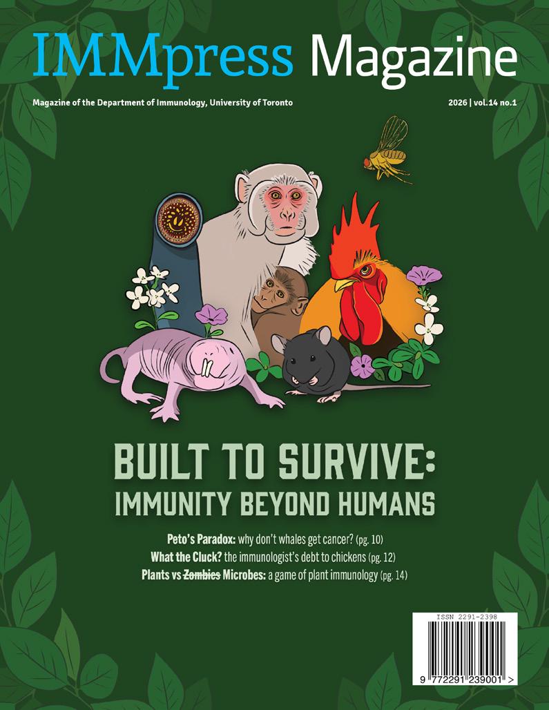
This marks the sixth and final issue cover I will design for IMMpress Magazine. It has been such a rewarding experience to bring together all of the articles over the past two years. Ana Sofia Mendoza Viruega will be stepping in as the newest Design Director, and I’m excited to see the creativity and fresh ideas she will bring to future issues.
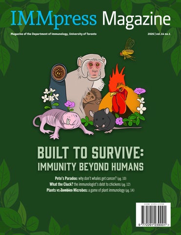
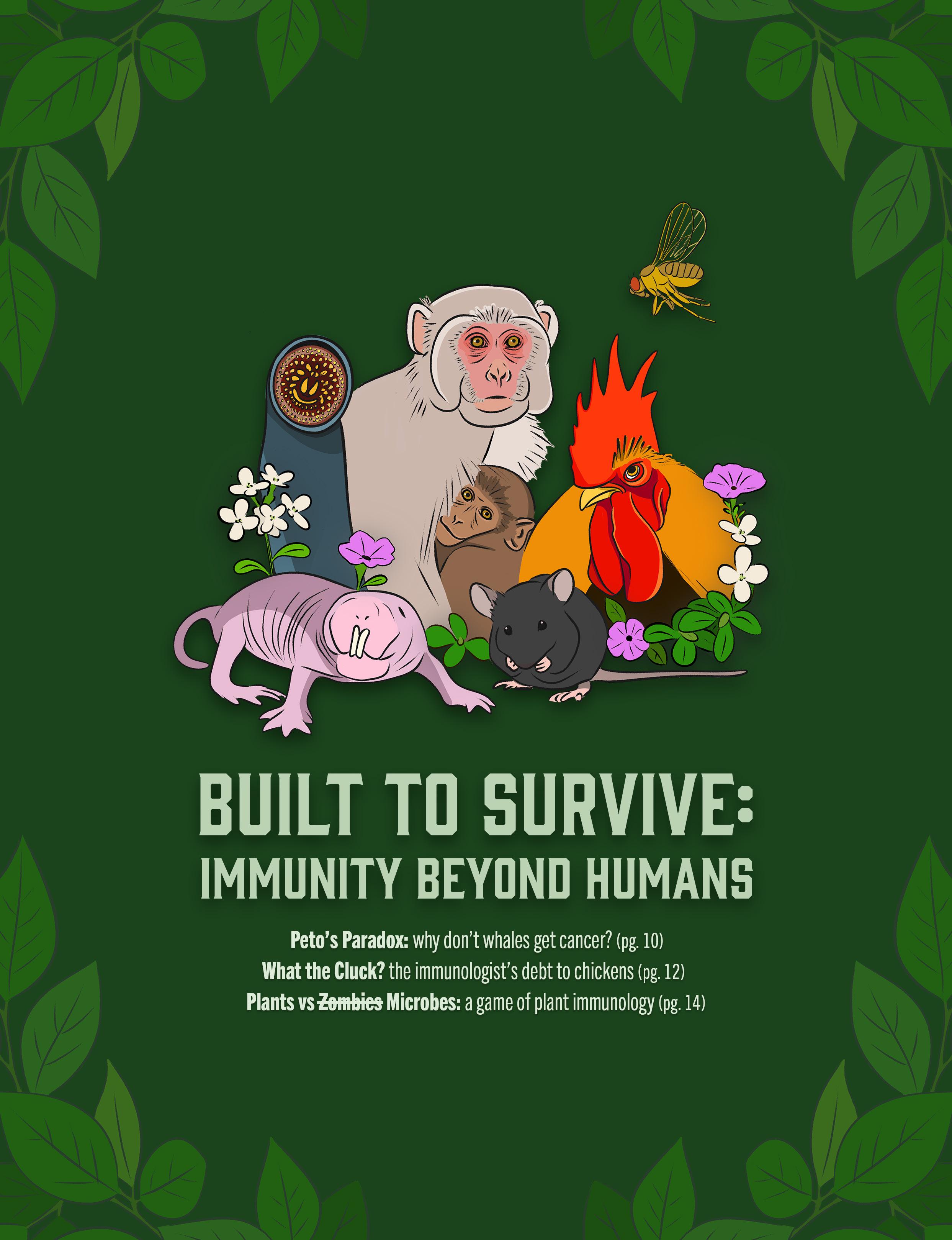
For this issue’s cover, I wanted to illustrate some of the incredible animals and model organisms featured in our students’ articles. The cover portrays the naked mole-rat, lamprey, rhesus macaque, fruit fly, chicken, and of course the familiar C57BL/6 mouse. These animals are surrounded by petunias (featured in the RNAi article) and the white flowers of Arabidopsis thaliana , one of the most widely used plants in genetics and cell biology research. The cover and opening pages of this magazine feature a vibrant green palette to pay homage to the diverse flora and fauna that shape our understanding of biology, immunity, and the interconnected systems of life that make scientific discoveries possible.

Jennifer Ahn Design Director
Copyright © 2013 IMMpress Magazine. All rights reserved. Reproduction without permission is prohibited. IMMpress Magazine is a student-run initiative. Any opinions expressed by the author(s) do not necessarily reflect the opinions, views or policies of the Department of Immunology or the University of Toronto.
EDITORS-IN-CHIEF
Manjula Kamath
Tianning Yu
DESIGN DIRECTORS
Jennifer Ahn
Ana Sofia Mendoza Viruega
SOCIAL MEDIA COORDINATOR
Victoria Sephton
SENIOR EDITORS
Adriana Zutic
Ana Sofia Mendoza Viruega
Annie Pu
Baweleta Isho
Deeva Uthayakumar
Manjula Kamath
Milea DiPonzio
Tianning Yu
Vera Lynn
Zi Yan Chen
DESIGN ASSISTANTS
Ana Sofia Mendoza Viruega
Annie Mitchell
Annie Pu
Baweleta Isho
Christopher Ryan Tan
Mariam Parashos
Milea DiPonzio
Sophie Sun
Tianning Yu
Victoria Sephton
Yoobhin Park
Zi Yan Chen
Zoeen Carter
WRITERS
Alina Mehra
Ana Sofia Mendoza Viruega
Annie Mitchell
Annie Pu
Christopher Ryan Tan
Deeva Uthayakumar
Jonathan Monteiro
Manjula Kamath
Mariam Parashos
Milea DiPonzio
Preya Patel
Tianning Yu
Vera Lynn
Victoria Sephton
Yasmin Anning
FOUNDING EDITORS
Yuriy Baglaenko
Charles Tran
ontributors

Join our mentorship Join our mentorship team team
WE’RE RECRUITING MOTIVATED MENTORS AND MENTEES
THE GRADUATE PEER SUPPORT NE T WORK IS A FACULT Y OF MEDICINE INITI ATIVE TH AT AIMS TO IMPROVE WELLBEING OF GRADUATE STUDENTS.
OUR MENTORSHIP PROGRAM PAIRS TRAINED STUDENTS WITH MENTEES TO PROVIDE SUPPORT WITH CH ALLENGES TH AT STEM BEYOND THE SPHERE OF ACADEMIC AND CAREER GOALS.
SIGN UP USING THE QR CODES BELOW:
MENTOR: MENTEE:





Primates: Better Models for Human Disease? Opportunities and Challenges
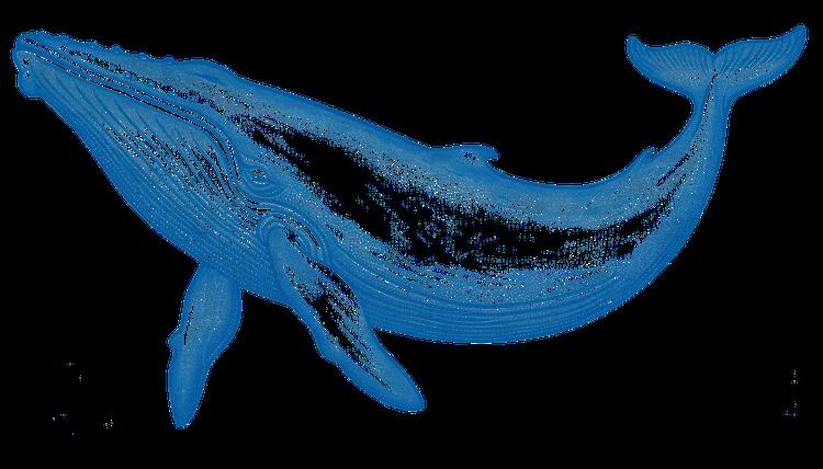
Peto’s Paradox: Why Don’t Whales Get Cancer?
What the Cluck?
The Immunologist’s Debt to Chickens
Plants vs Zombies Microbes: A Game of Plant Immunology
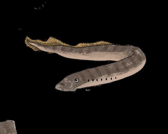
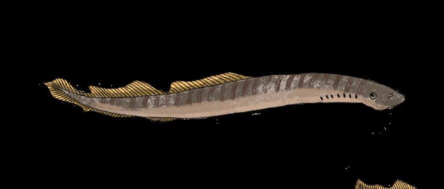
VLRs: The Jawless Fish Strike Back!
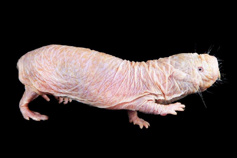
What the Humble Naked Mole-Rat can Teach Us about Immunity
Fur and Fortified Immunity: How Growing Up with Pets may Shape the Immune System
Spray-Induced Gene Silencing:
Mini Organoids vs Mouse Models: What will Shape the Future of Immunology Research? Dr. Götz Ehrhardt
Mapping Human Illness Across Time through the Lens of Zoonosis
Comparative Immunology: Why Investigation Across Species Still Matters
The Enormous Impact of a Tiny Immune System: a Book Review of Walter Isaacson’s “The Code Breaker”


Dr. Justin Tze Ho Chan Swimming through ImmunityWhat fish can teach us about the immune system



FROM THE
CHAIR L etter
And now for something completely different…
Iloved this latest issue of IMMpress brought to you by our talented Department of Immunology graduate students entitled “Built to Survive: Immunity
Beyond Humans”. I think almost any immunology project can benefit from the evolutionary perspective. Asking –“why is this beneficial to the host” can be a catalyst to important immunological insights. And for this reason, it is paramount that we think about other organisms beyond the beloved specific-pathogen-free C57Bl/6 Mus musculus. For me, as someone in their mid-fifties, I am all about the incredible health span of the naked mole-rat. It can live to 30-40 years of age! (although the trade-off appears to be spending its life underground in pathogen-poor environments, a state of existence that has its appeal). Kidding aside, there is much richness in comparative immunology that can be gleaned to better understand human immunology.
I was recently asked by Juan Carlos Zúñiga-Pflücker, the course coordinator of IMM429/1429, to take over the comparative immunology lecture for-

merly taught by Dr. Michael Ratcliffe. From this, and echoed in one of this issue’s articles, I learned about the fundamental role chickens played in our understanding of the immune system. It was from our humble Swiss Chalet friends that Dr. Max Cooper discovered two arms to the adaptive immune system, one originating from the thymus (T cells) and one from the bursa, which is located close to the chicken cloaca. If you don’t know what the cloaca is, …. Well, just look it up. In IMM429 I teach about B cell diversification in the bursa which starts before hatch (a bit ass-backwards if you ask me), and this is later driven by microbiota after hatch. Chicken immunologists were way ahead of the mouse people when it came to understanding the influence of the microbiota on adaptive immunity.
We also learn in this issue the value of human organoids for clinical modeling. A former student from our Department, Dr. Lisa Wagar, is pioneering work in this area out of her lab at UC Irvine. She is a guest editor for a Journal of Immunology review series coming out on human organoids – so look out for that one! While organoids have limitations, they can be a highly useful parallel track for drug discovery. Indeed, there is no singular model sys-
tem that will get us from discovery to drug. Typically, a combination of in silico, in vitro (sometimes organoids) and in vivo (including mice, but also typically requiring toxicology assessment in non-human primates) are required for a complete preclinical package before going to humans.
In the words of Dr. Ehrhardt whose love of lampreys is derived from his time with the venerable Dr. Max Cooper and who is interviewed in this issue, studying other organisms one can “encounter very unusual and exciting science” Goetz praises these creatures as not only an immunological treasure trove, but also a gastronomic delicacy (with a nice chianti on the side).
I hope you enjoy this issue –bon appétit!

Jen Gommerman, PhD
Canada Research Chair in Tissue Specific Immunity
Professor and Chair, Department of Immunology
L etter
FROM THE
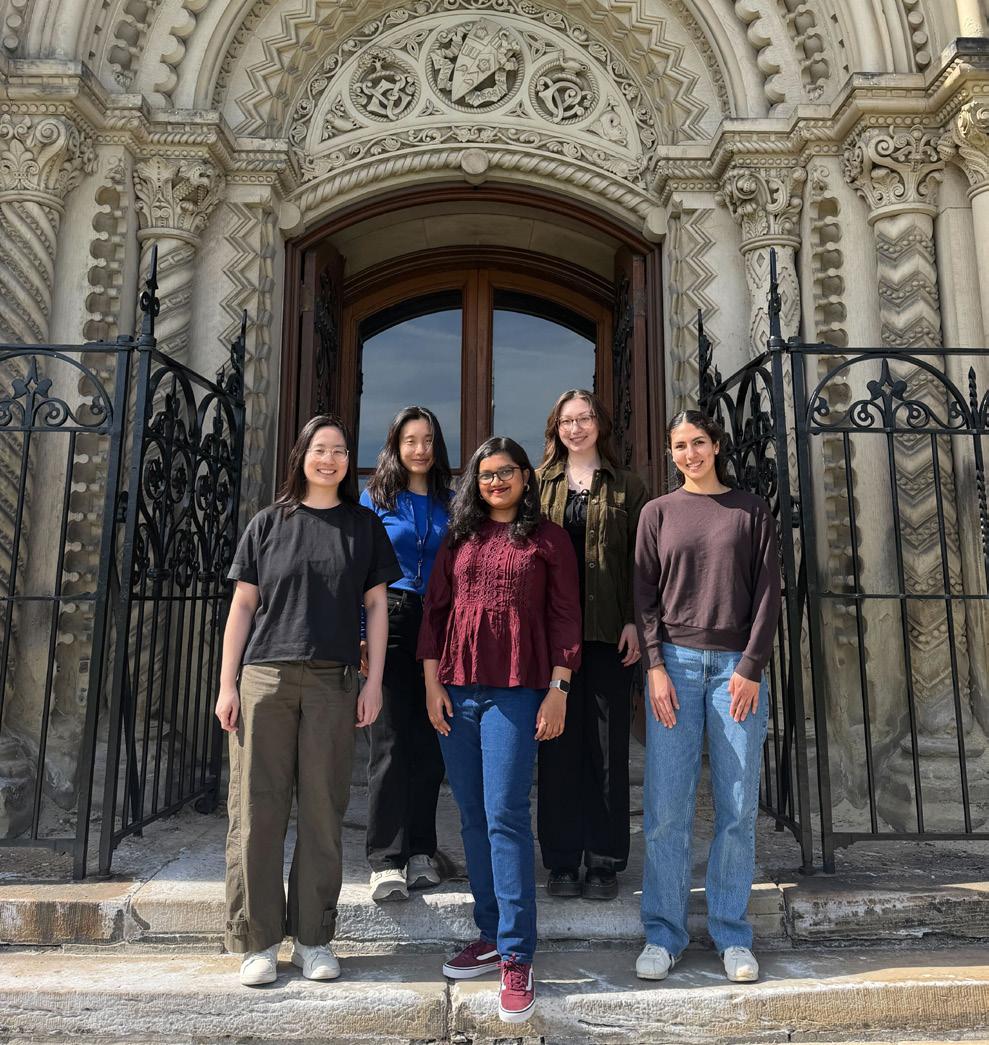
In order of left to right:
Tianning Yu (Editor-in-Chief), Jennifer Ahn (Design Director), Manjula Kamath (Editor-in-Chief), Victoria Sephton (Social Media Coordinator) & Ana Sofia Mendoza Viruega (Incoming Design Director)
As scientists working at the cutting-edge of immunology discovery research , we often turn to the familiar: the wellestablished mouse models, drawing parallels to the human immune system, the canonical pathways and conventional cell types. These form the foundation of modern biomedical research. Yet immunity did not emerge in a single organism, nor does it follow a single blueprint. Across hundreds of millions of years of evolution, life has developed remarkably diverse strategies to survive infection, tolerate environmental stress, and coexist with microbes. In this issue titled “Built to Survive: Immunity Beyond Humans”, we invite you to step beyond the conventional and explore immunity through the lens of comparative biology.
EDITORS
We begin with tracing the evolution of immune systems across the tree of life, from single-celled organisms to modern vertebrates (pg.8). We follow up with some of the most fascinating questions in evolutionary immunology: Why do whales, despite their enormous size and long lifespans, appear remarkably resistant to cancer? (pg.10)
How did chickens help uncover the fundamental organization of adaptive immunity? (pg.12) And how have plants evolved sophisticated immune systems without specialized immune cells at all? (pg.14) Through articles spanning organisms from plants to primates (pg.16), this issue highlights how studying diverse organisms continues to challenge assumptions and expand our understanding of immune biology.
Our interviews with Dr. Götz Ehrhardt (pg.20) and alumnus Dr. Justin Tze Ho Chan (pg.22) explore the unusual and elegant immune systems of jawless fish, particularly lampreys and their variable lymphocyte receptors (VLRs) ( pg. 28). This issue also examines the changing landscape of biomedical research. Comparative immunology is not only about evolutionary curiosity, but it also continues to shape modern medicine. Articles on non-human primates (pg.16), organoid systems (pg.18), and zoonotic disease (pg. 30) explore the opportunities and ethical challenges of studying immunity across species. Elsewhere, readers will encounter the hidden immunological influence
of growing up with pets (pg.24), the molecular arms race between fungi and RNA interference (pg.26), and all about the fascinating immune systems of the naked mole rats (pg.29).
This issue also reflects on how discoveries in comparative biology have transformed science itself. Our review of The Code Breaker revisits the story of CRISPR-Cas9, a revolutionary gene editing technology rooted in bacterial immune defense systems (pg.34). It is perhaps one of the best modern examples of how studying seemingly obscure biological systems can profoundly reshape medicine, biotechnology, and society.
Ultimately, the stories in this issue share a common message: there is no single “correct” immune system to study. Evolution has produced countless ways to survive. By studying these diverse systems, we not only deepen our understanding of immunity, but also uncover ideas that may inspire the next generation of therapies and technologies.
We hope this issue encourages you to look beyond the familiar and appreciate the remarkable diversity of immune systems across life.
We would like to thank all the wonderful writers, editors, and designers that made this issue possible! Last but not least, we would like to extend our deepest gratitude and warm farewell to our design director, Jennifer Ahn, as she steps out of her role onto bigger and better things. Thanks for helping design beautiful issues of IMMpress over the years, we will miss you!
Editors-in-Chief


Tianning Yu
Almost human: Tracing the evolution of the immune system
1. Single Celled Organisms
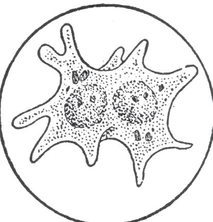
While single-celled organisms may not possess what we traditionally consider an immune system (i.e., one made up of specialized immune cells), they do possess a whole host of innate defense mechanisms to protect themselves from pathogens. One example in which they do this is via phagocytosis, a process in which a cell engulfs and degrades an extracellular pathogen. Both specialized mammalian immune cells and many single-celled organisms possess this ability. In fact, an organelle called the mitochondria, nicknamed the “powerhouse of the cell”, was theorized to have originated from the phagocytosis of an ancient bacteria by a larger single-celled organism!
2. Invertebrates
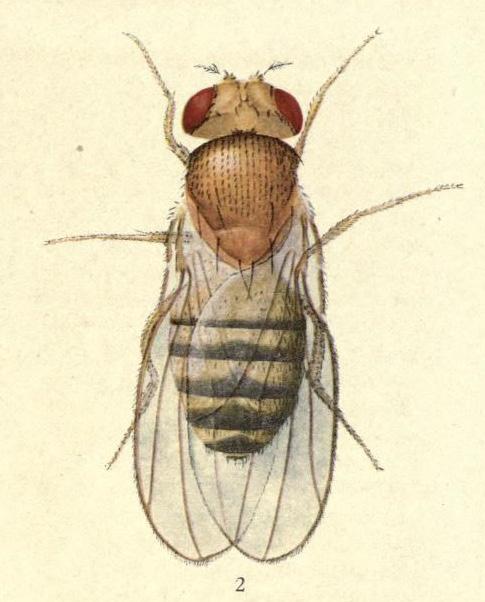
It turns out a backbone isn’t the only thing invertebrates lack – they also lack an adaptive immune system! Thankfully, they make up for it with a diverse range of innate immune mechanisms to defend themselves from pathogens.
As mentioned previously, where an adaptive immune system recognizes specific pathogens, an innate immune system is a generalized, non-specific immune response. As such, rather than detecting specific pathogen characteristics, it relies on sensing danger signals that indicate the presence of an infection, namely pathogen-associated molecular patterns (PAMPs) and danger-associated molecular patterns (DAMPs). These signals are recognized by pattern recognition receptors (PRRs), and emerged alongside the evolution of early invertebrates. For example, PRRs have been detected in different species of sponges, some of the most primitive invertebrates (and by extension, animals). Later invertebrates have evolved their innate immune systems to possess more complex PRR systems and specialized immune cells. For example, fruit flies (Drosophila melanogaster) have three types of immune cells, with one population, plasmatocytes, possessing the same phagocytic capabilities as both amoebae and some human immune cells. Interestingly, while innate immunity is not generally considered to have memory, recent research has shown that many invertebrate immune systems can possess some aspects of immune memory.
3. Jawless Vertebrates
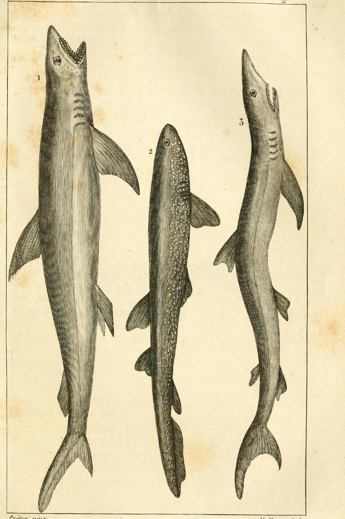
Jawless vertebrates comprise the next evolutionary step from inver tebrates, and it is here that we begin to see the earliest origins of adaptive immunity. Modern examples of jawless ver tebrates, such as the hagfish and lamprey, possess hallmarks of adaptive immunity, such as the production of antigen-binding molecules similar to human antibodies. What puzzled scientists was that the genes that define human adaptive immunity, as well as the organs (thymus, spleen) that allow for the production of these molecules, were absent in these organisms.
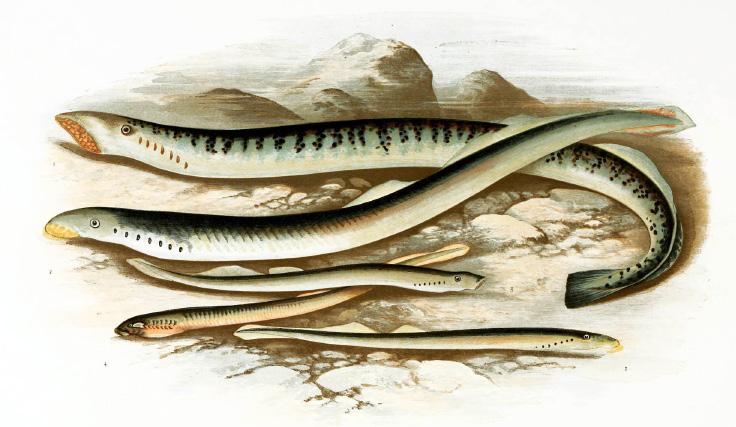
This all changed in 2004, when scientists discovered the presence of a family of variable lymphocyte receptor (VLR) genes. VLR genes contain the information needed for the production of VLR proteins, which recognize specific antigens. What allows these proteins to be specific to pathogens is that they are susceptible to becoming randomly modified by the insertion of repetitive sequences. The end result is the generation of trillions of different VLR proteins, all of which could potentially recognize different antigens. While distinct from antibodies, this process is likely the earliest origins of a mammalian adaptive immune system.

4. Jawed Vertebrates
Jawed vertebrates are where the adaptive immune system, as it is defined in humans, finally emerges. Rather than the modification of VLR genes resulting in trillions of VLR proteins, jawed vertebrates rely on a system called variable-diversity-joining (VDJ) rearrangement to create trillions of T cell receptors (TCRs) and B cell receptors (BCRs), the evolutionary equivalent of VLR proteins. One event that made this possible was the RAG transposon event.




The RAG transposon event refers to the invasion of a segment of DNA into our genome that allows for the production of the proteins RAG1 and RAG2 (i.e., the RAG1/2 gene). When and how this gene invaded our genome (the collection of all the genes in our body) is unknown. It was initially thought to have entered our genome in a vertebrate ancestor, possibly through a virus, but recent evidence has identified RAG genes in sea urchins, meaning it could have invaded much earlier. As to why it only became active in jawed vertebrates, we still do not know. Either way, this underscores how essential RAG1 and RAG2 proteins are to our adaptive immune system. They are so essential that deleting them from mice results in the deletion of the entire adaptive immune system!

From here on, the jawed vertebrate immune system was further sculpted by evolutionary pressures to where we are now, characterized by the development of different antibody subtypes and changes in the structure of the organs where the adaptive immune system predominantly resides.


5. What’s next for the human immune system?
While one might expect the human immune system to have stopped evolving, human populations continue to adapt to new pressures through genetic variation, environmental exposures, and social factors that shape immune function.
For example, it has been well-established that two groups of long-extinct distant human ancestors, the Neanderthals and Denisovans, often interbred with modern humans (i.e., Homo sapiens), and modern technology has enabled us to understand how this has shaped our modern immune system. Crucially, Neanderthals and Denisovans differed subtly from modern humans in the ways their immune systems communicated and recognized pathogens. Examples include Neanderthal- and Denisovan-specific variants in genes coding for cytokines and transcription factors, molecular messengers and regulators of the immune system, and even in some PRRs. When considering that different ethnic groups possess varying amounts of Neanderthal and Denisovan DNA, it becomes all the more important that research into the immune system is inclusive of people of all different ethnic backgrounds.
However, genetics are not the only driver of human immune evolution. Throughout history, as major pandemics have swept the world, the immune system of subsequent generations have become defined by the genomes of the survivors. For example, the frequency of the gene ERAP2 increased in human populations after the Black Death because it conferred protection, even if it is now linked to increased risk for autoimmune disease. Only time will tell whether modern pandemics, such as the recent SARS-CoV-2 pandemic, will have the same effect.
So, the next time you get a chance to look at a lymphocyte under the microscope, just remember that you are facing a miraculous product of cell engineering, shaped by millennia of evolutionary pressures.
Written & Designed by
Christopher Ryan Tan
PETO’S PARADOX: WHY DON’T
WHALES GET CANCER?
Cancer is often described as a uniquely human tragedy. But it isn’t. Cancer affects nearly every class of vertebrate and is especially common in mammals. It is, in many ways, a universal biological problem; a consequence of what it means to be multicellular.
Every cell in your body is part of an extraordinary cooperative society. Cells build tissues, generate energy, divide when needed, repair damage, and communicate constantly to maintain homeostasis. These biochemical networks are so intricately connected and precisely regulated that it almost seems miraculous they function flawlessly at all. Until they don’t.
Across one’s lifespan, billions upon billions of cellular reactions take place. DNA is copied, proteins are built, signals are transmitted. With so much happening, the real question is not if something will go wrong, but when. Genetic mutations accumulate. Metabolic errors occur. Environmental exposures leave their mark. Over time, these small changes can corrupt the machinery that keeps cells behaving properly.
When a cell begins to malfunction, the body has safeguards. Cells can trigger a “self-destruct” program known as apoptosis, sacrificing themselves for the greater good. If that fails, the immune system acts as a vigilant patrol, identifying and eliminating abnormal cells before they become dangerous. But cancer cells are, in a sense, evolutionary escape artists. If they acquire the right combination of mutations, they can evade these surveillance systems. They multiply, accumulate further mutations, hijack nutrients, and outcompete their neighbouring healthy cells. What began as a single rogue cell becomes a tumor.
Species Comparison
Lifespan, Cell Number, and Cancer Incidence
Mutations build up with age, which is why cancer risk increases over one’s lifetime. Additionally, the more cells an organism has, the greater the opportunity for one of them to go rogue. Larger bodies mean more cells and longer lifespans mean more time for mutations to accumulate. By that logic, the largest, longest-living animals on Earth should be riddled with cancer, but they aren’t. This contradiction is known as Peto’s Paradox, formulated by epidemiologist Richard Peto in 1977. The paradox asks a simple yet profound question: if cancer arises from mutations in cells, and larger animals have far more cells dividing over longer lifespans, why don’t they experience astronomically higher rates of cancer?
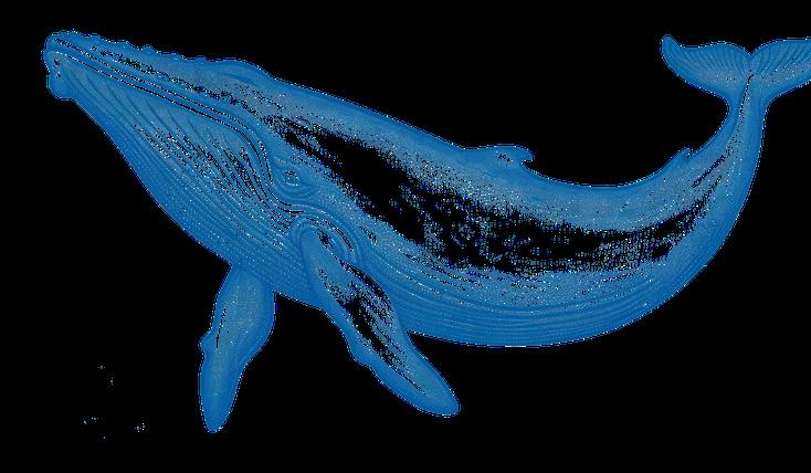
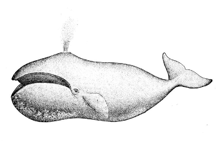
Consider a mouse. A mouse has far fewer cells than a human and lives only two to three years. By all reasoning, its cancer risk should be dramatically lower than ours. Yet mice develop cancer at rates comparable to humans. Now consider whales. Their bodies contain 1000 times more cells than humans and can live for over a century. If cancer risk scaled with cell number and lifespan, whales should be overwhelmed by malignancy, yet they are not. In fact, whales appear to have a much lower cancer risk compared to humans. How is this possible? Did whales simply get lucky in the evolutionary lottery? Or have they developed biological defenses that far surpass our own? Researchers have proposed several intriguing theories.

Mouse
Lifespan: 2-3 years
Cells: ~10 billion
Cancer Incidence: 30-50%

THEORY 1: REDUNDANCY OF TUMOR SUPPRESSOR GENES
One leading explanation is surprisingly straightforward: whales may simply be better equipped at preventing cancer in the first place.
Tumor suppressor genes act as their name suggests – they prevent tumors from developing by controlling cell growth, repairing damaged DNA, and triggering apoptosis when cellular damage becomes too severe. In humans, when these genes fail or mutate, cancer risk rises dramatically. In large, long-lived animals, relying on a single protective pathway would be risky. One proposed solution is genetic redundancy: the idea that whales may possess additional or functionally overlapping tumor-suppressing mechanisms that act as biological “backup systems.” If one pathway fails, another can compensate. For example, researchers have identified 71 duplicated tumor suppressor genes in cetaceans (whales, dolphins, porpoises), which are involved in anti-cancer processes such as DNA repair, metabolism, apoptosis, aging and cellular senescence. Thus, genetic mutations in one gene would not collapse the entire tumor suppression mechanism. Should one gene fail to control a cancerous cell, another redundant gene can step in to suppress it. In other words, whales may not avoid cancer by chance, they have evolved more robust systems for maintaining genomic integrity from the start.
THEORY 2: HYPERTUMORS
Cancer cells are inherently competitive. Unlike normal cells, which cooperate to maintain tissue function, cancer cells prioritize their own proliferation. One hallmark of tumor growth is angiogenesis, the recruitment of blood vessels to supply oxygen and nutrients. This vascular network supports continued tumor expansion.
Some researchers have proposed the concept of hypertumors, tumors that arise within existing tumors. This hypothesis proposes that, as cancer cells continue to accumulate mutations, an individual cell within a growing tumor acquires changes that cause it to diverge from the main tumor population. Rather than remaining part of a coordinated mass, this mutated cell establishes its own proliferative focus and begins competing for the same blood supply. By redirecting nutrients and oxygen toward itself, it deprives neighbouring tumor cells of resources, ultimately starving and killing the original cells. In essence, cancer is killing cancer.
In large organisms such as whales, tumors would require a prolonged period to reach a size capable of compromising the host. This extended timescale may allow such internal competition to emerge, effectively constraining tumor expansion before it becomes lethal. In contrast, in smaller organisms, tumors can reach life-threatening size more quickly, leaving insufficient time for this type of internal resource competition to significantly alter the outcome.
While these hypotheses offer plausible mechanistic explanations to Peto’s paradox, they are not without limitations. Large-bodied animals have evolved independently over the course of history, meaning different lineages may have developed distinct, rather than universal, anti-tumor mechanisms. A strategy observed in whales may not necessarily apply to other large-bodied animals. In addition, many models exploring the paradox assume that the rate-limiting step in cancer development is the accumulation of oncogenic point mutations. However, tumor initiation and progression are also influenced by factors such as environmental exposures, diet, infections, and aging. As it stands, no single explanation fully resolves the mystery.
Peto’s paradox is a fascinating phenomenon that piques scientific interest, but deciphering how large animals maintain cancer resistance has implications that reach far beyond evolutionary curiosity. By identifying the molecular and cellular strategies that protect these species, researchers may uncover mechanisms that could ultimately reshape how we approach improving human health and longevity – should we succeed in solving it.
Written & Designed by
Mariam
Parashos

Human
Lifespan: 70-100 years
Cells: ~30,000 billion
Cancer Incidence: 20-40%
Whale
Lifespan: 100+ years
Cells: ~100,000,000 billion
Cancer Incidence: 1.4%
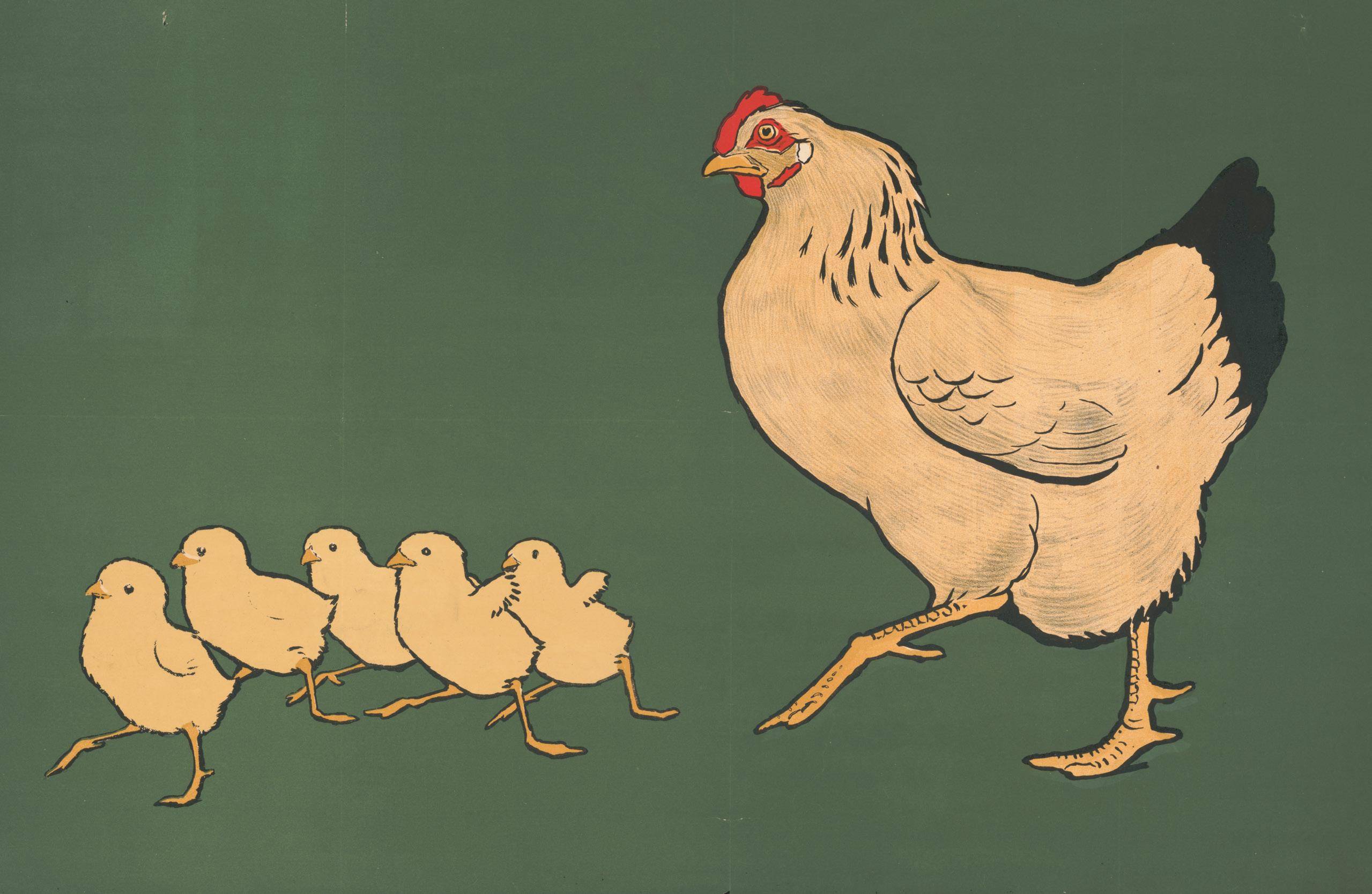
The Immunologist’s Debt to Chickens Whatthe Cluck?
Animal models are the cornerstone of basic research, providing simple, easy-to-study and ethical biological systems that help us better understand how the human body works. Immunologists tend to rely heavily on mice and rats for their research, owing to their similarity with human physiology, small body size, accelerated life spans, and ease of genetic modification. But it would be unwise of us to put all our eggs in one basket.
Though it may seem counterintuitive at first, many of our greatest scientific discoveries have arisen from studying animals with key physiological differences from humans. Though, broadly speaking, the way chickens and humans fight off pathogens are very similar, the variation between our immune organs and molecules provides easier ways to study the mechanistic underpinnings of the immune response. The chicken is the unlikely hero of immunological research – and not just through its contribution as tasty snacks to help fuel researchers through long days of experiments. It is precisely because of our differences that chickens have given rise to some of immunology’s most impactful breakthroughs, from fundamental adaptive immunity to vaccine development.
From Pasture to Pasteur: The Chicken’s Contribution to Vaccinology
Louis Pasteur is widely considered the father of immunology for his great contributions to our understanding of microbiology, microbial disease, and vaccination as both a preventative and prophylactic means of protection. Prompted by the rampant devastation of livestock by different infectious diseases, Pasteur started studying chicken cholera and its causative bacteria, Pasteurella multocida , in 1877. He isolated and began to grow the Pasteurella with the intention of injecting bacterial cultures into chickens, but accidentally left the samples out unattended on his laboratory bench for a month while on holiday. When he returned, he found that the bacteria had lost their virulence over time: chickens injected with these “attenuated” bacteria only developed mild symptoms, made full recoveries, and were also protected against
new infections when subsequently injected with fresh, more virulent cultures.
We now know that this live, attenuated vaccine conferred protection by allowing the immune system to mount a response against the pathogen without the risk of severe disease. After this “practice round”, the now-trained immune system can respond quickly and robustly when it encounters the same pathogen again. Pasteur and his colleagues applied this finding to other animal diseases, such as anthrax in cows or rabies in dogs, sheep, and eventually humans. These studies concluded that dead bacteria, which they inactivated with heat, could confer protection against future infection too. While newer versions of vaccines exist today, often using subunits of pathogens instead of whole organisms to train the
immune system, vaccines for measles, mumps, and rubella, chickenpox, and influenza still use the same model of live-attenuated or inactivated pathogens that Pasteur discovered.
The impact of chickens on vaccinology doesn’t end there. Every year, in preparation for flu season, scientists use chicken eggs to grow candidate influenza viruses, which they then isolate for preparation into both live-attenuated and inacti-
vated versions of a vaccine. The next time you get your flu shot – or any vaccine, for that matter – remember that you have chickens to thank.
What Came First: The Chicken or the Ig?
All multicellular organisms rely on the innate immune response as a rapid but non-specific first line of defense against pathogens, toxins, and other threats, but what happens when invaders slip through the cracks? In many vertebrates, this is when the adaptive immune response takes over. Adaptive immunity offers a more sophisticated, targeted offense against specific pathogens that not only clears current infections but offers long lasting immunity against future infections too.
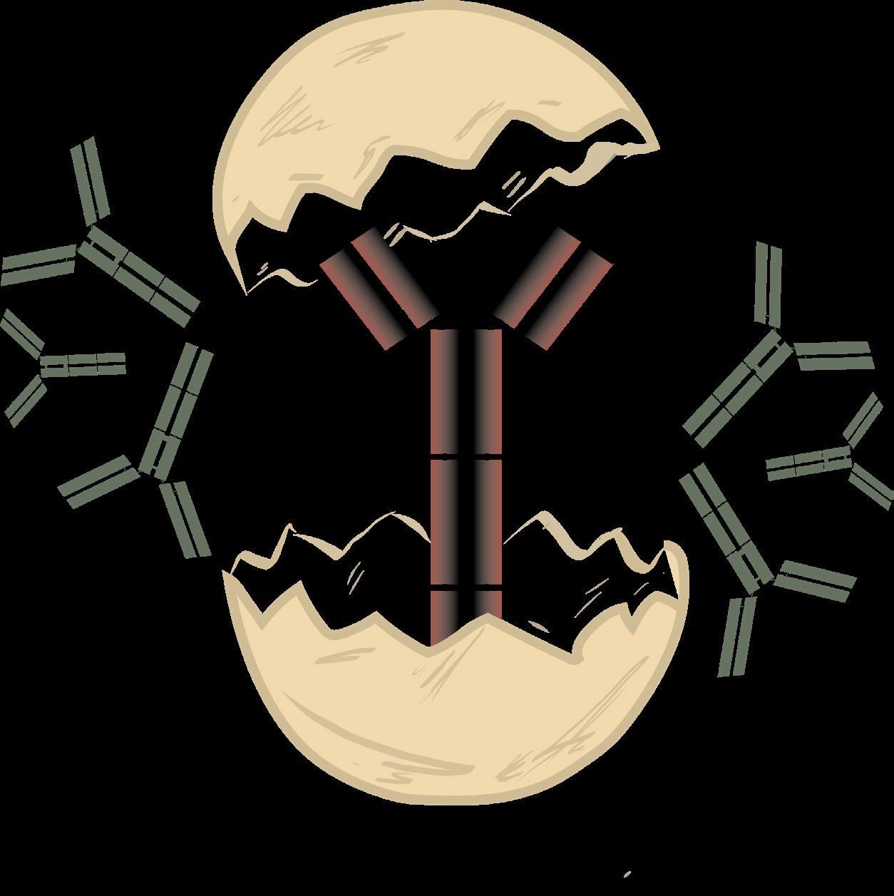
By 1948, there was some evi dence to suggest that antibod ies – otherwise known as im munoglobulins, which are proteins that specifically bind to and neutralize foreign antigens – were produced by plasma cells, but nobody knew where or how this occurred. It wasn’t until 1956 that Bruce Glick and Timothy Chang accidentally discovered that the bursa of Fabricius of chickens, a small and poorly characterized organ close to the cloaca, was critical for antibody pro duction. While routinely injecting chickens with Salmonella bacteria to generate antibodies for a separate experi ment, Glick and Chang saw that almost all bur sectomized chickens died soon after they were exposed, and those that survived produced almost no antibodies against Salmonella at all. Chickens with intact bursas maintained high levels of Salmonella-specific antibodies and all remained healthy.
This observation laid the groundwork for Max Cooper’s research in the 1960s. After Jacques Miller found that removing the thymus from newborn mice severely reduced lymphocyte production and resulted in extreme immune deficiencies, most of Cooper’s contemporaries thought lymphocytes were generated in the thymus and then seeded the rest of the body,
where they could then become antibody-generating plasma cells. Cooper disagreed: he thought thymus-derived lymphocytes and plasma cells might come from different sources instead of following one lineage path. So, he turned to the chicken (and its bursa) for answers. His experiments found that chickens without a thymus had very low lymphocyte counts, but could still produce normal levels of antibodies; however, chickens without a bursa produced no antibodies, but still had plenty of other lymphocytes. This was the first work that suggested there were two different arms of the adaptive immune system: one based in the bursa and one based in the thymus. Cooper’s further research found this was true in mammals as well, except mammalian antibody-producing cells are generated in the bone marrow instead of a bursa. We now refer to these two cell types as T cells, named after the thymus, and B cells, named not after the bone marrow, but after the bursa of Fabricius.
The main antibody isotype chickens produce is immunoglobulin Y (IgY), which is functionally similar to, but structurally distinct from, any of the antibodies that humans produce. As a result, IgY could be used to target mammalian structures without activating unwanted components of the mammalian immune system, which is a common struggle with many immune-based therapeutics today. Since IgY is easy to harvest from chicken eggs, it makes an excellent candidate for antibody-based diagnostic or therapeutic tools. Indeed, scientists are already studying IgY in the context of preventative and prophylactic antimicrobial treatment, cancer immunotherapy, and allergy therapies with promising results.
When it comes to immunology’s favourite model organisms, chickens still may not rule the roost. Nevertheless, immunologists remain deeply indebted to chickens for their contributions to the field, both as a model system to understand the mechanisms underlying the immune response and as tools for disease diagnosis, prevention, and treatment.
Written by Victoria Sephton
Designed by Ana Sofia Mendoza Viruega




EPLants PLants
















microbes
a game of Plant immuno Logy
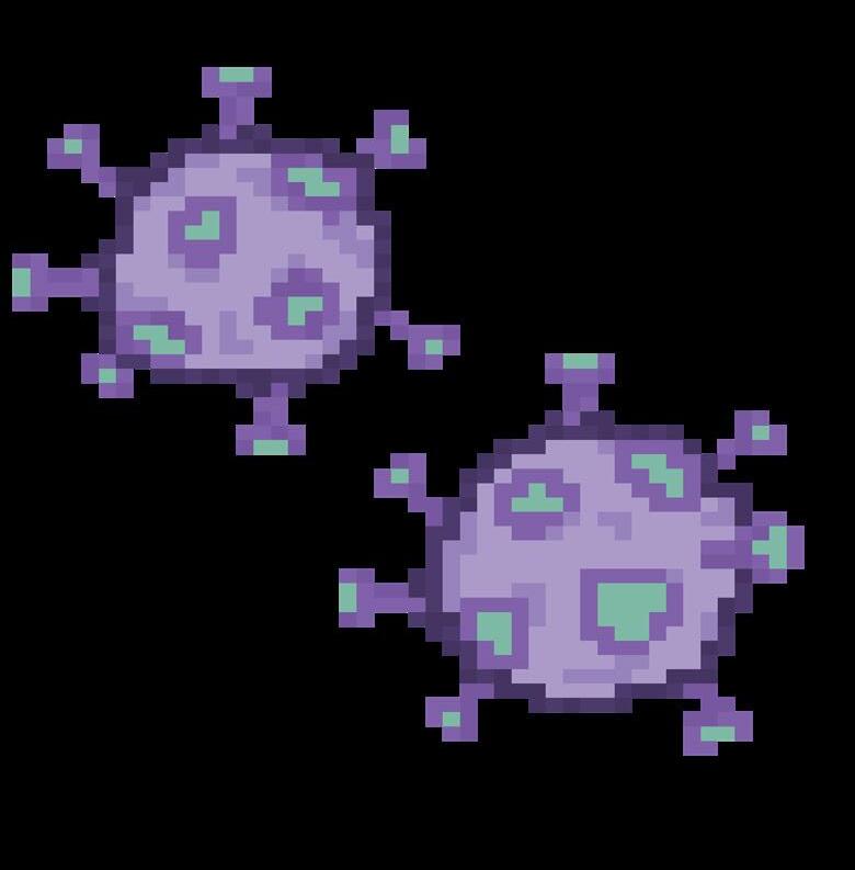
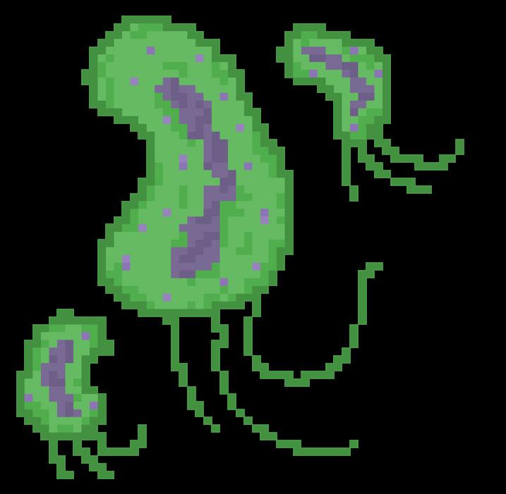
very year, approximately 40% of global food crops valued at over $200 billion USD are lost to plant disease. Staples like wheat, rice, potato, soybean are constantly fighting against pathogens, posing a strain to food security. Ever since the first plants colonized land, an endless evolutionary race against microbes started. In this immunity game, plants are not alone, as they have developed amazing strategies to defend against infectious fungi, bacteria, viruses, insects, and worms. Not only are the mechanisms underlying plant immunology fascinating, but understanding this immunology game rules provides valuable insights to build a more sustainable future.
Before the game begins, it is important to clarify the board. Plant immunity does not consist of a series of isolated events. Rather, it is a complex interconnected network with multiple players. Some moves will take place at the point of initial contact, local immunity, while others will travel to distant tissues, distal immunity. Both are also in constant communication with external stimuli to ensure the proper character and move are selected. As a plant levels up, previous encounters prime the system, ensuring strong and durable fights.
LeveL 1: PhysicaL and ChemicaL Barriers
While it often goes unnoticed, the first level of immunity is the battle for entry, where physical and chemical barriers prevail. Despite the diversity of plant species, all plants must sense and adapt to the dynamic environments surrounding them. Think of raindrops sliding off a leaf during a storm. Leaves have a waxy cuticle that creates a water repellent surface. Among other roles, this low humidity layer makes it difficult for bacteria and fungi penetration. Beyond structural armor, plants use chemical warfare against the disguised villains: insects. Insects not only feed on crops but often act as carriers of germs. In response, aromatic plants release scented organic compounds, like oils, that either deter dangerous pests or work directly as toxins.


LeveL 2: loCal immunity
If barriers fail or are hijacked by microbes exploiting natural openings or wounds, the second game’s round, local immunity, begins. The main goal is to detect the threat and contain the pathogen at the site of infection. Unlike animals, which rely on specialized immune cells and organs, every plant cell, no matter its location or nature, is immuno-potent. Thus, any cell may detect a threat and trigger a proper defense strategy against the intruding pathogen. Local immunity relies on two core mechanisms: pattern-triggered immunity (PTI) and effector-triggered immunity (ETI).
PTI is initiated by receptors residing on the surface of cells. These receptors sense components of microbes (pathogen-associated molecular patterns; PAMPs) or host modified molecules that are released during pathogen attack or stress (damage-associated molecular patterns; DAMPs). For example, flagellin sensing 2 receptor recognizes a conserved region of bacterial flagellin, a protein that forms the bacterial tail used for motility. Unlike PTI, ETI associated receptors are located intracellularly, meaning they are found inside the cell, and identify pathogen effector molecules directly or indirectly. In the latter case, intracellular receptors are guards surveilling the cell for any perturbations. An alarm is raised when guardees, which consist of the plant’s own derived molecules are modified as a consequence of pathogen effector activity.
Upon sensing a modified guardee, a series of alarms are amplified, and the appropriate response is activated. The guard model allows plants to specifically respond to a variety of infectious agents with a defined set of receptors, which is highly valuable in a world of rapidly evolving pathogens. Importantly, PTI and ET function cooperatively. While the initial PTI response provides rapid protection and primes downstream defenses, activation of ETI ensures that a


stronger and longer-lasting response is maintained to stop pathogens that potentially breached previous defense lines. In some instances, this leads to localized cell death as a way of sacrifice.
Local immunity networks are regulated at multiple instances to prevent overactivation of the immune system, which is an energetically expensive process. Additional “power-ups” include decoy proteins, in which plants produce molecules that mimic pathogen targets. Occasionally, the game may take an unexpected turn when plants become “paranoid” and activate defense mechanisms even in the absence of a threat. Such “nonplayer characters” highlight the relevance of regulatory mechanisms overseeing immune activation and provide an interesting parallel to animal autoimmune conditions, in which the immune system mistakenly attacks healthy tissues.
LeveL 3: distaL immunity
Simultaneously, the plant activates distant signaling events with the end goal of protecting healthy tissues by reducing pathogen proliferation and disease. This two-player stage is known as systemic acquired resistance (SAR), in which non-infected cells are prepared to enter the battle. Communication is mediated through electrical waves, in which signals can travel at high speeds for long distances, and messengers including peptides, proteins, RNA, and hormones. Each chemical messenger carries a specific note for the type of shields to deploy for defense. Salicylic acid (SA) is a critical hormone in SAR. Upon infection, SA levels increase at the site of attack and travel to uninfected tissues. The accumulation of SA in distal sites reprograms the cells to enter a primed state ensuring fast and robust responses.
Written by Ana Sofia Mendoza Viruega





LeveL 4:
keeping
your
‘enemies’ cLose
Not all microorganisms are opponents. Many beneficial bacteria reside on the plant in exchange for nutrients without causing disease. These allies facilitate immune development directly by training antimicrobial pathways and regulating plant morphogenesis, or indirectly by inhibiting proliferation of pathogens through microbe-microbe interactions. Interestingly, plants grown in sterile, controlled environments that lack exposure to microbes show abnormal growth and defective immune responses compared to those grown with natural microbiomes. Elucidating the optimal microbial alliances involved in proficient immunity could provide a novel alternative to plant disease control.
endgame: why should we care?
For more than ten thousand years, plant epidemics and diseases have caused economic losses, political instability, and famine. Currently, the abrupt environmental dynamics imposed by climate change further compromise plant immunity. An understanding of plants’ natural immune defenses can aid the development of pesticide alternatives and sustainable agriculture. Theoretically, “super-resistant” plants could be achieved through gene editing technologies, where the superpowers discussed above could be chosen and tailored for a set of common threats. However, this “final boss” comes with ethical and ecological questions. Could such modifications have unintended effects on the ecosystem and harm other organisms? Will pathogens overcome the evolutionary pressure leading to stronger strains? Will private sectors take control of seeds and agriculture? As we move forward, we must balance biotechnology advances with clear regulatory frameworks to ensure global food security for all.






Designed by Victoria Sephton
Primates: Better Models for Human Disease? Opportunities and Challenges
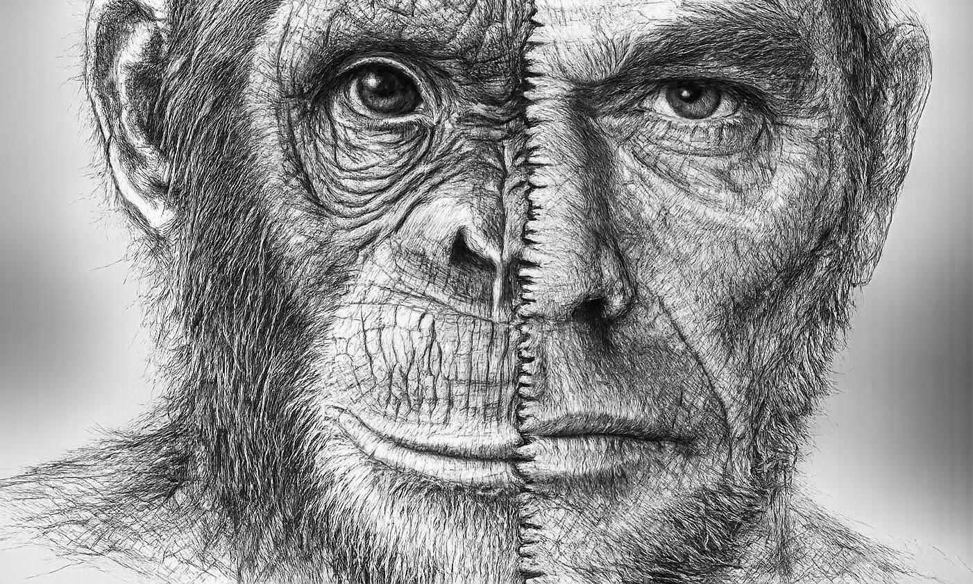
Non-human primates (NHPs) occupy a unique niche in biomedical research. The earliest documented scientific use of primates takes us back to the United Kingdom in 1698, when the Royal Society commissioned the first dissection of a juvenile chimpanzee for studies in comparative anatomy. Research on NHPs over the past 328 years has since led to medical advances in many fields, including vaccination, neurobiology, neonatology, and drug development.
NHPs can be broadly divided into Old World monkeys, including macaques, and New World monkeys such as marmosets, the latter of which are native to the Americas. While New World monkeys are sometimes useful to model the biology of specific diseases, Old World monkeys have been the most used species in biomedical research. As the theory of evolution became more widely accepted in the late 1800s, scientists became motivated to study biology in species perceived as ‘distant cousins’ of humans. Their motivations were well-founded. In 1908, the first experiments inoculating macaques with tissue from a child who had succumbed to a paralytic death led to the discovery of the poliovirus. This established the virology of polio and set the stage for the eventual eradication of the virus through vaccination.
The value held by Old World monkeys in biomedical research comes from our shared evolutionary origin. NHPs are a rare pre-clinical species that exhibit cross-reactivity to human therapeutic antibodies. This is mainly due to structural similarity between therapeutic targets. This similarity becomes particularly useful in research when designing therapies

against pathogens that interact with human proteins in unique ways. As such, establishing NHP models of infection led to the successful discovery of a certain yellow fever vaccine, translated to human use in the late 1930s. This discovery later earned Max Theiler the 1951 Nobel Prize in Physiology or Medicine for his work developing a vaccine against yellow fever, the first Nobel Prize awarded for the development of a vaccine against a viral disease.
NHPs have since become critical for their use in understanding virus pathogenesis in cases where human trials are unethical. Old World macaques are the standard model for understanding pathogenesis of the simian immunodeficiency virus (SIV) and the Ebola virus, and for testing vaccines that protect against the insidious diseases they cause. This has proven useful in scenarios involving global biomedical crises - success of the Moderna and Pfizer/BioNTech COVID-19 vaccines in rhesus macaques enabled rapid clinical translation during the 2019–2022 pandemic. So close are our primate cousins to us on the evolutionary tree that we even share similar blood. The study of rhesus macaque blood takes the credit for the discovery of the Rh+ blood group system, our understanding of which enables lifesaving blood transfusions.
Evolutionary proximity to humans also allows for the use of NHPs to understand neurobiology. Like us, primates have a prolonged neurodevelopmental period, which gives rise to complex social behaviour, fine motor control, and similarly organized neuronal circuits. In 1976, a 23-year-old chemistry graduate student in the United States synthesized and ingest-
ed an illicit drug that triggered the onset of a Parkinson’s-like disease within days. Researchers later discovered that the compound, ‘methylphenyl-tetrahydropyridine’, could trigger Parkinsonism in macaques. The later use of this model to test experimental deep brain stimulation therapies led to the approval and adoption of the latter as a standard of care. These therapies have also replaced neurosurgery that involved destroying large regions of brain tissue with a more conservative approach, improving patient quality of life.
The intricacy of the primate brain spurred its popularity as a model in 20 th century cognitive neuroscience. Famous experiments on maternal-child attachment by Harry Harlow helped establish the concept that maternal bonding is driven by comfort and sensation rather than only through feeding. Behavioural studies on primates initially aimed to understand complex human traits. These provocative experiments notoriously captured public attention when they claimed that apes

make a strong case to justify the experimental usage of NHPs over other small animal models.
Of particular interest today is the use of NHPs for the study of neuroimmunological diseases, since most of these diseases have no viable treatment options and represent a growing threat to the aging population. Rodents provide limited insight into the true triggers of these diseases, which are thought to be accrued with lifetime exposure to inflammatory stimuli. The common marmoset has become especially attractive in this area, as it naturally accumulates toxic proteins with age that also appear in humans. Similarities in the pathogenesis of brain diseases have thus led to the establishment of a chronic experimental model in marmosets that closely resembles human multiple sclerosis compared to existing models in rodents. The smaller size of these monkeys makes them easier to house and more cost-effective than their Old-World counterparts. Uniquely, marmosets are more frequently born as dizygotic twins with natural hematopoietic chimerism. This genetic similarity allows for the one-of-a-kind use of these monkeys for matched-pair immune cell exchange studies without the unwanted rejection of transplanted cells.
If our primate cousins resemble us behaviourally, can it be conscionable to use them for psychological experimentation, especially those involving psychological suffering?
were able to learn, understand, and communicate in sign language. This was particularly shocking, as it argued that the human-primate divide was thinner than ever imagined. However, Herbert Terrace’s later analysis of the American chimpanzee ‘Nim Chimpsky’ later refuted these findings. They in turn revealed that much of the apparent sentence-like signing that these animals were using instead seemed to reflect imitation, learned interpretation of cues from trainers, and very limited grammar, rather than true human-like language. Notably, these findings taught us to account for the unnoticed influence of controlled, human-imposed conditions on NHP behaviour. Large monkeys are often housed in confined spaces, which may not allow them to behave naturally. Controlling them by physical restraint can also convolute behavioral inferences by inducing psychological distress. The discovery of cognitive processing in NHPs and the remarkable behavioural similarity they bear to humans eventually also raised challenging ethical questions about their usage. If our primate cousins resemble us behaviourally, can it be conscionable to use them for psychological experimentation, especially those involving psychological suffering?
The imposition of ethical restrictions taking this into consideration began in the late 1990s to restrict NHP research. Today, primate work is concentrated in highly specialized centres. In Canada, NHP research is notably limited, with the Canadian Council on Animal Care (CCAC) reporting only 6,818 NHPs used in 2021, representing less than 0.2% of all research animals. Scientific necessity for the use of NHP models is distinct from other animal models. Investigators must
NHPs also share natural cognitive decline trajectories and age-associated neuropathology with aging humans. This is not observed in rodents. Aged marmosets also show comparable decline in brain immune cell functions with age, suggesting complex effects of lifetime pathogenic insults (and associated immune action) on the aging brain. Immunological aging in primates also mimics humans in terms of increase in low-grade systemic inflammation and loss of naïve adaptive immune cell reserves. NHPs are abundant in lifelong latent herpes viruses, which are thought to trigger several neurodegenerative diseases in humans. While these are known to relate in humans, whether they do so in NHPs, and how this might be therapeutically manipulated is yet to be understood.
The modern primate researcher thus holds the ability to ask questions of unusual scientific power while bearing great ethical responsibility. The evolutionary proximity which makes NHPs so valuable for modeling human immunology, neurobiology, and aging is also what demands their most careful and justified use. What will our primate cousins teach us next about human disease?
Written by Jonathan Monteiro
Designed by Tianning Yu
Mini Organoids vs. Mouse Models: What Will Shape the Future
of Immunology Research?
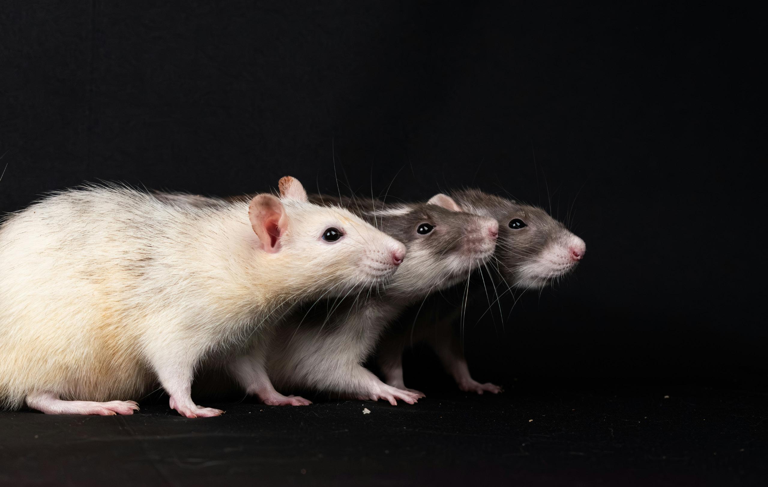
For decades, one of the most prominent tools in preclinical research— ranging from drug testing to understanding fundamental science and mechanisms of disease— has been the laboratory mouse. Many major breakthroughs in immunology and medicine are owed to early experiments performed in mice. While other animal models do exist, the mouse has been the gold standard in immunology for several reasons. For one, the mouse immune system is strikingly similar to our own: many immune cell types, signaling pathways, and functions closely resemble those of humans. Combined with the feasibility of housing, breeding, and genetic manipulation, mice have long been strong candidates for studying immunology outside of human subjects.
That said, human and mouse immunology do not directly mirror one another. Laboratory mice are also nearly genetically identical to one another as a result of inbreeding, which contrasts the genetic diversity seen across human populations. Furthermore, laboratory mice are typically housed in sterile, pathogen-free environments, meaning they lack the lifelong exposure to bacteria, viruses, and other microbes that continuously shape the human immune system. These collective limitations help explain why so many treatments succeed in preclinical mouse models but fall short in human trials, and they highlight the need for research tools that better reflect human immunology. To address this, researchers have developed modified mouse models that better approxi-
mate the human immune system. One example includes “humanized” mice, which are engineered to carry human rather than mouse immune cells. Another example includes “dirty” mice, which possess more diverse communities of microorganisms to better reflect the human microbiome.
But is there a better alternative? Recent efforts in biotechnology and bioengineering have been searching for the next best thing, of which, organoids have taken center stage. As its name suggests, an organoid is essentially a miniature organ-like structure, albeit substantially less complex and incapable of fully recapitulating the function of human organs (at least not yet). Still, organoids offer an exciting opportunity

for immunologists to study immune responses in systems that more closely resemble human biology.
Organoids can be grown from stem cells, which are essentially like blank canvases that, under certain conditions, can be “instructed” to develop into a specific cell type that composes a tissue of interest. Alternatively, organoids can be derived from human tissue samples, where small pieces of tissue obtained during surgeries or biopsies can be placed in specialized conditions where they continue to grow while maintaining many of the structural features of the original organ. In both cases, the goal is the same: to create an effective model of human tissue that can be studied in highly controlled laboratory settings rather than in the body.
TONSILS AS ORGANOIDS
Organoids have been explored in various contexts, ranging from drug testing to understanding basic principles of human immunology. One particularly creative application with organoids involves tonsils. Typically thought of as an inconvenience to be removed, excised tonsils that would otherwise be discarded after surgery are proving to be surprisingly useful for research. Tonsils are densely populated with immune cells and maintain their structure remarkably well in laboratory conditions. A 2021 study leveraged tonsil organoids to study immune responses to vaccines, successfully identifying the components needed to produce influenza-specific immunity. These results offer a compelling proof of concept for using organoid models to model complex immune processes.
INTESTINAL ORGANOIDS
Another application for human organoids is to better understand the function of tissue resident immune cells. These cells, which live inside the tissue, can functionally differ from the immune cells that circulate throughout the blood. While understanding their function provides insights into their role in disease and potential treatments, studying these cells in humans continues to be a challenge. One study investigated this by creating an intestinal organoid that encapsulates immune cells as well. This model allowed the researchers to understand how cancer treatments triggered inflammation in
TUMOR ORGANOIDS
Perhaps one of the most rapidly growing applications of organoids lies in cancer research, particularly in the study of immunotherapy. Immunotherapies work by harnessing the immune system to recognize and destroy cancer cells. While these treatments have shown remarkable success in some patients, they are not successful in all cases, and predicting who may respond to certain treatments remains a challenge. Tumor organoids can be grown directly from patient tumor samples obtained during surgery or biopsy, and they often retain many of the genetic mutations and characteristics of the original tumor when cultured in the laboratory. Researchers introduce immune cells into these systems to observe how they interact with the cancer cells. By testing different therapies on tumor organoids, scientists can study why some cancers respond to immunotherapy while others resist treatment. In the future, this approach may be revolutionary for personalized medicine, where doctors could potentially test treatments on a patient’s tumor organoid to determine which therapy is most likely to work before administering it. Researchers are also using organoid systems to better model human immune responses in ways that are difficult to achieve in traditional animal models. For instance, former University of Toronto trainee Lisa Wagar, now at University of California, Irvine, has pioneered the use of human organoid- and tissue-based systems to study vaccine responses and human immunology in more physiologically relevant settings.
THE PATH FORWARD
Despite their exciting potential, organoids are not a perfect substitute for animal models. The human body is extraordinarily complex, and we are not yet at the stage where we can mimic its function entirely in a petri dish. Furthermore, organoids are not suitable for studying connections between multiple organs (e.g. the gut-brain axis). For this reason, animal models still play an important role. While organoids cannot currently replace animal models entirely, they still serve as a valuable complementary approach. It’s possible that with time, these tiny organoids may one day have a tremendous impact on the future of immunology research.
Written & Designed by
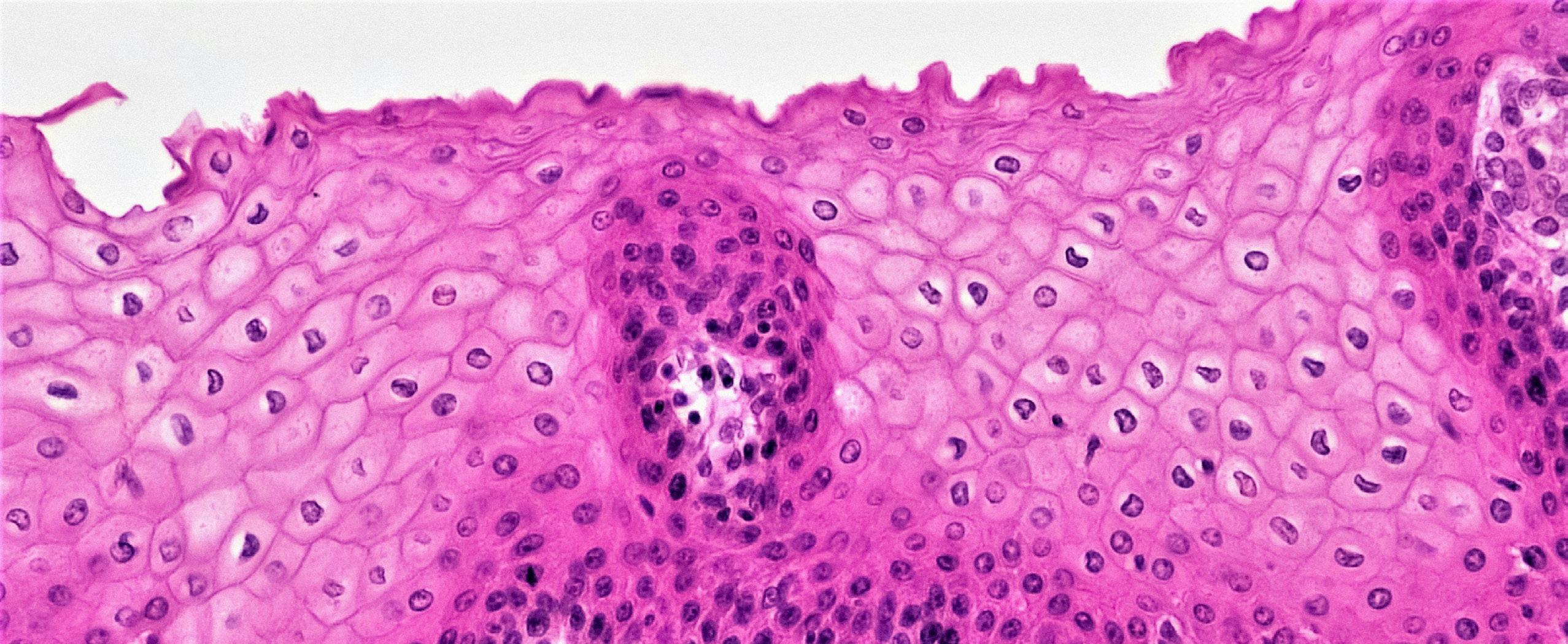
Milea DiPonzio
The Lamprey Time Machine
Unlocking the secrets of jawless immunity:

Ian interview with dr. Götz ehrhardt
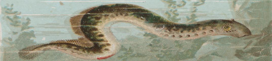
n the murky depths of the Great Lakes, the sea lamprey is often seen as a nightmare, an invasive parasite that latches onto prey and refuses to let go. But to Dr. Götz Ehrhardt, an Associate Professor in the Department of Immunology at University of Toronto, these “living fossils” are more of a biological time machine.
While humans and all other jawed vertebrates rely on the adaptive immunity toolkit of antibodies and T-cell receptors, lampreys took a different evolutionary path 500 million years ago. We sat down with Dr. Ehrhardt to discuss how studying these ancient creatures is revolutionizing our approach to biomarkers, oral therapeutics, and the very definition of “memory” in the immune system.
You have spent much of your career studying lampreys. What drew you to such an unconventional model, especially given its reputation as an invasive pest?
Chance, really. I did my postdoc in the lab of Max Cooper. And Max had worked as a fellow in Bob Good’s lab. Bob Good’s work on lampreys showed that, when immunized with heat-killed bacteria, the animals produced agglutinins, a fancy word for “clumping”, essentially indicating that there were antibody-like molecules in the serum. They also showed skin allograft rejection with accelerated kinetics upon repeat grafting, suggesting a cell-mediated immune response with memory.
For a long time, the adaptive immune system was considered an invention of the jawed vertebrates, from humans all the way back to cartilaginous fish, like sharks and rays. While jawless vertebrates behaved as if they had adaptive immune systems, investigators could not find adaptive immune counterparts like antibodies (or B cell receptors) and T cell receptors in lampreys despite searching for decades. That
Zeev Pancer joined the Cooper lab. He was the perfect example of ‘right person at the right time doing the right thing’. He did the founding work that led to the identification of what we now call variable lymphocyte receptors, the VLRs, which are antibody-like counterparts in lampreys. Call it chance, but I was in that lab when it happened.
How do these VLRs differ from the “conventional” antibodies we have in our own bodies?
Our antibodies are built on the immunoglobulin fold: a modular beta-sheet sandwich, that forms the basic structural unit of anticipatory receptor systems across virtually all jawed vertebrates. VLRs, on the other hand, use the leucinerich repeat as basic structural unit, which assembles into a curved solenoid shape. The antigen engages residues lining the inner, concave surface, as well as a flexible and variable loop protruding from the otherwise conserved C-terminus, a bit like our thumb if we were to form a “C” shape with the palm of our hand. Because the architecture is so different, we hypothesize that VLRs can “see” structures that our antibodies do not readily recognize. These are truly distinct solutions to the same problem of antigen recognition.
Given structural differences, can VLRs recognise antigens that conventional antibodies cannot?
With a large enough library, you can probably make a conventional antibody to almost anything. But there are two meaningful reasons to think VLRs can access targets that are practically difficult. First, the different protein architecture may allow recognition of certain epitopes that may be less accessible to immunoglobulin structures. Second, the mammalian immune system must maintain tolerance to self; given the large evolutionary distance of jawless and jawed vertebrates, the tolerogenic constraints that apply to humans and mice may not apply to lampreys (and vice versa).
I can answer this with an example. My lab works on memory B cells, adaptive immune cells that help the body “remember” past infections. For a long time, there was simply no reliable marker to isolate them. Justin Chan, a former graduate student in my lab, used a VLR display library to search for a binder that could distinguish memory B cells and plasma B cells from everything else. He isolated a VLR antibody that interacted specifically with memory B cells and plasma cells, a staining pattern that is inconsistent with any conventional antibody. The marker turned out to be HLA class I, a molecule found on almost every nucleated cell in the body. The result seemed nonsensical at first. Why would it be selective? It took considerable work to unravel, but the VLR was recognising HLA class I specifically in the context of tyrosine sulfation, a post-translational modification that had never been described in this context before. This opened an entirely new line of investigation that we are still actively pursuing.
Immunology as a field is quite mouse-centric. What would you say to students who want to work outside that paradigm?
Firstly, keep your mind open. And go to conferences that are a little off the mainstream. There is a meeting called the North American Comparative Immunology Workshop (NACIW) where you will encounter very unusual and
exciting science.
“sometimes the most useful tool is not the closest relative.”
Immunology had its origins in comparative biology; there are beautiful model systems across the tree of life. Developmental biology kept its zebrafish and Drosophila. Genetics kept its yeast. Immunology, for understandable reasons, focused on the development of the mouse models. You need a model that reflects human physiology, with a rapid reproductive cycle. And a mouse is perfect for those reasons. While it has served us extraordinarily well, it narrows the scope. Sometimes the most useful tool is not the closest relative. Our lamprey work began because we were trying to solve a problem in our own research area, finding a marker for memory B cells, and we looked elsewhere for the tool. And that worked out!
The theme of this issue is “Built to Survive.” You have found that VLRs are remarkably hardy. How does that translate to medicine?
This is where it gets really exciting. When we first tried to purify VLRs, we used the standard protocol of eluting an antibody from its antigen using a low pH (~ pH 2.5). Usually, that’s enough to make the antibody let go. With VLRs, nothing happened. We tried 3M magnesium chloride, 5M lithium chloride, and even hydrochloric acid at pH 1.5. Surprisingly, the VLR stayed stuck. We eventually had to go up to pH 12.5 using sodium hydroxide to get it to let go. And even then, once we neutralized the pH, the VLR could bind again. This is something a conventional antibody simply cannot do.
Because they are so resistant to low pH, they may be ideal candidates for development of oral therapeutics. They could potentially survive the transit through the stomach and act directly in the gut to treat gastrointestinal diseases.
Fun Fact!
In Portugal, lampreys are a seasonal delicacy and served as ‘lamprey stew on rice’. If you have worked with the animals in the lab, you see precisely what ends up on your plate. It does not help improve your appetite. The accompanying red wine, however, is exceptional!
Moral of the story: lampreys are great to have in the lab, not as great on the dinner plate.
Written by Manjula Kamath
Designed by Victoria Sephton
LAST QUESTION: What is one thing about lamprey immunity you wish more immunologists knew?
That lampreys are an excellent system for generating really interesting antibodies!
Dr. Justin Tze Ho Chan: Swimming through immunity—What fish can teach us about the immune system

Across your PhD and post-doctoral work, your research has revolved around immunity beyond humans. What first drew you to studying fish immunology?
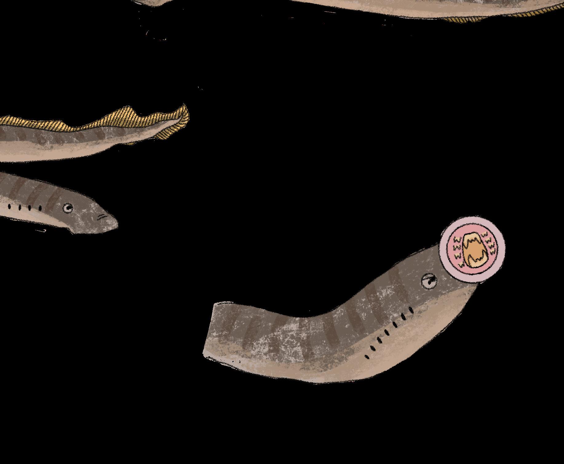
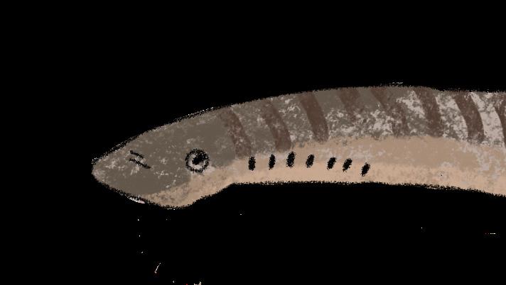
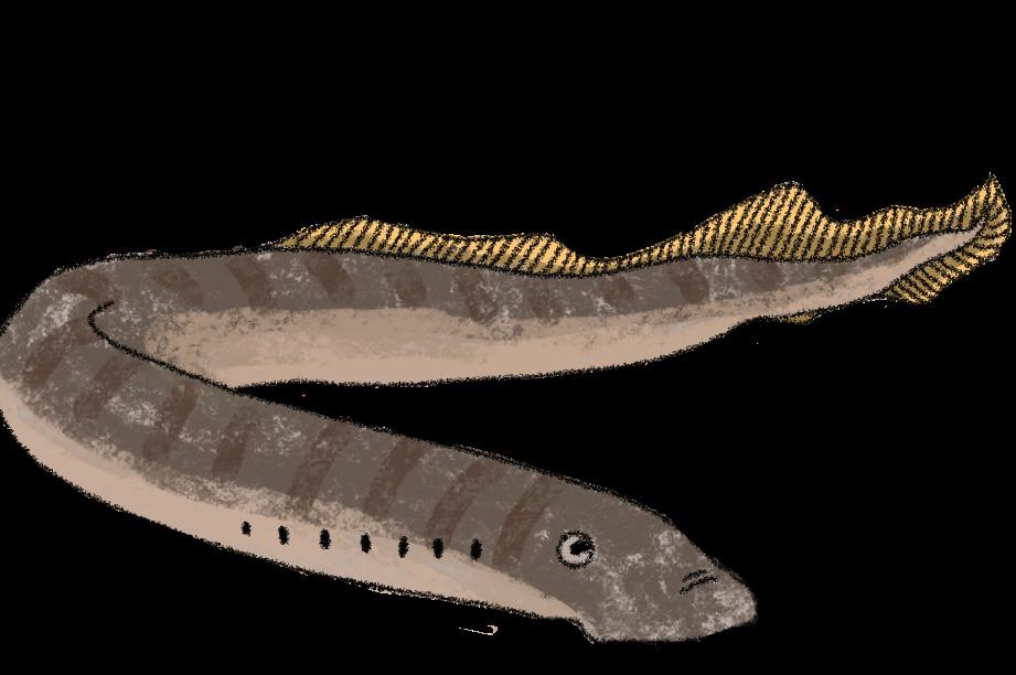
This issue, we are joined by Dr. Justin Tze Ho Chan, an alumnus of the University of Toronto Department of Immunology and currently a postdoctoral researcher at the University of Veterinary Medicine Vienna, whose research trajectory has been shaped by a curiosity for immunology in members of the animal kingdom beyond mammals—centred on the inhabitants of freshwater rivers and lakes. Diving into the world of fish, Justin’s work explores how vertebrates have evolved diverse strategies to fight infections and adapt to their environments. His contributions to the field include work with sea lamprey antibodies, teleost fish immune cells and immunological memory, offering a glimpse into how studying evolutionarily distant organisms can reshape our understanding of immunity in humans.
“The earliest exposure I had to fish immunology was a MIMM314 lecture at McGill University, in 2011. Dr. Roger Palfree introduced us to alternative immunity in the lamprey and the work of Dr. Max Cooper and colleagues, supplemented by a video ‘not for squeamish members of the audience’. Seeing the lamprey, first thing in the morning in a dimly lit lecture hall, was certainly captivating and eyeopening.”
Justin explains how this moment opened the door to a research area where many foundational questions remain unanswered and the thrill of discovery still felt possible: “Studying fish immunology, I feel like an early explorer in uncharted waters, having the chance to study fundamental and basic topics about immunology, like those I was born too late to study in humans.” He adds that he is also drawn to comparative research, as it contributes to a sense of shared biological history: “No matter how uncanny or unfamiliar someone may look, [through the lens of immunology] we all have something in common, we are all related and have a shared history.”
Following this initial spark, he credits his mentors, Drs. Ehrhardt and Tomáš Korytár, for further cultivating his interest. “Stars aligned and I am eternally grateful to have pursued a PhD in the laboratory of Dr. Götz Ehrhardt,” he says. His experience as a graduate student, focusing on lamprey antibody biology, plunged him deep into the depths of fish immunology.
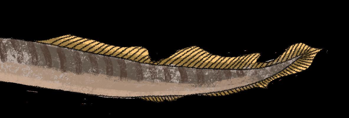
How has working with fish changed how you think about human immunology?
“If anything, working with fish has only heightened my appreciation for human immunology.” He adds “it goes both ways,” echoing a sentiment from fellow fish immunologist, Dr. Brian Dixon: “We are all comparative immunologists because without human immunology, there would be nothing to compare to.”
Working with fish also reframed how he sees humans within the evolutionary landscape. “It is humbling that we are just one node on a map. Human immunity is also just one alternative immune system in an ocean of alternatives.”
He emphasizes that “primitive” species “did not stop evolving as soon as new ones emerged. They are ‘primitive’ in terms of evolutionary distance, but it does not mean that their solution to immunity is featureless or lacking. I think it is just that we are not adapted to the same needs and environment.”
What advice would you give to students interested in exploring research in organisms beyond humans and mice?
While Justin encourages students to follow their curiosity first and foremost, he argues, “Humans already pay an inordinate amount of attention to humans.” He points to the long history of discoveries made through studying unconventional organisms: “The rich history of immunology is adorned with and in debt to discoveries from non-mammalian and non-conventional models: from Metchnikoff’s starfish, Hoffmann’s fruit flies, back to Cooper’s chickens. Overall, from a One Health perspective, animal health is intertwined with human wellbeing, livelihood, and socioeconomics.”
Justin reassures students that “The field has never been more alive,” and that their “scientific and problem-solving skills will always be transferable to all walks of life.” He adds, “You can see for yourself, the community and support at meetings such as the North American Comparative Immunology Workshop. At the end of the day, we’re all immunologists.”
For readers who may be unfamiliar, what are some of the key similarities and differences between the human and fish immune systems?
Beginning with the features shared among vertebrates, Justin references how vertebrates evolved from a common ancestor over 500 million years ago. He explains, “It is likely that an evolutionary ‘Big Bang’ gave our ancestors the ability to somatically diversify antigen receptors, the main lymphocyte lineages responsible for cellular and humoral immunity, and compartments within which these cells can develop, or mount responses.”
On the other hand, teleost fish, his current area of study, differ immunologically not only from humans, but between species due to tremendous diversity. “You can have a carp and a salmon whose last common ancestor diverged 200 million years before that of humans and mice, making it challenging to generalize findings from one fish species to another.” However, he highlights temperature dependence and mucosal surfaces as some major differences: “Fish are cold-blooded and temperature drastically impacts their physiology, biochemistry, and behaviour.”
He also states, “If you like mucosal immunology, the entire external body of a fish is mucus, from skin to gills to gut, and it is unclear how homeostasis is maintained across all these surfaces.”
And finally, among the organisms you’ve worked with—humans, fish, or any others—which immune system do you find the most fascinating?
“On the one hand, I have a soft spot for the lamprey because of my upbringing, but also because of my upbringing, any organism with B cells or B-like cells has my admiration.”
At the same time, he adds that part of his fascination with lampreys stems from their place in our own evolutionary history, explaining, “It’s a miracle to exist at the same time as lampreys and hagfishes, that we are living descendants of an ancient jawless fish.”
He ends with a sentiment that captures the heart of comparative immunology: “Despite the [evolutionary] distance, immunology prevails… as a tried, true, and refined solution to co-exist with microbes.”
Through his lens, Justin shows us how immunity forms a universal thread connecting vastly different species. His reflections also remind us that the answers we seek to better understand ourselves can be found beyond what is familiar, or in this case, above water.
Written by Yasmin Anning
Designed by Sophie Sun
Fur and Fortified Immunity: how growing up with pets may shape the immune system
For generations, parents have debated whether pets belong in homes with young children. Traditional advice often warned that exposure to animal fur and dander might trigger allergies or autoimmune disease. However, science is now revealing new ways in which growing up with a pet, especially during infancy, can build a resilient immune system.
Evidence linking pets to reduced allergy
Several large-scale epidemiological studies have found associations between early pet exposure and reduced rates of allergic disease. In one study involving over 4,000 adults, individuals who had grown up with dogs or cats were significantly less likely to develop asthma later in life. Those without childhood pets were roughly twice as likely to report asthma compared with those exposed to dogs during early childhood. Similarly, research following children from birth has found that dog exposure during infancy is associated with lower rates of wheezing and atopic dermatitis (eczema) by early childhood. Other cohort studies echo these findings. The PIAMA study followed nearly 3,000 children in the Netherlands from birth to age eight. Researchers found that children who had a cat or dog in the home during early infancy were less likely to develop sensitization to inhalant allergens by age eight. Another study found that contact with dogs was linked to lower rates of allergic sensitization to environmental allergens by age six.
Studies also suggest that pet exposure may also influence food allergies. For example, a large Japanese cohort study involving more than 65,000 infants found lower rates of certain food allergies like egg, milk, and nut allergies among children exposed to cats or dogs early until the age of 3. While these studies cannot always prove direct causation, the consistency of the results across different populations has strengthened the hypothesis that pets play a protective role in immune maturation.
The hygiene hypothesis and the “mini-farm effect”
The hygiene hypothesis, proposed by David Strachan in 1989, suggests that reduced microbial exposure in early life impairs immune development. Early exposure to microbes like bacteria, fungi, and other microorganisms helps the immune system learn to distinguish between harmful pathogens and harmless environmental substances such as pollen or
dust. In modern urban environments, children have reduced microbial exposure. Pets can bridge this gap by bringing in microbes into the household through their fur, saliva, and outdoor activity. These exposures may help “train” immune responses and reduce the likelihood of overreactions that lead to allergies or asthma.
This pet-related immune benefit has been dubbed the “mini-farm effect,” based on research showing that children raised on farms where they regularly encounter livestock, soil microbes, and animal environments have significantly lower rates of asthma and allergies. Pets may replicate part of this effect on a smaller scale by introducing environmental microbes into the home environment. Studies of household microbiomes have found that homes with dogs, for example, contain a greater diversity of bacteria compared with pet-free homes. This microbial diversity appears to correlate with lower rates of allergic disease. One proposed mechanism involves endotoxins; molecules derived from certain bacterial cell walls. Exposure to small amounts of endotoxins may help calibrate the immune system in early life, making it less likely to react to everyday exposures like pollen, food proteins, or pet dander.
How pets shape the gut microbiome
Another way that pets may influence immunity is through the gut microbiome, the complex ecosystem of microorganisms that live in our gut and influence immunity and overall health. Large cohort data from the Canadian CHILD study found that pet exposure during pregnancy and infancy was linked to higher abundance of bacterial species associated with lower risks of atopic dermatitis and obesity.
Since the gut microbiome also helps regulate inflammation and maintain the intestinal barrier, these changes may have implications beyond allergies. A large study led by researchers at the University of Toronto, found that children that lived with a dog between ages 5 and 15 showed signs of healthier gut barrier function and were significantly less likely to develop
Crohn’s disease, a chronic condition in which the immune system drives gut inflammation. Interestingly, bird ownership was linked to greater gut inflammation and higher Crohn’s disease risk. Another study found that adults who grew up without pets showed more signs of intestinal barrier leakiness and weaker immune regulation later in life. Together, these results suggest that the effects of childhood pet exposure may extend into long-term gut health.
Timing matters:
the critical early-window life
One consistent finding across many studies is that timing of exposure matters. The strongest protective effects against allergy and asthma are observed when pets are present during pregnancy or in the first year of life. Early infancy is a period of rapid immune system development, when immune cells learn to regulate inflammatory responses and learn to discriminate real threats from everyday environmental antigens. Exposure to animals during this window may influence how immune pathways mature. For instance, research shows that children who interact with dogs during their first year of life may have lower rates of developing an allergy later in childhood. Starting pet exposure at age three or later does not appear to produce the same protective effect.
Effects on stress and immune regulation

The benefits may extend beyond allergies and asthma. Emerging research suggests that childhood exposure to animals may influence how the immune system responds to stress decades later. In studies examining adults who grew up with pets, researchers found that early animal exposure correlated with more balanced inflammatory responses to stress. These individuals appeared less likely to mount exaggerated immune reactions when confronted with psychological stressors. Since chronic inflammation is implicated in conditions ranging from cardiovascular disease to depression, these findings hint at long-term health implications that extend well beyond childhood respiratory illness.
Caveats and ongoing debates
Despite promising evidence, we still do not have all the answers about the relationship between pets and immunity. First, as most studies are observational, they identify correlations rather than direct causal effects. Families with a history of allergies may be less likely to own pets in the first place, a phenomenon known as avoidance bias, which can influence study outcomes. Additionally, pets may have different effects depending on individual genetic predisposition. Children already allergic to animals may experience worsening symptoms if exposed to pet allergens. Finally, the type and number of animals may matter. Some research suggests that living
with multiple animals or animals that spend time outdoors may increase microbial diversity more than indoor-only pets. Given these complexities, medical professionals generally do not recommend pet ownership solely as an allergy-prevention strategy.
A new perspective on household pets and immunity
Overall, the relationship between pets and human health is more nuanced than once believed. Growing up with pets, particularly during infancy, appears to expose children to a wider range of environmental microbes that can shape immune development. Through mechanisms linked to the hygiene hypothesis, microbial diversity, and training the immune system, early-life pet exposure may reduce the risk of allergic sensitization and certain immune-mediated conditions. However, the effects vary between individuals, and further research is needed to clarify the biological mechanisms involved. For now, the evidence suggests that for most families, sharing a home with pets is unlikely to harm children’s immune health and may even provide health benefits that extend well beyond companionship.
Written by Preya Patel
Designed by Baweleta Isho
Spray-induced gene silencing: Using RNAi to fight fungi
Fungi are no fun for crops
The fungus Botrytis cinerea (B. cinerea) derives its name from the Latin term for “grapes of ash.” Also known as gray mold, this harmful crop pathogen coats damaged plant tissue with an ashy fuzz made from small, grape-like spores. The fungus can infect more than 1,400 types of plants including strawberries, tomatoes, and grapes. Chances are, you’ve seen B. cinerea growing on old produce in the back of your fridge.
Fungi like B. cinerea pose a major threat to the agricultural industry and global food security. Fungal diseases kill 10-20% of crops worldwide prior to harvest, and another 10-20% are lost after harvest. This amount of lost food could feed up to 4 billion people a year.
These crop losses occur despite anti-fungal farming strategies. Farmers will clear diseased plant tissue, use chemical fungicides, and genetically engineer crops to resist fungal disease. However, negative environmental effects of fungicides, the emergence of fungicide-resistant strains of fungi, and wariness toward genetically modified organisms challenge the application of these strategies.
To develop improved methods to combat fungal diseases, agricultural scientists have been looking to plant immunology for inspiration. In the last decade, a novel technique known as spray-induced gene silencing (SIGS), which sprays plants with inhibitory RNAs that silence vital fungal genes, has shown promise in multiple studies.
SIGS works by harnessing the powerful genetic regulation system known as RNA interference (RNAi). In all living organisms as well as in some viruses, the genetic code of DNA is transcribed into messenger RNA (mRNA), which is then translated into functional protein. To avoid excessive amounts of host protein or to destroy viral proteins, RNAi disrupts protein production in an organism by targeting specific mRNA for degradation. The key player in RNAi is double-stranded RNA (dsRNA), which locates mRNA with the same sequence and guides a group of proteins called the RNA-induced silencing complex (RISC) to the mRNA. Then, RISC chops up the mRNA, preventing it from being translated into protein. The term RNAi refers to the way dsRNA and RISC “interfere” with mRNA to silence proteins.
The floral history of RNAi
RNAi regulates gene expression and provides viral defense in almost all animals, plants, and fungi, but the phenomenon was first discovered in plants in 1990, when an experiment with petunias produced curious results. A group of plant biologists led by Carolyn Napoli had been trying to enhance the color of the petunias by adding DNA encoding violet pigment protein. To their surprise, the flowers turned white, and the mRNA levels of the pigment protein diminished. Although the petunia biologists did not realize at the time, the added DNA had been converted to dsRNA and had guided RISC to destroy the pigment protein mRNA.
Over the next five years, scientists extended the scope of RNAi to fungi and animals. One lab produced albino versions of the fungus Neurospora crassa (red bread mold) by targeting fungal pigment proteins with RNA. Multiple groups injected RNA into the worm Caenorhabditis elegans and observed the loss of the corresponding mRNA and proteins. One of these worm experiments was the first to prove that dsRNA caused RNAi, awarding Andrew Fire and Craig Mello the Nobel Prize in Physiology or Medicine in 2006. These experiments helped show that RNAi is a highly conserved gene regulation system that functions in many organisms.


Plants use RNAi to regulate their own proteins but also to silence foreign proteins from pathogens like viruses and fungi. Indeed, plants can transfer dsRNAs directly into insects and fungi to disrupt the pests’ genes in a process called “cross-kingdom RNAi.” In the mid-2010s, scientists began to recognize that since pests could take up dsRNA secreted by plants, spraying plants with dsRNA targeting vital fungal genes (SIGS) could be an effective pesticide technique. SIGS is also an appealing anti-fungal method since dsRNA does not persist in the environment like other chemical fungicides, nor does it require the expensive and often controversial method of genetic engineering.
In 2021, the company GreenLight Biosciences showed that potato leaflets were protected from the Colorado potato beetle after being dipped in a solution of dsRNA targeting PSMB5, a vital protein for the insect. In 2023, this product became the first pesticide to be approved for SIGS by the U.S. Environmental Protection Agency, sparking interest in developing SIGS products for other pathogens.
Indeed, SIGS has shown promise in controlling fungal infections, including B. cinerea. One group coated tomatoes, strawberries, grapes, lettuce, and onions with dsRNA targeting the B. cinerea proteins Bc-DCL1 and Bc-DCL2, drastically reducing fungal growth in 2016. In the following years, another group confirmed that grapes sprayed with dsRNA targeting three different vital B. cinerea proteins were protected from the fungus. A third group produced similar results in tobacco plants by targeting the gene Bc-DCL1 with SIGS in 2024.
The promise of using SIGS to control B. cinerea infections demonstrates the power of immunology research. By recognizing that plants and fungi communicate via the complex gene regulation system of RNAi, plant biologists were able to develop these exciting new fungicides that may receive regulatory approval in the near future.
Thanks to SIGS, we may soon see the last of those ominous ‘’grapes of ash’’ that haunt the back of our fridges.
Written & Designed by
Annie Mitchell

VLRS:

Thethe jawless fish strike back!
intricate web of organs, cells, and molecules that make up our immune system can be viewed as the product of evolutionary optimization, designed by nature itself. Our adaptive immune system, in particular, is a marvel; it is comprised of B cells and T cells, which recognize specific moieties on pathogens or other agents of disease, allowing for recognition and clearance of the threat, as well as protection against future exposures. These immune cells possess both secreted and cell-surface molecules that allow for identification of their target: antibodies produced by B cells and T cell receptors (TCRs) expressed on T cells. Both antibodies and TCRs belong to the immunoglobulin (Ig) family of proteins.
B cells are our body’s antibody-producing cells and encode in their DNA the ability to generate an extremely diverse repertoire of antibodies. Millions of B cell clones exist, each producing a unique antibody type designed to recognize a specific target. This army of B cells can expand or contract as an individual encounters various environmental factors throughout life. Similarly, a T cell army also exists, where each TCR recognizes a specific tar-
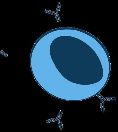
ANTIBODY

This highly adaptive machinery is ancient, first emerging around 450500 million years ago in jawed fish – our vertebrate ancestors! Given this relatively “sudden” emergence of a functional adaptive immune system in jawed fish on the evolutionary timeline, which has not changed much over the years, scientists fondly refer to this evolutionary event as the “Big Bang” of the adaptive immune system; the jawed fish possessed all the core adaptive immune ma chinery found in today’s mammals, but the jawless fish were thought to lack such a system entirely.
The jawless fish, however, had a wellkept secret that was only unearthed rela ly recently – the existence of an alternative adaptive immune system, which relied on cells that produced similar molecules to our Igs. Termed the variable lymphocyte receptor (VLR) family, jawless fish also have ingrained in their genome the ability to create a diverse repertoire of these VLR molecules (at more than 1014 unique VLRs) – an army size that can rival the B cell repertoire in mammals. Remarkably, the jawless fish devel oped an adaptive immune system centred around the VLRs, which although is functionally similar to jawed vertebrates, uses entirely different molecular building blocks. Through out these millions of years, these two adaptive immune systems emerged and evolved in parallel.
Written by Deeva Uthayakumar
Designed by Zoeen Carter
Through studies in lampreys and hagfish, two members of the jawless fish taxon, we have learned a lot about the function of these VLRs in comparison to the mammalian adaptive immune system. Instead of the Ig protein units found in antibodies and TCRs, VLRs consist of various leucine-rich repeat (LRR) components. LRRs are ancient protein compo nents in plants and animals that make up many other mole cules of the immune system and can be triggered by an excep tionally diverse array of ligands. There are three types of VLRs: VLRA, VLRB, and VLRC. Cells possessing these receptors have roles analogous to those of T and B cells in jawed vertebrates, allow ing jawless fish to recognize and respond to foreign pathogens.
The discovery of these alternative immune molecules has potential applica tions in biotechnology. Antibodies have been engineered to not only target infectious microbes, but also to target other disease-causing proteins and macromolecules within our bodies. Since jawless and jawed fish diverged millions of years ago, VLRs have an advantage of recognizing molecular targets that mammalian Ig-based antibodies struggle to recognize, notably carbohydrates. The unique shape and structure of these molecules also allow for further fine-tuning of their targeting capabilities.

These VLRs are, therefore, an excellent example of convergent evolution in immunology; while excluded from the immunological “Big Bang” that brought about the adaptive immune system in jawed fish, the jawless fish strike back with their own unique VLR solution that can achieve a similar

BIG BANG
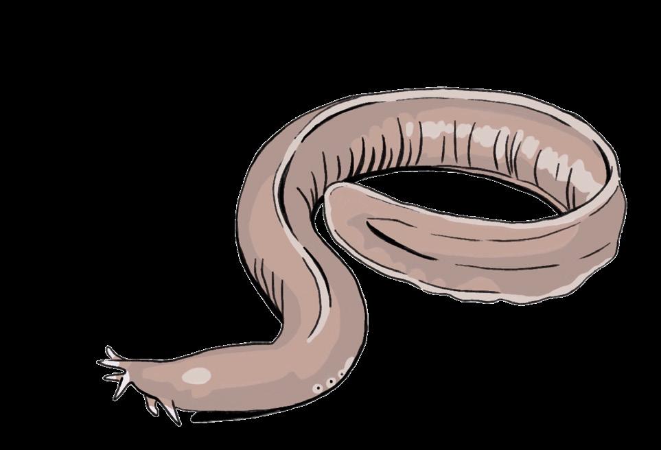
LAMPREY HAGFISH
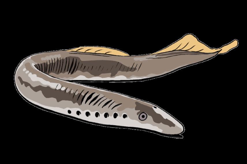


What the humble naked mole-rat can teach us about immunity
Heterocephalus glaber

In the world of immunology, the human and murine models are often viewed as the “gold standard.” As such, we (humans) define a robust immune system by two intricately linked arms: the innate system, which provides rapid, broad-spectrum defense, and the adaptive system, which offers exquisitely targeted responses and long-lasting memory. Conventional wisdom suggests that the coordinated activity of both is essential; to lose one is to invite systemic collapse.
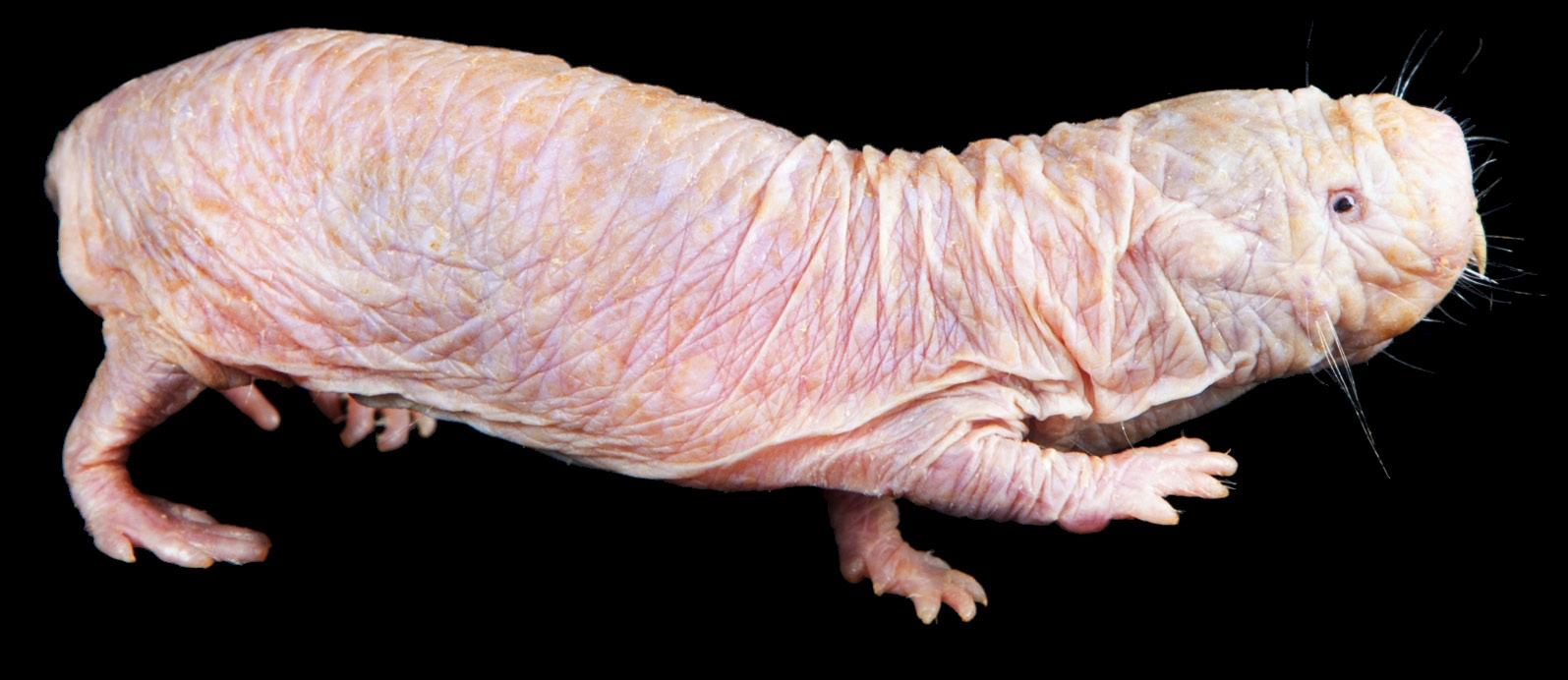
Naked mole -rats live in underground colonies that are relatively pathogen poor. In such an environment, the evolutionary pressure to maintain a metabolically expensive adaptive immune system may have been low.



Enter the naked mole-rat (Heterocephalus glaber), a nearly hairless, functionally blind, buck-toothed subterranean rodent. At first glance, their immune system appears strangely pared down compared to that of humans or lab mice. The primary cellular components of the adaptive arm, T cells and B cells, are both much more limited in numbers and variety of repertoire in the naked mole-rat. Their thymus (the source of T cells) shrinks early in life, reducing its capacity for producing T cells in adulthood. Instead of conventional T cells, naked mole -rats rely heavily on gamma- delta T cells, an unconventional subtype that may compensate for the near absence of natural killer cells. Their B cells also appear to have highly limited antibody diversity. Otherwise, their immune system is dominated by innate immune cells, with high numbers of myeloid cells, highly inflammatory macrophages, and neutrophils with unique functions. On paper, this looks like a recipe for vulnerability. And indeed, studies show that naked mole -rats mount relatively weak responses to many viral infections.
Despite this apparent “weakness”, naked mole-rats are astonishingly long-living compared to similarly sized rodents. Whereas lab mice and rats live up to about three years, naked mole-rats easily live up to three to four decades, defying the typical rules of mammalian aging. So how do naked molerats defend against pathogens and live such long lives when they are missing important components of the immune system? This may be due to an evolutionary trade - off.
Importantly, this reduced reliance on adaptive immunity might contribute to their longevity. In humans, aging is accompanied by chronic inflammation, immune dysregulation, and rising rates of cancer. Naked mole -rats seem to sidestep much of this decline. While humans often suffer from immune overreactions that can inadvertently promote tumor growth, naked mole-rats exhibit a dampened immune response to carcinogenic damage. Paradoxically, this “weakness” may be a protective attribute, as it restrains the chronic inflammatory environment that tumors exploit to thrive. When combined with highly robust DNA repair mechanisms and exceptionally low genetic mutation rates, the result is an animal that is virtually cancer resistant. Other changes that accompany the aging process as humans or lab mice age, such as metabolic decline or loss of fertility, also have not been found in aging naked mole-rats.
The naked mole-rat challenges our “more is better” approach to immunology. Despite this, the full picture of how they maintain such a prolonged health span and whether or how their unique immune system contributes to this phenomenon is still unclear. But ultimately, why does this matter? Comparative immunology is critically important to identify which immune features are universal and which are evolutionarily flexible, allows scientists to evaluate the pros and cons of the most commonly used animal models (such as the lab mouse), and identifies immune mechanisms that do not necessarily exist in humans, but could inspire new approaches to treat disease or maintain health in aging.
Written & Designed by
Annie Pu
mapping human illness across time through the lens of zoonosis
Animal to human disease transmission is a cause of numerous pandemics, epidemics, and severe illnesses we have seen throughout history. These diseases are zoonotic and make up around 60% of emerging human infections. The transmission process, known as zoonosis, can occur bidirectionally and through vertebrate animals, often from a wide variety of wildlife and livestock. Zoonotic pathogens can be bacteria, viruses, fungi, parasites, and prions, which are not viruses or living cells, but rather infectious proteins. Zoonosis has a serious impact on human health worldwide as it causes over 2.4 billion cases of illness and 2.7 million deaths per year.
To better understand the long-standing impact of zoonotic diseases, examining historical examples provides valuable insight into how animal-to-human transmission has shaped human health. Looking back, malaria and rabies were some of the first ever described zoonotic diseases in written text. Neither Plasmodium parasites nor Anopheles mosquitoes were identified as the cause and transmission vector of malaria until the late 1800s. As a result, early writings on malaria did not describe the disease as a zoonotic disease. The emergence of the disease, however, dates to 2700 BCE. Ancient medical writings in China, Greece, and Rome describe characteristic symptoms of malaria and reference medicines that are still
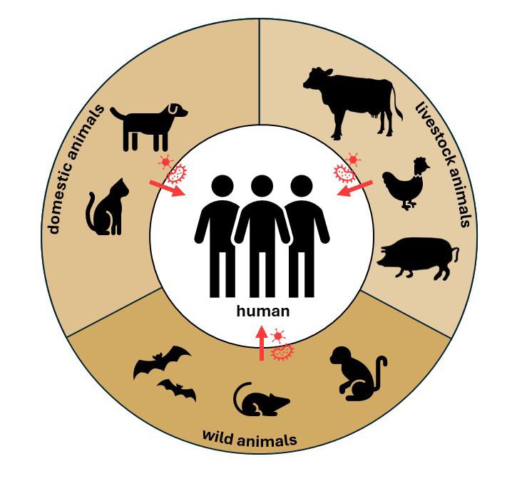
used today as antimalarial drugs. In 1930 BCE, Babylonia, excerpts from the Eshnunna Code describe the link between the bite of a rabid animal and human death, specifically noting dogs and emphasizing the need for caution in their domestication and ownership. Malaria and rabies were described very early on in human history yet continue to cause modern illness to this day.
While early zoonotic diseases were documented individually, later outbreaks demonstrated how animal-borne pathogens could reshape entire civilizations. The Bubonic Plague is a very well-known event in history that is associated with zoonotic disease. Caused by the bacterium Yersinia pestis, and transmitted through rodents, this zoonotic disease has been identified to be the cause of three plagues in history: the Black Death in Europe in the 1340-50s, the Justinianic plague in the Byzantine Empire in 540 CE. It was even traced back to the Bronze Age and was responsible for the plague that swept across Eurasia 5000 years ago, making it another example of ancient zoonotic diseases.
As microbiology advanced during the 1800s, researchers gained a deeper understanding of infectious diseases with animal reservoirs or animal-associated transmission, including tuberculosis (TB), ringworm, and salmonella . The mycobacterium tuberculosis bacterium responsible for causing TB was identified by Robert Koch in 1882, but when delving into history, it is yet another ancient disease. The first documentation describing TB was over 3000 years ago in India, but it is theorized an early ancestor of the bacterium infected hominids millions of years ago, making it one of the oldest infectious diseases in human history. Ring worm, despite the name, is caused by a fungus. It was discovered in the 1830s, through microscopic observation, but is another disease that was described in ancient texts in Rome and India. Salmonella is commonly associated with food poisoning linked to raw eggs and contaminated poultry, but these bacteria can also be transmitted through other food-producing animals, including cattle and swine. Certain species of Salmonella can cause typhoid fever, and salmonella infections remain among the most common causes of bacterial food-borne illness worldwide.
In addition to bacterial and fungal zoonoses, the 20th century saw the emergence and recognition of viral zoonoses such as avian influenza or “bird flu”. The first documented human infection was in 1997 but was identified as the cause of “fowl plague”, i.e. disease in poultry, in 1955. Ebola
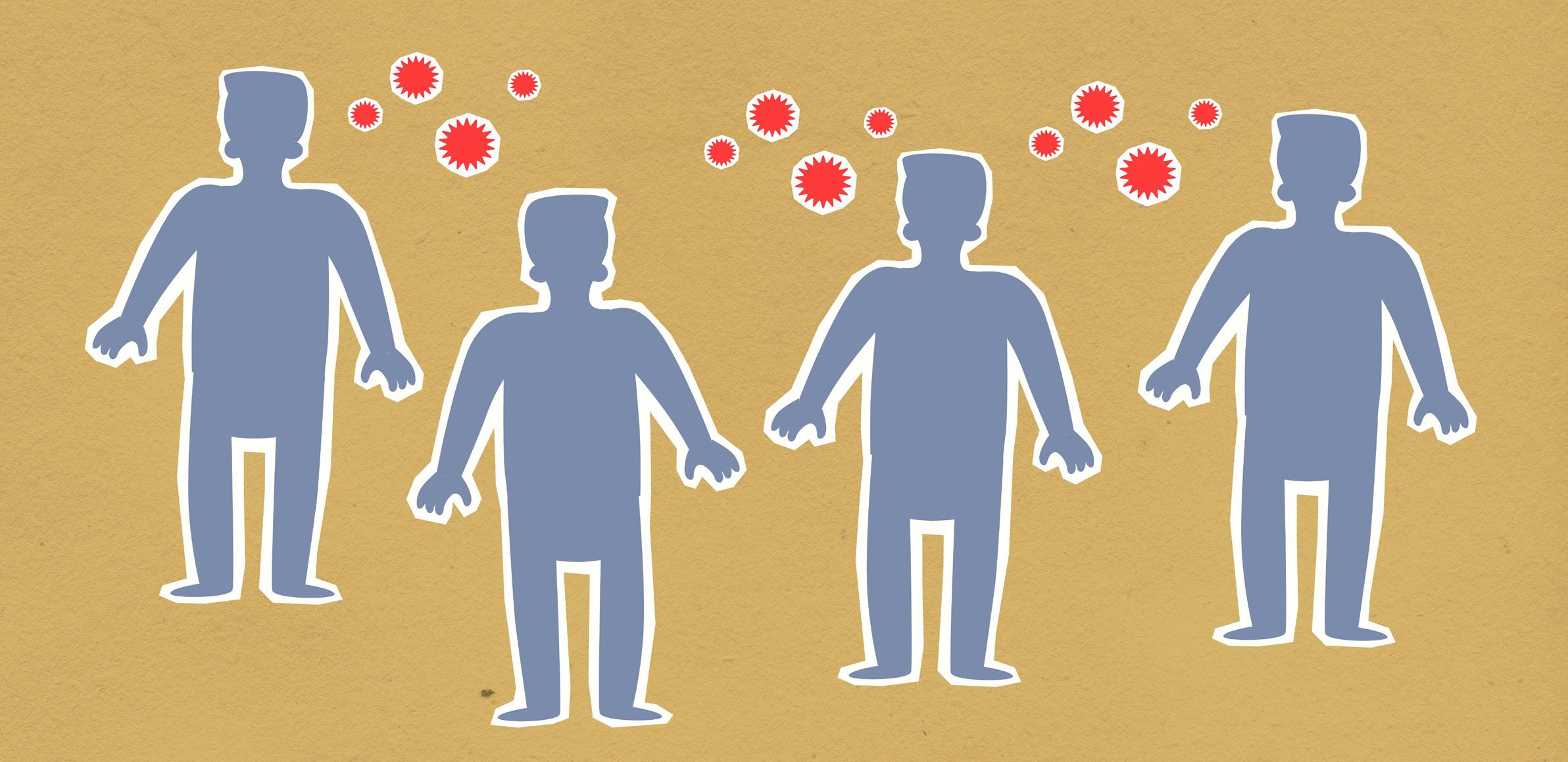
was also discovered in 1976, when it caused two outbreaks in Northeastern and Central Africa. The virus is believed to originate from animal reservoirs, likely bats, with transmission occurring through contact with infected wildlife. It made a reappearance as a devastating epidemic in Central Africa in 2014-2016.
Building upon these historical patterns, zoonotic transmission remains a major public health concern in modern society. Humanity’s most recent experience with a zoonotic disease was through the COVID-19 pandemic. Caused by a specific coronavirus, SARS-CoV-2, the first case occurred in 2019. Although originating from a horseshoe bat in China, human-to-human transmission led to a global spread of the diseases leading to a worldwide pandemic.
Beyond historical examples, it is important to examine the underlying factors that contribute to the emergence and spread of zoonotic diseases. Contributing factors can include anything from climate change, animal migration, animal product trade, travel and tourism, or other human activities that drive environmental changes, like urbanization. Major contact with wildlife or livestock has been a primary cause for zoonotic transmission, and people with high exposure through their job, lifestyle, or location are considered are at increased risk for these types of diseases.
Beyond their causes and historical significance, zoonotic diseases are characterized by a wide range of clinical presentations in humans. The specific symptoms are dependent on the type of disease that is contracted. A variety of organ systems can be affected such as respiratory (lung, airway), gastrointestinal (stomach, intestine), neurological (brain, spinal cord, nerves), skin, etc. Many bacterial and viral zoonoses can cause rash, aches, flu-like symptoms such as a fever or fatigue. This includes examples like Lyme disease, dengue fever, and AIDS. Gastrointestinal symptoms consist of abdominal pain, diarrhea, and vomiting, and come from diseases such as the plague and Ebola. Some more severe illnesses, such as rabies or mad cow disease, an extremely rare prion disease, cause neurological symptoms including seizures, hallucinations, paralysis, and memory loss.
Once symptoms appear, appropriate treatment depends on the type of pathogen involved. Many zoonotic diseases are treatable, and medications include anti-pathogen, so antibiotics, antivirals, antifungals, antiparasitic medications. Sometimes, more severe cases can use monoclonal antibodies to neutralize the pathogens, and in some cases, surgery is used for the removal of manifestations of the disease (example: cysts), such as Ebola or parasitic infections. There are a few zoonoses that are considered fatal or have a high mortality rate, but rabies is an example of one of the deadliest infectious diseases as once symptoms present, survival is extremely unlikely (1% or less).
While treatment is essential, preventing zoonotic transmission remains the most effective strategy for reducing disease burden. Vaccination is the most well-known medical preventative measure for many zoonoses. In Canada, the vaccine against SARS-CoV-2 is an example of a widely recommended vaccine against zoonotic diseases. Other infectious organisms such as rabies or Ebola also have vaccines recommended in Canada for high-risk cases. In addition to vaccination, protection from exposure by avoiding contact with infected fluids, bug spray, avoiding animal scratches/bites, and appropriate animal handling are also all methods of prevention.
Disease transmission through animals to humans has been the cause of many historical events, the reason for lifestyle food practices today, and the way we treat domesticated animals. It is a vast categorization of diseases, ranging from treatable illness to severe debilitating or life-threatening conditions. Caused by numerous types of pathogens and transmitted through various kinds of animals, these diseases are fascinating products of nature. As most emerging infectious diseases come from animals, zoonosis highlights how human health is dependent on not only the well-being of humanity, but that of animals, ecosystems, and environmental stability.
Written by Alina Mehra
Designed by Baweleta Isho
Comparative Immunology Why Investigation Across Species Still Matters
Lessons from Animal Models
Comparative medicine has long played an important role in scientific discovery. Many early breakthroughs in immunology and infectious disease came from studying similarities and differences between species. Edward Jenner’s observation about milkmaids exposed to cowpox being protected from smallpox, led to the discovery of the first vaccine. Robert Koch identified the organisms responsible for diseases such as anthrax, tuberculosis, and cholera through studies that involved different animal models. This led to the formation of Koch’s postulates, which are still used today to determine whether a microorganism can cause disease.
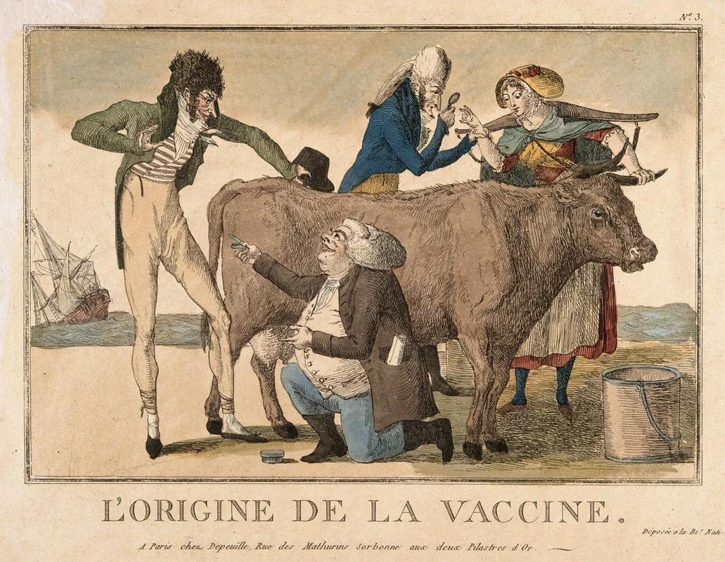
Animal models continue to be central to immunology research. Studies in mice have helped scientists understand how immune cells develop, how infections are controlled, and how tumors interact with the immune system. At the same time, research has also revealed important biological differences between species. A response observed in mice does not always translate to humans. For example, mouse models have often been poor predictors for treatments for sepsis, a severe condition in which the body’s heightened response to infection damages its own organs and may cause death. Many therapies that were proven to be promising in rodents ultimately failed in human trials.
“Even within humans, immune responses vary widely.”
Age, sex, genetics, and geographic environment can all influence how individuals respond to infections or vaccines. Because of this variation, it is difficult for any single experimental model to fully capture human immunology. Nearly 90 percent of therapies tested in early clinical trials do not reach the market, and limitations in preclinical models are often considered one of the top contributing factors.
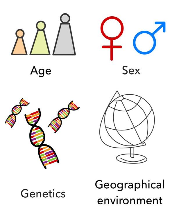
The Changing Landscape of Preclinical Research
Ethical considerations have also shaped how animal models are used. Researchers must justify every aspect of animal use in their studies and follow strict regulations. With the rising public awareness of animal welfare, these ethical considerations have become an integral part of discussions surrounding biomedical research.
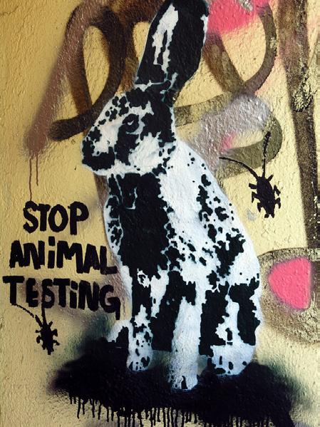
Recent policy changes reflect this shifting landscape. In April 2025, the United States Food and Drug Administration (FDA) announced plans to reduce or potentially eliminate animal testing requirements for certain drugs. Instead, FDA proposed shifting focus towards the use of human relevant approaches such
Written by Tianning Yu
Designed by Yoobhin Park
as computational toxicity models and in vitro testing using human cell lines and organoids. These approaches are commonly referred to as ‘New Approach Methodologies’ or NAMs.
The announcement drew significant attention from both the scientific community and saw immediate impact on financial markets. In the following days, shares of Charles River Laboratories, a company that provides preclinical research services including animal testing, dropped by 28% amid concerns that NAMs might eventually replace traditional animal models.
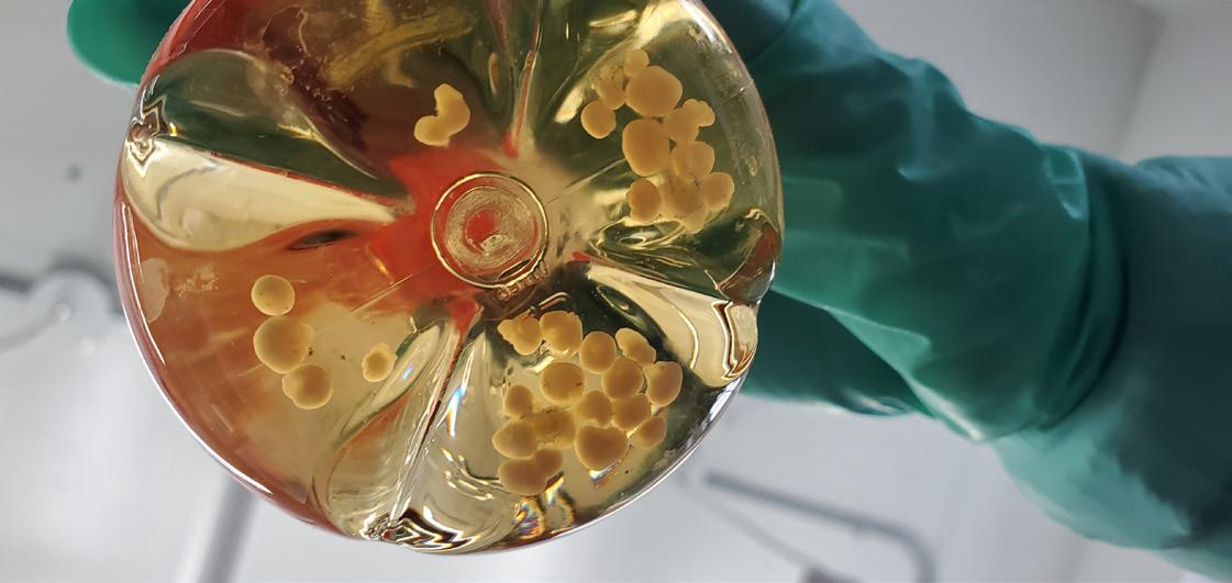
Animal Models Beyond Translation
The debate around animal models often centers on their limitations in predicting human outcomes. However, this overlooks a second, equally important role: their contribution to fundamental discovery. Animal models are not only tools for translation, but also for understanding how immune systems work. Even as efforts improve their clinical relevance, studying immune systems across species remains essential.
While new technologies such as organoids and computational models offer valuable tools, they cannot yet capture the full complexity of living biological systems. Interactions between organs, tissues, and immune pathways often require whole-organism models to be fully understood. At the same time, significant efforts have been made to improve the translatability of animal studies. These include the use of more physiologically relevant models, such as humanized mice that incorporate components of the human immune system; the selection of species or disease models that more closely mirror human pathology; and the use of “dirty” or rewilded mouse models that better reflect real-world environmental exposures. Together, these approaches aim to bridge the gap between preclinical research and clinical outcomes, making animal models more predictive rather than obsolete.
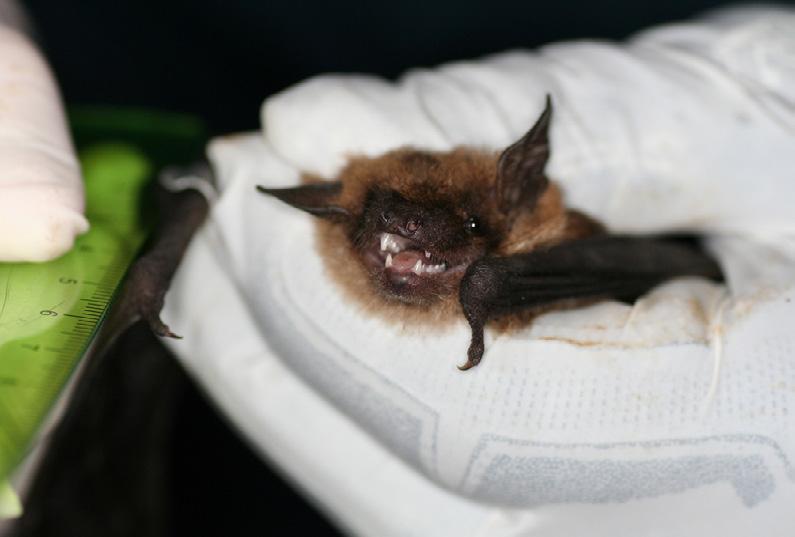
What Other Species Can Teach Us
Despite the changing landscape in preclinical research, studying immune systems across species remains critical for providing important biological insights. These insights span multiple aspects of immune function, from pathogen toler-
ance to immune system architecture and tissue compatibility. For example, bats can carry viruses such as coronaviruses and Ebola viruses with little illness. Understanding how their immune systems control inflammation may help scientists develop better treatments for viral infections. Other species show very different immune adaptations. For instance, sharks and lampreys possess alternative forms of adaptive immunity that rely on molecular systems distinct from antibodies. These alternative systems can inspire novel therapeutic tools, such as engineered binding proteins or new approaches to immune modulation.
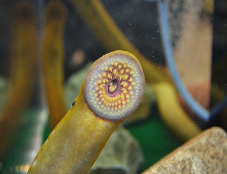
Moreover, studying immunology across species can shed light on tissue transplantation. For example, deep-sea anglerfish exhibit one of the most extreme reproductive strategies in vertebrates: tiny males permanently fuse to females, forming a shared circulatory system and functioning as lifelong sperm providers. To enable this, species with permanent attachment have lost key components of adaptive immunity, including MHC molecules, functional killer T cells, and, in some cases, antibodies— systems that would normally drive tissue rejection. Instead, they rely on modified innate defenses. This remarkable adaptation, which evolved multiple times independently, suggests that under strong reproductive pressure, vertebrates can survive without classical adaptive immunity, offering unexpected insights into transplant tolerance and immune system flexibility. Overall, these examples illustrate how studying diverse organisms can expand our understanding of immune biology.
Conclusion
The debate over animal models often focuses on what they cannot do. Yet their greatest value may lie not only in prediction, but in discovery. Comparative immunology shows that immune systems are more diverse and adaptable than any single model can capture. Rather than choosing between animal models, we might benefit from combining insights from different immune systems across species to build a more complete and holistic picture of immune biology.
THE CODE BREAKER:

Jennifer Doudna, Gene Editing, and the Future of the Human Race
The ENORMOUS Impact of a Tiny Immune System

HOW CAN I CONVINCE YOU TO READ THIS BIOGRAPHY?
As a student in the Department of Immunology, I tend to be overly critical when reading books about Immunology and I often choose instead to read about topics unrelated to science altogether. Despite this, I was pleasantly surprised while reading The Code Breaker, a biography about Jennifer Doudna. While the book focused on the discovery of CRISPR-Cas9 technology, the author included multiple interesting subplots to the main story while remaining faithful to the importance and application of science. To help you decide if this will be your next read, skim through this book review first!

WHO IS JENNIFER DOUDNA? AND WHO IS WRITING ABOUT HER?
If you’ve stepped into a bookstore in recent years, you may have seen the book covers featuring the unsettling stares of Elon Musk or Steve Jobs peering at you from the shelves. These are biographies written by the well-known American historian and journalist Walter Isaacson. In 2021, Isaacson wrote his first biography about a woman: the scientist Jennifer Doudna.
Jennifer Doudna is a Nobel Laureate in Chemistry, whose research in RNA biology led to the discovery of CRISPR-Cas9. This technology has been an indispensable
Credit: Christopher P. Michel / Wikimedia Commons / CC BY-SA 4.0
tool used in genome editing and is a technique which gives mortals the power to change the very essence of an organism’s biology and influence all its future descendants. This has the potential to eradicate genetic diseases yet raises important ethical questions on how far science should be able to influence human biology.

CRISPR-CAS9 AND ITS APPLICATIONS IN REAL LIFE AND IN INDUSTRY –IF YOU LIKE TO READ ABOUT FACTS AND SCIENCE
In this biography, Isaacson thoroughly researches the minor and major events leading up to the origin of CRISPR-Cas9 technology, and further delves into its biological and ethical impacts. Although the biography broaches a topic well-known to those studying the life sciences, Isaacson explains concepts with easy-to-understand analogies that can be understood by most readers. He describes the microscopic struggles for survival happening right under our noses – long before humans even came into the planetary picture. The number 1031 is inconceivable to us, but that is how many bacteriophages there are on earth. This concept is broken down by the author thus: “there are a trillion bacteriophages for every grain of sand...and in one milliliter of seawater, there can be as many as 900 million”. Bacteriophages are viruses that target bacteria, creating a never-ending evolutionary race as bacteria develop complex defense mechanisms while bacteriophages seek to improve their attacks. If you want to have a refresher on bacterial immunity and learn more about the implications of this technology, Isaacson’s visual storytelling on the drama surrounding the origins and consequences of CRISPR-Cas9 will keep you entertained.
A SIDE QUEST FOR YOGURT – IF YOU LIKE TO READ FUN TIDBITS AND SEE A MYSTERY UNRAVEL
One of my favorite anecdotes relating to the discovery of CRISPR-Cas9 involves a common breakfast item: yogurt. In the $40 billion global market of the yogurt and cheese industry, bacteriophages are pesky obstacles that target bacteria such as Streptococcus thermophilus, important for the fermentation process. French food scientists Rodolphe Barrangou and Philippe Horvath showed that bacteria can incorporate fragments of viral DNA into CRISPR regions of their genome, allowing them to recognize and defend against future viral infections. They used this discovery to vaccinate their yogurt-making bacteria and published their findings in Science in March 2007. Even though the book highlights the findings of Jennifer Doudna, Isaacson threads multiple such anecdotes about different contributing individuals to the overall fabric of the story. If you’re the type to enjoy reading slow mysteries with subplots that slowly intertwine into a larger picture, you’ll enjoy Isaacson’s style of writing.

THE STRUGGLE IS REAL – IF YOU LIKE TO SEE YOUR STRUGGLES REFLECTED AND FEEL REASSURED ABOUT YOUR OWN DECISIONS
Jennifer Doudna’s work has revolutionized modern science and her academic career as both a student and as a professor have been outstanding. Isaacson depicts Dr. Doudna as a passionate and charismatic scientist who not only strives to push herself towards her own goals but also deeply cares about her colleagues and students. While Isaacson did a wonderful job showing the strengths of the scientists featured in his writing, he also shows a realistic depiction of their hardships and worries. For instance, he highlights the uncertainties that Doudna faced even at the height of her career. He writes about her temporary stint in an industry job and the resulting anxiety she suffered in a different setting. He describes the doubts Doudna felt after making a major decision that impacted not only herself but also the members of her lab, and how she navigated through these changes and sought help from those around her. It was reassuring to read these very real and relatable stories and see that even well-known scientists face indecisiveness, doubts, and failures. If you’re the type to enjoy finding similarities between yourself and the main character of a story, then reading the Code Breaker will feel at once familiar and reassuring.
BOOK REVIEW


Written by Vera Lynn
Designed by Tianning Yu
Department of Immunology
University of Toronto
1 King’s College Circle
Toronto, ON M5S 1A8
Canada

Scientists examining fish on dock (1930?)
Photo Credits: University of Toronto Archives.