What’s the Diagnosis – Case 70
1
What’s the Diagnosis – Case 70
2
What’s the Diagnosis – Case 70
3
What’s the Diagnosis – Case 70
4
What’s the Diagnosis – Case 70
5
What’s the Diagnosis – Case 70
6
What’s the Diagnosis – Case 70
7
What’s the Diagnosis – Case 70
8
What’s the Diagnosis – Case 70
9
What’s the Diagnosis – Case 70
10
What’s the Diagnosis – Case 70
11
What’s the Diagnosis – Case 70
12
What’s the Diagnosis – Case 70
13
What’s the Diagnosis – Case 70
14
What’s the Diagnosis – Case 70
15
What’s the Diagnosis – Case 70
16
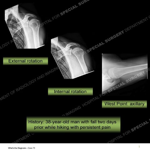
Findings • The initial 3 radiographs presented are unrevealing but the Stryker notch view shows cortical irregularity of the posterior glenoid and impaction of the anteromedial humeral head. These findings are confirmed on the MRI as are a tear of the posterior inferior labrum with a hyperintense posterior labral capsular junction. There is a hyperintensity of the posterior band of the inferior glenohumeral ligament (IGHL) but there is no disruption of the humeral attachment. • There is a defect seen of the anterior labrum which is contiguous with a sublabral foramen. A small amount of synovitis has insinuated into this area. No cartilage shearing injury has been sustained. There is a heterogeneous and hyperintense biceps pulley situated about the rotator interval but the rotator interval is not patulous.
What’s the Diagnosis – Case 70
17
What’s the Diagnosis – Case 70
18
What’s the Diagnosis – Case 70
19
What’s the Diagnosis – Case 70
20
What’s the Diagnosis – Case 70
21
What’s the Diagnosis – Case 70
22
What’s the Diagnosis – Case 70
23
What’s the Diagnosis – Case 70
24
What’s the Diagnosis – Case 70
25
What’s the Diagnosis – Case 70
26
What’s the Diagnosis – Case 70
27
What’s the Diagnosis – Case 70
28
What’s the Diagnosis – Case 70
29
What’s the Diagnosis – Case 70
30
What’s the Diagnosis – Case 70
31
Diagnosis: Posterior Shoulder Dislocation Posterior shoulder dislocations are much less frequent than anterior dislocations and are as a result of axial loading to an adducted and internally rotated shoulder. In an acute setting, the humeral head may be persistently internally rotated and posteriorly positioned, if the head does not reduce. Once reduction is achieved typical findings are as shown in this case. These findings include an impaction fracture of the anteromedial humerus referred to as a reverse Hill Sachs or McGlaughlin lesion. Posterior labral and glenoid injuries are analagous to their anterior counterpart as relates to the myriad of soft tissue Bankart injuries or an osseous Bankart injury. Although not present in this case there may be a concealed chondral injury or Kim’s lesion. Evaluation of the capsule is paramount particularly with concern of the posterior band of the IGHL and a humeral detachment which frequently necessitates an open stabilization. As shown in this case, a posterior translation can yield injury about the rotator interval. This is also critical to evaluate as this area may also require stabilization at the time of surgery to achieve a satisfactory outcome.
What’s the Diagnosis – Case 70
32