What’s the Diagnosis – Case 221
1
What’s the Diagnosis – Case 221
2
What’s the Diagnosis – Case 221
3
What’s the Diagnosis – Case 221
4
What’s the Diagnosis – Case 221
5
What’s the Diagnosis – Case 221
6
What’s the Diagnosis – Case 221
7
What’s the Diagnosis – Case 221
8
What’s the Diagnosis – Case 221
9
What’s the Diagnosis – Case 221
10
What’s the Diagnosis – Case 221
11
What’s the Diagnosis – Case 221
12
What’s the Diagnosis – Case 221
13
What’s the Diagnosis – Case 221
14
What’s the Diagnosis – Case 221
15
What’s the Diagnosis – Case 221
16
What’s the Diagnosis – Case 221
17
What’s the Diagnosis – Case 221
18
What’s the Diagnosis – Case 221
19
What’s the Diagnosis – Case 221
20
What’s the Diagnosis – Case 221
21
What’s the Diagnosis – Case 221
22
What’s the Diagnosis – Case 221
23
What’s the Diagnosis – Case 221
24
What’s the Diagnosis – Case 221
25
What’s the Diagnosis – Case 221
26
What’s the Diagnosis – Case 221
27
What’s the Diagnosis – Case 221
28
What’s the Diagnosis – Case 221
29
What’s the Diagnosis – Case 221
30
What’s the Diagnosis – Case 221
31
What’s the Diagnosis – Case 221
32
What’s the Diagnosis – Case 221
33
What’s the Diagnosis – Case 221
34
What’s the Diagnosis – Case 221
35
What’s the Diagnosis – Case 221
36
What’s the Diagnosis – Case 221
37
What’s the Diagnosis – Case 221
38
What’s the Diagnosis – Case 221
39
What’s the Diagnosis – Case 221
40
What’s the Diagnosis – Case 221
41
What’s the Diagnosis – Case 221
42
What’s the Diagnosis – Case 221
43
What’s the Diagnosis – Case 221
44
What’s the Diagnosis – Case 221
45
What’s the Diagnosis – Case 221
46
Findings The MRI demonstrates extensive injury of the right sided anterior abdominal wall with complete avulsion of the right sided rectus adductor aponeurosis, injury extending into the adductor musculature, obturator musculature, pectineus, and gracilis. A large pelvic hematoma has formed and displaces the urinary bladder. A large hematoma extends into the scrotum as well. CT examination performed without contrast, with contrast administration for arterial enhancement, and delayed imaging for venous evaluation demonstrates a small irregularity adjacent to the right side of the pubis.
What’s the Diagnosis – Case 221
47
What’s the Diagnosis – Case 221
48
What’s the Diagnosis – Case 221
49
What’s the Diagnosis – Case 221
50
What’s the Diagnosis – Case 221
51
What’s the Diagnosis – Case 221
52
What’s the Diagnosis – Case 221
53
What’s the Diagnosis – Case 221
54
What’s the Diagnosis – Case 221
55
What’s the Diagnosis – Case 221
56
What’s the Diagnosis – Case 221
57
What’s the Diagnosis – Case 221
58
What’s the Diagnosis – Case 221
59
What’s the Diagnosis – Case 221
60
What’s the Diagnosis – Case 221
61
What’s the Diagnosis – Case 221
62
What’s the Diagnosis – Case 221
63
What’s the Diagnosis – Case 221
64
What’s the Diagnosis – Case 221
65
What’s the Diagnosis – Case 221
66
What’s the Diagnosis – Case 221
67
What’s the Diagnosis – Case 221
68
What’s the Diagnosis – Case 221
69
What’s the Diagnosis – Case 221
70
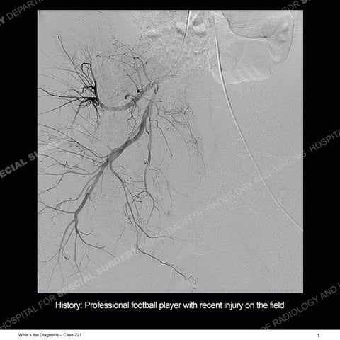
Diagnosis: Traumatic Pseudoaneurysm of the Obturator Artery An extremely rare occurrence, and one that we encounter particularly infrequently, is the development of a pseudoaneurysm. Pseudoaneurysms occur following injury to a blood vessel, when blood leaking from the vessel becomes contained by a surrounding fibrous wall. In the trauma setting, these commonly occur following high-energy mechanisms of injury, such as motor vehicle collisions or significant blunt trauma. There is extremely sparse literature about pseudoaneurysms in professional sports, although they are known to occur. In this case, the massive shearing injury sustained by the anterior abdominal wall also led to injury of the blood vessel. This patient sustained their injury during a professional football game, although the exact mechanism is not entirely clear. The patient was admitted to the hospital because of pain and difficulty urinating. Serial examinations demonstrated a declining hematocrit level, prompting the CT angiography study. On the angiography study, the decision was made to perform multiple phases of imaging to allow detection of what could be a subtle but important finding. This proved to be the case as the subtle blush of the obturator artery on the post contrast studies became much more conspicuous with the precontrast imaging. This patient was transferred to the interventional radiology service and underwent selective embolization of the vessel. He is now hemodynamically stable and will undergo repair of the extensive anterior abdominal wall injury imminently.
What’s the Diagnosis – Case 221
71
What’s the Diagnosis – Case 221
72
What’s the Diagnosis – Case 221
73
What’s the Diagnosis – Case 221
74
What’s the Diagnosis – Case 221
75
What’s the Diagnosis – Case 221
76