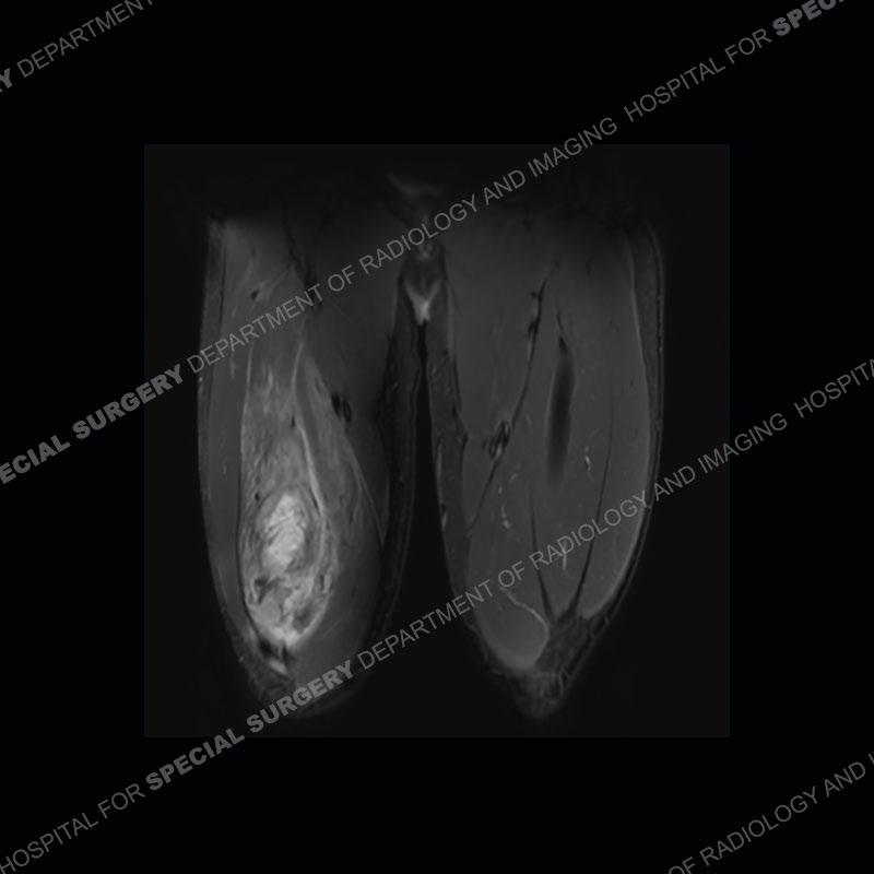
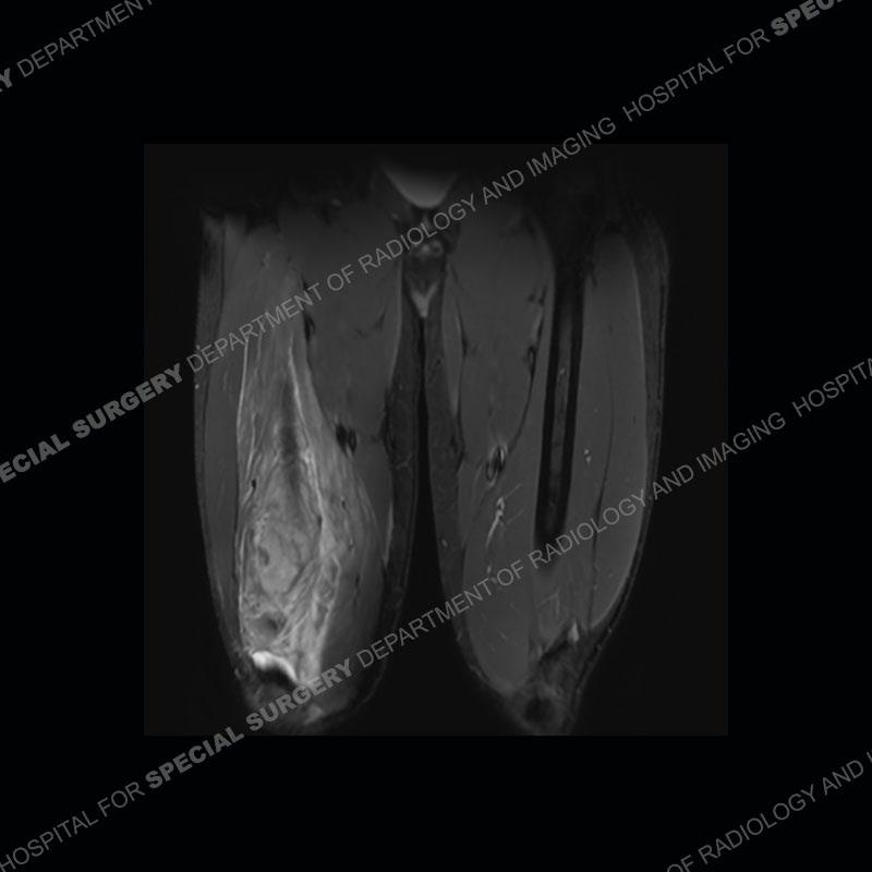
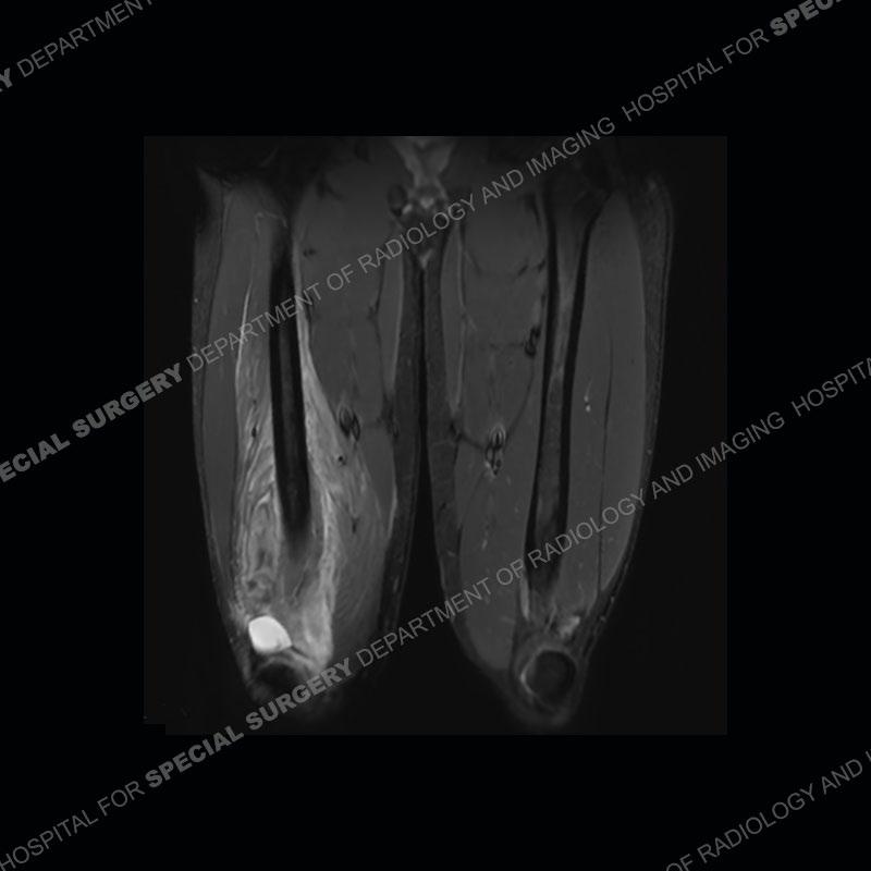
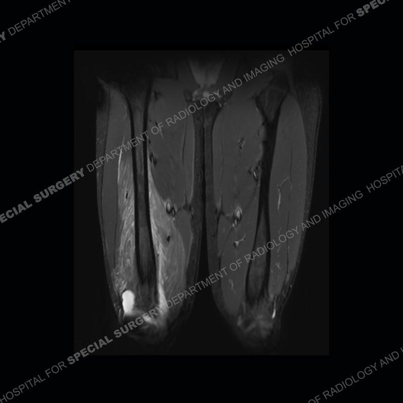
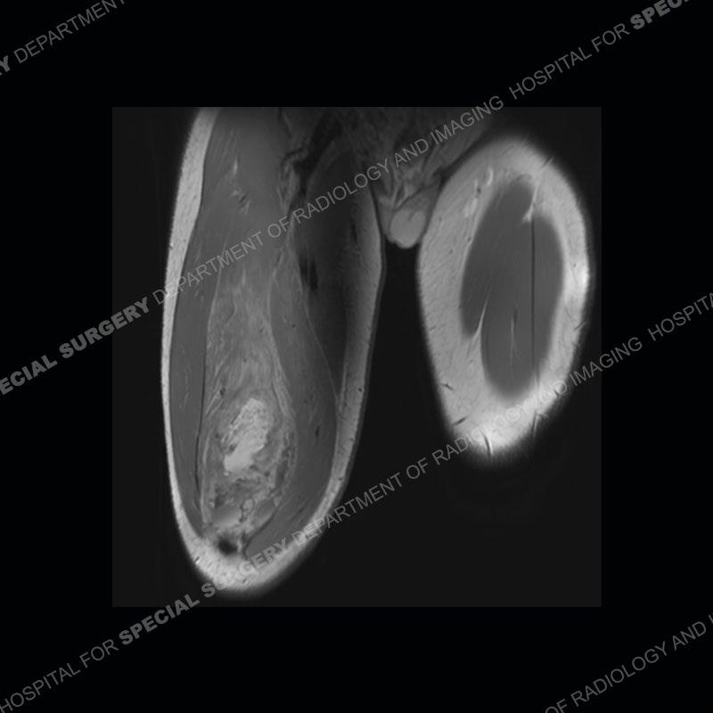
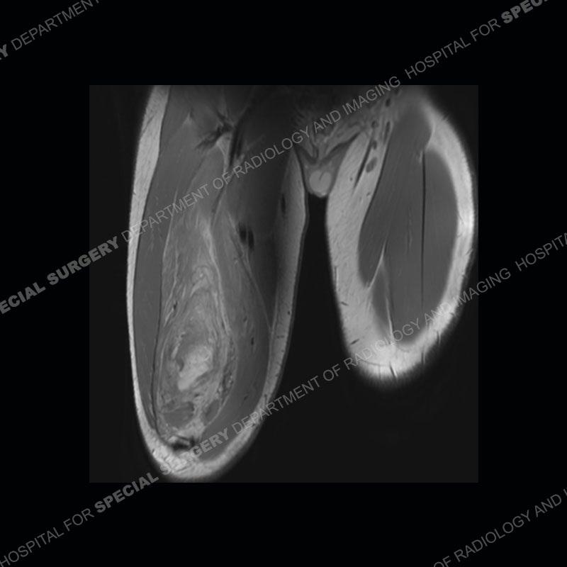
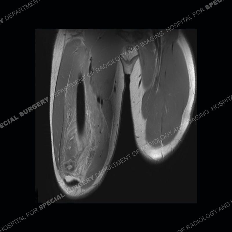
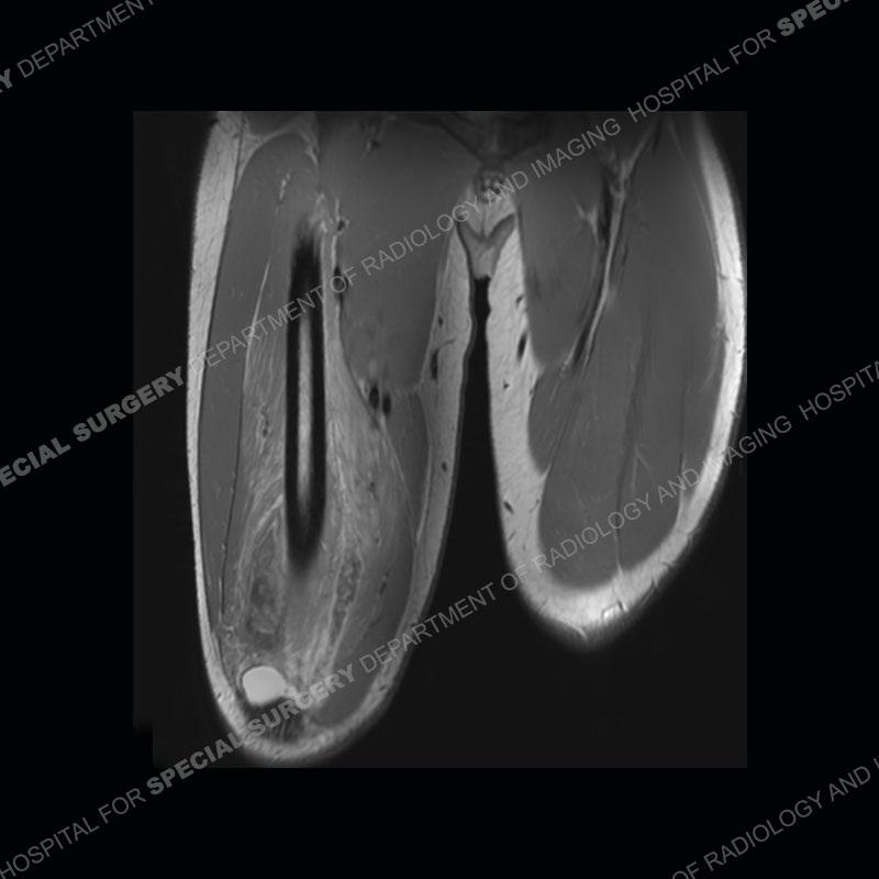
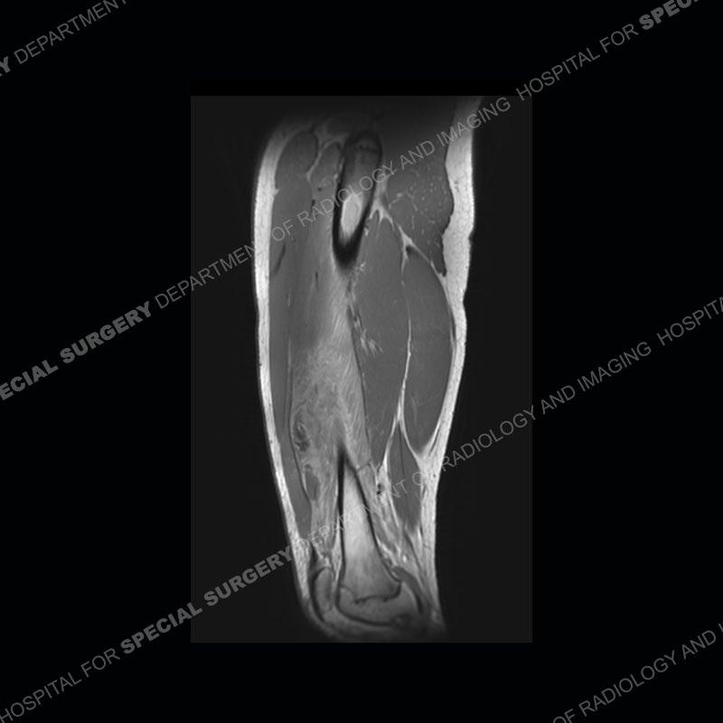
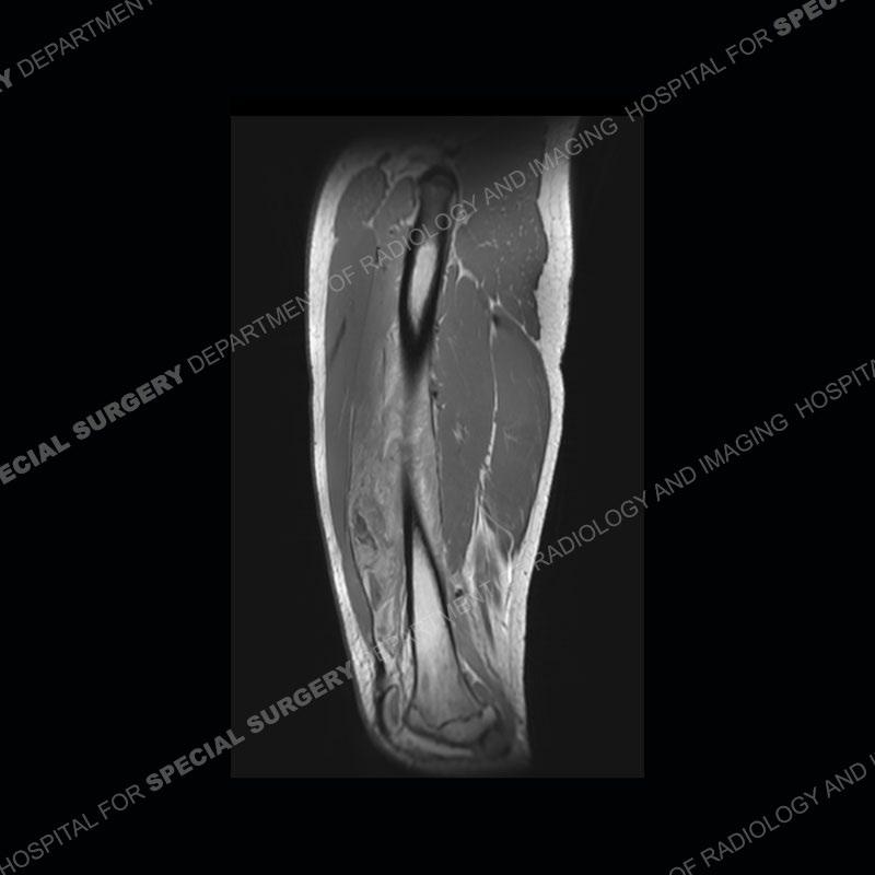
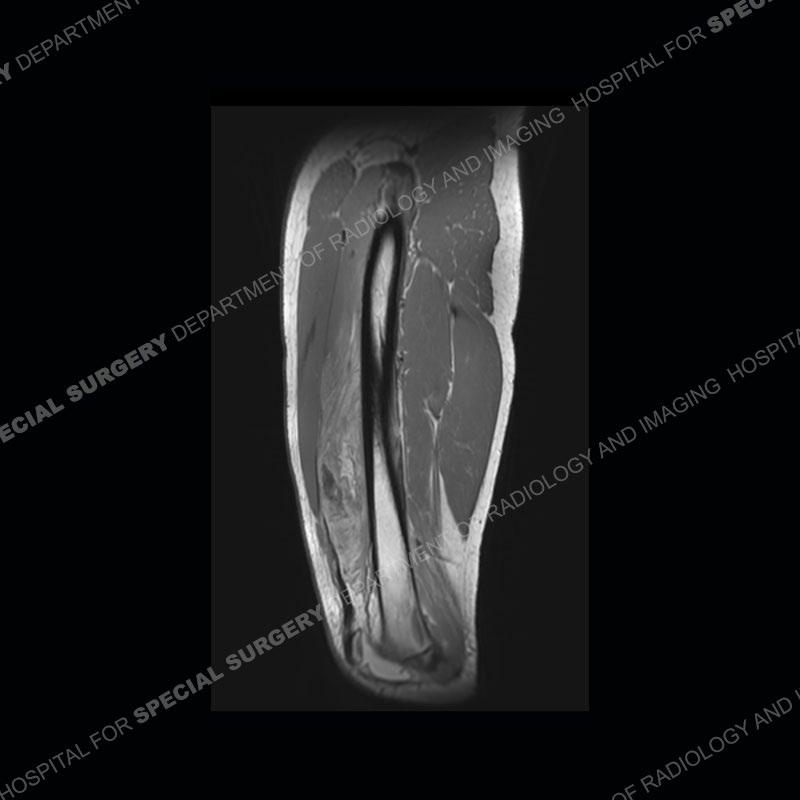
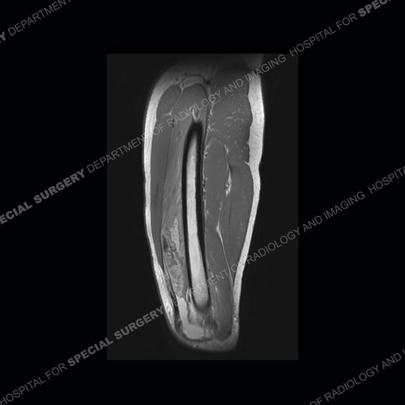
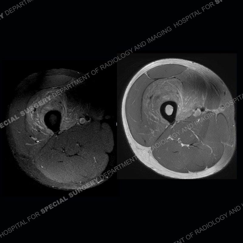
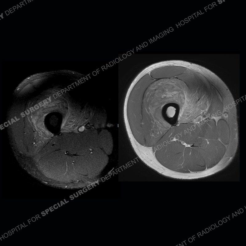
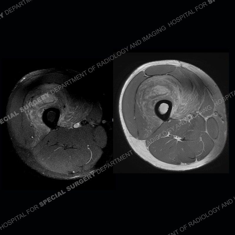
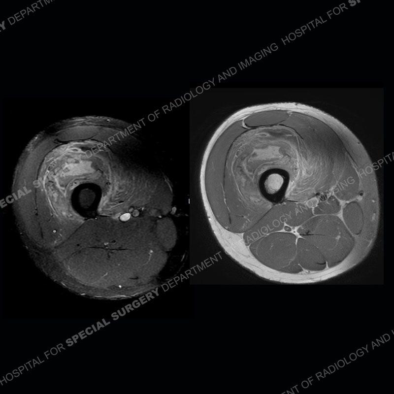
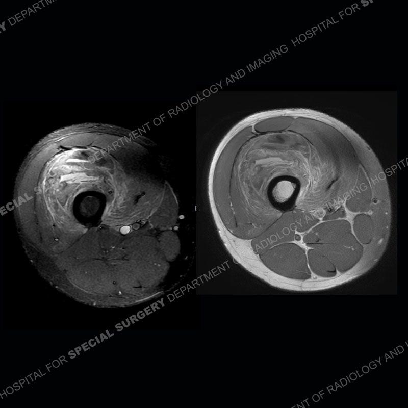
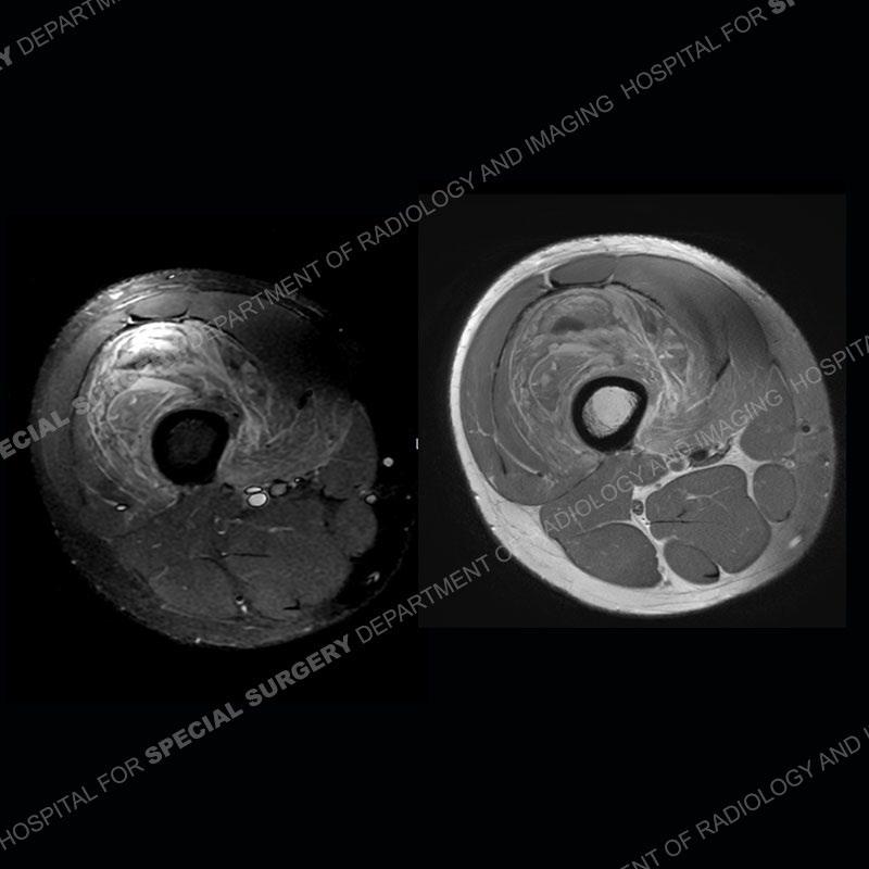
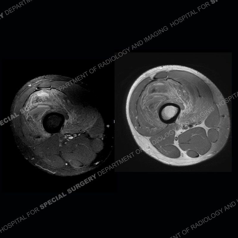
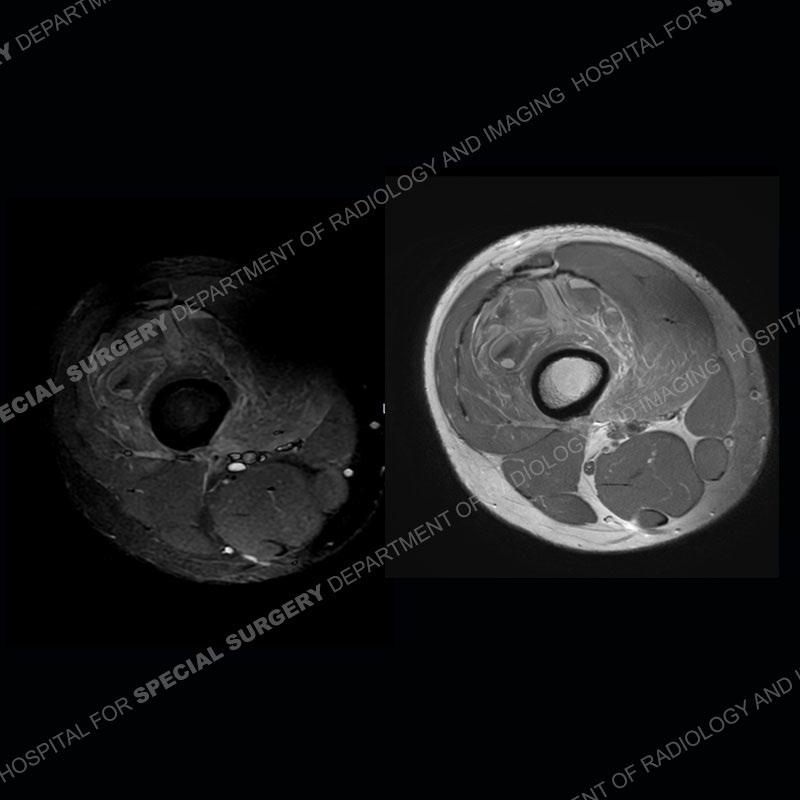
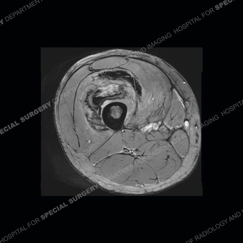
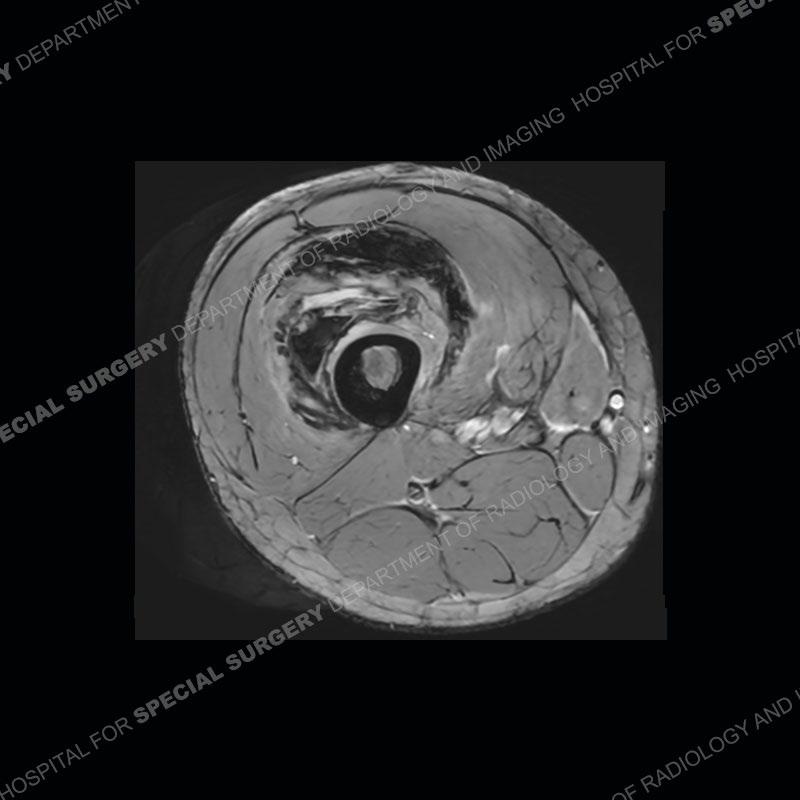
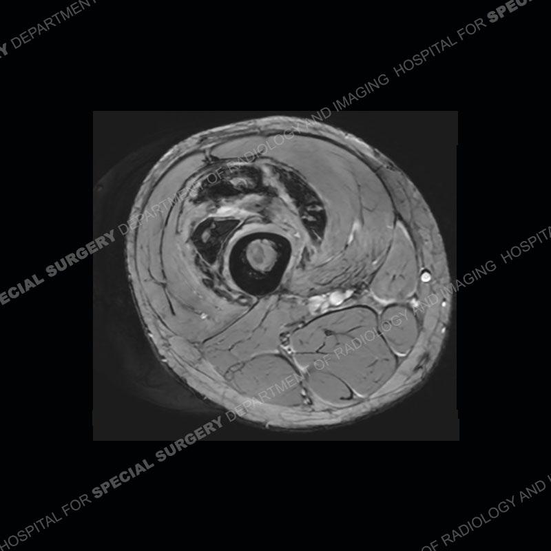
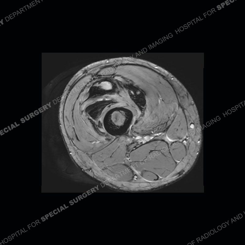
Findings
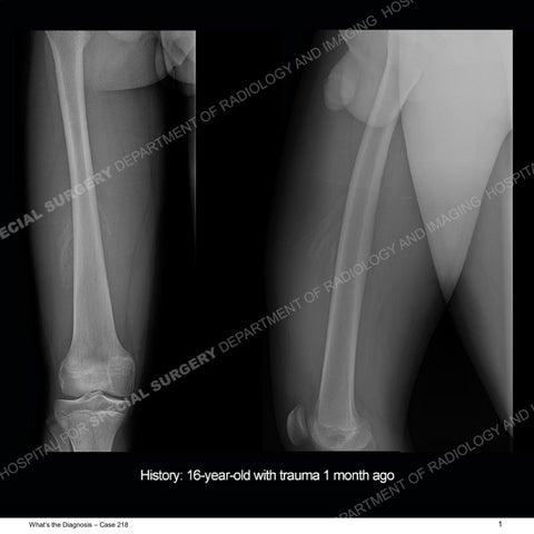
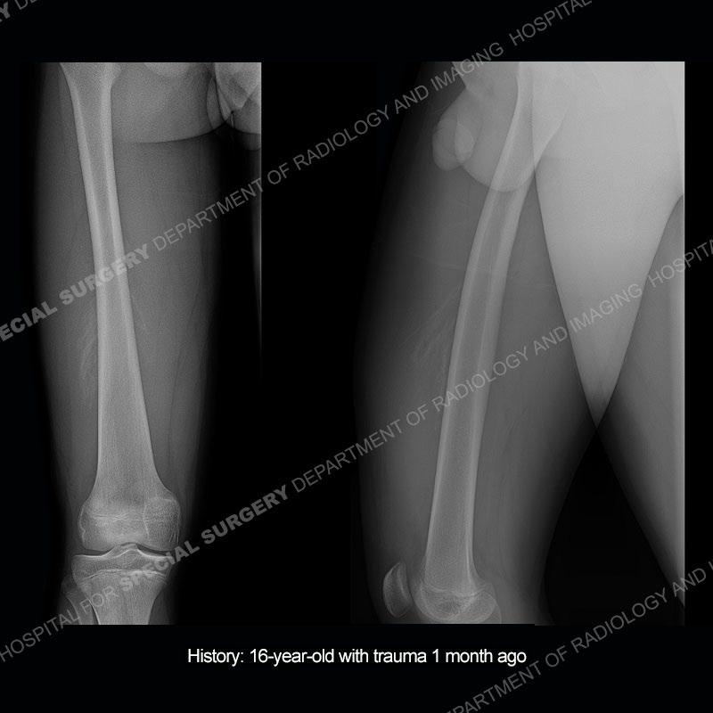
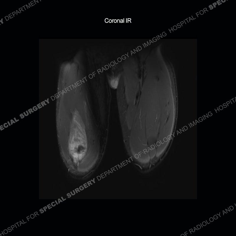
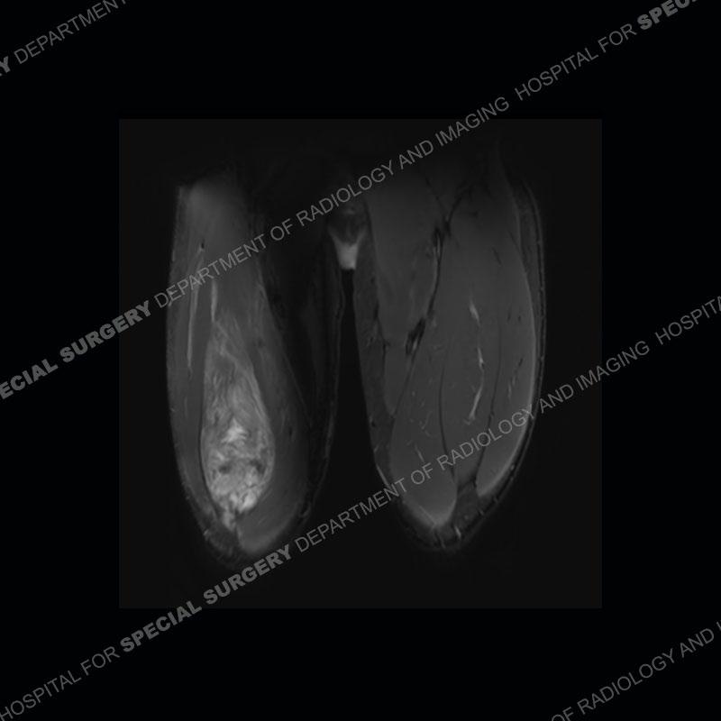
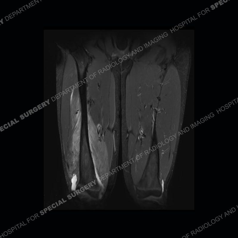
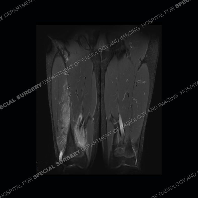
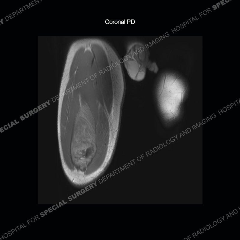
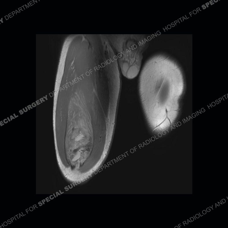
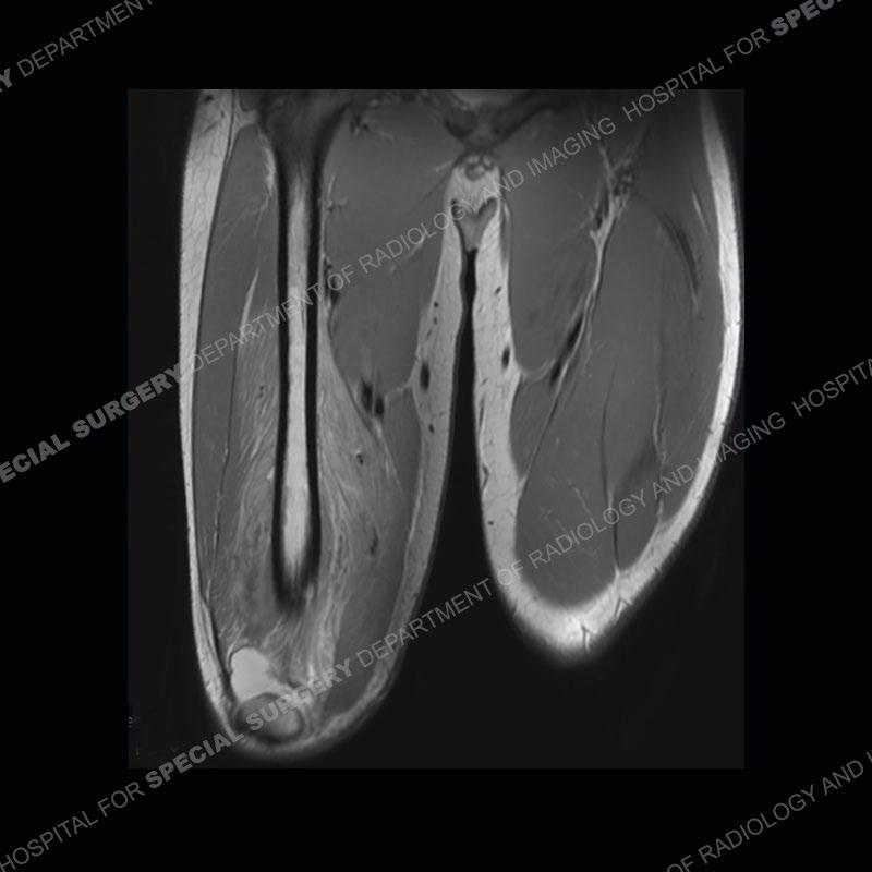
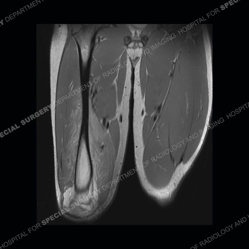
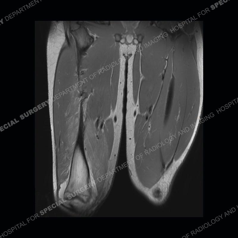
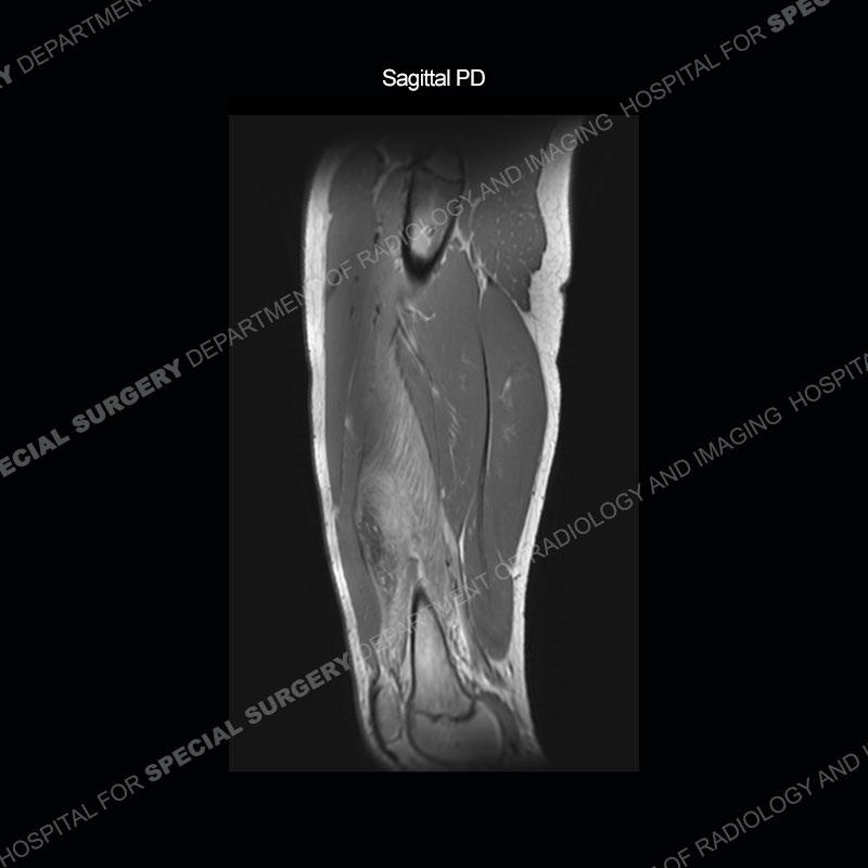
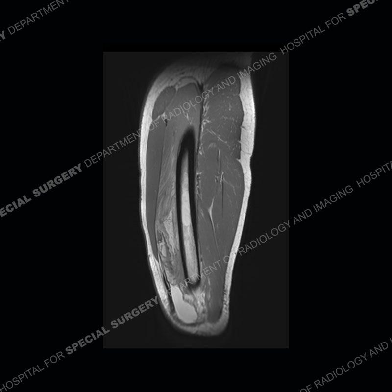
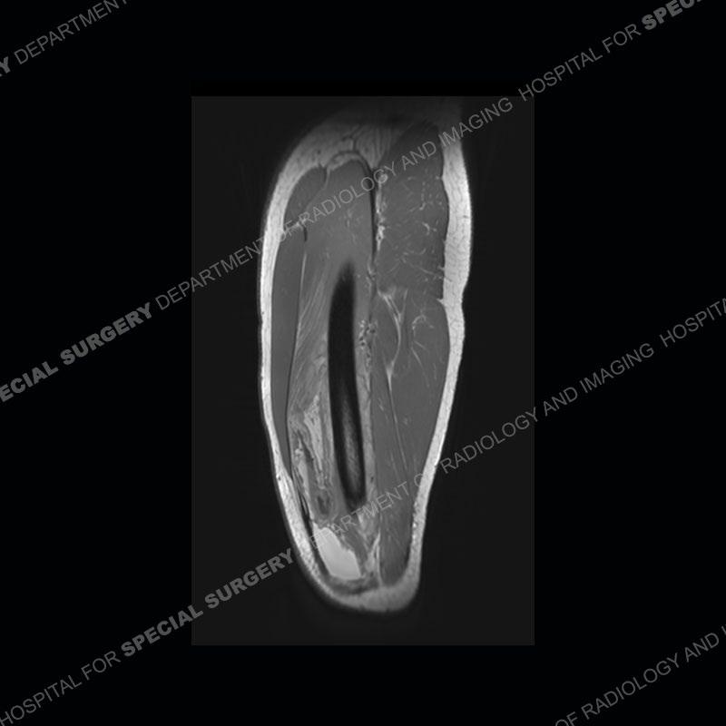
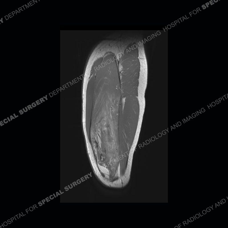
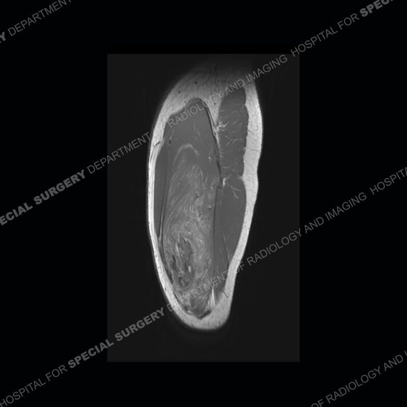
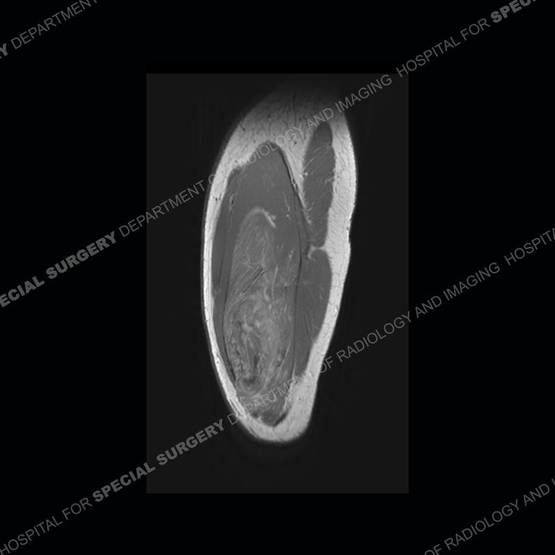
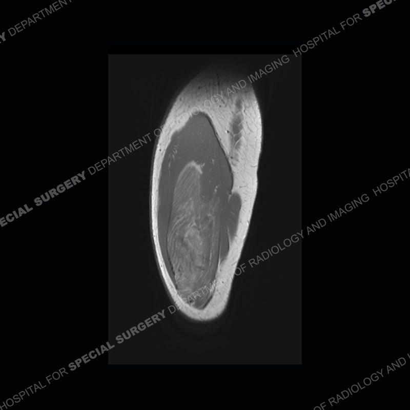
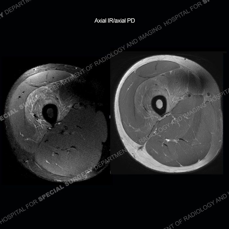
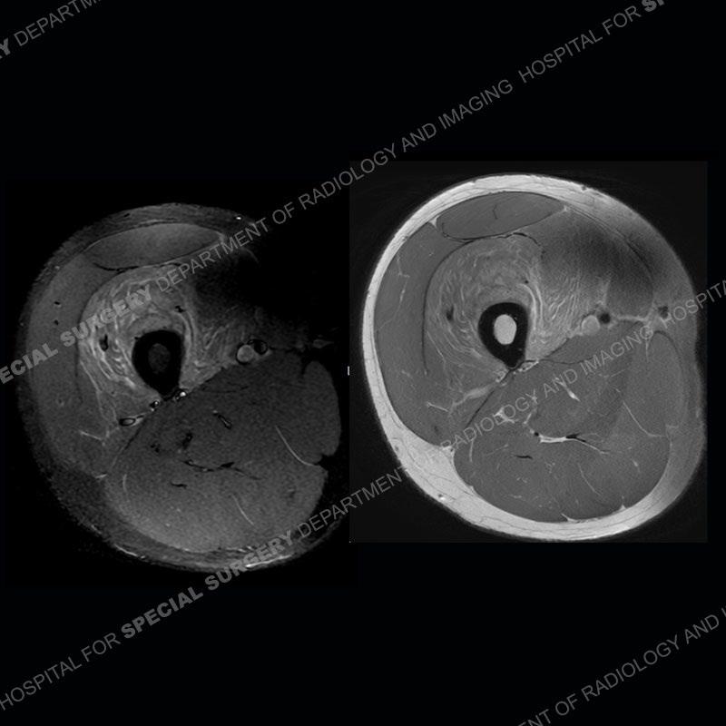
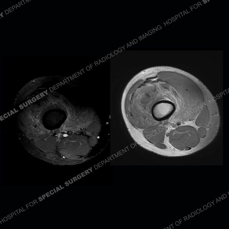
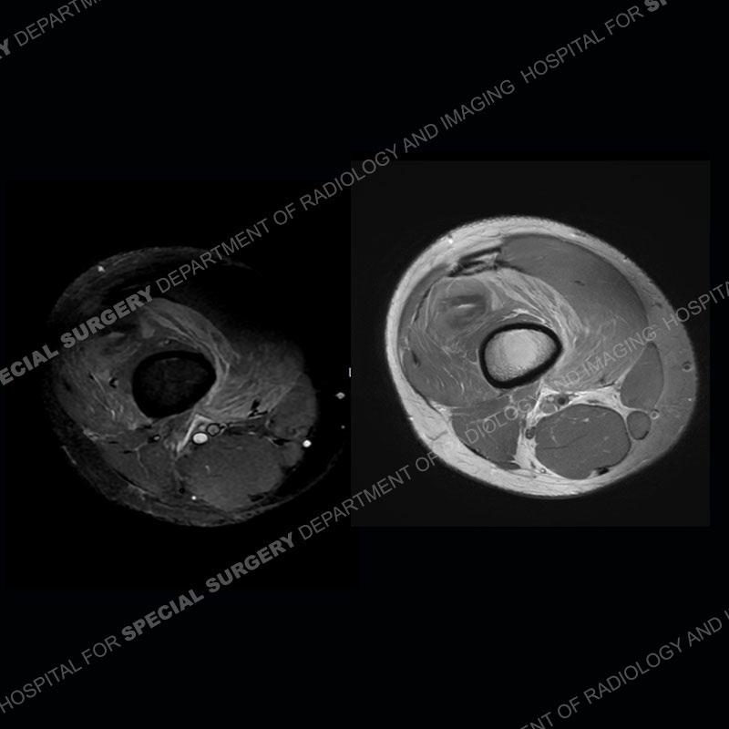
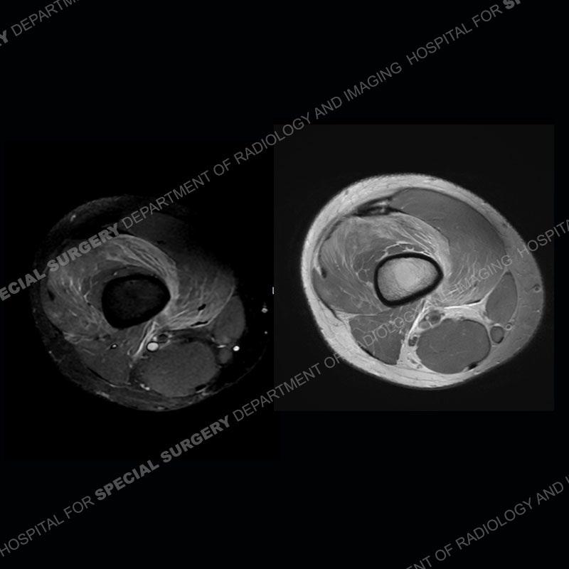
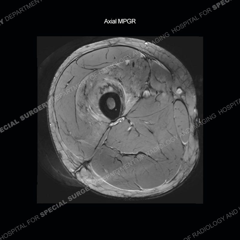
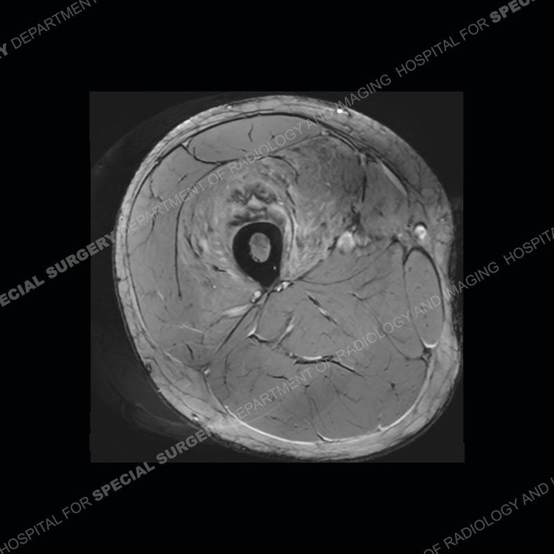
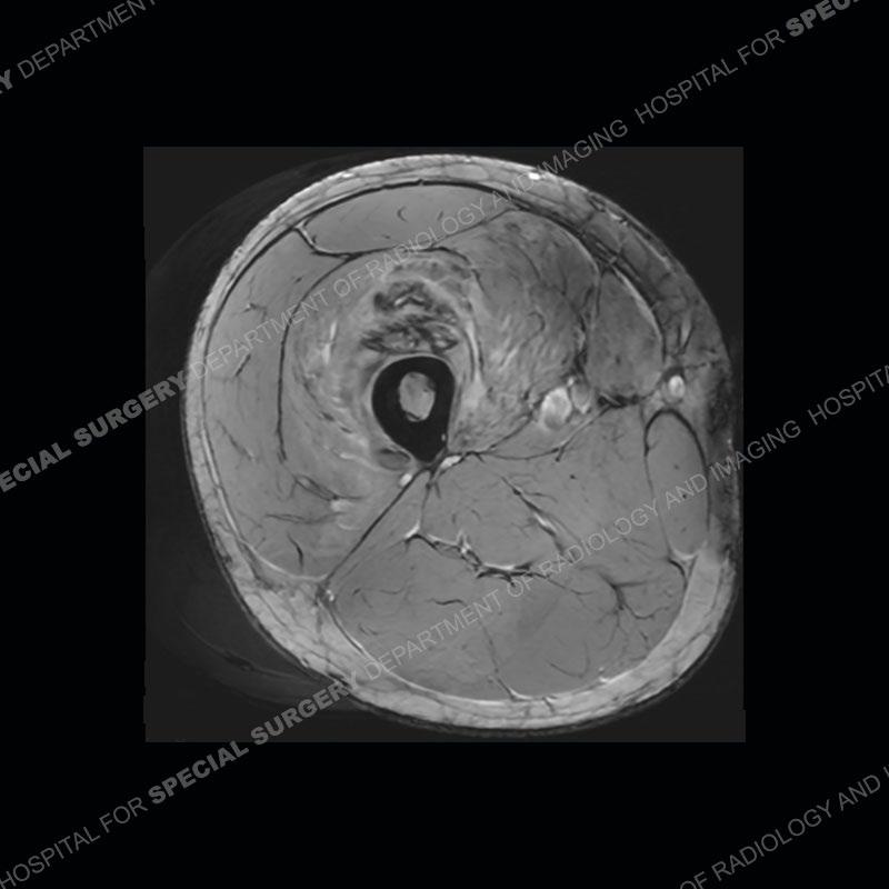
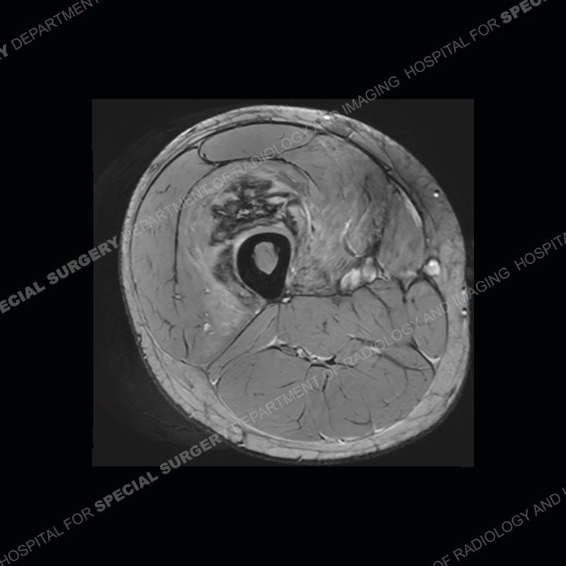
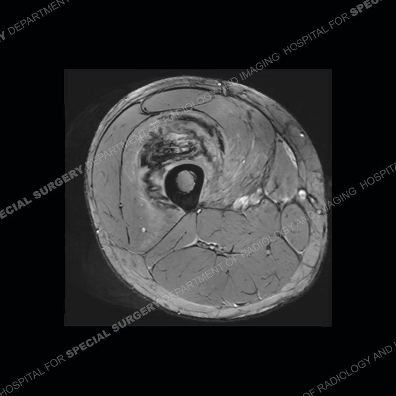
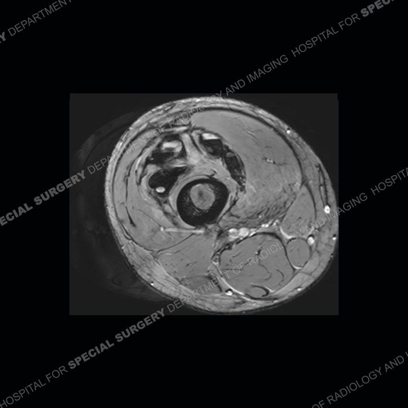
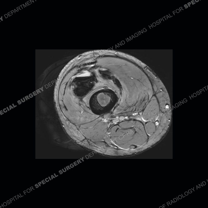
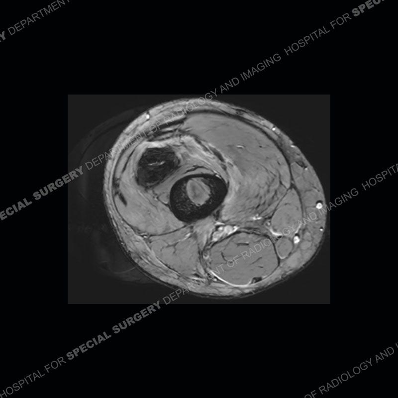
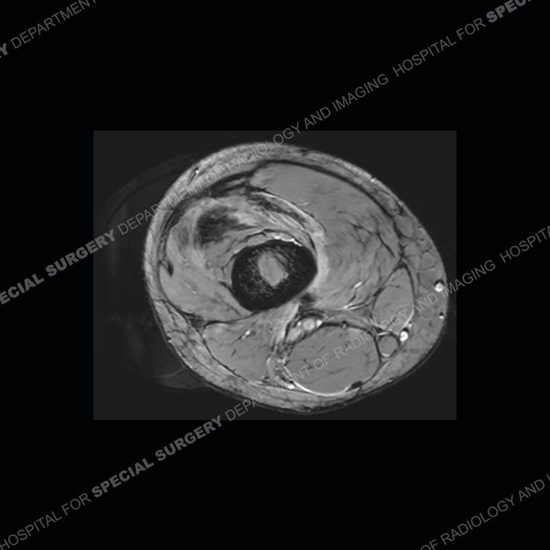
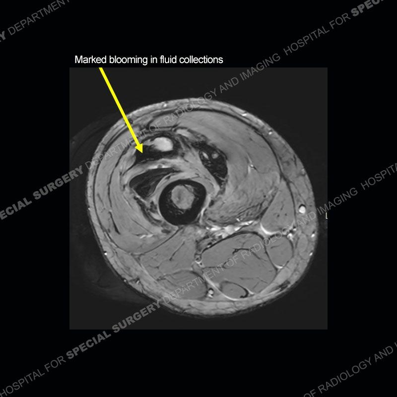
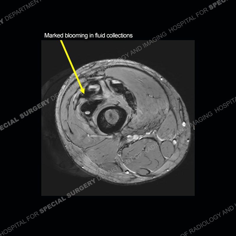
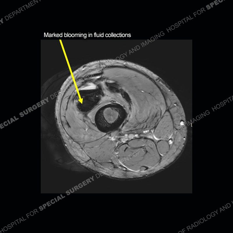
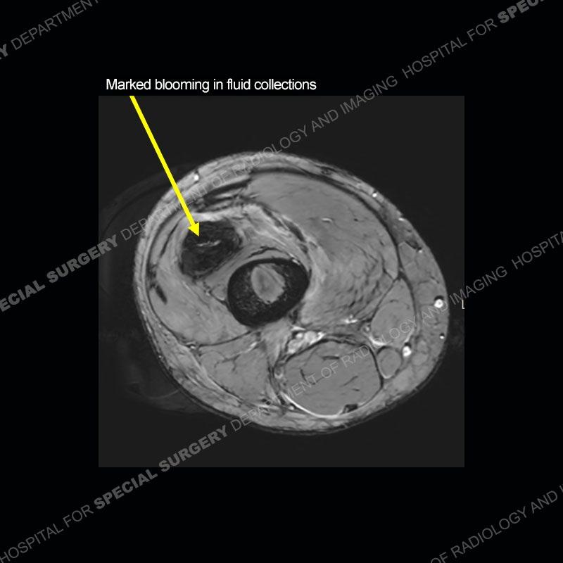
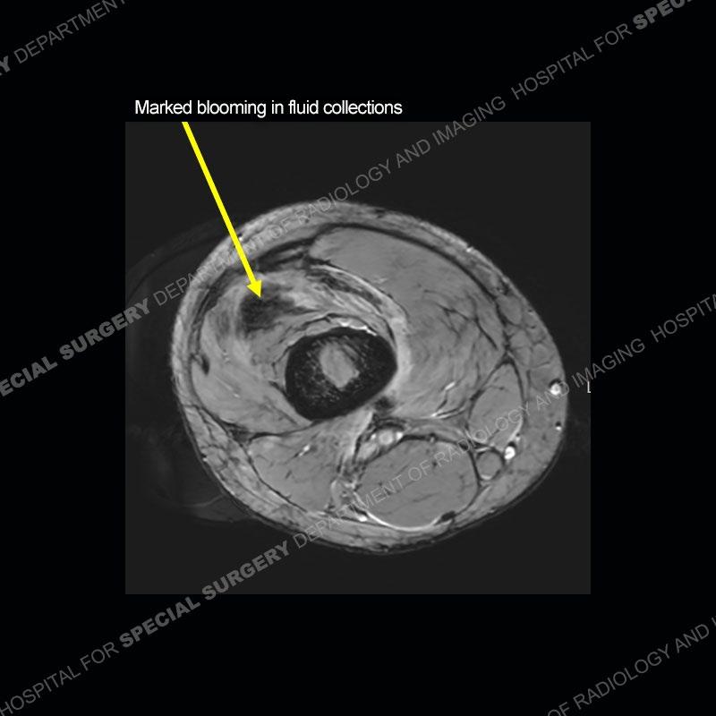
The radiographs demonstrate spicules of bone about the level of the femoral shaft extending into the soft tissue. The MRI demonstrates a marked amount of edema of the soft tissue with a complex architecture. Fluid collections are present containing areas of intermediate signal, and a marked amount of low signal/blooming phenomenon is present on the gradient echo images in the soft tissue.

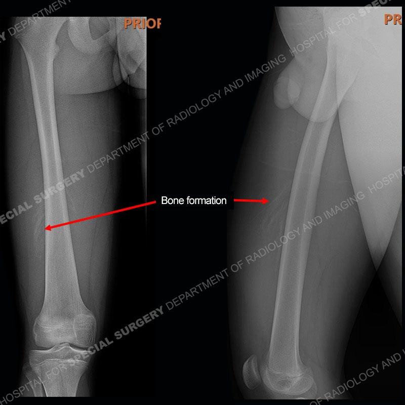
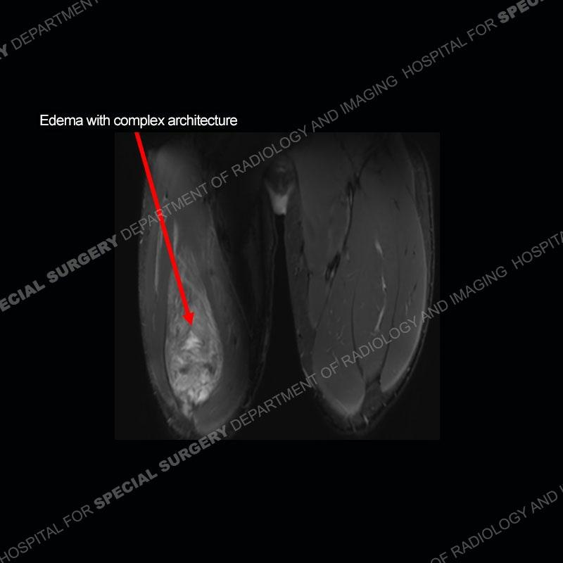
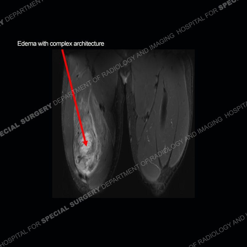
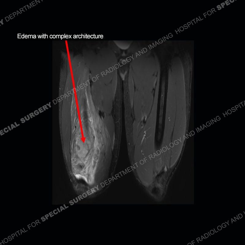
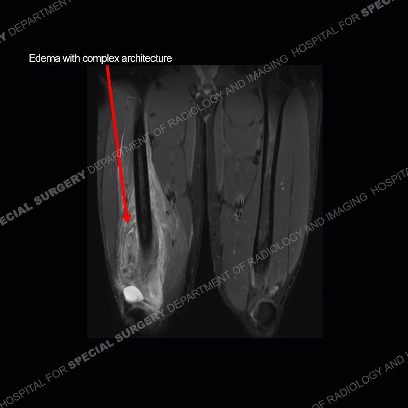
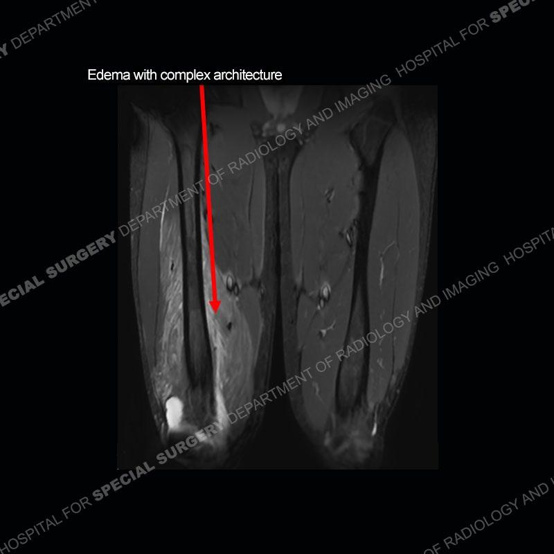
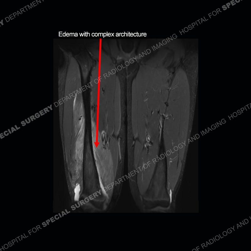
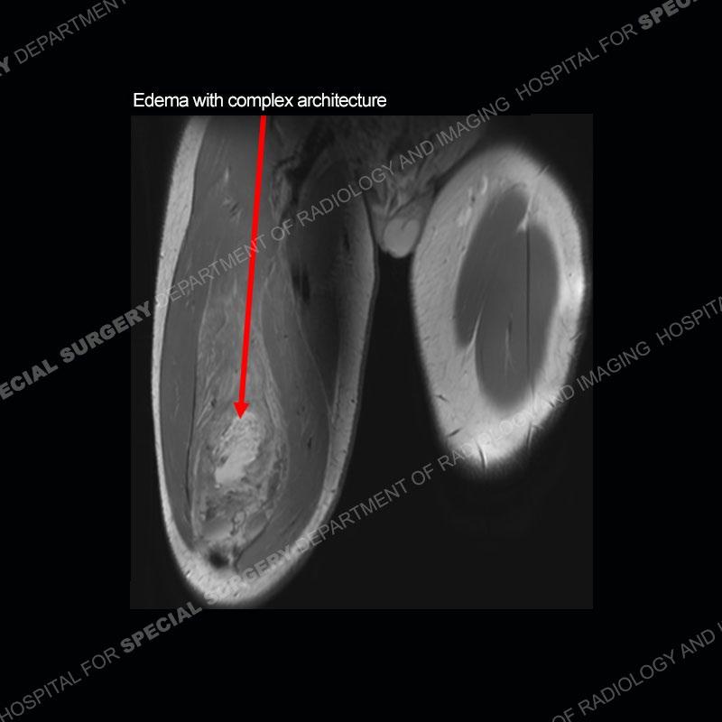
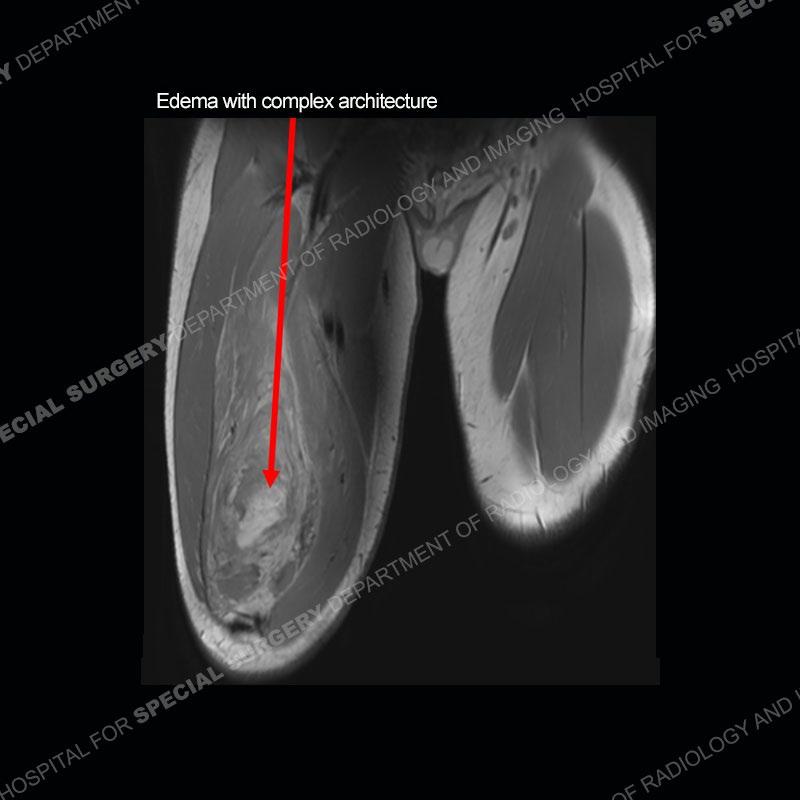
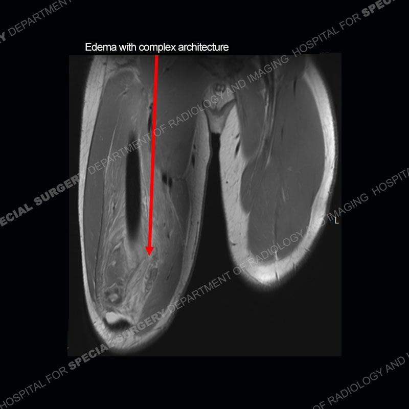
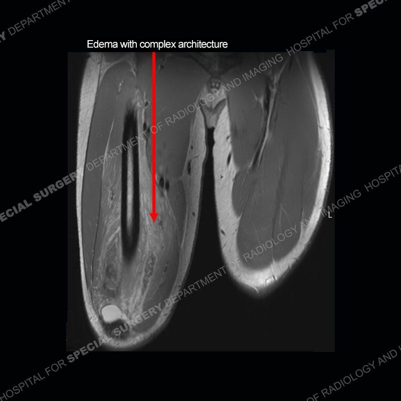
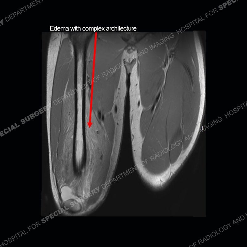
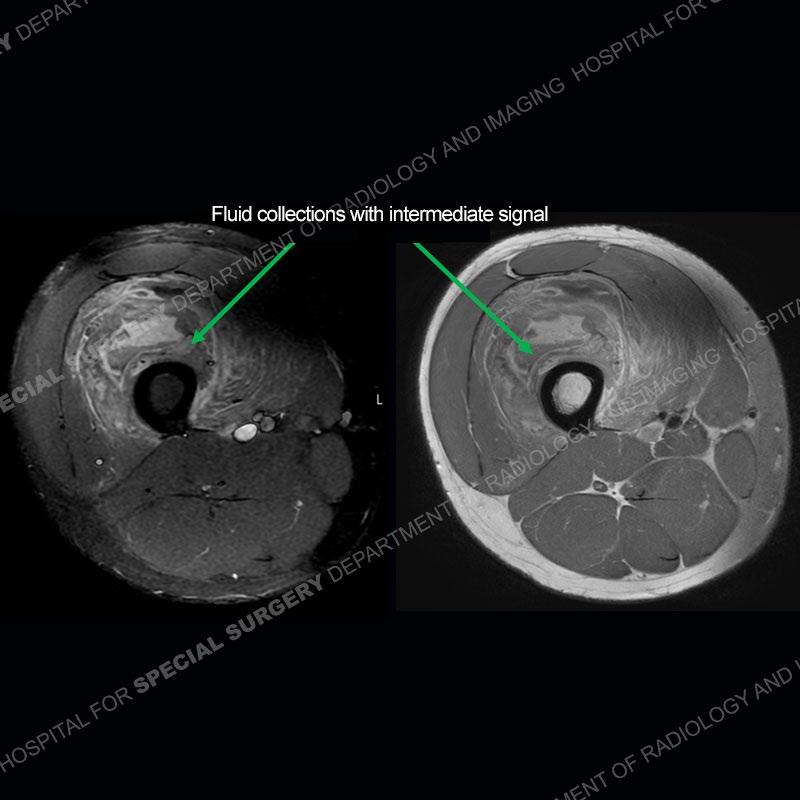
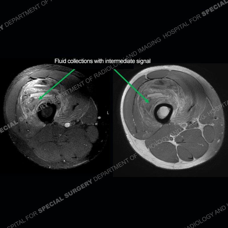
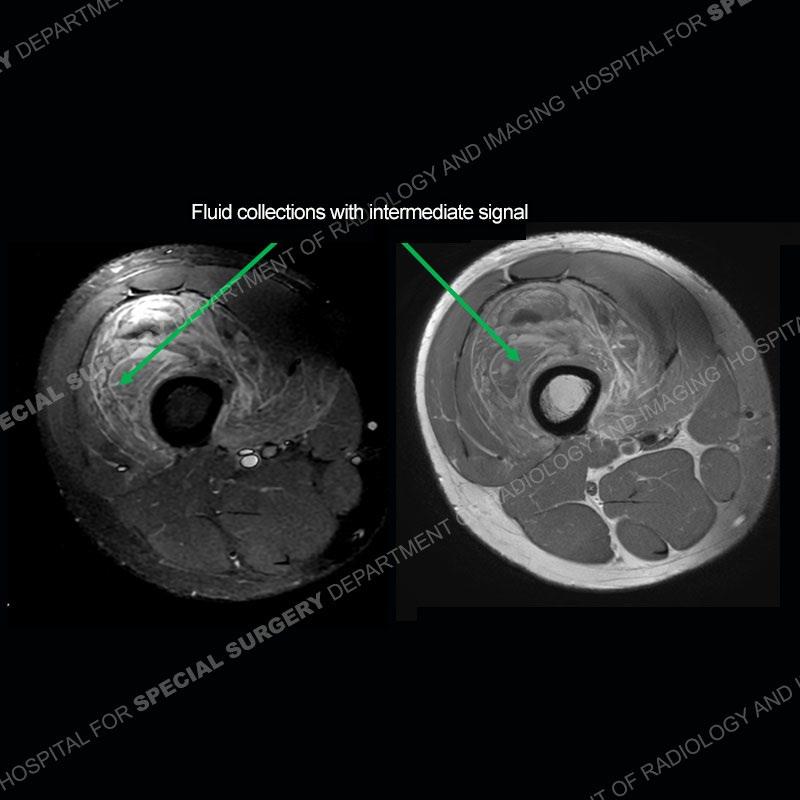
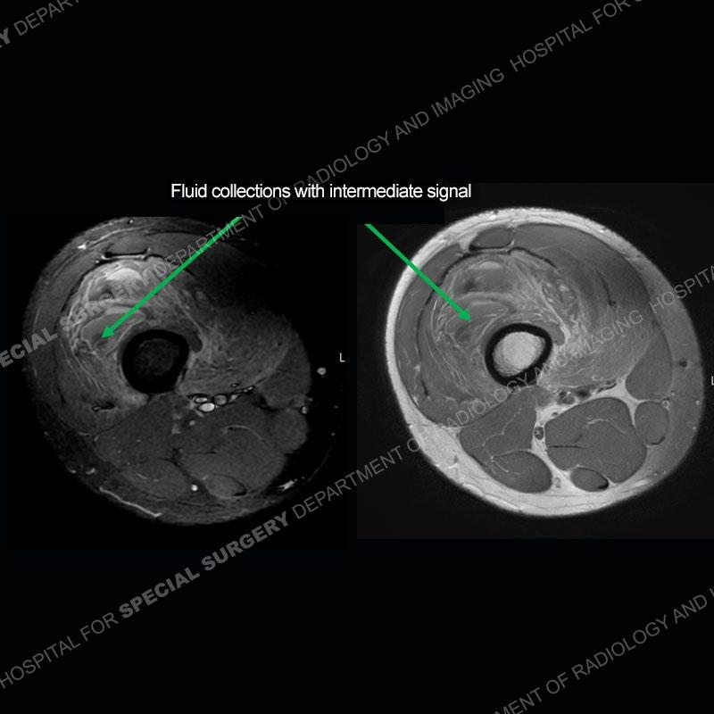
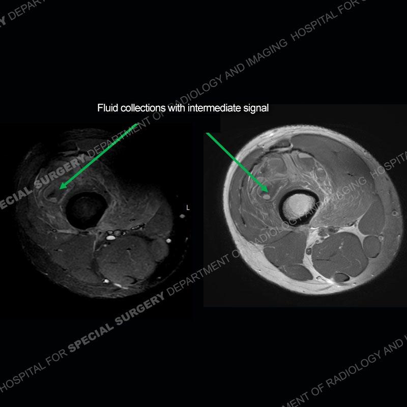
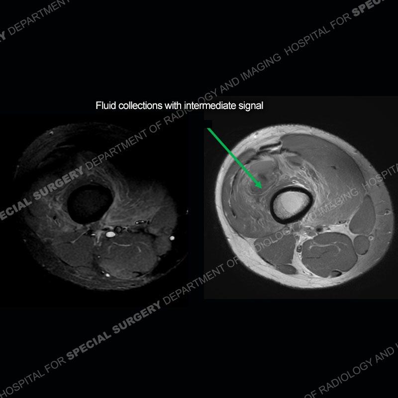
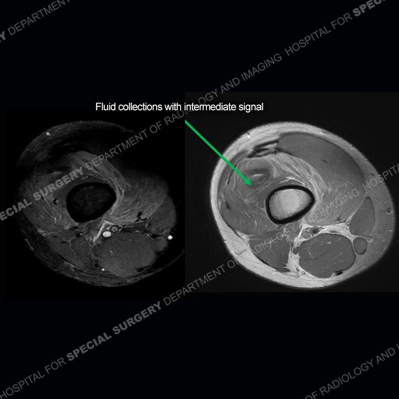
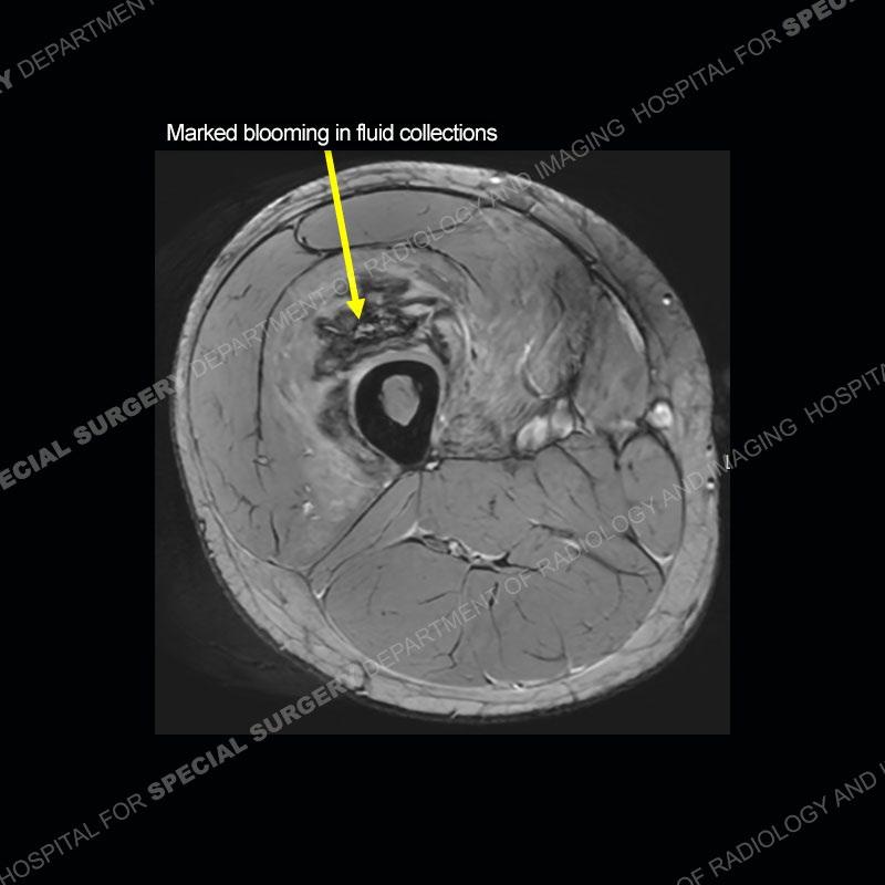
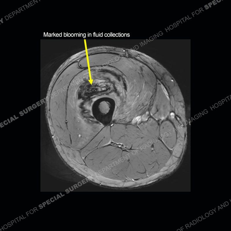
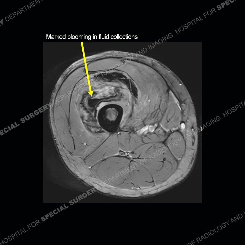
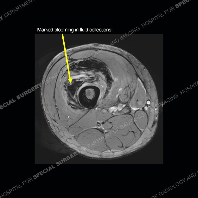
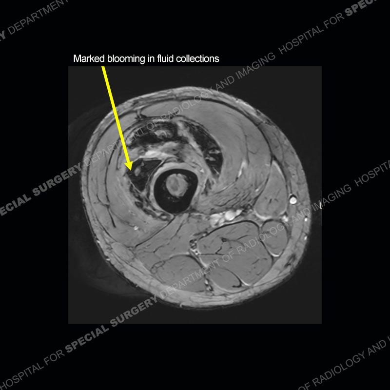
Diagnosis: Heterotopic Ossification (HO)/Myositis Ossificans (MO
What can be a very difficult if not impossible diagnosis to make early on. This case highlights how complex the imaging can look in these cases particularly relatively early in the post-traumatic time frame. Early on, the MRI as in this case, will show a marked amount of edema. The fluid collections in this case are hematoma/blood filled with intermediate blood being denser than fluid and positioned at the posterior aspect o the collections. The prominent areas of blooming phenomenon on the gradient echo sequence mostly represent areas of hemosiderin deposition. A portion of this may represent the bone formation but most represents the hemosiderin from the blood product in the setting of early trauma.
The bone formation is much more readily identified on the radiographs. In the traumatic situation, the bone becomes denser and better organized with defined cortical and medullary bone from the periphery of the area of injury and extending centrally. When a neoplastic process is present, the bone tends to be more sporadic in form, does not demonstrate a zonal architecture with maturation from periphery to central, and will not show well demarcated cortex and medullary cavities.
In the early stages of developing HO/MO the bone formation and bone pattern may be very difficult to assess. In these situations, it is often advisable to wait a number of weeks or even months to allow the bone formation to mature and for the process to declare itself.
One of the main clinical scenarios to separate is if an underlying neoplasm is present and trauma has been sustained to the neoplasm causing the bleeding. In those cases, a mass can often be seen but not always as the degree of edema and hematoma in the soft tissue may make this difficult. Also, bone formation may be present in the setting of neoplasm or trauma. Again, sometimes just with waiting and repeat imaging the bone formation and architecture may help in coming to the diagnosis of HO/MO. If after an appropriate time frame and repeat imaging, the bone formation is not clearly HO or an underlying mass persists, then a biopsy may be warranted.