What’s the Diagnosis – Case 213
1
What’s the Diagnosis – Case 213
2
What’s the Diagnosis – Case 213
3
What’s the Diagnosis – Case 213
4
What’s the Diagnosis – Case 213
5
What’s the Diagnosis – Case 213
6
What’s the Diagnosis – Case 213
7
What’s the Diagnosis – Case 213
8
What’s the Diagnosis – Case 213
9
What’s the Diagnosis – Case 213
10
What’s the Diagnosis – Case 213
11
What’s the Diagnosis – Case 213
12
What’s the Diagnosis – Case 213
13
What’s the Diagnosis – Case 213
14
What’s the Diagnosis – Case 213
15
What’s the Diagnosis – Case 213
16
What’s the Diagnosis – Case 213
17
What’s the Diagnosis – Case 213
18
What’s the Diagnosis – Case 213
19
What’s the Diagnosis – Case 213
20
What’s the Diagnosis – Case 213
21
What’s the Diagnosis – Case 213
22
What’s the Diagnosis – Case 213
23
What’s the Diagnosis – Case 213
24
What’s the Diagnosis – Case 213
25
What’s the Diagnosis – Case 213
26
What’s the Diagnosis – Case 213
27
What’s the Diagnosis – Case 213
28
What’s the Diagnosis – Case 213
29
What’s the Diagnosis – Case 213
30
What’s the Diagnosis – Case 213
31
What’s the Diagnosis – Case 213
32
What’s the Diagnosis – Case 213
33
What’s the Diagnosis – Case 213
34
What’s the Diagnosis – Case 213
35
What’s the Diagnosis – Case 213
36
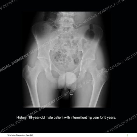
Findings The radiograph demonstrates the patient is skeletally mature at some locations and immature at others. There is subtle irregularity seen of both greater trochanters. The MRI demonstrates prominent edema pattern particularly about tendinous attachments to the greater and lesser trochanters. Bony abnormalities are seen of the greater trochanters as well. Edema pattern is seen about a lower lumbar facet joint. The sacroiliac joints are not completely mature but with additionally areas of edema pattern of the bone and subchondral bony abnormalities. Both hip joints demonstrate joint effusions with synovitis and areas of edema pattern of both femoral heads.
What’s the Diagnosis – Case 213
37
What’s the Diagnosis – Case 213
38
What’s the Diagnosis – Case 213
39
What’s the Diagnosis – Case 213
40
What’s the Diagnosis – Case 213
41
What’s the Diagnosis – Case 213
42
What’s the Diagnosis – Case 213
43
What’s the Diagnosis – Case 213
44
What’s the Diagnosis – Case 213
45
What’s the Diagnosis – Case 213
46
What’s the Diagnosis – Case 213
47
What’s the Diagnosis – Case 213
48
What’s the Diagnosis – Case 213
49
What’s the Diagnosis – Case 213
50
What’s the Diagnosis – Case 213
51
What’s the Diagnosis – Case 213
52
What’s the Diagnosis – Case 213
53
What’s the Diagnosis – Case 213
54
What’s the Diagnosis – Case 213
55
What’s the Diagnosis – Case 213
56
What’s the Diagnosis – Case 213
57
What’s the Diagnosis – Case 213
58
What’s the Diagnosis – Case 213
59
What’s the Diagnosis – Case 213
60
What’s the Diagnosis – Case 213
61
What’s the Diagnosis – Case 213
62
What’s the Diagnosis – Case 213
63
What’s the Diagnosis – Case 213
64
Diagnosis: Enthesitis related Arthritis Enthesitis related arthritis (ERA) is one of the seven categories of juvenile inflammatory arthritis (JIA). JIA is a heterogeneous group of arthritides. ERA is a process related to ligament and tendinous attachments. Although this can be found in the other types of JIA, in ERA it is a prominent feature. In ERA, sacroiliitis and peripheral arthritis particularly of lower extremity joints may also be found. ERA is more common in boys who frequently are HLA-B27 positive. Uveitis is also frequently found. It is important to try and classify this arthritis as unfortunately, patients with ERA often have a higher burden of disease and overall worse outcome. In the case presented, the subtle irregularities of the greater trochanters on the radiograph represented erosions. The edema pattern at the entheses represent ongoing inflammatory change. The findings at the sacroiliac joints in part may relate to skeletal immaturity but the subchondral edema pattern and extent of abnormality indicate there are also erosions and an active sacroiliitis. Although not the dominant feature, inflammatory change of the facet joints as seen in this case can occur and inflammation of the hip joints (also as seen in this case) can be present. The edema pattern of the femoral heads was likely about subtle capsular attachment sites.
What’s the Diagnosis – Case 213
65
References Mistry RR, Patro P, Agarwal V, Misra DP. Enthesitis-related arthritis: current perspectives. Open Access Rheumatol. 2019 Jan 25;11:19-31. doi: 10.2147/OARRR.S163677. PMID: 30774484; PMCID: PMC6354696.
What’s the Diagnosis – Case 213
66