What’s the Diagnosis – Case 210
1
What’s the Diagnosis – Case 210
2
What’s the Diagnosis – Case 210
3
What’s the Diagnosis – Case 210
4
What’s the Diagnosis – Case 210
5
What’s the Diagnosis – Case 210
6
What’s the Diagnosis – Case 210
7
What’s the Diagnosis – Case 210
8
What’s the Diagnosis – Case 210
9
What’s the Diagnosis – Case 210
10
What’s the Diagnosis – Case 210
11
What’s the Diagnosis – Case 210
12
What’s the Diagnosis – Case 210
13
What’s the Diagnosis – Case 210
14
What’s the Diagnosis – Case 210
15
What’s the Diagnosis – Case 210
16
What’s the Diagnosis – Case 210
17
What’s the Diagnosis – Case 210
18
What’s the Diagnosis – Case 210
19
What’s the Diagnosis – Case 210
20
What’s the Diagnosis – Case 210
21
What’s the Diagnosis – Case 210
22
What’s the Diagnosis – Case 210
23
What’s the Diagnosis – Case 210
24
What’s the Diagnosis – Case 210
25
What’s the Diagnosis – Case 210
26
What’s the Diagnosis – Case 210
27
What’s the Diagnosis – Case 210
28
What’s the Diagnosis – Case 210
29
What’s the Diagnosis – Case 210
30
What’s the Diagnosis – Case 210
31
What’s the Diagnosis – Case 210
32
What’s the Diagnosis – Case 210
33
What’s the Diagnosis – Case 210
34
What’s the Diagnosis – Case 210
35
What’s the Diagnosis – Case 210
36
What’s the Diagnosis – Case 210
37
What’s the Diagnosis – Case 210
38
What’s the Diagnosis – Case 210
39
What’s the Diagnosis – Case 210
40
What’s the Diagnosis – Case 210
41
What’s the Diagnosis – Case 210
42
What’s the Diagnosis – Case 210
43
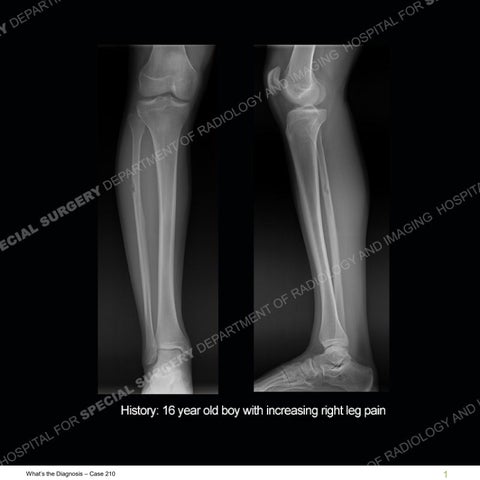
Findings The radiographs show a bubbly lesion of the shaft of the fibula with a loss of cortex laterally. The MRI shows a tangle of low signal structures in the soft tissue that have extended into the fibula at the area of the bubbly lesion. In the soft tissue and in the shaft of the fibula are multiple, prominent low signal structures. The low signal of the lesion is slightly more prominent on the gradient echo imaging. On the axial IR images in particular, there is a marrow edema pattern of the fibula adjacent to the bubbly lesion.
What’s the Diagnosis – Case 210
44
What’s the Diagnosis – Case 210
45
What’s the Diagnosis – Case 210
46
What’s the Diagnosis – Case 210
47
What’s the Diagnosis – Case 210
48
What’s the Diagnosis – Case 210
49
What’s the Diagnosis – Case 210
50
What’s the Diagnosis – Case 210
51
What’s the Diagnosis – Case 210
52
What’s the Diagnosis – Case 210
53
What’s the Diagnosis – Case 210
54
What’s the Diagnosis – Case 210
55
What’s the Diagnosis – Case 210
56
What’s the Diagnosis – Case 210
57
What’s the Diagnosis – Case 210
58
What’s the Diagnosis – Case 210
59
What’s the Diagnosis – Case 210
60
What’s the Diagnosis – Case 210
61
What’s the Diagnosis – Case 210
62
What’s the Diagnosis – Case 210
63
What’s the Diagnosis – Case 210
64
What’s the Diagnosis – Case 210
65
What’s the Diagnosis – Case 210
66
What’s the Diagnosis – Case 210
67
What’s the Diagnosis – Case 210
68
What’s the Diagnosis – Case 210
69
What’s the Diagnosis – Case 210
70
Diagnosis: AVM or Arteriovenous Malformation (High Flow Vascular Lesion) AVM or arteriovenous malformation is a high flow vascular lesion. Vascular lesions of soft tissue are a heterogeneous group of lesions with an often confusing classification. A somewhat simplified classification is the separation info infantile hemangioma, slow flow vascular lesion, and high flow vascular lesion. An infantile hemangioma is a true neoplasm which appears very early in life but typically involutes in childhood. Slow flow vascular lesions are much more common than high flow vascular lesions. Slow flow vascular lesions on MRI are often lobulated or bubbly with high T2 or IR signal. Small phleboliths or areas of dephasing may be seen on gradient echo imaging. They frequently insinuate about the musculature of the extremities. Again, high flow vascular lesions are much less common than the slow flow lesions. High flow lesions are typically separated into arteriovenous fistula (AVF) or AVMs. AVMs are the more common of these two entities. The high flow of these lesions is such that the signal in the vessels flows out of the field during MR acquisition and hence the lesions/vessels are low in signal. The AVM has a central tangle or nidus of vessels, but then multiple other high flowing vessels are present bringing blood to and draining the AVM. This is what accounts for the multiple areas of low signal adjacent to the tangle of vessels in this AVM in the soft tissue and the medullary cavity of the fibula.
What’s the Diagnosis – Case 210
71
Diagnosis: AVM or Arteriovenous Malformation (High Flow Vascular Lesion) Many entities can produce bubbly lesions of the bone but one of the things that has to always be thought about is a vascular lesion. Lymphatic or vascular lymphatic lesions can also do this. This case was unusual for two reasons. First, high flow soft tissue lesions are very uncommon. Second, in this case the lesion extends into the bone causing the bubbly lesion seen on the radiographs. That is also an unusual circumstance but one that is seen in this case. In this case, the lesion has eroded the bone and likely the surrounding bone is seeing increased load or stress. Therefore, the edema in the adjacent bone is thought to represent a stress response from the wearing away of bone from this lesion.
What’s the Diagnosis – Case 210
72
References MR Imaging of Soft-Tissue Vascular Malformations: Diagnosis, Classification, and Therapy Follow-up. Lucía Flors, Carlos Leiva-Salinas, Ismaeel M. Maged, Patrick T. Norton, Alan H. Matsumoto, John F. Angle, MD Hugo Bonatti, Auh Whan Park, Ehab Ali Ahmad, Ugur Bozlar, Ahmed M. Housseini, Thomas E. Huerta, and Klaus D. Hagspiel RadioGraphics 2011 31:5, 1321-1340
What’s the Diagnosis – Case 210
73