Clinical History
30-year-old woman with right distal thigh pain 4.5 months after right femoral derotational osteotomy.

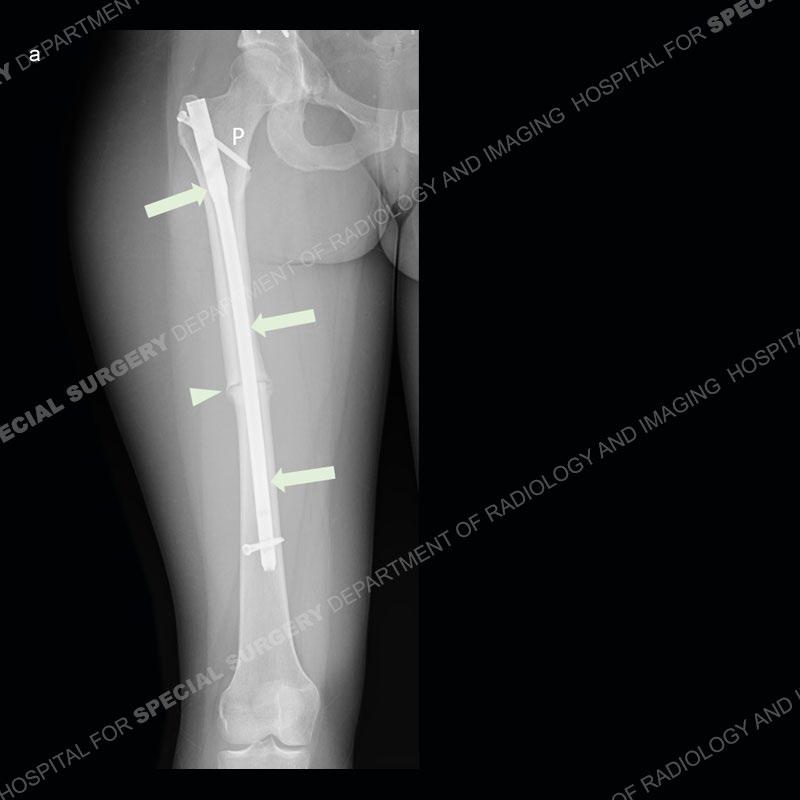
A recently-acquired radiograph of the right femur shows the intramedullary nail (arrows) with proximal (P) and distal (D) interlocking screws transfixing the femoral osteotomy (arrowhead).
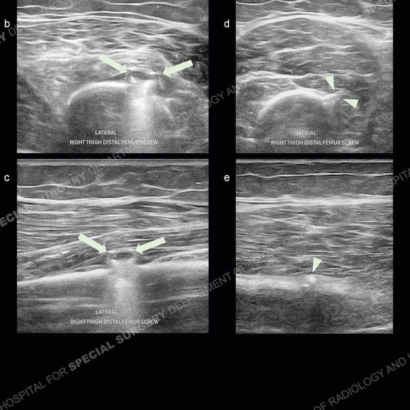
Sonographic evaluation of the right distal thigh was performed in the region of patient’s reported pain. Along the lateral aspect in the (b) transverse and (c) longitudinal orientations, imaging reveals the flat head of the distal interlocking scr (arrows) with associated dirty shadowing. Along the medial aspect in the same respective orientations (d) and (e) we see the pointy screw tip (arrowheads). The screw head and tip are proud to the bone and extend into the overlying muscle.
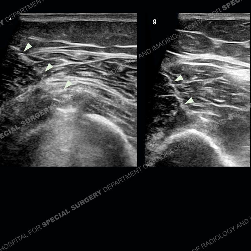
Ultrasound-guided injection of both ends of the distal screw was performed using anesthetic and cortisone, with images acquired during injection of the (f) screw head and (g) screw tip. Arrowheads indicate the needle track during each injection. Patient reported complete symptomatic relief immediately following the injections due to anesthetic effect.
Diagnosis: Pain from Hardware Impingement
Fixation hardware often extends beyond the cortex of the transfixed bone.
Sometimes the proud hardware can extend into muscle, which sometimes can be painful.
Ultrasound-guided injection can be used to confirm that the hardware is truly the pain generator.
Injection can also be done for symptomatic relief if the transfixed bone is not yet healed enough to allow hardware removal.