
Clinical History
64-year-old woman with new left ring finger soft tissue swelling.

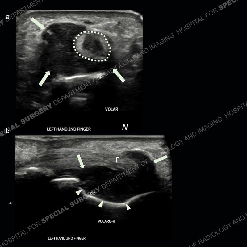
Sonographic imaging over the volar aspect of the left index finger in the transverse plane (a) reveals a hypoechoic lesion (arrows) deep to and extending along the radial aspect of the flexor tendons (dotted oval), with associated mass effect on the tendons. A sagittal image just radial of midline to the index finger (b) shows associated indolent erosion of the proximal phalangeal shaft and neck (arrowheads) with the lesion (arrows) wrapping over the flexor tendons (F).
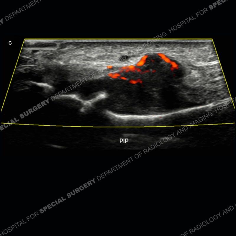
Power doppler imaging of the lesion showed intralesional vascularity (red).
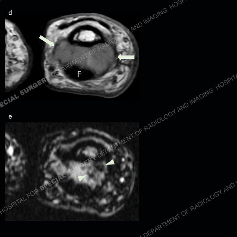
The patient underwent MRI of the left hand two months later. The axial proton density image (d) shows the mass (arrows) deep to the index finger flexor tendons (F). Foci of susceptibility artifact (arrowheads) are seen within the mass on the axial gradient echo sequence (e).
Diagnosis: Giant Cell Tumor of Tendon Sheath
Giant cell tumor of tendon sheath is a neoplasm arising from, as the name implies, the tendon sheath.
This is the most common neoplasm within the hand.
Sonographic appearance is usually hypoechoic and containing vascularity.
There may be indolent erosion of adjacent bone due to chronic mass effect.
Though MRI can additionally demonstrate blooming on gradient echo sequences, ultrasound is also very suggestive due to location and morphology.