
What’s the Diagnosis – Case 191 1
What’s the Diagnosis – Case 191 2
What’s the Diagnosis – Case 191 3

What’s the Diagnosis – Case 191 4
What’s the Diagnosis – Case 191 5
What’s the Diagnosis – Case 191 6

What’s the Diagnosis – Case 191 7
What’s the Diagnosis – Case 191 8
What’s the Diagnosis – Case 191 9
What’s the Diagnosis – Case 191 10
What’s the Diagnosis – Case 191 11

What’s the Diagnosis – Case 191 12
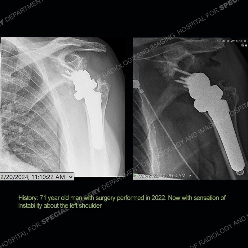
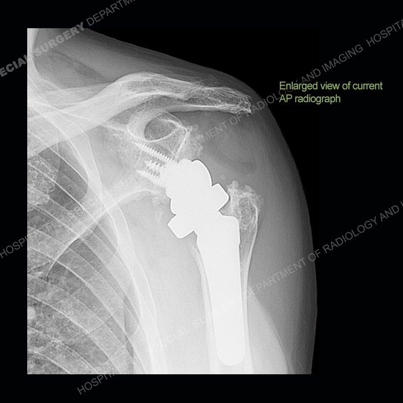
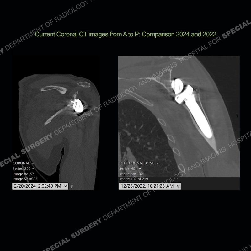
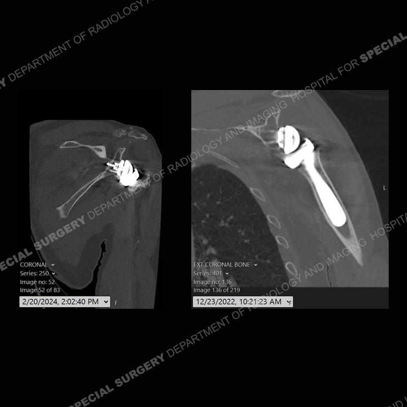
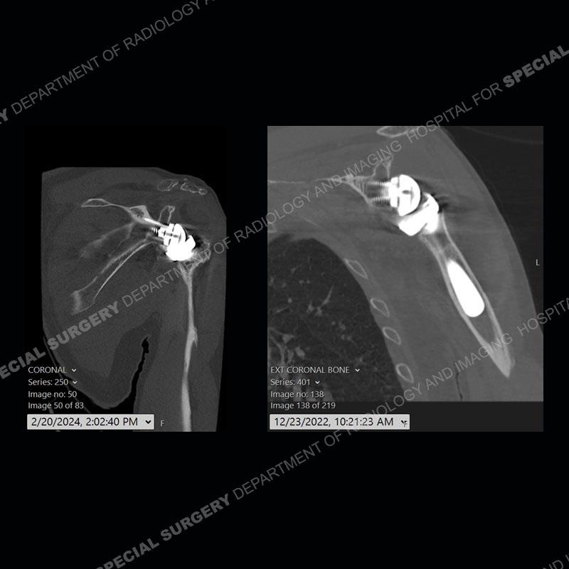
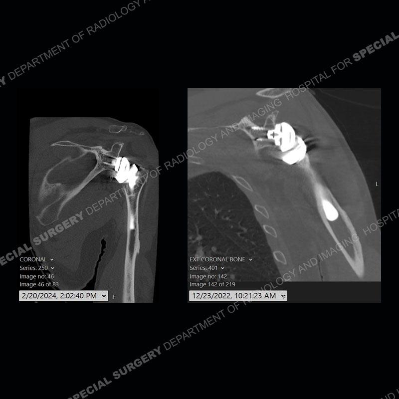
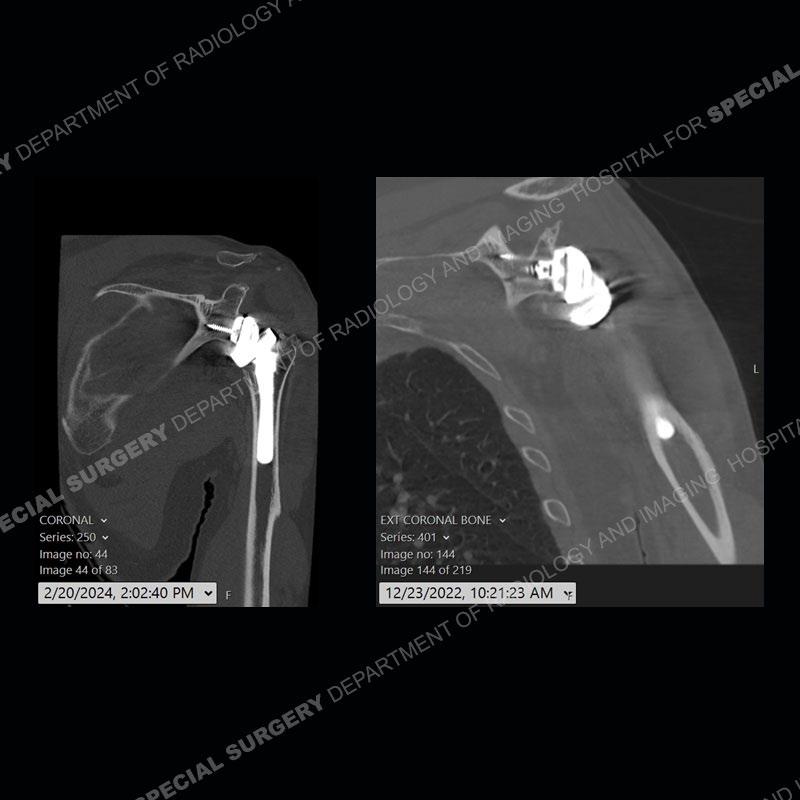
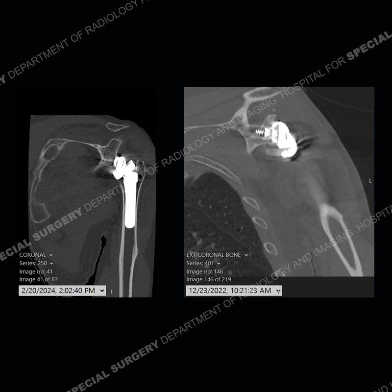
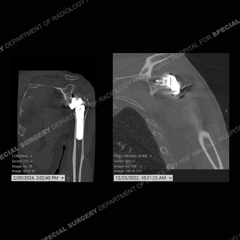
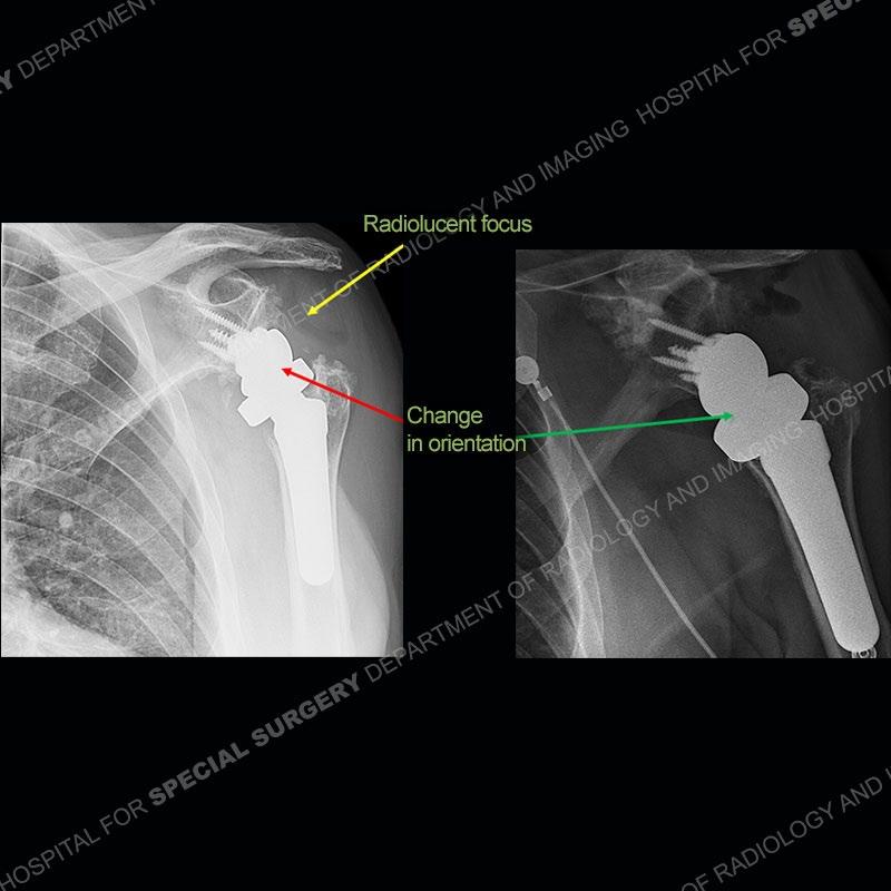
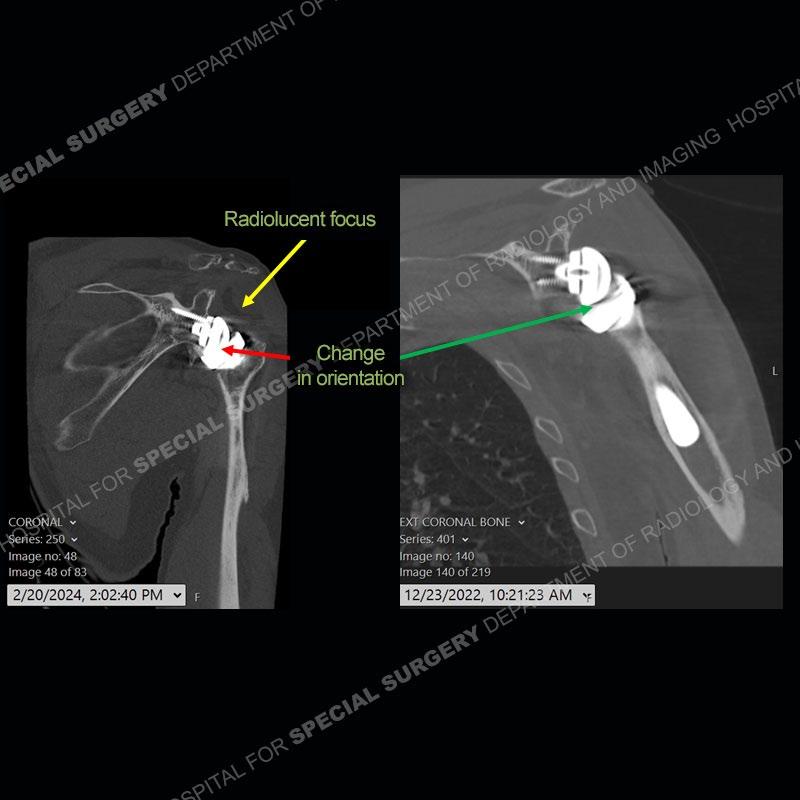
Findings
When comparing the radiographs, there is a slight change in the alignment in the components of the prosthesis. On the current radiographs, a radiolucent, crescentic focus is present at the level of the glenoid. When comparing the CT exams, there is a change at the alignment of the glenosphere and the humeral component and now a direct apposition of the components. On the current CT, the radiolucent, crescentic focus is again identified.
What’s the Diagnosis – Case 191
13
What’s the Diagnosis – Case 191 14

What’s the Diagnosis – Case 191 15

What’s the Diagnosis – Case 191 16
What’s the Diagnosis – Case 191 17

What’s the Diagnosis – Case 191 18