What’s the Diagnosis – Case 84
1
What’s the Diagnosis – Case 84
2
What’s the Diagnosis – Case 84
3
What’s the Diagnosis – Case 84
4
What’s the Diagnosis – Case 84
5
What’s the Diagnosis – Case 84
6
What’s the Diagnosis – Case 84
7
What’s the Diagnosis – Case 84
8
What’s the Diagnosis – Case 84
9
What’s the Diagnosis – Case 84
10
What’s the Diagnosis – Case 84
11
What’s the Diagnosis – Case 84
12
What’s the Diagnosis – Case 84
13
What’s the Diagnosis – Case 84
14
What’s the Diagnosis – Case 84
15
What’s the Diagnosis – Case 84
16
What’s the Diagnosis – Case 84
17
What’s the Diagnosis – Case 84
18
What’s the Diagnosis – Case 84
19
What’s the Diagnosis – Case 84
20
What’s the Diagnosis – Case 84
21
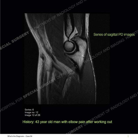
Findings Multiple planes of imaging demonstrate two tendons at the expected location of the distal biceps tendon. The more medial tendon demonstrates a low grade injury/strain with otherwise continuity to the radial tuberosity. The more lateral tendon demonstrates complete disruption with retraction of approximately 2.5cm from the radial tuberosity. A marked amount of edema is seen in the adjacent soft tissues.
What’s the Diagnosis – Case 84
22
What’s the Diagnosis – Case 84
23
What’s the Diagnosis – Case 84
24
What’s the Diagnosis – Case 84
25
What’s the Diagnosis – Case 84
26
What’s the Diagnosis – Case 84
27
What’s the Diagnosis – Case 84
28
What’s the Diagnosis – Case 84
29
What’s the Diagnosis – Case 84
30
What’s the Diagnosis – Case 84
31
What’s the Diagnosis – Case 84
32
What’s the Diagnosis – Case 84
33
What’s the Diagnosis – Case 84
34
What’s the Diagnosis – Case 84
35
What’s the Diagnosis – Case 84
36
Diagnosis: Complete disruption of one tendon in the setting of a bifurcated/bifid distal biceps tendon Disruption of the distal biceps tendon is much less common than at the proximal attachment and tends to occur in middle aged men, typically older than 40. The short and long heads of the biceps yield tendons at approximately 7cm proximal to the radial tuberosity attachment site. There is a vast variation in the degree of intertwining or decussation between the two tendons. As in this case, at times there is a persistent bifurcated or bifid architecture of the tendons with two separate tendons extending to the radial tuberosity. When two tendons are present, the short head of the biceps is the more medial of the tendons and has a more distal insertion on the radial tuberostiy. The long head of the biceps is the more lateral tendon and inserts more proximally. In the setting of a selective disruption of one of the tendons, by in large it is the short head but in this example it is the more lateral tendon or long head tendon that is disrupted. However, there has been a strain of the short head seen best on the axial IR images and hence this is not completely, an isolated or selective injury. The importance of these findings is to be aware that injury can have still been sustained even if a tendon attachment is present at the radial tuberosity and surgery may still be necessary for the selective disruption.
What’s the Diagnosis – Case 84
37
What’s the Diagnosis – Case 84
38
References Bifid insertion of the distal biceps brachii tendon with isolated rupture: magnetic resonance findings. Koulouris G, Malone W, Omar IM, Gopez AG, Wright W, Kavanagh EC. J Shoulder Elbow Surg. 2009 Nov-Dec;18(6). http://radsource.us/pathology-of-the-long-head-of-the-biceps-tendon/
What’s the Diagnosis – Case 84
39