What’s the Diagnosis – Case 220
1
What’s the Diagnosis – Case 220
2
What’s the Diagnosis – Case 220
3
What’s the Diagnosis – Case 220
4
What’s the Diagnosis – Case 220
5
What’s the Diagnosis – Case 220
6
What’s the Diagnosis – Case 220
7
What’s the Diagnosis – Case 220
8
What’s the Diagnosis – Case 220
9
What’s the Diagnosis – Case 220
10
What’s the Diagnosis – Case 220
11
What’s the Diagnosis – Case 220
12
What’s the Diagnosis – Case 220
13
What’s the Diagnosis – Case 220
14
What’s the Diagnosis – Case 220
15
What’s the Diagnosis – Case 220
16
What’s the Diagnosis – Case 220
17
What’s the Diagnosis – Case 220
18
What’s the Diagnosis – Case 220
19
What’s the Diagnosis – Case 220
20
What’s the Diagnosis – Case 220
21
What’s the Diagnosis – Case 220
22
What’s the Diagnosis – Case 220
23
What’s the Diagnosis – Case 220
24
What’s the Diagnosis – Case 220
25
What’s the Diagnosis – Case 220
26
What’s the Diagnosis – Case 220
27
What’s the Diagnosis – Case 220
28
What’s the Diagnosis – Case 220
29
What’s the Diagnosis – Case 220
30
What’s the Diagnosis – Case 220
31
What’s the Diagnosis – Case 220
32
What’s the Diagnosis – Case 220
33
What’s the Diagnosis – Case 220
34
What’s the Diagnosis – Case 220
35
What’s the Diagnosis – Case 220
36
What’s the Diagnosis – Case 220
37
What’s the Diagnosis – Case 220
38
What’s the Diagnosis – Case 220
39
What’s the Diagnosis – Case 220
40
What’s the Diagnosis – Case 220
41
What’s the Diagnosis – Case 220
42
What’s the Diagnosis – Case 220
43
What’s the Diagnosis – Case 220
44
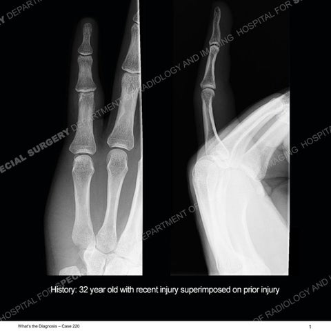
Findings The radiographs show no abnormality and particularly no fracture. On the MRI, along the volar aspect of the PIP of the small finger there is a focus of proximally displaced soft tissue and fluid of the volar recess of the PIP of the small finger. The adjacent pulleys and flexor tendons are intact. There is a thickening of both collateral ligaments of the small finger PIP but they are intact. No recent osseous injury is present.
What’s the Diagnosis – Case 220
45
What’s the Diagnosis – Case 220
46
What’s the Diagnosis – Case 220
47
What’s the Diagnosis – Case 220
48
What’s the Diagnosis – Case 220
49
What’s the Diagnosis – Case 220
50
What’s the Diagnosis – Case 220
51
What’s the Diagnosis – Case 220
52
What’s the Diagnosis – Case 220
53
What’s the Diagnosis – Case 220
54
What’s the Diagnosis – Case 220
55
What’s the Diagnosis – Case 220
56
What’s the Diagnosis – Case 220
57
What’s the Diagnosis – Case 220
58
What’s the Diagnosis – Case 220
59
What’s the Diagnosis – Case 220
60
What’s the Diagnosis – Case 220
61
What’s the Diagnosis – Case 220
62
What’s the Diagnosis – Case 220
63
What’s the Diagnosis – Case 220
64
What’s the Diagnosis – Case 220
65
Diagnosis: Volar Plate Rupture A well recognized but still poorly understood entity with necrosis and collapse representing the end stage of the pathology. The reasons for the necrosis are likely multifactorial and relate to the anatomy of the wrist inclusive of ulnar variance and the anatomy of the vasculature to the lunate. Other inflammatory, biological processes and overuse/trauma also likely play a role in the development of the necrosis. The disease manifests by sclerosis of the lunate followed by collapse and fragmentation. This is then followed by abnormal orientation of the scaphoid in a palmar flexed position and then adjacent arthritis. These latter processes were not the focus of this case presentation. The MRI will show areas of low signal on all pulse sequences indicating the areas of devitalized bone. The foci of slightly increased signal are in keeping with areas of maintained perfusion and highlight the variable architecture and findings of Kienbock’s. The fragmentation and collapse of the bone are seen well on both CT and MRI. This case highlights the need for knowing and understanding the detailed anatomy of the hand and fingers. Multiple, small structures are present and the intricate, complicated anatomy is difficult for most of us. In this case, having a “control finger” with the ring finger to show the normal anatomy of the volar plate was very helpful in making the injury of the volar plate at the small finger PIP much more conspicuous. This technique is often very helpful particularly with detailed anatomy in areas which may not be as familiar to us as many other areas of the body.
What’s the Diagnosis – Case 220
66