Clinical History
72-year-old woman with sharp wrist pain upon active wrist extension, status post volar fixation of a distal radius fracture 5 years ago.

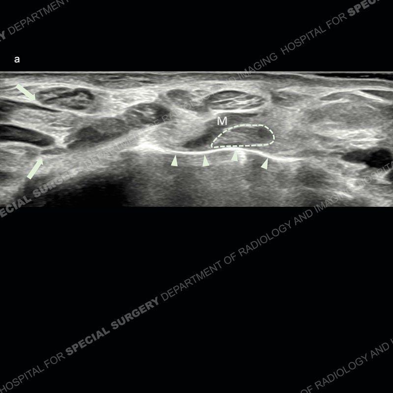
Sonographic imaging along the volar aspect of the left wrist in the transverse plane shows curvilinear echogenic line (arrowheads) with reverberation artifact extending posterior, representing the distal margin of the metal plate. Multiple flexor tendons course superficial to the plate, one of which (dashed oval), along with its associated muscle (M) is in direct contact with the plate.
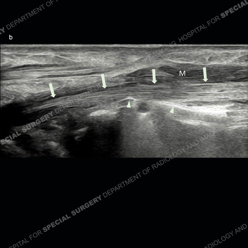
Longitudinal view of the same tendon (arrows) shows its course draping over and in contact with the distal margin of the plate (arrowheads). M again indicates the muscle associated with the tendon.
Diagnosis: Accessory Flexor Carpi Radialis Brevis Tendon
contacting a Distal Radius Volar Fixation Plate
The flexor carpi radialis brevis (aka flexor carpi radialis brevis vel profundus) is a rare accessory muscle of the distal forearm and wrist.
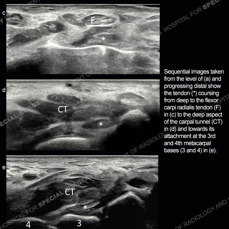
The tendon arises from the distal radius, courses deep to the flexor carpi radialis tendon, and inserts on the 3rd and 4th metacarpal bases.
This anomalous muscle is usually asymptomatic but becomes clinically significant due to its location in the region of distal radius fixation.