Joel Thomson - Student Research and Creativity Forum - Hofstra University

Page 1

TINAGL1 is an EGFR antagonist that suppresses the aggressive phenotype associated with head and neck squamous cell carcinoma Joel Thomson and Matthew L Fisher Department of Biology, Hofstra University, Hempstead NY, 11549 INTRODUCTION Head and neck squamous cell carcinoma (HNSCC) is comprised of cancers of the epithelial cells that line the mucosal surfaces inside the head and neck, predominantly the mouth, nose and throat. HNSCC occurs frequently in response to mutagens, including alcohol, tobacco and HPV exposure. Treatment typically includes surgery, radiation, and chemotherapy. More recently, Cetuximab, a targeted monoclonal antibody against Epidermal Growth Factor Receptor (EGFR) has been used. EGFR is a transmembrane glycoprotein frequently overexpressed in HNSCC, where it activates intracellular signaling pathways involved in cell proliferation, invasion, angiogenesis and metastasis. Signal transducer and activator of transcription 3 (STAT3) is a crucial downstream effector of EGFR signaling in HNSCC. STAT3 is involved in tumor-related immunosuppression, metastasis and drug resistance and is often overexpressed in aggressive HNSCC cases. While the function and importance of EGFR/STAT3 signaling has been clearly demonstrated, there is still much to be learned about how this pathway is regulated. Tubulointerstitial nephritis antigen like 1 (TINAGL1) is an extracellular matrix (ECM) protein involved in adhesion, and modulates several pathways linked to proliferation, migration and differentiation. While TINAGL1 is implicated in suppressing tumorigenesis in breast cancer, it is linked to enhanced tumorigenesis in several other cancers. Because of the tissue specific role of TINAGL1, it is essential to elucidate the role of this novel protein, which has never been studied in HNSCC. Here, we identify TINAGL1 as an interacting partner of EGFR, as well as integrins β1 and β4. We show that treatment with recombinant TINAGL1 (R-TINAGL1) suppresses EGFR activation and subsequent STAT3 signaling. This suppression of the EGFR-STAT3 signaling axis by R-TINAGL1 produces a similar phenotype to the EGFR inhibitor Cetuximab, mitigating HNSCC spheroid formation, invasion and migration. We propose that treatment with R-TINAGL1 represents a potential therapeutic strategy to target EGFR-STAT3 signaling and impair the aggressive phenotype associated with HNSCC.

MATERIALS AND METHODS Immunoprecipitation: CAL33 cells were maintained in DMEM (Dulbecco’s Modified Eagle’s Medium) supplemented with 10% fetal calf serum (FCS). For immunoprecipitation, protein lysates were collected on ice using Cell Signaling Lysis buffer, quantified and 500 μg of protein were incubated overnight with 5 μg of primary antibody against TINAGL1. Lysates were then incubated with protein A/G beads for 5 hours, washed and prepared for immunoblot. Immunoblot: Equivalent amounts of protein were electrophoresed on denaturing and reducing 10% polyacrylamide gels and transferred to nitrocellulose membrane. Membranes were blocked in 5% nonfat dry milk and then incubated with the appropriate primary (1:1000) and secondary antibody (1:5000). Secondary antibody binding was visualized using chemiluminescence detection technology. Cell Invasion Assay Matrigel was diluted in 0.01 Tris-HCL/0.7% NaCl, filter sterilized and 0.1 mL was used to coat individual BD BioCoat inserts (Millicell- PCF, 0.4 mm, 12 mm, PIHP01250). Cells (2.5 x 10⁴) were plated in 100 ml growth medium supplemented with 1% FBS into the upper chambers of the inserts. The lower chambers contained growth medium supplemented with 10% FBS. After invasion, membranes were harvested and the surface of upper membranes were rinsed with PBS to remove unattached cells. Membranes were fixed in 4% paraformaldehyde, stained with 1 mg/mL DAPI, and the underside of the membrane was photographed using an inverted fluorescent microscope and cells were quantitated. Spheroid Formation Assay: CAL33 cells were dissociated with trypsin, then centrifuged at 1000 RPM for 5 minutes. Cells were resuspended in a DMEM solution supplemented with FCS to inactivate the trypsin, then centrifuged again and washed in spheroid media to remove the FCS. These washed cells were then plated for spheroid growth in ultra-low attachment dishes containing spheroid medium consisting of (DMEM/F12 (1:1) with 2% B27 supplement, 20 ng/ml EGF, 0.4% bovine serum albumin and 4 μg/ml insulin. Spheroids number was monitored over 9 days of growth.

A

C

A

B

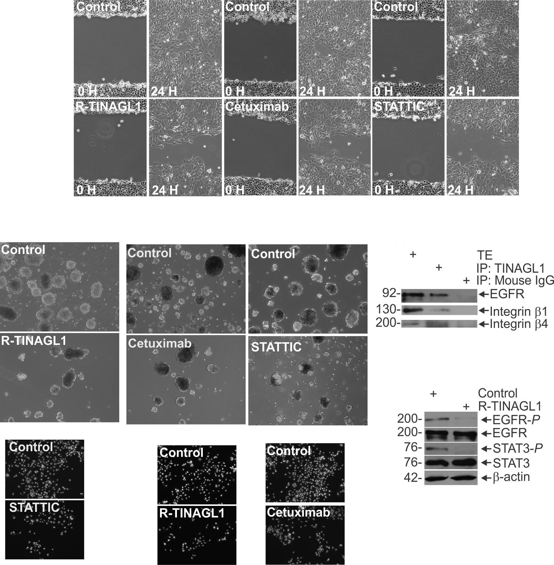
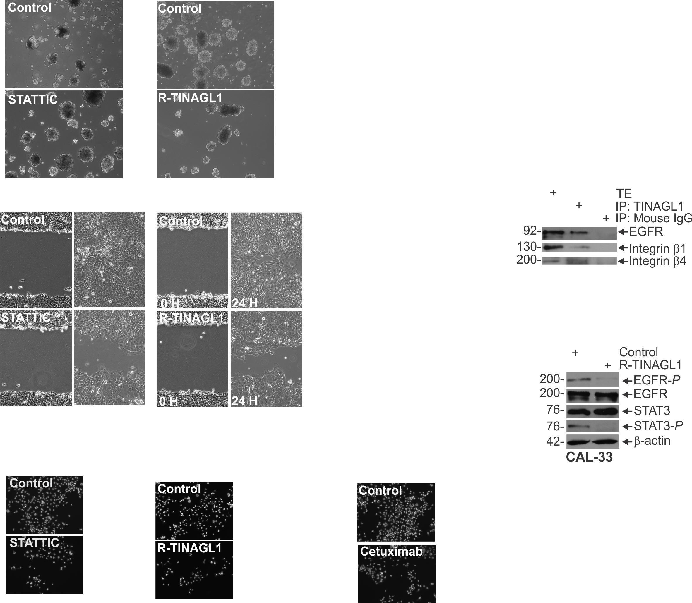
B Fig 1 TINAGL1 interacts with EGFR and Integrin β1 and β4 to suppress their activity A Cal33 cells were harvested for protein extraction for immunoprecipitation of TINAGL1 and immunoblot of the indicated epitopes. B Protein lysates were collected from CAL33 cells treated with or without R-TINAGL1 and used for a dot blot array. Relative intensities were compared to the control group. C Control and R-TINAGL1 treated cells were harvested for protein lysates and immunoblotted for the indicated epitopes. These findings suggest that extracellular TINAGL1 antagonizes EGFR, β1 and β4 integrin signaling

A

B

C D

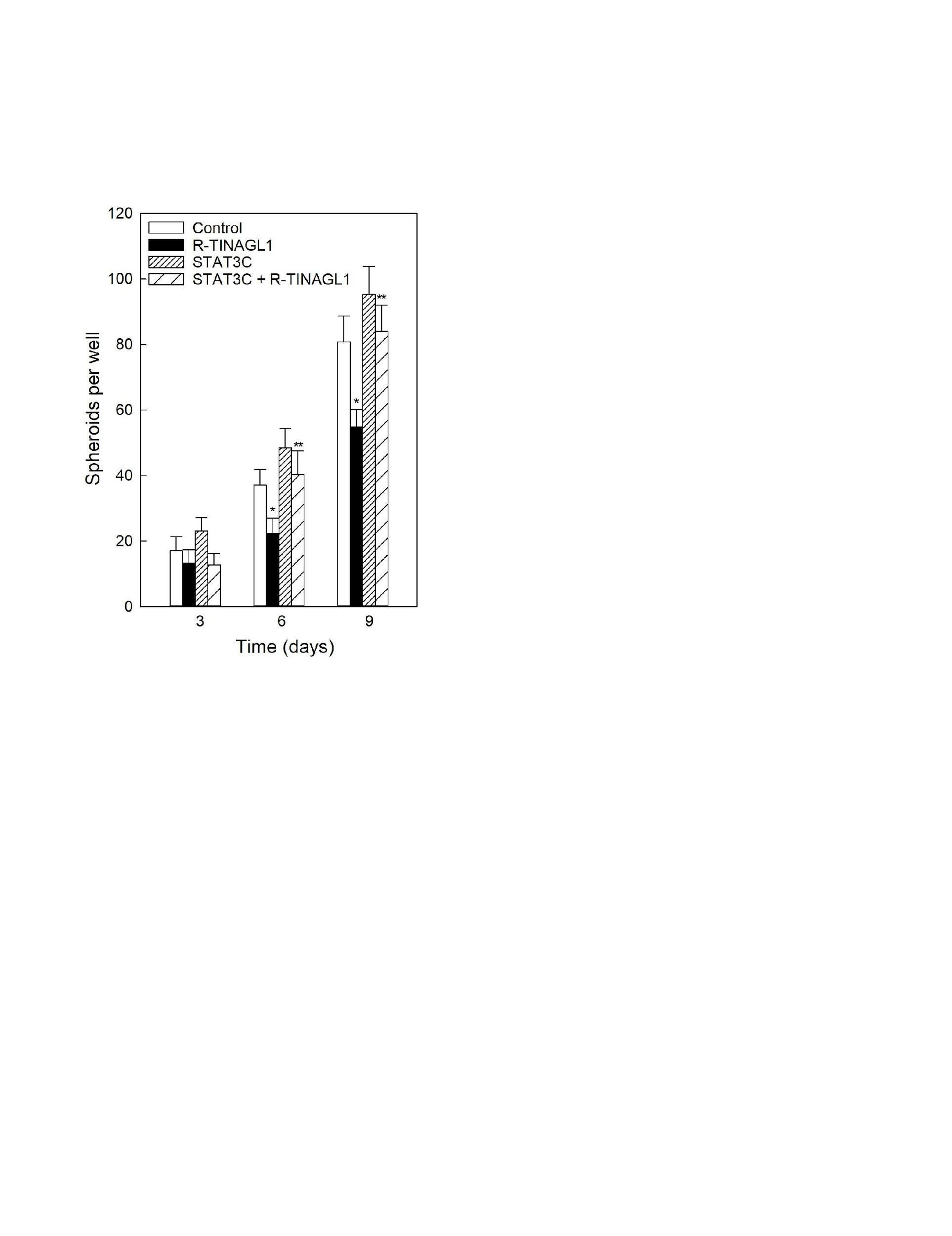
C Fig 3 R-TINAGL1 suppresses spheroid formation in HNSCC CAL33 cells (40,000) were seeded in non-adherent plates containing spheroid medium and treated with A R-TINAGL1 B Cetuximab, or C. STATTIC. Spheroid number was counted over 9 days. Images were taken on day 9. D Cal33 cells stably expressing constitutively active STAT3 were seeded for invasion assays, and then treated with R-TINAGL1 and monitored over 9 days of growth. *, ** , P < 0.05 (the double asterisk indicates significance compared with the single asterisk group) These results suggest that R-TINAGL1 suppresses spheroid growth via suppression of STAT3 activity.

D

Conclusions

E

F

• TINAGL1 interacts with EGFR, β1 and β4 integrin and suppresses their activation • Treatment with R-TINAGL1 impairs formation, invasion and migration

spheroid

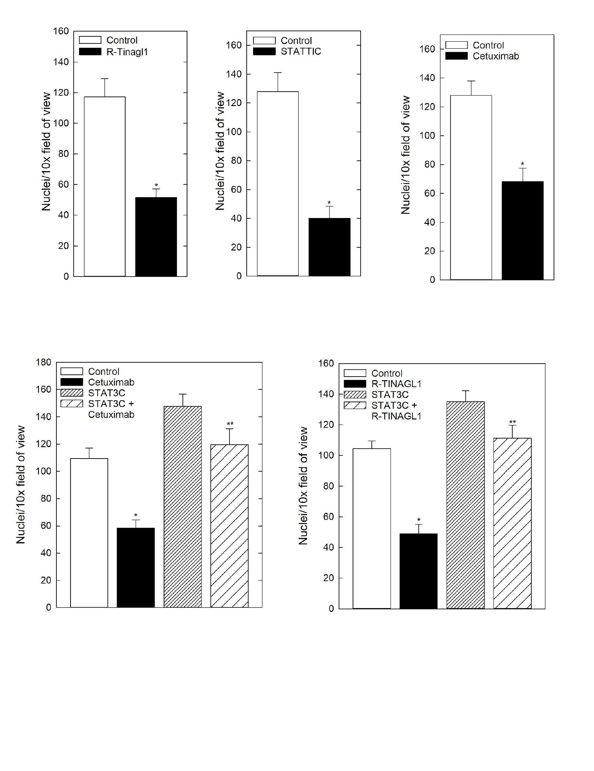
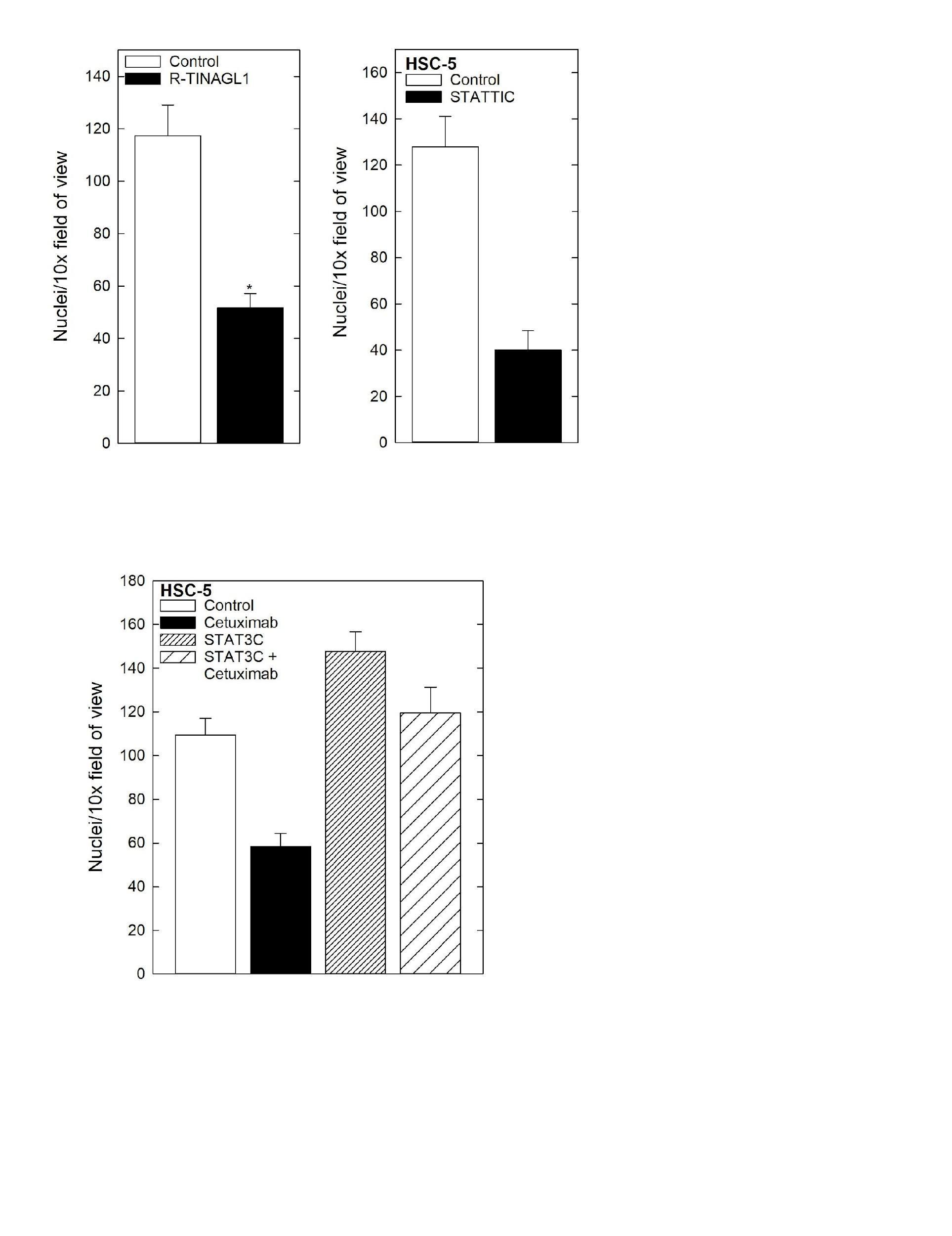
• R-TINAGL1 suppression of the aggressive phenotype mimics phenotypic changes seen with Cetuximab treatment • R-TINAGL1 suppression of the aggressive phenotype is mediated by suppression of STAT3 signaling, with STAT3C rescuing the aggressive phenotype in the presence of R-TINAGL1 Fig 2 R-TINAGL1 suppresses HNSCC invasion and migration by suppressing STAT3 activation. Cells were seeded on the upper chamber of a Matrigel coated membrane and treated with either A R-TINAGL1, B Cetuximab, or C STATTIC. After 24 h the membrane was removed, rinsed and fixed. Nuclei were DAPI stained and counted on the underside of the membrane. D Confluent cultures of Cal33 cells were scratched with a 10 μl pipette tip, and then treated R-TINAGL1, Cetuximab or STATTUC and migration into the scratch was monitored E Cal33 cells stably expressing constitutively active STAT3 were seeded for invasion assays, and then treated with Cetuximab or F R-TINAGL1. After 24 h the membrane was removed, rinsed and fixed. Nuclei were DAPI stained and counted on the underside of the membrane. *, ** , P < 0.05 (the double asterisk indicates significance compared with the single asterisk group) These findings indicate that R-TINAGL1 suppression of HNSCC invasion is dependent on suppression of STAT3 activity.

• R-TINAGL1 treatment represents a potential therapeutic strategy in HNSCC to impair EGFR/STAT3 signaling and suppress the aggressive phenotype


Turn static files into dynamic content formats.

Create a flipbook
Joel Thomson - Student Research and Creativity Forum - Hofstra University by Hofstra University - Issuu