DIGITAL SOLUTIONS
USING DIGITAL IMPRESSIONS OF THE UPPER JAW FOR A FULL-ARCH DENTAL BRIDGE TREATMENT
Taking digital impressions of the soft tissues of the mouth can be challenging - but not if you are using Planmeca Emerald™ intraoral scanners. The following case report by Dr Koby Sharvit
illustrates how intraoral scans, captured of the mucosa in full natural colours, can help designing advanced dental restorations quickly and accurately. Dr Koby Sharvit, MD, DMD, is an Israeli dentist and expert in CAD/CAM technology. He is currently working in PerioCenter Ltd. A dental centre, managed by Dr Yaniv Mayer and Dr Eran Gabay, both specialists in periodontology. The clinic is located in Haifa, Israel.
Emerald™ S intraoral scanner. With the scanner, Dr. Sharvit could easily capture the patient’s mucosa and available dentition for the design of an implantsupported full-arch dental bridge. Compared to traditional impressions, intraoral scanning would be much more comfortable and faster for the patient.
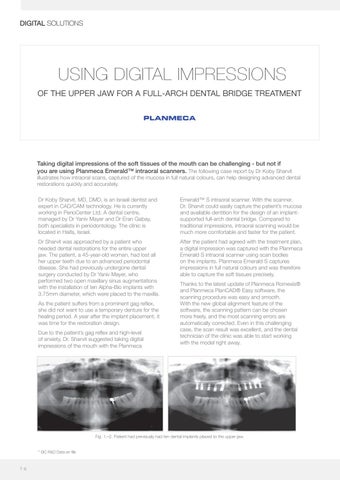
Dr Sharvit was approached by a patient who needed dental restorations for the entire upper jaw. The patient, a 45-year-old woman, had lost all her upper teeth due to an advanced periodontal disease. She had previously undergone dental surgery conducted by Dr Yaniv Mayer, who performed two open maxillary sinus augmentations with the installation of ten Alpha-Bio implants with 3.75mm diameter, which were placed to the maxilla.
After the patient had agreed with the treatment plan, a digital impression was captured with the Planmeca Emerald S intraoral scanner using scan bodies on the implants. Planmeca Emerald S captures impressions in full natural colours and was therefore able to capture the soft tissues precisely.
As the patient suffers from a prominent gag reflex, she did not want to use a temporary denture for the healing period. A year after the implant placement, it was time for the restoration design. Due to the patient’s gag reflex and high-level of anxiety, Dr. Sharvit suggested taking digital impressions of the mouth with the Planmeca
Thanks to the latest update of Planmeca Romexis® and Planmeca PlanCAD® Easy software, the scanning procedure was easy and smooth. With the new global alignment feature of the software, the scanning pattern can be chosen more freely, and the most scanning errors are automatically corrected. Even in this challenging case, the scan result was excellent, and the dental technician of the clinic was able to start working with the model right away.
Fig. 1.–2. Patient had previously had ten dental implants placed to the upper jaw. * GC R&D Data on file
7 6


