W W W. O R A L H E A LT H G R O U P. C O M
SPECIAL ISSU E
LASER DENTISTRY Publications Mail Agreement No. 40063170 • ISSN 0030-4204
PHOTO-ACOUSTIC ROOT CANAL DECONTAMINATION
Advanced Implant Impression Solutions for Every Office Implant impressions should perform in all areas – precision transfer of implant position, detailed reproduction of surrounding soft tissue and dentition, handling, and patient experience. Designed by experts in both implants and impressions, Dentsply Sirona offers both traditional and digital impression solutions for every office.
Aquasil Ultra+ & digit® Power Dispenser
CEREC® Omnicam Connect
Traditional Impression Solution
Stand-alone Digital Impression Solution
www.dentsplysirona.com | 1-877-393-3687
A NEWCOM Media Inc. Publication
SPECIAL ISSUE: LASER DENTISTRY Clinical Editor: Dr. Marina Polonsky Managing Editor: Jillian Cecchini 416-510-5125 jillian@newcom.ca Assistant Editor: Krysten McCumber 416-510-6777 krysten@newcom.ca
Advertising Services: Karen Samuels 416-510-5190 karens@newcom.ca Director, Business Development: Tony Burgaretta 416-510-6852 tonyb@newcom.ca
Art Direction: Barbara Burrows
Account Manager: Holly Power 416-510-6846 holly@newcom.ca
Circulation: Mary Garufi 416-614-5831 mary@newcom.ca
Classified Advertising: Karen Shaw 416-510-6770 karen@newcom.ca
Contents FEATURES —LASER DENTISTRY —
8
Managing Refractory Endodontic Disease with Radial Apical Cleansing: Report of Clinical Cases Justin Kolnick, DDS
16
The Role of Erbium and Diode Laser System in the Treatment of Oral Frenula and its Contribution to Breastfeeding: Is This Vision Important for Dentistry? Dr. María del Pilar Martín Santiago, DDS, MSc Lasers in Dentistry, Expert in Dentistry for Newborns
24
Peri-Implantitis Solea CO2 Laser: Clinical Case Report
30
An Underutilized Treatment Modality in the Dental Industry: Low Level Laser Therapy
Ed Kusek, DDS
MEDIA INC.
Chairman and Founder: Jim Glionna President: Joe Glionna Vice President, Publishing: Melissa Summerfield CFO: Peter Fryters Director of Circulation: Pat Glionna
Gerry Ross, DDS; Alana Ross, BScH
38
Laser Treatment of Benign Vascular Lesion: A Clinical Case Report Andre Chartrand, BSc, DMD
Oral Health, published by Newcom Media Inc., is an independent, monthly professional journal, written and edited for the practicing dentist in Canada, and supervised by an Editorial Board of Consultants from both general practice and major specialties of the profession. The editorial content consists of clinical articles and abstracts from the world’s finest dental literature and monthly departments. The editorial purpose is to provide information on clinical advances in all phases of dentistry. Oral Health is not responsible for the quality of graphic images submitted by the authors. The Editorial Board of Oral Health does not necessarily agree with the claims made for any product advertised. Nor should it be construed that the appearance of any product advertisement in Oral Health implies that the Board either approves or accepts the product. Oral Health reserves the right to edit departmental submissions for content and length. The contents of this publication may not be reproduced either in part or in full without the written consent of the copyright owner. ISSN 0030-4204 Yearly subscription rates: Canada 1 year $64.95 + taxes, 2 years $107.95 + taxes (GST/HST #103862405RT0001); U.S. 1 year $70.95; Foreign 1 year $107.95; Single copy Canada & U.S. $10.00; Single copy Foreign $10.00. Printed in Canada. All rights reserved. From time to time we make our subscription list available to select companies and organizations whose product or service may interest you. If you do not wish your contact information to be made available, please contact us via one of the following methods: Phone: 1-800-668-2374; Fax: 416-442-2191; E-mail: mary@newcom.ca; Mail to: Privacy Officer, Newcom Media Inc., 5353 Dundas St. W. Suite 400, Toronto, ON M9B 6H8. Oral Health is published monthly. CANADA POST Publications Mail Agreement No. 40063170. Changes of address notices and orders for subscriptions are to be faxed to (416) 510-6875 or mailed to Circulation Department – Oral Health, 5353 Dundas St. W. Suite 400, Toronto, ON M9B 6H8.
42
A Novel Approach to Veneer Removal Using Er,Cr:YSGG Laser: A Clinical Case Report Amir Azzat, DDS, MSc
DEPARTMENTS —EDITORIAL —
4 46
One Must Open the Window to Let The Light In… Marina Polonsky, DDS, MSc Lasers in Dentistry
Dental Marketplace
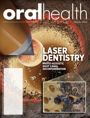
Cover Credits ART DIRECTION: Aarti Inamdar, MScBMC 3D ARTISTS: Mad Mouse Studio, madmousestudio.com CONTENT EXPERT: Dmitri Boutoussov, PhD The cover depicts a radial firing tip removing smear layer inside the root canal. Thanks to Biolase Inc. for their assistance with the cover.
www.oralhealthgroup.com
3
—EDITORIAL— —VIEWPOINT—
One Must Open The Window To Let The Light In… Marina Polonsky,
W
DDS, MSc Lasers in Dentistry
e all remember graduating dental school and thinking to ourselves: “What’s next?” The future is full of possibilities; we must make decisions which will ultimately affect the rest of our careers and lives. Some of us knew right away that general practice was not for them, whereas specialties like surgery, periodontics or endodontics presented opportunities to maximize one’s talents in a narrow area of expertise. The rest of us, myself including, who did not hear a particular calling towards any specialty and actually enjoyed a great variety of procedures general dentistry has to offer, left dental school and entered into the reality of private practice. We all thought that our newly acquired knowledge of the most recent concepts, products and technologies, which we learned in university, would compensate for the lack of clinical experience. WERE WE IN FOR A SURPRISE! The real world offers many valuable learning opportunities, and it’s up to us to grab them and pursue the never-ending thirst for knowledge and discovery. Every year we take continuing education courses in order to learn about new materials and technologies which promise to make our lives easier, our procedures better and our patients happier. Laser technology is no different. Depending on the university, students receive little or no education in laser applications in dentistry, which is likely due to the fact that schools pride themselves on teaching only wellproven techniques and concepts supported by decades of double-blind, controlled, and randomized clinical trials published only in reputable peer-reviewed publi4
oralhealth
cations. And it is a good philosophy to follow. However, as we put more emphasis on higher levels of evidence, this higher level is becoming more and more difficult to achieve due to the requirement of compliance with ethical standards described in the Helsinki declaration, of 1975. There exists a multitude of in-vitro studies supporting different laser applications in all clinical aspects of dental practice, however very few are well designed double-blind randomized clinical trials. A number of peer-reviewed scientific journals focus exclusively on laser research: Lasers in Medical Science; Lasers in Dental Science; Photo-medicine and Laser Surgery; and Lasers in Surgery and Medicine, to name a few. Sometimes clinical case reports are the only evidence which we have available to prove the validity or efficacy of a new treatment. Does that mean we should reject this treatment modality, or should we wait another 30 years for higher levels of evidence to become available? Who, but the patient stands to lose in this scenario? My first eye-opening experience with lasers happened in 2006, when I saw a cavity preparation performed with a Waterlase MD laser. The carious lesion was selectively evaporated from the tooth using nothing but the water spray and invisible laser light. No contact. No vibration. Minimally invasive and supposedly painless and not requiring anesthesia. My second eye-opener was when I found out how much this technology costs! And so, I set out on my road to discovery about lasers and their applications in dentistry. Not having the courage to spend close to $100,000 on my first laser, I initially pur-
FIG 1
Neurochirurgie CO2, Nd:YAG
HNO Nd:YAG, CO2, Er:YAG Pulmology Nd:YAG
Ophthalmology Ar, Kr, Excimer, Ho:YAG Er:YAG, Nd:YAG
Dental Nd:YAG, Er:YAG, Er, Cr:YSGG, CO2, Ar4, Dioden Kardiology Nd:YAG
Gastroenterology Nd:YAG, Ar
Onkology Nd:YAG, CO2, Farbstoff Gynakology Nd:YAG, CO2, Er:YAG Angioplasty Nd:YAG, Er:YAG, Dye
Urology Nd:YAG, Alexandrit, CO2, Er:YAG Orthopedic Ho:YAG, CO2, Er:YAG, Nd:YAG
Dermatology Alexandrit, Ar, Er:YAG. Excimer, Farbstoff, CO2, Kr, Kupfer, Nd:YAG
Courtesy of AALZ, Germany.
chased a much more reasonably priced diode laser. It was cute, portable, had disposable tips which sounded much better than having to cleave the fiber, and most importantly- it didn’t break the bank at a price tag of under $9,000. After all, a laser is a laser, right? WRONG! I quickly realized that I can’t prepare cavities with it, must still use anesthetic, and that it is much slower than cutting with a scalpel. So, what’s the benefit? Moreover, the only training offered to me was from the company representative, who arrived at my office with -literally- a package of hot dogs to teach me the set-up and buttonology, after which I was on my own. I started with some simple surgical procedures including gingivoplasty, fibroma removal and frenectomy. Later on, the need for packing a retraction cord became a thing of the past, as I mastered the troughing for impressions technique to help with subgingival margins and hemostasis. Finally, adding sulcular decontamination and periodontal pocket bacterial reduction to my hygiene program, enabled me to use this small laser device to its fullest potential. What amazed me was the patient response: faster healing; no stitches; no antibiotics; and better control of chronic conditions (like periodontal disease) with minimally invasive, non-surgical methods. The word “laser” itself was magic for easier treatment plan acceptance.
Patients are educated by both the media and advertisements on laser technology within other fields of medicine: laser eye surgery; laser hair removal; laser skin re-surfacing and many other applications (Fig. 1). In fact, lasers are utilized in every major branch of medicine with better clinical patient-reported outcomes, so why not dentistry? It took close to three years of practice and continuing education courses to become familiar with and fully comfortable with my diode laser, with respect to its applications, advantages and limitations. Who could predict that such a small devise would open such an enormous world of possibilities. One must open the window to let the light in, and this light will change your world. Clear bloodless surgical field, disinfection without antibiotics, hemostasis without chemicals or electro-surgery and healing without scar formation is just the beginning…then add pain control, faster healing and anesthesia without the use of pharmaceuticals, through photo-bio-modulation (PBM), and minimally invasive cancer treatment using photodynamic therapy (PDT)! In our everyday practice, we encounter many conditions for which modern medicine lacks any definitive treatment. We are trained to treat the www.oralhealthgroup.com
5
• Oral, inhaled and IV dental sedation courses • Canadian Dental Anaesthesia Assistant Certification (CDAAC) • Dental Office Medical Emergencies Workshop (DOME) • ACLS, PALS and CPR Courses
symptoms when we cannot offer a cure. We prescribe analgesics, steroids and anti-depressants to our patients which often provides the greatest benefit to the drug companies and leaves us dealing with the side-effects. Neuralgias, xerostomia, Bell’s Pulse, Lichen Planus and other mucocutaneous diseases, bisphosphonate-related-osteo-necrosis of the jaw (BRONJ), oral mucositis in cancer patients, are just a select few of the medical conditions where low-level-laser-therapy (LLLT) offers us a new and better way to address not just the symptoms, but rather to provide healing modalities for our patients. The uninformed, the sceptics, might call this voodoo, but the patients whom we help heal call us miracle workers and become ever grateful and loyal. The scientific evidence and support for the concept of Bio-Stimulation goes back to 1964 in Hungary and Prof. Endre Mester, the father of PBM (photo-bio-modulation). He experimented with carcinogenic effects of low-power ruby and HeNe lasers on rats and found that instead of cancer, there was faster hair growth and wound healing. His paper “Effects of laser rays on wound healing” was published in 1971, in The American Journal of Surgery. In 1985, he reported an 85% success rate in healing wounds resistant to conventional treatment in 875 patients. “The biomedical effects of laser application” was published in Lasers in Surgery and Medicine Journal. Since the invention of the first working ruby laser by Theodore Maiman in 1960, based on the concept by Gordon Gould in 1959, many different types of lasers have been created and utilized for various applications, from underwater welding with Argon lasers to calculating distances between planetary bodies. Our medical counterparts have embraced CO2 lasers as scalpels and excimer lasers in eye surgery, aesthetic medicine is relying on diode lasers for hair removal, and Nd:YAG and pulsed dye lasers for the treatment of vascular malformations and tattoo removals. Fractional technology with erbium lasers is evolving to address skin resurfacing and recontouring applications. So why is dentistry so resistant to accepting lasers into the scope of practice? This edition of Oral Health will showcase many different uses of laser technology in an attempt to open you to the world of possibilities, and all you have to do is keep an open mind. I’d like to finish by quoting Arthur Schopenhauer, “All truth passes through three stages. First, it is ridiculed. Second, it is violently opposed. Third, it is accepted as being self-evident.” OH
• Sedation supplies, monitors and emergency equipment sales
www.dentaled.com Contact us at info@dental-ed.com or call 778.984.0915
6
oralhealth
Dr. Marina Polonsky DDS, MSc is a gold medal University of Toronto ’99 graduate, she maintains private general practice in Ottawa, Ontario with focus on multi-disciplinary treatment utilizing lasers of different wavelengths. She holds a Mastership from World Clinical Laser Institute (WCLI), Master of Science in Lasers in Dentistry from RWTH University in Aachen, Germany and Advanced Proficiency Fellowship from ALD (Academy of Laser Dentistry). She is the founder of the Canadian Dental Laser Institute (CDLI), study club affiliated with the Academy of Laser Dentistry. She serves on the Executive Committee for Oral Health and is the editor of the Laser Dentistry issue.
ENDODONTICS —LASER DENTISTRY—
Managing Refractory Endodontic Disease With Radial Apical Cleansing Report Of Two Clinical Cases
Justin Kolnick, DDS
Keywords: Refractory endodontic disease, endodontic, radial apical cleansing, Er,Cr:YSGG, diode, laser, biofilm bacteria.
INTRODUCTION
One of the defining attributes of an astute endodontist is the ability to successfully treat refractory endodontic disease. Refractory disease is defined as disease that is recalcitrant, unresponsive, stubborn, unmanageable or resistant to treatment or cure. While the pathogenesis of refractory endodontic disease is not clearly comprehended, it is highly likely that microbiological and host immune influences play an important role. Unsuccessful endodontic outcomes are often attributed to persistent infection perpetuated by entombed bacteria or by reinfection of a previously disinfected root canal system, commonly via coronal leakage or tooth fracture. Extra-radicular causes are less common and include periapical actinomycosis, cholesterol crystals, foreign body reactions, unresolved cystic lesions and extra-radicular biofilm and usually require surgical intervention or extraction of the tooth. Refractory Endodontic Disease
Below is an example of such a clinical case (Fig. 1). It was posted online by an endodontist and is presented with his authorization. It involves retreatment of a lower right first molar tooth with a diagnosis of symptomatic apical periodontitis. Periodontal probing measurements were normal with no evidence of a tooth fracture. A CBCT 8
oralhealth
was taken but it was not posted. The treatment followed a standard endodontic protocol with long-term application of calcium hydroxide that was reapplied twice over a period of seven months. As the patient’s symptoms improved somewhat, the canals were obturated and the tooth restored. A week later the tooth was extracted due to persistence of symptoms! Although the etiology of the failed treatment and inability to resolve symptoms was never ascertained, a strong possibility is that the protocol used was ineffective in reducing sufficiently the bioburden within the tooth. Lower molars are known to have a complicated root canal system, especially in the mesial root (Fig. 2) and current instrumentation and irrigation techniques fall short of adequately addressing this anatomy. Case Report #1
The patient was a 58-year-old male dentist with a history of thyroid cancer. He had been diagnosed with recurrent endodontic disease in his lower right second molar. Retreatment was initiated by his endodontist. He presented in my office complaining of pain to percussion in the tooth. He had seen his endodontist multiple times over a period of several months with no improvement in his symptoms. Occlusion was light on the tooth. Periodontal probing depths were normal and trans-illumination showed no signs of a tooth fracture. A diagnosis of symptomatic apical periodontitis was made. The CBCT scan (Fig. 3) showed no evidence of untreated root canal anatomy or root fracture.
FIG 1A Pre-op
FIG 1B
FIG 1C Calcium Hydroxide Dressings
FIG 1D Post-Op
FIG 2 Typical complex root canal system found in the mesial roots of lower molars (Courtesy Dr. Francisco Balandrano).
Under local anesthesia, the tooth (Fig. 4) was treated with the Radial Apical Cleansing (RAC) protocol and re-medicated with calcium hydroxide paste. A prescription for Amoxicillin 500mg TID for seven days was prescribed. At the second visit three weeks later, the tooth was asymptomatic and was obturated with a bioceramic sealer and single cone gutta percha technique (EndoSequence® BC Sealer and Points™, Brasseler, USA). At his one and a half year follow up, the tooth was functional and asymptomatic with normal probing and radiographic evidence of complete healing. The tooth had not yet been permanently restored. Case Report #2
A 77-year-old female patient presented with pain and swelling associated with her lower left first molar tooth. She had previously been treated by two endodontists who were unable to relieve her symptoms (Fig. 5). Examination revealed swelling in the adjacent buccal fold and the tooth was percussion sensitive. Periodontal probing was normal except for a narrow 7 mm pocket on the buccal aspect of the mesial root. Trans-illumination showed no signs of a cracked tooth. Radiographic examination showed a J-shaped radiolucency on the mesial root and a smaller apical radiolucency on the distal root. The CBCT scan (Fig. 6) confirmed the extent of the findings as well as evidence of loss of buccal plate on the distal root. A diagnosis was
made of symptomatic apical periodontitis with a buccal draining sinus along the periodontal ligament space. Under local anesthesia, the tooth was treated with the Radial Apical Cleansing (RAC) protocol and re-medicated with calcium hydroxide paste (Fig. 7). The patient was prescribed Amoxicillin 500 mg TID for seven days. At the second visit, three weeks later, the tooth was asymptomatic and was obturated with a bioceramic sealer and single-cone gutta percha technique (Endo-Sequence® BC Sealer and Points™, Brasseler, USA). At recall, nine months later, probing depths were normal and the tooth was functional and asymptomatic with evidence of osseous healing. Radial Apical Cleansing (RAC)
RAC is a treatment protocol that consistently achieves superior cleaning and disinfection of complicated root canal systems, utilizing a chemo-mechanical protocol assisted by the application of radially firing laser energy. RAC Main Elements Instrumentation:
1. Glide path is established with .06 and .08 hand files
and rotary NiTi path files. 2. Deep apical shaping with heat-treated NiTi files,
always preserving root structure, especially in the coronal third and peri-cervical zone. www.oralhealthgroup.com
9
FIG 3 CBCT slices.
FIG 4
Pre-op
Post-op
Irrigation:
1. Effective apical negative pressure irrigation with
EndoVacTM system (Kavokerr) using 6% sodium hypochlorite solution. 2. Sonic activation of irrigant. Cleansing:
1. Laser Activated Irrigation (LAI) with Er,Cr:YSGG
laser (Waterlase iPlus, Biolase, Irvine, CA) using RFT2 and RFT3 laser tips with settings 1.25 Watts, H mode, 20 Hz (PPS), 30% air, 10% water, 62.5 mJ/ pulse.
1.5-year recall
the USA as no FDA clearance has been issued for this application. Ideally, for both cleansing and disinfection, the laser tip is placed 1 mm short of working length and activated on withdrawal of the tip, in a circular motion, at a rate of 1-2 mm per second. This process is repeated four times in each canal. Placement of the laser tip is influenced by root canal anatomy, diameter and flare of the prepared canal and presence or absence of canal patency; it will remain effective even at distances of 5 mm or more from the apical foramen.
Disinfection:
Erbium Lasers in Endodontics
1. Laser disinfection with Er,Cr:YSGG laser (Waterlase
Erbium lasers have emerged as the most promising laser wavelength in endodontics. They are both hard and soft tissue capable and have the most FDA clearances for a multitude of dental procedures. Their primary chromophore is water and to a lesser degree, hydroxyapatite. Photo-thermal interactions prevail in soft tissue and photo-disruptive in hard tissue procedures. When proper parameters are followed, thermal relaxation is
iPlus, Biolase, Irvine, CA) using RFT2 and RFT3 laser tips with settings of 1.00 Watt, H mode, 20 Hz (PPS), 10% air, 0% water, 50 mJ/pulse. 2. Deep dentin disinfection with 940 nm diode laser (Epic X, Biolase, Irvine, CA), with an uninitiated laser tip, using settings of 1 Watt, continuous wave, in a wet canal. This is an off-label use of the diode in 10
oralhealth
OPTIM 33TB ®
Protect Against it All. *
Tuberculosis Mycobacteria Terrae
Viruses Poliovirus, Norovirus, Rotavirus, HIV, Hepatitis B, Hepatitis C, Influenza A
Vegetative Bacteria Methicillin-resistant Staphylococcus aureus (MRSA), Vancomycin-Resistant Enterococci, Escherichia coli, Pseudomonas aeruginosa
Fungi Trichophyton mentagrophytes
Learn L Le arn how how fast fast O OPTIM PTIM PT IM M 33TB 33T 3TB B kills kilillls bacteria, viruses and fungi at:
www.scicancanada.ca/optim-33tb
* Specific organisms and contact times are listed on website at www.scicancanada.ca/optim-33tb. Distributed by: SciCan Ltd. | 1440 Don Mills Road, Toronto, ON M3B 3P9 Tel: 416-445-1600 | Fax: 416-445-2727 | Toll Free: 1-800-667-7733 | customerservice@scican.com. Manufactured by: Virox Technologies Inc. | 2770 Coventry Rd, Oakville, ON L6H 6R1 | Phone: (905) 813-0110. OPTIM is a registered trademark and Your Infection Control Specialist is a trademark of SciCan Ltd.
FIG 5 Images from previous endodontist.
FIG 6 CBCT slices showing a J-shaped finding on the mesial root and a smaller apical finding on the distal root.
FIG 8 Vapor bubble expansion and implosion at laser tip following a single pulse of laser energy (Courtesy Alina Sivriver).
FIG 7 Pre-op; Interim Ca(OH)2 dressing; Post-op and nine-month recall radiographs.
excellent with minimal collateral thermal damage in surrounding tissues. Action in Root Canal Systems
When activated in the presence of water, instantaneous vaporization occurs, creating a vapor bubble at the end of the radially-firing laser tip (Fig. 8). The rapidly expanding and imploding bubbles create a cavitation effect with high velocity water jets forming shear stress along the canal wall. Secondary cavitation effects from canal irregularities also contribute to the cleaning and sterilization potential of the treatment. At liquid-solid boundaries (canal walls) microscopic bubbles are generated by the shear forces from the passing acoustic wave resulting in a micro-streaming and 12
oralhealth
micro-cavitation effect that can permeate canal ramifications and dentinal tubules (Fig. 9). Expansion and collapse of intra-tubular water is possible at a depth of 1,000 microns or more, capable of producing acoustic effects strong enough to disrupt biofilm and kill bacteria.18 Discussion
Endodontic disease is essentially a biofilm-mediated disease and the success of endodontic therapy depends to a large extent on the ability to remove biofilm and to kill biofilm bacteria. To achieve this end, endodontic therapy has relied on a chemo-mechanical debridement of the root canal system. Due to the complexity of root canal anatomy, about 30-45% of the root canal system remains
FIG 9 Micro-streaming and micro-cavitation effect at liquid/solid boundaries (Courtesy Alina Sivriver).
untouched by mechanical instrumentation1 and overinstrumentation will further weaken the tooth and may influence apical crack initiation. 2 As a result, more reliance has been placed on the efficacy of disinfecting agents for the killing of biofilm bacteria as opposed to planktonic bacteria. Biofilm bacteria can be up to 1,000 times more resistant to antibacterial agents than their planktonic counterparts. 3 Previous studies have shown that instrumentation and antibacterial irrigation with NaOCl eliminated bacteria in 50–75% of the infected root canals at the end of the first treatment session, whereas the remaining root canals contained recoverable bacteria.4,5 In their study, Nair et al. showed that 88% of root canal– treated mandibular molars showed residual infection of mesial roots after instrumentation, irrigation with NaOCl, and obturation in a one-visit treatment.6 For antimicrobial agents to be effective, they need to reach the canal terminus, carry undissolved particles away, create a current and continuously replenish themselves. Chow illustrated that there is little flushing effect beyond the tip of a side-vented needle.7 In addition, the dissolving action of sodium hypochlorite on intracanal tissue releases bubbles that can coalesce to form apical vapor lock that promotes poor apical cleaning by preventing irrigants from reaching the canal terminus. Apical negative pressure irrigation has been shown to be extremely effective in overcoming these obstacles8 but is becoming increasingly more difficult to use with the smaller canal shapes being advocated with minimally invasive endodontic principles. Laser Activated Irrigation (LAI) generates stress waves strong enough to disrupt biofilm, thereby releas-
ing bacteria into their planktonic state.9,10 This may occur either due to cohesive failure, disrupting superficial layers, or adhesive failure, completely removing the biofilm. This makes the bacteria more susceptible to the biocides (intra-canal irrigants and medicaments) used for canal disinfection. There is also a reported direct effect on the bacteria themselves, increasing bacterial permeability by creating temporary pores in their membranes and damaging cell surfaces.11,12 If the shear forces generated are insufficient to break down the cohesive bonds of the viscoelastic biofilm matrix, the biofilm will simply deform and return to its original state.9 Insufficient forces may be generated with the use of sonic or ultrasonic agitation or if the laser tip placement is too distant from the biofilm. LAI has also been shown to effectively remove smear layer13 and dentinal plugs,14 thereby playing an important role in maintaining and re-establishing canal patency. Removal of apical vapor lock is another advantage of LAI and occurs by disruption of the surface tension at the solution-air interface.15,16 Laser disinfection is an important element of RAC and occurs with the application of the Er,Cr:YSGG laser in the dry mode.The laser energy seeks out the water in infected tissue, the highly hydrated biofilm matrix as well as the bacteria themselves, resulting in ablation of the targeted tissues and microorganisms.The end result is an effective disinfection to a depth of 200 microns into dentin.17,18 Deeper dentin disinfection has been reported with the diode laser19,20 and the dual laser approach has been showing promise in vitro.21,22 The primary chromophore for the diode laser wavelength is pigment (melanin and hemoglobin) and to a lesser degree, water. This results in greater light penetration through dentin with little interaction with it, making it possible to seek out and destroy microorganisms deeper in the dentinal tubules. Conclusion
The challenge presented by refractory endodontic disease can be summed up by Ricucci23: “(We need) to develop strategies, instruments or substances that can reach those areas distant from the main root canal to achieve sufficient reduction in the infectious bioburden to permit predictable periradicular healing”. A treatment protocol, Radial Apical Cleansing, (RAC) is presented for non-surgical management of refractory endodontic disease. The protocol relies primarily on a synergistic effect between Er,Cr:YSGG laser irradiation and subsequent apical negative pressure irrigation with 6% sodium hypochlorite, that promotes disruption and destruction of biofilm bacteria within www.oralhealthgroup.com
13
complex root canal systems and dentinal tubules. While several studies have focused on identifying root canal microflora in recalcitrant cases in an attempt to explain the pathogenesis of refractory disease, it is the contention of this author that RAC is a valuable tool capable of successfully treating the infectious bioburden, irrespective of the makeup of the biofilm itself. In the two case reports presented, the only significant deviation from standard endodontic protocols was the introduction of laser-assisted, endodontic cleaning and disinfection. OH
For the past 36 years, Dr. Kolnick has been in private practice, limited to endodontics, in Westchester County, New York. He received his dental training in South Africa and specialized in endodontics at Columbia University in the City of New York. He lectures extensively on endodontics on a local, national and international level. Dr. Kolnick is a Clinical Mentor for Biolase, Inc.
Oral Health welcomes this original article.
References: 1.
Peters OA, Schönenberger K, Laib A. Effects of four Ni-Ti preparation techniques on root canal geometry assessed by micro computed tomography. Int Endod J. 2001;34:221–30.
2. Çapar İD, Uysal B, Ok E, Arslan H. Effect of the Size of the Apical Enlargement with Rotary Instruments, Single-cone Filling, Post Space Preparation with Drills, Fiber Post Removal, and Root Canal Filling Removal on Apical Crack Initiation and Propagation. J Endod. 2015 Feb;41(2):253-6. 3. Ceri H, Olson ME, Stremick C, Read RR, Morck D, Buret A. The Calgary Biofilm Device: New technology for rapid determination of antibiotic susceptibilities of bacterial biofilms. J Clin Microbiol. 1999;37:1771–6. 4. Byström A, Sundqvist G. Bacteriologic evaluation of the effect of 0.5 percent sodium hypochlorite in endodontic therapy. Oral Surg Oral Med Oral Pathol. 1983;55:307–12. 5. Peters LB, van Winkelhoff AJ, Buijs JF, Wesselink PR. Effects of instrumentation, irrigation and dressing with calcium hydroxide on infection in pulpless teeth with periapical bone lesions. Int Endod J. 2002;35:13–21. 6. Nair PN, Henry S, Cano V, Vera J. Microbial status of apical root canal system of human mandibular first molars with primary apical periodontitis after “one-visit endodontic treatment.” Oral Surg Oral Med Oral Pathol Oral Radiol Endod. 2005;99:231–52. 7. Chow TW. Mechanical effectiveness of root canal irrigation. J Endod. 1983 Nov;9(11):475-9. 8. Nielsen BA, Baumgartner JC. Comparison of the EndoVac System to Needle Irrigation of Root Canals. J Endod 2007;33:611-5. 9. Y. Krespi, et al. Laser Disruption of Biofilm. Laryngoscope 2008;118:1168-1173. 10. Gnanadhas, D. P. et al. Successful treatment of biofilm infections using shock waves combined with antibiotic therapy. Sci. Rep. 2015; 5,17440; doi: 10.1038/srep17440
14. De Moor RJ, Meire M, Goharkhay K, Moritz A, Vanobbergen J. Efficacy of ultrasonic versus laser-activated irrigation to remove artificially placed dentin debris plugs. J Endod. 2010 Sep;36(9):1580-3. 15. Peeters HH, De Moor RJ, Suharto D. Visualization of removal of trapped air from the apical region in simulated root canals by laser-activated irrigation using an Er,Cr:YSGG laser. Lasers Med Sci. 2015 Aug;30(6):1683-8. 16. Peeters HH, Gutknecht N. Efficacy of laser-driven irrigation versus ultrasonic in removing an airlock from the apical third of a narrow root canal. Aust Endod J. 2014 Aug;40(2):47-53. 17. Gordon W, Atabakhsh VA, Meza F, Doms A, Nissan R, Rizoiu I, Stevens RH. The antimicrobial efficacy of the erbium, chromium:yttrium-scandium-gallium-garnet laser with radial emitting tips on root canal dentin walls infected with Enterococcus faecalis. J Am Dent Assoc. 2007 Jul;138(7):9921002. 18. Schoop U, Barylyak A, Goharkhay K, Beer F, Wernisch J, Georgopoulos A, Sperr W, Moritz A. The impact of an erbium, chromium:yttrium-scandium-gallium-garnet laser with radialfiring tips on endodontic treatment. Lasers Med Sci 2009 Jan;24(1):59-65. 19. Beer et al. Comparison of two diode lasers on bactericidity in root canals-an in vitro study. Lasers in Med Sci. 2012;27;361-4. 20. Hedge MN, Bhat R, Shetty P. Efficiency of a semiconductor diode laser in disinfection of the root canal system in endodontics: An in vitro study.J Int Clin Dent Res Organ 2015;7:35-8. 21. Gutknecht N, Al-Karadaghi TS, Al-Maliky MA, Conrads G, Franzen R. The bactericidal effect of 2780 and 940 nm laser irradiation on Enterococcus faecalis in bovine root dentin slices of different thicknesses. Photomed Laser Surg. 2016 Nov;34(1);11-6.
11. Arnabat J, Escribano C, Fenosa A, Vinuesa T, Gay-Escoda C, Berini L, Viñas M. Bactericidal activity of erbium, chromium:yttrium-scandium-gallium-garnet laser in root canals. Lasers Med Sci. 2010 Nov;25(6):805-10.
22. Al-Karadaghi TS, Gutknecht N, Jawad HA, Vanweersch L, Franzen R. Evaluation of temperature elevation during root canal treatment with dual wavelength laser: 2780 nm Er,Cr:YSGG and 940 nm diode. Photomed Laser Surg. 2015 Sep;33(9):460-6.
12. Lopez-Jimenez L, Arnabat J, Vinas M, Vinuesa T. Atomic force microscopy visualization of injuries in Enterococcus faecalis surface caused by Er,Cr:YSGG and diode lasers. Med Oral Patol Oral Cir Bucal. 2014;Doi:10.4317/medoral.19991
23. Ricucci D, Loghin S, Siqueira JF Jr. Exuberant biofilm infection in a lateral canal as the cause of short-term endodontic treatment failure: report of a case. J. Endod. 2013;39(5):712-8.
13. De Moor RJ, Blanken J, Meire M, Verdaasdonk R. Laser induced explosive vapor and cavitation resulting in effective irrigation of the root canal. Part 2: evaluation of the efficacy. Lasers Surg Med. 2009 Sep;41(7):520-3.
14
oralhealth
HEALTH & WELLNESS —LASER DENTISTRY—
The Role of Erbium and Diode Laser System in the Treatment of Oral Frenula and its Contribution to Breastfeeding.
Is This Vision Important for Dentistry? Dr. María del Pilar Martín Santiago,
DDs. MSc Lasers in Dentistry, Expert in Dentistry for Newborns
INTRODUCTION
Breastfeeding has an important role in the development of the orofacial structures in babies. For this reason, we need to review our knowledge and the importance of the mouth in the success of breastfeeding and in the growth of the infants and, on the other hand, the role of laser systems in a minimally invasive management of oral disorders and pathologies. In the first world countries of the 20th century, we observed a significant decrease in breastfeeding, which contributed to an increase in orofacial imbalances. Fortunately, from the end of the 20th century and continuing now, we are experiencing the resurgence of breastfeeding. In many cases, breastfeeding problems for mothers and babies are attributed to abnormal attachments of the tongue or the upper lip (such as tongue tie and lip-ties). Other problems like epulis, tumors, angiomas or natal teeth can be considered to be primary factors in creating breastfeeding difficulties, they alter the correct function of the mouth of the baby. This means that pediatricians and breastfeeding experts, together with expert dentists in neonatology, must create multidisciplinary teams to study cases in which the mouth becomes an impediment to the proper development of breastfeeding. We must carefully study each case and check for a restrictive pattern that is the cause of the difficulty in breastfeeding, as this is vital for the integral development of the newborn. There is a strong belief or axiom that we must wait 16
oralhealth
Complete sealing during breastfeeding, shape of the lips in C.
until the child has grown to perform a frenectomy, but sometimes when we do not do it, it may affect the future growth process itself and limit the infant’s own development. Disturbances caused by not being breastfed
We have to take into account that oral health is an
important part of the breastfeeding experience 1,2,3,4,5,6,7 and breastfeeding is not only a lifestyle choice, but rather a basic health need. When we do not have proper type of nutrition, we can observe some problems in the development of babies. We associate the following problems with babies who were not breastfed: dental malocclusions, high palates, narrow dental arches, anterior & posterior dental crossbites, inadequate nasal respiration, alterations in the deglutition and in the management of the tongue, inadequate facial development, incorrect mastication pattern, parafunctions, thumb sucking or problems in the emotional sphere.4,5,8 Newborns often present with ankyloglossia (tongue tie) or lip-tie which cause problems in breastfeeding. Ankyloglossia is associated with 25-60% of the incidence of difficulties during breastfeeding for mothers and babies.9,10 The importance of breastfeeding. The nipples as expanders of the palate.
Alterations in oral structures allow us to understand the importance of the breastfeeding experience, because the “Nipples are considered like Mother Nature’s palate-expanders”. As babies push the nipple behind the front teeth and push on the palate, they develop a wide and forward palate and enough room for the permanent teeth. On the other hand, peristaltic movements (in bottle feeding, piston movements)11 produce the first physiological advance of the mandible, connecting the jaw, hyoid bone and cervical spine in a harmonic three-dimensional position. The benefits of breastfeeding in the development of orofacial structures, allow infants to naturally breathe FIG 2
Breastfeeding and mandibular advancement
and swallow at the same time without learning a complicated protective mechanism. Breastfeeding benefits to infants: Q Q Q Q Q
Q
Q Q
Q
it improves the infant’s immune system prevention of allergies no preservatives as it is always fresh emotional attachment to the mother it protects against gastroenteritis, constipation and other stomach illnesses it reduces the risk of SIDS (sudden infant death syndrome) promotes proper facial development reduces the risk of heart disease or obesity as teenagers and adults Babies who are breastfed have fewer indicators of malocclusion problems, oral breathing or poor orofacial development.8
In the future, adults with ankyloglossia will have more problems with snoring and nocturnal apneas, because the tongue goes backwards during sleep, closing more airway space. Symptoms in babies:
We can observe: Q Restricted tongue movement where the baby may be unable to poke his tongue out or lick his lips, and during crying the tongue may remain in the floor of the mouth or just the edges may curl up forming a ‘dish’ or a whale tail shape. Q Poor wave motion Q It is possible that baby is unable to open the mouths widely when attaching to the breast, resulting in a biting/grinding behavior and a tight labial frenulum which does not allow flanging of the lips Q Unsettled/fussy behavior when latching to the breast and during feeds Q Coughing on the milk flow Q Difficulty staying attached to the breast. The nipple slides off Q No latch or un-sustained latch Q Falling asleep at the breast before the end of feeding Q Frequent or very long feeds Q Excessive early weight loss/poor weight gain/faltering growth. Excessive weight loss in the first few days of life, or slow weight gain later on, despite constant feeding Q Clicking noises and/or dribbling during feeds Q Failure to thrive Q Colic, wind, hiccups Q Reflux (aerophagia) www.oralhealthgroup.com
17
There are many other symptoms and signs in the neonatal period and during childhood.
There is no universally agreed upon way to classify tongue or lip-ties, therefore, there are different frenula classifications. However, in our experience, we believe it is more appropriate to simplify everything and classify the presence of ankyloglossia in:
Diagnosis of frenulum disorders
1. Normal:
For the diagnosis of frenulum and oral disorders in babies, we need to study the clinical history of baby and mother, type of delivery, maturity…etc. and a clinical exploration of all the structures of the child and mother which are involved in Breastfeeding. For example, checking the condition of the baby’s mouth by assessing: tongue, gums, jaws, lips, epiglottis and soft/hard palate. It is also important to take a look at the oral-nasal respiratory circuit and pattern. We need the important information provided by the pediatrician about the baby. In addition, we need to carefully examine oral frenula, breathing and the neurological reflexes correlated with breastfeeding and with the maturity of babies. A child with neurological immaturity cannot perform adequate suction, even if we control his frenulum.6
Q
Q Q
Gumming or chewing the nipple Unable to hold pacifier
Q
Q
Q
2. Mild restriction: Q Q
Q Q
FIG 3
Normal appearance. Insertion of the frenulum well below the tip of the tongue and closer to the base than to the tip (approximately 1 cm) and inserted into the floor of the mouth close to the insertion of the genioglossus muscle. Correct mobility both to the front and in lateral movements. No symptoms of restriction.
Q
Normal appearance. Insertion of the frenulum under the tip of the tongue and towards the floor of the mouth, being able to have a “trident” that anchors it in the lingual part of the alveolar ridge. No important symptoms of restriction. The length of mobile tongue is 12-16 mm. The Restriction is between 0.22 - 0.39 with good mobility of the tongue.
3. Moderate restriction: Q
Q
Q
Q
Nasal respiratory pattern and movement of mandibular advancement when the baby is sucking
Q
How to determine if your newborn infant is tongue-tied
Ankyloglossia or lip tie can be defined in different ways: Q Anatomic & clinical appearance. Q Restrictive movements and functional ability. Q Infant and mother symptoms: loss of weight, inability to breastfeed properly, pain in the nipple, mastitis… etc.
18
oralhealth
Q Q Q Q
The appearance of the tongue presents slight retraction of the tip or tendency to have a shape of the tail of a whale, and to position itself down when resting. The insertion of the frenulum is very close to the tip of the tongue and towards the floor of the mouth with a strong trident towards the alveolar ridge or in the alveolar ridge. Limited mobility both to the front and in lateral movements. When we provoke the suction introducing the finger in the mouth of the baby, we appreciate that the tongue has difficulties to surround and suck the finger, sometimes with a complete loss on the back of the finger, where there is a reverse movement (peristalsis), like a wave that is sailing backwards. Reversion of the sound or clicking occurs when the tongue loses contact with the finger and, therefore, loses the negative pressure. It is not very elastic. With symptoms of restriction. Length of mobile tongue 8-11 mm. The Restriction is between 0.14- 0.22 with limited mobility of the tongue.
KEEP YOUR
ENDO
IN-HOUSE WITH BIOROOT RCS • Pure mineral tri-calcium silicate • Hydrophilic can set in moisture • Resin-free - zero micro-leakage1
BioRoot RCS ™
AN OUTSTANDING BIOCERAMIC ROOT CANAL SEALER
FAST - EASY - EFFECTIVE 1. K. Bentley, S. Janyavula, D. Cakir, P. Beck, L.C. Ramp, J.O. Burgess. "Mechanical and Physical Properties of Vital Pulp Materials". School of Dentistry, University of Alabama at Birmingham, Birmingham, AL.
800-872-8305 • septodontusa.com From the manufacturers of Septocaine® anesthetics • materials • endodontics • infection control
FIG 4
Double upper Labial frenulum in a baby
4. Severe restriction: Q
Q
Q
Q
Q
Q
Q
Q Q
Q
Q Q Q Q
The appearance of the tongue presents strong retraction of the tip with complete hold of the tongue to the floor of the mouth or to the alveolar ridge. Heart-shaped, with the central part of the tongue very depressed, with tendency to place itself down in normal position. The insertion of the frenulum is at the tip of the tongue. Very limited mobility both to the front and in lateral movements. The tip of the tongue cannot ascend without closing the mouth and with a broad (heart-like) shape, with an important depression in the center of the tongue. The frenulum has less than 1 cm when the tongue is extended. There are signs of ischemia in the insertion of the tongue when we pull from it. It cannot be positioned on the lower lip. When we provoke the suction introducing the finger in the mouth of the baby, we appreciate that the tongue has serious difficulties to surround and suck the finger. It is very often seen the reversion of the sound or clicking that occurs when the tongue loses contact with the finger and, therefore, loses the negative pressure. It is relatively inelastic. With severe symptoms of restriction. Length of mobile tongue is smaller than 3 mm. The Restriction is close to and below 0.14 with very limited mobility of the tongue.
Treatment options for lip and tongue-ties Q Q Q
Myofunctional treatment. Frenotomy, Frenectomy or Z-plasty: With conventional surgery, these procedures are usu-
20
oralhealth
Q
ally more aggressive and more uncomfortable after surgery. There is more bleeding and need for sutures. There is high incidence of recurrence/relapse. With laser systems, the treatment is minimally invasive, little to no bleeding, no sutures, quick recovery, low to no recurrence of the problem.
Frenotomy and Frenectomy Techniques with laser systems Q
Q Q
Q Q Q Q
Q Q
The child is swaddled with a blanket to control movements. Good control of patient’s airway. Safety glasses with elastic strap according to Laser Safety Standards: ANSI 136.1(USA) and EN207/ EN208/EC60825 (Europe). Assistant holds and stabilizes head. Stabilize and elevate tongue. No anesthetics. Clip centre of frenum (~1 cm). Frenum is poorly vascularized and is poorly innervated Infant can breastfeed immediately. Completed in dental chair in 2-3 minutes.
It is very important for the infant’s safety and for a good visualization of the surgical area to have a welltrained staff to properly hold the infant and assist in maintaining a good airway. It is imperative to locate the mamelons where the holes of the outflow of the Wharton ducts are located, as they are excretors of the submaxillary salivary glands. We can use the fingers or the Lorenz tongue holder or groove director (Tongue lifter) to help position the tongue. Differents laser systems, like the diodes or erbium lasers, can be used to perform this surgical procedure, as reported by several authors in recent publications on this subject. 12,13,14,15,16,17
FIG 5
Surgery for tongue tie release with Er,Cr:YSGG laser
In my opinion, Erbium laser is more comfortable for the babies. We don’t need anesthetic or sutures, we don’t have bleeding, we have precise control over the important surrounding structures, like the glands, and there is no recurrence. Q Diode Laser 940 nm: pre-initiated tip is used in direct contact with tissue at a power of 2.0W CW using a 400 μm fiber. Q Er,Cr:YSGG, 2780 nm: 2-3 Watts, 50 Hz, H/S Mode, 20% air, 20% water, tip MZ5 or MC3. Around 50-70 mJ/cm2 of fluence.
Q Q Q Q Q
Rinsing the mouth with warm salt water. Hyaluronic gel. Auxin A+E, oil pearls. Cranial-Sacral therapy. Pediatric ibuprofen, if the baby has some discomfort.
Our mouths are very important for our lives! Dentists are in the unique position to help our youngest patients to start their lives on the right foot (or with the right mouth). Are we as a profession ready for this challenge? A note from the editor
In most cases, 2-8 mm of freedom is adequate to allow an improved and comfortable nursing. Post-operative care instructions Q
Q
1 month after surgery, control by a myofunctional therapist expert in neonatology. Pull in the morning and at night. 2-3 times per day for 2 weeks.
Performing baby frenectomies to help release tongue tie and lip tie is a very important service we can provide to young mothers and to give babies the right start in life. Many other laser systems, including Fotona or LiteTouch Er:YAG 2940nm, LightScalpel 10,600nm CO2, Solea 9300nm CO2, 1064nm Nd:YAG, 810nm diode Picasso, 980nm diode Zolar, Sirolase and Gemini, can safely and effectively perform this www.oralhealthgroup.com
21
procedure. It is important to be properly trained by a certified and manufacturer approved trainer, as the machine settings and laser parameters vary among
different laser systems and the parameters listed in this article cannot be applied to other laser systems. OH Oral Health welcomes this original article.
References: 1.
Palmer B. The Influence of Breastfeeding on the Development of the Oral Cavity: A Commentary. J Hum Lact. 1998;14: 93-98.
2. Palmer B. For Better Health!!. Breastfeeding and frenulum presentation. En ponencia presentada en DDS Kansas City, Missouri. USA; 2003. 3. Martín Santiago MP. Lactancia materna y desarrollo oclusopostural bajo una visión kinesiologica - Allattamento materno e sviluppo occlusoposturale da un punto di vista kinesiologico. En: ponencia presentada en Reunión Nacional de la SIKMO (Sociedad Italiana de Kinesiología Médica Odontológica). Nápoles. Italia; 2005. 4. Kotlow LA. The influence of the maxillary frenum on the development and pattern of dental caries on anterior teeth in breastfeeding infants: prevention, diagnosis, and treatment. J Hum Lact. 2010 Aug; 26(3):304-8. doi: 10.1177/0890334410362520. Epub 2010 Mar 22. PubMed PMID: 20308621.
14. Aras MH, Göregen M, Güngörmüş M, Akgül HM. Comparison of diode laser and Er:YAG lasers in the treatment of ankyloglossia. Photomed Laser Surg. 2010 Apr; 28(2):173-7. doi: 10.1089/pho.2009.2498. PubMed PMID: 19743963. 15. Olivi G, Signore A, Olivi M, Genovese MD. Lingual frenectomy: functional evaluation and new therapeutical approach. Eur J Paediatr Dent. 2012 Jun;13(2): 101-6. PubMed PMID: 22762170. 16. Lamba AK, Aggarwal K, Faraz F, Tandon S, Chawla K. Er, Cr:YSGG laser for the treatment of ankyloglossia. Indian J Dent. 2015 Jul-Sep;6(3):149-52. doi: 10.4103/0975962X.163049. PubMed PMID: 26392733; PubMed Central PMCID: PMC4558751. 17. Kumar G, Rehman F, Chaturvedy V. Soft Tissue Applications of Er,Cr:YSGG Laser in Pediatric Dentistry. Int J Clin Pediatr Dent. 2017 Apr-Jun;10(2):188-192. doi: 10.5005/ jp-journals-10005-1432. Epub 2017 Jun 1. PubMed PMID: 28890621; PubMed Central PMCID: PMC5571390.
5. Kotlow LA. The influence of the maxillary frenum on the development and pattern of dental caries on anterior teeth in breastfeeding infants: prevention, diagnosis, and treatment. Eur J Paediatr Dent. 2015 Dec;16(4):262. PubMed PMID: 26785517. 6. Martín Santiago MP. Lactancia Materna y el papel de los láseres en la intervención de frenillos bucales en recién nacidos y niños. En: Charla dirigida a pediatras, matronas y especialistas en Lactancia materna. Santa Cruz de Tenerife. Departamento de Pediatría, Hospital Universitario de Canarias; 2016. 7. Convissar R, Hazelbaker AK, Kaplan M, et al. Color Atlas of Infant Tongue-Tie and Lip-Tie Laser Frenectomy. Columbus, OH: PanSophia Press; 2017. ISBN: 978-0-9845445-3-0. Disponible en https://www.laserfrenectomybook.com/. 8. Rondón R, Zambrano G, Guerra ME. Relación de la lactancia materna y el desarrollo Dento-Buco-Máxilo-Facial: Revisión de la literatura latinoamericana Revista Latinoamericana de Ortodoncia y Odontopediatría. 2012. Disponible en: https:// www.ortodoncia.ws/publicaciones/2012/art-19/. Consultado el 06/03/2018. 9. Walsh J, Tunkel D. Diagnosis and Treatment of Ankyloglossia in Newborns and Infants: A Review. JAMA Otolaryngol Head Neck Surg. 2017 Oct 1;143(10): 1032-1039. doi: 10.1001/ jamaoto.2017.0948. Review. PubMed PMID: 28715533. 10. Walsh J, Links A, Boss E, Tunkel D. Ankyloglossia and Lingual Frenotomy: National Trends in Inpatient Diagnosis and Management in the United States, 1997-2012. Otolaryngol Head Neck Surg. 2017 Apr;156(4): 735-740. doi: 10.1177/0194599817690135. Epub 2017 Feb 7. PubMed PMID: 28168891. 11. Kotlow LA. Diagnosing and understanding the maxillary liptie (superior labial, the maxillary labial frenum) as it relates to breastfeeding. J Hum Lact. 2013 Nov;29(4):458-64. doi: 10.1177/0890334413491325. Epub 2013 Jul 2. PubMed PMID: 23821655. 12. Kotlow L. Lasers and pediatric dental care. Gen Dent. 2008 Nov-Dec;56(7): 618-27. PubMed PMID: 19014020. 13. Martín Santiago, MP. Estudio del Frenillo Labial Superior (FLS) y la cirugía láser con YSGG en la población metropolitana del norte de Tenerife. Master Europeo “Aplicaciones del Láser en Odontología” (European Master “Laser Applications in Dentistry”). Barcelona. EMDOLA. Facultad de Odontología. Universidad de Barcelona; 2010.
22
oralhealth
Dr. Pilar Martín graduated in 1987 from the Universities of La Laguna (Spain) and Universidad Odontológica Dominicana (Santo Domingo) with degrees in Medicine, Surgery and Dentistry. In 1996, Dr. Pilar completed her Post Graduate Course in Cosmetic Dentistry at the Baylor Collage of Dentistry in Texas. She has also been trained in the field of surgery with the Master in Implantology, Surgery, Periodontics and Prosthodontics in the University of Bern (Switzerland) in 2002. In 2010 she finished the European Master Degree in Oral Laser Applications in the University of Barcelona and EMDOLA. After that, she received her Master of Laser in Dermo-aesthetic Pathology in the University of Barcelona. She holds Master of Science degree in Laser Dentistry from RWHT University in Aachen, Germany, and is the Official Representative for AALZ (Aachen Dental Laser Center) in Latinamerica and Spain. Dr. Pilar is a member of WCLI (World Clinical Laser Institute) and recently, she has been named the Official Representative of WFLD (World Federation for Laser Dentistry) in Spain.
PERIODONTICS —LASER DENTISTRY—
Peri-Implantitis Solea CO2 Laser: Clinical Case Report Ed Kusek, DDS
INTRODUCTION
According to the Seventh European Workshop, 15-30% of all implants placed will develop peri-implantitis. Three million people have implants and this number is growing by 500,000 a year. Thus, peri-implantitis cases will grow up to 450,000 with an additional 75,000 which are expected to occur annually.1 Clinicians will need to find appropriate treatment modalities to solve this exponentially increasing problem. Treatment modalities have traditionally consisted of treating with chemical means, phosphoric acid, citric acid and tetracycline. These means have produced inconsistent results at best. In 2006, Dr. Robert Miller published in “Implant Dentistry” on the use of Er,Cr:YSGG to disinfect the implant surface with the use of laser for disinfection. The use of laser therapy has shown to have a greater effect than chemical means. According to Miller’s publication, the demonstrated depth of disinfection to over 1000 microns is possible, compared to around 100 microns2 when chemicals are used. Treatment by dental laser has not produced consistent results by itself. On April 8th, 2017, the annual meeting of the Academy of Laser Dentistry invited companies to participate in developing a consensus on a treatment of peri-implantitis that can work consistently. Companies that were invited were: Convergent, Biolase, Lightscalpel, Fotona and Millenium. Companies that elected to participate were: Convergent, Biolase, Lightscalpel and Fotona. Various criteria were discussed, and the group gave treatment recommendations on using lasers as an 24
oralhealth
adjunct for peri-implantitis. A presentation using the 9300nm CO2 (Convergent dental, Boston, Massachusetts) laser featured Edward R Kusek. Dr. Kusek is a Diplomate of ABOI, FAAID MALD MAGD. Dr. Kusek presented the use of Oral DNA as a pre-determinate to find pre-disposition for peri-implantitis/gum disease. Kusek showed two separate cases in which patients presented with advanced periodontal disease (Fig. 1), but their Oral DNA did not indicate a predisposition to periodontal disease. Kusek reported a correlation between patients with high risk genotypes and higher pre-disposition for some type of peri-implant disease. Kusek reported on a detailed study he had conducted in his office using anaerobic swabs around infected implants. The study consisted of swabbing three different times. The first swab was taken when the soft tissue was reflected around the granulation tissue. Second swab was taken after all granulation tissue was removed around the failing implants and a titanium brush had been used to clean the entire surface of exposed implant threads. The final swab was completed after using irradiation from the 9300nm CO2 laser. The swabs were then taken to a local lab to plate. This lab was instructed to find periodontal pathogens (Porphyromonas gingivalis, Prevotella intermedia, Beta Hemolytic streptococci, Cam-pylobacterWolinella, Capnocytophaga species, Fusobacterium, Peptostreptococcus microns, enteric gram-negative rods, Enterococcus species, Provetella melaninogenica and non-pigmented Prevotella.), if present around a failing
FIG 2
FIG 1
FIG 3
FIG 4
implant. A majority of cases showed high levels of bacteria on the ďŹ rst swab, moderate amounts on the second swab and all, but one, showed no bacterial growth on surfaces irradiated by the 9300nm CO2 laser (Fig. 2). The one of the four plated cases showing high amounts of bacterial growth was a heavy smoker (Fig. 3). Kusek presented eight cases of patients treated with the CO2 laser set to different energy levels. Two cases demonstrated that treatment using lower energy values had treatment failures and recurrence of infection. When higher energy levels around 14 W were used for at least 40 seconds (over 550J of energy), all cases showed success. Subsequently, the two failed cases have been re-treated with higher energy values and are now successful, with one case having a post-op time of over one-year.
FIG 5
Laser-Assisted Peri-implantitis Treatment Protocol 1. Flap extention is made two teeth beyond the infected
site using a 9300nm CO2 laser with only one releasing incision in a non-aesthetic position (Fig. 4). 2. Elevation of the flap to expose any granulation tissue over the implant body. 3. A surgical hand-piece is used to remove the granulation tissue around the threads of the implant body (Fig. 5). The treatment of three- or four-wall defects has a good prognosis when at least half of the implant body is ďŹ xed into solid alveolar bone.
FIG 6
4. The use of a titanium brush (Salvin Dental Specialties,
Charlotte, NC) to scrub the surface of the implant to the point where there are no tissue tags on the implant surface with visible loupes (Fig. 6). www.oralhealthgroup.com
25
FIG 7
FIG 9
FIG 8
FIG 10
Kusek presented eight cases of patients treated with the CO2 laser set to different energy levels. Two cases demonstrated that treatment using lower energy values had treatment failures and recurrence of infection. When higher energy levels around 14 W were used for at least 40 seconds (over 550J of energy), all cases showed success. 5. The use of 9300nm CO2 laser (Convergent dental
Boston, Massachusetts.) set to 60% police which allows an average of 14 W of energy to the surface of the implant body. The laser tip is positioned approximately 15 mm away from the implant surface with 1 mm spot size and 100% water mist (Fig. 7). The use of a water cooling system is necessary to keep the implant and bony wall from overheating the site, which would cause surrounding bone to necrotize.3 6. The use of Er,Cr:YSGG laser (Biolase, Irving, CA) around the bony walls with setting of 4.5W 31A/21W with a MZ8 (800 micron) tip to create bleeding points around the bony walls (Fig. 8). This stimulates ďŹ broblasts and aids in the healing process (micro-RAP phenomenon).4 7. Placement of Bio Oss (Geistlich Pharm AG, 26
oralhealth
Wolhusen, Switzerland) bone graft material soaked in PRF (platelet rich ďŹ brin) plasma and cut into small pieces to place around the implant body (Fig. 9) followed by coverage with an extended collagen interlocking membrane placed to cover the interproximal portion of the implant. 8. Placement of a PRF membranes (Intra-spin, Intralock, Boca Rotan, Florida) to cover the Collagen membrane and implant body (Fig. 10). 9. The area is sutured using 3.0 PTFE (Salvin Dental Specialties, Charlotte, NC) sutures with a deep horizontal mattress for coronally re-positioned flaps and followed by interrupted sutures to attain tight primary closure. 10. The use of Er,Cr:YSGG laser 4.0W, 20Hz, 31A/0W to de-epithelialize the tissues up to 1 cm past the
NEW! kage
Shrin
Beautifil II LS (Low-Shrink) composite provides
0.8%
dentists with the means to reduce polymerization shrinkage and shrinkage stress while creating predictable and functional aesthetics.
Low volumetric shrinkage (0.8%) and shrinkage stress (2.72 MPa)
Great strength and high wear resistance Tooth-like aesthetics with natural fluorescence and chameleon effect
Polishes in an instant Award winning handling Sustained fluoride release/recharge with bioactive Giomer chemistry
Product Sample Scan QR Code
LS Shofu Dental Corporation | San Marcos, CA
SNBIILS-0417
Visit www.shofu.com | Call 800.827.4638
isher (Air Flow or HuFriedy) or Titanium brush (Salvin). 3. Use of either Erbium or CO2 lasers is recommended.
FIG 11
six weeks post op.
These have proven to be the best lasers to detoxify implant surface without creating excess heat that would lead to de-bonding of the implant/bone contact. The least effective lasers for this treatment are diodes and Nd:YAG, as they create excess heat on the metal implant surface. 4. Decortication of the bone surrounding the implant (Erbium) or piezo and de-epithelization of soft tissues outside of the flap (CO2 or Erbium) should be performed. 5. Use of Biologics (PRF) in combination with particulate grafting (usually Xenograft) or slowly resorbable graft material is recommended. 6. Before closing the site, a Collagen membrane that is interwoven to prevent tissue invagination and then a PRF membrane covering the Collagen membrane, should both be affixed.6 OH Oral Health welcomes this original article.
FIG 12
References: 1. Berglundh T, Claffey N, Lindhe J et.al. Peri-implant diseases; Consensus report of the sixth European workshop on periodontology. J Clin Periodontol 2008;35(8):282-285. 2. Miller RJ. Treatment of the Contaminated Implant Surface using the Er,Cr:YSGG Laser. Implant Dentistry. 2004. June 13(2): 165-170.
entire surgical site 5 to aid in its healing (Fig. 11). 11. Sutures are removed around two weeks post-op. 12. The use of photo-bio-modulation (PBM) first at the time of surgery, again one week after surgery, and a final time after two weeks or at the suture removal appointment. 13. The patient needs to keep the area clean by manual toothbrush and should not use oral irrigators. Two months of healing is recommended before an oral irrigator or electric toothbrushes can be used (Sonic Care, Philips Care, Andover, MA/ Oral B Cincinnati, OH). The tissues appearance at six weeks post-op can be seen in Figure12. 14. Radiographs can be taken at a recall appointment after six months. Using this protocol, Kusek has reported 100% success rate for treatment of three and four wall defects. Summary 1. Flap reflection is necessary to gain access when treating
a three- or four-wall defect. These are the only defects that have been shown to be treatable. 2. Biofilm must be removed by either a Glycerin Air Pol28
oralhealth
3. Mang T, Rogers S, Keinan D, Honma K, Baier R. Antimicrobial photodynamic therapy (aPDT) induction of biofilm matrix architectural and bioadhesive modifications. Photodiagnosis Photodyn Ther. 2016 Mar;13:22-8. 4. Verna C, Regional Acceleratory Phenomenon. Front Oral Biol. 2016:18:28-35. 5. Kusek ER. Soft tissue management following implant placement. Perio/Implant. Product showcase A-B. 6. Kusek et al. Peri-implant Disease Management. Dentistry Today. June 2018 to be published.
Dr. Edward R. Kusek is a Diplomate of American Board of Oral Implantology/Implant Dentistry, Fellow of American Academy of Implant Dentistry, Masterships in Academy of General Dentistry and Academy of Laser Dentistry, Recognized Course provider from Academy of Laser Dentistry since 2012. Diplomate of International Congress of Oral Implantology.
Get More Patient THANK YOUs with Minimally Invasive Waterlase Laser Dentistry! ™
RESTORATIVE Provide a gentler patient experience by removing cavities with little to no anesthesia.
PERIODONTITIS Perform flapless, sutureless procedures with minimal recession, less post-operative discomfort, and faster healing.
IMPLANTS & PERI-IMPLANTITIS Remove >98% of biofilm on infected titanium implants without reflecting a flap or significantly affecting surface temperature.1
ESTHETICS Perform patient-friendly, minimally invasive esthetic procedures, such as Closed Flap Osseous Crown Lengthening and Gingival Recontouring, with less recession and greater predictability. 1 Data on file. ©2018 BIOLASE, Inc. 18-1823
Learn more at biolase.com or call 877.743.3086 today!
PAIN MANAGEMENT —LASER DENTISTRY—
An Underutilized Treatment Modality in the Dental Industry
Low Level Laser Therapy Gerry Ross,
DDS;
Alana Ross, BScH
I
magine a tool that could reduce pain, improve wound healing, relieve muscle tension and regenerate nerves without the use of pharmaceuticals and achieved without any side effects. Although it may sound like magic, it is actually a therapeutic technology that has been used clinically for decades: Photobiomodulation (PBM). Historically, other terms have been used to describe these PBM devices, including Low Level Lasers, Soft Lasers, and Cold lasers. However, a recent meeting of Key Opinion Leaders in Washington coined the MeSH (National Library of Medicine’s controlled vocabulary thesaurus used for indexing articles for Medline/PubMed) term that best describes this technology: PBM. The term Photobiomodulation is derived from the following breakdown: Photo – light from either a Laser or Light Emitting Diode (LED) Bio – eliciting a biological response Modulation – response to the light energy, resulting in either stimulation or inhibition. Light has been used for decades as a treatment modality for various pathological conditions. In 1903, Niels Finsen won the Nobel Prize for the treatment of Lupus Vulgaris with concentrated light. Early in the 20th century, many physicians recognized the benefit of using light to improve the health of hospital patients and would frequently take their patients to the hospital roofs to sit in the sunlight. To this day, many people suffering from 30
oralhealth
Seasonal Affective Disorder (SAD) utilize light therapy during the winter months to combat depression that can occur when sunlight is at a minimum. One of the first people to demonstrate the use of light as a therapeutic tool was Dr. Endre Mester in 1966. During an experiment investigating whether a low level laser could cause cancer, he shaved the bellies of two groups of rats and irradiated the skin. This study had two significant findings: 1) laser irradiation did not cause the growth of any cancerous cells; and 2) in the laserirradiated group, the hair on the stomachs of the rats grew back significantly faster. In a study the following year, he treated wounds on rats with laser devices and demonstrated that laser irradiation resulted in accelerated healing of the wounds. These findings were some of the first indications of the benefits of PBM and inspired hundreds of research groups to investigate the effects of PBM in both the lab and clinic setting. PBM is the use of light energy from lasers or LEDs to elicit cellular and biological responses in the body. Light photons act on a number of cell processes to stimulate the release of cellular energy (in the form of ATP), which can be used to facilitate the restoration of normal cell morphology and function (Fig. 1). The cellular response from cells, which follows the Arndt-Schultz law, is referred to as the Biphasic Dose Response.1 From a clinical standpoint, the biphasic dose response means that a low dose will result in cellular stimulation (e.g. stimulation of fibroblasts for improved tissue healing), whereas a higher dose results in a cellular
Table 1 - Secondary Clinical Effects PBM Result
Clinical Effect
Increased lymphatic flow
Decreased edema (swelling)
Stimulation of β-endorphins (the bodies natural pain killers)
Reduction of Pain
Reduction in the conduction of c-fibres, which carry pulpal pain
Reduction of pain
Reduction in the release of Histamine, Bradykinins, and Acetylcholine
Reduces the pain associated with inflammation
Stimulation of Osteoblasts, Odontoblasts and Fibroblasts
Stimulate the growth of bone, dentin an soft tissue, respectively
Increased activity of Neutrophils and Macrophages
Assist in the resolution of inflammation and tissue damage
FIG 1
inhibition (e.g. reduction of inflammatory chemicals) (FIG 2). This additional cellular energy is also available to power many secondary side effects, which are noted in Table 1. One of the first FDA approvals for PBM was for the treatment of repetitive strain injury, also referred to as Carpal Tunnel Syndrome (CTS).2 This condition is incredibly prevalent in Western society as a result of the large amount of time people spend at computers or working in positions that require repetitive movements. CTS is a significant problem in dental offices due to the position in which dentists, hygienists, assistants and receptionists work throughout the day. CTS presents as extreme pain and tingling in the arm, wrist and hand, often requiring anti-inflammatory and pain medications. In more severe situations, surgery is required, which can result in a number of side effects and potential complications. PBM is very effective in the treatment of CTS and can alleviate the pain and tingling in the arms, hands,
FIG 2
and fingers.3 A study investigating the effect of laser therapy in the treatment of CTS found that laser irradiation completely resolved symptoms in 77% of patients and the nocturnal complaints in 93% of patients.4 PBM in Dentistry
When most dentists hear the word laser, they often think of a hard or soft tissue lasers that are used to cut enamel or soft tissue. Low Level Lasers and LEDs are another subset of lasers that are relatively unknown to many dental practitioners, yet have the potential to be an invaluable tool to a dental practice and its patients. PBM can benefit almost every aspect of a dental practice, regardless of whether it is a general dental or a dental specialty practice. Stimulation of endorphins; reduction in the conduction of nerve fibres that carry pulpal pain (C-fibres); stimulation of fibroblasts, osteoblasts and odontoblasts (which creates soft tissue, bone and dentin, respectively); and increased circulation and lymphatic drainage can all aid in improving clinical outcomes www.oralhealthgroup.com
31
FIG 3
FIG 4
H 7LU 9P 6-
and decrease the pain felt by a patient after dental appointments. Some of the most frequently used clinical applications in dentistry include: POST SURGICAL
Laser irradiation will significantly decrease pain, swelling, bruising and inflammation after an extraction thus reducing or eliminating the need for post-operative analgesics.5 Clinically, the use of PBM allows difficult surgical extractions to be performed without the need for post-operative narcotic analgesic prescriptions. This can have a large societal impact due to the growing epidemic of opioid addiction. The speed of healing of the extraction site is also increased, primarily due to the stimulation of fibroblasts. In addition, there will be less likelihood of a dry socket, though in cases where it does occur, PBM will dramatically decrease the pain and stimulate the epithelial covering of the exposed bone through the stimulation of fibroblasts (FIG 2). Restorations
PBM is being used by a number of dentists and pedodontists for analgesia for primary tooth restorations and small crown applications. Laser irradiation to the apex decreases the conduction of c-fibres, which are thinly myelinated fibres that conduct nerve impulses from the pulp of the tooth to the brain, and the release of Ă&#x;-endorphins both contribute to decreased pain sensations (analgesia)6 (FIG 3). Though this technique works best on primary teeth, laser irradiation will allow for comfortable air abrasion treatments and crown and bridge cementations. Without the effect of anesthetic, it is also easier to achieve a more accurate bite adjustment. Nausea and Gagging
Many patients have a sensitive gag reflex making dental 32
oralhealth
work very stressful for both the patient and dentist. Laser application to the P6 acupuncture point on the wrist will decrease the gagging and nausea sensations many patients feel during dental treatments, impressions and x-rays.7 The P6 acupuncture point is one of a triad of points that calms your parasympathetic nervous system (FIG 4). Application of these points is also effective for patients who are anxious and nervous. Dental Infections
In cases of a dental infection, the laser can be applied to the submandibular lymph nodes to increase lymphatic flow, modulate inflammatory cells and bring neutrophils to the site of infection for faster healing8 (FIG 5). PBM will not preclude the use of antibiotics in most cases but will help to potentiate the uptake of the antibiotic into the blood stream. Implants
Laser irradiation during the preparation and placement of implants will decrease the pain and inflammation caused by the surgical procedure and improve the integration of the implant into the bone through the stimulation of the osteoblast/osteoclast turnover. Many studies investigating the effect of PBM during implant placement or bone grafting have demonstrated an improved bone formation around the site with a significant decrease in post-operative pain.9 FIG 5
GIVE YOUR PATIENTS
THE "WOW" FACTOR
Dentistry and photography courtesy of Dr. Marcos Vargas. EVANESCE case using BL3 and Enamel White
nano-enhanced TM
universal restorative
For Resttoratio ons That Diisappea ar
from
Simple or complex. Single shade or layering technique. Anterior or posterior. Whatever your technique or indication, EVANESCE Universal Restorative is designed for truly beautiful, naturally lifelike esthetics. Evanesce, a nano-enhanced universal restorative material from Clinician’s Choice®, is designed with unique nano pigments and a proprietary process to optimize the refractive index of both fillers and resin. You’ll be amazed with its ability to match natural tooth structure – creating an invisible composite/ enamel interface. High strength coupled with low shrinkage, Evanesce features exceptional, slump-free handling and a beautiful shade match; while providing an ultra-high, long-term polish.
TEACHING BETTER DENTISTRY
1-800-265-3444 clinicalresearchdental.com
CDLI 2ND ANNUAL LASERS IN DENTISTRY SYMPOSIUM
NY to Bermuda cruise AUG 04 TO 11, 2019
SEVEN DAY BERMUDA FROM NEW YORK VIEW ALL PORTS ITINERARY DAY
CRUISE PORTS
ARRIVE
DEPART
SUN
NEW YORK, NEW YORK (EMBARK)
–
4:00 PM
MON
AT SEA
–
–
TUES
AT SEA
–
–
WED
ROYAL NAVAL DOCKYARD BERMUDA
8:00 AM
–
THURS
ROYAL NAVAL DOCKYARD BERMUDA
–
OVERNIGHT
FRI
ROYAL NAVAL DOCKYARD BERMUDA
–
3:00 PM
SAT
AT SEA
–
–
SUN
NEW YORK, NEW YORK (DISEMBARK)
8:00 AM
BOOK EARLY TO ENSURE ROOM AVAILABILITY. LAST DATE TO BOOK/PAY IS MAR 15, 2019, BY 3PM
Group Rate negotiated for Balcony cabins double occupancy is CAD $2,747.36 pp Flight is additional $ Included in the price, select any two items below: • Ultimate Beverage Package* – 1st and 2nd guests only • 250 Internet Package (per cabin) • $50 Per Port Shorex Credit – per cabin • Dining Package – 1st and 2nd guests only (three nights, you pay the gratuity only) • 20 Photo Package – per cabin FOR SEMINAR DETAILS, PLEASE VISIT THE CDLI WEBSITE Your Travel Agents are:
CDLI CANADIAN DENTAL LASER INSTITUTE
Jeetu Gill (613) 866-0436 E-mail: jgill@tpi.ca Approved PACE Program Provider FAGD/MAGD Credit Approval does not imply acceptance by a state or provincial board of dentistry or AGD endorsement 5/1/2017 to 4/30/2019 Provider ID# 375531
cndlaserinstitute.com 613-979-LASE info@cndlaserinstitute.com
Mickey Gill (613) 295-1432 E-mail : mickey.gill@rogers.com Travel Professionals International Ontario Regional Office 1131 Nottinghill Gate, Suite 203 Oakville, ON L6M 1K5 TICO#: 1576226
FIG 6
Case Study: Implants
One of my patients had implants placed in #25 and #26. At a subsequent follow up appointment after the implants were placed, the bone quality was so poor that the implants could have been removed by unscrewing them with finger pressure. The implants were covered and the patient returned twice a week for two weeks for PBM therapy. A dose of 4J/cm2 (low dose for stimulation) was placed the Buccal and Lingual surface of the implant to stimulated osteoblasts to obtain maximum possible bone density. At a 5-year follow-up, there has been no bone loss surrounding the implant (FIG 6: September 9th, 2012; Fig. 7: November 1st, 2017) Facial Pain/Neuralgia
PBM can be used very effectively for the treatment of acute and chronic facial pain. A major complaint many patients have after long dental appointments is facial pain and muscle tension in the jaw.10 Application of the laser to the masseter muscle after the appointment will reduce or eliminate the muscle trismus and joint pain (Fig. 8). In chronic TMJ cases, PBM can be used in conjunction with other therapeutic tools to reduce the pain and muscle tension, while stimulating healing within the joint. There have also been a number of clinically documented cases of successful treatment of neuropathic pain/ neuralgias using PBM instead of very strong medications that are commonly prescribed in these cases. Case Study: Nerve Regeneration and Neuropathic Pain
A referral patient presented with paresthesia in the lingual nerve. The patient had received a mandibular block and at the time felt a burning sensation in his tongue. Four weeks later, the lingual gingiva and one side of the tongue were still numb. In addition, the patient com-
FIG 7
plained about a loss of taste sensations. This involved a number of different cranial nerves and was neuropathic (centrally mediated) in nature. A low dose to stimulate nerve regeneration was used at the site of the injury and all along the path of the nerve. The tongue was irradiated with a large cluster probe containing 80 diodes. In addition, the trigeminal ganglion was also irradiated to help with the neuropathic pain. This was done three days a week for three weeks, upon which the patient reported a partial return of sensation in the lingual nerve but only a slight improvement in the loss of taste. The patient was dismissed for three weeks to allow continued healing from the initial PBM treatments. At the end of this period the patient was re-assessed and all nerve sensations had retuned and there was some improvement in the loss of taste. Two months later, full taste sensation returned. Dentin Hypersensitivity/Endodontics
PBM will reduce the pain and inflammation after endodontic procedures and surgery, treat pulp hyperemia, help diagnose irreversible pulpitis and reduce the need to post-operative analgesics. Further, laser irradiation is very effective in treating dentine hypersensitivity. Soft Tissue Lesions
Soft tissue lesions, such as herpes lesions, denture sores, angular chelitis and lichen planus, respond very well to PBM. Research has indicated that laser irradiation can prevent cold sores from erupting if treated in the prodromal stage (when the tingling starts) and speed the healing in cases where the sore has erupted. More recently PBM has been shown to be an excellent modality in treating or preventing mucositis which is often a debilitating side effect of chemotherapy.11 Patients present with sores (similar to herpes lesions) along the lips, mucosa, palate and pharynx, making eating and drinking www.oralhealthgroup.com
35
TEXTBOOKS: Two of the best PBM textbooks are Handbook of Low-Level Laser Therapy13 and The New Laser Therapy Handbook.14
World Association of Laser Therapy (WALT), North American Association for Photobiomodulation (NAALT) and Academy of Laser Dentistry (ALD), which has a large PBM section at their meetings.
ORGANIZATIONS:
FIG 8
Conclusion
incredibly painful. Medicine has not found a satisfactory treatment for this debilitating condition and it is one of the two major complications that lead to stopping or postponing chemotherapy treatments. Nerve Regeneration
PBM has been demonstrated to be a very effective modality in stimulating the regeneration and healing of damaged nerves. Clinical successes have been demonstrated in cases of nerve damage following wisdom tooth removal, orthognathic surgery and injection complications.12
A huge benefit of PBM is that patients see their dental practitioner doing something extra to reduce the pain or discomfort that may have been caused during the appointment. The very nature of what dentists do causes pain and inflammation. PBM requires a paradigm shift; instead of using drugs to treat the pain after it has started, a dentist now has the opportunity to treat the pain immediately in the dental office and in many cases, mitigate the pain sensations before they are felt by the patient. In an aging population that is becoming wary of the over-prescription of pharmaceuticals, which often carry negative and harmful side effects, PBM is an effective alternate or adjunctive tool to achieve improved clinical effects. It is a benefit to the dental practitioner and patient to investigate PBM, an untapped and underutilized resource in the dental industry. OH
Is PBM Safe?
In several thousand studies done on Photobiomodulation, no clinical side effects have been reported. Photobiomodulation merely restores ATP levels in the mitochondria to normal levels, so it is the body’s normal healing mechanisms which deliver the clinical result. Return on Investment
Many of the procedures discussed in this article have direct billing codes (e.g. dentin hypersensitivity, acute muscle/nerve, etc.). In addition, there are a number of indirect reimbursements. In many procedures, PBM results in a reduction of post-operative complications, which result in patients returning to the office for a follow up procedures, usually performed at no charge. Instead, this chair time can be used for fee producing procedures. Where Can I Learn More JOURNALS: The two leading PBM Journals are Photomedicine and Laser Surgery and Lasers in Medical Science. Both are evidence-based, peer-reviewed journals that are a tremendous resource for any practitioners utilizing, or thinking of including, PBM in their practices.
36
oralhealth
Conflict of Interest Statement: Dr. Gerry Ross does lecturing and training courses for Zolartek, which produces surgical/ low level lasers. A fee is paid for conducting these courses; however, Dr. Ross has no financial interest in the company. Dr. Ross also contributes to the development of training manuals and instructional videos for MedX Health, which produces several PBM devices. A fee is paid for these activities; however, Dr. Ross has no financial interest in the company. Dr. Gerry Ross has been practicing General Practice Dentistry with a special interest in Facial Pain in Tottenham, Ontario, since 1971. He has been using Lasers in his practice since 1992. He has given over 200 courses and lectures on the subject both nationally and internationally and published 15 articles. He has written chapters for two textbooks and does Peer Review for four Laser Journals. He holds Advanced Certification and is a Certified Course Provider from the Academy of Laser Dentistry (ALD), as well as fellowship status in American Society of Lasers Medicine and Surgery. In 2016, he was awarded the Leon Goldman Award from ALD as the year’s outstanding laser clinician.
Oral Health welcomes this original article. References: 1.
Huang Y.Y.,Chen A.C.H.,Carroll J., Hamblin M (2009) Biphasic dose response in low-level light therapy. Dose Response 7 (4): 358-383.
2. Anderson TE, Good WT, Shumaker B (1995) Low level laser therapy in the treatment of carpal tunnel syndrome. www-ml830laser.com/gmstudy. 3. E Wong, J Z Chennan, D P Mason (1997) Cold laser successfully treats carpal Tunnel Syndrome. Laser Therapy:131-136
9. Zein R, Selting W, Benedicenti S (2017). Effect of low-level laser therapy on bone regeneration during osseointegration and bone graft. Photomed Laser Surg, 35(12). 10. Eckerdal A, Lehmann BH (1996). Can low reactive-level laser therapy be used in the treatment of neurogenic facial pain? A double-blind, placebo controlled controlled investigation of patients with trigeminal neuralgia. Laser Therapy 8(4): 247-252.
4. Weintraub M. I. Noninvasive Laser Neurolysis in Carpal Tunnel Syndrome. Muscle Nerve 1997: 20(8) 1029-1031
11. Yoshida K (2017). Current considerations for low-level laser therapy/photobiomodulation therapy in the management of side effects of chemoradiation therapy for cancer. Photomed Laser Surg. 35(9).
5. Kahraman SA, Cetiner S, Strauss RA (2017). The effects of transcutaneous and intraoral low-level laser therapy after extraction of lower third molars: a randomized single blind, placebo controlled dual-centre study. Photomed Laser Surg; 35(8).
12. Guarini D, Gracia B, Ramirez-Lobos V, Noguera-Pantoja A, Sole-Ventura P. Laser biophomodulation in patients with neurosensory disturbance of the inferior alveolar nerve after sagittal split ramus osteotomy: a 2 year follow-up study. Photomed Laser Surg. 36(1).
6. Chow R., Armati P., Laaksoo E.L., Bjordal J.M., and Baxter G.D. (2011) Inhibitory effects of laser irradiation on mammalian nerves and relevance to analgesic effect. A systemic review. Photomed Laser Surg,29(6): 365-381
13. Handbook of Low-Level Laser Therapy Michael R Hamblin, Marcelo Victor Pires De Sousa, Tanupriya Agrawal 2017 Plan Sanford Publishing
7. Enzo J., Streitberger K., and Scneider A. (2006) Cochrane systematic reviews examine P6 acupuncture- point stimulation for nausea and vomiting J Altern Complement Medicine 12(5) pp 489-495
14. The New Laser Therapy Handbook Jan Tuner, Lars Hode 2010 Prima Books
8. Lopes L.A., Lopes A., Tuner J., and Calderhead R.G. (2003) The use of Laser Therapy in the treatment of inflammation through lymphatic drainage. Lasers Med Sci 18: 02-08
SEE WHAT ALL THE
CHATTER IS ABOUT!
Dentalcard has been serving the dentists of Canada for over 20 years. We are your simple, fast and convenient financing option. Register at the conference and you will be automatically enrolled in our gift card promo.
'HQWDOFDUG Ê 7DNLQJ WKH ELWH RXW RI ê QDQFLQJ ®
1-888-689-9876
dentalcard.ca www.oralhealthgroup.com
37
ORAL PATHOLOGY —LASER DENTISTRY—
Laser Treatment of Benign Vascular Lesion: A Clinical Case Report Andre Chartrand,
F
BSc, DMD
or some years now, dentistry has shaped people’s lifestyles. The well-being, the self-esteem and the image that we project have become very important. Television, increasingly present in our homes, is helping to boost our confidence through series and beauty programs. Although dentistry is only one part of the transformations performed, the fact remains that the general population is discovering many opportunities available to them in our dental practices to improve their oral health while upgrading the projected image. Advertising, both institutional and professional, has given new hope to some patients struggling with unusual pathologies. The demands of our patients are becoming increasingly aesthetic oriented and require a more elaborate treatment plan than simply putting veneers or crowns in place. During the intra-oral and extra-oral examination, the practitioner must look at the smile as an integral part of the face. Whether it is the shape of the teeth, their hue, the gingival margin, the smile line, the shape of the lips, all these criteria are important to create harmony. Over the years, lasers have undergone a remarkable evolution. More and more research has proven beyond any doubt the very great utility of these devices. Increasingly informed, the general population uses various laser treatments for all kinds of health problems (vision correction, dermatological and anti-aging treatments, etc.). It is, therefore, common to receive these new patients in our offices for a “laser consultation” for various reasons.
38
oralhealth
FIG 1
Pre-op Photo
Considered as a common lesion by the practitioner, the venous lake and/or the oral haemangioma (Fig. 1) is often relegated to the “unimportant lesion” section during a complete or routine examination of our patients. However, when patients are questioned, they will give us some comments about the apparent and unsightly lesion. Since this is a vascular-type lesion, the general practitioner is typically not equipped (in knowledge and instrumentation) to deal with this problem and, perhaps, not aware of the treatments available under these circumstances. What’s more, this lesion is often found at the level of the lips, which are the centre of the aesthetic zone (face). Multiple different approaches, such as sclerotherapy, embolization, cryotherapy, and surgery have been used to remedy this situation despite the risk of excessive bleeding and formation of residual scar tissue, thus
FIG 2
FIG 4
Trans-mucosal Thermo-coagulation (TMT). Uninitiated Tip
FIG 5 FIG 3
Initiated Tip Intra-lesional Photo-coagulation (ILP).
FIG 6
discouraging the dental surgeon from any action on this type of lesion. Now, thanks to the advent of lasers and the many studies governing their use1-8, we can treat this type of lesion more safely, non-surgically, while minimizing the risk of scar tissue formation. On the other hand, for this type of (non-surgical) treatment, not all lasers and wavelengths are effective. In the last decades, CO2, Nd:YAG and diode lasers have enhanced both treatment and prognosis of vascular malformations, but the diode laser seems to have a significant advantage in treating this type of pathology. Due to the wavelength absorption characteristics of this laser, targeting entities with higher pigment, the energy absorption is rapid at the level of the vascular membrane of the lesion, as well as by haemoglobin, while leaving the surrounding tissues unscathed. The use of a low energy density is essential and particularly important for preserving the vermilion of the lip or other mucosal constituents covering the vascular lesion. This approach combines two different techniques, the trans-mucosal thermo-coagulation (TMT) and the intralesional photo-coagulation (ILP).6-8 The TMT is achieved when the laser irradiation is transmitted without contact of the fibre (uninitiated tip) with the tissue (Fig. 2). With
Post-op two weeks.
this technique, the energy is applied in a circular motion or scanning movement without keeping the fibre fixed on the same point for more than 5-10 seconds, thus allowing the energy to be absorbed partly by the chromophores of the vascular wall and the haemoglobin.3,4 The ILP is achieved when the laser irradiation is transmitted by contact of the fibre (initiated tip) with the tissue (Fig. 3) and released directly into the lesion. We will, therefore, perforate the lesion and force the evacuation of its contents (blood). This technique is useful for deep and large lesions located on the lip, tongue, cheek and mucosa. www.oralhealthgroup.com
39
FIG 7
FIG 8
Benign Oral Vascular Lesion – Pre-Op
With this approach, the unwanted post-operative risks and complications, such as abundant or prolonged bleeding, scar tissue formation in the mucosa or aesthetic areas, are minimized. This procedure is more efficient with a diode of short wavelength (810 nm). The energy delivered will be absorbed more rapidly at the targeted tissue, compared to a longer wavelength (940, 980, 1064 nm). The procedure is very simple, but must be done in a very precise order without compromising the integrity of the surrounding tissues.1 The TMT approach is the first step of this non-invasive treatment of the vascular lesion.8 When the energy has been absorbed at the level of the vascular walls (rich in chromophores), the lesion becomes lighter and a shade of grey or white colour is observed. When this lesion is “bleached”, it has reached its ideal degree of absorption and is now ready to be emptied of its contents (Fig. 4). We will then apply the IPL approach and perforate the lesion (initiated tip) and force the evacuation of its contents (blood), by simple pressure of the fingers (Fig. 5). The contraction of the vascular walls of the lesion, combined with the evacuation of its contents, will lead to the complete regeneration of the treated (targeted) tissues without any formation of scar tissue.
Transmucosal Thermocoagulation (TMT)
With this approach, the unwanted post-operative risks and complications, such as abundant or prolonged bleeding, scar tissue formation in the mucosa or aesthetic areas, are minimized.
is transmitted into the lesion in a circular motion, thus allowing the energy to be absorbed by the chromophores of the haemangioma membrane and haemoglobin. Subsequently, with the tip initiated and with the same minimum energy dosage (0.9W CW), we proceed to the perforation of the lesion. Following this perforation, by simple pressure of the fingers, we will try to empty the lesion of its contents (Fig. 9).
The Procedure
This procedure should be done under local anaesthesia for patient’s comfort and to minimize bleeding during drainage of the lesion (Fig. 7). The type of anaesthesia used will be established according to the practitioner’s choice and should be made at the level of the fold of the labial mucosa and attached gingivae. Using a minimum energy dose of 0.9 W CW (continuous wave) with a 400 μm uninitiated tip, the energy 40
oralhealth
Once the drainage has been obtained, the patient must be repositioned in a sitting position to stop the bleeding (gravitational effect) NOTE:
Thereafter, the postoperative instructions will be given to the patient, thus allowing the vascular and mucosal system to regenerate well. A follow-up of postoperative control will be done after two to three weeks following the intervention (Fig. 10).
FIG 9
FIG 10
Intra-lesional Photocoagulation (ILP)
2 week post-op
Conclusion
while the use of the diode (with adequate parameters) minimizes the risk of unwanted side effects and should be considered as the gold standard technique, permitting results unattainable with conventional treatments. OH
The use of the diode laser provides significant advantages over more conventional treatment methods such as sclerotherapy, excision, cauterization and cryotherapy. The risks associated with these types of treatments are much greater and often result in permanent side effects,
Oral Health welcomes this original article.
References: 1. Capon, Souil, Gauthier & al. Laser assisted skin closure by using a 815nm diode laser system accelerates and improves wound healing ; Laser Surg Med 2001; 28 : 168-75 2. Azevedo et al. Laser phototherapy in the treatment of periodontal disease. Laser Med Sci 2010; 21(2):86-89 3. Mordon, Capon, Fournier,Iarmarcovai. Laser thermique et cicatrisation cutanée; Médecine/Science, 2010 ; 89-4 4. Wilmink, Opalenik, Beckham & al. Molecular imaging assisted optimization of HSP70 & HSP47 expression during laser inducted thermal preconditioning of wound repair enhancements; J. Invest Dermatol. 2009; 129 :205-16 5. Angiero, Benedicenti, Romanos, Croppa. Treatment of haemangioma of the head and neck with diode laser and forced dehydration with introduced photocoagulation ; Photomedecine and Laser Surgery 2008; 26 : 113-8 6. Burstein, William, Schwenker, Nahai. Intralesional laser therapy treatment for haemangiomas; technical evolution; J. Cranofacial Surg. 2006; 17: 756-60 7. Sarig, Kimel, Orenstein. Laser treatment of venous malformations; Ann Plastic Surg. 2006; 57: 20-4 8. Romeo, Del Vecchio, Russo, Gaspare, Gaimari, Arnabat, Espana Laser treatment of 13 benign oral vascular lesions by three different surgical techniques; Med Oral Pato Oral Cir Bucal 2013; 18: 279-84
Dr. Chartrand graduated from the University of Montreal, School of Dentistry in 1984. He maintains a full-time private practice in cosmetic and restorative dentistry in Longueuil, Canada while having as a landmark incorporation of laser technology in dentistry. He bought his first laser in 1989 and now owns several different lasers. Dr. Chartrand is an invited professor at the University of Montreal Dental School, teaching laser dentistry to undergraduate students and doctors for the Continuing Dental Education program. Dr. Chartrand has enhanced his laser knowledge and experience by implementing the most advanced technologies, including performing routine and complex soft tissue surgical procedures in virtually bloodless field. He has several published articles in national and international dental journals. Recognized as a renowned dental laser user, he is frequently invited as a speaker to participate at numerous conferences and congress nationally and internationally.
www.oralhealthgroup.com
41
RESTORATIVE —LASER DENTISTRY—
A Novel Approach to Veneer Removal: A Clinical Case Report Using Er,Cr:YSGG Laser Amir Azzat, DDS, MSc
INTRODUCTION
In the last decade, as a result of patients’ desires to get the HOLLYWOOD SMILE, full-ceramic restorations have become more popular due to their superior aesthetic appearance and are frequently used in the restorative and prosthetic fields. The development of new materials and improvements in manufacturing technologies have led clinicians to utilize ceramic restorations in more and more cases. Ceramic crowns and veneers aid clinicians in achieving patient satisfaction, because of their excellent optical characteristics and biocompatibility. However, these kinds of restorations are usually adhesively cemented, which makes their removal for the purposes of remake, a challenging and time-consuming process.1 Generally, the removal process is performed by cutting or grinding off the restoration using rotary burrs instead of using potentially painful and damaging mechanical crown-bridge removers. The risk of harming the underlying tooth structure due to the lack of color contrast between tooth, adhesive resin interface and the restoration, makes the remake of ceramic restorations a delicate and time-consuming process. In cases of replacement due to fractures, secondary caries, or gingival recession, the integrity of the veneer following its removal is not important. However, when the removal of the ceramic restoration is required shortly after its insertion because of misalignment during cementation, poor color selection of the luting cement or unexpected inflammatory pulpal responses, keeping the integrity of the restoration becomes critical. Avoiding the cost involved and the time 42
oralhealth
required for re-manufacturing of the crown or veneer is beneficial for both the clinician and the patient. With the conventional removal techniques, it is almost impossible to remove the adhesively luted ceramic restoration in one piece. To overcome these conventional limitations, the use of laser technology was recently introduced as a more comfortable and conservative technique for veneer removal.1 Laser de-bonding technique was first described for the removal of ceramic orthodontic brackets, and has been experimentally used since early 1990s.2 Tocchio et al3 reported that de-bonding of ceramic brackets happens because of the degradation of adhesive resin, and that laser energy degrades the resin by three different mechanisms: thermal softening, thermal ablation, and photo-ablation. Thermal softening occurs when the laser energy heats up the bracket and resin until the resin becomes soft enough to remove the bracket, but this relatively slow method can lead to a large increase in both bracket and tooth temperature. Thermal ablation is the rapid vaporization of adhesive resin caused by the laser energy that heats up the resin instantly. The last mechanism of photo-ablation occurs when very powerful laser energy interacts with the adhesive resin to raise the energy levels of the bonds between resin atoms above their dissociation levels, resulting in decomposition of the material.3 The latter two are considered to be the more favorable mechanisms because they proceed so rapidly, the temperature of the resin and tooth surface remain within the physiologic range.
A You go with the laser tip on all the margins of the veneer B You go on the veneer from the buccal surface horizontally C You go on the veneer from the buccal surface vertically
FIG 2
FIG 1
Rechman P. et al4 showed that transmission of Er:YAG Laser wavelength in Emax CAD was 60.4%±4.2% at 1 mm veneer thickness and 20.5% ± 1.8% at 2.5 mm thickness. Empress Esthetics ranged from 48.6% ±1.7% to 20.5% ±1.8%, and Emax ZirCAD showed a range from 9.9%±0.9% to 4.9%±0.6%.4 This study also showed that resin bonding cements, such as Variolink Veneer, Variolink II, Multilink and SpeedCem, have high content of H2O/OH-, which matches with Er:YAG and Er,Cr:YSGG peak absorption in H2O/OH-. The ablation threshold for these cements was tested and determined to be 126-700mJ/pulse depending on the thickness of the veneer and the type of cement. Case Report
A 34-year-old female patient presented to our clinic complaining of recently cemented veneers on the upper six anterior teeth done in another clinic. The patient’s chief complaint was the misalignment of the two central veneers and the shape of the canines bilaterally because they were longer than the other veneers (Fig. 1). The patient was concerned with the time needed for the re-fabrication of the new restorations and the likelihood of fracturing the existing veneers while attempting to remove them, because she had an important social event. The process of veneer removal while preserving the restoration and the underlying tooth structure using the erbium laser technology was explained to the patient, informed consent was acquired. Provided everything went according to plan, the existing veneers would be re-cemented with improved alignment and better aesthetic result.
Procedure
The removal of the existing veneers is to be performed using Er,Cr:YSGG laser without preliminary administration of local anesthesia. The injection is to be provided in case of sensitivity, if requested by the patient. Laser Parameters: 4.5
W average power, 30Hz, 150mJ/ pulse, 60μs pulse duration (H-mode), 80%W/60%A, MZ8 tip. 1. Remove each veneer separately by outlining the margins first in contact mode, followed by sweeping motion on the veneer from cervical to incisal edge vertically and mesial to distal line angle horizontally (Fig. 2). 2. Keep repeating the procedure until the strokes under the veneer sound and feel differently (as if there is an air bubble or it is no longer a solid unit). 3. Push the veneer gently using an excavator, but do not use force to avoid breakage. Alternate between laser ablation and gentle pressure on the veneer until it is removed. 4. Repeat steps one to three for the remaining veneers (Figs. 3-6). 5. Removal of the remaining resin cement from the tooth surface using finishing tapered stone (infiltration anesthesia was administrated for this part). 6. Surface treatment of the inner surface of the veneer using silane application. 7. Re-cementation of veneers was done using Resin Cement (G-Cem from GC) Capsules. 8. New impressions for the canine veneers to be changed, then cementation of the temporary veneers, so the patient could attend her social event without any www.oralhealthgroup.com
43
FIG 3
FIG 4
FIG 5
FIG 6
noticeable change in her appearance. 9. Next appointment: the removal of the tempo-
rary veneers using the same procedure with the Er,Cr:YSGG laser, followed by cementation of the new veneers. Discussion
The laser de-bonding procedure for all-ceramic restorations has been shown to be easy and safe, in comparison to the conventional techniques. The ceramic materials and hard tissues were evaluated under light microscope after the de-bonding and it was concluded that the bond between the ceramic restoration and the tooth is disrupted mainly at the ceramic/cement interface, leaving the majority of the inner surface of ceramic veneer free of resin cement. Moreover, no ablation craters or even slight marks of ablation were found on the tooth surface. It was also reported that laser de-bonding does not change the chemical surface composition of dental ceramics.4-8 The clinical protocol described in this case report confirms the findings described in the literature. During the de-bonding process of all ceramic restorations, erbium laser energy is transmitted through the ceramic and is absorbed in the resin cement. The mechanism of veneer removal using Erbium lasers is mainly based on thermal ablation and photo-ablation of the composite resin cement.7 Theoretically, in photon-in44
oralhealth
duced thermo-mechanical ablation process, the water in the media or within the material absorbs the energy, vaporizes and rapidly expands, causing subsurface pressure within the enclosed environment of the irradiated material.9,10 This explains how the process of veneer removal in this case worked without affecting the underlying tooth structure. Conclusion
We can safely conclude that Erbium laser energy is transmitted through the all-ceramic materials and the amount of transmitted energy depends on the ceramic thickness and the composition. The bonding cement absorbs the energy transmitted through the veneer resulting in the selective ablation of the cement and loosening of the restoration within seconds. This novel technique offers a significant advantage to the clinician in cases where preservation of the existing veneer is preferred. There is also a valuable savings of chair time necessary for the removal and remake of the old ceramic restorations. The use of old veneers as temporary restorations during the interim waiting period may be an interesting option to consider in the interest of saving clinician’s chair time and additional expense for the patient. OH Oral Health welcomes this original article.
References: 1.
Morford, C.K., Buu, N.C., Rechmann, B.M., Finzen, F.C., Sharma, A.B., and Rechmann, P. (2011). Er:YAG laser debonding of porcelain veneers. Lasers. Surg. Med. 43, 965– 974.
2. Azzeh, E., and Feldon, P.J. (2003). Laser debonding of ceramic brackets: a comprehensive review. Am. J. Orthod. Dentofacial. Orthop. 123, 79–83. 3. Tocchio, R.M., Williams, P.T., Mayer, F.J., and Standing K.G. (1993). Laser debonding of ceramic orthodontic brackets. Am. J. Orthod. Dentofacial. Orthop. 103, 155–162. 4. Rechmann P, Buu NC, Rechmann BM, Le CQ, Finzen FC, Featherstone JD.(2014) Laser all-ceramic crown removal-a laboratory proof-of-principle study-phase 1 material characteristics. Lasers Surg Med. 46(8):628-35. 5. Sari T, Tuncel I, Usumez A, Gutknecht N. Transmission of Er: YAG laser through different dental ceramics. Photomed Laser Surg 2014;32:37–41
THE MISSING LINK!
6. Rechmann P, Buu NCH, Rechmann BMT, Finzen FC. Laser all-ceramic crown removal-a laboratory proof-of-principle study-Phase 2 crown debonding time. Lasers Surg Med 2014;46:636–643.
((Doxycycline (Doxycyclin y y ne Hyclate) y )
7. Morford CK, Buu NC, Rechmann BM, Finzen FC, Sharma AB, Rechmann P Er:. YAG laser debonding of porcelain veneers. Lasers Surg Med 2011;43:965–974. 8. Pich O, Franzen R, Gutknecht N, Wolfart S. Laser treatment of dental ceramic/cement layers: Transmitted energy, temperature effects and surface characterization. Lasers Med Sci 2015;30:591–597. 9. Mundethu AR, Gutknecht N, Franzen R. Rapid debonding of polycrystalline ceramic orthodontic brackets with an Er: YAG laser: An in vitro study. Lasers Med Sci 2014;29: 1551–1556. 10. Apel C, Franzen R, Meister J, Sarrafzadegan H, Thelen S, Gutknecht N. Influence of the pulse duration of an Er:YAG laser system on the ablation threshold of dental enamel. Lasers Med Sci 2002;17:253–257.
ADDING PERIOSTAT TO SCALING & ROOT PLANING INCREASES CLINICAL ATTACHMENT GAIN BY 71%* Periostat + SRP is proven to be more effective than SRP + Placebo, even in periodontitis patients with these complicating conditions: • Severe and rapidly progressing cases 1-12,19
Dr. Amir Ezzat, DDS, MSc is a graduate of MIU (Misr International University) in Egypt. He is a part owner and a Vice President of DCC (Dental Care Centre) offices in Cairo specializing in laserassisted treatments. Dr. Ezzat received his MSc degree in Dental Lasers from RWTH University in Aachen, Germany in 2016. He is currently a laser clinical instructor at MIU in collaboration with Aachen University. Dr. Ezzat’s scientific research has been published in peer-reviewed journals, such as Lasers in Dental Science (LIDS)
• Genotype-positive 25
• Smokers 7
• Rheumatoid arthritis
• Diabetes 22,23
13
• Cardiovascular disease 14-17 • Post-menopausal osteoporosis 18,21,24 The safety RTQƒNG KU UKOKNCT VQ RNCEGDQ 1-6
FOR THE REFERENCES & YOUR PRESCRIPTION KIT:
NAME / NOM ADDRESS / ADRESSE TEL
1 888 442.7070 OR
DATE
PERIOSTAT Capsules 20 mg Quantity / QuantitĂŠ 60 capsules (1 mois / 1 month)
WWW.ORALSCIENCE.COM
Sig : One capsule B.I.D. to be taken 1 hour before or 2 hours after breakfast and dinner (12 hour intervals). Sig: Une capsule BID à prendre 1 heure avant ou 2 heures après le dÊjeuner et le souper (12 heures d’intervalle). PERIOSTAT DIN 02247104 PENDOPHARM, a division of Pharmascience Inc. : 1 886 926-7653 Product Number / NumÊro de produit : 5760602305 SIGNATURE REPEAT RÉPÉTITION
LIC. #
1
2
3
4
5
NR
No replacement / Pas de substitution
*JADA 1 JVVR LCFC CFC QTI ,WN[ R 6CDNG
www.oralhealthgroup.com
45
D E N T A L
M A R K E T P L A C E
O P P O R T U N I T I E S I N T H E D E N T I S T RY P R O F E S S I O N Contact: Karen Shaw • tel: 416-510-6770 • cell: 437-991-7187 • e-mail: karen@newcom.ca • fax: 416-510-5140 Toll free: CDA 1-800-268-7742 ext 6770 • Toll free: USA 1-800-387-0273 ext. 6770
P R O F E S S I O N A L
S E R V I C E S
A S S O C I A T E S H I P S
WESTLOCK, ALBERTA A boutique law firm servicing dental professionals since 1974 t %FOUBM 1SBDUJDF 4BMFT BOE 1VSDIBTFT t *ODPSQPSBUJPO PG EFOUJTUSZ QSPGFTTJPOBM DPSQPHoward Kutner SBUJPOT t &TUBUF QMBOOJOH GPS EFOUJTUT t "TTPDJBUF FNQMPZNFOU QBSUOFSTIJQ BOE DPTU TIBSJOH BHSFFNFOUT t -FBTF BOE SFBM FTUBUF USBOTBDUJPOT 5FM "DBEJB "WF 4VJUF .BSLIBN 0OUBSJP - 3 # www.kutnerlaw.ca Michael Kutner
Dental Practice Advisory Services • Practice Purchase Consulting • Financial Planning • Professional Corporation • Review of Dental Practice Tax and Investment Strategies Appraisal Reports Victor Staniewski CA victor@fgsaccountants.com 416-222-3221
Alglobe
Fedder, Gurau & Staniewski CHARTERED PROFESSIONAL ACCOUNTANTS
Construction Company
20 years construction experience s $ENTAL OFFICE CONSTRUCTION AND DESIGN CABINET MAKING s 0ROVIDE TURN KEY OPERATION TO DENTAL OFFICE s 2ELIABLE CONSTRUCTION TIMING
s 'OOD FOLLOWING UP SERVICE AFTER CONSTRUCTION Call: Stanley s .EW DENTAL LOCATION AND LEASE NEGOTIATION Tel: (416) 321-3313 s 3AVE TIME SAVE MONEY www.alglobe.com
Contact: Karen Shaw Tel: 416-510-6770, Fax: 416-510-5140 Cell: 437-991-7187 E-mail: karen@newcom.ca
YOUR ADVISOR IS IN A S S O C I A T E S H I P S
FULL TIME ASSOCIATE Large established Group Practice in Rural Alberta (one hour north of Edmonton) requires a Part-Time/ Full-Time Associate for a very busy fee-for-service practice. The principle dentist has a Full-Time Dental Implant Only Practice (mentoring available), with patients from all across Western Canada. The office is equipped with a CBCT Scanner (i-CAT), Digital X-Rays, Implant Motors, Drill Kits for numerous Implant Systems, Implant Surgical Tools / Equipment and Supplies, Soft Tissue Laser, Piezosurgery Machine, Canon Digital Cameras, and Computers & Monitors in every operatory. Experience in molar endo, surgical extractions, Botox, Invisalign and / or sedation are assets. Join our highly trained, terrific, friendly team, with an opportunity for a long-term full-time Associateship or future buy-in. Join our Team of three experienced Dentists. Please email your resume to: anitalgh@gmail.com or Phone: 1-888-877-0737 and ask for Office Manager Corie.
KELOWNA, BC WELL ESTABLISHED FAMILY PRACTICE LOOKING FOR A DENTIST TO JOIN OUR TEAM FULL TIME/PART TIME ASSOCIATE to take over from the retiring associate dentist. Ideal candidate will be highly motivated, dynamic, with excellent interpersonal skills and experienced in general dentistry. Buy-in opportunity will be offered to the right candidate. Great opportunity to live and work in one of the best places in Canada. Located next to the biggest mall of the region, this well established general practice is a very busy, modern high-tech office, with experienced staff and very successful management. Please e-mail resume to doitforyou@shaw.ca and specify if you’re interested in the Buy-in option.
GRANDE PRAIRIE, AB
FERGUS, ON
OSHAWA, ON
Seeking an enthusiastic, motivated associate dentist for a longterm, full time position in our well established, growing practice. The position will include some evening hours with weekends off. Applicants, please forward your resume to gpfdcadm@telus.net
We are a very busy office located downtown Fergus. We are looking for an Oral Surgeon to practice one day a month in our office. Starting date would be Tuesday July 10th. If interested please contact Sandra at: reception@drmauramckeown.ca
We are a large, modern, established office looking for a Full Time associate. Ideal candidate must provide excellent patient care, strong communications skills, and must work well with a large team. E-mail: trish.eastvillagedental@gmail.com
P R O FA ES SS SOI C O INAAT LE SS HE I RP VS I C E S
REGINA AND SURROUNDING AREAS, SK Multiple Full-time associates required for a 30-year-old well-established, family-oriented dental practice in Regina and surrounding areas. The position replaces the former owner who is transitioning into retirement. Above average earning potential, busy schedule from day one. Email a cover letter and resume in confidence to: dentistSK17@gmail.com
OWEN SOUND, ON Seeking a part time Associate to join established, very busy dental clinic (4 Dentist) in Owen Sound, Ontario. This is an excellent opportunity for a dentist seeking 3 days weekly Monday to Wednesday with a possibility of a 4th day down the road and enjoy a wonderful lifestyle in the Georgian Bay area whilst practicing dentistry at its best. The position will be available July 1 2018. Please e mail your resume to guychouinard@hotmail.com or phone at 519-376-4244.
MILTON, BRAMPTON, MISSISSAUGA & VAUGHAN Associates needed for our busy dental practices. Our locations are surrounded by thousands of newly built homes. We have in-house specialists servicing our patients. Mentorship program available for new grads. Please email dentist@thedentalteam.ca
MISSISSAUGA, ON
GREAT LAKES DENTAL CARE Mississauga, Oakville, Milton Associate opportunities for pt/ft. Opportunity for mentorship to learn and grow. e-mail: jungzilla@hotmail.com
HAMILTON, ON LABRADOR CITY, NL Enthusiastic dentist required for a family dental practice, in Labrador City, Newfoundland,friendly staff to assist you. For further information, please contact Rehan at 709 944 4294 or e-mail rmalik@crrstv.net
LONDON, ON Well-established family practice in London, ON looking for a full time Associate to join our team. This is a long term opportunity for a motivated, EXPERIENCED Dentist. Excellent communication and clinical skills a must. Please email your CV/resume to maryannew@bellnet.ca
LLOYDMINSTER, AB Modern and nicely equipped clinic with a well established and growing patient base seeking a full-time, long-term, experienced Associate. Paid on PRODUCTION! Contact: admin@oriondentalgroup.ca
KITCHENER/CAMBRIDGE, ON We are looking for a part-time associate dentist for one to two days per week for a family dental practice. Email resumes to kwdentist66@gmail.com
Part time to full time Associateship Available Established group family practice looking for an experienced associate to replace a retiring dentist. Certification in N2O and Invisalign a plus. One evening a week and 1- 2 Saturdays per month September to June. Nine year old office with digital radiography. Please send resume and cover letter to: hamdent99@gmail.com
CENTRAL ALBERTA General dentist needed immediately for our central Alberta location. Very busy full time schedule. Contact: robmartin@adental.ca
EDMONTON, AB Full time associate required for busy dental group servicing Edmonton and surrounding community. Long term associateships preferred. Please email resume to omar@infinitydental.ca
SUDBURY, ON PT Dental Associate required for busy and established dental office in Sudbury. Excellent support team in place and high new patient flow. Position available immediately. Kindly send your resume to Claudia@smile-express.ca
Our busy dental office is looking for an enthusiastic, caring and proficient general dentist to replace dentist with 30+ years experience – previous owner. Modern and digital practice offering full scope dentistry. One early morning, two evenings a week and 1-2 Saturdays a month. Mondays & Fridays OFF – flexible. Please email resume to: smiledentists10@gmail.com
STRATFORD, ON Very Busy Office Looking for a full time Associate. FT associate opportunity available in a very busy high tech, digital office with a fully booked schedule and high billing. Candidate should be motivated and patient-care focused to join our excellent, skilled and friendly team. Email: dentalgroupswo@gmail.com
CALGARY, AB Our busy Calgary downtown clinic is seeking a part time associate dentist. The successful candidate will be selfmotivated, friendly and service oriented. We offer competitive compensation and exceptional possibilities. Please contact dentaloppt@gmail.com or 403-991-8522 for more details.
KITCHENER, ON Part Time Associate Required for Busy, Well-Established Office Looking for a part time associate for Sundays in a very busy general practice clinic in the Kitchener/Waterloo area. Candidate must have at least 1 year experience. Ideal candidate should be proficient in all phases of dentistry, particularly emergency dentistry. Please email resume to: Kitchenerdentist01@gmail.com
VICTORIA, BC Established family practice is seeking a part-time associate for 2 days per week. Healthy new patient flow supporting 2 days/wk associate for 8 years to date. Please email CV to: dentistinvic@gmail.com
REGINA, SK Seeking motivated and dynamic Associate for long-term, full-time position in a growing, modern and well-appointed clinic. Paid on PRODUCTION! New grads welcome, mentoring available. Contact: admin@courtsidedental.ca
A S S O C I A T E S H I P S
ORILLIA, ON Looking for a part time associate. Please send resumes to dental_2010@live.ca
TORONTO, ON Specialities needed for Busy TO office: Endodontist, Periodontist, Pedodontist and Orthodontist. Surgeon for 8”s removal 1/2 day a month. Reply to Oral Health Box #58 – karen@newcom.ca
SCARBOROUGH, ON Dental associate needed for Mondays and Saturdays for a Scarborough dental office. Extractions of impacted wisdom teeth is a necessary asset. Must have a minimum of 2 years experience in general dentistry. E-mail: torontodentist@yahoo.com
YONGE AND 16th – RICHMOND HILL, ON Seeking PT Periodontist and GP to join a modern practice with strong new patient flow. Chinese speaking is an asset. Flexible days per month. Please email resume: info@ywdental.ca
WINNIPEG, MB Plessis Dental Centre is looking for a self motivated, energetic full time Dental Associate with at least one year of experience. Willingness to do all fillings, crown/bridges, RCT’s and extractions. Please email your resume to r.c.sandhu19@gmail.com or stop in the office.
OAKVILLE, ON Part time female associate wanted in Oakville. We do all aspects of dentistry. Tuesdays (10am to 4pm) and alternating Saturday (9am to 3pm) to start. Email: oakvilledds@gmail.com
NEWMARKET, ON FULL TIME ASSOCIATE WANTED Seeking full time dental associate for busy Newmarket general practice. Looking for a caring individual with strong clinical and communication skills. Strong restorative schedule. Experience in extractions and endo also an asset. Please email your resume to newmarketfamilydentist@gmail.com
OAKVILLE, ON
TRENTON, ON
Oakville Dental Office is currently hiring a part-time Associate Dentist to join our team. Expertise in providing wide scope of dentistry basic restorative procedures, endodontics, prosthodontics, extractions and Invisilign an asset. Future purchase opportunities available for the right candidate. Job Types: Parttime, Permanent. E-mail: romanbaksa@bellnet.ca
Full-time Associate Position Well established dental practice is currently looking for a Full-time dental associate to join our friendly and dedicated team, for long team commitment. Candidates must be a great communicator, possessing strong clinical skills in all aspects. E-mail: trentonfamdental@gmail.com
C O N T I N U I N G
E D U C A T I O N
THE NEXT BIG THING IN DENTAL Transformational Dental Training! In the Dental Practice, we manage the day to day problems without a second thought. Is there a better way? Can your team be transformed into a better version of themselves? Can a Dentist have a success driven team even when they are absent? Yes, they can, with our Transformational Dental Training Course.
Angie Drinic Dental Leadership Trainer with 38 years experience
1. TRANSFORM patients into your most loyal promoters and grow your practice. 2. Convert Marketing efforts to actual patients while receiving BETTER GOOGLE REVIEWS. 3. Grow your PATIENT BASE, in 30 days or less. 4. Increase CASE ACCEPTANCE by 65% within months. 5. “STOP CANCELLATIONS” immediately. 6. Increase RECALL SUCCESS by 45% immediately after the course. 7. Master the art of ASKING for referrals, and get a YES 95% of the time! 8. Transform the practice team into THE DREAM TEAM with the “WIG.” 9. “FREE” one-hour in-office training when you register for the course. 10. Learn why BRANDING is essential with our guest speaker Carolyn Van Brussel from sproutcommunication.ca 11. Learn what NATURAL LAWS are, the impact they have, and how they apply to teams and patients.
ENROLL NOW; SPACE IS LIMITED
Carolyn Van Brussel Guest Speaker
Course Date: October 27 & 28 McMaster University Burlington Campus 4350 S. Service Rd. Burlington ON L7L 5R8
Is travelling with your whole team daunting? ASK us about our in-office training!
info@dent-cents.com • (647) 967-3093 • www.dent-cents.com
P R A C T I C E S
&
O F F I C E S
IMPROVING DENTISTS’ SMILES FOR OVER 40YEARS. We know and understand the business of buying and selling dental practices. As Canada’s professional practice appraisal and sales leader since 1974,
our record of proven results is second to none. With a dedicated team offering appraisal, consulting and brokerage services, we’re here to make sure you end up smiling. Contact us at (888) 764 - 4145 PS JOGP!SPJDPSQ DPN
BROKERAGE
roicorp.com
AD INDEX Biolase . . . . . . . . . . . . . . . . . . . . 29 Clinical Research Dental . . . . . 33 dentalcorp . . . . . . . . . . . . . . . OBC DentalEd . . . . . . . . . . . . . . . . . . . 6 Dentsply Sirona . . . . . . . . . . . . IFC iFinance . . . . . . . . . . . . . . . . . . 37 Millennium Dental . . . . . . . . . . 23 National Dental . . . . . . . . . . . 7, 15 Oral Science . . . . . . . . . . . . . . . 45 SciCan . . . . . . . . . . . . . . . . . . . . . 11 Septodont . . . . . . . . . . . . . . . . . 19 Shofu Dental Corporation . . . . 27 VOCO Canada . . . . . . . . . . . . . IBC
SOUTHWESTERN ONTARIO PRACTICE FOR SALE
WHY LOSE 10% TO A BROKER? NO PRODUCTION GUARANTEES!
Modern, state of the art practice with 7 operatories. Annual revenue in excess of $2.5 Million, with over 3000 active patients. Office is equipped with all new technology (Cerec, lasers, digital xray, pan, intraoral camera in each Op and etc). Serious enquiries only to owner’s representative. Email: swetstein@gmail.com
Looking to sell your practice for full market value? We welcome the owner staying on too. Confidentiality is guaranteed.
H Y G I E N I S T
THE PAS, MB Hygienist Wanted New, modern, state of the art dental clinic located in The Pas, Manitoba is looking for a full time hygienist. Option to work longer days if you like. We can help you find accommodations. If interested please send resume to laura@springhilldentalthepas.com You can also check us out on facebook or call 204-623-1999.
E-mail: gtadentist@yahoo.com
C A R E E R S
LOCUM WANTED – ACROSS CANADA Orthodontist with 15+ years experience in all aspects of orthodontics available to do short or long term locums. AB licensed, but willing to travel out of province if license can be provided. Please contact juanita.mills@gmail.com for more information. LOCUM DENTIST AVAILABLE IN ONTARIO Locum dentist with over 17 years in general practice providing locum services anywhere in Ontario. Available from July 2018 to December 2018 only. Please contact Dr. Sidhu at dentalpractitioner@hotmail.com
MAXIMUM PRECISION IN UP TO 30% LESS CHAIR TIME
OPTIMAL WORKING TIME SHORT SETTING TIME
r Up to 2-minutes of adjustable working time gives control over the pace of the procedure, while the abbreviated 2-minute thermo-activated set time, saves time and improves patient comfort r The rapid snap set reduces the time in the critical zone by up to 50% minimizing potential distortion due to human error resulting in less re-takes r Heavy Soft Fast material offers an optimal viscosity to load the tray but remains soft enough after it sets for easy removal from the mouth r Due to its low polyether-like contact angle of less than 10° V-Posil’s UWRGTKQT J[FTQRJKNKEKV[ FKURNCEGU ĆƒWKFU UCNKXC DNQQF GVE ETGCVKPI CP enhanced contact to the dental anatomy – yielding more precise impressions r Exceptional tear strength and elastic recovery ensures dimensional accuracy of the impression during and after removal
Call 1-888-658-2584 VOCO Canada ¡ toll-free 1-888-658-2584 ¡ Fax 905-824-2788 ¡ infousa@voco.com ¡ www.voco.com
V-Posil Time Optimized VPS Impression System
Focus on what you love and do best—dentistry. Our Partners have complete clinical autonomy and professional independence while beneďŹ ting from unlimited access to strategic services.
Learn about the expert support available to you at dentalcorp.ca/resources
dentalcorp.ca