1


1
The pathogenesis of Haemorrhagic Bowel Syndrome (HBS, Fig. 1) is not well understood. An inquiry has been raised by veterinarians and stakeholders regarding a potential correlation between HBS and clinical Lawsonia intracellularis (L.i.) infection (Fig. 2 and 3). This study aims to elucidate the occurrence of HBS throughout the year in combination with clinical L.i. infections in post-mortem examination submissions from Dutch pig farms to Royal GD.
Material and methods
For this retrospective data analysis, all results from pigs necropsied at GD over a five-year period (November 2018 to October 2023) were included. A qPCR test and/or Warthin-Starry staining was performed either upon request or when suspicion of infection was raised based on macroscopic or histologic findings. Cases were filtered to encompass those with HBS, which also involved manifest torsio mesenterialis/intestinalis and colon torsion. Within the study population, three groups were defined (Fig. 4): A) pigs with confirmed clinical L.i. infection (based on histology of ileum/colon, Warthin-Starry-staining (Fig. 5), and/or qPCR with ≥105 bacteria/gram faeces), B) pigs in which a clinical L.i. infection could not be confirmed through further examination (histology of ileum/ colon, Warthin-Starry-staining, and/or qPCR with <105 bacteria/gram faeces), and C) pigs without macroscopic signs of L.i. infection without further investigation. Furthermore, associations with pig age group (sows/boars versus younger animals) and season were explored by calculating relative risks.
Results
Out of 10,516 total submissions, HBS was identified in 202 cases in pigs ≥25kg (Tab. 1). Out of these, 127 evaluations relied only on macroscopic evaluation (group C). In 68 cases, clinical L.i. infection could not be confirmed through further examination (group B), and only in seven cases, clinically relevant amounts of L.i. were detected (group A). Cases of group A were not clustered in a specific month, and no such case was linked to sows and boars. In total, 65 out of 202 HBS cases were associated with sows and boars, with a comparable distribution across the months. The relative no. of total HBS cases per month indicates a clustering in autumn (Tab. 1), with a relative risk of 1.93 (95% CI: 1.42 to 2.62) in Sep/Oct compared to the remaining time of the year.
Discussion and Conclusion
Within the HBS case study population, clinical L.i. infection was only proven in the minority of HBS cases. The clustering of HBS cases in autumn may indicate other etiological factors such as feeding freshly harvested grains and temperature fluctuations. It is suggested that L.i. infection may contribute to adverse effects
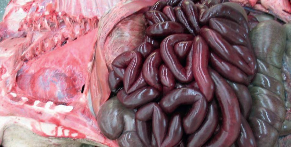
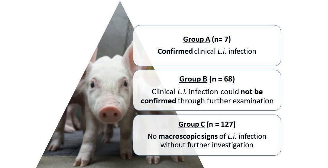
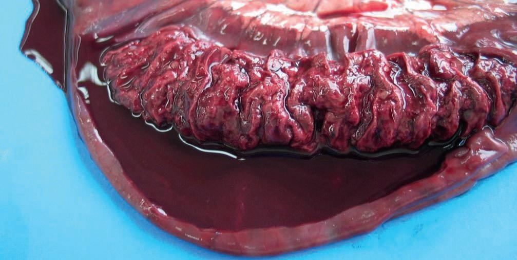
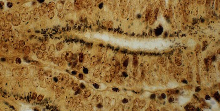
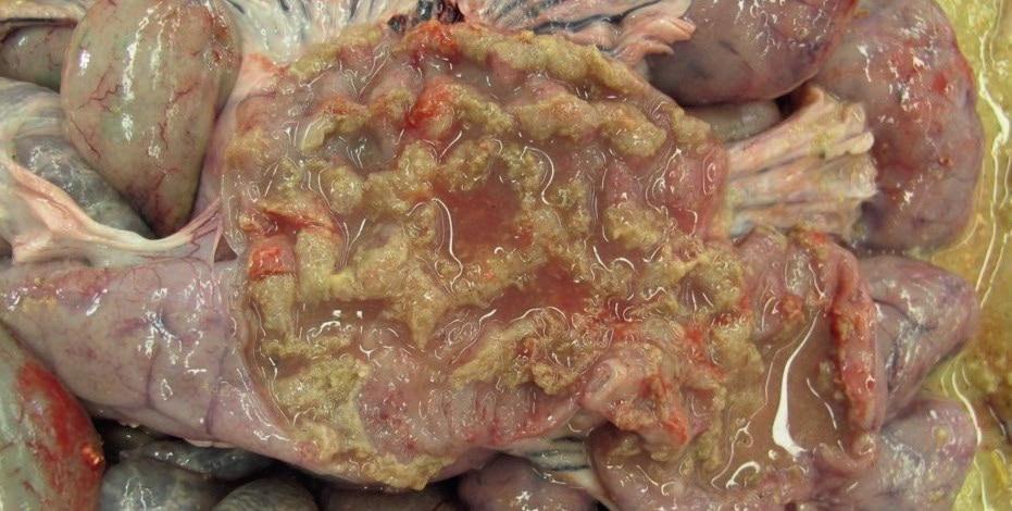
 Fig. 1: Macroscopic evidence of Haemorrhagic Bowel Syndrome. Organs in situ in the abdominal cavity, both small and large intestines are affected.
Fig. 2: Proliferative haemorrhagic enteropathy with fibrinohaemorrhagic cast in the intestinal lumen due to a Lawsonia intracellularis infection.
Fig. 4: Overview of the study groups within the study population (202 cases of haemorrhagic bowel syndrome in pigs ≥25kg within five years). L.i. = Lawsonia intracellularis.
Fig. 5: Warthin-Starry silver staining of an ileum section. Affected intestinal crypt cells showing large numbers of curved small L. intracellularis concentrated in the apical cytoplasm of hyperplastic mucosal epithelial cells.
Fig. 1: Macroscopic evidence of Haemorrhagic Bowel Syndrome. Organs in situ in the abdominal cavity, both small and large intestines are affected.
Fig. 2: Proliferative haemorrhagic enteropathy with fibrinohaemorrhagic cast in the intestinal lumen due to a Lawsonia intracellularis infection.
Fig. 4: Overview of the study groups within the study population (202 cases of haemorrhagic bowel syndrome in pigs ≥25kg within five years). L.i. = Lawsonia intracellularis.
Fig. 5: Warthin-Starry silver staining of an ileum section. Affected intestinal crypt cells showing large numbers of curved small L. intracellularis concentrated in the apical cytoplasm of hyperplastic mucosal epithelial cells.