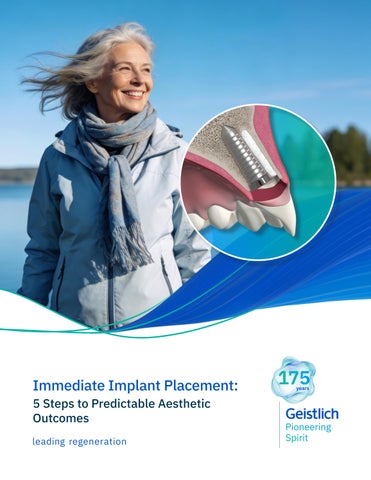
Immediate Implant Placement: 5 Steps to Predictable Aesthetic Outcomes
Predictable Immediate Implant Placement Starts with Biology When biologic principles are respected, aesthetic-zone outcomes become consistent and stable.
THE BIOLOGIC ADVANTAGE Know facial bone anatomy to improve case selection. Manage remodeling to preserve contour and papillae. Control soft tissue phenotype to support long-term aesthetic stability. Immediate implant placement becomes predictable when biology and protocol work together.
Your Roadmap to Predictable Aesthetic Outcomes
1
page 3
Identify the Right Case Recognise ideal and manageable risk situations
2
page 4
Apply a Structured Decision Framework Use biologic criteria to guide treatment planning
3
page 5
Follow the 10 Keys Protocol Execute a sequential roadmap for stability
4
page 6
Manage Gap & Soft Tissue Strategically Support long-term volume and contour
5
page 8
See Real-World Applications Learn from expert-led aesthetic zone cases
3
STEP 1: Identify the Right Case
The criteria below help you quickly assess risk, guide decision-making, and determine when immediate placement in the aesthetic zone is appropriate – or when it’s not.1
Core Eligibility Criteria ↪Ideally, these should be fulfilled.
ࠦ Both the buccal and palatal walls should be intact ࠦ ≥3–4 mm of apical bone for primary stability ࠦ Facial bone of at least 1 mm in thickness ࠦ No acute infection with pus or bone destruction ࠦ At least 35 Ncm insertion torque or ISQ of 70 (if aiming for immediate loading)
Dr. Stephen Chen on the importance of risk-aware case selection
Biological Predictability Boosters hard tissue
ࠦ Adequate facial bone thickness ࠦ No vertical or horizontal bone defects ࠦ Favorable socket anatomy soft tissue
ࠦ Thick gingival biotype ࠦ Adequate width of keratinised tissue ࠦ Intact papillae and no recession
Dr. Chen’s insights on the risk factor of a thin bone phenotype
Potential Risk Modifiers ↪Refine the decision aesthetics ť Low–medium smile
line preferred ť Highly scalloped architecture increases recession risk
prosthetic & occlusal ť Immediate contour
management is preferred (fixed provisional or custom healing abutment) ť Absence of parafunctional habits
1. Hamilton A, et al.: Clin Oral Implants Res. 2023 Sep:34 Suppl 26:304-348. (Specific criteria determined from a review of type 1A protocols for single tooth sites in the maxillary aesthetic zone.)
leading regeneration
patient factors ť Good oral hygiene
and compliance ť Non-smoker ť Realistic aesthetic expectations
STEP 2: Apply a Structured Decision Framework
site-related
patient-related
This summarised table helps you combine biologic factors and risk modifiers into one overall risk view.2 BADGE-CHECK LOW RISK
⚠ MEDIUM RISK
OCTAGON-XMARK HIGH RISK
Uneventful healing expected (non-smoker, no diabetes or immunodeficiencies)
Healing potentially compromised (e.g. light smoker, controlled diabetes, immune-suppressive medications)
Compromised healing (e.g. heavy smoker, post radiotherapy cancer treatment, poorly controlled diabetes)
Realistic patient expectations
High lip line
Significant aesthetic compromise expected, unrealistic patient expectations
BADGE-CHECK LOW RISK
⚠ MEDIUM RISK
OCTAGON-XMARK HIGH RISK
Intact socket walls
Buccal defect <5 mm
Marked buccal defect (>5 mm)
Thin phenotype
Severe thin phenotype
Apical bone for primary stability
Lack of apical bone for primary stability
Thick phenotype Adequate bone volume
Chronic periodontal infection and controlled periodontal disease
Absence of infection and periodontally healthy Absence of recession
Minor recession
Gingival recession >2 mm
Gap between facial bone and planned implant position 1–2 mm
Gap between facial bone and planned implant position <1 mm
BADGE-CHECK LOW RISK
⚠ MEDIUM RISK
OCTAGON-XMARK HIGH RISK
Minimally invasive extraction
Damage to surrounding soft tissues including severed and detached papillae
Significant damage to soft tissues and bone
Gap between facial bone and planned implant position >2 mm
intra-operative assessment
Acute infection and periodontal disease
Primary stability achieved Ideal 3-dimensional position achieved
BADGE-CHECK Ideal IIP Case • Predominantly
favorable factors • High predictability • Standard IIP Protocol
Lack of primary stability Facially positioned or overangulated implant or excessive implant depth
⚠ IIP with Caution
OCTAGON-XMARK Avoid IIP
• Multiple medium-risk factors • Advanced techniques required • Grafting and contour
• One or more high-risk factors • High aesthetic or biologic risk • Consider early or
management critical
Dr. Stephen Chen on the ITI treatment guide for immediate implant placement
delayed placement
Immediate implant placement begins with the right decision—but long-term success is driven by how hard and soft tissues are managed. The following 10 Keys outline the principles that support predictable regeneration and aesthetic stability.
2. Lambert F, et al.: Pre-operative analysis and treatment planning. In D. Wismeijer, Bart, & N. Donos (Eds.), ITI treatment guide 14: Immediate implant placement and restoration in partially edentulous patients (Vol. 14). Quintessence, 2023. (Table above is a truncated list, for full risk assessment refer to ITI treatment guide 14.)
leading regeneration
4
STEP 3: Follow the 10 Keys to Success Your practical checklist from planning → surgery → restoration.3-5
surgical
treatment planning
The following 10 Keys, described by Dr. Robert A. Levine, outline a practical, sequential checklist for predictable aesthetic-zone immediate implant placement.
1
Aesthetic risk assessment • Smile line • Gingival phenotype • Patient’s expectations
2
CBCT analysis CONSIDER: • Intact buccal bone wall — the thicker, the better (>1mm if possible) • Alveolar process at least 8 mm wide • Position of the alveolar socket inside the bone envelope • Implant selection and positioning to ensure a buccal gap >2 mm
3
Minimally traumatic tooth extraction (flapless if possible) • Assess buccal and palatal plates after extraction • If buccal height is compromised, consider an alternative protocol6
4
3D implant placement (using a surgical guide) • Good bone availability for palatal wall implant positioning Use of a narrow or regular diameter implant • Achieve primary stability and maintain a buccal gap >2 mm
5 6
prosthetic
Virtual surgical and restorative-driven treatment planning
Buccal gap bone grafting (with low substitution material)
7
Buccal soft-tissue grafting (CTG or volume-stable collagen matrix)
8
Immediate or delayed emergence profile management • Customised healing abutment or temporary crown
9
Use of a custom impression coping technique • To duplicate the created transition zone
10
Screw-retained final restoration (when possible)
If a key cannot be achieved at any stage, clinicians should pause and consider early or delayed implant placement instead. 3. Levine RA, et al.: Clin Implant Dent Relat Res. 2022 Aug;24(4):403-413. 4. Araújo MG, et al.: Clin Oral Implants Res. 2026 Mar 13. 5. Levine RA, et al.: Compend Contin Educ Dent. 2017 Apr;38(4):248-260. 6. Cardaropoli D, et al.: Dent J (Basel). 2025 Nov 12;13(11):529.
leading regeneration
Hear Dr. Robert Levine describe the 10 Keys Checklist
5
6
STEP 4: Manage Gap & Soft Tissue Strategically Strategic gap and soft-tissue management preserves long-term aesthetic outcomes.
Why Gap Management Matters While implant positioning is critical, it does not prevent post-extraction remodeling. Biomaterials play a key role in managing the peri-implant gap and maintaining longterm hard- and soft-tissue stability. Dr. Stephen Chen discusses the role of biomaterials and grafting in managing the peri-implant gap in immediate implant placement.
WATCH WEBINAR
Without filling the gap
“Placing an implant does not stop facial bone remodeling. The implant is a passive occupant — reconstruction with biomaterials is required to maintain volume and long-term hard- and soft-tissue stability.”
Clinical Impact of Gap Grafting
Filling the gap with Geistlich Bio-Oss Collagen®
After 1 year
92%
horizontal bone preserved7 Bone resorption after 1 year1
Evidence supporting this approach
Geistlich biomaterials for immediate implant placement 50+ peer-reviewed IIP studies covering 1,100+ patients and 1,300+ implants8 High long-term implant survival demonstrated with Geistlich Bio-Oss® and Geistlich Bio-Oss Collagen® Largest evidence base in immediate implant placement with long-term follow-up 7. Cardaropoli D, et al. Int J Periodontics Restorative Dent. 2014 Sep-Oct; 34(5):631-7. 8. Data on file. Geistlich Pharma AG, Wolhusen, Switzerland.
leading regeneration
SEE FULL EVIDENCE
7
Tissue Grafting Options for Long-term Stability Selecting the right grafting strategy supports long-term hard- and soft-tissue stability in immediate implant placement. When grafting is indicated, the choice of technique and biomaterials depends on whether the buccal bone wall is preserved or compromised.
Preserved buccal wall ↷
Fill the Gap: stabilise the clot and support ridge contour stability.
Defective buccal wall <5 mm ↷
See how it works!
↪
Fill + Protect: stabilise the clot and limit soft‑tissue ingrowth to support aesthetics.
See how it works!
↪
Soft-Tissue Augmentation Buccal soft-tissue augmentation becomes relevant when the thickness of keratinised tissue is <2 mm, as thin phenotypes are associated with a higher risk of recession and contour loss. A collagen matrix such as Geistlich Fibro-Gide® can be used to increase tissue thickness and support long-term aesthetic stability.8
leading regeneration
See how it works!
↪
STEP 5: See Real-world Applications Follow the roadmap in action — from diagnosis to stable aesthetic results.
You’ve got the principles, now see how they’re applied in real-world clinical scenarios. Immediate Implant Placement and Provisionalization for Anterior Aesthetics David E. Urbanek, DMD, MS | St. Louis, MO A healthy, 56-year-old female presented with fractured, endodontically treated tooth #9. The tooth was fractured at the gingival level and asymptomatic. Both the patient and the restorative dentist had high aesthetic expectations, and preferred immediate implant placement with provisionalization if possible.
1
2a
2b
3a
3b
4a
4b
5a
5b
6a
6b
7a
7b
8
1. Pre-operative image showing a fractured, endodontically treated tooth #9. 2. Image 2a: Pre-operative sagittal CBCT image demonstrating a Kan Class 1 sagittal root position with initial digital implant planning conducted during the consultation appointment. Image 2b: Definitive digital implant planning. 3. Image 3a: Tooth #9 was extracted with minimal flap elevation, preserving the papillae. Image 3b: A 3.6 x 15 mm implant was placed in a fully guided manner with a palatal bias to maximize the facial gap dimension, achieving a +2 mm gap. 4. Image 4a: The underside of the periosteum was incised just apical to the mucogingival junction, and a supra-periosteal pocket was created using sharp dissection with a #15 blade. Image 4b: The facial gap was packed with 50 mg of Geistlich BioOss Collagen®. A cover screw was temporarily placed to prevent bone graft granules from entering the implant chamber. 5. Image 5a: A 15 x 20 x 3 mm Geistlich FibroGide® collagen matrix was inserted into the supra-periosteal pocket, with the coronal portion positioned to cover the facial bone crest. Image 5b: The provisional crown was placed, and the facial mucosal flap was coronally advanced to cover the Geistlich Fibro-Gide® collagen matrix. 6. Images 6a & 6b: 2-week post-operative follow-up. 7. Images 7a & 7b: Final restoration at 16 months post-operative. 8. Periapical radiograph at 16 months postoperative demonstrating stable crestal bone levels.
“To obtain the best result with challenging cases, such as this one, I always approach them with thorough pre-surgical assessment, proper hard- and soft-tissue management, and the use of high-quality, evidence-based materials.” — Dr. David E. Urbanek
leading regeneration
8
9
Phenotype Modification Using Geistlich Fibro-Gide® for Immediate Implants in the Aesthetic Zone Robert A. Levine DDS | Philadelphia, PA A healthy non-smoking 50-year-old female patient who desired a single-tooth solution to replace a non-restorable tooth, #12. A root fracture at the level of the palatal post was diagnosed in a root canaled tooth. Maintaining aesthetics of the adjacent teeth was important as they are also restored with single full coverage porcelain crowns. 1
2
3
4
5
6
7
8
1. Pre-operative assessment demonstrates minimal zone and thickness of buccal keratinised gingiva, with a medium periodontal phenotype. 2. Pre-operative CBCT with virtually planned implant placement. A thin buccal plate (<1 mm) is measured. Good apical bone is noted for the placement of a Straumann® 12 mm x 4.1 mm bone level tapered implant. 3. Minimally invasive removal of #12 using only a buccal approach mini-flap showing an intact buccal plate with immediate placement of the implant (1 mm below the intact buccal wall) in a screw-retained position. A 3 mm buccal gap is measured and a 1.5 mm palatal gap. 4. Both the buccal and palatal gaps have been packed with Geistlich Bio-Oss Collagen® hydrated with Gem 21S®. I prefer to squeeze Geistlich Fibro-Gide® between thumb and forefinger, prior to placement. A dry-carved piece of Geistlich Fibro-Gide® is in position thinned approximately 2 mm with beveling laterally and coronally with a new #15
blade. 5. Geistlich Fibro-Gide® in place facial to the intact buccal wall under a full thickness buccal approach mini-flap. Immediate contour management was completed using an Anatotemp® for a maxillary bicuspid tooth. 6. Suturing completed using 4-0 PTFE and 5 -0 polypropylene nonresorbable sutures. Anticipated short-term 25% postoperative swelling is discussed with the patient. 7. 3 months post-operative appointment showing a welldeveloped subgingival transition zone created with immediate contour management. A reverse torque test was completed, and the case proceeded to completion. 8. 14-month post-operative view with final screw-retained crown in place. Good interproximal papilla healing is noted with thickening of the buccal periodontal phenotype compared with Fig. #1. (Restorative Therapy: Drew Shulman DMD, MAGD; Philadelphia, PA)
“The importance of the ‘one-two punch’ of ROUTINE phenotype modification using Geistlich Fibro-Gide® in conjunction with bone grafting the >2 mm buccal gap with Geistlich Bio-Oss Collagen® provides excellent buccal convex tissue maintenance long-term.” — Dr. Robert A. Levine
leading regeneration
Key Takeaways from the 5-STEP Roadmap
1
Learn
STEP 1:
Identify the Right Case Predictability starts with disciplined case selection and biologic assessment.
STEP 2:
Apply a Structured Decision Framework
Explore additional clinical cases.
Combining biologic factors and risk modifiers reduces uncertainty.
2
STEP 3:
Follow the 10 Keys to Success Structured execution — from planning to restoration — minimises complications.
STEP 4:
Manage Gap & Soft Tissue Strategically
Your next steps
Buy
Support your protocol with evidence-based biomaterials.
Hard- and soft-tissue management preserves contour and long-term aesthetics.
STEP 5:
Apply in Real Clinical Cases When the protocol is respected, predictable aesthetic outcomes follow.
Affiliate Australia and New Zealand Geistlich Pharma Australia Pty Ltd. The Zenith – Tower A Level 21, Suite 21.01 821 Pacific Highway Chatswood, NSW 2067, Australia Phone AU +61 1800 776 326 Phone NZ +64 0800 500 043 info@geistlich.com.au info@geistlich.co.nz www.geistlich.com/en-au
8377_01
Precision in planning. Discipline in execution. Confidence in outcomes.
Manufacturer Geistlich Pharma AG Bahnhofstrasse 40 6110 Wolhusen Switzerland Phone +41 41 492 55 55 info@geistlich.com www.geistlich-pharma.com