Unlocking the Full Potential of AI-Driven Robotic Cataract Surgery
Robotic cataract surgery systems now entering the clinic could offer advantages such as greater precision and more predictable outcomes.
Accommodating
Simultaneous Vision Lenses
Understanding neuroplasticity can improve refractive outcomes with modern IOLs.
Top ophthalmologists share their experiences treating elite athletes, with practical insights that apply in regular vision care.
The Continuing Evolution of Corneal Allogenic Intrastromal Ring Segments
Farhad Hafezi discusses the advantages of ECO-CAIRS, including faster surgery and better hydration.
https://congress.escrs.org/
Drive the Research That Drives Surgery
Good surgical practice is based on solid research evidence— and there’s no better way to build research expertise than the ESCRS Peter Barry Fellowship.
The Peter Barry Fellowship is a research fellowship opportunity for young ophthalmologists who have finalised their training in ophthalmology and want to increase their knowledge and/or research skills in anterior segment surgery (cataract, cornea, or refractive). The €60,000 fellowship enables European trainees to spend a year at a global centre of excellence to build research expertise in cataract and refractive surgery.
The fellowship is named for Peter Barry (1948–2016), a founding member of ESCRS who served as president and director of the Society and helped shape modern cataract care. His leadership of the ESCRS endophthalmitis prophylaxis study—at the time the largest antibiotic study in ophthalmology—demonstrated that intracameral cefuroxime at the end of cataract surgery reduces postoperative infection five fold, changing practice across Europe and beyond. He also championed outcomes benchmarking via the EUREQUO registry and promoted education and opportunities for younger ophthalmologists.
Eligibility
Applicants must be young ophthalmologists in their second year of sub-specialty experience in anterior segment surgery and are full members of ESCRS. (This will include only ophthalmologists working in Europe and exclude trainees.) International applicants are considered but must be ESCRS members at the point of application, and their application must clearly outline a benefit to Europe.
The Peter Barry Fellowship only accepts research fellowship applications. The Fellowship is announced at each ESCRS Annual Congress, to start the following year.
Application deadline: 15 July
Keep Your Eye on the Ball!
Top ophthalmologists share their experiences treating elite athletes as well as practical insights that apply in regular vision care.
18 Managing Concurrent Cataract and Vitreomacular Traction
Ananth Sastry MD
20 Unlocking the Full Potential of Robotic Cataract Surgery
Yu-Hsuan (Alex) Huang MD, PhD
21 Closing the Presbyopia Gap for Younger Patients
Anas-Alexis Benyoussef MD, MSc
22 The Living Framework
Joaquín Fernández Pérez MD, PhD
24 When Digital Training Enhances Neuroadaptation
Susana Marcos PhD
25 Soft Skills Help with Hard Patients
29 Presbyopia: One Condition with Many Solutions
Laura Maubon FRCOphth, BMBS, BMed Sci, PGCert (Surg Ed)
CORNEA
30 A New Era for Keratoconus Diagnostics
Renato Ambrósio Jr MD, PhD, FWCRS, PCEO
32 Droopy Lids? The Bleph Boom
Clare Quigley MD
34 The Evolution of CAIRS
Farhad Hafezi MD, PhD, FARVO
DIGITAL OPHTHALMOLOGY
Ruggiero Paderni
Erik
Pei-Fen Lin MBBS,
Başak Bostancı MD, FEBO
26 Robotics in Cataract Surgery: Coming Soon?
Pavel Stodůlka MD, PhD
27 Phakic IOL Follow-Up: What to Watch For
Kjell Gunnar Gundersen MD, PhD
28 Determining the Best Patient for the IPCL
Pavel Stodůlka MD, PhD
36 Digital Integration to Meet Future Challenges
Bruce Allan MD, FRCS
GLAUCOMA
37 Raising the Bar for Cataract Surgery in Glaucoma Patients
Reza Alizadeh MD
Publisher
Tom Ogilvie-Graham
Executive Editor
Stuart Hales
Editor-In-Chief
Sean Henahan
Senior Content Editor
Kelsey Ingram
Creative Director
Kelsy McCarthy
Graphic Designer
Jennifer Lacey
Circulation Manager
Lucy Matthews
Contributing Editors
Cheryl Guttman Krader
Roibeárd O’hÉineacháin
Contributors
Laura Gaspari
Soosan Jacob
Timothy Norris
Clare Quigley
Andrew Sweeney
Colour and Print
Advertising Sales
Roo Khan
Tel: +44 203 530 0100 | roo.khan@wearemci.com
EuroTimes® is registered with the European Union Intellectual Property Office and the US Patent and Trademark Office.
Published by the European Society of Cataract and Refractive Surgeons, Building 4000, Langstone Park, Langstone Road, Havant, PO9 1SA, UK. No part of this publication may be reproduced without the permission of the executive editor. Letters to the editor and other unsolicited contributions are assumed intended for this publication and are subject to editorial review and acceptance.
ESCRS EuroTimes is not responsible for statements made by any contributor. These contributions are presented for review and comment and not as a statement on the standard of care. Although all advertising material is expected to conform to ethical medical standards, acceptance does not imply endorsement by ESCRS EuroTimes. ISSN 1393-8983
Learn more about EuroTimes or connect with ESCRS at ESCRS.org
Helping Patients Win in the Game of Life
With an estimated viewership of six billion people, the 2026 FIFA World Cup has focused a lot of attention on optimal human performance under the stress of competition. And as with all sports, from elite competition to a neighbourhood pickleball game, this event reminds us of the key role of vision in maximising performance.
Vision care is involved in so many aspects of sports, from vision assessment, rehabilitation, and eye protection to refractive interventions and the treatment of eye injuries. Athletes’ concerns for optimal acuity and good contrast vision are more urgent, but perhaps not that different from those of the typical cataract or refractive surgery patient.
This all serves as a welcome reminder not only of the role of ophthalmologists in sport, but in everyday life.
When kids see elite athletes wearing eye protection, they are encouraged to view it as a safe and useful option. When Dutch footballer Edgar Davis, who played at the international level, arrived on the pitch wearing goggles following glaucoma surgery, this not only encouraged the idea of eye protection but also reminded young people who might have diabetes to not limit their endeavours because of an eye disease.
Who can say how many people may have been inspired to consider laser refractive surgery after Tiger Woods had LASIK in 1999? Since that time, it has become common for professional athletes to undergo refractive surgery. Fellow golfers Vijay Singh and Berhard Langer have undergone LASIK. Other examples include tennis players Rafael Nadal, Andy Murray, and Juan Martín del Potro. NBA stars Lebron James and Derek Fisher, and footballer Lionel Messi, have undergone refractive surgery as well.
EDITORIAL BOARD
Adi Abulafia (Israel)
Bruce Allan (UK)
Noel Alpins (Australia)
Juan Alvarez de Toledo (Spain)
Gerd Auffarth (Germany)
Başak Bostanci (Türkiye)
John Chang (Hong Kong SAR, China)
Béatrice Cochener-Lamard (France)
Burkhard Dick (Germany)
Mor Dickman (The Netherlands)
Joaquín Fernández (Spain)
Oliver Findl (Austria)
Sri Ganesh (India)
Christina Grupcheva (Bulgaria)
Farhad Hafezi (Switzerland)
Nino Hirnschall (Austria)
Soosan Jacob (India)
Jack Kane (Australia)
Yao Ke (China)
Georgios Kymionis (Greece)
Our cover story by Andrew Sweeny looks at the essential role ophthalmologists play in treating trauma in sports. It reminds us that whether treating celebrity athletes or normal patients, the approach is the same.
Sport-related injuries are not limited to summer activities. In a related article, Laura Gaspari caught up with Dr Ruggiero Paderni, who was an ophthalmologist consultant at the Milan Cortina 2026 Winter Olympic Games. The article reviews the potential issues and injuries associated with high altitude sports, from minor conjunctivitis to major trauma associated with high impact injuries.
Sport has its heroes, and ophthalmology has its own heroes, but rarely do the two come together as dramatically as in the case of Henry Stallard. A medallist in the 1924 Olympics who was later depicted in the film Chariots of Fire , Stallard went on to lead a remarkable life in ophthalmology, refining many surgical techniques, writing a major textbook, and leading clinical studies on the treatment of retinoblastoma. We’ve reprinted an article on Dr Stallard that we ran in 2024 as a reminder of the life of this remarkable individual.
Eye care in sports highlights the value of optimising visual function on the playing field. This serves to underscore the obstacles practising ophthalmologists face when trying to help patients function at the highest levels in the challenging environments of everyday life.
Sean Henahan Editor-in-Chief
David Lockington (UK)
Artemis Matsou (Greece)
Cyres Mehta (India)
Jod Mehta (Singapore)
Sorcha Ní Dhubhghaill (Belgium)
Rudy Nuijts (The Netherlands)
Catarina Pedrosa (Portugal)
Konrad Pesudovs (Australia)
Nic Reus (The Netherlands)
Filomena Ribeiro (Portugal)
Andreia Rosa (Portugal)
Giacomo Savini (Italy)
Julie Schallhorn (US)
Sathish Srinivasan (UK)
Paola Vinciguerra (Italy)
Shin Yamane (Japan)
Ron Yeoh (Singapore)
Mihail Zemba (Romania)
Thomas Kohnen
José Güell
Paul Rosen
An Opportunity of a Lifetime
Interested in taking a giant step forward in your surgical career?
ESCRS is launching the Emanuel Rosen Fellowship for young ophthalmologists who have finalised their training in ophthalmology and want to increase their knowledge and/or surgical skills in anterior segment surgery (cataract, cornea, or refractive).
The Fellowship of €60,000 is designed to allow the applicant to work abroad at a centre of excellence for clinical experience in the field of cataract and refractive surgery, anywhere in the world, for one year.
The fellowship celebrates the first president of ESCRS, Emanuel Rosen, who helped ignite the Society’s founding and growth, co-launched the Journal of Cataract & Refractive Surgery, and championed advances ranging from intraocular lenses and phacoemulsification to laser refractive surgery, all while serving patients across the UK National Health Service and in private practice.
Eligibility
Applicants must be young ophthalmologists who are full members of the ESCRS (this will include only ophthalmologists working in Europe by definition and also exclude trainees) who are in their second year of sub-specialty experience in cataract, cornea, or refractive surgery.
The successful applicant for the Fellowship will be announced at the ESCRS Annual Congress in London.The fellowship will start in 2027.
Apply by 30 June for the opportunity of a lifetime!
SAVING SIGHT IN UKRAINE WITH BALLISTIC EYE PROTECTION
Supplying eye protection to Ukraine faces additional complications with changing cultural perceptions.
ANDREW SWEENEY REPORTS
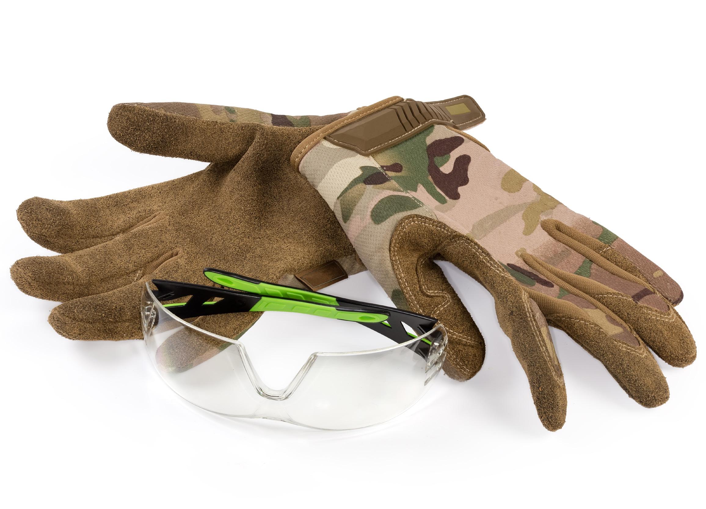
Bulletproof vests, helmets, and first aid kits are all essential equipment for military personnel these days, and all are well known among the general public. What people do not regularly associate with first responders, medics, and soldiers, however, is ballistic eye protection, yet it is just as important as military gear (if not more so).
Also known as military combat eye protection (MCEP), these glasses are made from a lightweight, impact-resistant thermoplastic designed to withstand explosive blasts, shrapnel, and other debris. While they cannot stop everything, they can help to save sight.
In Ukraine, where Russia’s full-scale invasion continues after more than four years, eye protection is crucial. Shelling, close-quarter combat, and first-person view (FPV) fibre-optic-guided drone warfare have all made this war particularly cruel to sight.
“Your entire future is impacted if you lose your vision,” said Brian True, the managing director at Eye Care for Ukraine. “If you lose your leg, you can get a prosthetic one and you can run in a road race, you can climb up a mountain, but if you go blind, you’re blind.”
Your entire future is impacted if you lose your vision.
Mr True’s organisation launched in 2021 and came into its own after the war began. Today, its first priority is still supplying corneal tissue through corneal banking—despite considerable legislative challenges—while providing valuable equipment such as glaucoma valves and artificial irises.
The project to supply MCEPs to first responders and medics came about as a result of the war. They are ‘required wearing’
for Ukraine’s soldiers, but supply is intermittent and disorganised, and the quality of the glasses can be poor.
“Some soldiers are provided eye protection, some are not. We’re currently providing in the low 1,000s of pairs of eye protection,” Mr True said.
“Soldiers are often forced to find and pay for the protection themselves. It’s expensive: for instance, I can buy glasses in the US for about $55 that would cost me $125 in Ukraine.”
Mr True’s foundation has joined forces with the US-based BBH Eye Foundation to provide NATO-grade antiballistic eyewear to many medical first responder organisations. A notable example is the Hospitalliers, a Ukrainian voluntary organisation of paramedics, many of whom volunteered to serve at the front. They are routinely, and illegally, targeted with drones as they evacuate wounded troops, making eye protection even more important, as these new weapons cause significant shrapnel damage.
“We provide what we can, and we also try to raise awareness about the importance of eye protection,” he said. “We urge everyone we meet to wear eye protection.”
Unfortunately, the need to drum this message home is considerable. Many first responders and soldiers prefer not to
wear them, citing a variety of reasons, including a perception that they prevent peripheral vision, are annoying for people unaccustomed to wearing spectacles, and are ‘not cool’.
Overcoming this resistance to wearing MCEPs is challenging, but Mr True and his team are making considerable progress. Acceptance is up thanks to their education efforts as well as their practical demonstrations.
“We took three different types of lenses that we work with, placed them at 10 metres distance from us, and a soldier shot them with a high-powered gun using a fragmentation bullet. None of the lenses broke,” Mr True said.
“It was not exactly scientific, but it demonstrated that the lenses do work at protecting the eyes from damage and absorbing impact. For the minor inconvenience of wearing glasses, you could save your sight.”
Brian True is the managing director at Eye Care for Ukraine. Major support for providing ballistic eyewear is through a partnership with the BBH Eye Foundation, a US non-profit. briantrue@eyecareforukraine.org
Top ophthalmologists share their experiences treating elite athletes as well as practical insights that apply in regular vision care.
BY ANDREW SWEENEY
he World Cup kicks off this summer and the Commonwealth Games are coming. Add international tennis tournaments, motor racing, and cycling and 2026 is shaping up to be a summer of sports.
That means it’s likely to be a season of ocular trauma, too; clinicians can expect to see more patients presenting with sports-related injuries. From the humble football (or ‘soccer ball’ to American audiences) to the well-timed left-hand jab, danger to the eyes is omnipresent in sport.
“One of the most common injuries is blunt ocular trauma from a football kicked at the face, or a tennis/squash ball that hits the ocular region,” said James E Neffendorf MD. “The nature of the injury can vary widely—from a mild, isolated commotio retinae to severe intraocular bleeding (such as a total hyphaema) or a retinal detachment.”
“Posterior segment injuries are much rarer and more severe: for instance, commotio retinae with Berlin’s oedema, retinal tear or detachment, and vitreous haemorrhage,” said H Burkhard Dick MD, PhD. “It is crucial to perform dilated fundoscopy and to examine the patient immediately if they report photopsia, floaters, or any visual field defect.”
‘Harmless’ equipment can threaten sight
The noted ophthalmologists interviewed for this article stressed the importance of remembering how ocular trauma can result from the most innocuous actions. Simple, everyday sports equipment can cause catastrophic damage capable of threatening an athlete’s career.
José L Güell MD, PhD described treating a well-known tennis player who was slowly developing secondary cataract following ball impact. The patient had visited several surgeons, but none of them wanted to propose surgery because the patient was young and amongst the 50 top players in the world.
One of the most common injuries is blunt ocular trauma from a football kicked at the face, or a tennis/squash ball that hits the ocular region.
“The question was: should I use a multifocal lens? The eye works and he doesn’t have presbyopia, but he would have it if I did surgery,” Professor Güell said. “Would he need perfect near vision as well as distance vision?
“The decision to do surgery was hard because his vision was quite good, despite that when the sun was in front of him, he had disturbances due to posterior capsule opacity. Fortunately, everything went well.”
Dr Neffendorf described a “particularly unfortunate case” involving a young adult who had a sports scholarship at a top university. The patient sustained an accident with an elastic resistance band, which, under tension, slipped off a bar and snapped into their face, with catastrophic results.
“It resulted in a bilateral retinal detachment that required multiple surgical procedures,” Dr Neffendorf said. “The retinal detachments were fixed, but the reduced vision meant their sporting career was over. It was an eye opener about what’s at stake for athletes and the risk posed by seemingly harmless equipment.”
Managing myopia in athletes
The risks posed to athletes by harmless equipment are one concern, but common conditions can also have an oversized impact on their sight. The most notable example is myopia, as even small, uncorrected refractive errors can significantly affect visual performance.
“When examining a high-performing athlete with uncorrected myopia, in addition to the usual procedures, we pay particular attention to contrast sensitivity, binocular vision and stereopsis, and dynamic visual acuity and oculomotor function,” Prof Dr Dick said.
“Soft contact lenses are preferable because of better optical quality and a smaller possibility of visual field constriction than rigid gas permeable lenses. I also emphasise that a strict hygiene regime should be adhered to (‘no water and no overnight wearing’).”
According to Prof Güell, myopic athletes need to maximise their perceptual processing and decision making, reaction
time, and spatial awareness. This can make them demanding patients as a result—but rewarding ones, too.
“One professional padel player I treated had become intolerant to contact lenses. Surgery was successful, but there was residual low myopia in the left eye, and she felt her shot precision on her left side had been reduced,” Prof Güell said.
“We needed to re-operate twice to achieve an extremely low residual error. The second time was just 0.25 dioptres of a residual cylinder to reach the comfort level for her left-hand shots. It shows how much precision athletes demand.”
Always keep an open mind
Whether the patient is an athlete at the top of their game or a patient who walked in after a five-a-side game went wrong, the approach towards treatment remains the same. The attitude of athletes—and their ability to appreciate injury as a challenge to be overcome— is motivating for the surgeons interviewed, but insights apply to any patient with a sports-related eye injury.
“Keep an open mind when assessing a patient with trauma during sport,” Dr Neffendorf said. “All patients should have a dilated fundal examination—it is not uncommon that I will find a small retinal tear lurking in the periphery of the retina.”
“Inform these patients about the increased long-term risk of glaucoma, cataract, and retinal detachment, which need to be documented for insurance purposes—not only for professional sports people. In more complicated cases, surgery such as anterior chamber washout becomes necessary,” Prof Dr Dick said.
James E Neffendorf MA (Cantab), MBBS (Lond), MD (Res), FRCOphth is a consultant ophthalmologist, cataract, and retinal surgeon based in London. james.neffendorf@nhs.net
José Luis Güell MD, PhD is head of the Cornea, Cataract, and Refractive Surgery Department at IMO Grupo Miranza in Barcelona, Spain. jose.guell@imo.es
H Burkhard Dick MD, PhD is Chairman and Head of the University Eye Clinic, Bochum, Germany, and President of the ESCRS. dickburkhard@aol.com
ESCRS UPDATE
ESCRS Launches New Learning Hub and Membership System
ESCRS has launched a new platform that combines membership and education and makes it easier to access resources for professional development and networking.
Upon logging into the platform, members can access their membership record, manage their communication preferences, and explore a range of ESCRS member benefits and resources, including the following:
• access to the ESCRS Learning Hub, featuring interactive, accredited educational content, surgical videos, animations, and on-demand sessions from past Congresses;
• access to reduced registration fees for ESCRS Congresses and events; and
• subscriptions to ESCRS publications, including EuroTimes and the Journal of Cataract & Refractive Surgery.
Members may need to reset their password when logging in for the first time.
Recording Devices to Assist with Surgeons’ Development
ESCRS is partnering with a medical technology company to provide recording devices to help ophthalmologists record their surgeries on a secure, organised platform, allowing mentors to review, comment on, and discuss surgical videos with trainees.
The recording devices will be shared with affiliated national ophthalmology societies to host and use with their members. Leaders of affiliated national societies are urged to contact escrs@escrs.org to request the MicroRec devices.
Under the arrangement, a fresh cohort of approximately 30 ophthalmologists in four training centres will receive MicroREC Kit hardware and MicroREC Connect access each quarter. Using these devices, surgeons will record their procedures, organise their video libraries, and receive structured feedback from assigned ESCRS mentors. At the end of each quarter, the devices will rotate to four new groups of trainees.
All told, the devices will be shared with 12 training centres. At the end of the 3-year programme, ESCRS will have built a network of 360+ surgeons with documented, mentored surgical experience.
The recording equipment is being provided by Custom Surgical, a medical technology company based in Munich, Germany, that specialises in surgical video recording and digital education solutions for ophthalmology.
The Olympian Ophthalmologist
Henry Stallard’s remarkable career had many milestones.
SEAN HENAHAN REPORTS
The 2024 Olympiad in Paris marked the centenary of the last time the games took place in the City of Light. The 1924 Olympics were later made famous in the 1981 film Chariots of Fire, featuring some British athletes’ quest for gold, particularly in running events. Among the select athletes competing for Britain was Hyla (Henry) B Stallard, who would earn a medal in Paris. He would also go on to reach Olympian heights in his chosen field of ophthalmic surgery.
As detailed in an excellent video assembled by Hugh Williams1, Henry Stallard had become one of the fastest middle-distance runners in British history years before the Paris Games. In 1920, he was part of a combined Oxford-Cambridge University team that travelled to the United States for a twomile relay race against America’s fastest runners. The British team won, setting a new world record in the category. His amateur athletics career continued as a member of the Cambridge University Athletics team. He went on to win the one-mile race against Oxford three years in a row, in 1920, 1921, and 1922. He was also part of the Oxford-Cambridge team that set a world record in the 4×880-yard relay in 1922. His mile record held until Roger Bannister broke the four-minute-mile mark in 1954.
Stallard was selected to run the 800 and 1500 metres at the 1924 Olympics. He injured his right foot in the final of the 800 m, finishing fourth. He then ran in the 1500 m despite having what turned out to be a metatarsal stress fracture in the right foot. He ran against the doctor’s advice, saying famously, “I am going to run tomorrow if I never run again.” He nonetheless received a bronze medal, later apologizing for his “poor show.”
His running career continued alongside his medical training at St Bartholomew’s Hospital and Moorfields Eye Hospital in London. He eventually held positions at both hospitals and maintained a private practice on Harley Street. He was obliged to end his running career in 1928, having suffered recurrent stress fractures of the navicular bone in both feet.
It was at St Bartholomew’s and Moorfields that he became interested in retinoblastoma treatment under the mentorship of Mr Robert Foster Moore. Moore pioneered the use of radioactive radon seeds in treating retinoblastomas in children. Stallard improved on this idea, developing focal radiotherapy, applying a radioactive cobalt 60 plaque to the surface of the eye, overlying the tumour, and giving a
measured dose of radiation. Thanks to this research, it became possible to save both the lives and the vision of many children with the disease.
His busy career was interrupted in 1939 by the onset of the Second World War. He served first in Cairo, where he performed more than 600 operations on wounded soldiers and civilians, meticulously logging and describing the injuries and treatments, often with carefully drawn illustrations. He somehow found time to run and climb over the top of the Great Pyramid of Giza, Mr Williams noted.
He was later transferred to Europe, arriving on the beaches of Normandy only days after D-Day, eventually performing eye surgery in France and Belgium. A major in the British Army, he steadfastly refused promotion, fearing he would become more bureaucrat than surgeon.
After the war, he built on his considerable surgical experience, publishing Eye Surgery, the first modern comprehensive guide to eye surgery, which set the standard for ophthalmic surgical training for many years.
The British Journal of Ophthalmology, to which he was a frequent contributor and one-time assistant editor, noted in memoriam: “Stallard’s distinction lay in the quality of his surgical skills and his ability to impart them to others. Every single operation was in fact a research project to be performed as perfectly as possible. He was the complete master. There was a natural reserve, a great humility combined with a high ideal of service and a great compassion which made him an outstanding doctor. [He was] artistic, warm-hearted, and perceptive, with a genial and kindly humour.”2
Stallard is also credited with developing many surgical techniques. As described in a comprehensive article in the Indian Journal of Ophthalmology, this included “partial cyclectomy and its modifications for iris neoplasia, techniques of eyelid reconstruction and partial transplantation (middle third) of levator palpebrae superioris muscle in patients with superior rectus palsy, management of ocular emergencies, and surgeries for epiphora.”3 He also developed several surgical instruments, including a corneal grafting knife, a ptosis spatula, and a non-magnetic foreign body extractor.
“Henry Stallard was an outstanding athlete and gifted surgeon, a truly remarkable man,” Mr Williams noted. “While he had no children of his own, the hundreds of children throughout the world who had their lives and their sight saved and those who will need our help in the future—they are Stallard’s children.”
For citation notes, see page 40.
This article originally appeared in the July/August 2024 issue of EuroTimes
Hugh Williams DO, FRCS, FRCOphth is an Honorary Consultant Surgeon, Moorfields Eye Hospital, London. drhpwilliams@yahoo.co.uk
Turn your clinical exper tise into business success.
A new ESCRS programme for ophthalmologists look ing to build, grow, and lead successful practices
Weekend One: Business & Financial P lanning
30 O ctober – 1 November 2026 | M arriot Budapest
Scan to learn more
Snow, Speed, and Sight
From high-speed trauma to UV exposure, eye safety deserves greater attention in winter sports and elite competitions like the Milan Cortina 2026 Winter Olympics.
LAURA GASPARI REPORTS
Winter sports are commonly associated with lower limb injuries, while eyes—frequently exposed to stress and risks—remain underestimated and need more recognition, according to Ruggiero Paderni MD.
Dr Paderni served as an ophthalmologist consultant at the Milan Cortina 2026 Winter Olympic Games, where he managed ocular emergencies for athletes and their families. The healthcare organisation for such an event was massive: a top-level healthcare system capable of ensuring safety and rapid response in every context, covering an area of more than 22,000 km² across Lombardy, Veneto, and the Autonomous Provinces of Trento and Bolzano and from metropolitan areas to high-altitude competition sites.
The designated Olympic hospital was the Niguarda Hospital in Milan, where an Olympic ward has been inaugurated with 11 inpatient rooms directly connected to the emergency room via protected tunnels. Some Olympic polyclinics were established on site, along with a well-coordinated collaboration with regional paramedics. Each competition venue had at least two medical stations (one for athletes and one for spectators) with dedicated protocols for rapid ‘on snow’ triage.
This network supported Dr Paderni’s efforts to protect eye safety, which he said is crucial in winter sports. Understanding ocular risks is essential for prevention, safeguarding visual function, and ensuring optimal performance during competition.
“The main risk factors in high-altitude mountain environments include high speed, atmospheric events like wind, cold, and UV radiation, and rapid altitude changes,” Dr Paderni explained.
High speed or physical contact sports—such as skiing, snowboarding, skeleton, luge, hockey, and ice skating—expose the eye to blunt traumas caused by collision with other athletes, accidental falls, or impacts with equipment. These injuries can lead to a sudden increase in IOP and permanent visual damage due to intraocular haemorrhages, retinal detachment, or vascular occlusions. Frontal impacts may also result in orbital fractures (blow-out), which can trap eye muscles and cause diplopia. Trauma may also be associated with potentially critical systemic conditions such as concussion (due to intracranial haemorrhage) and systemic infections (caused by lacerated wounds), which must be promptly managed to ensure the athlete’s safety.
Wind and cold can irritate and dehydrate the ocular surface. Moreover, wind may carry small debris that can penetrate the eyeball and cause corneal abrasions and infections. UV rays may cause damage to the cornea, such as photokeratitis, because snow reflects up to 80% of them, amplifying exposure. Altitude also plays a role, since UV intensity increases by about 10% every 1,000 metres. Extreme environmental conditions can alter ocular physiology, requiring the eye to adapt to atmospheric changes in pressure and oxygen levels: rapidly ascending from 500 to 3,000 metres may lead to temporary decreases in visual performance, and corneal hypoxia and high-altitude retinopathy are potential risks.
Helmets and goggles
For these reasons, protective equipment is essential to prevent irreparable damage to the eyes at high altitude. Helmets and goggles must meet strict standards, including UV-400 certification to block 100% of UVA and UVB rays; category 3 or 4 filters with polarized lenses to shield against radiation reflected from snow; shatterproof materials (polycarbonate or Trivex) to prevent splintering; and anti-fog systems with double lenses and a sealed air chamber to prevent condensation and maintain clear vision in extreme conditions.
As Dr Paderni remarked, the ophthalmologist assisting an injured athlete must be able to manage ocular emergencies by applying specific diagnostic and therapeutic protocols in accordance with accepted guidelines for immediate risk assessment. In ophthalmic emergencies, the guidelines of the AAO are typically followed. Also considered valid are the
“Consensus statement on injury and illness in sport” by the International Olympic Committee (IOC), useful for assessing craniofacial injuries, and the “F-MARC Medical Assessment Guidelines” developed by FIFA during the 1994 World Cup, useful for immediate on-field evaluation of visual capacity.
Among the ‘warning signs’ requiring immediate ophthalmological evaluation are severe eye pain after trauma (indicating a foreign body or sudden increase in the IOP, especially if associated with nausea), photopsia, floaters, ‘curtain-like’ visual shadows (suggestive of retinal lesions), diplopia in orbital fractures, and altered pupillary reflexes or anisocoria. Photokeratitis presents with severe pain and burning, redness, intense tearing, and reduced visual performance (even for 24 to 48 hours).
Dr Paderni said he was lucky to face only some cases of conjunctivitis, which he believes were likely caused by foreign bodies, photokeratitis, and a corneal abrasion. More severe traumas were evaluated at the Niguarda Hospital in Milan. However, such an experience is really something unique and once in a lifetime.
“It was both a great responsibility and an honour,” Dr Paderni said. “Being part of such a complex international event gave me the chance to contribute to athlete safety while applying specialised medical expertise in a challenging and dynamic environment.”
Ruggiero Paderni MD is head of the neuro-ophthalmology service at the Centro Diagnostico Italiano, Milan, Italy. He was a member of the Olympic medical staff for the Milan Cortina 2026 Winter Olympics.
Tools to Guide Treatment Choices
Aligning patient expectations with the best available options.
TIMOTHY NORRIS REPORTS
When examining a new patient with presbyopia, using objective tests to predict quality of vision and subjective tools to align expectations and tolerance are both essential. According to Erik L Mertens MD, FEBOS-CR, it is important to transfer all of this into a shortlist and to have a plan B ready.
The first step to consider is the modern patient’s journey through the digital sphere and the front desk, he said. There is only one chance for a first impression, and it is important to make it count. The second step is for optometrists and technicians to actually speak and listen to the patient, considering their motivations and frustrations about their vision, collecting useful data on their lifestyle, work, and hobbies, and understanding their expectations.
The subjective tools available in the clinical armamentarium are the basic quality of vision questionnaire and an interview focused on patients’ top three daily tasks, assessing their needs, their level of spectacle tolerance, and whether they are perfectionists or easy-going. The interview should be no longer than five minutes to avoid annoying the patient too much, he noted.
Another important subjective tool is the simulation/trial phase, in which Dr Mertens includes a monovision trial and explains dysphotopsia and what to expect from plan B to the patient. When the patient is going to receive a multifocal IOL, dysphotopsia needs to be explained honestly from the outset,
he emphasised. The patient needs to be informed that halos and glare can be experienced for up to six months. Moreover, he pointed out, the surgeon should always discuss what is going to happen if the first surgery is not successful.
The third step is a full examination using the objective tools. Dr Mertens explained a full examination includes checking the ocular surface and considering a thorough preoperative treatment, performing an OCT evaluation of the macula, measuring astigmatism, and examining the cornea looking for conditions such as Cogan dystrophy or map-dot-fingerprint dystrophy as well as the risk of ectasia. It is also important to look for coma, trefoil, and spherical aberration, he said.
The pupil also needs to be analysed in scotopic, mesopic, and photopic circumstances, with evaluation of the angle kappa and alpha.
The fourth step is where the surgeon becomes directly involved. Once all the subjective and objective data have been gathered, Dr Mertens analyses the whole picture, determining the options for each candidate. He then decides which of those options will come closest to the patient’s expectations without exceeding the level of tolerance they are willing to accept.
“I [form a plan in] my mind [about] what I will do with the patient. By step five, I only make one specific recommendation to the patient—because when you give more options, you create doubt,” he concluded.
Dr Mertens presented at the 2026 ESCRS Winter Meeting in Helsinki.
Erik L Mertens MD, FEBO, PCEO, FWCRS, FEBOS-CR is Director, Founder, and Ophthalmic Surgeon of Medipolis, Antwerp, Belgium. E.Mertens@Medipolis.be
Accommodating Simultaneous Vision Lenses
Understanding
neuroplasticity can improve refractive outcomes.
TIMOTHY NORRIS REPORTS
Acknowledging neuroplasticity and neuroadaptation as two key factors in selecting patients for a premium lens implant can greatly improve refractive outcomes, sensibly reducing the risk of unhappy patients, according to Pei-Fen Lin MBBS.
“What type of patient gives you the heebie-jeebies, the bad vibes that make you think, no trifocal for you?” Dr Lin asked.
When dealing with the realm of premium and, more specifically, trifocal lenses, she recalled her mentors’ wise words, warning her of the specific types of patients that can be troublesome to work with: the perfectionist and the hyper focused. However, so-called builder patients, thanks to their flexibility, can adapt more quickly to a trifocal lens.
How do these different personalities deal with refractive outcomes? It is widely known that 50% of patients implanted with advanced technology IOLs experience some form of dysphotopsia as well as reduced contrast sensitivity, but most people adapt, she said. Even patients with 20/20 outcomes sometimes complain.
One classic study reported that between 4% and 12% of patients implanted with simultaneous vision lenses (SVLs) retain some level of dissatisfaction, with 4.7% being so unhappy they will eventually ask for IOL explantation.1 This leaves 45% of patients that do adapt, but what is the reason? According to Dr Lin, the answer lies in neuroadaptation.
The human crystalline lens is naturally monofocal; as a result, the entire optic system has evolved around this monofocality, with distance and depth of focus provided by binocularity and accommodation. Humans are the only species that ask their brain to switch between systems halfway through life, she noted.
For many people, vision is monofocal until the implantation of a multifocal cataract and presbyopic correction lens, forcing the brain to relearn and adapt to something that is not natural. This, however, is not an impossible task. As Dr Lin explained, this is where neuroplasticity comes into play.
Neuroplasticity is the ability to change the activity in the brain in response to intrinsic or extrinsic stimuli by reorganising its structure, functions, or connections. The same process can be involved in the rehabilitation or recovery of functionality following stroke, brain injuries, or amputations. Learning to use a prosthesis can initially feel clunky but eventually will become second nature.
To overcome dysphotopsia in multifocality, however, a patient needs to have a good level of neuroadaptation, a type of plasticity that refers to the decaying of neuronal activities
in response to repeated or prolonged stimulation. According to a study from the NECSUS group, patient functional magnetic resonance imaging after three weeks from simultaneous binocular implantation of multi- and trifocal lenses showed an increase in blood flow in the areas of the brain dedicated to task planning, perceptual learning, and attention.2
It is our job to find the easiest way for the brain to adapt to a new technology you want to provide for your patients.
Generally, this phenomenon returns to normal after six months, which is why Dr Lin suggested six months as the ‘magic number.’ If a patient complains of postoperative dysphotopsia and is dissatisfied with visual clarity, she recommends having them wait at least six months before doing something, giving the brain time to learn how to deal with this new setting.
To maximise the positive outcomes of this process, she suggests carefully selecting the patients that are eager to neuroadapt to multifocality. Neuroadaptation can be encouraged if the learning curve is smaller, such as in the case of patients who are tolerant to changes and highly motivated, she said. Patients who are hyper focused on fine details and rigid in their lifestyles will struggle with the process of adaptation. Therefore, a deeper neurological understanding can be especially useful for the ophthalmologist.
“It is our job to find the easiest way for the brain to adapt to a new technology you want to provide for your patients,” Dr Lin said. “Technology hopefully will advance in the future to help us do that.”
Dr Lin presented at the 2026 ESCRS Winter Meeting in Helsinki.
For citation notes, see page 40.
Pei-Fen Lin MBBS, MA (Cantab), PGDip, FEDIP, FRCOphth is Consultant Ophthalmic surgeon at Moorfields Eye Hospital NHS foundation trust, London. p.lin@nhs.net
Managing Concurrent Cataract and Vitreomacular Traction
Decision making recognises that not all VMT are alike.
Cataract surgery can be performed safely in eyes with vitreomacular traction (VMT), but not all eyes with VMT require vitrectomy, said Ananth Sastry MD.
As a retina specialist who sees many patients referred for clearance before cataract surgery, Dr Sastry encouraged cataract surgeons to include a macular OCT in their preoperative workup to avoid missing pathology that is invisible on clinical examination. He noted the question of how phacoemulsification affects VMT occurred to him when examining a patient with VMT who had been sent for evaluation by their cataract surgeon.
“It has been well described in the literature that cataract surgery can induce a posterior vitreous detachment (PVD). However, we found there was very scant evidence about what happens when phacoemulsification is performed in the context of VMT,” Dr Sastry said. “Ideally, the traction would release and there would be spontaneous restoration of the foveal contour. However, there is also the possibility that there would be aggressive pulling of the vitreous, leading to a full thickness macular hole.”
To investigate whether phacoemulsification affects the rate of VMT release and VMT-related complications, Dr Sastry and colleagues performed a single-centre retrospective cohort study including 310 eyes with concurrent VMT and cataract.1 The study compared outcomes in eyes that underwent cataract surgery (phacoemulsification) versus a control group with no intervention.
“Fascinatingly, our results showed no significant difference in the rate of VMT release between the two groups, and counterintuitively, the time to VMT release was longer in the phacoemulsification group than in the eyes that were simply observed,” he said. “Similarly, there was no significant difference between the phacoemulsification and
observed groups in the rate of VMT-related complications, but there was a longer time to complication development in the phacoemulsification group.”
Multivariable analyses identified age as a significant predictor of VMT release with the probability of release decreasing with increasing age, which was another counterintuitive finding, Dr Sastry remarked. In addition, eyes with a larger adhesion diameter had a lower probability of release versus those with a focal adhesion.
When to perform vitrectomy
Dr Sastry said the decision to perform vitrectomy in eyes with VMT requiring cataract surgery is relatively straightforward, as it is guided by whether the VMT is clinically significant.
“It is important to understand that not all VMT is created equal, and not all eyes with VMT are candidates for vitrectomy,” he said.
Illustrating his point, Dr Sastry presented three cases of patients with concurrent cataract and VMT representing different levels of retina involvement and symptomatic impact. The first case described a patient with a focal adhesion whose visual complaints were judged to be cataract related.
“It would be perfectly safe to consider cataract surgery alone in this patient and to expect that their focal adhesion would likely release spontaneously,” he said.
OCT in the second case showed VMT with a slightly increased adhesion diameter and the ‘cotton ball sign’ indicating involvement of deeper outer retinal layers. Dr Sastry said he would consider the patient’s visual complaints in such a case.
“A patient noticing metamorphopsia or scotoma might be a good candidate for combined surgery, but if the patient’s symptoms seem to be just cataract related, it would be perfect-
CHERYL GUTTMAN KRADER REPORTS
ly reasonable to consider a staged procedure beginning with cataract surgery and then considering vitrectomy later as needed,” he explained.
The third case showed a patient with a more advanced VMT associated with subretinal fluid. Patients with this presentation will almost certainly be symptomatic from their VMT and may be considered for vitrectomy. The question then becomes, should the vitrectomy be performed at the time of phacoemulsification or using a staged approach?
It is important to understand that not all VMT is created equal, and not all eyes with VMT are candidates for vitrectomy.
Noting that a combined procedure is the standard of care for surgeons outside the United States, Dr Sastry said it has advantages in being cost effective and efficient and sparing patients from having two different surgeries.
“In our retrospective study, we found a 15% rate of VMT-related complications regardless of whether eyes had cataract surgery or were observed. Doing vitrectomy at the time of cataract surgery could lower that rate of VMT-related complications,” Dr Sastry suggested.
A downside of combining the two surgeries is that vitrectomy in VMT poses difficulties and risks. Inducing a PVD can be challenging, and the risk of creating a retinal break or macular hole intraoperatively increases, thereby shifting VMT-related risks from the postoperative period intraoperatively. Then, if a retinal break, retinal detachment, or macular hole occurs intraoperatively, patients may need a gas tamponade that could cause IOL dislocation through a fresh capsulorhexis.
Finally, Dr Sastry suggested surgeons should consider whether vitrectomy may be unnecessary in cases with a higher probability of spontaneous release.
“If we can risk-stratify patients to get a good sense of which patients are more likely to have spontaneous release, doing a vitrectomy in such patients may be unnecessary,” he said.
Dr Sastry presented at the 2026 ASCRS annual meeting in Washington, DC.
For citation notes, see page 40.
Ananth Sastry MD is an attending surgeon and faculty member of the vitreoretinal service at the Cleveland Clinic Cole Eye Institute and assistant professor of ophthalmology, Cleveland Clinic Lerner College of Medicine, Cleveland, Ohio, US. ananth.sastry@gmail.com or sastrya@ccf.org
GO ON THE RECORD
Every surgery that isn’t recorded is a missed opportunity. A new initiative from ESCRS will help close that gap.
ESCRS is partnering with Custom Surgical, a medical technology company, to provide recording devices to help ophthalmologists record their surgeries on a secure, organized platform. This will allow mentors to review, comment on, and discuss surgical videos with trainees.
The recording devices will be shared with affiliated national ophthalmology societies to host and use with their members. Leaders of affiliated national societies are urged to contact escrs@escrs.org to request the MicroRec devices.
Unlocking the Full Potential of Robotic Cataract Surgery
Leveraging AI to develop capability for robot interpretation and decision making.
CHERYL GUTTMAN KRADER REPORTS
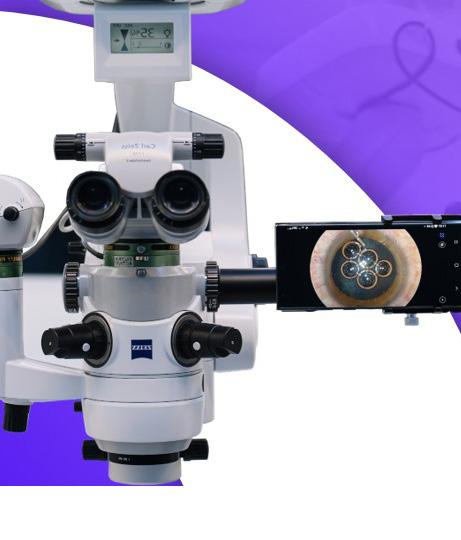
Two teleoperated robotic cataract surgery systems have already been used clinically, demonstrating advantages such as greater precision, faster response times, tremor cancellation, and improved standardisation. However, the technology still faces significant limitations, cautioned Yu-Hsuan (Alex) Huang MD, PhD, speaking at a symposium on artificial intelligence, robotics, and surgical simulation.
“Currently, robotic cataract surgery involves longer procedure times and is limited to certain surgical steps. It also lacks real-time monitoring of surgical conditions, cannot manage complications, and still requires human supervision. The absence of true decision-making capability remains the key bottleneck holding the field back,” Dr Huang said. “Advancing to a level where the system can fully perceive, understand, and respond to complications is the major challenge. The future of cataract robotics lies in perception and intelligence. That is the next frontier, and we are all building it together.”
Reaching its full potential
Dr Huang explained robotic surgery platforms can be classified by the level of human versus robotic control. At one end are robot-assisted tools that enhance the surgeon’s hand. The next level includes teleoperated systems in which a human remotely controls the robot—such as the platforms developed
by ForSight Robotics and Horizon Surgical Systems that have already been used in humans.
Further along are cooperative systems where humans and robots share control. At the far end are systems capable of partial or full autonomy.
The feasibility of fully autonomous systems hinges on their ability to identify and manage complications. That ability fuelled Dr Huang’s vision in founding Xensur Medical, where artificial intelligence (AI) is being applied to advance robotic cataract surgery.
Dr Huang, who holds a doctorate in computer science in addition to being an ophthalmic surgeon, used Tesla’s Optimus robot to illustrate the strategy.
“Optimus can perform a wide range of physical tasks using a single neural network, having learned these skills directly from human demonstration videos. This approach enables and accelerates the acquisition of new capabilities,” he said.
“This is the direction AI is heading in the realm of surgical robotics. To train a robot, the first step is to capture how human experts perform the surgery.”
At Xensur Medical, the approach leverages two complementary technologies: AI-driven analysis of surgical recordings to interpret fluidics changes and real-time, high-precision tracking of surgical instrument trajectories to learn techniques directly from expert surgeons. These capabilities translate complex surgical data into actionable medical insights.
In contrast to teleoperated systems, where a human uses joysticks or controllers to manipulate a robotic arm, the Xensur system directly tracks surgeons operating real surgical instruments. In its current iteration, the platform achieves real-time, micron-level motion tracking inside the eye, capturing every tilt and rotation with high precision.
“Our technology generates ground-truth surgical data to train both robots and surgeons,” Dr Huang said, adding that “this high-quality data will define the future of surgery—transforming how surgeons learn and unlocking true robotic autonomy.”
Dr Huang spoke at the 2026 ASCRS meeting in Washington, DC.
Yu-Hsuan (Alex) Huang MD, PhD is director at Universal Eye Centre, Taipei, Taiwan, a board member of the Ophthalmological Society of Taiwan, and founder of Xensur Medical.
Closing the Presbyopia Gap for Younger Patients
A reversible, high-quality vision strategy delivers strong outcomes in young presbyopic patients through precise protocols and patient selection.
LAURA GASPARI REPORTS
Presbyopia correction with phakic implantable collamer lenses (ICLs) using a monovision approach is safe and efficient, especially for young presbyopic patients, Anas-Alexis Benyoussef MD reported.
Young presbyopic patients, or those between 40 to 55 years old, find themselves in a “presbyopia gap”, a surgical no man’s land.
“They are slowly crossing a bridge, starting with active accommodation, going through an accommodative decline, and finally reaching the total loss of accommodation,” Dr Benyoussef said.
Current standard surgeries like LASIK and refractive lens exchange have limitations: the former can induce ocular surface diseases or higher-order aberrations, while the latter is too invasive for patients who still have partial accommodation, carrying the risk of retinal detachment for those with high myopia.
An additive, reversible solution is needed, and phakic ICLs offer clear potential advantages. They preserve optical quality, maintain corneal integrity for future cataract calculations, and ensure adequate safety thanks to their central port design. However, they are contraindicated in the majority of hyperopic patients due to shallow anterior chambers, which increase the risk of endothelial damage and glaucoma.
How can ophthalmologists create a correct monovision design? According to Dr Benyoussef, the “sweet spot” is between -0.75 D and -1.25 D. In fact, a low target results in poor near vision, while targets above -1.50 D lead to a loss of stereopsis. The optimal target is 1.00 D, which allows patients to maintain a good 3D vision around 60 arcseconds.1
A 2025 study on 31 patients with an average age of 49 years showed positive clinical results with ICLs, with most patients achieving UDVA better than 20/20 and an even better CDVA, with a near perfect IOL predictability.2 Visual gains were significant, with 33 eyes improving by one to two lines, attributed to the magnification effect of ICLs for myopic patients. At three months post-surgery, there were no incidences of cataract development, pupillary block, or IOP spikes, and vault stability was good. The patients’ satisfaction with the surgery was high.
Another study from 2023 compared ICLs with femtosecond LASIK in myopic patients, showing excellent safety and efficacy for both groups.3 Despite having a similar safety index, the efficacy index was significantly better for ICLs. Binocular balance was affected in both groups, with most patients experiencing imbalanced vision at various
distances, primarily driven by age-related presbyopia and the anisometropia created by the surgeries.
Dr Benyoussef stressed the importance of establishing a protocol to identify suitable patients and determine the best surgical approach. Sensory dominance testing should replace unreliable motor tests, with the dominant eye corrected for distance vision. Defocus and simulation tests, along with contact lens trials, help assess tolerance before surgery. However, not all patients are suitable, especially those in professions or activities requiring strong depth perception, such as airline pilots, some military personnel, law enforcement agents, firefighters, and certain athletes. But in general, the benefits of ICLs are strong, with a bright future ahead.
“Expand the indication, trust the data, and respect the protocols,” Dr Benyoussef concluded.
Dr Benyoussef spoke at the 2026 ESCRS Winter Meeting in Helsinki.
For citation notes, see page 40.
Anas-Alexis Benyoussef MD, MSc is an ophthalmologist at the University Hospital of Brest, France. anas-alexis.benyoussef@chu-brest.fr
The Living Framework
One global language for vision: the new functional IOL classification reshapes eye care, research, and patient trust.
LAURA GASPARI REPORTS
The recently achieved global consensus on the evidence-based functional classification of simultaneous vision IOLs (SVIOLs) is a crucial milestone for focusing on what really matters to surgeons and patients: clinical outcomes, according to Joaquín Fernández MD, PhD, ESCRS secretary and European coordinator for the global consensus group.
Initially an ESCRS project, the evidence-based functional classification of IOLs spread to other regional sister societies such as the ASCRS, the Asia-Pacific Association of Cataract and Refractive Surgeons, and the Latin American Society of Cataract and Refractive Surgeons. It developed into a collective and synergistic effort to create a global framework to classify SVIOLs based on the similarity of their clinical results, improving the understanding for ophthalmologists, patients, and stakeholders.
The societies formalised their efforts at the ESCRS Annual Meeting in Copenhagen, where all groups recognised overlapping objectives and a need for harmonisation. The global consensus was finally published in the Journal of Cataract & Refractive Surgery (JCRS) in March 2026.1
As Professor Fernández highlighted in his presentation at the ESCRS Winter Meeting in Helsinki, the global consensus aimed to advance a new classification based on evidence rather than opinions, as the latter can vary according to prior knowledge and interests. Instead, evidence is objective. The weight of strong evidence and more than a decade of scientific literature naturally propelled the consensus.
“Scientific societies across the world didn’t need to be convinced of its validity; they recognised it,” Prof Fernández commented.
Science is objective and solid, but how can the consensus transmit the same concepts to different languages, cultures,
and targets? The semantics behind the terminology was the main topic of discussions, but in the end, the group reached consensus, replacing the term ‘range’ with ‘depth of field’ and maintaining precise language that represents mutually exclusive categories.
Collaboration will expand the evidence base, refine models, and ensure the classification evolves with global input—not as a static system, but as a living framework.
Learning a new language
For a language to be widely understood, it must be used regularly, and the same is true of a new scientific framework that is robust, reproducible, and clinically meaningful. According to Prof Fernández, this is how the new classification will spread in the scientific literature, gradually becoming the language of research as authors find it useful. What truly matters, he said, are the current endpoint thresholds that establish the functional categories, as those come from the scientific method, which allows an objective classification based on clinical outcomes.
Moreover, the understanding provided by the new functional classification serves not only professionals, but also patients and the relationship of trust they should have with their surgeon. Before, preoperative explanations relied on subjec-
tive language and subjective reception of concepts. A significant and dangerous gap may form when there is a mismatch between what the surgeon intended and what the patient understood, inevitably leading to failed expectations, dissatisfaction, and, in a way, failure. The functional classification lays the groundwork for conversations about probabilities and measurable outcomes, with surgeons clearly explaining the level of visual acuity a patient is likely to achieve at certain distances.
As Prof Fernández pointed out, when a patient sees that the outcomes align with the preoperative prediction, confidence in technology, science, and surgeons grows. Trust in medicine is one key ingredient in the recipe.
Such transformative work does not involve physicians alone, but other stakeholders, such as those in industry. While the ISO classification is focused on a pre-market assessment meeting with strict, highly controlled, and often limited approval standards, the functional classification embraces variability, integrating real-world experiences, postmarketing studies, and a broader spectrum of outcomes.
Industry members may have a different perspective due to their priorities. The functional classification, however, is more for clinicians and patients than manufacturers, and the two views are complementary rather than contradictory. And while the starting point might differ, collaboration and convergence become logical, especially if the new patient-centred approach guides innovation to meet clinical needs, Prof Fernández said.
The path forward is mapped out, and the work to realise it is underway. There are still some unanswered questions that may directly or indirectly include other endpoints such as contrast sensitivity, photic phenomena, dysphotopsia (and how often they occur), patient-reported outcome measures, and the psychometric validation of visual quality. The same scientific approach may be applied, and this will not be done in isolation.
By now, constructive collaboration between regional and sister societies around the world is well established and anchored to the same principle of how well professionals will understand what patients will see, rather than how they name the lenses. This is the true meaning of functionality, and this global consensus demonstrated that it can be reached, shaped, and allowed to grow and expand together.
“Collaboration will expand the evidence base, refine models, and ensure the classification evolves with global input— not as a static system, but as a living framework,” he concluded.
For citation notes, see page 40.
Joaquín Fernández Pérez MD, PhD is CEO and Medical Director in the Ophthalmology Department at Qvisión in Vithas Virgen del Mar Hospital, Almería, Spain. joaquinfernandezoft@qvision.es
Observership Grants Available for Young Ophthalmologists
The ESCRS Young Ophthalmologist (YO) Observership programme supports European trainee ophthalmologists who wish to observe clinical practice in a hospital or university setting. Ten €2,000 observership grants are available annually.
Application Details
Applicants must be ESCRS members and residents in the second half of their training programme. Applicants must make their own application to an approved clinic or hospital centre listed on the ESCRS website.
The observership duration is two to three weeks. The host clinic or centre must agree to host you for your proposed time and dates and provide evidence of this agreement in your application.
Participating Clinics/Hospital Centres
More than 20 clinics have observership programmes available for visiting ESCRS YOs. ESCRS will not provide contact details for the named hosts due to privacy regulations. You must contact them by publicly available methods.
Application Deadline
The application deadline is 17 July 2026. For application details and more information, scan the QR code.
When Digital Training Enhances Neuroadaptation
Visual simulators can be a powerful tool to improve vision outcomes.
LAURA GASPARI REPORTS
Anew wave of visual simulators is proving useful in helping understand neuroadaptation in the real world of IOL performance, training, and pre- and postoperative visual function, according to Susana Marcos PhD.
Neural adaptation is a very rapid process that occurs over seconds to minutes and can be observed through after-effects of colour, blur, facial expressions, or orientation, reflecting the brain’s effort to maintain perceptual constancy. For instance, exposure to certain visual patterns can temporarily alter how subsequent images are perceived, she explained.
“This is something that can be demonstrated with the simulated images, but this is also something that happens with a standard astigmatic correction,” Professor Marcos said.
Astigmatic correction can lead to a shift in perception, making neutral images appear blurred along the astigmatic axis. A study demonstrated that patients with astigmatism who were tested before and after wearing corrective lenses showed rapid adaptation to neutral perception.1 However, these rapid changes in perception do not necessarily reflect immediate improvements in visual function, as measured by visual acuity over six months. In fact, individuals may have a long-term bias toward their original visual condition, she noted.
Innovation makes it possible to simulate presbyopic correction through adaptive optic systems or temporal multiplexing-based simulators, such as the SimVis Gekko (2EyesVision), allowing patients to experience real-world vision with different lens corrections, she explained.2 The SimVis Gekko is wearable, wireless, programmable, and has a wide field of view, making it a realistic representation of different lens designs.
A recent paper published in Ophthalmology Science showed the system accurately simulates postoperative vision with real IOLs, comparing preoperative and postoperative visual acuity.3 Also, the system can simulate halos produced by different lens designs and assess how patients are able to adapt to these simulated corrections. Neuroadaptation to presbyopic corrections can be measured, as a study shown by Prof Marcos demonstrated.4 However, careful testing methods, such as smooth transitions between images, are needed to avoid bias from adaptation to a previous image blur or sharpness.
Over weeks to months, changes in vision are better explained by perceptual learning rather than neuroadaptation, which involves specific improvements in visual tasks through training and experience. This can lead to permanent improvements in tasks such as hyperacuity, orientation discrimination, and contrast sensitivity. The underlying mechanism is the plasticity of the visual cortex, which happens even in adults, along with changes in receptive visual fields.
Studies show targeted visual training can enhance acuity, even in adults with conditions like amblyopia or in patients with multifocal lenses.5 Visual simulators can be combined with perceptual learning to enhance vision training. A new generation of binocular see-through simulators with advanced features such as convergence control, spatial lens representation, dynamic aberrometry, and accommodation tracking enable viewing of the real world in a realistic environment, as will future applications.
“Visual simulators may expand to include detailed training for myopia control, early presbyopia correction, and also in multifocal or EDOF intraocular lenses for presbyopia,” Prof Marcos concluded.
Prof Marcos spoke at the 2026 ESCRS Winter Meeting in Helsinki.
For citation notes, see page 40.
Susana Marcos PhD is Director of the Center for Visual Science at the University of Rochester, New York, US. smarcos2@ur.rochester.edu
Soft Skills Help with Hard Patients
Preoperative counselling a key point for a happy patient.
TIMOTHY NORRIS REPORTS
The line between a patient’s satisfaction and dissatisfaction can potentially be drawn in the consultation room. According to Başak Bostancı MD, the surgeon can be a master in corneal and refractive surgery, but if the patient is not treated as a human being with a neurosensorial system and a lifestyle, the most excellent result can still produce an unhappy patient.
“Satisfaction and dissatisfaction are psychological states and not visual acuity measurements,” she said.
Dr Bostancı explained that dissatisfaction is driven by five main factors: expectation-outcome mismatch, personality-technology mismatch, ocular surface instability, incomplete lifestyle analysis, and the absence of structured preoperative counselling.
For these reasons, when dealing with new patients, Dr Bostancı always starts with defining priorities: not by asking if they want to be spectacle free, but rather what matters the most between distant, intermediate, or near vision. If everything is important, nothing is going to be prioritised, she said. Openly explaining concepts like trade-off and neuroadaptation can scale down expectations to a level where the patient will no longer demand surgical perfection from the surgeon, while avoiding panic in the postoperative phase. After that, she usually asks the patient to summarise what they expect from the surgery.
Confirming the alignment is the third step. If the patient cannot articulate what they expect, there is no alignment, and the surgeon should stop and repeat the process. In her consultation, Dr Bostancı uses a three-level counselling model, explaining to the patient what will happen postoperatively and why it will happen before asking what this means for the patient, preparing for alternatives if the patient is not happy with the trade-off.
Satisfaction and dissatisfaction are psychological states and not visual acuity measurements.
Too many options could overwhelm the patient, and this can increase anxiety, she cautioned. Her method consists of openly ruling out what is unsuitable for the patient, narrowing down to two realistic choices, weighing their pros and cons to reduce the risk of regret.
Red flags such as reporting low tolerance to visual changes, tiny imperfections, or asking for perfect vision at all distances must be considered, but not to the point of completely dismissing certain lenses. Perfectionism is not a contraindication—unrecognised perfectionism is, she said.
Expectations are also set by the surgeon’s language during the counselling phase. Absolute language like ‘perfect’ and ‘no halos’ should be avoided, and more probabilistic language should be used instead. This does not decrease the surgeon’s credibility; on the contrary, it shows trustworthiness, she said.
Postoperative counselling is also important to define the narrative. Instead of being dismissive, Dr Bostancı recommends being more open, telling the patient the effects are part of the neuroadaptation process.
“Dissatisfaction does not come from the optics alone,” she noted. “Dissatisfaction comes from misalignment during conversation. For this reason, counselling is not a soft skill, but something we must all be working on.”
Dr Bostancı spoke at the 2026 ESCRS Winter Meeting in Helsinki.
Başak Bostancı MD, FEBO is Assistant Professor of the Bahçeşehir University of Istanbul, and a cataract and refractive surgeon at World Eye Hospital, Istanbul, Türkiye. drbbostanci@gmail.com
Robotics in Cataract Surgery: Coming Soon?
A look at what the future holds for robotic surgery in Europe.
CHERYL
GUTTMAN KRADER REPORTS
Developments in robotic technology are poised to handle the growing volume of cataract surgery patients and meet their increasing expectations for precision outcomes. One day, the field may progress to reach a level of fully autonomous robotic procedures, but that is not likely to occur any time soon, said Pavel Stodůlka MD, PhD.
Providing an update on the current state of automation in cataract surgery and a look to the future, Dr Stodůlka said femtosecond laser cataract surgery (FLACS) represents an existing level of automation, and experience with FLACS has resulted in important lessons for the success of future innovations.
“We learned that OCT guidance brought reproducibility to capsulotomy and laser fragmentation reduces effective phaco time,” he said. “However, several clinical studies showed no clear superiority of FLACS, and we learned that cost and time matters.”
Robotic positioning with laser control represents the next step in automation of cataract surgery and is embodied by the investigational FemtoMatrix laser from Keranova. The laser provides robotic docking and has real-time OCT guidance to optimise energy delivery to different parts of the lens. Its beam, transmitted through the robotic arm onto a face mask, is multiplied to enable faster lens fragmentation and faster surgery.
“The lens is fragmented into 20,000 cubes in 20 seconds, and the majority of cases have been completed with zero ultrasound,” said Dr Stodůlka, who, together with Gilles Thuret MD, PhD (France), completed clinical studies to support regulatory approval of the laser in Europe.
The robotic arm has six axes of freedom, operates with 50 μm precision, and controls force on the eye during the procedure. The device also enables surgical workflow because, once the laser steps are done, the surgery can proceed by repositioning the robotic arm rather than the patient.
Robotic tool manipulation describes the next level of automation in cataract surgery and is represented by the robotic platform from ForSight Robotics. With this system, the surgeon sits at a console and performs surgery with real-time three-dimensional image guidance and advanced visualisation. The robotic arm has 14 degrees of freedom and mimics the surgeon’s hand movements. It offers motion scaling and tremor filtering. A first study in Europe is planned.
Mini fingers and multiple hands
Dr Stodůlka said that supervised autonomy in cataract surgery is on its way and is anticipated for its promise of delivering consistency and precision exceeding human physical limitations. Eventually, fully autonomous surgery may become a reality with the possibility that the robots will have microfingers capable of manipulating new surgical microtools and even be equipped with more than two hands, Dr Stodůlka proposed.
Looking back to the lessons learned with FLACS, he outlined several considerations for the successful introduction and adoption of robotic cataract surgery.
“Registries matter—all companies will need to get through the regulatory pathway with real-world data, benchmarking, transparency, and long-term monitoring. It will be important to define endpoints early, and it will be necessary to measure system impact, demonstrate value, and engage payers,” he said.
“Core takeaways from experience with FLACS showed that precision alone fails, workflow wins, economics decide, and there is definitely a psychological barrier on both the patients’ and surgeons’ side for fully autonomous robotic cataract surgery.”
Dr Stodůlka spoke on this topic during a symposium on AI, robotics, and surgical simulation co-sponsored by the ESCRS, ASCRS, and APACRS that was held during the 2026 ASCRS annual meeting in Washington, DC.
Pavel Stodůlka MD, PhD is founder, chief surgeon, and CEO of the Gemini Eye Clinic, Zlin, Czech Republic, and senior lecturer, department of ophthalmology, Third Faculty of Medicine, Charles University, Prague, Czech Republic. stodulka@lasik.cz or Pavel.stodulka@lf3.cuni.cz
Phakic IOL Follow-up: What to Watch For
Current follow-up data on monofocal lenses can make up for lack of literature on phakic presbyopia IOLs.
TIMOTHY NORRIS REPORTS
Phakic presbyopic IOLs are mainly indicated for presbyopic patients who want spectacle independence but still have a clear crystalline lens and are not ideal candidates for laser or lens exchange surgery. According to Kjell Gunnar Gundersen MD, PhD, these lenses come with a spectrum of complications not dissimilar to those of monofocals.
So, what kind of follow-up do these kinds of patients require? Dr Gundersen suggested follow-up at day 1, week 1, month 1, and 3 to 12 months to evaluate for potential complications such as IOP spikes, iritis, toxic anterior segment syndrome, endophthalmitis, and pupillary block with persistent IOP rise.
Long-term follow-up every 12 months should evaluate endothelial cell loss and corneal decompensation, pigment dispersion glaucoma, retinal tears, dysphotopsia, and lens tilt and decentration, he added.
Dr Gundersen noted that there is a limited number of publications in the literature on presbyopia-correcting posterior chamber IOLs, with small study cohorts and short observation times. Moreover, PubMed is not as reliable as it was previously. On the other hand, a more extensive review by Reinstein (et al.) on posterior chamber phakic IOLs (PIOLs) provides an ample overview of the safety profile, he noted, as it considers the accuracy, precision, and the defocus curve of these lenses.1
The incidence of cataract in implantable phakic contact lens (IPCL)-implanted patients is on average reported to be 0.95% of cases. The cataract formation rate drops to 0.46% in newer lens designs, with no cases requiring explantation due to cataract during the reported follow-up. According to Xu and Song (et al.), the risk of retinal detachment remains low and comparable to highly myopic controls in long-term follow-ups.2
When trying to avoid refractive lens exchange in high myopes, phakic IOLs seem to be a very safe option. Considering endothelial cell loss, Visian ICL (STAAR Surgical) trial data showed a 3.3% mean endothelial cell loss at one year and roughly 9.7% rate at four years, including both surgical trauma and progressive loss. Similarly, the IPCL showed 5.5% loss in the first year and less than 1.5% in the following two years.3 If the patient starts with a good endothelial cell count, a dramatic decrease can be avoided, he said.
The incidence of secondary glaucoma after posterior chamber phakic IOL implantation is very low, but reported cases vary by definition, follow-up time, and ICL design. Glaucoma monitoring should begin immediately after surgery and maintained in the long term.
When counselling patients, it is reasonable to insist on annual life-long monitoring of IOP, vault, endothelial cell
disease, lens status, and retina, as well as discussing the basic monofocal lens for standard PIOLs and stressing that presbyopic optics are newer, with limited long-term data.
“At the moment, only a few presbyopic PIOL series have a robust follow-up beyond two years, with most evidence gathered in more than five years coming from non-presbyopic versions of the same platforms,” Dr Gundersen concluded.
Dr Gundersen presented during the 2026 ESCRS Winter Meeting in Helsinki.
For citation notes, see page 40.
Kjell Gunnar Gundersen MD, PhD is a practising ophthalmologist at the iFocus Eye Clinic in Haugesund & Stavanger, Norway. KGg@ifocus.no
Determining the Best Patient for the IPCL
Seven years of data show good and safe outcomes, even in younger patients.
LAURA
GASPARI REPORTS
Implantable phakic contact lenses (IPCL) provide effective long-term vision correction for both distance and near vision, with overall results that are stable, safe, and well-received by patients, according to Pavel Stodůlka MD, PhD.
In recent years, presbyopia-correcting phakic IOLs have been used in Europe, with consequent long-term clinical results reported, including those presented by Dr Stodůlka on the IPCL made by EyeOL UK. The lens is made in hydrophilic acrylic with a small incision size (1.8 to 2.2 mm) and features trifocal structures on the anterior surface of its optic provided in different additions, with a standard addition of +3 D for all patients. The new models include a central opening that is slightly different from other lenses in the market, and it is conical, which helps provide less light dispersion.
The very first lens was implanted by Dr Stodůlka in 2014. “At that time, we were seeking an appropriate group of patients for this implant, and I was also considering hyperopic patients, but now I use it primarily in myopes, including pre-presbyopes. It gives them the chance to stay spectacle free for longer,” he said.
The data presented included 26 eyes after seven years of follow-up. Patients enrolled were 48 years old on average, with an equal proportion of male and female patients. Distance refraction and uncorrected distance visual acuity (UDVA) remained stable over the seven years, with mean values of -0.3 and 1.0 logMAR, respectively. Cumulative UDVA data show 71% of eyes have at least 0.1 logMAR and 83% of eyes at least 0.2 logMAR, which indicates a particularly good distance visual acuity. The cumulative corrected distance visual acuity shows at least 0.0 logMAR in 96% of eyes at seven years, with a progression over time, he reported.
Regarding the cumulative uncorrected near visual acuity
at seven years, 71% of eyes could read J2 or better, providing a high chance of long-term spectacle independence for near vision. Contrast sensitivity results in a small subgroup of 14 eyes showed normal photopic and mesopic values, with borderline mesopic values with glare. Despite the diffractive structure, contrast sensitivity is fine, with stable IOP and high subjective satisfaction. Overall, 90% of patients reported seeing halo and glare, but with minimal disturbance.
Complications in past models included some bleeding from iridectomy, which is no longer performed thanks to the centre opening. Pressure spikes were observed in 10% of cases using ophthalmic viscosurgical devices (OVDs), which Dr Stodůlka said he now avoids. No refractive surprise, proper sizing, or requiring lens exchange were observed.
Some useful tips to manage the implantation include folding the phakic IOL optic up, pushing the IPCL in the cartridge with the spatula before pushing it by the injector, and pharmacological miosis.
The lens provides fast distance visual rehabilitation, but it may take up to three months for near vision rehabilitation, he cautioned.
“It is an excellent indication for myopes more than 35 years old, which is a little bit earlier than most surgeons might consider,” he noted.
Dr Pavel Stodůlka spoke at the 2026 ESCRS Winter Meeting in Helsinki.
Pavel Stodůlka MD, PhD is founder, chief surgeon, and CEO of Gemini Eye Clinics in the Czech Republic and Vienna, Austria. stodůlka@lasik.cz
Presbyopia: One Condition with Many Solutions
The quest to tackle presbyopia involves finding a compromise between the best focal range, patients’ needs, and retinal contrast image.
LAURA
GASPARI REPORTS
The quest to find an effective presbyopia solution continues, with ongoing research driving the development of new treatments and highlighting the need for personalised solutions to meet the needs of every patient, according to Laura Maubon BMBS.
Presbyopia is a problem affecting around 1.8 billion people worldwide, which is expected to become 2.1 billion by 2030. “It is really a burden, because the people that are affected are economically the working age group,” Dr Maubon said. In this situation, the challenge lies in identifying the right target and range in a heterogeneous population with diverse needs and an expanding array of options.
The onset of presbyopia starts around the age of 40 and is a gradual decline, with most patients experiencing more difficulties focusing on near objects. However, it is a very personal variable, with some people maintaining a good level of near vision through their 60s, and others experiencing blurred vision, headaches, eye strain, fatigue, poor recognition, loss of independence, and embarrassment—all affecting their daily activities and hobbies.
As Dr Maubon explained, there are two main theories on the mechanism of presbyopia: the Young–Helmholtz theory suggests there is a zonular relaxation and lens contraction, with ageing leading to a loss of elasticity and flexibility due to protein accumulation. In contrast, the Schachar theory focuses on peripheral changes and lens widening, with an increasing tension in the equatorial zonules. Both theories sparked many debates, but lens stiffness and protein accumulation are
established and well-accepted factors in presbyopia, she said. So, what are the solutions now available? IOLs remain the most common surgical treatment for presbyopia. New treatments that have emerged, such as pilocarpine and aceclidine eye drops, have shown good adaptation in some studies but are associated with side effects. Contact lenses are another option focusing on the concept of asphericity to manipulate aberrations in the eye. Other options include corneal inlays and scleral treatments.
“Our future challenge is an ongoing ageing population, with an increasing number of presbyopes to come. Just look at how big the screens on our phones are getting. However, the most important thing to bear in mind when treating presbyopia is that patients have different needs in their lives according to their professions and lifestyles,” Dr Maubon concluded. “The real quest for presbyopia is to find an individualised option for each patient, respecting their uniqueness and finding a compromise. There is no perfect range and no perfect lens. It is all about getting the best focal range but without losing the retinal contrast image.”
Dr Maubon spoke at the 2026 ESCRS Winter Meeting in Helsinki.
Laura Maubon FRCOphth, BMBS, BMedSci, PGCert (Surg Ed) is a consultant ophthalmologist specialising in anterior segment surgery, ocular surface disease, and surgical education in the UK. lauramaubon@nhs.net
A New Era for Keratoconus Diagnostics
Multimodal data and AI are transforming early detection, prevention, and personalised care.
LAURA GASPARI REPORTS
The in-depth study of keratoconus and ectatic corneal diseases is undergoing a major paradigm shift, moving from late recognition of advanced disease toward earlier detection, prevention, and individualised care. According to Renato Ambrósio Jr MD, PhD, this shift depends on multimodal diagnostics, artificial intelligence, and a clearer distinction between corneal ectasia diagnostics (CED) and ectasia risk assessment (ERA).
Professor Ambrósio delivered this message during the Lorenzo il Magnifico Medal Lecture at the 3rd World Keratoconus Congress. The honour was particularly fitting, as Lorenzo il Magnifico became a symbol of Renaissance patronage and cultural integration in Florence. For Prof Ambrósio, the new diagnostic era in keratoconus also requires integration of technology, clinical reasoning, education, and a patient-centred purpose.1
In the past, patients with keratoconus were often diagnosed only when the disease was already advanced, when keratoplasty was the primary and only surgical option. Today, improved contact lens rehabilitation, cross-linking, intrastromal corneal ring segments, phakic IOLs, and refractive therapeutic strategies have changed the timing and meaning of diagnosis. “A paradigm shift is when new evidence changes how the field sees, thinks, and decides,” Prof Ambrósio said. “It changes the meaning of the data, not just the data.”
It is for this reason he places strong emphasis on the ‘why’ of multimodal diagnostics. CED and ERA are complementary, but they are not the same. CED is diagnosis-oriented: it includes population screening, diagnosis confirmation, classification, staging, prognosis, individualised management, and clinical follow-up. ERA is risk-oriented: it goes beyond detecting early keratoconus to characterise the cornea’s
susceptibility to biomechanical decompensation and ectasia progression, particularly when elective refractive surgery or laser vision correction procedures are considered.
This distinction matters because ‘normal topography’ is not equivalent to ‘no susceptibility.’ Placido disk topography remains important, but it is only one surface-based expression of a deeper structural and biomechanical reality. Multimodal evaluation integrates directed clinical history, slit-lamp examination, Placido topography, Scheimpflug tomography, epithelial or layered tomography with OCT, corneal biomechanics, ocular wavefront, biometry, and, in the future, genetic and molecular data.
The conceptual framework also connects with McGhee’s two-hit hypothesis, which proposes keratoconus and ectatic progression result from the interaction between intrinsic susceptibility (the first hit) and extrinsic influences (the second hit).2 Prof Ambrósio suggested that this concept should increasingly be recognised as a theory, considering the accumulated clinical evidence and the biomechanical cycle of corneal decompensation described by Roberts and Dupps.3 In practical terms, ectasia should be understood as a dynamic interaction between corneal resistance and environmental or surgical impact.
Eye rubbing is one of the most important preventable extrinsic factors. As Prof Damien Gatinel has emphasised, eye rubbing can be understood as a sine qua non factor for ectasia progression.4 Prof Ambrósio suggested refining this concept: eye rubbing and ocular trauma may be considered sine qua non extrinsic triggers for ectasia progression because they can aggravate keratoconus or, in selected situations, induce secondary ectasia. This distinction is important because aggravating
keratoconus or causing secondary ectasia is not the same as initiating primary keratoconus. Even if the role of eye rubbing in disease initiation remains debated, patient education about eye rubbing, allergy, inflammation, and ocular trauma is central to prevention and follow-up.
In refractive surgery screening, the field has evolved from classical structural parameters toward more relational and individualised measures.5 Residual stromal bed (RSB) remains a classical parameter, while percent tissue altered (PTA) introduced by Santhiago helped express the proportion of tissue affected by LASIK.6,7 More recently, relational tissue altered (RTA) was developed by Aydano P Machado using machine learning to provide an objective, data-driven metric of the structural impact induced by laser vision correction.8 RTA is incorporated into BEES (the BrAIN Enhanced Ectasia Software) as part of a broader strategy to integrate surgical impact with intrinsic susceptibility to ectasia.
Prof Ambrósio stressed that RTA should not be interpreted as a standalone predictor of ectasia among laser vision correction candidates. Its value lies in quantifying the procedural impact on the cornea. The stronger clinical model emerges when surgical impact, as measured by metrics such as RTA, is combined with intrinsic susceptibility, characterised by tomography, biomechanics, epithelial mapping, and other multimodal parameters.
AI is, therefore, not simply a new diagnostic label.9 Its value lies in helping clinicians interpret large, complex data sets while preserving clinical judgement. In Prof Ambrósio’s framework (AI²), applied artificial intelligence provides the ‘how,’ while clinical purpose, what he calls “applied ancient intelligence,” provides the ‘why.’² AI should support the physician’s discernment, not replace it.
Enhanced ectasia risk assessment represents the real paradigm shift in corneal diagnostics. It moves the field beyond, but not over, detecting early keratoconus—beyond topography and even tomography alone—and toward a multimodal understanding of ectasia susceptibility.
“What this field needs is to continue evolving through science,” Prof Ambrósio concluded. “We must go beyond detecting early keratoconus to characterise ectasia susceptibility, beyond topography and tomography toward multimodal diagnosis, and beyond artificial intelligence alone toward careful, patient-centred clinical judgement.”
Prof Ambrósio spoke at the 3rd World Keratoconus Congress 2026 in Florence, Italy.
For citation notes, see page 40.
Renato Ambrósio Jr MD, PhD, FWCRS, PCEO is Adjunct Professor of Ophthalmology at the Federal University of the State of Rio de Janeiro, Affiliated Professor at the Federal University of São Paulo, Refractive Surgery Director of Rio Vision Hospital in Rio de Janeiro, and CEO of BrAIN, Brazilian Artificial Intelligence Networking in Medicine. dr.renatoambrosio@gmail.com
UPCOMING EVENTS
Sept 11–15
ESCRS Annual Congress
London, UK
October 1–4
EURETINA
Vienna, Austria
October 9–12
American Academy of Ophthalmology
New Orleans, Louisiana, US
November 18–20
UKISCRS Annual Conference
London, UK
Oct 1
Sept 11
Oct 9
Nov 18
Droopy Lids? The Bleph Boom
An oculoplastic update on bleph surgery.
BY CLARE QUIGLEY MD
The Bleph Boom: Are We Obsessed with Eyelids?” At the 2026 annual Future Beauty and Health Show at the Royal Dublin Society, the seats were full, with standing room only for those attending the bleph talk. Three oculoplastic surgeons practising in different sites in Ireland—Michéal O’Rourke, Nikolina Budimlija, and I—spoke in a panel chaired by the co-founder of Future Beauty, Liz Dwyer. Who is suitable for surgery, and who should be cautious? What should a patient look for in a prospective surgeon? The discussion varied, moving from bland practicalities, including timing of surgery and recovery, to video recordings of a bilateral upper blepharoplasty surgery performed on the chair of the panel herself. A careful warning was given to the audience members that blood and real-life surgery were about to feature on the big screen. When the video played, I saw a mix of responses: some shielded their gaze with their hands in an involuntary ‘ick’ response, while others watched Michéal’s handiwork closely.
There are different sides to oculoplastics, a recently developed ophthalmology subspecialty. Oculoplastics is concerned with sight-threatening pathology, such as orbital cellulitis and orbital inflammation, and management of rare deadly infections such as invasive mucormycosis. At the Future Beauty show, the focus was on the aesthetic side of oculoplastics, whereby eyelid changes and malpositions (mainly age-related) can be managed with surgery.
Blepharoplasty is the headline procedure on the aesthetic side of oculoplastics and is one of the five most popular cosmetic surgeries in the UK, per the BBC. After bilateral upper blepharoplasty surgery, patient satisfaction rates typically exceed 90%. As with any surgery, there are risks, and dissatisfaction or regret are estimated to occur in up to 15%.
I am keen, when I see any patient who is complaining of droopy lids, to zero in on their exact complaint. Are they bothered about excess upper lid skin changing their appearance or blinkering their vision, or is the upper lid position lower than normal, i.e., ptotic?
Making a distinction is essential, as dermatochalasis and ptosis are two
distinct entities that patients may not be able to distinguish between. Dermatochalasis, or excess upper lid skin, can be managed through bilateral upper blepharoplasty, a relatively straightforward procedure involving excision of excess skin. For an abnormally low upper lid, or upper lid ptosis, careful consideration needs to be given to a broad list of diagnoses prior to any surgical planning. Potential underlying conditions including vascular, neoplastic, and autoimmune disorders, among others, can present for the first time with a complaint of a droopy eyelid in the clinic. We do not want to miss a secondary ptosis due to Horner’s syndrome, Myasthenia gravis, or a third nerve palsy. We also do not want to misidentify ptosis in contralateral lid retraction or proptosis.
My approach to a patient with droopy lids is informed by my time in practice as a consultant, my time spent training in ophthalmology, and my fellowships in oculoplastics. I can remember Andy Gibson, consultant oculoplastic surgeon and
military clinical director at James Cook University Hospital, Middlesbrough, UK, giving me a memorable framework for assessing ptosis. He instructed me on pertinent negatives that should be noted in the clinical exam. Specifically documenting important negatives in each ptosis exam is helpful, as the ingrained habit is protective when running a busy clinic. I think of this as broadly analogous to documenting ‘no tobacco dust’ in an acute posterior vitreous detachment.
We do not want to miss a secondary ptosis due to Horner’s syndrome, Myasthenia gravis, or a third nerve palsy.
Is there fatiguability or variability of lid position? After 60 seconds of sustained upgaze, worsening of a ptosis should raise suspicion for myasthenia. Ice-pack testing and bloods including acetylcholine receptor and muscle-specific kinase antibodies can be helpful. Are eye movements full and pupils equal?
Careful pupil inspection with room lights dimmed can enhance the subtle anisometropia of Horner’s syndrome; look for the smaller pupil on the ptotic side. Iopidine drops are useful to confirm this by inducing a reversal of the anisometropia, and the drops are usually readily available in clinic. A painful Horner’s is a reason to send a patient for an emergency CT angiogram head and neck, looking for carotid dissection.
Is levator function reduced? This helps with determining cause of ptosis, identifying a congenital ptosis or a potential third nerve palsy. Levator function is also essential to measure in surgical planning, as the technique to correct a ptosis with significantly reduced function is distinct from a routine aponeurotic ptosis repair.
A further caution: after determining that your patient has a straightforward problem like dermatochalasis with no lurking undiagnosed pathology, have a conversation about concerns and expectations before proceeding with surgery. In Australia, patients who seek cosmetic surgery must be screened for body dysmorphia—another condition not to miss.
This is the third in a series of columns discussing conditions of eyelids. Previous columns are available on the EuroTimes website
Apply for the New Sustainability Research Award!
Ophthalmologists and researchers (MD and/or PhD) as well as experienced ophthalmic nurses are encouraged to apply for the ESCRS Sustainability Research (SURE) Award, which will fund projects that investigate meaningful, practical ways to promote environmental responsibility in ophthalmic care.
Two awards will be granted; each award will provide up to €10,000 per project. Applicants must be active ESCRS members at the time of application and must hold a current full- or part-time clinical or research position at a clinical or academic institution. Early-career researchers and young ophthalmologists are especially encouraged to apply.
The application period closes on 19 June. Award recipients must submit an article to a peer-reviewed journal within six months after the research period concludes. The article should be made open access if accepted and submitted to the Journal of Cataract & Refractive Surgery in the first instance.
Get funding for projects to promote environmental responsibility in ophthalmic care.
Dublin, Ireland.
Clare Quigley MD is a Consultant Eye Surgeon in private practice in Progressive Vision and in public practice in the Royal Victoria Eye and Ear Hospital and St James’s Hospital,
The Evolution of CAIRS
ECO-CAIRS provide a high-volume, sterile, easy-to-insert segment with minimal biological risk.
TIMOTHY NORRIS REPORTS
Dr Soosan Jacob’s corneal allogenic intrastromal ring segments (CAIRS) have provided surgeons worldwide with a valuable option in keratoconus treatment. Since then, the technique has been further developed to facilitate insertion and reduce the risk of ocular graft versus host disease and postoperative infections.
“When I started implanting CAIRS, my team and I asked ourselves: How can we make the insertion easier? How can I put more volume into the cornea? How can I reduce the risk of rejection and infection?” said Farhad Hafezi MD, PhD. “We were wondering if it was the volume or the stiffness that gave the topographical effect. Last and not least, we were also considering the risk of implanting a graft from a donor with an unchecked keratoconus.”
One of the first difficulties a new surgeon may encounter when dealing with the original CAIRS technique or with precut segments (Keranatural/Optigraft/CTAK) relates to the soft and floppy nature of the untreated allogenic graft, Professor Hafezi observed. Even the ingenious solution proposed by Dr Shady Awwad of the “jerky technique”, from Prof Hafezi’s perspective, comes with a challenge: while working like a charm for the first few seconds, as soon as the graft comes in contact with some humidity on the ocular surface, the graft thickness starts to vary and the segment can rapidly lose stiffness if not implanted quickly enough. In the hands of a true master like Dr Awwad, this is not a problem, and it can be an incredibly fast technique, Prof Hafezi noted.
Although rare, anterior stromal melt can occur with CAIRS implantation. The implantation of CAIRS segments is a transplantation of living keratocytes. It has lower risk of postoperative dysphotopsia or glare, extrusion, and erosion when compared to synthetic alternatives. The gamma ray sterilisation of precut segments, on the other hand, sensibly increases the cost for any graft personalisation option, as the tissue is processed in the eye banks.
While troubleshooting CAIRS, Prof Hafezi selected five pivotal questions that needed one answer, one solution: How to make the insertion easier? How to insert more volume? How to avoid rejection? How to avoid infection? And, finally, what if my donor had keratoconus? The answer to all of these questions is cross-linking, he said. Cross-linking is a procedure that modifies the physiology of the cornea in many ways, largely addressing the five questions about CAIRS, he added.
To refine his version of CAIRS, called ECO-CAIRS, Prof Hafezi carefully analysed the properties of cross-linked stro-
mal tissue in search of the best possible procedure to achieve the correct stiffness—especially a stable thickness of the graft. It is the volume that drives the topographical effect, he pointed out.
With his team at the ELZA Institute, he analysed corneal stroma from porcine eyes cross-linked at different fluences, from 15 joules/cm2, up to 30 joules and even 60 joules. Considering cross-linking is made outside the eye, Dresden protocol 5.4 joules/cm2 were not considered. The fastest protocol was used, with 30 mW for 16 minutes, 40 seconds. The results were published in the Journal of Refractive Surgery 1
The second clear advantage of ECO-CAIRS is represented by hydration. The segment resists rehydration and stays super stiff for several minutes, Prof Hafezi noted.2
The modified procedure kills the keratocytes and stabilises the surface, making sure that no bacteria or living keratocytes remain on or within the segment. This drastically reduces risk of infection and rejection, giving the surgeon the possibility to cross-link and customise the segment at the same time in a less costly fashion.
Once treated, graft insertion is extremely fast, Prof Hafezi observed. By his fourth surgery, he implanted the graft in less than a minute due to the maintained stiffness and lack of resistance to the recipient stroma. To facilitate the insertion, he moistens one tip of the graft enough to grasp it with the forceps.
“It makes my blood pressure and my pulse low,” he said. The overall procedure takes 20 minutes in total.
This procedure can also be helpful for the whole procurement system. In countries where it is legal to use the same donor cornea for different recipients, it is possible to provide multiple patients with grafts using just one cornea, decreasing costs and increasing supply.
“From the same graft we can provide two patients with CAIRS implants,” Prof Hafezi said.
Dr Hafezi presented at the 2026 ESCRS Winter Meeting in Helsinki.
For citation notes, see page 40.
Farhad Hafezi MD, PhD, FARVO is Medical Director at the ELZA Institute of Zurich, Switzerland, and Professor at the Universities of Geneva (Switzerland), New York (US), Los Angeles (US), and Wenzhou (China). farhad@hafezi.ch
Exclusive learning. Unlimited access for ESCRS members.
Digital Integration to Meet Future Challenges
Using AI-integrated models and interoperability to improve diagnostic efficacy can save time and money.
TIMOTHY NORRIS REPORTS
The ageing population, combined with the demographic decrease of fertility, has created a demographic time bomb for the next decade. In the UK, the government has implemented a 10-year plan to deal with ever-increasing healthcare costs, divided into three pillars. Moving from treatment to disease prevention is the first pillar, followed by moving from hospital to community care, and finally moving from analogue to digital healthcare systems.
Digital transformation is crucial. AI modelling as part of digital transformation will empower early diagnosis and accurate risk stratification, supporting disease prevention through early intervention and safe transfer of patients to community care, Bruce Allan MD reported. The first key ingredient in digital transformation is interoperability.
“We are all familiar with Bluetooth and Wi-Fi that enable us to pick up music from the internet with one device and reproduce it using another device from a different manufacturer. This is interoperability that we experience every day,” he said. “Our devices, our workstations, our EHR systems, our PACS (picture archiving and communication system), and our decision tools and guidance systems all need to speak the same language, and now we have a suite of emerging interoperability standards that is worth being at least aware of.”
This suite is comprised of SNOMED CT, defined by Dr Allan as a language-independent hierarchical system of eight-digit codes for systematic medical nomenclature; FHIR (fast healthcare interoperability resources), a system that allows web-based EHR (electronic healthcare record) systems to communicate; and DICOM (digital imaging and communications in medicine), a standard for transferring medical images and associated metadata, which is particularly important in an image-rich specialty like ophthalmology. He praised the tireless efforts of Flora Lum MD, who has rallied cross-society support for full DICOM introduction in ophthalmology from AAO, ARVO, ASCRS, and ESCRS.
The second key ingredient for digital transformation Dr Allan highlighted is protocol-driven care. Protocol-driven care involves the design of patient pathways to ensure each patient has the right investigations at the right time. In combination with interoperable EHR and PAC systems, protocol-driven care elevates the quality of data collected in routine care to a level comparable with clinical trials. Moreover, under EU GDPR article 9 exemptions, de-identified clinical data collected during routine clinical care can be used in healthcare research without Institutional Review Board oversight or special measures for consent.
Systematic healthcare data collection in protocol-driven care is infinitely less costly than interventional trials, allowing the build-up of high quality, rich labelled image data sets for AI modelling, he said.
Using keratoconus as an example, he explained that AI modelling can provide accurate disease and risk classification, offering a rational basis for either early intervention (disease prevention) for high-risk patients or safe transfer to annual monitoring in the community for low-risk patients. He presented data showing that only one in eight patients currently monitored regularly in the Early Keratoconus Clinic at Moorfields Eye Hospital will still require regular hospital-based monitoring once the new AI models of progression risk are implemented.
“So which model will we use to make the prediction? All of them,” Dr Allan said. In AI modelling, the same data set can be revisited as newer, more powerful, predictive models emerge. The key is to organise care pathways to build high-quality data sets. After a single diagnostic review, it is possible to assign most keratoconus patients to a high-risk category (>90% risk of progression) and early intervention with corneal cross-linking or a low-risk category (<10% risk of progression in two years) and annual monitoring in the community. After just two visits, patients can be classified even more accurately. “Imagine the cost savings in that,” Dr Allan concluded.
Dr Allan spoke at the 2025 ESCRS Annual Congress in Copenhagen.
Bruce Allan MD, FRCS is consultant ophthalmic surgeon at the Moorfields Eye Hospital and Professor of Anterior Segment and Refractive Surgery at the University College of London, both in the UK. bruce.allan@ucl.ac.uk
Raising the Bar for Cataract Surgery in Glaucoma Patients
MIGS, femtosecond laser, and LAL can help improve outcomes.
CHERYL GUTTMAN KRADER REPORTS
Performing cataract surgery in patients with glaucoma is a common occurrence. When patients with glaucoma require cataract surgery, surgeons face the dual challenge of having to simultaneously manage intraocular pressure (IOP) and ensure optimal visual outcomes.
“Looking at my schedule, I found that 40% to 50% of my glaucoma patients who are phakic develop cataracts and need surgery in about two years, and 25% to 30% of my cataract patients have coexisting glaucoma,” said Reza Alizadeh MD.
Sharing his perspectives on performing cataract surgery in patients with glaucoma at a recent conference, Dr Alizadeh’s key takeaway messages highlighted the benefits of combining minimally invasive glaucoma surgery (MIGS) with cataract surgery and using a femtosecond laser-assisted approach for the cataract procedure (FLACS). He also reviewed considerations for IOL selection and optimising refractive outcomes.
Dr Alizadeh listed several benefits for performing a FLACS procedure in cataract patients. He said the procedure may have a particular role for patients with complex anterior segment anatomy, particularly those with pseudoexfoliation, where the laser’s ability to create a precise capsulotomy will enable better IOL centration. Potentially minimising ultrasound energy usage and inflammation can translate into reduced endothelial cell loss, a consideration in patients with glaucoma whose corneal endothelium may be compromised from chronic medication use. There may also be less need for postoperative steroid treatment.
For patients requiring astigmatism management, the ability to make precise corneal incisions is another advantage of using the femtosecond laser. When implanting a toric IOL, however, and particularly in patients with a history of corneal refractive surgery, Dr Alizadeh said he often uses intraoperative aberrometry for real-time IOL power and positioning optimisation.
Lens selection
“A toric IOL can correct 0.75 to 6.00 D of astigmatism to help patients become more spectacle-independent, which is important when checking the visual field and also enhances contrast sensitivity,” Dr Alizadeh said.
However, for patients wanting presbyopia correction, he recommended against using multifocal IOLs because they reduce contrast sensitivity, which is a particular problem for patients with ganglion cell loss.
Better alternatives, according to Dr Alizadeh, include simultaneous vision lenses (SVLs), a monofocal IOL with a mini monovision approach, or the Light Adjustable Lens (LAL, RxSight).
“[SVLs] improve intermediate vision, are associated with less glare, halos, and positive dysphotopsia versus multifocal IOLs, and maintain contrast sensitivity,” he said.
The opportunity to correct the residual refractive error is the main reason for choosing the LAL in patients with glaucoma, and therefore it has a particular role in circumstances where the patients are unsure of what they want or if the outcome is unpredictable. The latter situation includes patients who had prior corneal refractive surgery but also those undergoing combined glaucoma incisional surgery that can affect refractive status.
When counselling patients on the LAL, Dr Alizadeh emphasises that it is an investment in both time and money.
“Patients must return for multiple postoperative visits and use protection against ultraviolet light until lock-in,” he said. “Compliance with these needs is essential.”
In general, for patients with glaucoma asking for an ‘upgraded’ IOL option, Dr Alizadeh said he only offers one to individuals with well-controlled IOP, mild to moderate glaucoma, and realistic expectations about visual and IOP outcomes who are expected to have good compliance with postoperative care and follow-up.
Dr Alizadeh also advocated for using intraoperative aberrometry to optimise outcomes when implanting a toric IOL and stated his preference for choosing an SVL and avoiding a multifocal IOL for patients hoping to achieve a better range of vision postoperatively.
Dr Alizadeh spoke during the 2026 ASCRS annual meeting in Washington, DC.
Reza Alizadeh MD is an Assistant Professor in Ophthalmology, Stein Eye Institute, David Geffen School of Medicine UCLA, Los Angeles, California, US.
European launch for Bausch +
Lomb vitrectomy tool
Bausch + Lomb announced the launch of its Bi-Blade+ dual-port vitrectomy cutter in Europe. The product allows an increased cutting speed of 25,000 cuts per minute and is designed to minimise retinal traction, increase vitreous flow, and reduce infusion pressure fluctuations when used with adaptive fluidics, according to the company. The Bi-Blade+ provides an average flow rate increase of 25%, enabling more efficient vitreous removal compared to the original Bi-Blade. The announcement follows the 510(k) US FDA clearance for the tool earlier in April. bausch.com
EMA OKs Fast Track status for retinitis pigmentosa treatment
Ray Therapeutics’ experimental retinitis pigmentosa treatment RTx-015 received a PRIME (Priority Medicines) designation from the European Medicines Agency. RTx-015 is a genotype-independent optogenetic gene therapy administered intravitreally. Early clinical trials showed promising effects on vision in retinitis pigmentosa patients. PRIME status is reserved for treatments that meet significant unmet medical needs and is intended to speed development. RTx-015 also recently received a regenerative medicine advanced therapy (RMAT) designation from the US FDA. raytherapeutics.com
Enhanced monofocal from Alcon
Alcon announced the US launch of Clareon® TruPlus, its monofocal and toric intraocular lens (IOL), during the annual ASCRS meeting in Washington, DC. TruPlus is the latest addition to the Clareon portfolio. The company says the enhanced design IOL enables increased depth of focus without compromising the high-quality distance vision patients expect from a monofocal IOL. The new IOLs leverage the proprietary Opti-Balance Technology in the centre of the lens to slightly extend the depth of focus, the company reports. alcon.com
US FDA approves OcuRing-K clinical trial
LayerBio announced OcuRing-K, a treatment for postoperative ocular inflammation and pain, had received investigational new drug (IND) status. OcuRing-K is a bio-erodible ring that attaches to the haptic of an intraocular lens and delivers ketorolac in a sustained release form. The company plans to proceed to a multicentre Phase II clinical trial this year. layerbio.com
BVI Virtuoso platform gets CE Mark
Having debuted at the 2025 ESCRS Annual Congress, the BVI Virtuoso platform received the CE mark under the new European Union Medical Device Regulation (MDR). The cataract-only and dual function cataract-vitrectomy system supports both anterior and posterior segment procedures on a single platform, with features including IOP management, integrated irrigation and aspiration, and a high-speed, dual-pneumatic, dual-blade vitrectomy drive. bvimedical.com
Corneal regeneration study underway
Aurion Biotech announced the enrolment of patients in the ASTRA Phase 3 study, evaluating AURN001 in patients with corneal oedema secondary to corneal endothelial dysfunction. Patients in the US study will receive a single administration of regenerative cell therapy. AURN001 is a potential evolutionary step ahead from conventional keratoplasty procedures, consisting of unmodified human corneal endothelial cells (neltependocel) and a rho-kinase inhibitor (Y-27632).
“Dosing the first ASTRA study participants marks an important milestone for patients, physicians, and Aurion as we work together to advance new solutions for vision loss from corneal endothelial disease,” said Edward J Holland MD, Chief Medical Officer, Aurion Biotech. “While modern endothelial keratoplasty procedures have transformed the treatment of the disease, there still is a need to reduce complications and improve our patients’ experience.” aurionclinicaltrials.com
JCRS AWARD WINNERS ANNOUNCED AT THE 2026 ASCRS MEETING
Recipients of the annual Journal of Cataract & Refractive Surgery (JCRS) awards for top full-length article and top laboratory science article were named at the 2026 ASCRS meeting in Washington, DC, US. The announcements were made at the start of the JCRS/JCRS: Case Reports joint symposium.
Jay S Pepose MD, PhD, first author of an article titled “Assessing Ocular Dominance: Rethinking the Paradigm” was the winner of the Obstbaum Award for the best full-length article published in 2025. Dr Pepose is founder and former medical director of the Pepose Vision Institute, St Louis, Missouri, US, and collaborated with multiple other investigators in the research.
Appearing in the March 2025 issue of JCRS, the article described a prospective, multicentre, double-masked, non-interventional, comparative study assessing near and far ocular dominance using the ‘hole-in-the-card’ binary motor sighting test versus a new sensory test performed with a visual simulator simulating monovision. The researchers found that the results of the two tests often failed to align and suggested the need for further study to determine whether both strength and localisation of ocular dominance in planning monovision are important factors in predicting patient satisfaction and adaptation to monovision.
Announcing the award, session co-moderator William J Dupps Jr MD, PhD, commented that choosing the Obstbaum Award recipient is always a very difficult task given the number of fulllength articles published each year and the challenge of defining what is ‘best’.
“We use our gut instincts and think about impact,” Dr Dupps said. “This year’s winner was a favourite amongst all those who voted.”
Christian M Hammer DSc, MME, was named as the winner of the JCRS Mamalis Award for best laboratory science paper. Titled “Improvement of Keratorefractive Lenticule Creation by Application of a Vortex Beam in a UV Femtosecond Laser System” and published in the June 2025 issue of JCRS, the article described an experimental study in which a 347 nm ultraviolet femtosecond laser was used to create refractive corneal lenticules in ex vivo porcine eyes. The laser was operated with a vortex beam (Laguerre-Gaussian) generated by a spiral phase plate in 20 eyes and with a normal beam (Gaussian) in 20 eyes. Evaluations based on histological quantification and scanning electron microscopy showed advantages associated with the vortex beam, including decreased intraoperative gas bubble production and smoother, more regular lenticule beds. The researchers concluded that operating a UV femtosecond laser with a vortex beam could increase surgical safety and precision. The research was a collaboration between investigators at the University of Fribourg, Switzerland; University of Erlangen-Nürnberg, Erlangen, Germany; and WaveLight GmbH, Erlangen, Germany.
The JCRS Obstbaum Award honours Stephen Obstbaum MD, who was a founding editor of JCRS. The JCRS Mamalis Award honours Nick Mamalis MD, an editor emeritus of the journal. A third JCRS award, the Rosen award, is given annually for the best technical paper. The award honours Emanuel Rosen MD, who helped to launch the JCRS and was the founding father of ESCRS. Fittingly, the winner will be announced in September at the 2026 ESCRS Annual Congress in London, UK.
Research Education Innovation
ESCRS’s vision is to educate and help our peers excel in our field. Together, we are driving the field of ophthalmology forward.
Cited in this Issue
The Olympian Ophthalmologist
Page 12
1. Museum, The Royal College of Ophthalmologists. www.rcophth.ac.uk/about-the-college/museum
2. Bullock JD, Henry B, Stallard MD. “The 1924 Paris Olympics, and Chariots of Fire,” Survey of Ophthalmology, 2011; 56: 466–71.
3. Mrittika Sen, et al. “Hyla Bristow Stallard: Citius, Altius, Fortius,” Indian Journal of Ophthalmology, 2021 Sept; 69(9), 2252–2255.
Accommodating Simultaneous Vision Lenses
Page 17
1. Rosen E, Alió JL, Dick HB, Dell S, Slade S. J Cataract Refract Surg, 2016 Feb; 42(2): 310–328. doi:10.1016/j.jcrs.2016.01.014. PMID: 27026457.
2. Rosa AM, Miranda ÂC, Patrício MM, McAlinden C, Silva FL, Castelo-Branco M, Murta JN. J Cataract Refract Surg, 2017 Oct; 43(10): 1287–1296. doi:10.1016/j.jcrs.2017.07.031. PMID: 29120714.
Managing Concurrent Cataract and Vitreomacular Traction
Page 18
1. Bala S, et al. Am J Ophthalmol, 2026; 283: 176–187.
Closing the Presbyopia Gap for Younger Patients
Page 21
1. Gawęcki M. J Ophthalmol, 2019 May 6; 2019: 2654170. doi:10.1155/2019/2654170
1. Vinas M, et al. PLoS One, 2012; 7(9): e46361. doi:10.1371/ journal.pone.004361
2. Marcos S, et al. Biomed Opt Express, 2025 Feb 13; 16(3): 1025–1042.
3. Papadogiannis P, et al. Ophthalmology Science, 2026; 6. doi:10.1016/j.xops.2026.101140
4. Radhakrishnan A, et al. PLoS One, 2014; 9(3): e93089. doi:10.1371/journal.pone.0093089
5. Polat U, et al. Proc Natl Acad Sci USA, 2004 Apr 27; 101(17): 6692–6697. doi:10.1073/pnas.0401200101; Kaymak H, et al. J Refract Surg, 2008: 24(3): 287–293. doi:10.328/1081597X-20080301-11
Phakic IOL Follow-up: What to Watch For Page 27
1. Reinstein DZ, MacGregor C, Archer TJ, Gupta R, Potter JG. Curr Opin Ophthalmol, 2024 Mar 1; 35(2): 138–146. doi:10.1097/ ICU.0000000000001018. Epub 2023 Dec 6. PMID: 38059758.
2. Xu W, Song Z, Huang Y, Tao Y, Wang J, Wang L, Li Z. Front Med (Lausanne). 2020 Dec 15; 7: 582633. doi:10.3389/ fmed.2020.582633. PMID: 33425935; PMCID: PMC7793859.
3. Subudhi P, Patro S, Agarwal P, Khan Z, Subudhi BNR, Mekap C, Padhi A. Clin Ophthalmol, 2020 Oct 30; 14: 3681–3689. doi:10.2147/OPTH.S270690. PMID: 33162752; PMCID: PMC7642691.
A New Era for Keratoconus Diagnostics Page 30
1. Ambrósio R Jr, et al. Eye Vis (Lond), 2023; 10(1): 45. doi:10.1186/s40662-023-00363-0.
2. McGhee CNJ, Kim BZ, Wilson PJ. Cornea, 2015; 34 Suppl 10: S16-S23. doi:10.1097/ICO.0000000000000504.
3. Roberts CJ, Dupps WJ Jr. J Cataract Refract Surg, 2014; 40(6): 991–998. doi:10.1016/j.jcrs.2014.04.013.
4. Gatinel D. Int J Kerat Ect Cor Dis, 2016; 5(1): 6–12. doi:10.5005/jp-journals-10025-1114
5. Randleman JB, Woodward M, Lynn MJ, Stulting RD. Am J Ophthalmol, 2008; 145(5): 813–818.e2. doi:10.1016/j. ajo.2007.12.033.
6. Santhiago MR, et al. Am J Ophthalmol, 2014; 158(1): 87–95.e1. doi:10.1016/j.ajo.2014.04.002.
8. Machado AP, et al. Ophthalmol Ther, 2025. doi:10.1007/ s40123-025-01206-y.
9. Esporcatte LPG, Salomão MQ, Machado AP, Ambrósio R Jr. Expert Rev Ophthalmol, 2025. doi:10.1080/17469899.2025.2594 819
The Evolution of CAIRS
Page 34
1. Aydemir ME, et al. Journal of Refractive Surgery, 2025; 41(11). 2. Kollros L, et al. submitted.
Drive Your Career Forward
Want to improve your clinical skills, stay on top of developments in the field, and have an active voice in the evolution of cataract and refractive surgery?
As an ophthalmologist in training, all these things (and much more) are within your grasp—and at no cost to you— by joining ESCRS!
Since 1991, ESCRS has promoted education in the field of implant and refractive surgery and supported research into the practice of intraocular lens implantation and refractive surgery. With more than 7,000 members across Europe and the world, ESCRS wields a powerful voice in the global discourse about ophthalmology and provides members with a rich and diverse network of professional connections.
Drive ophthalmology—and your career—forward. Take advantage of free ESCRS trainee membership today!
ESCRS YO membership has given me a strong international community, real integration across cultures and languages, and meaningful connections that extend far beyond meetings. It has opened opportunities to present on stage, build confidence, and grow as a leader within ESCRS and the wider ophthalmology community.
—LAURA MAUBON
As a young ophthalmologist, ESCRS membership opened doors to high-quality education, mentorship, and opportunities that have shaped my early career. Being an ESCRS YO member has connected me with an inspiring global community and given me access to resources that continually improve my clinical practice.
—VALENTIN HOOIJER
Apply for the
John Henahan Writing Prize
How Has the ‘Human Touch’ Inspired You?
Ophthalmology, like all medical disciplines, is about improving the lives of people. That ‘human touch’ is fundamental to our work and transcends the technological and scientific advances that have transformed medical care.
The 2026 John Henahan Writing Prize seeks to capture the human touch and the impact it has on ophthalmic surgeons.
Young ophthalmologists are invited to compete for the Henahan Prize by submitting an essay (800 words maximum). The author of the winning essay will receive a €500 bursary and a specially commissioned trophy. The winning essay will also be published in EuroTimes.
Henahan Prize contestants must answer this prompt: The digital OR, AI algorithms, and robotics notwithstanding, cataract and refractive surgery involve a human relationship between you and your patient. Please describe how an experience or experiences in your early training reminded you of the importance of the human touch, and how this has inspired you in your clinical practice.
Eligibility
The competition is open to ESCRS members (including the free membership available to trainees) age 40 or younger on 1 January 2026.
Writing Standards
Please compose your essay on your own without the use of any AI tools. We suggest you ask a mentor or teacher to review your essay prior to sending it.
Deadline
The closing date for entries is 30 June 2026. Send your essay with cover page to seanh@eurotimes.org.