SEPTEMBER/OCTOBER 2026 | VOLUME 31 | ISSUE 7
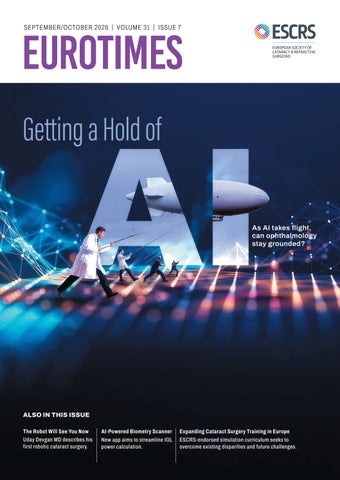
Getting a Hold of As AI takes flight, can ophthalmology stay grounded?
ALSO IN THIS ISSUE The Robot Will See You Now Uday Devgan MD describes his first robotic cataract surgery.
Sept-Oct_2026_EuroTimes.indd 1
AI-Powered Biometry Scanner New app aims to streamline IOL power calculation.
Expanding Cataract Surgery Training in Europe ESCRS-endorsed simulation curriculum seeks to overcome existing disparities and future challenges.
8/18/26 11:21 AM
We’re willing to bet most eye care professionals don’t realise just how prevalent Demodex blepharitis is. In fact, ~54% of eye care patients in Europe may have Demodex blepharitis (DB).1* *Data from an evaluation of 804 patients from 6 countries in Europe, including 15 clinicians each with ~50 consecutive patients. DB defined by the presence of collarettes.
WHAT ABOUT YOUR PATIENTS? LEARN HOW DB CAN FLY UNDER THE RADAR AT
Find us at ESCRS in London - Booth E.118 Reference: 1. Nanavaty MA, Findl O, Carones F, et al. Proportion of patients with Demodex blepharitis in ophthalmology clinics in Europe: the Eos study. Eye. 2026:40:165-167.
© 2026 Tarsus Pharmaceuticals, Inc. All rights reserved. TARSUS, LOOK AT THE LIDS, THINGS START LOOKING UP WHEN YOU GET YOUR PATIENTS LOOKING DOWN, and any associated logo(s) are trademarks owned exclusively by Tarsus Pharmaceuticals, Inc.
GL--2600026 03/26
Sept-Oct_2026_EuroTimes.indd 2
8/18/26 11:21 AM
Hear Ye, Hear Ye: ESCRS Annual Congress is coming to London!
S
eptember has always been an important month for ESCRS, and this year promises to be truly unique. The 2026 ESCRS Annual Congress is coming to London, and it is expected to be the biggest, most international, and most diversified Congress ever. As ESCRS President, I am proud of the programme that we have developed and am very much looking forward to welcoming colleagues from all over the world. London, a city born of science, medicine, and global interaction, is an ideal stage for a Congress that pays tribute to our history while looking ahead to the future. Education and scientific exchange remain the foundation of all we do. Delegates can explore cutting-edge evidence through our Main and Clinical Research Symposia, Near Live Surgery, video sessions, wet labs, free papers, and presented posters. More than 100 instructional courses—with 688 presentations covering surgery for every specialty, optics, retina therapy, artificial intelligence, practice management, and more—will be waiting. One of my key priorities is to design Congress formats that are scientifically sound as well as interactive and memorable. I am very excited about one of these innovations, created especially for London: Global Saves in 3D. It will feature challenging surgical cases, where experience, wisdom, and composure turn potential complications into success. The 3D format will take attendees as close as possible to those high-pressure moments. The Operating Room Microscope Showdown will allow attendees to compare available surgical visualisation techniques and their effect on the quality of surgical procedure, ergonomics, and decision making. Artificial intelligence will be one of the key themes of the Congress. During our AI Summit, we will discuss not only the huge potential of this technology but also its responsibilities in terms of patient care and validation.
The Global Refractive Summit will bring together outstanding international faculty to debate emerging trends and challenges in refractive surgery—from surgical techniques to diagnostics and outcomes. The fifth annual ESCRS iNovation Day will once again provide a dynamic platform for surgeons, researchers, industry representatives, entrepreneurs, and investors to shape the future of eye care. Delegates can listen to engaging debates, learn about new technologies, and meet people with ideas that can transform ophthalmology. At the World Café roundtables, delegates can discuss clinical cases and practical issues with experts and colleagues. The parliamentary-style Arena Debates will address controversial questions in cataract and refractive surgery. A live vote will show how the discussion can influence opinions in the room. These formats reflect one of the core principles of ESCRS: we should not be afraid of constructive disagreement. Evidence-based and respectful debate is one of the main drivers of progress. As exciting as the programme is, I encourage you to do more than attend scientific sessions. The Congress is a great opportunity to meet colleagues and make friends from all over the world, listen to new voices, challenge assumptions, and celebrate the engaging international community that ESCRS has become. While selected Congress sessions will be accessible through our online platforms and covered in EuroTimes, I strongly encourage you to attend the Congress in person. Come to London to enjoy the science. Come for innovation, ideas, debates, and new formats. But most importantly, come to enjoy and engage with the ESCRS community. London is calling. I look forward to seeing you there! H Burkhard Dick President, European Society of Cataract and Refractive Surgeons
2026 SEPT/OCT | EUROTIMES
Sept-Oct_2026_EuroTimes.indd 1
1
8/18/26 11:21 AM
Contents September/October 2026 | Vol 31 Issue 7
12 Cover
Getting a Hold of AI As AI takes flight, can ophthalmology stay grounded?
01
Welcome to ESCRS 2026
04
Editorial: Doing AI the Right Way
08
ESCRS Update: New Features Added to ESCRS IOL Calculator; Pioneer Research Awards Open for Applications
CATARACT & REFRACTIVE 16
2
Presbyopia Drops: Worth the Hype? Sotiria Palioura MSc, PhD, CEBT, FEBO, FEBOS-CR and Giovanna Benozzi MD
18
Expanding Phakic Possibilities Víctor Lázaro-Rodríguez MD, FEBO, FICO
19
Targeting More Accurate IOL Power Prediction Mitchell P Weikert MD, MS
20
Embracing AI Kerry D Solomon MD
22
Optimising ELP Prediction with AI Woong-Joo Whang MD
23
Welcoming the Era of AI Simulation Training Anshu Arundhati MD
24
New AI-Powered Biometry Scanner App Issac Levy MD; Mayank Nanavaty MBBS, DO, FRCOphth, PhD; and H Burkhard Dick MD, PhD
26
A New Era in Cataract Surgery Uday Devgan MD
32
Antimicrobial Resistance Rising Alvin L Young MBBCh, BAO (NUI), MMedSc (Hons), FRCOphth, FHKAM (Ophth)
34
Rethinking Aberrations: Theory to Real Life Soosan Jacob MS, FRCS, DNB and Damien Gatinel MD, PhD
36
Lenticules: Picking Up the SLAK Jod Mehta MBBS, FRCOphth, FRCS(Ed), FAMS, PhD
38
Useful Answers from AI Start with Asking the Right Questions Béatrice Cochener-Lamard MD, PhD, FEBO
40
Endophthalmitis Incidence After Vitrectomy Robert McGrath MD
41
More Collaboration Needed to Unlock AI’s Potential Jesper Hjortdal MD
42
Paediatric Keratoconus David Alves Berhanu MD, PhD
RETINA 44
AI Analysis for Retinal Imaging Katherine E Talcott MD
46
AI and Gene Therapy as the Next Frontier for Uveitis Andrew Dick BSc, FRCOphth
DIGITAL OPHTHALMOLOGY 48
Prioritising Cataract Surgery Training H Burkhard Dick MD, PhD
CORNEA 31
Has AI Transformed Keratoconus Care? Mazen Sinjab MD, MSc, ABO, PhD, FRCOphth (London), CertLRS, FRCSEd, SSBO-Ed
EUROTIMES | SEPT/OCT 2026
Sept-Oct_2026_EuroTimes.indd 2
8/18/26 11:21 AM
16
31 Publisher Tom Ogilvie-Graham Executive Editor Stuart Hales Editor-In-Chief Sean Henahan Senior Content Editor Kelsey Ingram Creative Director Kelsy McCarthy Graphic Designer Jennifer Lacey Circulation Manager Lucy Matthews
44
48
50
Contributing Editors Cheryl Guttman Krader Roibeárd O’hÉineacháin Contributors Laura Gaspari Soosan Jacob Priscilla Lynch Timothy Norris Clare Quigley Andrew Sweeney Colour and Print CitiPost Advertising Sales Roo Khan MCI UK Tel: +44 203 530 0100 | roo.khan@wearemci.com
ALSO ALSOIN INTHIS THISISSUE ISSUE 50
Henahan Prize The Symphony of AI in Ophthalmology
52
Industry News
53
JCRS Highlights
54
Citation Index
57
Upcoming Events
EuroTimes® is registered with the European Union Intellectual Property Office and the US Patent and Trademark Office. Published by the European Society of Cataract and Refractive Surgeons, Building 4000, Langstone Park, Langstone Road, Havant, PO9 1SA, UK. No part of this publication may be reproduced without the permission of the executive editor. Letters to the editor and other unsolicited contributions are assumed intended for this publication and are subject to editorial review and acceptance. ESCRS EuroTimes is not responsible for statements made by any contributor. These contributions are presented for review and comment and not as a statement on the standard of care. Although all advertising material is expected to conform to ethical medical standards, acceptance does not imply endorsement by ESCRS EuroTimes. ISSN 1393-8983
Learn more about EuroTimes or connect with ESCRS at ESCRS.org 2026 SEPT/OCT | EUROTIMES
Sept-Oct_2026_EuroTimes.indd 3
3
8/18/26 11:21 AM
EDITORIAL
Doing AI the Right Way
O
phthalmology loves technology. This is the field that pioneered prosthetics (spectacles), microsurgery, imaging, organ transplants, artificial implants, ultrasound, and laser surgery. Before most of the medical world had heard of artificial intelligence, ophthalmology was using it to screen for diabetic retinopathy. Now we find every area of ophthalmology is touched by AI. The cover article in this issue provides an overview of AI in ophthalmology. A series of related articles take a deeper look at current and future AI applications, including better biometry, effective lens position prediction, simulators, corneal imaging, and keratoconus prediction. This issue also reports Dr Uday Devgan’s first AI-assisted robotic cataract laser surgery experience. We are already profiting from AI, but it is worth asking what we stand to lose. Sir Harold Ridley had the insight to develop an implantable artificial lens based on his combat surgery experience. Dr Charles Kelman famously conceived how ultrasound could be the key to phacoemulsification while having his teeth cleaned. Femto-LASIK was developed after an eye injury in a laser lab at the University of Michigan. One of the commonly advertised AI features is that it can free us from tedious tasks, allowing us to focus more on higher-level pursuits. This sounds good, but is it possible the offloading of cognitive tasks could reduce the purely human ability to become inspired with new ideas based on random coincidence, observation, and daydreaming? This is linked to another risk: deskilling. If a resident’s first 10,000 fundus images are pre-sorted by an algorithm, what happens to the pattern recognition that once came from struggling through ambiguous cases unaided?
The 2026 Henahan Prize essay prompt encouraged young ophthalmologists to describe an early personal experience in their training that reminded them of the importance of the human touch, all without relying on AI tools. The responses were heartening, making clear that young ophthalmologists continue to value human ingenuity. There is also a perennial fear that AI and robotic technology will take jobs. As Professor Béatrice Cochener-Lamard noted in her recent EuCornea Medal Lecture, “Remember, it is not AI that will replace you, but rather the person who knows how to use it.” The cover story is the first step in a monthly series that will take a deeper look at AI in our field, speaking with clinicians and researchers who are developing and validating new tools. EuroTimes will ask how the profession will shape the use of AI so it improves care, supports clinicians, protects patients, and strengthens rather than weakens the human connection. The 2026 ESCRS Annual Congress in London (11–15 September) will be a great place to learn more about AI. An AI summit will explore how the technology is reshaping ophthalmic practice worldwide, a symposium will cover “Fake Science in the AI Age,” and several digital health and research symposia will cover important AI aspects. Attendees will be able to explore cutting-edge applications, learn from real-world experiences, and join in discussions on the opportunities, limitations, and ethical considerations of AI. If you are unable to attend, fear not—EuroTimes will provide extensive coverage in future issues. H Burkhard Dick MD, PhD, FEBOS-CR President, ESCRS
C
M
Y
CM
MY
CY
CMY
K
EDITORIAL BOARD
4
Thomas Kohnen
José Güell
Adi Abulafia (Israel) Bruce Allan (UK) Noel Alpins (Australia) Juan Alvarez de Toledo (Spain) Gerd Auffarth (Germany) Başak Bostanci (Türkiye) John Chang (Hong Kong SAR, China) Béatrice Cochener-Lamard (France) Burkhard Dick (Germany) Mor Dickman (The Netherlands)
Joaquín Fernández (Spain) Oliver Findl (Austria) Sri Ganesh (India) Christina Grupcheva (Bulgaria) Farhad Hafezi (Switzerland) Nino Hirnschall (Austria) Soosan Jacob (India) Jack Kane (Australia) Yao Ke (China) Georgios Kymionis (Greece)
Paul Rosen
David Lockington (UK) Artemis Matsou (Greece) Cyres Mehta (India) Jod Mehta (Singapore) Sorcha Ní Dhubhghaill (Belgium) Rudy Nuijts (The Netherlands) Catarina Pedrosa (Portugal) Konrad Pesudovs (Australia) Nic Reus (The Netherlands) Filomena Ribeiro (Portugal)
Andreia Rosa (Portugal) Giacomo Savini (Italy) Julie Schallhorn (US) Sathish Srinivasan (UK) Paola Vinciguerra (Italy) Shin Yamane (Japan) Ron Yeoh (Singapore) Mihail Zemba (Romania)
EUROTIMES | SEPT/OCT 2026
Sept-Oct_2026_EuroTimes.indd 4
8/18/26 11:21 AM
GalaxyH-F-Eurotimes-O.pdf
1
05/08/2026
09:48
C
M
Y
CM
MY
CY
CMY
K
Sept-Oct_2026_EuroTimes.indd 5
8/18/26 11:21 AM
Advertisement Feature
WHEN “CONTROLLED” ISN’T THE WHOLE STORY
By Ms Shreya Haldar, MBBS BSc AICSM MRCP FRCOphth
RETHINKING GLAUCOMA MANAGEMENT THROUGH THE LENS OF QUALITY OF LIFE, ADHERENCE AND LONG-TERM DISEASE CONTROL TREATING MORE THAN INTRAOCULAR PRESSURE As glaucoma specialists, we have traditionally focused on one primary objective: lowering intraocular pressure (IOP). While IOP remains central to glaucoma management, I believe our conversations with patients are increasingly about something more comprehensive—how we can preserve vision while minimizing the impact treatment has on their everyday lives. One of the most noticeable changes in my practice is the profile of patients I see. Many glaucoma patients today are younger and lead active, demanding lives. They are building careers, raising families, travelling, exercising and planning for the future. Being diagnosed with a chronic eye disease during this stage of life can create concerns that extend well beyond pressure measurements and visual field tests. For these patients, long-term treatment burden becomes an important part of the discussion. While topical medications continue to play a valuable role, adherence can be challenging, and treatment fatigue is a reality for many individuals facing decades of therapy ahead. Ocular surface symptoms, complex treatment regimens and the cumulative burden of chronic medication use can all influence the patient experience. This is one reason why I have embraced the concept of Interventional Glaucoma.
WATCH IT NOW
intervention. Rather than waiting for disease progression to force our next step, we can consider procedural options earlier and more strategically within the treatment pathway. The goal is not simply to lower pressure, but to create a long-term treatment approach that aligns with each patient’s needs and lifestyle. Importantly, this evolution has been supported by a growing body of evidence. More than a decade of real-world experience with trabecular microbypass technologies has increased confidence among surgeons that earlier procedural intervention can be both predictable and durable. Real-world outcomes matter because they reflect what we see in everyday clinical practice.
HOW CAN I HELP THIS PATIENT PROTECT THEIR VISION WHILE REDUCING THE LONG-TERM BURDEN OF TREATMENT? That perspective has fundamentally changed the way I approach care.
“Modern glaucoma management is not just about controlling pressure. It’s about giving patients a treatment strategy that fits their lives.”
To me, Interventional Glaucoma represents a shift from reactive escalation to planned procedural
Sept-Oct_2026_EuroTimes.indd 6
8/18/26 11:21 AM
Advertisement Feature
THINKING EARLIER. THINKING LONGER TERM One of the greatest opportunities in glaucoma management comes when patients present for cataract surgery. Historically, cataract surgery and glaucoma treatment were often considered separately. Today, many surgeons are increasingly looking at the entire disease journey and asking whether this surgical moment offers an opportunity to address both conditions simultaneously. For patients with mild-to-moderate glaucoma, early intervention can provide benefits that extend far beyond the immediate postoperative period. By addressing glaucoma earlier in the disease pathway, we may help slow disease progression more effectively than medical therapy1. Just as importantly, we can begin managing the disease with a longer-term perspective from the outset. In my view, one of the most important shifts in glaucoma care is how we define success. A patient may appear “controlled” when their pressure reaches target on topical therapy. However, pressure alone does not tell the entire story. We must also consider treatment adherence, fluctuations that occur outside clinic visits, ocular surface health and the practical realities of maintaining lifelong therapy. This has led many surgeons to reconsider the traditional drops-first paradigm. The conversation is increasingly moving beyond what is possible today toward what is best for the patient over the next decade and beyond. Ultimately, I believe Interventional Glaucoma is about preserving options, preserving vision and supporting quality of life.
As glaucoma specialists, our responsibility extends beyond preventing disease progression. We also have an opportunity to help patients live confidently with their condition, minimizing the extent to which treatment itself becomes a burden. The future of glaucoma care is not simply about intervening earlier. It is about intervening thoughtfully, at the right time, for the right patient. And for many patients, that future is already here.
“The question is no longer whether we can intervene earlier. The question is whether earlier intervention may help us better support the long-term needs of our patients.” Glaucoma management should fit into patients’ lives—not the other way around
SHREYA HALDAR, MBBS BSC AICSM MRCP FRCOPHTH Consultant Ophthalmologist specialising in Glaucoma and Cataract Surgery Glaucoma Lead and Clinical Lead for Ophthalmology, Royal Berkshire Hospital Private practice at The London Clinic Eye Centre and Reading Expertise in cataract surgery, glaucoma surgery and laser treatments Email: shreya@shreyahaldar.com
References 1. Radcliffe NM et al. Ophthalmol Ther. 2023;12(6):2823-2839.
© 2026 Glaukos® Corporation. Glaukos® is a registered trademarks of Glaukos Corporation. PM-EU-0453
Sept-Oct_2026_EuroTimes.indd 7
8/18/26 11:21 AM
ESCRS UPDATE
New Features Added to ESCRS IOL Calculator
T
he ESCRS IOL Calculator is set to become even more essential to the ophthalmic community thanks to new features that have been added. A new ‘Settings’ interface, now live in the web application, can be accessed by clicking the cog icon in the upper-right corner. The goal in adding this feature is to streamline calculation workflows by pre-filling common choices such as surgeon name, IOL models, refractive targets, incisions, and surgically-induced astigmatism. Users can also adjust how IOL constants are sourced from IOLCon, including whether optimised constants are used and the threshold number of cases used for optimisation. These preferences will be stored locally in your browser. Another new feature is BiomAPI integration, a significant update that solves a longstanding usability issue of all web IOL calculators—tedious manual transcription. BiomAPI is an interoperability and data transport service for optical
biometry data. It extracts, validates, normalises, exports, and temporarily shares structured biometry data. The recent update also includes the following enhancements: • added toric support for the Pearl DGS formula; • added post-LASIK support for the Cooke K6 and Pearl DGS formulas; • enabled post-radial keratotomy calculations using the Barrett True K, EVO, Cooke K6, and Pearl DGS formulas; and • enhanced support for different toric IOL step sizes. To explore these new features, scan the accompanying QR code.
ESCRS IOL Calculator
Pioneer Research Awards Open for Applications
E
SCRS members 45 and younger interested in clinical research are encouraged to apply for an ESCRS Pioneer Research Award (PRA), which will fund a single project up to €50,000 with a duration no longer than two years. The Pioneer Research Awards are designed to support and encourage independent clinical research in the field of cataract and refractive surgery. The Pioneer Awards aim to fund various new initiatives, which may include: • a novel research idea for the development of clinical trial studies; • a non-interventional or observational study; • a natural history/epidemiological study; • a comprehensive series of retrospective case-control studies; or • a patient or disease registry. The competition is open to young ophthalmologists who are less than 10 years post-MD, have been ESCRS members for at least one year, and hold a full-time clinical or research
8
position at a clinical or academic centre within the European region. A PRA application requires clear reference to a current systematic review and meta-analysis, if available, published on the topic of which the applicant wishes to apply. The supervision of an established researcher is required. Only one application per lead investigator will be accepted. Should more than one application be submitted, all will be rejected unless the lead specifies which to keep in peer review. The application deadline is Thursday, 24 September. For more information, scan the QR code.
ESCRS - Pioneer Research Award
EUROTIMES | SEPT/OCT 2026
Sept-Oct_2026_EuroTimes.indd 8
8/18/26 11:21 AM
2023 MONTH | EUROTIMES
Sept-Oct_2026_EuroTimes.indd 9
9
8/18/26 11:21 AM
SPONSORED CONTENT
SUPPORTED BY
BEYOND GUTTAE
Quantitative Assessment of Corneal Edema in Fuchs Endothelial Corneal Dystrophy Using Pentacam® Tomography – From Biomarkers to Edema Classification By Prof. Katrin Wacker, MD, University Medical Center, Freiburg, Germany Introduction Fuchs endothelial corneal dystrophy (FECD) is the most common primary corneal endothelial disorder and leading indication for endothelial keratoplasty worldwide. Although guttae are the morphologic hallmark of FECD, their extent does not directly reflect endothelial function. Progressive endothelial dysfunction leads to corneal edema, a main cause of visual impairment, and one of the main determinants for surgical intervention1. The healthy corneal endothelium maintains stromal deturgescence through its barrier and pump function. As endothelial function declines, stromal hydration increases, altering corneal thickness, transparency, and optical quality. Corneal edema develops gradually and may cause glare, fluctuating vision, and reduced contrast sensitivity before becoming clinically apparent3,4,5. FECD has traditionally been assessed by slit-lamp grading of guttae, endothelial cell density, and central corneal thickness. However, slit-lamp grading is subjective, endothelial cell counts become unreliable in areas of confluent guttae, and corneal thickness alone cannot distinguish a physiologically thick cornea from edema-related thickening. Scheimpflug tomography enables objective detection of early corneal changes associated with endothelial dysfunction. Validated tomographic biomarkers include loss of regular pachymetric isopachs, displacement of the thinnest corneal point, focal posterior corneal depression, and increased corneal backscatter. Together, these features provide a functional assessment of corneal decompensation beyond central corneal thickness alone2,3,4. From Individual Biomarkers to Integrated Edema Assessment Each validated biomarker reflects a distinct aspect of endothelial dysfunction in FECD, but interpretating several parameters simultaneously can be challenging in routine clinical practice. The FECD Analysis display integrates these complementary tomographic biomarkers into a single assessment of edema severity in routine clinical practice. The FECD Analysis display addresses this need by integrating morphological, pachymetric, and optical information into a structured assessment of corneal edema. Its primary output is a model-derived estimate of corneal edema thickness based on multiple tomographic biomarkers. These features regress after endothelial function is restored, supporting their interpretation as markers of reversible corneal edema rather than fixed structural change5. The estimated edema thickness is categorized on a four-level scale (Scores 0-3) to facilitate interpretation. By integrating established biomarkers into a clinically meaningful classification system, the FECD Analysis display supports objective disease staging, longitudinal monitoring, and surgical decision-making. This approach does not replace individual tomographic parameters; instead, it integrates their complementary information and translates complex quantitative data into a practical clinical framework. Thus, the FECD Analysis display connects advanced tomography with routine FECD management.
Endothelial dysfunction is also associated with displacement of the thinnest corneal point. The physiologic thinnest point is typically inferotemporal; remodeling often shifts it toward the corneal center.
Figure 2: Irregular isopach pattern associated with fluid accumulation, demonstrating the characteristic distortion of pachymetric contours caused by corneal edema.
Figure 3: Regular isopach pattern with round and parallel contours, demonstrating a homogeneous corneal thickness distribution without edema-related structural alterations.
Although central corneal thickness has historically served as a surrogate for corneal edema, the spatial distribution of thickness provides substantially more diagnostic information. Corneas with the same central thickness may have markedly different pachymetric profiles; thus, spatial pachymetric analysis is considerably more sensitive for detecting early endothelial decompensation2.
Figure 1: The new FECD Analysis display in the Pentacam®, showing uniformity of the isopachs, posterior corneal depression and corneal backscatter assessment.
Uniformity of Pachymetric Contour Lines (Isopaches) Progressive endothelial dysfunction causes stromal fluid accumulation and an increasingly irregular pachymetric distribution. In healthy corneas, lines of equal corneal thickness (isopachs) are smooth and approximately concentric, with nearly uniform spacing. As edema develops, the isopachs become progressively irregular and lose their parallel, concentric arrangement, irreflecting localized tissue swelling.
Assessment of Posterior Corneal Depression Conventional posterior elevation maps use a best-fit sphere as the reference surface. Physiologic variation in corneal asphericity and astigmatism can affect elevation values and obscure subtle posterior changes associated with endothelial dysfunction. To overcome these limitations, the FECD analysis uses a morphology-based reference model derived from the anterior corneal surface. By compensating for physiological differences between the anterior and posterior cornea, an individualized expected posterior surface is reconstructed and compared with the measured surface. The resulting difference map enables quantification of localized posterior depression caused by stromal swelling, independent of normal corneal shape variations. The maximum
10 EUROTIMES | SEPT/OCT 2026
Sept-Oct_2026_EuroTimes.indd 10
8/18/26 11:21 AM
SUPPORTED BY
posterior depression within the central cornea serves as a quantitative biomarker of endothelial decompensation. Corneal Backscatter Increasing stromal hydration alters corneal optical properties and increases light scatter. Scheimpflug densitometry quantifies this backscatter as an objective measure of corneal transparency. In FECD, increased backscatter predominantly occurs adjacent to the endothelium and within the anterior corneal layers. This characteristic distribution is frequently visible in the densitography profile as the socalled “camel sign”2. Corneal backscatter was shown to be significantly associated with the edema severity in FECD, highlighting its potential for FECD classification2.
Figure 4: Corneal densitometry assessment demonstrating the characteristic “camel sign” associated with Fuchs’ endothelial corneal dystrophy (FECD), reflecting the typical pattern of increased backscatter caused by corneal structural changes and endothelial dysfunction.
For quantitative assessment, the cornea is subdivided into ten anatomical layers. Rather than relying on absolute densitometry values, the FECD analysis evaluates the individual backscatter of the layers most relevant for endothelial dysfunction (Layers 2 and 10) relative to patientspecific threshold values derived from the mean corneal densitometry. This individualized normalization improves robustness against physiological interindividual variability.
SPONSORED CONTENT
Calculated Edema Thickness Each biomarker reflects a specific aspect of endothelial dysfunction, but no single biomarker fully represents the overall extent of corneal edema. Therefore, the FECD analysis integrates multiple validated tomographic parameters into a multivariable regression model to estimate corneal edema thickness. The model combines corneal thickness, pachymetric distribution, posterior corneal depression, corneal backscatter into a single quantitative edema estimate. The calculated edema thickness is mapped to four prespecified severity categories (Scores 0-3) for longitudinal comparison. Relevance for Cataract Surgery Planning In patients with FECD and visually significant cataract, the surgeon must decide between phacoemulsifaction alone and a combined procedure with endothelial keratoplasty. This decision has historically relied on central corneal thickness thresholds, endothelial cell density, and slit-lamp appearance – parameters with limited prognostic value. In a cohort followed for a median of five years, the cumulative risk of disease progression or intervention was 7%, 48% and 89% when none, one or two, and all three pachymetry and posterior elevation map features were present, respectively, whereas central corneal thickness was not a significant risk factor in univariate analysis5. Tomographic assessment therefore stratifies risk in a way that thickness measurement alone cannot. By quantifying these features automatically, the FECD analysis display makes this stratification accessible at the point of preoperative planning, without requiring subspeciality experience in interpreting pachymetry and elevation maps. The output informs, but does not determine, the choice between staged and combined surgery, which remains a s clinical decision incorporating symptoms, cataract density, fellow-eye status, and patient preference. Conclusion Scheimpflug tomography has transformed FECD assessment by enabling objective visualization of structural changes associated with endothelial dysfunction. Integrating validated tomographic biomarkers into a quantitative estimate of edema represents the next step beyond interpreting individual parameters in isolation4,5,6. The FECD Analysis display organizes tomographic findings into a standardized classification that may support disease staging and follow-up. As therapeutic options evolves, objective assessment of corneal edema may become increasingly important for individualized FECD management.
1. Figure 5: Example of significant corneal edema (85 µm) despite a relatively thin cornea (514 µm), highlighting the importance of objective edema assessment beyond individual tomographic parameters such as corneal thickness alone.
2.
3.
4.
5.
Ong Tone S, Kocaba V, Böhm M, Wylegala A, White TL, Jurkunas UV. Fuchs endothelial corneal dystrophy: the vicious cycle of Fuchs pathogenesis. Prog Retin Eye Res. 2021;80:100863. doi:10.1016/j.preteyeres.2020.100863 Alves-Ambrósio J, Miranda V, Aguiar CP, Chibante-Pedro J, Almeida I. Assessment of tomographic parameters and detection of subclinical edema in Fuchs’ endothelial corneal dystrophy pre-cataract surgery. Int J Ophthalmol. 2025;18(4):590-597. doi:10.18240/ijo.2025.04.04 Sun SY, Wacker K, Baratz KH, Patel SV. Determining subclinical edema in Fuchs endothelial corneal dystrophy: revised classification using Scheimpflug tomography for preoperative assessment. Ophthalmology. 2019;126(2):195-204. doi:10.1016/j.ophtha.2018.07.005 Patel SV, Hodge DO, Treichel EJ, Spiegel MR, Baratz KH. Predicting the prognosis of Fuchs endothelial corneal dystrophy by using Scheimpflug tomography. Ophthalmology. 2020;127(3):315-323. doi:10.1016/j.ophtha.2019.09.033 Zander D, Grewing V, Glatz A, Lapp T, Maier PC, Reinhard T, Wacker K. Predicting edema resolution after Descemet membrane endothelial keratoplasty for Fuchs dystrophy using Scheimpflug tomography. JAMA Ophthalmol. 2021;139(4):423-430. doi:10.1001/jamaophthalmol.2020.6994
Figure 6: Example of minimal corneal edema (19 µm) in a relatively thick cornea (617 µm), demonstrating that increased corneal thickness does not necessarily reflect clinically relevant edema and emphasizing the need for an integrated assessment beyond individual tomographic parameters.
2026 SEPT/OCT | EUROTIMES 11
Sept-Oct_2026_EuroTimes.indd 11
8/18/26 11:21 AM
COVER ARTICLE
Getting a Hold of
As AI takes flight, can ophthalmology stay grounded? BY SEAN HENAHAN
12 EUROTIMES | SEPT/OCT 2026
Sept-Oct_2026_EuroTimes.indd 12
8/18/26 11:21 AM
R
eady or not, an AI boom is upon us, affecting nearly every aspect of daily life. Millions of people are now using AI-based apps trained on billions of data points, generating trillions of dollars for investors. Meanwhile, AI is moving rapidly from research promise to clinical reality in ophthalmology. With AI applications already entering clinical practice and many more in development, every ophthalmologist should have a working understanding of the technology. As a general summary, artificial intelligence is an umbrella term for software systems that can perform tasks normally requiring human intelligence. Machine learning allows systems to identify patterns from large data sets. Large language models analyse and generate language. Generative AI creates new content, including text, images, video, and code. Multimodal AI can combine images, text, and other forms of data. Together, these technologies are creating a growing range of clinical, administrative, educational, and research applications. As a specialty built on image analysis, structured data, and precision measurement, ophthalmology is well suited to taking advantage of AI. Indeed, ophthalmology was an early adopter. In 2018, IDx-DR (now Luminetics Core) became the first autonomous AI diagnostic system in medicine approved by the US Food and Drug Administration (FDA). That AI system screens large image libraries to identify patients at risk for diabetic retinal disease progression requiring referral. Retina remains one of the most active areas for ophthalmic AI. Applications are being developed for age-related macular degeneration, diabetic macular oedema, retinal vein occlusion, geographic atrophy, inherited retinal disease, and treatment-response prediction.
Beyond retina
AI has since expanded into all areas of ophthalmology, including cataract and refractive surgery, cornea, and glaucoma. It is being used for everything from advanced research to smoothing patient flow in the office setting. The most noticeable appearance of AI in the cataract and refractive field is in IOL power calculation. AI-based formulas including Hill-RBF, PEARL-DGS, Kane, and Zeiss-AI use machine learning to produce guidance on IOL selection. The ESCRS IOL Calculator (https://iolcalculator.escrs.org/) includes AI-based systems among its options. A study by Woong-Joo Whang MD and colleagues (see page 22) indicates that AI could not only help improve IOL power calculation but also solve one of the most stubborn issues in cataract surgery—predicting an effective lens position (ELP). Their study suggests that using attention-enhanced deep learning models can optimise ELP prediction. Another exciting development in which AI plays a big role is robotic-assisted cataract surgery. In late 2025, Uday Devgan MD performed the first robot surgeries in humans using the Horizon Polaris system (in which he has declared a financial interest). That system uses microrobotic arms, 3D visualisation, and real-time machine learning to assist in routine cataract surgery. “The next step for robotics is, can the robot give me another benefit? Can it help me visualise or see things I couldn’t otherwise see? Can the robot actually make me a safer surgeon? Like, no matter how dumb I am, don’t let me make this mistake. We can tell the system, don’t let me bring my instrument within 5 or 10 microns of the posterior capsule, ever,” Dr Devgan told EuroTimes.
2026 SEPT/OCT | EUROTIMES 13
Sept-Oct_2026_EuroTimes.indd 13
8/18/26 11:21 AM
COVER ARTICLE
The near future is not a robot replacing the cataract surgeon. Rather, it is a digitally guided operating room in which imaging, planning software, machine learning, and robotic motion control make surgery more predictable, measurable, and (potentially) scalable, he noted. “I anticipate that before we realise it, sooner rather than later, we’ll have the robot with the ability to do an entire cataract case,” he predicted.
risks, including a lack of accountability, hidden bias, patient safety concerns, privacy risks, cybersecurity vulnerabilities, plagiarism, overreliance on automated outputs, and uncertainty about who is legally responsible when something goes wrong. The changes wrought by AI have been so rapid and dramatic that governments and professional groups have been slow to respond. Yet most agree medical AI needs guidelines, even guardrails.
Cornea
The next step for robotics is, can the robot give me another benefit? Can it help me visualise or see things I couldn’t otherwise see? Can the robot actually make me a safer surgeon?
The cornea field is also integrating AI into many aspects of practice. Its impact can be seen in keratoconus screening in particular. Convolutional neural networks trained on Scheimpflug tomography, topography, and other corneal imaging data are achieving high accuracy in detecting and grading keratoconus. Similar approaches are being explored for ectasia risk prediction, refractive surgery screening, and infectious keratitis triage. “Artificial intelligence is very important for teaching, research, and clinical use,” Andreia Rosa MD, PhD explained. “We are using it for keratoconus screening and establishing probability of progression and for corneal infections progression detection. Many tools we use already have AI incorporated.”
Glaucoma
Glaucoma is another area where AI may be particularly useful because diagnosis and monitoring depend on multiple data streams: optic disc photographs, OCT retinal nerve fibre layer and ganglion cell analysis, visual fields, IOP, pachymetry, and more. AI systems are being studied for glaucoma detection, visual field interpretation, progression analysis, and risk stratification.
Everyday clinical practice
For most surgeons, one of the least favourite parts of clinical practice is the endless paperwork and documentation. AI can take on these demands with digital scribes, patient-facing chatbots, screening tools, and office management platforms. “As ophthalmologists, we spend a significant proportion of our day on tasks that don’t require our clinical expertise: reviewing records, documenting consultations, navigating multiple imaging platforms, analysing data, and managing increasingly complex pathways,” Artemis Matsou MD said. “If AI can take on some of that work safely, it allows us to spend more time doing what only clinicians can do: examining patients, operating, communicating, and making complex decisions.” She is interested in using AI to improve clinical pathways. In cataract surgery, for example, her team has introduced AI tools that support virtual triage, identify patients who are suitable for streamlined pathways, and reduce unnecessary hospital visits. AI-powered follow-up systems allow routine postoperative patients to be monitored safely while ensuring those with potential problems are escalated for clinician review.
Better regulate than never
The rapid expansion of AI in medicine also brings significant
To that end, the EU AI Act, passed in August 2024, recognises the risks posed by AI and proposes transparency and clinical accountability for its use. Some AI tools are likely to fall under the purview of European Medical Device Regulation (MDR) rules on production, clinical testing, and distribution of medical devices in the EU. In the UK, AI devices are required to go through the existing regulatory system, but an AI-specific system is in development. “I am optimistic about AI in ophthalmology, but more cautious than excited. The specialty is clearly well suited to it because we rely so heavily on imaging and longitudinal data. But I don’t think the best version of this future is one where machines replace ophthalmologists,” commented Sian Liu MD, PhD. “The real value will probably be in less dramatic tools that reduce administrative tasks, help detect disease earlier, support triage, and give clinicians better information while keeping the doctor–patient relationship intact.”
Uday Devgan MD is founder of Devgan Eye Surgery, Los Angeles, US, and creator of cataractcoach.com. devgan@gmail.com Woong-Joo Whang MD is Assistant Professor of Ophthalmology, Catholic University of Korea, South Korea. olokl@nate.com Artemis Matsou MD, MRCP (UK), FEBO is Consultant Ophthalmologist and Cataract Lead at Queen Victoria Hospital, East Grinstead, UK. art.matsou@gmail.com Andreia Rosa MD, PhD is a specialist in the Cornea and Refractive Surgery Section of the University Hospital of Coimbra, Portugal. Sian Liu MD, PhD is NIHR Academic Clinical Lecturer, UCL Institute of Ophthalmology, London, UK. siyin.liu@ucl.ac.uk
14 EUROTIMES | SEPT/OCT 2026
Sept-Oct_2026_EuroTimes.indd 14
8/18/26 11:21 AM
The ABCs of AI Agentic system
Generative AI
Algorithm
Generative pre-trained transformer (GPT)
An AI tool that can plan stages, use tools, make decisions, and take steps to complete a task without human oversight. Includes everything from travel planning to military applications. Medical applications include triage, chart review, and scheduling. A set of instructions or rules used by a computer to solve a problem. In ophthalmology, an AI algorithm can classify images, estimate risk, predict progression, or provide a referral.
Artificial intelligence (AI)
A variety of computer software-based systems for actions normally requiring human intelligence. This includes image interpretation, pattern recognition, problem solving, and decision support.
Artificial general intelligence (AGI)
A hypothetical moment when AI possesses intellectual capabilities equivalent to a human being.
Assistant
A software program using AI to help with a range of activities, from general information search and task automation to more complex decision interactions with humans. Wellknown applications include ChatGPT (OpenAI), Claude (Anthropic), Gemini (Google), and CoPilot (Microsoft).
Black box model
A system in which large language models produce useful results without explaining how these results were determined. Concerns include lack of accountability, reliability, and hidden biases.
Convolutional neural network (CNN)
A deep learning model suited to image recognition. CNNs have been widely used for detecting diabetic retinopathy, AMD, glaucoma, and keratoconus.
Deep learning
A type of machine learning that uses multilayered neural networks to identify complex patterns in data. Deep learning is especially powerful for image analysis, including retina imaging and OCT analysis.
AI that creates new content, such as text, images, video, music, or computer code. In ophthalmology, it may help generate patient information sheets, clinic letters, teaching materials, and research summaries. A family of LLMs designed to generate human-like text. ‘Transformer’ refers to the model architecture. Hence, ChatGPT.
Ground truth
The accepted ‘correct’ answer used to train an AI system. In ophthalmology, this may come from expert graders, multimodal imaging, clinical follow-up, surgical findings, or histopathology.
Hallucination
When an AI system produces an answer that sounds plausible but is false, fabricated, or unsupported. This is a major concern when using LLMs for clinical or scientific information.
Large language model (LLM)
Large language models are trained on huge data sets using deep learning to summarise, draft, translate, answer questions, and generate clinical or educational text. ChatGPT is an example.
Machine learning
A branch of AI in which computers learn patterns from data rather than explicit programming. In ophthalmology, machine learning can be trained on OCT scans, fundus photographs, visual fields, or biometry data.
Natural language processing (NLP)
AI applied to human language. In medicine, NLP can extract information from clinical notes, referral letters, discharge summaries, or research papers.
Superintelligence
Refers to a hypothetical moment when machine intelligence surpasses human cognition and capability. Sometimes considered a doomsday scenario in terms of societal risks.
Foundation model
A large AI model trained on broad data sets that can be adapted to different tasks. In ophthalmology, retinal or multimodal foundation models could support multiple applications, including diabetic retinopathy detection, AMD assessment, and glaucoma risk prediction.
2026 SEPT/OCT | EUROTIMES 15
Sept-Oct_2026_EuroTimes.indd 15
8/18/26 11:21 AM
CATARACT & REFRACTIVE
Presbyopia Drops: Worth the Hype? New treatment option shows promise but is not transformative. TIMOTHY NORRIS REPORTS
M
iotic eye drops have joined the list of available options for presbyopia treatment. However, they have not yet reached their full potential, and there is still a long way to go in deciding where they fit in. Giovanna Benozzi MD and Sotiria Palioura PhD discussed the pros and cons at a recent conference. A universal human experience, presbyopia is expected to affect an estimated 2 billion people by 2030. The skyrocketing numbers are due to a longer life expectancy and more nearvision activities. “Eighty per cent of these people feel disabled by the reduced near vision, not only in the work environment but also for their daily activities,” Dr Benozzi said. Presbyopia has a high impact on productivity, with effects on working-age patients younger than 50 years old costing $11 billion annually, which increases to $25 billion for those younger than 65 years old, she added. The responsibility on the shoulders of ophthalmologists and researchers is therefore significant. Currently, the treatments offered to patients for presbyopia are glasses, contact lenses, and lenticular and refractive laser surgical solutions. “All these have trade-offs, especially for early presbyopes in their 40s and 50s and patients demanding reversible non-surgical options,” Professor Palioura commented. Presbyopia eye drops gained popularity in research following the assumptions of the most widely accepted theory
on accommodation from Hermann von Helmholtz, among others, which focused on two concepts: miotic drops and lens softening drops. Miotic drops use parasympathetic agents to induce miosis and a pinhole effect by pupillary constriction, avoiding ciliary muscle contraction and increasing the depth of focus, potentially stimulating limited accommodation in early presbyopia. Unlike pilocarpine, aceclidine selectively targets the iris sphincter only, without stimulating the ciliary muscle. Lens softening drops, in contrast, try to trick the root cause of the problem of presbyopia by breaking disulfide bonds, decreasing lens stiffness, and increasing true accommodation. However, conceptually attractive lens softening drops failed phase 2 clinical trials in 2022, Prof Palioura noted. Only miotic drops have obtained US FDA approval, with pilocarpine 1.25% and 0.4% approved respectively in 2021 and 2023, aceclidine in the summer of 2025, and the combination carbachol-brimonidine in January 2026. Clinical trials on these molecules showed positive results.1 Of the presbyopic patients receiving pilocarpine 1.25% once a day, 30% gained three lines of near vision at three hours, with an onset of 15–30 minutes and a duration of six hours. Nearly half of the patients receiving pilocarpine 0.4% twice a day gained three lines at one-hour post-dose, with an onset of 20 minutes and a duration of eight hours. Two-thirds of patients receiving aceclidine once daily gained three lines at three hours, with an onset of 30 minutes and duration of 10 hours.2
16 EUROTIMES | SEPT/OCT 2026
Sept-Oct_2026_EuroTimes.indd 16
8/18/26 11:21 AM
The combination carbachol-brimonidine once daily produced gains of three lines in 40% of patients at one to two hours, with an onset of 30 minutes and duration of 10 hours.3 Subgroup data for pseudophakic patients are not yet published. In all trials, distance vision was preserved under photopic conditions.
Reading the fine print
However, real-world data suggests that while patients report improved near vision for tasks such as reading phones, such benefits do not extend to prolonged reading activities. Early to moderate presbyopes (+1.00 D to +1.50 D) appear to do best. Side effects include head and brow ache, transient blur, dim vision, and some instillation discomfort with associated conjunctival hyperaemia. Moreover, there are some safety concerns about the risk of retinal detachment with a higher concentration of pilocarpine, especially in high myopes and patients with lattice degeneration, as a small pupil reduces light intake. Dr Benozzi and her group have worked the last 18 years on a combination of pilocarpine and the anti-inflammatory diclofenac, which modulated both miosis and ciliary vessel response, reducing side effects like inflammation while maintaining vision at all distances. Long-term follow-up showed no synechiae, no retinal detachment, and strong patient adherence. Presbyopia drops are a viable option under physician supervision, especially for chronic usage, Dr Benozzi said. “The professional evaluation of these drops is very important—the selection of patients, the evaluation of their ocular conditions, and the risk factors and the degree and progression of presbyopia, especially with a combination of parasympathetic agents and anti-inflammatory,” she added. “We may offer an option for patients who are not surgical candidates yet or prefer to avoid surgery.” However, they are not yet a game changer. “There was a lot of hype in the beginning, but expectations were too high and they outpaced the results,” Prof Palioura said. “But there is hope and a meaningful benefit for selected patients with honest counselling. It is a rapidly evolving field.” Prof Palioura and Dr Benozzi spoke at the 2026 ESCRS Winter Meeting in Helsinki. For citation notes, see page 54.
Sotiria Palioura MSc, PhD, CEBT, FEBO, FEBOS-CR is Professor of Clinical Ophthalmology at the Bascom Palmer Eye Institute, University of Miami, US. sotiria.palioura@gmail.com Giovanna Benozzi MD is Founder and Member of the Presbyopia Argentine Society and Director of the Presbyopia Advance Research Centre in Buenos Aires, Argentina.
2026 SEPT/OCT | EUROTIMES 17
Sept-Oct_2026_EuroTimes.indd 17
8/18/26 11:21 AM
CATARACT & REFRACTIVE
Expanding Phakic Possibilities Phakic IOLs show strong results in presbyopic patients. TIMOTHY NORRIS REPORTS
P
hakic IOLs offer several approaches for refractive correction of myopia and presbyopia in patients aged 40 to 55 years, with numerous advantages documented in the literature, according to Víctor Lázaro-Rodríguez MD. Phakic IOLs have emerged as a compelling option thanks to their strong performance, anatomical preservation, and reversibility compared to in-the-bag IOL exchange, making them especially suitable for presbyopia correction in myopic patients. “As myopia increases, so does the risk of retinal detachment,” Dr Lázaro-Rodríguez said. “To this, we should add the risk introduced by cataract or refractive lens exchange surgeries in myopic patients. Young age represents an additional factor.” However, the risk of retinal detachment in patients undergoing phakic IOL implantation remains close to the intrinsic risk associated with their myopia, making this approach safer. Phakic IOLs also have a low risk of endophthalmitis. Presbyopia can be managed with phakic IOLs either through monovision or by implanting presbyopia-correcting phakic IOLs. The safety and efficacy of this approach in patients older than 40 years was demonstrated in two Spanish publications.1 Studies have also shown good binocular vision at all distances and long-term safety and efficacy following monovision implantable collamer lens (ICL) implantation in presbyopic myopes.2 Currently, three phakic IOL options are available for presbyopia: the iris claw IOL (Artiplus, Ophtec) and two posterior chamber phakic IOLs (EVO Viva ICL, STAAR Surgical; IPCL V2.0 Presbyopic, Care Group). Implantation criteria include patients with myopia and symptomatic presbyopia with a transparent crystalline lens, sufficient anterior chamber depth, endothelial cell count greater than 2,000 cells, and an absence of iris anomalies or pupillary dysfunctions without associated ocular pathologies. It is important to meet the same criteria used for implanting a simultaneous vision IOL when considering refractive lens exchange, he noted. Patients with a history of inflammatory diseases of the anterior segment should be excluded, as well as those with any alteration of the endothelium, recurrent or chronic uveitis, any clinically significant cataract, glaucoma, or IOP higher than 21 mmHg. The anterior chamber depth for the iris claw IOL must be equal to or greater than 2.8–3.0 mm and equal to or greater than 2.8 mm for posterior chamber phakic IOLs. Many studies have shown presbyopia-correcting phakic IOLs are effective. A prospective multicentre clinical study on the EVO Viva ICL demonstrated improved distance, intermediate, and near visual acuity, increasing independence from glasses and a high level of patient satisfaction.3 Two prospective clinical studies evaluating the IPCL V2.0 showed the lens was safe and effective in achieving spectacle
independence for several distances in myopic patients with presbyopia.4 Regarding the iris claw IOL, a multicentre clinical trial showed it provides good visual acuity from distance to near with high levels of patient satisfaction and spectacle independence, despite some bothersome and nonbothersome optical disturbances.5 Considering all the advantages, a correct evaluation of the patient is crucial, he stressed. “An appropriate preoperative evaluation is essential for a good indication, as presbyopia-correcting phakic IOLs share similar considerations to other simultaneous vision intraocular lenses,” Dr Lázaro-Rodríguez concluded. Dr Lázaro-Rodríguez spoke at the 2026 ESCRS Winter Meeting in Helsinki. For citation notes, see page 54. Víctor Lázaro-Rodríguez MD, FEBO, FICO is a surgeon at Institut Català de Retina (ICR) and Hospital de Sant Pau, Barcelona, Spain.
18 EUROTIMES | SEPT/OCT 2026
Sept-Oct_2026_EuroTimes.indd 18
8/18/26 11:21 AM
Targeting More Accurate IOL Power Prediction AI-based tools offer some improvement. CHERYL GUTTMAN KRADER REPORTS
W
hile artificial intelligence (AI) has been playing an increasingly important role in the development of intraocular lens (IOL) power calculation formulas, refractive outcomes using pure AI or hybrid AI formulas are only incrementally better than those achievable with non-AIenhanced tools. Furthermore, achieving high levels of refractive predictability in extremely long and extremely short eyes remains a particular challenge, said Mitchell P Weikert MD. “The AI-based formulas are moving the bar a little bit in a positive direction, but certainly there is not a big sea change in the results they are producing compared to geometric optics and raytracing formulas,” he commented. “One limiting factor in the performance of the AI-enhanced formulas is that the supervised machine learning process used for their development and refinement is dependent on accurate postoperative refraction data.” Speaking at the 2026 ASCRS meeting in Washington, DC, Dr Weikert reviewed findings from published studies assessing the performance of different AI-enhanced formulas. A paper reporting on the development of the Nallasamy formula, a ‘pure’ AI formula, included data from eyes with axial lengths (ALs) ranging from 20 mm to 32 mm and found that the Nallasamy and EVO formulas predicted refractive outcomes within ±0.5 D of target in 80% of eyes.1 These performances were slightly better than comparators that included one hybrid AI formula and five other geometric optics formulas. However, Dr Weikert cautioned that this is a good example of how ophthalmologists need to dig a little deeper when assessing formula performance, as the Nallasamy formula was only trained for a single IOL (SN60WF, Alcon)—so it is unknown whether its results can be generalised to other IOL models. In addition, although these formulas performed relatively well across a broad range of ALs, their accuracy, as with other formulas, decreased in very long and very short eyes.
A study focusing on eyes with an AL greater than 30 mm also highlighted the limitations of AI-enhanced formulas in very long eyes.2 “Fortunately, these are not commonly encountered eyes,” Dr Weikert said. “And even though formulas are not highly accurate in this group, based on what we know about their performance, we can at least set appropriate patient expectations.” In another study, the same investigators evaluated the same set of formulas and found they were more accurate in medium-long eyes (AL 24.5–26.0 mm).3 The predicted outcome for all formulas in this investigation was ±0.5 D of target in 82% to 87% of eyes. The disappointing performance of newer formulas was seen in a study including eyes with an AL less than 22 mm.4 In this study, conducted by Dr Weikert and colleagues, the best-performing formula (Zeiss AI) predicted a refractive outcome ±0.5 D of target in only 73% of cases. “Mean absolute error with the Zeiss AI formula was 0.4 D, which is pretty good but still not where we would like it to be,” Dr Weikert said. He recommended using IOL power calculations with more than one formula (e.g., a geometrics optic and an AI-based), particularly in cases of ‘atypical’ eyes. Dr Weikert mentioned the ESCRS online calculator as a useful resource for this task, as it provides power calculations using seven modern formulas and requires users to input data only once. For citation notes, see page 54.
Mitchell P Weikert MD, MS is Professor of Ophthalmology, Baylor College of Medicine, Houston, Texas, US. mweikert@bcm.edu
2026 SEPT/OCT | EUROTIMES 19
Sept-Oct_2026_EuroTimes.indd 19
8/18/26 11:21 AM
CATARACT & REFRACTIVE
Embracing AI AI tools could hold the key for practices to survive and thrive. CHERYL GUTTMAN KRADER REPORTS
A
rtificial intelligence (AI) is already transforming ophthalmology and will continue to do so in ways that have not been seen before. With that in mind, Kerry D Solomon MD not only encouraged his colleagues to incorporate AI-enabled products into their practices but to become involved in shaping the transformation so it serves their best interests and those of their patients.
earlier, and soon, we may even prevent some diseases from occurring,” Dr Solomon said. “There are already great AI products now, and they will only get better with time. With AI, we will change the quality of life for our patients in ways we never dreamed. The future is not just bright; it is unimaginably bright.”
We are at an amazing time now where we can see things we could not see before because of AI.
Delivering the Cornelius D Binkhorst MD Lecture at the 2026 ASCRS annual meeting in Washington, DC, Dr Solomon proposed that adopting AI-enabled tools into practice is necessary to meet growing demands for increased practice efficiency. At the same time, while AI holds the key to economic growth across the board, Dr Solomon said this holds particularly true for cataract surgeons, as they need solutions for overcoming the challenges posed by growing workforce shortages and increasing patient volume. Discussing economics, Dr Solomon cited Medicare cataract reimbursement cuts and rising practice overhead costs in the US. “Finances are terrible. We need to become more efficient to handle growing patient volumes, but with AI we can also increase the culture of our practice, our customer service, and patient adoption of premium lenses, which is what we need to do to stay ahead of what is coming,” he explained. Dr Solomon believes that implementing AI tools will improve patients’ overall experience and suggested raising patient satisfaction levels will translate into growth in a practice’s cash pay business for premium IOLs. As more concrete advice, Dr Solomon offered using tools for clinical documentation (ambient scribes), surgical planning, call centre and patient engagement, and billing support as areas where practices can incorporate AI. He noted that ambient scribes can save 45 minutes per clinic day per provider, which can allow providers to dedicate more time to each patient. Cataract and refractive surgical planners improve clinic workflow, patient outcomes, and, based on Dr Solomon’s experience, premium IOL adoption rates. Call centre and patient engagement tools handling incoming calls, scheduling and rescheduling, and appointment reminders reduce no-shows and increase patient satisfaction. By reviewing insurance claim forms and catching discrepancies, billing support tools minimise payer denials and accelerate revenue return. “As a common thread, all these tools let us do more with less,” he said. “They free staff from repetitive tasks so we can all spend more time on patient care and complex practice issues. The result is a better patient experience and higher conversion to premium IOLs.” While he urged ophthalmologists to integrate AI into their practices now, Dr Solomon outlined future areas of growth for
“We are at an amazing time now where we can see things we could not see before because of AI. We can process information faster than humanly possible, we can treat diseases
Benefits of integration
20 EUROTIMES | SEPT/OCT 2026
Sept-Oct_2026_EuroTimes.indd 20
8/18/26 11:21 AM
the technology, including advances in oculomics, cataract and vitreoretinal robotic surgery, drug discovery, disease management, liquid biopsy, and vaccine development.
Getting involved
“Steve Jobs was right: Software is the magic, and AI is nothing more than sophisticated software,” Dr Solomon said. “The question is no longer whether to adopt AI into your practice, but how quickly can it be integrated into your practice, workflows, and patient care.” Dr Solomon reassured ophthalmologists that patients are accepting provider transitions to AI-enabled tools if the change results in more time spent in direct patient care. Moreover, he suggested patients will seek out practices that have implemented AI technologies. “Relevance is another reason for adopting AI,” he said. “Because patients are going to look for practices who offer the latest advancements, providers need to keep updated. Standing still is no longer safe—it risks falling behind.” Issuing a call to action, he urged ophthalmologists to visit the booths of AI tool vendors on conference exhibit floors and to trial the products in their practices. “Don’t just watch a demo,” he advised. “Implement what makes sense by starting simple, perhaps with an AI scribe or surgical planner, and resist the urge to quit early.” Emphasising that AI will never replace the human qualities
underpinning the patient-physician relationship, Dr Solomon also spoke to the importance of giving early and ongoing feedback to AI developers. “The companies building these tools need your clinical voice. Tell them what works, what doesn’t, and what you actually need,” he said. “We must be in the same room with software engineers and other industry shareholders to represent our profession, our patients, our humanity, and our passion. And for these products to have any meaningful effect in medicine and ophthalmology, they have to live and breathe inside our clinic workflow. Therefore, we need to collaborate with the developers from day one all the way through to commercialisation.”
Kerry D Solomon MD is Chief Medical Officer, US Eye, and Medical Director, Carolina Eyecare Physicians, Mount Pleasant, South Carolina, US. kds@cepmd.com
REF R AC T I V E & C ATAR AC T S URGERY
WI N THE
RACE AGA I N ST RI S K
E S CR S Meet us at OCULUS Booth D.166
POWERED BY OCULUS DATA-DRIVEN DIAG NOSTICS oculus.de/escrs The availability of products and features may vary by country. OCULUS reserves the right to change product specifications and design.
Eurotimes ESCRS Win the race against risk 178x130 e 06.26.indd 1
24.06.26 10:44
2026 SEPT/OCT | EUROTIMES 21
Sept-Oct_2026_EuroTimes.indd 21
8/18/26 11:21 AM
CATARACT & REFRACTIVE
Optimising ELP Prediction with AI Study finds value from implementing an attention mechanism. CHERYL GUTTMAN KRADER REPORTS
W
ith postoperative effective lens position (ELP) prediction remaining a major limiting factor in the accuracy of IOL power calculation, researchers have applied artificial intelligence (AI) techniques to improve ELP accuracy. Results of a study conducted by Woong-Joo Whang MD and colleagues indicate that enhancing deep learning models with an attention mechanism can optimise ELP prediction. “I think concepts of attention are very useful for improving ELP prediction and the accuracy of IOL power calculations,” Dr Whang said. “We know that the contribution of different ocular parameters to ELP changes depending on axial length (AL) range. This makes it challenging to define a single equation that applies across the entire spectrum of AL, and so we need alternatives.” Structural equation modelling is one option, but even with its introduction, results of a previous study showed the optimal combination of variables and the best-fitting equations change depending on AL.1 “Attention mechanisms identify the relationships between input variables, dynamically weigh which inputs are most important for prediction, and capture complex interactions between many variables,” Dr Whang explained. “For example, in IOL calculations, AL might be more significant when considered together with corneal power (K) in certain scenarios, while in others, the relationship of ELP with anterior chamber depth (ACD) could be more relevant. The attention mechanism automatically learns these contextual relationships, so it is ideal for understanding the relationships between various ocular parameters and finding the optimal combination.”
Testing the concept
The potential for improving ELP prediction using attention-enhanced deep learning models was investigated in a retrospective study involving 1,120 patients who underwent conventional cataract surgery at Yeouido St Mary Hospital in Seoul. Data included were AL, K, ACD, lens thickness, and white-to-white (corneal diameter)—all measured by sweptsource optical coherence tomography—and ELP was calculated by the vergence formula. ELP prediction performance was evaluated using 35 models that consisted of five standard deep learning models (multilayer perceptron, 1.0 D-convolutional neural network, recurrent neural network, gated recurrent unit, long shortterm memory) used alone and each model enhanced individually by one of six types of attention mechanisms (self attention, multi-head attention, temporal attention, gate attention, additive attention, scaled-dot attention), yielding a total of 30 attention-enhanced deep learning models.
A comparison of the results achieved with the various models versus traditional multiple regression analysis showed that 20 of the 35 deep learning models provided superior ELP prediction performance. A comparison of results obtained with the deep learning models alone versus in combination with an attention mechanism showed that 22 of the 30 attention-enhanced deep learning models offered better performance than the standard deep learning model. An analysis of the outcomes achieved with each of the five deep learning models showed that different architectures benefited from different types of attention. Two of the deep learning models performed best when enhanced with multi-head attention, the best results for two of the others were obtained using the additive and temporal mechanisms, while gated attention optimised performance of the fifth deep learning model. “These findings indicate that a tailored attention mechanism selection for each neural network is crucial,” Dr Whang said. Overfitting analysis was performed to verify the models learned genuine biometric–ELP relationships rather than memorising training data. The test-to-training mean squared error ratio remained below 1.2 across all 35 combinations, indicating true pattern recognition. Dr Whang spoke on this topic at the 2026 ASCRS annual meeting in Washington, DC. For citation notes, see page 54.
Woong-Joo Whang MD is an Assistant Professor of Ophthalmology, Catholic University of Korea, South Korea. olokl@nate.com
22 EUROTIMES | SEPT/OCT 2026
Sept-Oct_2026_EuroTimes.indd 22
8/18/26 11:21 AM
Welcoming the Era of AI Simulation Training Use as an educational tool accompanied by a broad range of benefits. CHERYL GUTTMAN KRADER REPORTS
S
imulation-based training represents a pedagogical shift in ophthalmic surgical education and is introducing a new era in which there can be improved patient safety and better surgical outcomes, said Anshu Arundhati MD. “AI-based simulation and haptic virtual reality (VR) replace the model of ‘see one, do one, teach one’ with a model of ‘see in 3D, practice until perfect, and teach with objective precision’,” she said. “These approaches make competence a data point, not a guess, and mean learning does not come at the patient’s expense. With these tools, we are not just teaching surgery, we are upholding our most sacred oath: primum non nocere.” Aiming to convince her colleagues of the necessity of simulation training, Dr Arundhati noted the existing crisis in training and growing disparity between demand and supply of ophthalmology services. “By 2035, ophthalmology will face the second most severe workforce shortage in medicine,” she said. “Augmented reality/virtual reality (AR/VR) is a critical tool for scaling education efficiently, and with telesimulation leveraging low-cost hardware, it will allow for global access and democratisation of surgical training. Geography will no longer be a barrier to education in low- to middle-income countries because with simulation, trainees can ‘scrub in’ with a world-class expert and practise surgery safely anywhere in a shared virtual space.”
Proven benefits and possible risks
Dr Arundhati observed that simulation training allows quantifiable success and has been documented in several studies to strengthen basic surgical skills, enhance surgical efficiency, and improve safety. “Simulation provides a safe setting where trainees can benefit from repeated practice that enables competence with
surgical manoeuvres before entering the OR, including for uncommon complications they may not have encountered during a standard rotation,” she said. Dr Arundhati acknowledged simulation training is not without potential limitations and safety considerations. Cost and accessibility are legitimate concerns, and there are challenges with realism. “While VR/AR technology has advanced, sensory fidelity may not fully replicate real surgical experience. Still, simulation training improves spatial awareness and helps to build muscle memory.” Another downside to consider is the likelihood for training to be disrupted by technical glitches or failures. However, as technology improves, these occurrences become less frequent. Cybersickness, a form of motion sickness caused by sensory conflict between visual input and physical movement, is a real phenomenon that can occur while using VR simulators. However, the risk for cybersickness can be minimised if users are careful to limit session duration to 15 to 30 minutes.
A broader role
Dr Arundhati pointed out that AI-driven simulation is not just a tool for ophthalmology residents. At Singapore National Eye Centre, use of the EyeSi slit lamp simulator to educate optometrists and nurses is reducing the training-related fatigue among the surgeons responsible for teaching them. In addition, surgical simulators can be of benefit for even the most senior consultants who can use surgical simulators to practise specific complex surgical steps or to learn new procedures. Oculomics is another area where artificial intelligencedriven simulation offers positive impact. “We are no longer just eye doctors,” Dr Arundhati said. “Instead, ophthalmologists are the forefront to insights on systemic health. Now, AI-driven simulations train us to identify biomarkers of retinal images that predict systemic conditions, such as Alzheimer’s disease, cardiovascular disease, and chronic kidney disease, long before physical symptoms occur.” Dr Arundhati spoke on this topic at the 2026 ASCRS annual meeting in Washington, DC.
Anshu Arundhati MD is Deputy Chief Executive Officer (Education) and a senior consultant in the cornea/external eye disease and refractive surgery services at Singapore National Eye Centre, Singapore. arundhati.anshu@singhealth.com.sg
2026 SEPT/OCT | EUROTIMES 23
Sept-Oct_2026_EuroTimes.indd 23
8/18/26 11:21 AM
CATARACT & REFRACTIVE
New AI-Powered Biometry Scanner App Clinicians can expect quick and efficient preoperative cataract surgery planning thanks to a new app.
I
surgical centres, even small inefficiencies can cascade into n the constantly evolving field of cataract surgery, the workflow bottlenecks. Errors during transcription, whether focus has increasingly shifted from vision rehabilitadue to number inversion, laterality mistakes, or missed fields, tion towards achieving excellent refractive correction. are a documented source of refractive surprises. Advancements in biometry technology, IOL design, and The Biometry Scanner App (OcuMetric Innovations modern calculation formulas have significantly improved LTD), available for iOS and Android, uses advanced optical postoperative refractive outcomes. character recognition (OCR) technology to streamline the Currently, modern formulas achieve refractive prediction process. It’s simple to use: open the app, scan the biometry accuracies within ±0.50 D in approximately 80–85% of cases, printout with a smartphone camera or upload a photo or particularly in patients with normal axial lengths and optimal PDF file, and the app will automatically extract the numerbiometric measurements. ical data to fill in the ESCRS IOL Calculator fields, saving Despite these achievements, comparing multiple formulas time and effort. before selecting the final IOL power remains critical. This is The app allows users to save data entry points, including especially important for eyes at the extremes of axial length, a default surgeon name, surgical induced astigmatism, those with irregular keratometry readings, or patients who have undergone previous corneal refractive surgery. Such cases demand special consideration due to corneal irregularity and elevated visual expectations. Each formula employs different algorithms, assumptions, and methods for estimating effective lens position (ELP), often suggesting slightly different IOL powers. These variations can result in clinically meaningful differences in predicted outcomes. Comparing multiple formulas enables surgeons to identify and resolve discrepancies, thereby mitigating the risk of refractive outliers. A variety of web-based platforms and digital tools have been developed to assist ophthalmic surgeons in performing accurate intraocular lens (IOL) power calculations. The ESCRS IOL Calculator (https://iolcalculator.escrs.org) is widely used, especially for complex cases. This platform integrates seven modern formulas, offering comparative outputs and high accuracy across a range of eyes. It also allows the option to calculate IOLs for a keratoconic patient after refractive surgery and even compares four different toric IOL calculators. When using the ESCRS calculator, the user must manually enter biometric values into web-based calculators. Manual data entry is time-consuming, introduces Figure 1. Illustration of the app’s screens: 1) Main screen for configuration settings and the risk of human error, and reduces clinbiometric scan, 2) surgeon settings, 3) lens settings, 4) surgeon settings, 5) upload bioical efficiency. In busy ophthalmology metric scan, 6) image processing, 7) data population on the ESCRS IOL Calculator, 8) final calculation presented. practices and exceptionally high-volume
24 EUROTIMES | SEPT/OCT 2026
Sept-Oct_2026_EuroTimes.indd 24
8/18/26 11:21 AM
incision location, and IOL model. It works with printouts from any biometric device and can process reports in multiple languages, recognising data regardless of the text labels used.
Proven efficiency and accuracy
A study recently validated the app’s performance by directly comparing it with manual data entry. The results highlighted significant efficiency gains, as the app reduced the average data entry time by 50%. In a pilot survey of ophthalmologists, the app received a mean rating score of 4.5/5 for both ‘ease of use’ and ‘accuracy of data extraction.’ The app not only speeds up the process but also reduces the mental load on the clinical team. As high-volume cataract clinics look to optimise every minute of the surgical day, the Biometry Scanner App offers a ‘low-hanging fruit’ solution. By eliminating manual transcription, it enhances safety by reducing error risk and significantly improves workflow efficiency. The app effectively bridges the gap between analogue printouts and digital calculation tools, allowing surgeons to focus less on data entry and more on surgical planning.
The Biometry Scanner App is available now. Visit www.biometryscanner.com. This article was prepared by Issac Levy, Mayank A Nanavaty, H Burkhard Dick, and Arbel Israeli, who participated in the development of the app. For citation notes, see page 54.
Issac Levy MD is one of the founding partners of OcuMetric Innovations and is currently a cornea Fellow at Sussex Eye Hospital, Brighton, UK. drlevy.eye@gmail.com Mayank Nanavaty MBBS, DO, FRCOphth, PhD is a cornea consultant based at Sussex Eye Hospital, University Hospitals Sussex NHS Foundation Trust, Brighton, UK. mayank.nanavaty@nhs.net H Burkhard Dick MD, PhD is professor, chairman, and director at Ruhr University Eye Hospital, Bochum, Germany. He is president of the ESCRS. burkhard.dick@kk-bochum.de
TELEON
2026 SEPT/OCT | EUROTIMES 25
Sept-Oct_2026_EuroTimes.indd 25
8/18/26 11:21 AM
CATARACT & REFRACTIVE
A New Era in Cataract Surgery First-in-human robotic procedures mark a milestone as rapid advances shape the future. CHERYL GUTTMAN KRADER REPORTS
I
n October 2025, Uday Devgan MD performed the first robotic cataract surgery procedures in humans using the Polaris platform from Horizon Surgical Systems. “The experience was amazing, and the outcomes were excellent,” Dr Devgan said. “There were no adverse events or complications, and visual acuity outcomes were excellent.” While the success of those first cases establishing the feasibility of robotic cataract surgery represents a milestone event, it is equally or even more remarkable to consider the improvements made to the system over the ensuing few months, he told EuroTimes. “This is a field that is changing rapidly,” Dr Devgan said. “Therefore, I am 100% certain that during my career I will be watching an autonomously functioning robot complete an operation better than the top 1% of cataract surgeons.”
Targeting full automation
Of the ten cases performed in October 2025, five were completed with Dr Devgan fully controlling the robot. The other five were performed through a combination of surgeon control and surgeon supervision. “Operating with only surgeon supervision represents the holy grail for robotic cataract surgery,” Dr Devgan explained. “It is analogous to ‘driving’ a Tesla with autopilot. The car is driving by itself, but a human is there to supervise the drive.” Progress along the path to full surgeon supervision is occurring quickly, judging from the multiple improvements made to the robotic system between October 2025 and June 2026. The accelerated development trajectory is being powered by the exponential rate at which artificial intelligence (AI) is advancing. Horizon Surgical’s robotic system uses a 4K 3D camera and real-time optical coherence tomography to image the surgical field. The captured data are continuously analysed by AI to identify intraocular tissues and safely guide the robotic arm movements. Improvement in AI predictive model accuracy is
allowing surgery to be performed with higher precision, leading to greater safety and enabling a higher level of automation. The latest iteration of the system also features physical upgrades from the platform used for the first in-human trial. While the robot Dr Devgan operated with in October had just two arms—one for fixating the eye and the other working inside—the current robot features a second intraocular arm. In addition, the footprint of the robot has been significantly reduced, which will allow it to integrate into more operating rooms, even those with less space, Dr Devgan said.
Professional impact
Dr Devgan reassured his colleagues that the eventual commercial roll-out of autonomously operating cataract surgery robots will not eliminate the need for human cataract surgeons. “Although the robot may be capable of operating without hands-on surgeon control, surgeons will still have to be involved in each procedure, providing supervision. They might be fulfilling that role at a station just several feet away from the patient or they may be located a few thousand miles away,” he said. “The promise of the robot is not that it will address workforce shortages by replacing human surgeons. Rather, the benefit of the robot is that it can equalise outcomes by making all surgeons as talented as the best surgeons in our field. I think that is something magical.”
Uday Devgan MD is founder of Devgan Eye Surgery, Los Angeles, California, US, where he specialises in cataract and refractive surgery. He is a former Clinical Professor of Ophthalmology at the UCLA Jules Stein Eye Institute, Los Angeles. devgan@gmail.com
Scan the QR code to watch one of these robotic-assisted procedures.
26 EUROTIMES | SEPT/OCT 2026
Sept-Oct_2026_EuroTimes.indd 26
8/18/26 11:22 AM
SPONSORED CONTENT
Innovating the Next Generation of IOL Performance Across the Full Continuum of Care As surgeons navigate expanding IOL choice and rising patient expectations, Alcon’s next-generation Clareon® portfolio is advancing performance across Full Range of Field, EDOF and enhanced monofocal categories to create the best possible quality of vision for each patient need.
Sept-Oct_2026_EuroTimes.indd 27
8/18/26 11:22 AM
SPONSORED CONTENT
Innovating the Next Generation of IOL Performance Across the Full Continuum of Care As surgeons navigate expanding IOL choice and rising patient expectations, Alcon’s next-generation Clareon® portfolio is advancing performance across Full Range of Field, EDOF and enhanced monofocal categories to create the best possible quality of vision for each patient need. A New Era in IOL Decision-Making Cataract surgery has evolved beyond restoring vision. As intraocular lens (IOL) technologies expand, surgeons are increasingly asked to optimize visual performance for the individual patient—balancing range of vision, image quality, visual comfort and confidence in realworld conditions. Against this backdrop, Alcon is introducing the next generation of its Clareon® IOL portfolio, including Clareon® PanOptix® Pro and Clareon® TruPlus™. Building on its legacy as the global leader in IOL innovation, Alcon can now push further by addressing physical limitations and existing trade-offs of the IOL selection process. Pushing the Boundaries and Reducing Traditional Trade-Offs Across the continuum, surgeons balance range, distance quality, contrast, visual comfort, tolerance for visual phenomena, and consistency across lighting conditions. The next frontier is not more IOL choices but shifting the tradeoff curve within the categories surgeons already rely on. Advances in optical design are elevating expectations within each category, improving quality of vision while mitigating traditional compromises. For monofocal IOLs, the trade-off centers on range. Traditional monofocals provide limited functional range, while enhanced monofocals aim to extend it, although some lens designs have reported differences in distance visual performance versus conventional monofocals.5 The opportunity is to extend useful range while preserving the distance-first visual quality and predictability surgeons expect from this category.6,7 Extended Depth of Focus (EDOF) technologies face a different boundary. Recent non-diffractive designs have extended depth of focus while preserving visual comfort, but near vision and spectacle independence remain limitations of the category.8,9 For EDOF, the next-generation challenge is how much the category can move closer to functional near while maintaining distance and intermediate quality, contrast and low visual-disturbance profile. Trifocal/Full Range of Field (FROF) IOLs address the other end of the continuum, where range and spectacle independence are prioritized.10,11 For this category, progress depends on improving how light is managed across the full range of vision, with the goal of reducing visual disturbances and scatter while improving the quality, contrast and comfort patients need.12 By addressing these trade-offs across the IOL continuum, the next generation of Alcon IOLs are allowing patients greater opportunity for higher quality vision and more functional range, regardless of category. The Science Behind Next-Generation IOLs Pushing the boundaries of IOL performance depends on the convergence of three components: material science, manufacturing precision and optical design. Together they help explain how Alcon is reducing trade-offs in its next generation IOLs. Across the Clareon® platform, Alcon’s advanced hydrophobic acrylic material and high refractive index support optimized optical
Clareon® IOL with FULLOPTICS Imaging Target (30x)
Tecnis† IOL with FULLOPTICS Imaging Target (30x)
Calculation of optical surface: Clareon® platform = π(6/2)² − 9π Tecnis† platform = π(4.9/2)² − 6.0025π Clareon® platform with 50% more usable optical surface
structures and a more than 49% larger usable optic surface versus TECNIS^, helping direct more light through the intended optical zone.13-15 Compared with lower-refractive-index materials, including TECNIS, Clareon®’s material enables a fully usable 6 mm optic designed to support high image quality, lower sensitivity to pupil size and lighting conditions, and a lower halo and glare profile versus TECNIS Eyhance, based on bench data.5,16 Spherical Dioptric Power Power Range
Industry Tolerance²²
Clareon® Tolerance²²
0 to 15 D
±0.30 D
±0.25 D
15 to 25 D
±0.40 D
±0.25 D
25 to 30 D
±0.50 D
±0.25 D
Greater than 30 D
±1.00 D
±0.50 D
Cylinder Dioptric Power (Toric) 0 to 2.5 D
±0.30 D
±0.25 D
2.5 to 4.5 D
±0.40 D
±0.25 D
Greater than 4.5 D
±0.50 D
±0.25 D
The second component is manufacturing. Advanced Clareon® manufacturing translates optical concepts into highly precise, reproducible lens designs, including targeted refinements at the sub-micron level. Alcon’s manufacturing allows for even less than 0.5-micron adjustments in step heights, while structured process controls, expert human oversight and extensive validation— including at least 41 inspections depending on model—help support consistency from lens to lens.17,‡ The third component is optical design. Each next-generation Clareon® IOL reflects an extensive design process in which multiple optical models are developed, tested and screened before a final design is selected. As stated by Alcon’s R&D Vice-President, Rob Scott, “Every new Clareon® IOL begins with a clear objective: deliver better visual outcomes for patients. Using advanced optical modeling, we evaluate hundreds of potential designs before narrowing them to the most promising candidates. Through rapid prototyping, extensive testing, and human vision simulation studies, we iteratively refine each design until we identify the solution with the greatest potential to improve real-world patient outcomes.”
28 EUROTIMES | MONTH 2023
Sept-Oct_2026_EuroTimes.indd 28
8/18/26 11:22 AM
SPONSORED CONTENT
Advancing the Full Range of Field Category For FROF IOLs, the defining challenge is continuity across the full range of vision and limiting visual disturbances. PanOptix® has already set the bar for continuous near-to-far vision, but visual disturbances remain a necessary consideration for the category when compared to EDOF or monofocal approaches.10-12 Clareon® PanOptix® Pro addresses this with ENLIGHTEN™ NXT Optical Technology, designed to optimize the PanOptix® diffractive profile for uninterrupted light distribution across the full range and the lowest level of light scatter among trifocal IOLs. PanOptix® Pro is also designed for better image contrast and harnesses 94% of total light energy compared with 88% for Clareon® PanOptix®.17,18,‡,§ Those optical refinements are reinforced by bench evidence demonstrating a 16% higher MTF image quality compared with Clareon® PanOptix® at -1D—supporting image quality and contrast in the far-intermediate range.17,‡ The benefits also extend to direct simulated comparisons with TECNIS^ Odyssey^: in simulated studies of 65 healthy phakic volunteers, more subjects reported a preference for PanOptix® Pro over Odyssey for measures of starburst and halo and for simulated intermediate vision. PanOptix® Pro subjects also reported higher simulated monocular intermediate VA than Odyssey.19, ∆,¶,Ω,**,††
important option because they can deliver distance and intermediate quality with a low visual disturbance profile, but the next challenge is to elevate performance further so that more patients can experience high-quality vision across a broader range of real-world conditions.8,9, 22-24,¶ Next-generation EDOFs should push the category beyond today’s boundaries, moving closer to functional near while preserving the distance and intermediate quality, contrast and visual comfort surgeons value.8, Elevating the Enhanced Monofocal Category For patients who are not opting for a PCIOL, enhanced monofocal IOLs can provide a modest extension in functional range.6,7 The tradeoff is that some current designs, distance image quality may be more sensitive to pupil size, lighting conditions and lens position, potentially affecting distance image quality.5-7 Clareon® TruPlus™ is designed to address this need by slightly extending depth of focus while maintaining the distance performance expected from a monofocal.6,7,26 In bench MTF testing, Clareon® TruPlus™ demonstrated enhanced intermediate performance versus Clareon® Monofocal, without compromising the quality of distance vision surgeons expect from monofocal IOLs.6,7
Halo Preference (N=63)
Starbursts Preference (N=63) 100
70 60 50 40 30
70%
20 10 0
Starburst
30% TECNIS Odyssey™
Clareon® PanOptix® Pro
Reported preference for measures of halo
Reported preference for measures of starbursts
80
80 60 40
89%
20 0
Halo TECNIS Odyssey™
Clareon® PanOptix® Pro
†Visual disturbance assessment through a visual simulation in phakic patients. Assessor masked to IOLs. 11% ††Subjects were presented a night scene set at 4 m and viewing LEDs and asked to say which IOL they prefer for 1) Starburst and 2) Halo. Alcon data on file, 2025; REF-27901.
Clinical data presented at ASCRS 2026 suggests these optical refinements preserve the expected 20/20 vision across the full-range while pointing toward quality-of-vision benefits.20,‡,§§ Specifically, Zavodni, Solomon et al. found that at six months, PanOptix® Pro showed numerically higher rates of patients achieving 20/25 or better vision at 4 m, 66 cm, and 120 cm, with comparable performance at 40 cm. It also showed preference trends favoring PanOptix® Pro for low-light halos and starbursts and photopic high-contrast vision at 4 m and 120 cm.21 “PanOptix® Pro is a thoughtful technological evolution of PanOptix®,” said Dr Francesco Carones. “The focus on light management, improving optical efficiency and reducing scatter, while preserving the full range of vision surgeons expect from PanOptix®, gives me greater confidence about recommending it to a broader range of patients compared to alternatives.” Shaping the Next-Generation Non-Diffractive EDOF Beyond FROF, EDOF lenses like Clareon® Vivity® have become an
At the design level, TruPlus™’s Opti-Balance™ Technology reflects a next-generation approach to optimizing visual performance by balancing range and quality while maintaining the Clareon® platform’s -0.2 microns spherical aberration.25 Built on the Clareon material and platform, TruPlus™ is designed to support predictable, high-quality distance and intermediate vision across pupil sizes.6,7,26 A fully usable 6 mm optic is designed to support high image quality and lower halo and glare profile versus TECNIS^ Eyhance^.5,6 In bench testing, TruPlus™ demonstrated higher distance image quality across pupil sizes and lighting conditions versus TECNIS Eyhance.5 Additionally, Eyhance’s small central optic and more pronounced edge profile demonstrated increased susceptibility to dysphotopsia when decentered.16 “Patients who choose a monofocal IOL are typically prioritizing distance vision, but many would still welcome a slight extension of functional range.” said Dr Morgan Micheletti. “The bench data and my early experience with Clareon® TruPlus™ have been encouraging, and part of what I value is that TruPlus™ builds on the
2023 MONTH | EUROTIMES 29
Sept-Oct_2026_EuroTimes.indd 29
8/18/26 11:22 AM
SPONSORED CONTENT
broader Clareon® platform, including the fully usable 6 mm optic shared across the portfolio. Compared with a 4.9 mm zone, the 6 mm optic provides nearly 50% more usable refracting area to focus incoming light onto the retina.” Pushing The Limits of Technology for The Benefit of Patients The practical significance is not simply more options, but clearer choices, helping surgeons match the right optical design to the right patient while preserving visual quality, consistency, and confidence. The Clareon® portfolio creates a patient-centered continuum based on visual needs, tolerance for visual phenomena, ocular profile, and lifestyle demands. Within that framework, PanOptix® Pro advances the FROF category through less light scatter while TruPlus™ is designed to bring intermediate vision with no compromise on distance image quality.6,7,17,18,26,§ Designing the Future of Real-World Vision Alcon’s leadership helped pioneer the current era of IOL technology by pushing the limits of optical design, material science, and manufacturing. Today, the future of IOL innovation lies not in a onesize-fits-all lens, but in a portfolio that better addresses diverse patient needs and expectations. Alcon’s next-generation Clareon® portfolio reflects this shift, setting a new bar for what’s possible in cataract refractive surgery.
Alcon medical device(s) comply with the current legislation for the medical devices. Please refer to relevant product’s instructions for use for complete list of indications, contraindications, warnings and serious incidents. Please find at: https://ifu.alcon.com/ Some product(s) may not be approved in your country. Please check the availability with your local sales representative. ^Trademarks are the property of their respective owners. *Based on worldwide unit sales of AcrySof IQ Vivity and Clareon Vivity IOLs, as of Q4, 2024. †Based on worldwide IOL unit sales, Q1-Q3, 2023. ‡Compared to Clareon PanOptix §Compared to Clareon PanOptix. PanOptix has 88% light utilization (12% scatter light)/PanOptix Pro 94% light utilization (6% scatter light). ¶ Visual disturbance assessment through a visual simulation in phakic patients. Assessor masked to IOLs. ∆ Subjects were presented a night scene set at 4m and viewing LEDs and asked to say which IOL they prefer for 1) Starburst and 2) Halo ΩBased on a Monocular defocus curve in a virtual simulation with phakic patients & subjective preference of IOL at intermediate defocus when viewing 0.2 logMAR. Assessor masked to IOLs. **Question: Which IOL do you prefer viewing the 0.2 logMAR line at intermediate distance? ††Based on a Monocular defocus curve in a virtual simulation with phakic patients. Assessor masked to IOLs. ‡‡Based on mean value of binocular defocus curve at near, intermediate, and distance at 6 months (n=127 §§Snellen VA was converted from logMAR VA. A Snellen notation of 20/20-2 or better indicates a logMAR VA of 0.04 or better, which means 3 or more of the 5 Early Treatment Diabetic Retinopathy Study chart letters in the line were identified correctly. ¶¶ Results from a prospective, randomized, parallel group, subject- and assessor-masked, multisite trial of 107 subjects bilaterally implanted with the AcrySof IQ Vivity Extended Vision IOL and 113 with the AcrySof IQ IOL with 6 months’ follow-up. © 2026 Alcon Inc. 08/26 IMG-CPO-2600022 Scan for references
30 EUROTIMES | MONTH 2023
Sept-Oct_2026_EuroTimes.indd 30
8/18/26 11:22 AM
Has AI Transformed Keratoconus Care? From diagnostics to education, the journey is not over yet. LAURA GASPARI REPORTS
A
I holds significant promise for keratoconus and ectatic corneal disease (ECD) management, but there are still some critical gaps, according to Mazen Sinjab MD. AI use in keratoconus and ECD is widely recognised and still under investigation, especially for diagnostics and classification, treatment planning, guidelines, research frontiers, and education. For some of these applications, AI has proven an effective and valuable ally alongside keratoconus and ECD specialists; in others, however, there is still a long way to go. For instance, as Professor Sinjab underlined, there are already some commercially available AI-based indices for keratoconus diagnosis and classification. “We have not yet reached absolute idealism,” he said. “AI could approach more than 0.99 area under curve (AUC) by a deep learning machine in the field of clinical keratoconus, while the sensitivity drops to 76% when talking about subclinical keratoconus due to the lack of universal definition and low certainty of evidence.” As for treatment planning, AI is used in six domains: cross-linking optimisation, intracorneal ring selection, contact lens fitting, IOL calculation, decision support, and progression prediction. When looking at the relevant literature, there is a concentration of evidence in corneal cross-linking optimisation and progress prediction. Despite promising results, Prof Sinjab noted no AI treatment planning tool has been validated in a prospective clinical trial, and no tool replaces clinical judgement. Several AI models have shown strong performance in predicting keratoconus progression. The Moorfields two-visit model raises area under the receiver operating characteristic curve from 0.84 to 0.93, well above age alone (0.63), while Ke Cao MD’s clustering approach identifies three natural progression phenotypes (fast, slow, and minimal).1,2 Naoko Kato MD predicts cross-linking needs directly, and Hassan Hashemi MD’s systematic review of 10 studies confirms AUC values consistently between 0.75 and 0.93.3,4 Together, two-visit models combined with phenotype stratification could reduce unnecessary follow-ups by 83% while maintaining 96% specificity, making it the strongest near-term case for AI adoption in keratoconus management. A significant gap also exists in the establishment of guidelines and research due to fears of inaccuracy and hallucination. However, traditional systematic reviews are time-consuming, and AI could help the process with automation, efficient lead application, and automated analysis. Prof Sinjab and his team are currently developing a valid AI system to speed up the process of conducting research.
Nevertheless, human research judgement remains paramount. In his project, a three-layer architecture is built to ensure AI complements rather than replaces human expertise. Finally, AI is useful in medical education. A medical education assistant can be fed with sources such as books and articles and used by students to get immediate answers to their questions through a reference-based systematic approach. Prof Sinjab’s team created Razeen, an AI medical assistant he uses in his teaching activities. Razeen is tailored specifically for refractive surgery. It assists doctors with personalised treatment planning, standard and customised laser profiles, preoperative assessments, and diagnostic investigation. Prof Sinjab spoke at the 3rd World Keratoconus Congress in Florence, Italy. For citation notes, see page 54.
Mazen Sinjab MD, MSc, ABO, PhD, FRCOphth (London), CertLRS, FRCSEd, SSBO-Ed is Adjunct Clinical Assistant Professor at University of Sharjah, UAE; Consultant Ophthalmologist Surgeon at Dr Sulaiman Al Habib Hospital, Dubai, UAE; and General Secretary of the International Keratoconus Society. prof.sinjab@sinjabacademy.org
2026 SEPT/OCT | EUROTIMES 31
Sept-Oct_2026_EuroTimes.indd 31
8/18/26 11:22 AM
CORNEA
Antimicrobial Resistance Rising Easy access to the eye makes it particularly vulnerable to microbial threats. ANDREW SWEENEY REPORTS
W
hat do global warming, microplastics, and antimicrobial resistance (AMR) all have in common? They are all growing global challenges, but AMR is positioned to be particularly problematic for ophthalmology. “AMR occurs when bacteria, viruses, fungi, and parasites no longer respond to antimicrobial medicines,” Alvin L Young MBBCh explained. “As a result of such resistance, antibiotics and antimicrobials will become ineffective, and infections become difficult or impossible to treat, increasing the risk of disease spread, severe illness, disability, and death. “Why is this happening? Excess consumption of broad-spectrum antibiotics, inappropriate dosages of standard antibiotics, poor patient compliance, external use outside the medical field (such as for veterinary or agricultural purposes), inappropriate disposal, etc., leading to higher antibiotic resistance globally.” Professor Young said AMR can occur in a number of ways, including through enzymatic inactivation. For example, Escherichia coli can inactivate penicillin and cephalosporins when they produce enzymes to destroy or chemically modify antibiotics. In target modification, mutations can alter the antimicrobial target site, reducing or preventing drug binding. One
example is herpes simplex virus (HSV), which modifies the thymidine kinase so acyclovir is no longer activated or incorporated. There are also efflux pumps, where transport protein pumps antimicrobials out to lower intracellular drug concentration, as is the case with Candida. “Resistant microbes will grow and become the dominant strain afterwards, and that is why you would end up with resistant bacteria with repeated antibiotics,” Prof Young said. “In ophthalmology, this affects blepharitis and conjunctivitis, as well as keratitis, uveitis, vitritis, retinitis, scleritis, orbital cellulitis, and other conditions.” AMR risk increases with patient age, Prof Young said, as it is associated with more time spent in hospitals and nursing homes. Ophthalmologists should therefore consider treatment options less likely to encounter AMR, like dropless cataract surgery, although he also stressed caution with this option, as there have been rare reports of endophthalmitis after dropless cataract surgery with moxifloxacin. Prof Young said the eye’s accessibility to antibiotics poses a unique challenge for ophthalmologists, as topical exposures may be higher than systemic ones. Minimum inhibitory concentrations (MICs), the standard for measuring microbial resistance, may struggle with monitoring this potential exposure as they are measured in the lab and have not been validated for keratitis. Ultimately, these challenges make prevention the best cure. “Prophylaxis should be considered as the first antibiotic,” Prof Young said, adding that every course of action with a patient requires good hygiene, too. He also recommends that ophthalmologists obtain microbial cultures and adjust antimicrobial and bacterial treatment based on sensitivity results. “We should follow local and international guidelines, observe antibiotic stewardship, and avoid unnecessary prolonged antibiotic prescription. We should also educate patients on adherence to antibiotic regimens,” Prof Young said. “If we do all that, we can protect the treatment effectiveness of antibiotics, and, with concerted efforts, we can limit the development of AMR. This will reduce the added cost in medical treatment, public safety issues, and patient mortality.” Prof Young presented at the EuCornea annual congress in Porto, Portugal.
Alvin L Young MBBCh, BAO (NUI), MMedSc (Hons), FRCOphth, FHKAM (Ophth) is Deputy Hospital Chief Executive & Chief of Service at the Prince of Wales Hospital, the Chinese University of Hong Kong. youngla@ha.org.hk
32 EUROTIMES | SEPT/OCT 2026
Sept-Oct_2026_EuroTimes.indd 32
8/18/26 11:22 AM
Meet us at the ESCRS 2026 in London:
booth D.346
CASIA2 at tomey.de
ICL Sizing with CASIA2
The CASIA2 ICL Sizing application delivers precise, OCT-based phakic ICL sizing using the trusted NK and KS formulas. High-resolution tomographic imaging combined with real-time anatomical measurements enables precise ICL size recommendations for both horizontal and vertical fixation. Crystalline lens tracing and exportable ICL sizing reports – all within an intuitive workflow.
Pre-OP: Confirm the correct ICL size using WTW, ACW, ATA, CLR, and other key parameters.
Post-OP: Visualize dynamic vaulting under mydriasis and miosis for quality control and outcome validation.
Meet the experts
Don't miss our series of live expert talks during ESCRS 2026 featuring real clinical cases, practical insights, and the latest applications of CASIA2 imaging – from diagnosis and surgical planning to postoperative management. Scan the QR code for the complete schedule, presentation topics, and speaker information. See you in London at our booth D.346!
Sept-Oct_2026_EuroTimes.indd 33
33
8/18/26 11:22 AM
CORNEA
Rethinking Aberrations: Theory to Real Life Our two experts conclude this three-part series with more on the Gatinel–Malet decomposition. SOOSAN JACOB MS, FRCS, DNB IN CONVERSATION WITH DAMIEN GATINEL MD, PHD
SJ: Moving from theory to a real patient, can you describe a few clinical situations where the Zernike framework becomes actively misleading?
DG: Of course! Let’s consider a few examples. Consider an example of a post-myopic LASIK patient who was objectively emmetropic but complained of halos and reduced contrast under mesopic conditions (pupil diameter 6.0 mm). The patient had essentially no true residual refractive error, yet their Zernike decomposition suggested both positive spherical aberration and a positive defocus term. This misleadingly suggested a myopic shift to their refraction. Also, the retinal image simulations generated from the Zernike low/high split exaggerated the amount of blur and made the visual performance appear worse than what they actually experienced. What happened was that the apparent myopia was not real but a compensatory artefact created by the Zernike framework itself. The decomposition was fabricating defocus to cancel the quadratic component hidden inside the spherical aberration mode. On reanalysis with the Gatinel–Malet (GM) basis, low-degree defocus became negligible while high-degree spherical aberration became properly prominent. This immediately resolved the contradiction: it was an emmetropic patient in terms of refraction, but with significant higher-order aberrations (HOAs) affecting contrast and halos.
Another example is Zernike decomposition in wavefront- and topography-guided ablation planning. When ablations are designed directly from ocular or corneal Zernike-based wavefront data, the hidden low-degree content within higher-order modes can produce crosstalk. So, surgeons may think they are treating an HOA, but in reality they may also be inadvertently changing defocus, astigmatism, or even prism-like effects. In fact, the C4/C12 adjustment in Alcon Wavelight EX500’s topography-guided ablations correct for the effect of the defocus term (Z20) on the spherical aberration (Z40). Such adjustments are often attempts to correct not the optical system but a problem created by the Zernike structure. Treating individual Zernike modes ‘as is’ may inadvertently modify low-degree terms—altering defocus, astigmatism, or inducing prism, compromising both the optical and geometric symmetry of the ablation. Coma is especially delicate, as Zernike coma contains a linear component equivalent to tilt or a prismatic effect. If one treats that mode ‘as is,’ subtle decentrations or geometric misalignments may be introduced. The low-degree/high-degree (LD/HD) approach avoids this by cleanly separating sphere, cylinder, and prism into the low-degree side and leaving the high-degree side paraxially neutral. That makes ablation planning not only optically cleaner but also geometrically safer.
34 EUROTIMES | SEPT/OCT 2026
Sept-Oct_2026_EuroTimes.indd 34
8/18/26 11:22 AM
Keratoconus is yet another setting where the distinction becomes useful. In the Zernike framework, secondary astigmatism can partially cancel regular astigmatism since it contains hidden quadratic content. As a result, the cylinder may appear deceptively low. Similarly, coma contains a hidden linear component that can exaggerate apparent tilt and make the wavefront seem more decentred than it really is, which can distort both the clinical reading and the simulated image quality. Surgeons may underestimate the refractive component and misunderstand the irregular component. With LD/HD, the hidden low-degree content is reassigned where it belongs. The low-degree component recovers the full clinically relevant sphere, cylinder, and tilt, while the highdegree component isolates the true irregularity of the ectatic cornea. This gives more realistic point spread function (PSF), more faithful image-quality prediction, and a much cleaner understanding of what part of the problem is refractive and what part is genuinely aberrational. Similarly, in PresbyLASIK and other aspheric presbyopia-correcting profiles, the intended change is often a controlled fourth-order modification of the corneal wavefront. But in Zernike reading, shifts in spherical aberration can drag compensatory changes in defocus along with them. That makes before-and-after interpretation quite treacherous, because it becomes harder to tell how much of the effect is true refractive change and how much is just an artefact of the basis. The GM framework separates these much more faithfully, and, for presbyopia-correcting strategies, where controlled multifocality is often the whole point, that clearer separation is particularly valuable. The fourth-order change stays in HD, while actual defocus remains in LD. That means the postoperative optical profile can be interpreted in a way that is much closer to the surgical intent.
Thanks for clear examples! However, many surgeons may worry that this requires a new machine or a new type of measurement. Does it?
No, the LD/HD approach is best understood as a new way of reading the wavefront, not a new way of measuring it. You do not need a new aberrometer, a new topographer, or a new acquisition system. If your device already gives you standard Zernike coefficients, then you already have the data you need and the process just reorganises the information that is already there. It can be implemented entirely in software without altering the optical hardware at all. The GM coefficients are obtained by a simple linear conversion from the Zernike set. All low-degree content is collected into LD, and the same low-degree content is removed from the HD side. The physical measurement itself remains completely unchanged.
For a busy surgeon, what would a practical LD/HD workflow look like in clinic?
Very simple! Acquire the wavefront in the usual way, ideally over a common pupil size such as 6.0 mm or the largest reliable shared diameter; then export the standard Zernike coefficients up to the highest practical order, often sixth order (higher if available), and convert those Zernike coefficients
analytically into GM coefficients. Then, interpret the result in two steps. First look at LD—that is the best estimate of the true spectacle-plane sphero-cylinder—because it contains the paraxial refractive content in a watertight way. Then look at HD—that is where to assess the sources of contrast loss, glare, halo risk, and the relative importance of coma versus spherical aberration or other HOAs. So, use LD for refraction and HD for quality.
Before we close, what are the most common questions surgeons ask when they first encounter the GM/LD-HD approach?
The first is usually ‘Do I lose orthogonality?’ The answer is: within LD, no; within HD, no; between LD and HD, yes—and that is intentional. We sacrifice global orthogonality in order to gain clinical independence between refraction and aberration.
The LD/HD approach is best understood as a new way of reading the wavefront, not a new way of measuring it. The second question is, ‘Do I need a special GM-enabled aberrometer?’ No, and we have already discussed that. The third is, ‘Why do the HOA coefficients sometimes look larger in GM?’ Because they are no longer being artificially minimised by having to coexist with hidden low-degree content. In GM, coma and spherical aberration often look larger not because the eye changed, but because their true contribution is finally being shown honestly.
If you had to leave the reader with a few key takehome messages from this whole discussion, what would they be?
First, with significant higher-order aberrations, the Zernike framework can fabricate spurious defocus and tilt, resulting in clinically confusing wavefront-based refraction and image simulations. Second, the GM/LDHD approach solves this by rendering the high-degree component paraxially flat and clinically purer, leading to more realistic HOA comparisons and more faithful PSF and retinal image simulations. Third, use LD for refraction and HD for quality.
Soosan Jacob MS, FRCS, DNB is Director and Chief of Dr Agarwal’s Refractive and Cornea Foundation at Dr Agarwal’s Eye Hospital, Chennai, India, and can be reached at dr_soosanj@hotmail.com. Damien Gatinel MD, PhD is Head of the Anterior and Refractive Surgery Department, Rothschild Foundation, Paris, France.
2026 SEPT/OCT | EUROTIMES 35
Sept-Oct_2026_EuroTimes.indd 35
8/18/26 11:22 AM
CORNEA
Lenticules: Picking Up the SLAK A technique originating from the 1960s is coming into its own. ANDREW SWEENEY REPORTS
M
any ideas that emerged during the 1960s have not withstood the test of time, from Beatles haircuts to avocado bathrooms and smoking on planes. One innovation from the decade, however, is just coming into its own: allogeneic stromal lenticule transplantation (SLAK). “The term SLAK derives from José Barraquer’s work where he described the procedure of stromal transplantation, which he called stromal keratophakia. His outcomes weren’t too bad, considering the technology he had available to him, though there were complications,” said Jod Mehta PhD. “One was a complication in the lenticule creation. Then there was the cutting accuracy of the microkeratome that he used, which was very variable. The cryopreservation techniques he used were also outdated. Of course, now, in 2026, we have much better technology available.” That improved technology can be seen in improved stromal keratophakia procedures, Professor Mehta said. As before, lenticules can be created using donated corneal tissue, but now with the aid of femtosecond lasers (VisuMax 800 and Ziemer platforms) and corneal lenticule extraction (KLEx). Prof Mehta also reported that it is now possible to create myopic and hyperopic lenticules as well. Donor tissues can undergo decellularisation with sodium dodecyl sulphate to remove keratocytes, and cross-linking can be included for increased structural robustness.
“This is a topic we’ve investigated over 15 years, and we did extensive proof-of-concept studies in rabbits, which showed it was possible to re-implant these lenticules after they were frozen,” Prof Mehta said. “In non-human primate studies, we could actually reverse the refractive error in these animals following lenticule creation and re-implantation.”
Multiple indications, multiple data centres
There are now four primary indications for SLAK, according to Prof Mehta. These are: tectonic, i.e., a glued patch graft in corneal microperforation; therapeutic usage in keratoconus and ectasia, as well as stromal expansion in cross-linking for thin and ultra-thin corneas; refractive treatment for hyperopia, aphakia, and presbyopia; and as a scaffold in drug delivery systems and tissue engineering. Using the presbyopia indication as an example, Prof Mehta described research published since 2017, including four papers and 120 eyes, conducted mainly in India. A multicentre study based in Europe was also included, using an allograft presbyopic inlay. “The procedure in the multicentre study involved inserting a 1.0 mm fluorescein-stained lenticule into a femtosecond pocket,” Prof Mehta said. “We saw an improvement in uncorrected visual acuity from 0.6 on average all the way up to 0.2.”
36 EUROTIMES | SEPT/OCT 2026
Sept-Oct_2026_EuroTimes.indd 36
8/18/26 11:22 AM
SLAK’s efficacy can also be demonstrated in therapeutic uses, including ectasia. Prof Mehta described how SLAK uses both a plano-shaped refractive inlay and a hyperopic, negative-shaped lenticule, something he said is “currently only available on the VisuMax femtosecond laser in order to create a hyperopic refractive outcome.
SLAK has reached more than 8 million procedures with different femtosecond lasers. “In 10 cases of keratoconus in patients that were contact lens intolerant, with curvature ranging between 55 D-CCT and 70 D-CCT, lenticules were obtained from corneal donor tissue suitable for penetrating keratoplasty and deep anterior lamellar keratoplasty. In 70% of postoperative eyes, corrected distance visual acuity reached 20/100, up from 20/200 preoperative best-corrected visual acuity.”
Banking on eye banking
SLAK has come a long way since the 1960s, yet further advancements are coming. Prof Mehta reported that bioengineered construct nanoparticles containing recombinant nerve growth factor (NGF) were sent to Singapore, where they were implanted into a rabbit eye through a pocket created using a femtosecond laser. “Other studies have looked at other drugs as well. Some are looking at NGF, one has been using silver nanoparticles, and another is examining using antibiotics. This is quite a nascent field; none yet have been translated into patients,” Prof Mehta said. The most ‘avant-garde’ area of SLAK is eye banking and lenticule storage. In Singapore, Prof Mehta has worked to set up an eye bank in cooperation with Cordlife, a consumer healthcare and stem cell banking company. The clinic will ensure a steady supply of lenticules for autologous and potentially allogeneic use, an initiative he’s working on with the Venice Eye Bank. “This will require some work with the regulatory authorities because we need to get consent from donors, as these people are still alive. It’s probably going to be easier for tectonic and therapeutic uses as opposed to refractive uses,” Prof Mehta said. “SLAK has reached more than 8 million procedures with different femtosecond lasers. The creation of these refractive human stromal lenticules offers a unique opportunity to use this stromal tissue for other applications such as tectonic, therapeutic, and refractive lenticule implantation.” Prof Mehta presented at the EuCornea annual congress in Porto, Portugal.
Jod Mehta MBBS, FRCOphth, FRCS(Ed), FAMS, PhD is a senior consultant at the Singapore Eye Centre, Singapore. jodmehta@gmail.com
Drive Your Career Forward Want to improve your clinical skills, stay on top of developments in the field, and make rewarding connections that can help drive your career forward? As an ophthalmologist in training, all these things (and much more) are within your grasp—and at no cost to you—by joining ESCRS! Join more than 7,000 fellow ophthalmologists in Europe and across the world who are advancing the practice of cataract and refractive surgery. Take advantage of free ESCRS trainee membership today!
ESCRS YO membership has given me a strong international community, real integration across cultures and languages, and meaningful connections that extend far beyond meetings. It has opened opportunities to present on stage, build confidence, and grow as a leader within ESCRS and the wider ophthalmology community. —LAURA MAUBON As a young ophthalmologist, ESCRS membership opened doors to high-quality education, mentorship, and opportunities that have shaped my early career. Being an ESCRS YO member has connected me with an inspiring global community and given me access to resources that continually improve my clinical practice. —VALENTIN HOOIJER
2026 SEPT/OCT | EUROTIMES 37
Sept-Oct_2026_EuroTimes.indd 37
8/18/26 11:22 AM
CORNEA
Useful Answers from AI Start with Asking the Right Questions AI technology is only as good as the ophthalmologist who knows how to properly train it. ANDREW SWEENEY REPORTS
A
I in ophthalmology is much like AI in any other field: It provokes excitement in some and dread in others. However, its applications are clear, according to Béatrice Cochener-Lamard MD, PhD, and its use should be well established. “Do you know that we have been talking about AI since 1940? But even in the early 2000s, nobody was even thinking that AI was already in our life. Machine learning was there, but nobody knew that it was AI,” she said. “Then there is the deep learning we are all using today— neural networks with many layers. Deep learning is no more than an algorithm that will answer questions. The key is asking the proper question. That is your responsibility.” Being able to ask the proper question requires clinicians to provide deep learning models with a significant quantity of data to create an algorithm comprising millions or billions of parameters to be calculated. Data hygiene is a must, according to Professor Cochener-Lamard, as the information fed to deep learning models must be relevant, accurate, and consistent. “AI can offer binary, multi-label, and multi-class classifications as responses. You can decide, for example, to ask your AI to say an image is normal or not, or if there are any kind of lesions, but you need to segment your inquiries and make them specific.”
Train your technology
Prof Cochener-Lamard defined three ways to train deep learning models: supervised, unsupervised, and self-supervised. Supervised is the most common. It is frequently used in medical imaging diagnostics and depends on the accuracy of manually labelled image sets to carry out automated disease classification and lesion segmentation.
The unsupervised and self-supervised approaches instead process vast amounts of raw, unlabelled data and learn autonomously by predicting gaps in the data and extrapolating answers. This is generative AI, the model used by programs like ChatGPT, and its autonomy can cause problems. “You need to understand what type of AI is engaged, the quantity and quality of data, the population on which the data was trained, etc.,” Prof Cochener-Lamard said. Despite these challenges, Prof Cochener-Lamard said that AI can be trusted when properly trained. Continuing her example of lesion analysis, she said deep learning models can highlight specific pixels on an anterior segment image or video frame to show exactly where a lesion or structural anomaly was flagged. “When AI starts to work on a big volume of data, you can go from ‘do I have diabetic retinopathy or not?’ to ‘yes, and I can tell you that I have identified some other lesions,’” she said. “Explainability is key—you have to give AI the right data to find the right answers.”
Taking over triage and testing
When mastered, AI’s clinical applications are significant, particularly in the anterior segment. Prof Cochener-Lamard attributed this significance to high patient volumes, standardised diagnostic imaging, and a clinical dependency on ‘structural micrometre accuracy,’ making ophthalmology an ideal environment for deep learning. “AI in the anterior segment is transitioning gently from diagnostic support to predictive analytics to intraoperative guidance. It is also moving to semi-autonomous microsurgery and will one day move to robotics too,” Prof CochenerLamard said.
38 EUROTIMES | SEPT/OCT 2026
Sept-Oct_2026_EuroTimes.indd 38
8/18/26 11:22 AM
Not only can AI outperform traditional screening for many anterior segment conditions, but Prof CochenerLamard said it can also significantly improve corneal treatment. It can create digital twin corneas, allowing clinicians to map biomechanical responses, customise laser ablation profiles, and map preoperative woundhealing dynamics.
AI ... has real added value for doctors. Remember, it is not AI that will replace you, but rather the person who knows how to use it.
“Keratoconus and ectasia prediction is the most mature AI application in the cornea. AI is transforming keratoconus screening from a static threshold to multidimensional risk prediction based on tomography, epithelial mapping, and biomechanical analysis,” Prof Cochener-Lamard said. “Infectious keratitis is also a clinically impactful example of AI because in certain parts of the world, where clinicians have low resources, AI can be used to triage patients. You can even use a smartphone to help you to differentiate infection agents.”
AI is here, ready or not
Yet AI is still dependent on national and international legislation, Prof Cochener-Lamard said. The EU’s European Directorate for the Quality of Medicines & HealthCare (EDQM) provides significant data privacy protection for patients when AI is used, but that means its development could also be stymied. Other regions, such as China, are reportedly far ahead. Whatever the regulatory requirements, however, Prof Cochener-Lamard emphasised that AI is here to stay; it is useful and effective, and those doctors who do not use it will get left behind. “AI is a tool that has real added value for doctors,” Prof Cochener-Lamard concluded. “Remember, it is not AI that will replace you, but rather the person who knows how to use it.” Prof Cochener-Lamard presented at the EuCornea annual congress in Porto, Portugal.
GO ON THE RECORD Every surgery that isn’t recorded is a missed opportunity. A new initiative from ESCRS will help close that gap. ESCRS is partnering with Custom Surgical, a medical technology company, to provide recording devices to help ophthalmologists record their surgeries on a secure, organised platform. This will allow mentors to review, comment on, and discuss surgical videos with trainees. The recording devices will be shared with affiliated national ophthalmology societies to host and use with their members. Leaders of affiliated national societies are urged to contact escrs@escrs.org to request the MicroRec devices.
Béatrice Cochener-Lamard MD, PhD, FEBO is Head of the Ophthalmology Department at Brest University Hospital, France, a recipient of the Legion of Honour, a former president of the ESCRS, and past president of EuCornea. beatrice.cochener@ophtalmologie-chu29.fr
2026 SEPT/OCT | EUROTIMES 39
Sept-Oct_2026_EuroTimes.indd 39
8/18/26 11:22 AM
CORNEA
Endophthalmitis Incidence After Vitrectomy Findings from the largest-ever study question the value of some widely used preventive measures. PRISCILLA LYNCH REPORTS
T
he overall incidence of post-vitrectomy endophthalmitis is higher than previously reported, and the routine use of prophylactic subconjunctival antibiotics as a preventative strategy is not supported, according to the key findings of the largest systematic review to date of post-vitrectomy endophthalmitis. Robert McGrath MD reported the results of a systematic review and meta-analysis of almost two million vitrectomy procedures across the globe to assess the incidence of postoperative endophthalmitis and identify factors associated with reduced risk. The review carried out by Dr McGrath and colleagues compared the main risk factors for endophthalmitis, including instrument gauge, prophylactic antibiotics, phacovitrectomy, and intraocular tamponade. “It is helpful to update the literature and have some actionable points that surgeons can take away to standardise practice for vitrectomy to minimise harm on a population-wide basis,” Dr McGrath said.
Key findings
The literature review identified 2,781 cases of endophthalmitis following 1,979,160 pars plana vitrectomies from 69 eligible studies, giving an overall incidence of 0.14%, or one case of endophthalmitis per 711 cases. This is higher than reported by previous reviews, Dr McGrath pointed out. The meta-analysis compared the value of some widely used preventive measures. For example, the use of non-fluid intraocular tamponade agents was associated with a significantly lower risk of endophthalmitis (OR 0.15, 95% CI 0.09–0.27). In contrast, the use of prophylactic subconjunctival antibiotics did not reduce the risk of endophthalmitis when compared with topical antibiotic prophylaxis (OR 0.77, 95% CI 0.36–1.64). The study also highlighted the impact of surgical technique. While smaller-gauge vitrectomy systems showed a trend towards higher rates of endophthalmitis compared with traditional 20-gauge surgery, the difference did not reach statistical significance.
However, when the usage of smaller-gauge instruments was compared directly, 25-gauge vitrectomy was associated with a significantly higher risk of endophthalmitis than 23-gauge surgery (OR 2.03, 95% CI 1.05–3.92). More research is needed to determine the reason, but it could be related to wound suturing, Dr McGrath commented. Combined cataract and vitreoretinal surgery (phacovitrectomy) was not associated with an increased risk of postoperative infection, the review also found.
Updated evidence base
Dr McGrath noted there remains a lack of consensus regarding best practice for preventing endophthalmitis following vitrectomy. He said the study findings reinforce that the biggest risk factor is the initial sealing of the incision in the immediate postoperative period when bacteria could enter the wound. “The biggest modifiable factor vitreoretinal surgeons can take away is to use an air or gas tamponade for any vitrectomy, even in cases that traditionally wouldn’t necessarily need it, such as epiretinal membrane peels, diabetic haemorrhages, or oil removal cases,” he said. “It leads to quicker sealing of the wounds, less hypotony, and less risk of endophthalmitis by a magnitude of about 85%.” While a tamponade may lead to some blurred vision in the days post-surgery, patients should be reassured that it is a safety protocol worth performing, he concluded. The review findings have been published in Retina. Dr McGrath spoke at the Irish College of Ophthalmologists 2026 annual conference in Galway. For citation notes, see page 54.
Robert McGrath MD is an ophthalmologist fellow at the Mater Misericordiae University Hospital, Dublin, Ireland. r.mcgrath0019@gmail.com
40 EUROTIMES | SEPT/OCT 2026
Sept-Oct_2026_EuroTimes.indd 40
8/18/26 11:22 AM
More Collaboration Needed to Unlock AI’s Potential AI holds great promise for ophthalmology, but significant challenges exist with full implementation. ANDREW SWEENEY REPORTS
C
hatGPT co-founder Sam Altman recently said he doesn’t expect AI to cause the mass layoffs he once expected. AI development in general has stalled—but in ophthalmology, it continues. Advanced AI-driven imaging is frequently used in keratitis diagnosis and treatment, but is this application as effective as possible? Jesper Hjortdal MD believes AI has potential but is not being used effectively, largely due to confusion over what it is and what it requires to work effectively. “AI is a broader term for creating systems that can perform tasks that normally require human intelligence. Machine learning is a subset in which computers create a model that learns from patterns from data (like images) instead of being explicitly programmed with fixed rules,” Dr Hjortdal said. “I looked at 10 studies of the use of AI in vivo confocal microscopy in keratitis. They report it is difficult to find high-quality images. They can include unrepresentative training sets with insufficient diversity, limited data sets to train machine learning models, and a lack of social acceptance and trust in the results.” If the machine learning models ophthalmologists create do not have high-quality data to input, then it is not surprising there is dissatisfaction with AI, Dr Hjortdal said. The solution, he said, is to expand data sets, foster improved collaboration within ophthalmology, and develop user-friendly AI tools. Dr Hjortdal pointed to a review examining machine learning AI in classifying infectious keratitis into clinically meaningful categories. The review included 37 studies, three of which came from the United States and the remaining 34 from across Asia, and the machine learning model was tasked with distinguishing bacterial versus fungal infection. The review found that in studies focused on bacterial versus fungal classifications, area under the receiver operating characteristic (AUROC) values ranged from 0.81 to 0.89, and accuracy ranged from 71% to 93%. This, Dr Hjortdal said, emphasises how AI shows “significant potential to improve pathogen detection in infectious keratitis, enhancing diagnostic accuracy and accessibility.1 “The conclusion is we must prioritise multicentre validation and create standardised methodologies to ensure reproducibility. Then we can begin to think ‘where are the needs for AI in patients with keratitis?’” Dr Hjortdal said. “If you can just take an iPhone-based image, send it somewhere, and find out if it is infectious or non-infectious, that’s one key aspect. In primary eye care departments without access to polymerase chain reaction and microbial culture testing, this could be very useful.”
To ensure AI-driven machine learning reaches its full potential in ophthalmology, Dr Hjortdal said rigorous data validation is required, as are standardised performance metrics and open data reporting. The ultimate key is to ensure AI training is provided, as implementation requires “interoperability with healthcare systems and compliance with regulations.” Dr Hjortdal presented at the EuCornea annual congress in Porto, Portugal. For citation notes, see page 54.
Jesper Hjortdal MD is a clinical professor and senior consultant at the department of ophthalmology at Aarhus University Hospital in Denmark and a former president of EuCornea. jesper.hjortdal@clin.au.dk
2026 SEPT/OCT | EUROTIMES 41
Sept-Oct_2026_EuroTimes.indd 41
8/18/26 11:22 AM
CORNEA
Paediatric Keratoconus Examining penetrating keratoplasty considerations for complex cases. ANDREW SWEENEY REPORTS
P
aediatric conditions often possess distinct clinical characteristics that differentiate them from their adult variants. Keratoconus is one such condition, and its initial diagnosis can be difficult for clinicians. “Paediatric keratoconus is often a more advanced disease at diagnosis, often with a higher frequency of acute corneal oedema, [which] may lead to the risk of amblyopia. This type of patient frequently requires a multidisciplinary approach to treatment due to a strong association with other atopic allergic conditions,” said David Alves Berhanu MD, PhD. “Between 7% and 10% of these patients require penetrating keratoplasty (PK) or deep anterior lamellar keratoplasty for treatment. This entails surgical challenges, both intraoperatively [due to] a less rigid sclera and the difficulty of trepanation and suturing, and postoperatively due to difficulty in follow-up and suture management.”
PK appears to be a safe and effective measure, with minimal complication rates and favourable graft survival in the mid-term, especially to the five-year mark.
was a marked preoperative corneal thinning, with a mean thinnest pachymetry of 332 μm that significantly increased to 521 μm postoperatively. Overall, the results showed PK as a highly effective treatment, Dr Berhanu said, with a clear reduction and flattening of the corneal curvature. With improved anterior and posterior curvature parameters, and a decrease in anterior astigmatism, this technique can be used effectively to treat paediatric patients, he believes. “We can say that paediatric keratoconus requiring PK presents normally at advanced stages with severe ectasia and, in some cases, episodes of acute corneal hydrops. PK provides both favourable anatomical and functional outcomes in the short and medium term,” Dr Berhanu said. “PK appears to be a safe and effective measure, with minimal complication rates and favourable graft survival in the mid-term, especially to the five-year mark. In our cohort of patients, only one required repeated keratoplasty.” Dr Berhanu presented at the EuCornea annual congress in Porto, Portugal.
David Alves Berhanu MD, PhD is a neuroradiology specialist at North Lisbon University Hospital Centre/Hospital de Santa Maria in Portugal.
Dr Berhanu wanted to examine the efficacy of using PK for paediatric keratoconus patients, so he set up a retrospective observational study on a group of paediatric patients undergoing PK treatment for keratoconus. The study sought to determine the best-corrected visual acuity (BCVA) outcomes and assess changes in tomography data and surgical outcomes over a minimum follow-up of 12 months. Twenty eyes of 17 patients affected by paediatric keratoconus were surveyed in the study, with only one patient having undergone previous ocular surgery. The review covered 21 PK procedures, including 15 due to advanced or severe disease, 5 due to acute corneal hydrops, and 1 due to previous graft failure. “Sixty per cent of the eyes were classified as stage 4 using the severity classification, and a quarter of the patients had experienced prior episodes of acute corneal oedema. Regarding surgical technique, the donor corneal spiral graft was oversized by about 0.25 mm, and the most frequently trephine size used was 7.00 mm,” Dr Berhanu said. He reported that BCVA improved significantly across the studied cohort, from 1.1 to 0.1 logMAR. Regarding the tomographic data and visual and surgical outcomes, there
42 EUROTIMES | SEPT/OCT 2026
Sept-Oct_2026_EuroTimes.indd 42
8/18/26 11:22 AM
2023 MONTH | EUROTIMES 43
Sept-Oct_2026_EuroTimes.indd 43
8/18/26 11:22 AM
RETINA
AI Analysis for Retinal Imaging Existing and forthcoming applications offer efficiency advantages and potential for improving patient outcomes. CHERYL GUTTMAN KRADER REPORTS
A
rtificial intelligence (AI) is transforming the interpretation of retinal images, with implications for improving disease diagnosis, management, and prognosis. “Macular imaging has a critical role in routine practice for the diagnosis and management of retinal disease, but image review can be time intensive, and subtle findings that are needed to guide referrals to specialists and treatment may be easily missed,” said Katherine E Talcott MD. “In addition, we are facing a rising burden of retinal disease, and our clinics are overloaded with patients receiving frequent intravitreal injections. Together, these issues make our field ripe for applying AI to retinal imaging, where it can help in clinical care and research for identifying new disease biomarkers and the effectiveness of next-generation therapies.” Screening for diabetic retinopathy using point-of-care fundus cameras coupled with autonomous AI diagnosis currently represents the most prominent clinical application of AI in macular imaging. “These systems eliminate the need for image interpretation
The transition that is being developed using AI offers the potential for improving efficiency, reliability, and accuracy. by an ophthalmologist,” Dr Talcott said. “And their deployment into offices of primary care physicians and endocrinologists, or even within pharmacies, can help address low screening rates for diabetic retinopathy and allow for referral of patients with earlier stage disease.” Artificial intelligence-powered home OCT imaging devices for monitoring macular fluid levels in patients with neovascular age-related macular degeneration (nAMD) are also available for real-world use. This technology, which is intended to be used on a daily basis, automatically generates and transmits an alert to the retina specialist if the macular fluid level reaches or exceeds the clinician’s preset threshold. “Home OCT has the potential to facilitate personalised treatment, improve patient outcomes by allowing earlier
detection of disease activity, and reduce treatment burden for patients and physicians. The ongoing 104-week DRCR Retina Network Protocol AO comparing home OCT-guided treatment of nAMD with treat-and-extend management is evaluating if the AI-powered tool results in better visual acuity outcomes and/or fewer number of injections,” she said. Artificial intelligence-powered tools that evaluate OCT image quality, detect various biomarkers, and identify abnormalities are also commercially available. Their programs label abnormal B-scans and recommend an interval for referral to a more specialised provider, if necessary.
Future applications
Other applications of AI to retinal imaging encompass an active area of research. These projects include research aimed at identifying new biomarkers that can serve as better efficacy endpoints in clinical trials. For example, a change in lesion area on fundus autofluorescence has been the gold standard for assessing the efficacy of treatments for geographic atrophy, but there is interest in applying AI to OCT images for quantitative analysis of the ellipsoid zone that reflects photoreceptor health. “Currently, an individual reader has to look at every B-scan and trace every layer of the retina. The transition that is being developed using AI offers the potential for improving efficiency, reliability, and accuracy,” she noted. Artificial intelligence-based analysis of ultra-widefield angiography imaging is another area of ongoing research. “Ultra-widefield angiography images contain a wealth of information that remains untapped. AI-based algorithms that could automatically count microaneurysms and assess and map out areas of leakage and ischemia could overcome limitations of current analysis that relies on subjective clinician interpretation,” she predicted. Dr Talcott spoke on this topic at the 2026 ASCRS annual meeting in Washington, DC.
Katherine E Talcott MD is a retinal surgeon and associate professor of ophthalmology at Cole Eye Institute, Cleveland Clinic, Cleveland, Ohio, US. talcotk@ccf.org
44 EUROTIMES | SEPT/OCT 2026
Sept-Oct_2026_EuroTimes.indd 44
8/18/26 11:22 AM
Be a Pioneer in Clinical Research Apply now for the ESCRS Pioneer Research Award The ESCRS Pioneer Research Award (PRA) aims to support and encourage independent The award competition is open to ophthalmologists up to the age of 45 (at the application deadline). Eligible participants must hold a full-time clinical or research position at a clinical or academic centre within the European region.
Purpose of the Award The Pioneer Award aims to fund various new initiatives, which may include: • a novel research idea for the development of clinical trial studies; • a non-interventional or observational study; • a natural history/epidemiological study; • a comprehensive series of retrospective case-control studies; or • a patient or disease registry. Successful applicants may receive up to €50,000 for a single project with a duration no longer than two years. A PRA application requires clear reference to a current systematic review and meta-analysis, if available, published on the topic of which the applicant wishes to apply. The supervision of an established researcher is required by ESCRS. Only one application per lead investigator will be accepted. Should more than one which to keep in peer review. Application Deadline: 24 September 2026
Sept-Oct_2026_EuroTimes.indd 45
8/18/26 11:22 AM
RETINA
AI and Gene Therapy as the Next Frontier for Uveitis Effective new treatments are entering the clinic. PRISCILLA LYNCH REPORTS
T
he use of artificial intelligence (AI) and taking a ‘cross-sciences’ research approach holds the key to unlocking better treatment pathways for uveitis, according to Andrew Dick BSc. He said uveitis is now on the cusp of major treatment progress, particularly in sustained disease control with fewer systemic side effects. This will be achieved by taking an increasingly individualised treatment approach, using better targeted agents to treat earlier-detected disease, and the eventual introduction of gene therapy into uveitis treatment protocols.
Evolution of treatment
“Despite being a leading cause of preventable blindness, uveitis remains underdiagnosed and undertreated. However, we have made significant inroads, treatment-wise, in the last two decades,” Professor Dick said. Historically, treatment for uveitis has relied heavily on the use of corticosteroids, but they carry the risk of both shortterm and long-term side effects and increased morbidity and mortality rates, he noted. “We know steroids are fantastic at inducing remission of acute inflammation, but you shouldn’t have patients on them for longer than three months as they start accruing side effects, with a change of mood and looks [in the short term],” he said. “Furthermore, the longer-term data is deeply worrying
on cardiovascular, diabetes, and osteoporosis risks, as well as increased glaucoma and cataracts.” However, increased disease understanding, particularly around cytokine inflammatory pathways, and recent advancements in treatment have significantly improved outcomes in uveitis. The use of disease-modifying antirheumatic drugs as first-line therapy, as well as biologic therapies (e.g., anti-TNF drugs such as adalimumab) for patients with severe or recurrent cases of uveitis, has proven highly effective. While these treatments mark a significant step forward, ensuring global access, optimising treatment protocols for the best responses, and predicting nonresponders remains a challenge, Prof Dick acknowledged. “There has certainly been a lot of progress, [but] there is a lot more to do. Among the key things we need to do are to identify patients that are going to fail current, very successful treatments, particularly anti-TNFs,” he told EuroTimes. “We know about 30–40% of patients do not respond adequately to anti-TNF therapies, but why is this? We need to know from the get-go who those patients are and [whether they] have the necessary biomarkers to predict treatment response.” Prof Dick and others are currently working on trying to identify the molecular and cellular signature driving patients’ individual uveitis disease. “If we can find that, then we could offer them better treatment earlier, so they don’t fail their first treatment.”
46 EUROTIMES | SEPT/OCT 2026
Sept-Oct_2026_EuroTimes.indd 46
8/18/26 11:22 AM
His talk stressed the importance of a tailored treatment approach depending on the cause of the inflammation driving the uveitis, as different subtypes require specific management strategies. While non-infectious uveitis often involves immunosuppressive medications to control inflammation, infectious cases must be treated with targeted antimicrobial therapies. “By tailoring treatment approaches, we can significantly decrease the likelihood of vision loss, which is the most serious potential outcome of untreated eye inflammation,” Prof Dick said.
The need for a multidisciplinary approach
Prof Dick also strongly advocated for a multidisciplinary approach to managing uveitis, as it is frequently linked to underlying systemic diseases. Collaboration between ophthalmologists, rheumatologists, and infectious disease specialists is essential to ensuring patients receive comprehensive care and improving earlier detection and targeted treatment, he said. Regarding research, he highlighted the advantages of taking a ‘cross-sciences’ approach to tackling the remaining uveitis challenges. Prof Dick is a co-investigator on CLUSTER, a consortium between four institutions in the UK bringing together expertise in immunology, clinical and genomic medicine, bioinformatics, and AI through computer science to define distinct juvenile idiopathic arthritis-associated uveitis ‘endotypes’ or ‘strata’ that reflect treatment response and disease course. “Now we are engaging much more with computational biologists and mathematicians, and we are bringing in deep learning and artificial intelligence because we need to unravel a huge amount of data—patient or biological data—that we are getting from the lab or patient.” Beyond the ongoing development of “far better targeted treatments”, the next frontier of uveitis treatment will be gene therapy, Prof Dick explained. Ongoing research aims to refine vector designs, delivery methods, and patient selection criteria to maximise benefits and minimise risks. “We are pushing really hard,” Prof Dick said. “We are now in the process of developing a human-ready gene therapy and will see where we get to. There have been some setbacks, but we are close.” All these ongoing advancements mean the future is bright for uveitis treatment, Prof Dick concluded.
Do Your Patients Know What to Expect? Helping your patients understand
what to expect from their cataract or refractive surgery is critical to
maximizing their satisfaction. ESCRS has developed a Patient Portal to educate
patients about their conditions relating to upcoming or recent cataract or refractive surgery.
The Patient Portal is split into two
sections: Cataract and Refractive. Each
section provides an easy-to-understand summary and clear diagrams of the
different types of conditions, including the benefits, risks, procedures, and aftercare of common conditions.
Posters are available to download, and we encourage you to print them and place in your clinic waiting rooms or
present them on screens as appropriate. Poster languages:
English / French / Italian
Prof Dick presented the Annual Mooney Lecture during the 2025 Irish College of Ophthalmologists (ICO) annual conference in Kilkenny on ‘Evolution of Treatment Regimens for Uveitis’.
Andrew Dick BSc, FRCOphth is Director of the Institute of Ophthalmology at University College London and Professor of Ophthalmology at the University of Bristol, UK. A.Dick@bristol.ac.uk
2026 SEPT/OCT | EUROTIMES 47
Sept-Oct_2026_EuroTimes.indd 47
8/18/26 11:22 AM
DIGITAL OPHTHALMOLOGY
Prioritising Cataract Surgery Training ESCRS-endorsed simulation curriculum in development to overcome existing disparities and cope with future burden. CHERYL GUTTMAN KRADER REPORTS
A
mid rising demand for cataract surgery services across Europe, worsening staffing shortages, and substantial intercountry differences in surgical exposure and trainees’ confidence, ESCRS is developing a pan-European formal training pathway in which simulation training will be a prerequisite for gaining OR autonomy and will be used to support competency maintenance throughout the surgeon’s career, said H Burkhard Dick MD, PhD. “Training capacity must match the expanding surgical volume associated with population ageing while maintaining safety and quality,” Professor Dick said. He noted that curricular structure and training vary widely across Europe, and in some countries there is no live cataract surgery training throughout the entire ophthalmology residency programme. A pan-European cataract simulation curriculum, developed under the guidance of Sarah Maling MD and Filomena Ribeiro MD, PhD, follows a stepwise progression, incorporates validated assessments, and is designed for continuous growth. It can define a formal training pathway, address the heterogeneous landscape, reduce variability, and ensure equitable access. To implement, enforce, and scale the curriculum across Europe, the strategy will combine standards, access, and accountability. While aiming to harmonise competencies, it will also provide local flexibility.
Prof Dick said the Society will publish the ESCRS-endorsed simulation curriculum, including competency gates and minimum standards, and will then work with national societies and the European Board of Ophthalmology to secure its recognition and adoption. Training sites will be accredited using transparent criteria, and the use of ESCRS Moving Simulators, together with regional simulation centres, will enable equitable access. Simulation training through virtual reality platforms will play a central role in the curriculum, enabling skill transfer to live cataract surgery. This training approach is supported by evidence demonstrating improved surgical performance. “Studies show that virtual reality training enhances technical skills among novice cataract surgeons and reduces complications,” Prof Dick explained. “Evaluations of available virtual reality platforms are ongoing, recognising that different systems cover different surgical steps.” The curriculum will also include wet lab practice with model eyes, as this approach offers distinct advantages over virtual reality simulation and addresses distinct learning objectives. “Our recommendation is that virtual reality be used for early repetition and benchmarking and wet lab training be used to build proficiency in haptics, instruments, and ergonomics,” Prof Dick said. “Nevertheless, although simulation training compresses the early learning curve, it is not a replacement for supervised surgery.” As a foundation for introducing a pan-European cataract surgery training pathway, ESCRS already has resources to support infrastructure and access. These include the ESCRS iLearn digital curriculum—an online modular learning programme comprising videos, cases, quizzes, and assessments— and two moving simulators (EyeSi Surgical) that are currently used as training tools for residents in low-access countries. ESCRS also offers various cataract educational programmes, such as the Complex Cataract Masterclasses, which provide structured faculty-led learning and mentoring. Its Cataract Surgery Guidelines are a reference standard for quality and safety expectations and provide evidence-based guidance for a clinical cataract pathway, Prof Dick said. Prof Dick spoke on this topic at the 2026 ASCRS annual meeting in Washington, DC.
H Burkhard Dick MD, PhD is professor, chairman, and director at Ruhr University Eye Hospital, Bochum, Germany. He is president of ESCRS and serves on the ESCRS Education Committee. burkhard.dick@kk-bochum.de
48 EUROTIMES | SEPT/OCT 2026
Sept-Oct_2026_EuroTimes.indd 48
8/18/26 11:22 AM
ESCRS
Leadership, Business & Innovation
Lead with Confidence
Catch up on the latest advances in strategic insight, operational excellence, and forward-thinking innovation from the ESCRS Leadership, Business & Innovation programme.
Enhance Your Presentation Skills
Are your presentation skills holding you back from sharing your knowledge and insights at ophthalmic conferences? Listen and learn as three experienced international speakers and educators describe their presentation tips and tricks. Join Drs Başak Bostanci, Artemis Matsou, and David Lockington for an informative discussion on delivering engaging presentations, structuring scientific talks effectively, communicating with confidence, and maximising audience interaction in both live and virtual conference settings.
Building a Multi-Surgeon Practice
Single-surgeon practices will become a thing of the past, with many surgeons building multi-physician/multi-disciplinary clinics to enhance growth and create value. Join Drs Paul Rosen, Vincent Qin, and Eric Donnenfeld as they explore the art and science of onboarding new doctors and structuring practices for growth.
Sept-Oct_2026_EuroTimes.indd 49
8/18/26 11:22 AM
HENAHAN PRIZE
Entrants in the 2023 John Henahan Writing Prize were asked, ‘What is the potential role for AI in ophthalmology and what are the negative implications and caveats?’. The winning essay by Siyin Liu MD raises important criticisms and reminds us of how far we have come since that time.
The Symphony of AI in Ophthalmology BY SIYIN LIU MD
M
edicine is at a critical inflection point for artificial intelligence (AI). With a whopping 3,327 new AI companies in the mix and a projected $37 billion splurge on AI by 2025, this tech is drastically transforming every industry, including healthcare. Ophthalmology, with its rich imaging data, presents an ideal setting for training algorithms in image recognition, segmentation, and disease detection. Current focus lies on prevalent ophthalmic conditions like diabetic retinopathy (DR), age-related macular degeneration (AMD), and glaucoma, leveraging large, standardised imaging data sets. The COVID pandemic accelerated the integration of AI into teleophthalmology, exemplified by the FDA-approved autonomous diagnostic device for DR, enabling point-of-care diagnosis without human oversight. Challenges exist in AI research for anterior segment diseases due to non-uniform slit-lamp images and limited data sets. Nevertheless, recent advancements demonstrate AI’s potential in the anterior segment, including early detection of keratoconus, post-refractive surgery ectasia screening, and diagnosis of infectious keratitis. AI in ophthalmology has primarily focused on image-based deep learning. Yet, the adoption of electronic health records has unveiled the untapped potential of unstructured free-text data. Natural language processing
(NLP) and algorithmic rule-based text extraction techniques have shown promise in leveraging this data to enhance care delivery and for big data analysis, predictive modelling, cohort identification, and stratification. NLP also standardises specialised ophthalmic terminology, facilitating interactions among healthcare providers and patients. With predictions that 85% of customer interactions will be managed without human agents by 2025, NLP-based chatbots hold tremendous potential in triaging symptoms, monitoring treatment adherence, and providing support in areas with limited ophthalmology services.
AI chasm
A core challenge in applying AI is the clinical validation of recently developed concepts and tools. Clinical AI research faces limitations due to retrospective design, leading to biased algorithms ‘overfitted’ to specific data sets. The conventional approach of pitting AI against clinicians may not demonstrate real-world performance, as its realistic application likely involves interaction between clinicians and algorithms. Clinicians, the end users, must grasp AI’s strengths and limitations to foster mutual learning. Envisioning AI’s role in high-risk real-time situations like surgery, where surgeons adapt their approach on the fly, is challenging.
50 EUROTIMES | SEPT/OCT 2026
Sept-Oct_2026_EuroTimes.indd 50
8/18/26 11:22 AM
AI regulatory approval is another challenging obstacle. Many algorithms rely on complex and opaque mathematical models, often referred to as ‘black boxes.’ The lack of transparency raises concerns about data mishandling and understanding of the algorithms’ inner workings. Agencies like the FDA/ EMA require extensive transparency in scientific methods, but researchers and companies may hesitate to expose proprietary algorithms publicly due to potential financial risks. Further, without a clear understanding of algorithmic processes, AI may struggle to gain patients’ trust and approval. Would it be worse for patients to be misdiagnosed by a human or a machine? What if the algorithm had demonstrated superior performance
Overall, more rigorous work is needed to combat the disparity between AI hype and application in healthcare, the so-called ‘AI chasm.’ in research settings? Ultimately, trust and confidence in algorithmic decision making play a pivotal role. Overall, more rigorous work is needed to combat the disparity between AI hype and application in healthcare, the so-called ‘AI chasm.’
The glass cage
The integration of AI into healthcare runs the risk of stripping medicine of its human touch. With advancing algorithms, we may see clinics where machines take the lead, like a macular clinic where an OCT machine decides on intravitreal anti-VEGF injections for AMD without ophthalmologist
involvement. Even with fancy AI voice synthesizers like Siri, the empathy and intuition of the doctor–patient relationship diminishes. After all, to these machines, patients are reduced to mere data points where efficiency and cost-effectiveness are the sole measures of success. Further, overreliance on AI-powered automated diagnosis or treatment decisions may stunt the clinicians’ development of critical thinking and decision-making skills, which may, with time, add them to the list of skills lost due to technology. Biases embedded in the training data can perpetuate inequalities by providing inaccurate or inadequate recommendations for certain groups or populations. With training data predominantly derived in the Western world, algorithms may struggle to effectively generalise to diverse groups. Unchecked, AI can reinforce and amplify existing disparities in healthcare outcomes. Also, the ethical implications of data ownership and privacy protection arise in an era of round-the-clock data collection from gadgets and wearables. Algorithms may exploit this data and stigmatise the chronically ill or those who don’t fit the ‘healthy lifestyle’ mould, potentially leading to unjust financial and health penalties, such as reduced access to insurance.
The rise of AI
We are told AI-powered healthcare is not about replacing ophthalmologists but rather augmenting their knowledge and expertise. The integration of genomic data, lifestyle factors, and imaging will lead to AI-powered decision support systems that can guide targeted therapies and tailored interventions. Although AI’s current capabilities fall short of the hype, overcoming challenges could unlock its potential in achieving the holy grail of personalised medicine. Yet, the question remains whether the rise of AI leads to the ‘machinification’ of medicine.
AI’S NEXT MOVEMENT EuroTimes caught up with Siyin Liu MBChB, MRes, PhD and asked him to reflect on AI three years after receiving the Henahan Prize for his essay on the topic.
I
t feels slightly strange looking back at the piece now, because the three years since I wrote it have covered the duration of my PhD. In that time, AI has moved quickly. In ophthalmology, the discussion has shifted from relatively narrow image-based tools, such as diabetic retinopathy detection or OCT segmentation, to broader systems that can draw on images, clinical notes, reports, and patient-facing language. I still think the main tension in the essay holds. We are very good at producing impressive retrospective studies, but much less good at showing these tools work safely and usefully in real clinics. The difficult questions have not gone away, like how we validate AI, how we avoid bias, how transparent these systems need to be, who is responsible when they are wrong, and how patients are meant to trust them. But I am optimistic about AI in ophthalmology; perhaps more cautious than excited. The specialty is clearly well suited to it because we rely so heavily on imaging and longitudinal data. But I don’t think the best version of this future is one where machines replace ophthalmologists. The real value will probably be in less dramatic tools that reduce administrative tasks, help detect disease earlier, support triage, and give clinicians better information while keeping the doctor–patient relationship intact.
2026 SEPT/OCT | EUROTIMES 51
Sept-Oct_2026_EuroTimes.indd 51
8/18/26 11:22 AM
INDUSTRY NEWS
Virtual perimetry
Haag-Streit announced the release of the EyeSi Slit Lamp SLT Module: a virtual reality training system for selective laser trabeculoplasty (SLT). The new system’s SLT courseware is broken down into training tasks, combined with interactive animations, and is designed to enable physicians to learn the proper technique and ensure optimum safety and efficacy as they transition to clinical cases. haag-streit.com
FDA rolling submission completed for Stargardt disease oral therapy
Belite Bio announced the completion of its rolling submission of a New Drug Application to the US Food and Drug Administration (FDA) for tinlarebant, an investigational, once-daily oral therapy for the treatment of Stargardt disease type 1 (STGD1). The drug is intended to reduce the accumulation of vitamin A-based toxins (known as bisretinoids) that cause retinal disease in STGD1 and contribute to disease progression in geographic atrophy (GA). The company has completed a phase 3 trial (DRAGON) in adolescent and adult subjects with STGD1, which met its primary endpoint, and the drug is currently being evaluated in a phase 2/3 trial (DRAGON II) in adolescent and adult subjects with STGD1 and a phase 3 trial (PHOENIX) in subjects with GA. belitebio.com
Prefilled syringes launched for Teva Eylea biosimilar
Teva Pharmaceutical Industries has launched Ahzantive (aflibercept), a biosimilar to Regeneron’s Eylea, in Europe. The roll-out of Ahzantive prefilled syringes has begun across several key markets, including France, Germany, Spain, and the Netherlands, with additional European markets expected to follow later this year. Ahzantive received European Commission approval in 2025 for the same ophthalmic indications as Eylea. The launch follows Teva’s semi-exclusive commercialisation agreement with Klinge Biopharma and Formycon, covering parts of Europe and Israel. tevapharm.com
First patient receives dose of new Stargardt treatment
Alkeus Pharmaceuticals announced the administration of the first dose in the first patient of oral gildeuretinol for the treatment of Stargardt disease in the global phase III NORTHSTAR study. The randomised, placebo-controlled, double-masked 24-month trial will evaluate the efficacy, safety, and pharmacokinetics of gildeuretinol in patients with advanced Stargardt disease and atrophic lesions. Gildeuretinol is a modified vitamin A derivative that reduces the dimerization of vitamin A in the retina, a process that contributes to retinal damage. alkeuspharma.com
52 EUROTIMES | SEPT/OCT 2026
Sept-Oct_2026_EuroTimes.indd 52
8/18/26 11:22 AM
JCRS HIGHLIGHTS
New Eylea biosimilar enters European market
Samsung Bioepis announced the European launch of its Opuviz 40 mg/mL solution, an Eylea biosimilar referencing aflibercept. Opuviz marks the fifth biosimilar product commercialised by Samsung Bioepis. Earlier this year, the company initiated commercial distribution of Byooviz (ranibizumabnuna) 10 mg/mL solution in Europe. samsungbioepis.com
Oculis drops DME drops
Oculis reported 29 May that it would not pursue US FDA approval for OCS-01, its dexamethasone eye drop candidate targeting diabetic macular oedema (DME), after the primary endpoint of visual improvement was not met in either of the DIAMOND phase III trials. Oculis said it would focus its developmental efforts and financial resources on the ongoing PIONEER registrational programme for privosegtor in optic neuropathies and the PREDICT-1 registrational trial for licaminlimab in dry eye disease. oculis.com
Virtual reality perimetry
Zeiss and Envision Health Technologies have entered a strategic collaboration focused on advancing virtual reality-based (VR) visual function testing for glaucoma care. The partnership combines Zeiss’ expertise and innovations in perimetry with Envision’s software-driven and virtual reality technologies to help shape the future direction of VR perimetry. zeiss.com envisionhealthtech.com
CATQUEST QUESTIONNAIRE MEETS VALIDATION CRITERIA In a prospective study of a US military veteran population, the Catquest 9SF questionnaire met validation criteria and demonstrated that cataract surgery improves patientreported outcomes, regardless of comorbidities. The study included all veteran patients undergoing routine cataract surgery without simultaneous secondary ophthalmic procedures and excluded those who experienced surgical complications. Among the 257 included veterans, an analysis of pre- and postoperative responses to the questionnaire showed that Rasch person-measure estimates improved from -1.37 to -4.22 logits, indicating higher visual functioning after cataract surgery. Additionally, Catquest9SF demonstrated satisfactory Rasch performance-ordered thresholds, including person reliability, person separation, and first contrast eigenvalue (1.65). The patients’ mean logMAR visual acuity improved from 0.41 to 0.18. J Flood, et al. “Patient-reported outcomes measure after cataract surgery using the Catquest-9SF questionnaire in a US Veterans Health Administration population,” 52(6): 530–536.
TORIC IOLS SHOW VALUE IN LOW ASTIGMATISM Low-cylinder-power toric IOLs can effectively correct astigmatism and improve visual outcomes in eyes with mild corneal astigmatism, a population previously excluded from toric IOL implantation, according to the findings of a singlecentre observational cohort study. The study’s authors divided 75 eyes of 46 patients into three groups of 25 eyes each. One group received a non-toric IOL, another group received a toric IOL with 1.0 D cylinder power at the IOL plane, and the third group received toric IOLs with 1.5 D cylinder power or more. At three months, the low-cylinder-power IOL group had significantly lower mean refractive and subjective astigmatism and better uncorrected distance visual acuity than the non-toric IOL group. The low-power toric IOL achieved astigmatism correction comparable with that of higher lower toric IOLs among eyes with mild astigmatism. Y Ninomiya. “Real-world outcomes of low-cylinder power toric intraocular lenses in eyes with mild corneal astigmatism,” 52(6): 537–543.
GOOD TWO-YEAR RESULTS WITH TORIC IOLS Toric IOL implantation effectively reduces astigmatism for up to two years, according to a new systematic review. Moreover, the IOLs demonstrated excellent rotational stability, and the reoperation rate was low. However, longerterm data remain limited. The authors conducted a search of Ovid Embase and PubMed for studies reporting long-term outcomes of toric IOLs. Their search identified 19 studies, including 15 cohort studies, three case series, and one case report, comprising a total of 1,564 eyes from 1,180 patients. The mean follow-up period was 29.9 months. The mean residual cylinder was -0.65 D at one year and -0.80 D at two years. The mean IOL rotation was 2.27 degrees at one year and 2.82 degrees at two years. A total of 12 IOLs (0.77%) required reoperation. C C-Y Lam, et al. “Long-term astigmatism reduction with toric intraocular lenses: systematic review,” 52(6): 616–622.
2026 SEPT/OCT | EUROTIMES 53
Sept-Oct_2026_EuroTimes.indd 53
8/18/26 11:22 AM
CITATION INDEX
Cited in this Issue Presbyopia Drops: Worth the Hype? Page 16
1. Singh M. Curr Ophthalmol, 2025 Jan 18; 36(2): 111–121. 2. Grzybowski A. Adv Ophthalmol Pract Res, 2024 Sept 3; 4(4): 220–225. 3. “Safety and Efficacy Study of BRIMOCHOL™ PF and Carbachol PF in Subjects with Emmetropic Phakic and Pseudophakic Presbyopia.” Clinicaltrials.gov. 2026. https://clinicaltrials.gov/study/NCT05135286
Expanding Phakic Possibilities Page 18
1. Tañá-Rivero P, et al. J Ophthalmol, 2020 Jun 29; 2020: 7457902. doi:10.1155/2020/7457902; Alfonso JF, et al. J Cataract Refract Surg, 2021; 47(4): 459–464. doi:10.1097/j. jcrs.0000000000000486. 2. Kamiya K, et al. Sci Rep, 2017 Sept 12; 7(1): 11302. doi:10.1038/s41598-017-11539-9; Ye Y, et al. Graefes Arch Clin Exp Ophthalmol, 2022; 260(8): 2763–2771. doi:10.1007/s00417021-05545-x. 3. Packer M, et al. Clin Ophthalmol, 2020 Sept 18; 14: 2717–2730. doi:10.2147/OPTH.S271858 4. Bianchi GR. Cesk Slov Oftalmol, 2020; 76(5): 211–219. doi:10.31348/2020/30; Stodůlka P, et al. J Cataract Refract Surg, 2020; 46(1): 40–44. doi:10.1097/j.jcrs.0000000000000033 5 Wanten JC, et al. J Cataract Refract Surg, 2025; 51(11): 963–971. doi:10.1097/j.jcrs.0000000000001722
Targeting More Accurate IOL Power Prediction Page 19
3. Stopyra W, et al. Life (Basel), 2025; 15(1): 45. 4. Kenny PI, et al. J Cataract Refract Surg, 2023; 49(7): 697–703.
Optimising ELP Prediction with AI Page 22
1. Yoo YS, Whang WJ. J Clin Med, 2022; 11(6): 1469.
Has AI Transformed Keratoconus Care? Page 31
1. Balal S. “Predicting Keratoconus Progression Through Multi-Modal Deep Learning,” presented at the European Society of Cataract & Refractive Surgeons Annual Congress, Copenhagen, Denmark, 14 September 2025. 2. Cao, et al. Intelligence-Based Medicine, 2023. doi:10.1016/j. ibmed.2023.100095 3. Kato N, et al. J Clin Med, 2021. doi:10.3390/jcm10040844 4. Hashemi H, et al. Int Ophthalmol, 2025. doi:10.1007/s10792025-03855-1
Endophthalmitis Incidence After Vitrectomy Page 40
1. McGrath R, Mulcahy L, Al Abri A, Whitlow S, Brennan N, Connell P. Retina, 2026 Feb 11. doi:10.1097/ IAE.0000000000004810.
More Collaboration Needed to Unlock AI’s Potential Page 41 1. Assaf JF, et al. Ophthalmol Sci, 2025 Jun 19; 5(6): 100861.
1. Li T, et al. Br J Ophthalmol, 2023; 107(8): 1066–1071. 2. Stopyra W, et al. Am J Ophthalmol, 2025; 271: 337–346.
The leading community and trusted source for SCIENCE, EDUCATION & PROFESSIONAL DEVELOPMENT in the fields of cataract and refractive surgery.
learn more at escrs.org 54 EUROTIMES | SEPT/OCT 2026
Sept-Oct_2026_EuroTimes.indd 54
8/18/26 11:22 AM
Exclusive learning. Unlimited access for ESCRS members.
Sept-Oct_2026_EuroTimes.indd 55
8/18/26 11:22 AM
https://congress.escrs.org/
Sept-Oct_2026_EuroTimes.indd 56
8/18/26 11:22 AM
Upcoming Events September 11–15
Sept 11
ESCRS Annual Congress London, UK October 1–4 EURETINA Vienna, Austria
October 9–12
American Academy of Ophthalmology New Orleans, Louisiana, US
November 18–20
UKISCRS Annual Conference London, UK
Oct 1
Oct 9
Nov 18
2026 SEPT/OCT | EUROTIMES 57
Sept-Oct_2026_EuroTimes.indd 57
8/18/26 11:22 AM
PREDICTABLE PROCEDURE
NEXT GENERATION INJECTOR SYSTEM UNLIMITED OPPORTUNITIES MAXIMUM PRECISION
TRULY MICROINVASIVE EXCELLENT SAFETY PROFILE1
POWERFUL IOP REDUCTIONS
1
81.8% OF PATIENTS ≥20% REDUCTION IN IOP 53% OF PATIENTS ≥30% REDUCTION IN IOP
WATCH THE VIDEO
1-Sarkisian Jr, Steven R., et al. “Effectiveness and safety of iStent infinite trabecular micro-bypass for uncontrolled glaucoma.” Journal of glaucoma 32.1 (2023): 9-18. iStent infinite® IMPORTANT SAFETY INFORMATION INDICATION FOR USE: The iStent infinite System is intended to reduce intraocular pressure safely and effectively in adult patients diagnosed with primary open-angle glaucoma, pseudo-exfoliative glaucoma or pigmentary glaucoma. The device is safe and effective when implanted in combination with or without cataract surgery in those subjects who require intraocular pressure reduction and/or would benefit from glaucoma medication reduction. The device may also be implanted in patients who continue to have elevated intraocular pressure despite prior treatment with glaucoma medications and/or conventional glaucoma surgery. CONTRAINDICATIONS: The iStent infinite System is contraindicated under the following circumstances or conditions: •In eyes with primary angle closure glaucoma, or secondary angle-closure glaucoma, including neovascular glaucoma, because the device would not be expected to work in such situations.• In patients with retrobulbar tumor, thyroid eye disease, Sturge-Weber Syndrome or any other type of condition that may cause elevated episcleral venous pressure. WARNINGS/PRECAUTIONS: • For prescription use only. • Intended users are trained ophthalmologists only. • This device has not been studied in patients with uveitic glaucoma. • Do not use the device if the Tyvek® lid has been opened or the packaging appears damaged. In such cases, the sterility of the device may be compromised. • Due to the sharpness of certain injector components (i.e., the insertion sleeve and trocar), care should be exercised to grasp the injector body. Dispose of device in a sharps container. • iStent infinite is MR-Conditional • Physician training is required prior to use of the iStent infinite System. • Do not re-use the stent(s) or injector, as this may result in infection and/or intraocular inflammation, as well as occurrence of potential postoperative adverse events • There are no known compatibility issues with theiStent infinite and other intraoperative devices (e.g., viscoelastics) or glaucoma medications. • Unused product & packaging may be disposed of in accordance with facility procedures. Implanted medical devices and contaminated products must be disposed of as medical waste. • The surgeon should monitor the patient postoperatively for proper maintenance of intraocular pressure. If intraocular pressure is not adequately maintained after surgery, the surgeon should consider an appropriate treatment regimen to reduce intraocular pressure. • Patients should be informed that placement of the stents, without concomitant cataract surgery in phakic patients can enhance the formation or progression of cataract. ADVERSE EVENTS: The most common postoperative adverse events reported in the iStent infinite pivotal trial included IOP increase ≥ 10 mmHg vs. baseline IOP (8.2%), loss of BSCVA ≥ 2 lines (11.5%), ocular surface disease (11.5%), perioperative inflammation (6.6%) and visual field loss ≥ 2.5 dB (6.6%). CAUTION: Please see DFU for a complete list of contraindications, warnings, precautions, and adverse events. Glaukos®, iStent®, iStent infinite® and TMB® are registered trademarks of Glaukos Corporation. All rights reserved. ©2026. PM-EU-0351
Sept-Oct_2026_EuroTimes.indd 58
8/18/26 11:22 AM