ALSO IN THIS ISSUE
Implanting Expectations
Shared decision making based on clear expectations leads to happier cataract and refractive surgery patients.
ALSO IN THIS ISSUE
Implanting Expectations
Shared decision making based on clear expectations leads to happier cataract and refractive surgery patients.
Challenges and opportunities await cataract surgeons interested in integrating early glaucoma treatment as part of comprehensive care.
The Femto Cataract Robots are Here Robotics systems in development and now in use can help improve safety and precision.
Expanding the Range of Treatment with KLEx
Dr Walter Sekundo provides a clinical update on the optimal treatment of hyperopia.
We’re willing
to bet most eye care professionals don’t realise just how prevalent Demodex blepharitis is.
In fact, ~54% of eye care patients in Europe may have Demodex blepharitis (DB).1*
LEARN HOW DB CAN FLY UNDER THE RADAR AT

Reference: 1. Nanavaty MA, Findl O, Carones F, et al. Proportion of patients with Demodex blepharitis in ophthalmology clinics in Europe: the Eos study. Eye. 2026:40:165-167. Find us at EuCornea in Porto - Booth 6
*Data from an evaluation of 804 patients from 6 countries in Europe, including 15 clinicians each with ~50 consecutive patients. DB defined by the presence of collarettes. © 2026 Tarsus Pharmaceuticals, Inc.


https://congress.escrs.org/

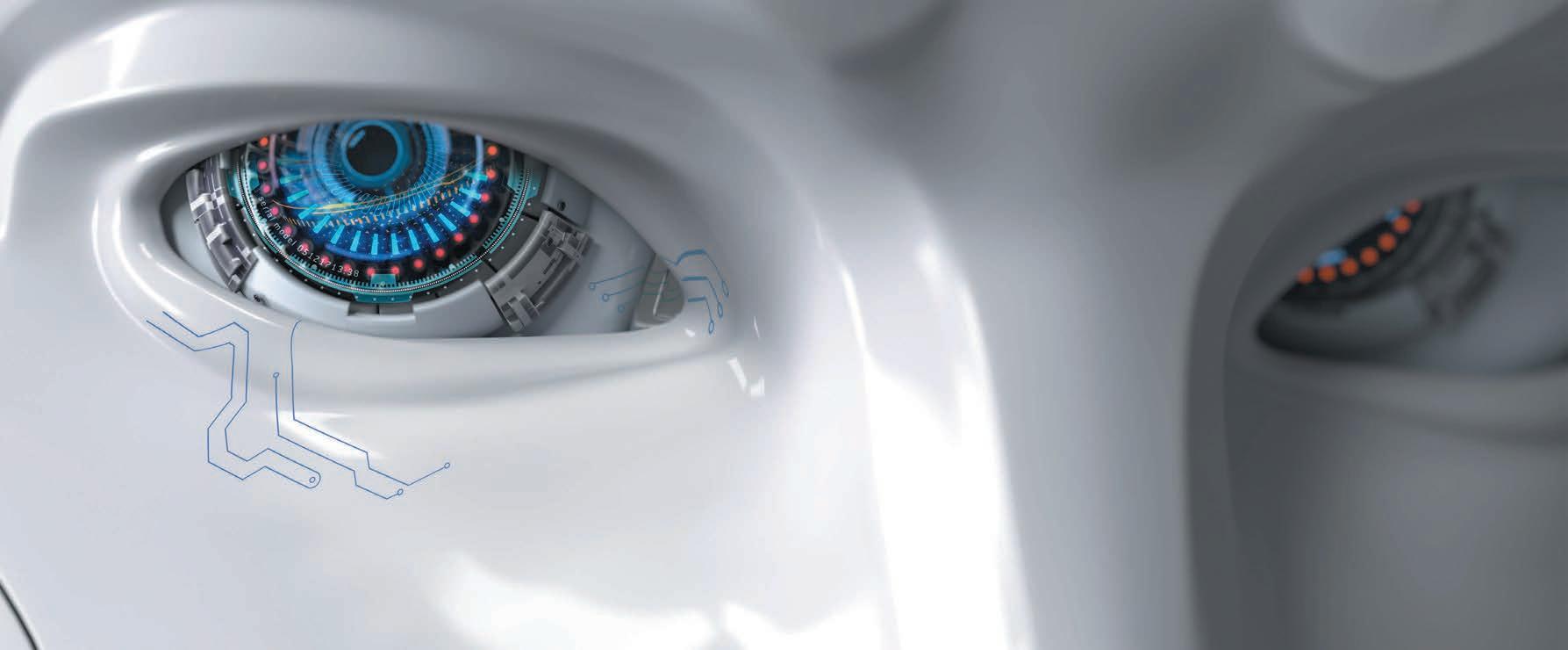
Debating the Future of Glaucoma Surgery
Challenges and opportunities await cataract surgeons interested in integrating early glaucoma treatment as part of comprehensive care.
04 Editorial: Hot Ideas, Cold Splash of Reality
07 ESCRS Update: Preliminary Congress Programme Now Online; ESONT ‘Bring Your Team’ Promotion Returning to Congress; New Videos Address Postoperative Surprises and Simultaneous Vision IOLs
15 First the Patient, Then the IOL
Nic Reus MD, PhD
16 Implanting Expectations
Andrea Janeková MD, PhD, FEBO, FEBOS-CR
17 Tips and Tricks to Avoid Refractive Surprise
Ruth Lapid-Gortzak MD, PhD
18 Dealing with Ocular Surface Disease in Cataract Patients
Sajjad Ahmad MBBS, FRCOphth, PhD
19 FLACS for Complex Cataracts
Soon-Phaik Chee MD
20 AI-Guided Robotics
Elevate Cataract Surgery
Dagny Zhu MD
Ronald R Krueger MD
22 Placing Ocular Surface Disease Under Observation
Béatrice Cochener-Lamard MD, PhD
24 DMEK Outcomes Using Tissue from Donors with Diabetes
Jonathan H Lass MD
25 A Closer Look at EndoArt
Mayank A Nanavaty MBBS, DO, FRCOphth, PhD and Leo Feinberg
26 Femtosecond LaserAssisted DALK
Mayte Ariño-Gutierrez MD
27 Possibilities and Limits of KLEx for Hyperopia
Walter Sekundo MD, PhD
28 PMLT: A New Frontier for Ocular Hypertension
Davide Borroni MD
30 Slightly More than MIGS
Rachel G Simpson MD
31 AI-Based Strategies for Predicting Glaucoma Progression
Cecilia S Lee MD, MS
32 Minimally Invasive Bleb Surgery
Ian Pitha MD, PhD
34 Reaching the Retina for Drug Delivery
Dominik Fischer MD, PhD
PAEDIATRIC OPHTHALMOLOGY
35 Watch Out for Adrenal Suppression in Congenital Cataract Patients
Diana Chabané Schmidt MD, PhD


Publisher
Tom Ogilvie-Graham
Executive Editor
Stuart Hales
Editor-In-Chief
Sean Henahan
Senior Content Editor
Kelsey Ingram
Creative Director
Kelsy McCarthy
Graphic Designer
Jennifer Lacey

Circulation Manager
Lucy Matthews
Contributing Editors
Cheryl Guttman Krader
Howard Larkin
Roibeárd O’hÉineacháin
Contributors
Laura Gaspari
Soosan Jacob
Timothy Norris
Clare Quigley
Andrew Sweeney
Colour and Print
CitiPost
Advertising Sales
Roo Khan
MCI UK
Tel: +44 203 530 0100 | roo.khan@wearemci.com
EuroTimes® is registered with the European Union Intellectual Property Office and the US Patent and Trademark Office.
Published by the European Society of Cataract and Refractive Surgeons, Building 4000, Langstone Park, Langstone Road, Havant, PO9 1SA, UK. No part of this publication may be reproduced without the permission of the executive editor. Letters to the editor and other unsolicited contributions are assumed intended for this publication and are subject to editorial review and acceptance.
ESCRS EuroTimes is not responsible for statements made by any contributor. These contributions are presented for review and comment and not as a statement on the standard of care. Although all advertising material is expected to conform to ethical medical standards, acceptance does not imply endorsement by ESCRS EuroTimes. ISSN 1393-8983


Finland hosted the very enjoyable ESCRS Winter Meeting this year. The Helsinki event was a great conference, offering warm Finnish hospitality in the cold northern climate. The programme offered a great choice of courses and presentations, many focusing on solutions for presbyopia.
One particularly popular format was the Sauna Sessions. The Nordic tradition of the sauna starts with a visit to the very hot, steamy environment of the sauna itself, followed by a cold plunge in the Baltic Sea or, at a minimum, an ice-cold shower. This process can be repeated until you are feeling relaxed and rejuvenated.
The Sauna Sessions used this as a metaphor for debate, with one speaker offering a hot take on a given controversy in cataract and refractive surgery and the second speaker pouring cold water on the topic in response—all in 10 minutes. Topics included the potential obsolescence of monovision IOLs, the utility of mix-and-match IOLs, presbyopia drops, and the value of training simulators.
The metaphor of the sauna underscored the never-ending evolution of clinical practice in ophthalmology: ideas are proposed, challenged, discarded, and developed. A lot of cold water was thrown on the idea of implanting intraocular lenses
when first proposed, leading to the development of the safe and predictable lenses we use today. Radial keratotomy had cold water thrown on it by the 10-year results of the PERK study, but corneal refractive surgery evolved into safe and effective treatments for ametropia.
This give and take is essential, eventually leading to consensus. As the Sauna Sessions demonstrated, there are many unresolved issues in clinical ophthalmology. Every issue of EuroTimes tackles the ongoing debates in the field. This issue takes a look at the evolving surgical approaches to glaucoma treatment and the potential role of the cataract surgeon. Other reports look at the role of AI and robotics in cataract surgery, optimal IOL choice strategies, KLEx and hyperopia, and the ESCRS IOL nomenclature project.
There is probably no better place to engage in these kinds of discussions than at major ophthalmology conferences. It is not too soon to start planning your attendance at the 2026 ESCRS Annual Congress in London, 11–15 September. ESCRS President Burkhard Dick gives a preview of the Congress in a Newsmaker interview in this issue. Hope to see you there!
Sean Henahan Editor-in-Chief
EDITORIAL BOARD

Adi Abulafia (Israel)
Bruce Allan (UK)
Noel Alpins (Australia)
Juan Alvarez de Toledo (Spain)
Gerd Auffarth (Germany)
Başak Bostanci (Turkey)
John Chang (Hong Kong SAR, China)
Béatrice Cochener-Lamard (France)
Burkhard Dick (Germany)
Mor Dickman (The Netherlands)

Joaquín Fernández (Spain)
Oliver Findl (Austria)
Sri Ganesh (India)
Christina Grupcheva (Bulgaria)
Farhad Hafezi (Switzerland)
Nino Hirnschall (Austria)
Soosan Jacob (India)
Jack Kane (Australia)
Yao Ke (China)
Georgios Kymionis (Greece)
David Lockington (UK)
Artemis Matsou (Greece)
Cyres Mehta (India)
Jod Mehta (Singapore)
Sorcha Ní Dhubhghaill (Belgium)
Rudy Nuijts (The Netherlands)
Catarina Pedrosa (Portugal)
Konrad Pesudovs (Australia)
Nic Reus (The Netherlands)
Filomena Ribeiro (Portugal)
Andreia Rosa (Portugal)
Giacomo Savini (Italy)
Julie Schallhorn (US)
Sathish Srinivasan (UK)
Paola Vinciguerra (Italy)
Shin Yamane (Japan)
Ron Yeoh (Singapore)
Mihail Zemba (Romania)

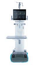
The best tools donʼt demand attention. They earn trust by supporting your surgical flow — from the first patient to the last. With Triple Pump Fluidics for advanced chamber stability, an intuitive interface, and a compact, wireless setup, Sophi is built for efficiency across your entire day.
So
you can focus on the surgery, not the system.

Rayner Group, all rights reserved. Sophi and Rayner are proprietary marks of the
10 Dominion Way, Worthing, West Sussex, BN14 8AQ. Registered in England: 615539. Sophi Registration number: 1401-8100-042-01. EC-2026-24 03/26

Extreme difficulty in extracting wounded soldiers from the front lines forces ophthalmologists to rethink their treatment procedures.
Many EuroTimes readers will know the experience of working in an emergency room, having to triage patients based on traumatic injuries. It’s a chaotic, stressful environment for doctor and patient alike.
Now imagine doing it on a battlefield as artillery rains down, all while tiny drones zip overhead looking for targets, their high-pitched whine somehow more terrifying than the booming shells.
This situation is the reality of modern combat in Ukraine, where doctors and medics on the front line are deliberately targeted as they try to extricate wounded Ukrainian soldiers. Under these conditions, preserving vision amid combat trauma is particularly challenging.
“Several factors working simultaneously make modern weapons so dangerous,” said Volodymyr Salamashchak MD. “Shock waves cause catastrophic damage to the eye’s structure, often causing multiple intraocular foreign bodies along with thermal and chemical burns and infection.”
In one survey involving 128 patients under Dr Salamashchak’s care, open eye injuries accounted for 72% and closed eye injuries accounted for 28% of trauma cases. The majority of patients (76%) experienced craniocerebral and facial injuries; 9% of cases were bilateral, and another 9% involved ‘eye ruin.’
The key to maximising outcomes for these patients is extracting them from the front lines as quickly as possible, ideally within the ‘golden hour’ of 24 hours. Dr Salamashchak explained this is very difficult, and not just for geographical reasons. For example, Dr Salamashchak is based in Lviv, more than 1,000 km away from the frontline city of Kramatorsk.
“Unfortunately, we can’t reach this ‘golden hour’ at the moment due to the extreme difficulty of removing the patient from the battlefield. The enemy has control of the airspace and uses drones to specifically target medical personnel,” Dr Salamashchak said.
He says the key factors in maximising outcomes under these conditions are stabilising vital functions in a timely manner, protecting the injury with a shield or antibiotic prophylaxis, and coordinating evacuation. This often relies on telemedicine via consulting an ophthalmic surgeon to determine triage priorities.
“Our basic principle is to repair now, refine later; to seal, then reconstruct. The main thing is to quickly restore the integrity of the eye, ensure its tightness, and prevent the loss of internal structures,” Dr Salamashchak said.
“If the patient is stable, then it’s ideal to perform surgery and reconstruction of the posterior segment within 24 hours after injury, but this can take weeks in Ukraine. Each day of delay worsens the patient’s outcome.”
The war in Ukraine is unlikely to abate in the near future, meaning these highly challenging conditions will persist for some time. Dr Salamashchak is working on a series of protocols to improve patient outcomes in the meantime, a strategy that includes an improved triage system for patients with ocular injuries.

ESCRS members and others in ophthalmology can contribute by providing training programmes for vitreoretinal surgeons. There’s also an equipment gap, and institutions working to treat patients in Ukraine urgently need more supplies.
“We need to implement rapid consultations via telemedicine for ophthalmic surgeons, as this will help us to rapidly coordinate our actions in the field,” Dr Salamashchak said. “Ultimately, we need a unified system of knowledge, skills, and standards to provide care for our soldiers who have experienced combat trauma.”
Dr Salamashchak presented at the 2025 Society of Ukrainian Ophthalmic Surgeons Congress in Kyiv.
Volodymyr Salamashchak MD holds the rank of major in the Armed Forces of Ukraine and is an oculoplastics specialist and surgeon based at the Military Medical Clinical Centre of Lviv. volodymyrua89@gmail.com

The preliminary programme for the 2026 ESCRS Annual Congress in London is now available on the Congress website. The programme offers a preview of what to expect at this year’s Congress, from plenary sessions to subspecialty days to instructional courses, all designed to help guide you on your medical journey. While the programme content is subject to change, it is a helpful starting point from which to begin planning your time at ESCRS 2026. Additional information about the Congress is also available on the website, including details about how ESCRS is working to limit carbon emissions from the event and advice about saving money on registration fees.
| ESCRS 2026
Following its inaugural success at last year’s Congress, ESCRS is once again offering the ‘Bring Your Team’ discount to encourage participation among firsttime attendees in the Ophthalmic Nurse/Technician registration category.
‘Bring Your Team’ provides targeted support to ophthalmic nurses and technicians who have never attended an ESCRS Annual Congress (dating back to Amsterdam 2021). Eligible first-time attendees can access a reduced registration rate of 30% off the regular Ophthalmic Nurse/Technician fee.
The discount is intended to foster engagement from ophthalmic nurses and technicians and encourage senior doctors to bring emerging talent to the Congress, thereby supporting professional development and collaboration.
Your Team | ESCRS 2026
In a recent episode of the ESCRS Independent Medical Education (IME) video interview series on refractive surgery, Drs Pooja Khamar and Béatrice Cochener-Lamard discuss postoperative surprises, focusing on how preexisting ocular surface conditions, such as dry eye, can affect surgical outcomes. They explore complications such as pseudoregression and glare caused by poor epithelial healing; they also highlight the importance of thorough preoperative assessment, targeted treatment, and patient education to optimise visual outcomes.
In addition, the IME Forum on Simultaneous Vision IOLs, held during the 30th Winter ESCRS Meeting in Helsinki, is now available on demand. This session brought together leading experts to explore the latest advancements in simultaneous vision IOLs and their evolving role in presbyopia management. Moderated by Professor Burkhard Dick, the faculty included Dr Joaquín Fernández, Dr Ruth Lapid-Gortzak, Dr Nick Reus, and Dr Andrea Janeková, who shared valuable insights into global functional classification, preoperative diagnostics, personalised lens selection, and patient communication.
IME Forum on Simultaneous Vision IOLS
Refractive Surgery: Communicating Benefits and Setting Expectations

Vision to Venture will expand ESCRS’s educational offerings beyond clinical training.
Business processes and practices increasingly are being integrated into healthcare to heighten cost efficiency, improve patient outcomes, and ensure regulatory compliance. Knowledge of business processes and practices, unfortunately, is generally lacking among healthcare practitioners, and ophthalmologists are no exception.
“Many ophthalmologists are not literate in economics, accountancy, and marketing,” says Christina Grupcheva MD, PhD. “Most of them think, ‘I have the patients, I have the skills, I can purchase the equipment. The rest will take care of itself.’”
To some extent, the European ophthalmology landscape is to blame—while clinicians receive extensive medical training, there is limited access to practical, structured education on the business, operational, and leadership aspects of running a clinical practice. Existing programmes in this area are largely US-based or tailored to a narrow audience and do not sufficiently reflect the regulatory, financial, and operational realities of European healthcare systems.
Beginning this year, ESCRS is bridging this gap by launching a structured business education programme
for ophthalmologists. Titled Vision to Venture (V2V), the programme is designed to serve as a business accelerator for ophthalmologists and practice managers, supporting individuals at different stages of their professional journey.
“Vision to Venture will equip clinicians to operate effective, sustainable, and well-managed practices within the European healthcare environment,” says Paul Rosen MBA, MD, who chairs the ESCRS Leadership, Business, & Innovation Committee (LBI), which is developing the programme.
“By partnering with top-tier business leaders, professional service firms, verified leaders of successful European clinics, and professionals from outside the industry, we will offer a programme with unparalleled substance.”
Programme format
The programme will feature an introductory online component followed by a series of five in-person weekend courses. Each weekend course will focus on a specific area of business and practice management:
• Business planning: Developing a viable clinical and
commercial strategy, including funding, market analysis, and financial modelling
• Legal, regulatory, and financial essentials: Understanding compliance requirements, contracts, company structures, and financial controls
• Leadership and human resources: Building and managing teams, leadership development, and organisational culture
• Operations and practice management: Establishing efficient clinical workflows, managing resources, and scaling operations
• Marketing and positioning: Developing a clear brand, communicating value, and managing patient acquisition within regulatory frameworks
Weekend courses will be presented at accessible European locations and will include a combination of expert-led sessions and practical, small-group work. Group sizes will be limited to encourage interactive learning. Assorted digital content (e.g., webinars and ongoing learning materials) will be delivered throughout the year to support the courses. Attendance at individual weekends is not mandatory, although the curriculum is designed to be progressive and encourage continued participation.
The initial launch, including introductory content and the first in-person course, is scheduled to occur this year. The programme will expand next year to deliver content across all five weekend modules. Further development will be based on participant feedback and programme evaluations.
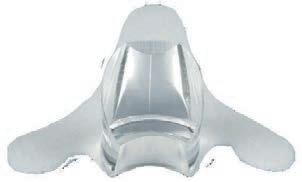
The GONIO ready® CL offers the surgeon a crystal-clear view of the anterior chamber angle during MIGS without having to hold a lens.
Ultra high clarity through OCULUS quality lens
Less adjustment due to large self-stabilizing foam fin
Single use eliminates sterilization time and infection risk
Contact the OCULUS team or your local distributor for more information and a no-obligation trial.
In addition to serving as a business accelerator for European ophthalmologists, Vision to Venture will support ESCRS’s broader strategic direction of expanding its educational offerings beyond clinical training.
“V2V is not separate from education—it is a natural extension of it,” says Başak Bostanci, a member of the LBI Committee. “It fits naturally within ESCRS because it expands the definition of education from not only how we operate, but also how we build and sustain what we operate in.”
“Vision to Venture perfectly fits within the existing ESCRS educational offer,” agrees Vincent Qin, MD, MBA. “The existing offer is mostly technical: toric IOLs, premium lens patient selections, and complications management. V2V offers the non-technical, business and management side of ophthalmology—the soft skills needed to run a department or a practice.”
“If ESCRS is committed to helping ophthalmologists deliver the highest standards of care, then supporting them in building strong, well-run practices is part of that mission as well,” says Artemis Matsou MD, another LBI Committee member. “The difficult decisions about business planning, finance, hiring, leadership, operations, and marketing are very real, but they are often made without much structure. Vision to Venture takes those areas seriously and turns them into something teachable, practical, and relevant.”


Challenges and opportunities await cataract surgeons interested in integrating early glaucoma treatment as part of comprehensive care.
BY SEAN HENAHAN
As cataract surgeons increasingly take on glaucoma procedures, the divide between the two fields is blurring, prompting one speaker at a recent ophthalmology conference to comment that someday there might need to be a ‘G’ for glaucoma added to the ESCRS acronym. Dr Robert Osher’s popular Video Journal of Cataract, Refractive, and (most recently) Glaucoma Surgery (VJCRGS) has already seen this change.1 While it is unlikely the ESCRS will add a new letter to its acronym any time soon, it is not unlikely that cataract surgeons will be including more glaucoma management in their practices.
The traditional approach in glaucoma treatment has been to prescribe drops early, reserving trabeculectomy surgery for later, with the goal of keeping intraocular pressure (IOP) under control to slow optic nerve damage. While this goal remains the same, dissatisfaction with poor compliance with drop regimens coupled with various bleb-free surgical options (particularly in combination with phacoemulsification) have driven a new interest in earlier intervention by cataract surgeons.
“The cataract surgeon has transformed from being solely a lens specialist to an active decision maker in early glaucoma management. We are no longer merely recognising glaucoma as a secondary condition; instead, we are managing glaucoma alongside cataract surgery, particularly during combined procedures like MIGS. This change indicates a shift towards earlier, more integrated, and patient-centred
care, where reducing medication load and enhancing longterm adherence are as vital as visual restoration,” ESCRS president Burkhard Dick MD, PhD told EuroTimes
In a recent edition of the ESCRS Clinical Trends Series, Professor Dick discussed key findings from the 2025 ESCRS Clinical Trends Survey, providing an in-depth analysis of how Society members now approach glaucoma management.2 One interesting finding was the steady decline in the reported percentage of cataract patients with glaucoma since 2018. He attributes this to earlier cataract surgery, combined procedures (e.g., combining cataract surgery and minimally invasive glaucoma surgery [MIGS]), and a denominator effect wherein the growing number of non-glaucoma patients undergoing cataract surgery lowers the overall proportion with coexisting glaucoma. Nonetheless, overall global demographic trends suggest glaucoma cases will only increase in coming years.
An ESCRS survey indicated that medications were a first-line therapy for 77.6% of survey participants, followed by 20.1% who perform selective laser trabeculoplasty (SLT) and 1.9% who choose MIGS as an initial approach. The survey noted that while 15% of cataract surgery candidates on topical therapy could be considered MIGS candidates, a majority of ESCRS respondents (58%) lack confidence in performing MIGS procedures on cataract surgery patients. 2
What percentage of all your cataract patients would you estimate have glaucoma?
average number of patients seen per month considered as having glaucoma
Medication remains the predominant first-line treatment for glaucoma (Figure 2). However, other modalities, particularly selective laser trabeculoplasty (SLT) and MIGS, are being notably utilized, especially following initial treatment.
What is your typical first-line therapy for newly diagnosed glaucoma?
What is your typical first-line therapy for newly diagnosed glaucoma?
of glaucoma patients prescribed more than two medications are not compliant
Figure 1. Percentage of estimated cataract patients with glaucoma from 2018 to 2025, showing steady decline over the years. 2
•E arlier cataract surgery
•C ombined procedures
•
The utility of standard SLT as an effective first-line treatment was confirmed in the prospective, multicentre, randomised, controlled Laser in Glaucoma and ocular Hypertension Trial (LiGHT) study, which concluded the procedure was more cost-effective than IOP-lowering drops.3 Consequently, SLT is now recommended as a first-line intervention for lowering IOP in open-angle glaucoma by the European Glaucoma Society.4
But it was the development of direct SLT (DSLT; Voyager, Alcon), which eliminates the need for a gonioscopy lens and coupling medium, that made this option available for non-glaucoma specialists. The prospective GLAUrious study compared DSLT with conventional SLT.5 The 6-month primary end point did not achieve statistical noninferiority compared with conventional SLT. However, DSLT did provide an effective reduction in IOP that was sustained for 12 months.
*The latest EGS Guidelines can be found at : https://eugs.org/educational_materials/6
Adopting DSLT offers a solution for helping glaucoma specialists and general ophthalmologists meet the increasing demand for services from the expected growing population of patients with glaucoma, said Christine Funke MD.6
“I am using DSLT frequently in my clinic, and I think and hope this machine will help push the envelope so SLT is used more as first-line therapy for IOP lowering,” she said. “DSLT
Medication and Patient
Survey respondents estimate coma patients prescribed non-compliant. This figure as multiple studies report patients ranging from on multi-drop regimens missed doses increasing
In my experience, any drops is at particularly varying dosing schedules, challenging, especially of non-compliance include medication costs, and such as low health literacy cant role.
To improve adherence, ing the treatment regimen:
Figure 2. Medication overwhelmingly remains the most common first-line therapy for newly diagnosed glaucoma. 2
“Poor adherence to multipledrop regimens is unfortunately very common.”
is a simple and user-friendly procedure. By eliminating the need for a gonio lens, it also means there should be no more excuses for not doing SLT.”
• Simplify the regimen: possible, reduce dosing ules with the patient’s
• Educate your patient: preserving goals of and encourage the
• Manage the ocular and switch to preservative-free reduce discomfort.
lack conÞdence in performing MIGS procedures on cataract surgery patients 58% of cataract surgery patients on topical glaucoma therapy are MIGS candidates 15%
• Offer laser/surgical even eliminate the going SLT or a simple drops markedly improves
Another innovative approach called femtosecond laser, image-guided, high-precision trabeculotomy (FLiGHT, ViaLase) adds to the trend towards laser-first therapy. The device uses a femtosecond laser to cut precise apertures in the trabecular meshwork, guided by real-time, ultra-high-definition gonioscopic and optical coherence tomography imaging. A clinical study found mean IOP was reduced by nearly 35% at 24 months with no serious adverse events reported.7
Pursuing a similar approach, the ELIOS procedure (Bausch + Lomb) uses an excimer laser to facilitate aqueous outflow via microchannels in the trabecular meshwork. The procedure is available in Europe but has yet to receive US FDA approval. Bausch + Lomb recently announced 24-month results from a new prospective, multicentre clinical study of the ELIOS System, with most patients medication free at 23 months.8
For citation notes, see page 40.
Please see related articles in this issue for an update on new MIGS procedures and the use of AI for glaucoma screening. 3
When do you usually initiate laser/surgical intervention for glaucoma patients?
When do you usually initiate laser/surgical intervention for your glaucoma patients?
First line After first line medication After two medications After three medications I do not perform laser/surgical interventions Laser Surgery
Figure 3. Laser therapy is typically used earlier in the glaucoma treatment process, whereas surgery is more often reserved for later stages—commonly after the use of three medications. 2 - -
Figure 3. Laser therapy is typically used earlier in the glaucoma treatment process, whereas surgery is more often reserved for later stages—commonly after the use of three medications.
incisional surgery becomes indicated. Importantly, the EGS emphasizes that surgery “should not be left as a last resort,” but considered whenever drops or SLT are unlikely to preserve vision.
Patient adherence and quality of life are central considerations. Achieving medication-free IOP control can substantially enhance quality of life by reducing costs and discomfort; benefits often seen following DSLT or surgical intervention.
• Provide follow-up use phone reminders, force consistent medication Laser vs. Surgical Intervention According to the Clinical dents reported starting vention, and just 3% initiated Following initial management come more inclined to 24% introduced laser treatment 20% after two medications. to surgery after first-line medications. Notably, patients already on three dents reported never using area with clear potential Deciding between and surgery depends on individual patient factors. I consider DSLT early, option, especially for patients angle glaucoma (OAG)
The EGS explicitly therapy in mild or moderate from trials such as LiGHT. safely achieved with medications a reduction greater than

Interested in taking a giant step forward in your surgical career?
ESCRS is launching the Emanuel Rosen Fellowship for young ophthalmologists who have finalised their training in ophthalmology and want to increase their knowledge and/or surgical skills in anterior segment surgery (cataract, cornea, or refractive).
The Fellowship of €60,000 is designed to allow the applicant to work abroad at a centre of excellence for clinical experience in the field of cataract and refractive surgery, anywhere in the world, for one year.

The fellowship celebrates the first president of ESCRS, Emanuel Rosen, who helped ignite the Society’s founding and growth, co-launched the Journal of Cataract & Refractive Surgery, and championed advances ranging from intraocular lenses and phacoemulsification to laser refractive surgery, all while serving patients across the UK National Health Service and in private practice.
Eligibility
Applicants must be young ophthalmologists who are full members of the ESCRS (this will include only ophthalmologists working in Europe by definition and also exclude trainees) who are in their second year of sub-specialty experience in cataract, cornea, or refractive surgery.
The successful applicant for the Fellowship will be announced at the ESCRS Annual Congress in London.The fellowship will start in 2027.
Apply by 30 June for the opportunity of a lifetime!


Technologies offering diffractive or lens position changes are entering human trials.
Laser-adjustable intraocular lenses (IOLs) are now entering clinical studies and have the potential for in vivo diffractive adjustment into any distance or multifocal profile, according to Ronald R Krueger MD. Further developments in laser-adjustable lenses, as well as the already commercially available light-adjustable lenses, are likely, he added.
While the refractive performance of IOLs has improved greatly in recent years, the accuracy of fixed power lenses is still limited by factors including biometry problems, mechanical IOL positioning, and wound healing, Professor Krueger noted. Indeed, a EUREQUO study of more than a quarter million surgeries published in 2018 found that while about 73% of eyes fell within 0.50 D of target refraction, the absolute mean prediction error in spherical equivalent was 0.42 D.1
Adjusting lenses after implementation has the potential to further reduce errors, Prof Krueger said. The Light Adjustable Lens (RxSight), which was approved in the US in 2017, has demonstrated how effective the approach can be, increasing the percentage of patients testing with 0.50 D to 98% in an early trial and 92% in the FDA approval trial.2,3 About 75% of recent patients opt for blended vision or monovision, according to company research.4
However, the Light Adjustable Lens requires a specific polymer lens designed to react to ultraviolet light to adjust its power. Lasers could alter the refractive power of any acrylic lens type and even induce multifocality through a process called Refractive Index Shaping (RIS).
RIS uses a 520-nm green femtosecond laser to create a thin layer of refractive index change, forming a diffractive Fresnel prism. Refractive index changes allowing corrections as fine as 0.10 D are made inside the IOL by laser bursts that do not reach ablation strength, so the shape of the lens is not affected and no photoablation occurs.
In an ongoing study by Pavel Stodůlka MD, PhD, 18 eyes of 9 patients implanted with monofocal Tecnis ZCB00 (Johnson & Johnson) lenses received a 40-second outpatient RIS
treatment that changed their lenses into diffractive multifocal lenses. One month after treatment, patients had good near vision without sacrificing distance visual acuity, Prof Krueger said. While the early results are positive, more patients and longer follow-up are required to further test the concept.
The global trend for personalised medicine will fuel further development in laser and light adjustability in the future.
Another approach to lens adjustment is argon laser-directed lens movement, Prof Krueger said. A hydrophobic IOL incorporating a spring-loaded nitinol cradle is implanted in the eye. An argon laser then allows the lens to be moved forward or backward by selectively releasing spring tension to correct errors after implantation. The lens has been tested in rabbits, and a human trial is about to start, Prof Krueger said.
“The global trend for personalised medicine will fuel further development in laser and light adjustability in the future,” Prof Krueger concluded.
Prof Krueger made his comments at the 2025 ESCRS Annual Congress in Copenhagen.
For citation notes, see page 40.
Ronald R Krueger MD is McGaw Professor and Chair of the University of Nebraska Medical Center Department of Ophthalmology and Visual Sciences, and Director of the Stanley M Truhlsen Eye Institute, both of Omaha, Nebraska, US. ronald.krueger@unmc.edu
Matching patients’ expectations with their needs is crucial for a good surgical outcome.
LAURA GASPARI REPORTS
For simultaneous vision lens implantation, the best outcome for patients can only be reached by prioritising their needs, according to Nic J Reus MD, PhD.
“There’s no free lunch in optics,” Dr Reus recalled, quoting Professor Gerd Auffarth’s ESCRS Binkhorst Lecture of 2021. If the depth of field is increased, there are effects on visual quality and dysphotopsias—surgeons cannot have everything.
“You have to match the IOL to the patient,” Dr Reus said. “It is all about equilibrium—look at the depth of field versus visual quality versus dysphotopsias. There is not a question of which lens is the best, but for whom is this trade-off acceptable.”
However, over time, lunch has become less expensive, Dr Reus observed. IOL optics have changed and optical profiles have improved, resulting in fewer dysphotopsias. There is also greater refractive accuracy, due in no small part to accurately performing preoperative biometry. Despite all the developments, sometimes what patients get from the lens does not match with their functional visual needs and expectations. Some of them have unrealistic expectations about the lens’s capabilities in various lighting conditions, which can be the cause of discontent with the surgery.
All aspects of patient life must be carefully considered when choosing the best simultaneous vision lens. Whether they work in dim light, drive at night, or rely on near vision, everything matters.
Dissatisfaction is around the corner, Dr Reus warned, recall ing his experience with an unhappy patient with a trifocal lens whose hobby was working on electronics in dim light. The pa tient had been counselled about the importance of good lighting for near tasks, but the extent to which they worked under such low-light conditions had not been fully appreciated before the surgery and could have made a difference in the outcome.
Patient awareness of lens options also varies, as the 2025 ESCRS Clinical Trends Survey revealed: most surgeons (54%) claimed their patients are aware but not well educated, while some others are educated (26%) or very well educated (12%).
Good counselling about photic phenomena is crucial to prevent dysphotopsias, which may become bothersome for patients. Photic phenomena, such as halos, starbursts, and streaks, can occur with IOLs and may develop into dyspho topsias, leading to patient dissatisfaction.
Dr Reus suggested a four-pillar checklist to keep in mind: First, note the patient’s lifestyle, needs, expectations, and previous visual corrections; second, collect a thorough history of the ocular condition, with contrast sensitivity and comor bidities, the binocular single vision, the stability of the cap sular support, or the axial length; third, gather accurate and consistent measurements of the eye and the IOL power, taking astigmatism and, in select cases, angle kappa into account; and, finally, establish the lens strategy.

Prioritising patient needs, customising lens choice, and managing expectations to maximise satisfaction—by balancing spectacle independence with visual acuity and framing photic phenomena as a trade-off rather than a complication—are the key rules to follow when preparing for surgery. A dissatisfied patient may also cause dissatisfaction for the surgeon.
“If there is a mismatch between the lens, the eye, and the expectations, then you will have complaints,” Dr Reus concluded.
Dr Reus spoke at the 2026 ESCRS Winter Meeting in Helsinki.
Nic Reus MD, PhD is an independent ophthalmologist at Amphia Hospital, Breda, Netherlands. nreus@amphia.nl

Effective communication and realistic expectations are essential for patient satisfaction in refractive and cataract surgery.
LAURA GASPARI REPORTS
To achieve patient satisfaction in refractive and cataract surgery, even a perfect surgical performance is sometimes not enough—effective communication plays an important role, according to Andrea Janeková MD, PhD.
This disconnect becomes clear in practice: despite all the perfect biometrics, refractive outcomes, and measurements, patients can still be unhappy after a surgery. The same surgeon with identical precision and skill set can implant the same type of IOL in two different patients and deliver different final outcomes.
“The difference is the expectation from the surgery,” Dr Janeková said. “When I start my consult with patients who want to be spectacle independent, I start with their lifestyle, not with the type of IOL, to better select the best options to solve their problems.”
Simple and direct communication, she stressed, can enhance the establishment of a good relationship between ophthalmologists and their patients, which helps in making the right IOL choice. Discussing complicated and technical optics with older patients is not useful in cataract surgery. Another effective strategy is to honestly reframe patient concerns about glare and halos as a brain adaptation to new optics rather than a problem, so they understand halos and glare are not necessarily postoperative complications.
Full transparency about costs and outcomes to manage patient expectations is fundamental. Key considerations about the economics of presbyopia vary for different patient groups, like moderate myopes, early presbyopic patients, or those

who underwent corneal refractive surgery. These considerations can pose challenges that can limit the surgeon’s choice of technology, even though the motivation to get rid of the glasses is high. Dr Janeková suggested written documentation as a useful and dependable tool to protect the bond of trust between surgeon and patient.
When I start my consult with patients who want to be spectacle independent, I start with their lifestyle, not with the type of IOL.
Overwhelming patients with too many options and promising absolute spectacle independence are counterproductive, according to Dr Janeková. A surgeon must be clear and honest about every risk and issue surrounding the surgery, as not all presbyopic patients are the same.
Age matters in the evaluation of the expectations and tolerance for trade-offs. Younger patients are more likely to be active, have lower tolerance for visual effects, and want to get rid of glasses as soon as possible. The best strategy to take with this demographic is to share the decision-making process and manage their expectations. Conversely, older patients will want to improve their visual outcomes and will be more accepting of visual effects, focusing more on procedure safety. They do require clarity, reassurance, and simple communication strategies. Yet, as Dr Janeková emphasised, personalisation according to the patient’s case and lifestyle—and not age alone—drives final satisfaction.
The long-term value of the surgery and the shared decision making between patients and doctors are invaluable in the process leading to successful ophthalmic surgery, she stressed. Bearing this in mind can greatly help patients to change their life for the better.
“We are not just implanting lenses, we are implanting expectations, and clear communication before surgery helps us make our patients happy after the procedure,” she concluded.
Dr Janeková spoke at the 2026 ESCRS Winter Meeting in Helsinki.
Andrea Janeková MD, PhD, FEBO, FEBOS-CR is Head Physician of Cataract, Refractive, and Vitreoretinal Surgery, Prague Eye Centre, Czech Republic. janekovaandrea@gmail.com
Ocular surface optimisation and reliable preoperative measurements are keys to better cataract surgery outcomes.
LAURA GASPARI REPORTS
Residual refractive errors can significantly affect visual acuity and patient satisfaction, so surgeons should develop strategies involving proper preoperative evaluation to avoid them, according to Ruth Lapid-Gortzak MD, PhD.
“From the ESCRS Clinical Trends Survey, 26% of cataract patients present to the preoperative consult with ocular surface disease (OSD) symptoms, and we think it might be slightly underestimated,” she noted.
The survey also revealed 48% of respondents routinely check the ocular surface in all preoperative cataract surgery examinations, but only two-thirds of those who find OSD act on it, meaning one out of three cataract surgeries might be affected.
According to Dr Lapid-Gortzak, good screening and optimisation of the ocular surface are the first steps in avoiding residual refractive errors. Of course, it is not possible to run every available test for every patient, but the most important thing for surgeons is to remain practical and use what works in their practice to act quickly, she remarked.
Patients with dry eye should receive a suitable treatment before proceeding with cataract surgery. Shared consent is a powerful tool to make sure patients understand their dry eye disease and its potential impact on their vision. Sometimes a ‘no’ is a proper answer, Dr Lapid-Gortzak emphasised, especially when dry eye disease will not yield good results with simultaneous vision correction.
Multi-tiered approaches, including disease control with anti-inflammatory therapy, technology-based therapies, and baseline care such as lid hygiene, warm compresses, and nutraceuticals, are a good way to optimise the ocular surface, she added.
Proper data validation using tools like keratometry, topography, biometry, and OCT is particularly important. Preoperative measurements should include a good identification of the patient and the IOL, as well as careful assessment of potential

26% of cataract patients present to the preoperative consult with ocular surface disease symptoms.
red flags such as keratometry abnormalities, astigmatism, long and short eyes, corneal irregularities in laser patients, and keratoconus. Specialists should repeat the validation process when there are inconsistencies or after dry eye treatment, using the results to drive their decision making. The days of performing cataract surgery without topography or tomography are over, according to Dr Lapid-Gortzak, adding manual keratometry is outdated.
Concerning posterior corneal astigmatism, Dr Lapid-Gortzak noted it can meaningfully affect outcomes in one in five patients. Since measurements are based on assumptions, it is difficult to make a precise prediction. For this reason, she emphasised the importance of using accurate calculators—such as the ESCRS online calculator—and applying them consistently, using the same formula with adjustments as needed. She added other formulas and calculators can help check for consistency.
Finally, achieving expert outcomes depends on meticulous preoperative precision.
“Not last and certainly not least, we should properly identify our patients and IOLs in the OR,” she concluded.
Dr Lapid-Gortzak spoke at the 2026 ESCRS Winter Meeting in Helsinki.
Ruth Lapid-Gortzak MD, PhD is a cataract, cornea, and refractive surgeon at the Amsterdam University Medical Centers, Netherlands. r.lapid@amsterdamumc.nl
Three challenging conditions can make it difficult to treat cataract patients.
ANDREW SWEENEY REPORTS
Which is harder to manage in cataract surgery patients: severe dry eye disease, cicatricial conjunctival disease, or limbal stem cell deficiency (LSCD)?
All three present their own challenges and difficulties, said Sajjad Ahmad MBBS, PhD, who discussed his experience with treating such complex patients.
Addressing dry eye disease, Dr Ahmad said the most important thing to do is identify the main “factor for the disease,” whether it is primarily evaporative or aqueous deficient. This will allow the clinician to optimise control before performing surgery.
Noting he is often asked whether patients on topical antiinflammatories like cyclosporine should have their treatment stopped before surgery, Dr Ahmad said surgeons should not advise stopping treatment, as it will help the inflammation after surgery.
Intraoperatively, hydration remains crucial, and Dr Ahmad recommended regularly using agents such as hydroxypropyl methylcellulose (HPMC) to coat and protect the surface. He also warned against using trifocal lenses and stressed the importance of rapid follow-up.
“Dry eye disease patients are the ones you want to see within the first week, especially if their wounds are doing something funny—if they’re melting, which occasionally they can do—or if they’re put on non-steroidals,” Dr Ahmad said.
As for cicatricial conjunctival disease, Dr Ahmad stressed the importance of managing inflammation in the preoperative stage, as postoperative inflammation is expected and may require immune system suppression and oral steroids. Anaesthetic considerations are also vital because the conjunctiva is scarred, so “topical and sub-Tenon anaesthetic would be inappropriate.
“It’s also really important that you don’t overdo your local anaesthetic, because you then get a tight space, which becomes even tighter with your injection. This is where peribulbar anaesthetic is very helpful, and sometimes a general anaesthetic may be possible,” he explained.
“Postoperative management here involves using oral steroids. So, I always give 20 milligrams of steroids with 20 milligrams of lansoprazole for two weeks because it’s easy to remember.”
LSCD patients require even more careful planning in the preoperative stage, as operating on these patients is high risk. Dr Ahmad said that severe or total LSCD may require staged surgeries or the “open sky” technique, where the cornea is removed via penetrating keratoplasty (PK) concurrent with cataract removal.
Dry eye disease patients are the ones you want to see within the first week, especially if their wounds are doing something funny.
“If there’s partial stem cell deficiency, what you can do is peel away the conjunctiva, do your cataract surgery, and then put an amniotic membrane on afterwards. However, if there’s no view, then Vision Blue sometimes helps. Light pipes, I find, do not help,” Dr Ahmad said.
“I still create a large exit, lift up the cornea, and do an open-sky cataract before putting it back. Some people would approach these as PK. So, if you do, remove the host cornea, do your cataract as an endothelial keratoplasty, and then put the same cornea back.”
Dr Ahmad presented at the 2025 ESCRS Annual Congress in Copenhagen.

Laser makes surgery easier and safer in complicated cases.
HOWARD LARKIN REPORTS
Dense cataracts. Intumescent cataracts. Ruptured posterior polar cataracts. Subluxated cataracts. Cataracts with severe zonulopathy. Cataracts with anterior chamber vitreous.
Femtosecond laser-guided cataract surgery (FLACS) can help with these and other difficult cases, according to SoonPhaik Chee MD. FLACS greatly aids capsulotomy, and laser lens fragmentation reduces the needed disassembly force and phaco power, helping preserve delicate ocular tissues.
Hard cataracts
For very dense cataracts, Professor Chee favours the laser to segment the nucleus into octants rather than a grid because it reduces phaco power needs and endothelial cell loss.1 Patients with dense and thick cataracts are common in the population she serves, frequently with weak zonules. Often no chopping is needed to remove the nucleus, but only a lateral separation to free the posterior plate, she said. “This enables us to do very complex surgeries and get very good outcomes.”
Intumescent cataracts
Given it simultaneously cuts the entire circumference, laser-assisted capsulotomy can keep intumescent cataract cases from tearing even with explosive fluid release.2
“You can see the explosion, and because of the simultaneous cut you can see it is complete,” Prof Chee said. She uses trypan blue to ensure a complete opening.

FLACS enables complex cataract surgeries, making them easier and safer.
Prof Chee also uses laser fragmentation to cut through fibrosis for intumescent cases. “It is advantageous in white or brown cataracts.”
Ruptured posterior polar cataracts
FLACS is useful for spontaneously ruptured posterior polar cataracts in several ways, Prof Chee noted. For nucleus segmentation, she increases the posterior thickness slightly to leave a thicker plate to protect the posterior capsule. To avoid rotating the nucleus and possibly extend a posterior tear, she uses a counter cross chop and manually tumbles out the fragments. She then aspirates the epinuclear plate, injects viscoelastic, and slowly aspirates the cortex.
Capsulotomy is also very important with posterior cataracts because an optic capture may be required to place the lens at the end of surgery, Prof Chee added. “In these complicated eyes, we can get very good outcomes.”
In acute angle closure glaucoma with anterior subluxation, FLACS helps segment the lens for removal without rotation and aids capsulotomy in very shallow anterior chambers.3 Prof Chee begins by removing some vitreous to decompress the eye, then severs synechia holding the subluxated lens before removing it.
In Marfan syndrome, FLACS enables capsulotomy centring on a tilted capsular bag while laser fragmentation allows nucleus removal without rotation, placing less stress on severely weakened zonules. Prof Chee inserts capsular tension rings early to support the bag during surgery. Capsular tension segments help stabilise and centre the lens.
FLACS is also helpful in subluxation cases with vitreous in the anterior chamber, Prof Chee noted. “The laser cuts right through.” Vitreous is removed first before stabilising the capsule with hooks for surgery and lens insertion.
“FLACS enables complex cataract surgeries, making them easier and safer,” Prof Chee concluded.
Prof Chee made her comments at the 2025 ESCRS Annual Congress in Copenhagen.
For citation notes, see page 40.
Soon-Phaik Chee MD is Distinguished Professor of Clinical Education at the Duke-National University of Singapore Medical School, professor at the National University of Singapore, and senior consultant at the Singapore National Eye Centre, all in Singapore. gmscsph@duke-nus.edu; cpac@ihhhealthcare.com

Femtosecond lasers bring automation to cataract surgery.
HOWARD
Robotic cataract surgery is no longer just a dream. With today’s femtosecond laser assistance systems, robotics are already in use in operating theatres.
Rather than take over surgery autonomously, modern robotics provide surgeons with precision guidance to optimise nearly every step of cataract surgery, explained Dagny Zhu MD. The technology helps surgeons analyse and plan surgeries and increase the safety of delicate manoeuvres inside the eye, keeping them firmly in control.
For example, the Lensar ALLY system uses iris registration guided by artificial intelligence to precisely direct incisions and toric placement. Capsulotomies are precisely centred on the optical axis, which is especially important for optimising multifocal lens performance, said Dr Zhu, who implants premium lenses in about 90% of her cataract patients and uses a femtosecond laser in virtually all cases.
AI also helps image cataracts and determine their density to recommend fragmentation patterns that research suggests can reduce phacoemulsification energy requirements and capsule ruptures, Dr Zhu added. Such automated arcuate incision planning and precise placement result in more accurate and stable astigmatism corrections.
“All of this is done semi-autonomously, meaning the surgeon is there to guide every step of the way and supervise this robotic intelligence,” Dr Zhu said. “Robotic cataract surgery exists, or at least robotic laser-assisted cataract surgery.”
Robotic cataract surgery conjures images of robots performing every step of surgery independently, “with perfect precision, without any input from the surgeon whatsoever. . . . There’s always this fear of this happening when you let robotics take over,” Dr Zhu said.
However, autonomous robotic cataract surgery lies far in the future, if it is possible at all, and will always require surgeon supervision, Dr Zhu said. Today’s cataract robotics are mostly surgeon-controlled, although semi-autonomous systems with combined robot and surgeon control are in use, with many more in development.
Current robotic surgery platforms fall into three categories, Dr Zhu noted. One type includes handheld smart tools with all the technology built into the handle, helping make surgical manoeuvres more precise. Another type are co-manipulation systems in which dedicated tools are controlled by robotic manipulators and the surgeon. And there are telemanipulation systems in which robotic manipulators separate from the surgeon are controlled from a surgical cockpit.
Robotic retinal surgery already exists, using both handheld and direct surgeon control devices, Dr Zhu said. They improve precision by reducing hand tremors, making delicate manoeuvres such as peeling epiretinal membranes or cannulising retinal arteries safer. “These are difficult to perform even for the best retinal surgeons.”
Other potential advantages of robotic systems include better visualisation and spatial perception, haptics for improved tactile feedback, greater manoeuvrability in the eye, and reduced reaction time. Robotics also enable better integration of OCT and other imaging technologies, both to guide surgery and create safety zones that prevent instruments from getting too close to fragile structures, such as the corneal endothelium and posterior capsule.
Robotic cataract surgery using telemanipulation is already in development, Dr Zhu said. One system has been used to perform entire cataract procedures on an animal model in a different room from the control console.
“Will robots ever replace surgeons?” Dr Zhu said. “Not completely. They will always work on the side of the good— assisting us in doing the best we can and making our current surgery even better.”
Dr Zhu made her remarks at the 2025 ESCRS Annual Congress in Copenhagen.
Dagny Zhu MD is medical director and partner at NVISION Eye Centers, Rowland Heights, California, US. Dagny.Zhu@nvisioncenters.com
Good surgical practice is based on solid research evidence— and there’s no better way to build research expertise than the ESCRS Peter Barry Fellowship.
The Peter Barry Fellowship is a research fellowship opportunity for young ophthalmologists who have finalised their training in ophthalmology and want to increase their knowledge and/or research skills in anterior segment surgery (cataract, cornea or refractive). The €60,000 fellowship enables European trainees to spend a year at a global centre of excellence to build research expertise in cataract and refractive surgery.
The fellowship is named for Peter Barry (1948–2016), a founding member of ESCRS who served as president and director of the Society and helped shape modern cataract care. His leadership of the ESCRS endophthalmitis prophylaxis study—at the time the largest antibiotic study in ophthalmology—demonstrated that intracameral cefuroxime at the end of cataract surgery reduces postoperative infection five fold, changing practice across Europe and beyond. He also championed outcomes benchmarking via the EUREQUO registry and promoted education and opportunities for younger ophthalmologists.
Eligibility
Applicants must be young ophthalmologists in their second year of sub-specialty experience in anterior segment surgery and are full members of ESCRS. (This will include only ophthalmologists working in Europe and exclude trainees.) International applicants are considered but must be ESCRS members at the point of application, and their application must clearly outline a benefit to Europe.
The Peter Barry Fellowship only accepts research fellowship applications. The Fellowship is announced at each ESCRS Annual Congress, to start the following year.
Application deadline: 15 July

Preoperative treatment of the ocular surface, pathognomonic observation, and selected treatment can lead to better refractive outcomes.
TIMOTHY NORRIS REPORTS
Ahealthy ocular surface is a key requirement for successful refractive surgery, reported Béatrice Cochener-Lamard MD, PhD.
“It is important to take the ocular surface into consideration if we want to get proper measurements and correctly predict the refractive outcomes,” Professor Cochener-Lamard stressed. “It is not only a matter of patient satisfaction, but of surgical accuracy.”
According to the 2025 ESCRS Clinical Trends Survey, 17% of patients undergoing laser vision correction and 7% of patients undergoing keratorefractive lenticule extraction (KLEx) presented with ocular surface disease (OSD), of which more than 40% experienced postoperative aberrations, fluctuation of vision, and functional symptoms that severely affected patient satisfaction, she noted.
These symptoms are not only a problem for patients within the typical presbyopia age group, but are increasingly affecting younger generations as well, Prof Cochener-Lamard said, noting how meibomian gland dysfunction (MGD) is frequently linked to an increase in average screen time
and the consequent decrease in tear break-up time due to reductions in tear quality and blink rates.
OSD is frequently asymptomatic, Prof Cochener-Lamard observed, with roughly one patient out of two presenting to the operating theatre without any knowledge of their condition. Without a preoperative diagnosis, she warned, surgery will reveal the OSD, which patients might consider a complication caused by the procedure.
One of the key but less appreciated factors contributing to MGD and dry eye disease is an infestation of Demodex. Demodex folliculum is the most common version, targeting ciliary follicles, followed by Demodex brevis, which burrows deep into the sebaceous and meibomian glands. Pathognomonic signs include collarettes, along with eyelid inflammation and ocular irritation.
The dandruff from these infectious mites carries bacteria and mycotic agents, causing inflammation that can become chronic. The infestation can impact meibomian gland function and tear film quality and worsen postoperative inflammation with discomfort and visual fluctuation. Prof Cochener-Lamard said it is quite common for these patients to have associated pathologies
such as blepharitis, rosacea, pterygium, and an active vicious cycle maintaining a chronic inflammation.
The limited available treatments to address and manage these ectoparasites helps explain why doctors have not routinely looked for them. According to Prof Cochener-Lamard, a severe underestimation of the problem is another explanation. A clinical survey in Europe showed 53% of patients have Demodex blepharitis (even higher in the elderly population), with 93% of those being negatively impacted by the parasite infection.
Preoperative treatment is key to improve the quality of surgical outcome, so screen, diagnose, treat, and reassess.
Prof Cochener-Lamard suggested conducting an eye exam through a single drop of fluorescein, followed by examination of visual acuity, refraction slit lamp, meibomian gland expression, pupil size, fundus, IOP, and a symptom questionnaire. She also advised ophthalmologists to look for tear film instability with topography and pachymetry and evaluate corneal sensitivity.
Medical history, contact lens use, medications, and life conditions should be included in the overall eye evaluation. Moreover, while biomarking is still under evaluation, imaging such as topography or anterior segment OCT for epithelial mapping and meibography can provide useful information, while confocal microscopy can be useful for a postoperative assessment in cases of neurogenic pain.
Prof Cochener-Lamard recommended treating OSD first and postponing or even cancelling surgery in some cases. Surface ablation may be preferable if OSD is present. During surgery, topical anaesthetic should be controlled to reduce toxicity, and limiting direct light with a suction ring and surface lubrication should be considered.
After surgery, the patient should be closely monitored, with a longer follow-up and treatment grading from artificial tears up to anti-inflammatory as needed.
“Preoperative treatment is key to improve the quality of surgical outcome, so screen, diagnose, treat, and reassess,” Prof Cochener-Lamard said. “Specific treatments for Demodex are still in the pipeline, but are coming soon.”
A 0.25% topical lotilaner ophthalmic solution has been approved in the US and China for Demodex blepharitis.
Prof Cochener-Lamard spoke during the 2025 ESCRS Annual Meeting in Copenhagen.

Evidence-based medicine supports quality science. That’s the premise behind the ESCRS Systematic Review Award (SRA)—to encourage high-quality review papers that document and codify existing knowledge in cataract, refractive, and corneal medicine and surgery.
ESCRS is accepting applications for the SRA through 15 May. Up to six (6) awards are available, with a total maximum of €10,000 per award.
The SRA is open to all ophthalmologists and researchers (MD and/or PhD or ophthalmic nurses with research experience) who currently hold a full or part-time clinical/research position at a clinical or academic institution.
For more information and to apply, scan the QR code.
Béatrice Cochener-Lamard MD, PhD is Professor and Chair of Ophthalmology, University Hospital of Brest, France. beatrice.cochener@ophtalmologie-chu29.fr
Results represent good news for expanding the donor pool.
CHERYL
GUTTMAN KRADER REPORTS
Results from the Diabetes Endothelial Keratoplasty Study (DEKS) show that Descemet membrane endothelial keratoplasty (DMEK) in eyes with uncomplicated corneal endothelial dysfunction has a very high success rate at one year, irrespective of donor diabetes status. In addition, analyses showed that neither endothelial cell loss (ECL) nor morphometric changes were related to donor diabetes status or severity.
The findings were published online in JAMA Ophthalmology. 1,2 Jonathan H Lass MD, the DEKS study chair, discussed the results at a recent conference.
“DMEK is the most common keratoplasty procedure performed in the United States, and the findings from the DEKS support the use of tissue across the full spectrum of donor diabetes severity,” Dr Lass said. “Acceptance of these results by the cornea community should expand the pool of donor tissue.”
A marked rise in the proportion of corneas from diabetics in the donor pool, along with studies reporting poorer outcomes using this tissue, provided the impetus for the DEKS. Sponsored by the National Eye Institute (NEI), the DEKS is a randomised trial that enrolled patients aged 30 to 90 years undergoing DMEK for uncomplicated Fuchs’ endothelial corneal dystrophy (FECD), pseudophakic cornea oedema, or failed endothelial keratopathy. It involved 46 surgeons from 28 clinical sites and donor corneas provided by 13 US eye banks. Two-thirds of the grafts were from normal donors and one-third from donors with diabetes as confirmed by postmortem HbA1c testing.
The study group included 1,421 eyes of 1,097 patients, of which 96% completed the one-year follow-up visit. Comparisons between characteristics of the diabetic and non-diabetic donor corneas and the non-diabetic and diabetic recipient groups showed they were well matched.
The primary endpoint analysis showed that at one year, the graft success rate was 96.3% in eyes receiving a non-diabetic donor and 97.1% in the diabetic donor group. Analyses of ECL were performed in a sub-cohort of 1,274 eyes with clear grafts at one year and analysable images at one month and/ or one year. Baseline donor and recipient characteristics were similar in the diabetic and non-diabetic groups. In both the recipient groups receiving donors without and with diabetes, the ECL rate was 24% at one month and 28% at one year.
“The ECL rate was also 28% in both the ‘double whammy’ subgroup consisting of cases with a donor and recipient with diabetes compared to the subgroup with both recipients and donors without diabetes,” Dr Lass said.

ECL was also not affected by diabetes severity, although consistent with previous studies, it was significantly higher (40%) in eyes with pseudophakic corneal oedema and failed endothelial keratoplasty compared to 28% with eyes with FECD.
Additional reports based on analyses at one year are forthcoming.
“Along with donor diabetes status, the DEKS prospectively collected over 60 preoperative and postoperative factors that will be examined in secondary analyses to identify predictors of DMEK outcomes,” Dr Lass said.
He also announced that the NEI has provided funding to support five-year follow-up of DEKS participants to validate the long-term findings regarding donor diabetes status and DMEK outcomes.
“And as a novel aspect, a genetics component has been added to assess if donor and recipient FECD and diabetes genetics affect longer term endothelial cell loss and graft failure,” Dr Lass said.
Dr Lass spoke at AAO 2025 in Orlando, US.
For citation notes, see page 40.
Jonathan H Lass MD is Study Chair of the DEKS and Charles I Thomas Professor, Department of Ophthalmology and Visual Sciences, Case Western Reserve University and University Hospitals Eye Institute, Cleveland, Ohio, US. jonathan.lass@uhhospitals.org
Implant offers solutions to the most complicated cases.
TIMOTHY NORRIS REPORTS
Recent clinical experiences and research outcomes with the EndoArt artificial endothelial layer (EyeYon Medical) played a central role during a session at the ESCRS Winter Meeting in Helsinki.
During their presentations, Mayank Nanavaty MBBS, PhD and Leo Feinberg analysed the device from different points of view, from scanning electron microscopy to valuable data, using clinical experience.
EndoArt is a 50-micron thin, flexible, hydrophilic material that creates a passive barrier. It covers an area of 6.5 mm and 33.1 mm2. As an artificial acrylic disc, the device has no risk of rejection, Professor Nanavaty observed.
He further explained that these implants are only used on high-risk patients, making visual outcomes relatively dependent on the patient’s preoperative vision. Even so, the logMAR improvement remains significant.
To study the device, Prof Nanavaty used scanning electron microscopy housed at the University of Sussex at 2048 x 1760 dpi resolution.1 He compared two freshly platinum-coated specimens that had not been implanted and one recently explanted from the patient. The extraction happened approximately 11 months after the transplant due to a perforation and ongoing ocular surface issues, Prof Nanavaty explained.
The specimens were scanned from top to bottom, horizontally and obliquely. The posterior surface demonstrated an exceptionally smooth topography with minimal irregularities across the full 6.5-mm diameter. The explanted specimen showed fibrous tissue attached to this surface.
According to Prof Nanavaty, the adhesions can be traced to three elements: a smooth device surface morphology, posterior corneal irregularity with an elevation of more than 200 micrometres, and annular fibrosis formation.
Dr Feinberg suggested that the EndoArt has the potential to be a valid alternative to keratoplasty in complicated cases. He shared data collected between September 2024 and October 2025 from 10 patients split between a visual rehabilitation group and a comfort group due to poor visual potential. These were complicated cases: Some 90% of implanted patients had previous glaucoma filtering surgery or a drainage device in situ. In addition, 30% were aphakic, 60% underwent a failed endothelial keratoplasty, and 3 patients out of 10 had experienced multiple graft failures prior to EndoArt implantation.
Dr Feinberg’s findings showed a 50% rebubbling rate, with 4 patients out of 10 requiring a suture revision and 4 out of 10 also requiring multiple procedures. Moreover, one case of endophthalmitis and one case of subconjunctival haemorrhage were observed.
However, the results showed some improvement. An 18-year-old aphakic patient with microphthalmia and nystagmus with bilateral Baerveldt implants went from 1.32
logMAR/CCT 868 to 0.34 logMAR/CCT 690 after implantation. A 55-year-old glaucoma patient with previous Descemet stripping automated endothelial keratoplasty (DSAEK) and multiple rejections in less than 2 years with preoperative 1.1 logMAR improved to a 0.50 logMAR visual acuity post implantation. A 53-year-old patient with a failed penetrating keratoplasty followed by a failed DSAEK, suffering from a painful bullous disease and multiple microbial keratitis, reported no pain after surgery and required no further intervention after the EndoArt implantation.
Both presenters spoke at the 2026 ESCRS Winter Meeting in Helsinki.
For citation notes, see page 40.
Mayank A Nanavaty MBBS, DO, FRCOphth, PhD is Consultant Ophthalmologist and surgeon at the University Hospitals Sussex NHS Foundation Trust UK. mayank.nanavaty@nhs.net
Leo Feinberg is a specialty registrar in ophthalmology at Musgrove Park Hospital, Taunton, UK. leo.fineberg@nhs.net

Faster recovery and less risk than manual dissection in many cases.
HOWARD LARKIN REPORTS
Using advanced femtosecond lasers to help dissect donor and recipient corneas in deep anterior lamellar keratoplasty (DALK) can improve outcomes compared with manual trephination, according to Mayte Ariño-Gutierrez MD.
Laser incisions can produce complex graft shapes that better match the donor and recipient tissue interface, improving fit and alignment, she explained. This may contribute to reduced risk of endothelial cell loss, rejection, and perforation and faster visual recovery in many cases.1–6
Modern femtosecond laser systems also integrate high-resolution swept-source optical coherence tomography (OCT) imaging, which allows for intraoperative procedure monitoring. Surgical planning software enables customised treatments according to specific patient parameters, and a curved interface allows large treatment zones to accommodate all treatment designs, Dr Ariño-Gutierrez said.
“The curved [interface] requires less pressure and avoids oval cuts when working with ectasias.”
Laser assistance supports a variety of graft cut shapes, from cylindrical side cuts to shapes with differently sized anterior and posterior surfaces joined by top hat, zigzag, anvil, or ‘Christmas tree’ shapes to hold the graft in place. Dr Ariño-Gutierrez typically uses a mushroom-shaped graft with a larger epithelial surface—up to 9.0 mm—stepped down to a smaller-diameter of 6.0 mm on the endothelial side.
“This allows us to treat larger superficial zones and is very convenient when working with ectasias,” she said.
The smaller posterior dissection zone also lowers the risk of perforation, she added. Creating an intrastromal tunnel at a depth of 50 to 80 microns from the endothelium enables air or gas to be injected to safely separate the layers. In addition to corneal ectasias (including keratoconus and post-LASIK), the procedure is useful for corneal scarring, corneal dystrophies, and previous penetrating keratoplasties where the endothelium is functional and intact.
For the donor, Dr Ariño-Gutierrez pressurises the anterior chamber with BSS and manually ensures the laser is centred before cutting. Similarly, she marks the centre of the recipient cornea before docking to ensure proper placement.
“It’s easier to be more accurate on the centration when you make your marks under the surgical microscope,” Dr Ariño-Gutierrez observed.
She also manually adjusts the depth of the posterior cut to make sure it is deep enough for a successful graft but not deep enough to penetrate, leaving about 50 to 60 microns of tissue anterior to the endothelium. Dr Ariño-Gutierrez then removes the superficial stroma to get a better view of the
deeper layers before manually completing the dissection. OCT helps give a better view to ensure cuts are complete and non-penetrating.
For advanced keratoconus cases, Dr Ariño-Gutierrez uses 16 sutures to close, but generally 8 with more superficial wounds. She is currently conducting a study on how different stitching approaches affect outcomes.
While laser-assisted DALK offers significant advantages, it also comes with some downsides, Dr Ariño-Gutierrez said. Extra time and resources are required.
In addition to the laser, preparing the donor cornea requires an artificial anterior chamber and a scleral-corneal rim of 3.0 mm. Donor and recipient preparation both take more time, Dr Ariño-Gutierrez noted.
Transparent tissues also are necessary. The procedure also entails suction, which may worsen glaucoma, and a laser with a flat interface risks oval cuts in ectasias.
Laser cuts also can lead to less regular surfaces in deep planes, Dr Ariño-Gutierrez said. The compression of docking systems can induce posterior deformation, with reduced collagen fibre compaction, potentially decreasing quality.
In general, however, laser assistance speeds recovery and reduces endothelial cell loss, rejection, and perforation risk, Dr Ariño-Gutierrez said.
Dr Ariño-Gutierrez made her comments at the 2025 ESCRS Annual Congress in Copenhagen.
For citation notes, see page 40.
Mayte Ariño-Gutierrez MD is an ophthalmologist at Hospital Clinico San Carlos and at Oftalvist Madrid, both in Madrid, Spain. arinomayte@gmai l.com

Critical issues and precautions for achieving best results with fewer postoperative complications.
TIMOTHY NORRIS REPORTS
One year after the introduction of hyperopia treatment with the VisuMax® (ZEISS) platform, some challenges and technical considerations remain when treating high hyperopic refractions, according to Walter Sekundo MD, PhD.
“The major obstacle is not just the surgery by itself, but what you are going to treat,” he explained.
Refracting a hyperopic patient is more difficult than dealing with a myopic one. Cycloplegic refraction is not ideal because the anterior chamber deepens and the diaphragm moves backwards. As such, he suggested ‘fogging’ to obtain accurate refractions, in which the patient wears a pair of spectacles with 6 D for 20 minutes and the sphere gradually reduces until reaching the best visual acuity.
Anatomy presents another issue, as hyperopic patients tend to have smaller corneas, requiring the use of the platform’s ‘M’-size treatment pack that carries a higher risk of suction loss.
Professor Sekundo stressed the importance of considering the white-to-white measurement, explaining the M-size treatment pack on the VisuMax is in fact standardised for corneas with a white-to-white distance of at least 11.7 mm, measured using the IOLMaster rather than the Pentacam (Oculus).
Moreover, the entire treatment zone needs to be shifted nasally due to hyperopic eyes having a large angle kappa. Surgeons should therefore add the angle kappa value to the whiteto-white measurement. A large optical zone is ideal because a 6.3-mm optical zone in SMILE is equivalent to a 7.0-mm optical zone in femto-LASIK performed with a MEL 90 laser.
Drawing on more than a decade of experience, Prof Sekundo noted the lenticule needs to be large, as not having a large transition zone results in a much higher regression. Central thickness can be adjusted in the laser settings, but he recommended keeping it no lower than 25 µm, which is the mini-
mum safe thickness. This can be adjusted further depending on the degree of correction.
With a large cap, the clearance zone becomes quite narrow, so Prof Sekundo suggested using a longer spatula and creating a slightly larger incision to extend the manoeuvring space within the cornea. From a practical standpoint, he further advised starting from the temporal side and using the surgeon’s dominant hand on the corresponding eye to more easily observe the boundaries of the cap.
Prof Sekundo recommended steering clear of performing surgery on high levels of hyperopia to avoid creating an iatrogenic keratoconus. The greater the correction, the larger both the transition and optical zones must be; otherwise, a very small optical zone will result in glare and halos, he added.
Dry eye is also a big issue. “If there is one type of procedure most associated with postoperative dryness, it is hyperopic ablation, regardless of the laser platform used,” he said. He reported that in his experience, thinner caps tend to yield better results. When performing hyperopic SMILE, he tends to use a 110-µm cap instead of the typical 120- or 130µm caps for myopic KLEx procedures. Surgeons should also plan for potential enhancements.
“Finally, starting from at least +5 D on, I think lenticule implantation is a safer and reversible technique compared to laser vision correction,” he concluded.
Prof Sekundo presented during the 2025 ESCRS Annual Congress in Copenhagen.
Walter Sekundo MD, PhD is Professor and Chairman, Department of Ophthalmology, Phillips University of Marburg, Germany. sekundo@med.uni-marburg.de


A new laser procedure with the potential to complement or surpass SLT.
TIMOTHY NORRIS REPORTS
For the first time, Pattern MicroSecond Laser Trabeculoplasty (PMLT) has been performed in Europe. Davide Borroni MD of the University of Riga performed the procedure. An evolution of Micropulse Laser Trabeculoplasty (MLT), PMLT is designed to lower intraocular pressure (IOP) with an even gentler impact on ocular tissues. Both treatments are effective for open-angle glaucoma and aim to reduce IOP by acting on the trabecular meshwork.
The two techniques differ substantially in their mode of energy delivery, Dr Borroni observed. As the current firstline laser treatment, selective laser trabeculoplasty (SLT) uses short, high-energy pulses of about 1 mJ that selectively target pigmented trabecular cells. In contrast, MLT delivers energy through rapid, low intensity ‘micropulses’ separated by cooling intervals, allowing treatment of the trabecular meshwork without causing visible tissue damage.
“Clinical outcomes are generally comparable in terms of IOP reduction, but MLT is typically associated with less post-treatment inflammation and represents a significant step forward in laser therapy for glaucoma,” he said in an interview with EuroTimes.
PMLT, developed by the Danish company Norlase, represents an evolution of MLT technology. It is designed to offer
greater precision and improved control of energy delivery while maintaining the same tissue-sparing, subthreshold treatment profile, the company says. PMLT allows surgeons to align the laser spots along the natural curvature of the trabecular meshwork thanks to a digital pattern alignment system.
The microsecond-pulsing laser system was initially developed for macular oedema, achieving results comparable to those of yellow laser technology. Because of its efficiency and portability, the developers went on to adapt it for microsecond MLT. According to Dr Borroni, what makes this platform especially valuable is its high level of automation and digital guidance, which streamline the procedure and optimise spot alignment, significantly reducing execution time while maintaining precision and safety.
PMLT is essentially an evolution of MLT, Dr Borroni explained, as it allows for digitally controlled orientation of laser spots in sequences of four. This means PMLT can automatically deliver four consecutive pulses to the trabecular meshwork. With the MEMS technology built into the device, it is also possible to create a sequencing pattern that enables automatic alignment, ensuring no untreated areas or overlapping spots. All of this greatly simplifies the procedure and significantly reduces treatment time, he said.
Dr Borroni’s decision to test PMLT first in Europe stemmed from his need to find a valid alternative to existing pharmacological and surgical options.
“The idea was to address a common clinical need among my patients with ocular hypertension, aiming to manage it better while reducing the use of eye drops, prompting research for a ‘micropulsed’ solution,” he explained.
Dr Borroni performed the first laser trabeculoplas ty with this device in Gallarate, in Lombardy, Italy. The patient had ocular hypertension and was using a hypotensive eye drop in the evening, which had caused dry and inflamed eyes as a side effect. After undergoing PMLT, the patient no longer needed the hypotensive drops. With the cessation of the medication, ocular inflammation resolved, and the patient’s symptoms improved, he said.
The procedure is performed in office and takes about three minutes per eye, using one drop of 1% pilocarpine. It is virtually painless, Dr Borroni noted. He used the Latina gonio lens from Ocular Instruments. One-month follow-up data showed an IOP reduction of about 18%.
In the meantime, he reported that 10 patients have already been treated with this device, adding that the follow-up results should be available shortly.
Patient selection must still be cautious, as with SLT, Dr Borroni stressed. He selected patients with ocular hypertension who were initiating IOP-lowering therapy, as well as those requiring an additional pharmacologic agent to achieve target pressure control.
“This procedure already has everything it needs to become the next first-line standard treatment,” Dr Borroni concluded.
Want to improve your clinical skills, stay on top of developments in the field, and make rewarding connections that can help drive your career forward?
As an ophthalmologist in training, all these things (and much more) are within your grasp—and at no cost to you—by joining ESCRS!
Join more than 7,000 fellow ophthalmologists in Europe and across the world who are advancing the practice of cataract and refractive surgery. Take advantage of free ESCRS trainee membership today!
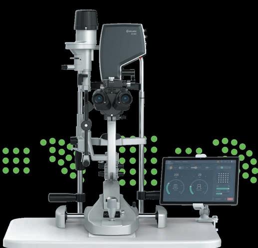


ESCRS YO membership has given me a strong international community, real integration across cultures and languages, and meaningful connections that extend far beyond meetings. It has opened opportunities to present on stage, build confidence, and grow as a leader within ESCRS and the wider ophthalmology community.
—LAURA MAUBON

As a young ophthalmologist, ESCRS membership opened doors to high-quality education, mentorship, and opportunities that have shaped my early career. Being an ESCRS YO member has connected me with an inspiring global community and given me access to resources that continually improve my clinical practice.
—VALENTIN HOOIJER
A host of promising options are on the horizon for the procedure.
CHERYL GUTTMAN KRADER REPORTS
Minimally invasive glaucoma surgery (MIGS) is a relatively new therapeutic category that has dramatically expanded since the term was first introduced in 2009, providing surgeons with an amazing set of tools. A look at the investigational pipeline reveals there is much more to look forward to, including a new category of surgical options that can be thought of as MIGS+ or ‘SMMIGS (slightly more than MIGS).’
The MIGS pipeline comprises surgical sustained release systems and procedures targeting the conventional outflow pathway, explained Rachel G Simpson MD.
Surgical sustained release devices under investigation include two devices from Glaukos: iDose TRIO and iDose TREX. iDose TRIO is a next-generation version of iDose TR that is designed to eventually facilitate in-office administration. It is being evaluated in a phase 3b trial and anticipated for possible approval in the US in 2027. iDose TREX has the same external footprint as iDose TR but carries twice the drug load with the promise of doubling the efficacy duration. It was investigated in a completed phase 2a study.
Other sustained release drug delivery systems are being developed by SpyGlass Pharma, including a bimatoprost-IOL system, which is in phase 3 trials, and a drug ring system delivering bimatoprost for at least three years. The ring is designed to be injected, removable, and replaceable, offering lifetime treatment potential. It is in early development.
“The ring is designed to answer the question, ‘What happens when the footplates on the IOL dissolve after three years?’,” Dr Simpson said.
The conventional outflow pipeline includes a device from eyeFlow that combines canal stenting with sustained drug delivery. In early clinical trials, the dual-action stent/drug delivery is designed to minimise tissue trauma during injection
and enhance aqueous access to the collector channels while releasing a prostaglandin analogue over a period of 12 months.
There are also three laser-based procedures in development. The laser options all aim to create full thickness channels in the trabecular meshwork, but they do so using different laser types and approaches. This category includes an excimer laser (ELIOS, Bausch + Lomb), which is approved outside the US and used in the operating room with or without cataract surgery; a femtosecond laser from ViaLase used in a standalone procedure termed LIGHT (Laser Image Guided High precision Trabeculotomy) that can be performed in an ambulatory surgery centre; and a pico-green laser (PicoLumen, EyeX Solutions) that also represents a standalone procedure that could be performed in the clinic.
“A nice thing about the pico laser is that it accesses all four quadrants of the eye, giving 360-degree access to the collector channels,” Dr Simpson said.
The ‘SMMIGS’ category has a single entry—a conjunctival sparing ab externo supraciliary implant (InterCil, Ciliatech) that uses the uveoscleral outflow pathway via controlled separation of the ciliary body and sclera. Dr Simpson described results of initial clinical trials as “very, very promising”, reporting they showed 41.6% reduction in IOP, 71% reduction in medication use, and 70% of patients becoming medication free.
Dr Simpson spoke at the 2026 ASCRS meeting in Washington, DC.
Rachel G Simpson MD is Assistant Professor of Ophthalmology and Visual Sciences, Moran Eye Center, University of Utah School of Medicine, Salt Lake City, US. rachel.simpson@hsc.utah.edu


Risk factor identification aims to guide timely surgical intervention.
CHERYL GUTTMAN KRADER REPORTS
Big data and artificial intelligence (AI) hold strong potential for improving glaucoma patient management by enabling individualised assessment and prediction of disease progression. However, validating these AI-based prognostic approaches will be essential before they are ready for clinical implementation, according to Cecilia S Lee MD.
“A major challenge that we face in glaucoma management today is that progression is highly variable,” Dr Lee explained. “Therefore, we can miss the rapid progressors, and surgery is done only because there is evidence of progression versus prevention.”
Interested in addressing this challenge, Dr Lee and the research team worked on training deep learning networks to forecast future 24-2 Humphrey Visual Fields (HVFs).1 They extracted about 1.7 million perimetry points from more than 32,000 HVFs collected over 20 years with the aim of predicting a future HVF from a single baseline test. This research used time as the undeniable ground truth, meaning the training relied on a single visual field as the input and a future visual field at a specified time as the output.
“We were surprised by how well the models performed,” Dr Lee said. “What was very interesting was the study showed deep learning can learn structural spatial patterns that are carried when glaucoma progresses.”
As a next step, Dr Lee and colleagues developed a policy-driven multimodal model that reflects clinical practice by fusing information from disc photos and OCT to predict the visual field.2
They found that when the two modalities were fused and the strategy was able to decide which modality was more
appropriate for its contribution to visual field prediction, the model performed better than when it relied on a single input.
The team is also implementing big data and AI to discover new modifiable factors for glaucoma development and gain new insights into which patients will experience glaucoma progression and why individuals progress the way they do— an exciting area in which various systemic biomarkers and environmental factors have already been reported.
Dr Lee said these initiatives require access to large, longitudinal multimodal data sets to train predictive models. In this realm, Dr Lee and colleagues are working on AI-READI (Artificial Intelligence Ready and Exploratory Atlas for Diabetes Insight), a US National Institutes of Health Bridge2AI programme that is recruiting about 4,000 participants and gathering a broad spectrum of data to optimise for AI/machine learning analysis. Included in the data set are OCT and OCT angiography volumes, macular and optic disc imaging, glucose measurements, and other health metrics as well as sleep and stress measurements and environmental data.
Dr Lee presented at the 2026 ASCRS meeting in Washington, DC .
For citation notes, see page 40.
Cecilia S Lee MD, MS is the Jane Hardesty Poole Endowed Professor in the Department of Ophthalmology and Visual Sciences, Washington University, St Louis, Missouri, US. She is also Program Director of the AI-READI project. cslee@wustl.edu
Growing array of options aim for robust IOP lowering while minimising downsides of filtering procedures.
CHERYL GUTTMAN KRADER REPORTS
Minimally invasive bleb surgery (MIBS) represents a family of devices and surgical techniques aiming to fill the gap between traditional filtering procedures and minimally invasive glaucoma surgery (MIGS) for patients at high risk of vision loss from glaucoma.
“We know that there is a trade-off between safety and effi cacy when choosing surgical intervention for these patients,” said Ian Pitha MD, PhD.
“MIBS addresses some of those trade-offs by aiming to achieve IOP reduction approaching that of traditional filtering surgery, but with a streamlined surgical approach, better safety profile, and faster recovery. Having said that, MIBS is not risk free, and its long-term success still relies on the sub conjunctival healing process. Therefore, the use of antifibrotics remains an essential part of all MIBS.”
Speaking at the Glaucoma Subspecialty Day during the 2026 ASCRS annual meeting, Dr Pitha reviewed currently available MIBS options and investigational approaches.
MIBS devices that have gained regulatory approval include the gel stents XEN 45 and XEN 63 (AbbVie) that create a con trolled outflow pathway between the anterior chamber and subconjunctival space. The newer XEN 65, which has the CE mark but is not approved by the US FDA, is made of the same material as the XEN 45 and inserted with the same injector system but has a wider internal lumen. Because resistance to outflow is decreased, the XEN 63 has potential to achieve a lower IOP target and for less occlusion and fouling versus the XEN 45, but also carries a higher likelihood for hypotony.
The PreserFlo MicroShunt (Glaukos) received the CE mark in 2012 but is just being investigated in a pivotal trial for approval in the US.
We know that there is a trade-off between safety and efficacy when choosing surgical intervention for these patients.
“Since this device has been around for some time outside the US, there are a lot of [insights from] using it, including that increasing mitomycin-C dosage generally leads to in creased success,” Dr Pitha said.
AquaLumen (PLU Ophthalmic) is an implant-free MIBS that creates a sclerotomy through a transconjunctival ab externo approach using a posteriorly directed cutting needle. It does not have the CE mark but is available in the US and has been evaluated in primary open-angle glaucoma, where it


Apply for the
How Has the ‘Human Touch’ Inspired You?
Ophthalmology, like all medical disciplines, is about improving the lives of people. That ‘human touch’ is fundamental to our work and transcends the technological and scientific advances that have transformed medical care.
The 2026 John Henahan Writing Prize seeks to capture the human touch and the impact it has on ophthalmic surgeons.
Young ophthalmologists are invited to compete for the Henahan Prize by submitting an essay (800 words maximum). The author of the winning essay will receive a €500 bursary and a specially commissioned trophy. The winning essay will also be published in EuroTimes.
Henahan Prize contestants must answer this prompt: The digital OR, AI algorithms, and robotics notwithstanding, cataract and refractive surgery involve a human relationship between you and your patient. Please describe how an experience or experiences in your early training reminded you of the importance of the human touch, and how this has inspired you in your clinical practice.
The competition is open to ESCRS members (including the free membership available to trainees) age 40 or younger on 1 January 2026.
Please compose your essay on your own without the use of any AI tools. We suggest you ask a mentor or teacher to review your essay prior to sending it.
The closing date for entries is 30 June 2026. Send your essay with cover page to seanh@eurotimes.org.
Suprachoroidal injection offers attractive alternative under certain circumstances.
CHERYL GUTTMAN KRADER REPORTS
Suprachoroidal injection (SCI) is a welcome tool in the vitreoretinal surgeon’s arsenal for achieving retinal drug delivery, but the pros and cons of the approach must be considered in relation to the condition being treated, the target location, and the therapeutic payload.
“Suprachoroidal injection can be helpful in certain conditions, but depending on the need, it might be superior or inferior to subretinal delivery or an intravitreal injection,” said Dominik Fischer MD, PhD.
Interest in targeting the suprachoroidal space for retinal drug delivery derives from several potential advantages, including the ability to target more of the retina (up to 100%) than local bleb creation and improved bioavailability compared with intravitreal injection. SCI also offers ease of application because it avoids vitrectomy and can be performed as an office-based procedure, and it provides a safety advantage relating to compartmentalisation.
“The proximity of the suprachoroidal space towards the outer retina minimises the effects on anterior segment tissues,” Professor Fischer explained.
Devices capable of delivering the drug to the retina via SCI include a standard 30-gauge needle, microcatheter-based devices, and purpose-built microneedles. Of these, the microneedles overcome limitations of the first two options.
“Compared with use of a 30-gauge needle, design features of the purpose-built microneedles minimise the risk of inadvertent intravitreal injection and reflux,” Prof Fischer said.
“Unlike the microcatheters, delivery with the purpose-built microneedles can be an office-based procedure.”
However, microneedle delivery is not without potential drawbacks, as this technique might limit targeting to the peripheral retina. In addition, there is some conflicting data from preclinical and clinical studies regarding the durability of AAV transduction when delivering gene therapy via suprachoroidal injection.
“Forthcoming data from the AAVIATE clinical trial of RGX314 for wet age-related macular degeneration will provide better insight on the latter issue because the protocol includes measurement of transgene levels in the aqueous humour,” Prof Fischer said.
A post hoc review of data sets from eight clinical trials provides insight into the risks and complications associated with SCI of a proprietary triamcinolone acetonide formulation delivered through a proprietary microneedle when used as monotherapy or combined with intravitreal injection.1
Encompassing three disease states, the trials included 621 patients who received the steroid SCI as monotherapy or in conjunction with an intravitreal injection of aflibercept and 449 control patients treated with aflibercept and sham SCI. The only serious adverse event related to the SCI was retinal detachment in a single patient being treated for non-infectious uveitis. There were no cases of lens injury, suprachoroidal haemorrhage, or endophthalmitis.
“Even though we operate close to the heavily perfused choroid, suprachoroidal haemorrhage does not seem to be much of a concern,” he said.
He added that recently published expert panel guidance on SCI states that patients and their caregivers should be informed about common or expected side effects, such as subconjunctival haemorrhage.2 In addition, they should be counselled about signs and symptoms of more serious potential complications, such as infection, retinal detachment, or suprachoroidal haemorrhage, and to call if they occur.
The guidance also states that intraocular pressure elevation, endophthalmitis, or scleritis should be managed in accordance with standard protocols for postinjection patients.
Prof Fischer spoke at EURETINA 2025 in Paris, France.
For citation notes, please see page 40.
Dominik Fischer MD, PhD is Professor of Ophthalmology, Oxford Eye Hospital, University of Oxford, UK. dominik.fischer@eye.ox.ac.uk
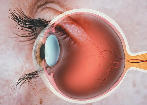
Treatment for the rare condition comes with its own risks, including potentially life-threatening complications.
ANDREW SWEENEY REPORTS
Congenital cataract is thankfully rare, but early surgical intervention is usually required and is frequently followed by postoperative inflammation. Although this inflammation is typically managed with topical steroids such as dexamethasone, steroid therapy, too, is associated with potentially serious side effects.
Diane Chabané Schmidt MD, PhD described the search for a means to avoid these side effects. She and her research group wanted to examine whether a reduction in the steroid dosage could reduce the risk of adrenal suppression.
Their study was a follow-up to one conducted by Dr Regitze Bangsgaard, who was inspired by personal experience of treating a young patient with adrenal suppression.
“The patient had congenital cataract surgery as an infant, and afterwards she received dexamethasone eye drops. Later, she developed anorexia, irritability, and lethargy, and she was then hospitalised for malaise after a routine vaccination,” Dr Chabané Schmidt said.
“She was seen at our clinic, where we saw she had big cheeks and a protruding tongue. The patient had Cushing syndrome because of the eyedrop treatment. She was followed for three years until her adrenal glands were functioning again.”
Dr Chabané Schmidt said cases like these happen because topical glucocorticoids trigger a negative feedback mechanism on the hypothalamic-pituitary-adrenal (HPA) axis. The mechanism causes decreased endogenous cortisol production, referred to as HPA-axis suppression, which usually presents with vague and non-specific symptoms such as fatigue, nausea, and poor growth. However, it can lead to the life-threatening Addisonian crisis if unrecognised.
The Copenhagen University Hospital – Rigshospitalet initiated a policy in 2017 to significantly reduce steroid exposure in congenital cataract patients. Dr Chabané Schmidt said eye drops were reduced from 203 to 72 dexamethasone eye drops in each patient.
A prospective cohort study was then conducted on infants younger than two years of age who underwent cataract or congenital glaucoma surgery between 2017 and 2021. All patients underwent adrenocorticotropic hormone (ACTH) testing to evaluate adrenal function.
“We found that 12 children had adrenal suppression, and 11 out of 12 children with adrenal suppression were under six months of age. Suppression lasted a median of 7.5 months,
Three infants had affected growth, one had Cushing syndrome, and none developed Addisonian crisis. In a five-kilogram infant, each additional daily drop increased the odds of adrenal suppression by 79%.
with some cases extending to three years,” Dr Chabané Schmidt said.
“Three infants had affected growth, one had Cushing syndrome, and none developed Addisonian crisis. In a five-kilogram infant, each additional daily drop increased the odds of adrenal suppression by 79%.
“During treatment, almost half of the children are suppressed. However, when we tested them after they stopped taking their eye drops, we found that almost none of the children were suppressed.”
Dr Chabané Schmidt said this highlights how the patients suppressed during treatment were able to quickly recover their own endogenous production of cortisol afterwards. She believes this should lead to an update of clinical protocols.
These include testing only infants younger than six months, conducting ACTH testing within one week after treatment cessation, and providing stress-dose steroids during surgery or illness. Vigilance and tailored monitoring in paediatric ophthalmology are also strongly recommended.
“We know that those currently treated might have adrenal suppression, and probably a lot of them have,” Dr Chabané Schmidt said. “Thus, we need to treat them as if they’re suppressed.”
Dr Chabané Schmidt presented at the 2025 ESCRS Annual Congress in Copenhagen.
Diana Chabané Schmidt MD, PhD is a specialist registrar in ophthalmology at the Department of Ophthalmology, Copenhagen University Hospital – Rigshospitalet, Denmark. diana.chabane.schmidt.01@regionh.dk
Her team includes ophthalmologists Professor Line Kessel, Associate Professor Daniella Bach-Holm, Dorte Ancher Larsen, and Regitze Bangsgaard, as well as endocrinologist Professor Katharina Main.
ESCRS President Burkhard Dick discusses new partnerships and new ideas for the ESCRS.
EuroTimes E ditor-in-Chief Sean Henahan caught up with ESCRS President Burkhard Dick at the ESCRS 2026 Winter Meeting in Helsinki. Professor Dick shared his vision and goals for the ESCRS in the next two years.
Q: Tell us a little about yourself. What is your history with the ESCRS?
A: I have known Emanuel Rosen (the first president of the ESCRS) since the late 1990s, and he was a highly influential educator and mentor to me. His active dedication to the Society inspired me, as he played a crucial role in transforming ESCRS from a small European initiative into a respected scientific society. Initially, I regularly attended the ESCRS conferences. Eventually, I was honoured to be asked by Professor Rudy Nuijts to chair the ESCRS Research Committee. At that time, I was an experienced cataract and refractive surgeon and Chair of the University Eye Hospital in Bochum [Germany], where we had a strong track record of publishing in basic and clinical research. With the support of some very clever people, I was able to redesign the committee and update the documentation and standard operating procedures. Next, I became Secretary of the Society, which enabled me to become involved with everything going on in the organisation.
In recent years, ophthalmology has become increasingly intertwined with the outside world. Each ESCRS president enters a new world, with new challenges and goals. What is your outlook as you begin your presidency? In 2026, I am looking forward with great confidence in the ESCRS. My predecessors have definitely created a solid foundation, and we have a lot of momentum. We are now expanding
our activities and reaching even further, strengthening our position as the leading global society for cataract and refractive surgery. My goal is to increase international cooperation and expand the ESCRS’s influence. To this end, we have recently strengthened our international efforts by establishing new partnerships, including with the Egyptian Ophthalmology Society, the Emirates Society, and with societies in Saudi Arabia and Mexico. Those societies have reached out to us, and we are glad to work with them.
The benefits of these new partnerships for our members are undeniable. These include greater scientific exchange, more research opportunities, and access to the latest technology. Keeping the recent changes in MDR
rules in mind, these affiliations will help us stay up to date with the global innovation pipeline at the earliest stages. It is also important to remember that these partnerships are truly bilateral and will strengthen the global acceptance of guidelines developed by the ESCRS. These include the cataract and refractive surgery guidelines, the newly developed IOL classification system, and the associated nomenclature. It is also interesting to study problem-solving across many different healthcare systems.
Education will remain central to all we do. ESCRS membership offers many benefits, including training resources and access to our conferences, the Journal of Cataract and Refractive Surgery and EuroTimes. We offer an expanding array of research support, including Digital
Research Awards, Clinical Research Awards, Pioneer Research Awards, the Systematic Review Award, and, most recently, the Sustainability Award.
In 2026, several key initiatives will be launched, including the new Masterclass Course in Refractive Surgery and educational programmes covering posterior segment topics and ocular comorbidities, in line with current clinical practice. Our relationship with others, such as EURETINA, will also strengthen. Together, we shall continue to highlight the integrated nature of ophthalmic practice in the modern era.
Artificial intelligence will also become a key focus for our Society in 2026, with specific courses, summits, and symposia designed to help members assess and utilise these new technologies responsibly and effectively. Professional development will extend well beyond the operating theatre with the launch of the ESCRS Insight Hub, covering medical writing,
presentation skills, and critical reading and review skills, among others. We will continue strengthening our relationships with industry partners in the fields of pharmaceuticals and interventional microsurgery.
Could you give us a preview of what’s in store for the 2026 ESCRS Annual Congress being held in London?
Our meetings shall continue to provide platforms for innovation and engagement. Indeed, the successful Winter Meeting in Helsinki on presbyopia set the stage for the Annual Congress to unveil new and dynamic initiatives, such as the 3D Surgery sessions, the OR Microscope Showdown, the Excimer Laser Museum, the Parliament Debates, the AI Summit, and the enhanced Young Ophthalmologists sessions. All these shall aim to blend the best of scientific content with innovation, youth, and interactivity.
Scan the QR code to learn more about the global reach of ESCRS.

The ESCRS Young Ophthalmologist (YO) Observership programme supports European trainee ophthalmologists who wish to observe clinical practice in a hospital or university setting. Ten €2,000 observership grants are available annually.
Application Details
Applicants must be ESCRS members and residents in the second half of their training programme. Applicants must make their own application to an approved clinic or hospital centre listed on the ESCRS website.
The observership duration is two to three weeks. The host clinic or centre must agree to host you for your proposed time and dates and provide evidence of this agreement in your application.
Participating Clinics/Hospital Centres
More than 20 clinics have observership programmes available for visiting ESCRS YOs. ESCRS will not provide contact details for the named hosts due to privacy regulations. You must contact them by publicly available methods.
Application Deadline
The application deadline is 17 July 2026. For application details and more information, scan the QR code.
EnVista Envy, Bausch + Lomb’s line of diffractive simultaneous vision IOLs, is now available in Europe. In a US multicentre, randomised, controlled clinical trial, 86% of patients reported little to no problem with dysphotopsia (e.g., glare, halo, starbursts). In a Canadian clinical study, 95.4% of patients reported little to no difficulty viewing close objects, and 93% were completely to moderately satisfied with their vision post-surgery, the company reported in a press release. The IOL uses the company’s EyeGillity delivery system, allowing injection through a 2-mm incision. A toric version is available in cylinder increments of 0.50 D or less. bauschsurgical.com/cataract/envista-envy/
The US Food and Drug Administration has approved Johnson & Johnson’s Tecnis PureSee™ simultaneous vision IOL. The company notes it is the only simultaneous vision IOL that maintains contrast sensitivity similar to what would be seen with a conventional aspheric monofocal IOL. Moreover, in clinical trials, 97% of lens recipients reported no ‘very bothersome’ visual effects. The proprietary Tecnis Simplicity™ delivery system is used to fold and assist in inserting the new IOL. A toric version is available. The IOL is expected to be available later this year in the US. jnjvisionpro.com/en-us/products/tecnis-puresee/
Citing a ‘lack of assurance of sterility,’ the US FDA issued a recall involving more than three million over-the-counter eye drops. The drops were sold under various brand names at leading retail outlets in the US including Walgreens, Kroger, and CVS. A full list, including lot numbers and expiration dates, is available on the FDA website. accessdata.fda.gov/scripts/ires/?Event=98533
With recent Medical Device Regulation (MDR) approval, the Cassini Guidance System is now available throughout the European Union. The system is designed to integrate with existing surgical set-ups, reducing manual marking, improving accuracy, and saving time, the company said. Specifically, the system allows IOL alignment within three degrees of target, with real-time tracking with overlay. It integrates with all major operating microscopes. The company recently announced it was advancing its open platform strategy through a collaboration with Leica Microsystems. cassini-technologies.com/guidance
Heidelberg Engineering announced that Hrvoje Bogunović was the latest recipient of its Xtreme Research Award. The award recognises his research in AI and retinal imaging with optical coherence tomography (OCT). His work uses AI to recognise biomarkers associated with different retinal diseases.
“By analysing large collections of high-quality OCT scans over time, AI can detect prognostic patterns that may be too subtle or too complex for human experts to recognise. Eventually, that can help us discover new imaging biomarkers, better understand disease progression, and ultimately improve our ability to predict what may happen next for a patient,” said Dr Bogunović, who is Director of the Christian Doppler Laboratory for Artificial Intelligence in Retina at the Medical University of Vienna. heidelbergengineering.com
The Global Functional Vision Working Group (FVWG), formed by international and national ophthalmic societies including ESCRS, ASCRS, APACRS, and LATAMSCRS, has created an intraocular lens (IOL) classification system focused on patient-centred outcomes such as depth of field and real-life visual function.
FVWG aims to set a global standard for evidence-based IOL research and offer a clinical framework that complements regulatory classifications and supports informed decision making. While the ISO classification for IOLs emphasises pre-marketing technical specifications and performance standards, the FVWG Functional Classification provides a post-market, evidence-based framework. Its purpose is to categorise IOL performance into mutually exclusive, clinically relevant, and progressively ordered groups based on patient-centred outcomes.
The FFVWG classification for IOLs is based on the range of field (ROF) and the visual acuity enhancement from intermediate to near distances. The group recommends using the term ‘depth of field’ (DOFi) to avoid confusion with peripheral visual field terminology and ensure clearer and more consistent communication among clinicians and researchers.
ISO classifies simultaneous vision lenses (SVIOL) into multifocal (MIOL), extended depth of focus (EDOF), and full visual range (FVR). The FVWG classification divides SVIOLs into six categories—three partial DOFi IOL types (narrowed, enhanced, and extended) and three full DOFi IOL types (steep, smooth, and continuous). The partial DOFi IOLs provide a progressive increase in DOFi, accompanied by a gradual compromise in far distance visual quality. The continuous categories represent a progressive enhancement in intermediate visual performance, which may be associated with a corresponding progressive reduction in near visual quality.
The working group also highlighted the importance of incorporating additional visual quality metrics, such as contrast sensitivity and patient-reported outcomes (PROMs), dysphotopsia, satisfaction, and spectacle dependence, as well as the influence of biometric parameters such as pupillometry and corneal aberrations.
The FVWG agreed that the Assessment of Intralocular Implant Symptoms (AIOLIS) questionnaire, a tool developed by the AAO, best captures a wide range of single-item questions related to the frequency and severity of visual symptoms, as well as grading visual quality, satisfaction, and spectacle independence.
Looking ahead, the FVWG agreed on the need for guidelines to assess evidence supporting IOL classifications, including a grading system for how confidently an IOL can deliver functional performance based on its classification. The group is developing these guidelines to ensure rigorous but straightforward evaluation, facilitating wide use by industry and ophthalmologists.
J Fernández, et al. “Global consensus on the evidence-based functional classification of simultaneous vision IOLs,” 52(3): 219–222.

Ophthalmologists and researchers (MD and/or PhD) as well as experienced ophthalmic nurses are encouraged to apply for the ESCRS Sustainability Research (SURE) Award, which will fund projects that investigate meaningful, practical ways to promote environmental responsibility in ophthalmic care.
Two awards will be granted; each award will provide up to €10,000 per project. Applicants must be active ESCRS members at the time of application and must hold a current full- or part-time clinical or research position at a clinical or academic institution. Early-career researchers and young ophthalmologists are especially encouraged to apply.
The application period closes on 19 June. Award recipients must submit an article to a peer-reviewed journal within six months after the research period concludes. The article should be made open access if accepted and submitted to the Journal of Cataract & Refractive Surgery in the first instance.
Get funding for projects to promote environmental responsibility in ophthalmic care.
Debating the Future of Glaucoma Surgery
Page 10
1. Video Journal of Cataract, Refractive, and Glaucoma Surgery. https://vjcrgs.com/
2. ESCRS Supplement. https://www.escrs.org/channels/ eurotimes-articles/supplement-escrs-clinical-trends-series-glaucoma
3. Wright DM, et al. Ophthalmology, 2020; 127(10): 1313–1321.
4. EGS Publications. “Terminology and Guidelines for Glaucoma”; “A guide on Surgical Innovation for Glaucoma.” Both are available in multiple languages at www.eugs.org
5. Gazzard G, et al. Ophthalmology, 2025 Oct; 132(10): 1091–1104.
6. Funke C. EuroTimes, 2025; 30(Q3): 28. https://issuu.com/ eurotimes/docs/eurotimes_online_q3
7. Nagy Z, et al. Ophthalmology Science, 2023 Apr; 3(4): 100313.
8. Bausch + Lomb. “Bausch + Lomb Announces Positive 24-Month US Data on the ELOIS System for Treatment of Glaucoma,” 2026 Mar 23. https://www.bausch.com/newsroom/news/?id=319
Laser IOL Power Adjustment
Page 14
1. Lundstrom M, et al. J Cataract Refract Surg, 2018 Apr; 44(4): 447–452.
2. Hengerer FH, Dick HB, Conrad-Hengerer I. Ophthalmology, 2011 Dec; 118(12): 2382–2388.
3. Summary of Safety and Effectiveness Data. US FDA 2017. PMA P160055.
4. RxSight PMCS-002 Clinical Outcomes of Patients Bilaterally Implanted with LAL.
FLACS for Complex Cataracts
Page 19
1. Chee SP, Yang Y, Wong MHY. Am J Ophthalmol, 2021 Sept; 229: 1–7.
2. Chee SP, Chan NS, Yang Y, Ti SE. Br J Ophthalmol, 2019 Apr; 103(4): 544–550.
3. Chee SP, Wang MH, Jap A. Am J Ophthalmol, 2017 Jan; 173: 7–15.
DMEK Outcomes Using Tissue from Donors with Diabetes
Page 24
1. Price FW Jr, et al. JAMA Ophthalmol, 2025 October 17; 143(12): 1043–1051. Online ahead of print.
2. Lass JH, et al. JAMA Ophthalmol, 2025 Oct 17; 143(12): 1053–1060. Online ahead of print.
A Closer Look at EndoArt
Page 25
1. Nanavaty M, et al. JCRS, online 25 Feb, 2026. doi:10.1097/j. jcrs.0000000000001921
Femtosecond Laser-Assisted DALK
Page 26
1. Shehadeh-Mashor R, Chan CC, Bahar I, et al. Br J Ophthalmol, 2014; 98: 35–39.
2. Salouti R, Zamani M, Ghoreyshi M, et al. Br J Ophthalmol, 2019; 103: 1716–1723.
3. Gonzolez A, Price MO, Feng MT, et al. Cornea, 2017; 36: 1076–1082.
4. Gadhvi KA, Romano V, Fernandez-Vega Cueto L, Aiello F, Day AC, Allan BD. Am J Ophthalmol, 2019; 201: 54–62.
5. Li H, Chen M, Dong YL, et al. Int J Ophthalmol, 2020; 13(4): 567–573.
6. Du K, Liu E, Li N, Yuan B, Peng R, Hong J. Am J Ophthalmol, 2023 Dec; 256: 126–137.
AI-Based Strategies for Predicting Glaucoma Progression
Page 31
1. Wen JC, et al. PLoS One, 2019 Apr 5; 14(4): e0214875.
2. Kihara Y, et al. Ophthalmology, 2022; 129(7): 781–791.
Reaching the Retina for Drug Delivery
Page 34
1. Sharma S, et al. Inv Ophthalmol Vis Sci, 2021; 62: 1209.
2. Wykoff CC, et al. Retina, 2024; 44(6): 939–949.
May 4–8
ARVO
Salt Lake City, Utah, US
May 9–11
French Society of Ophthalmology (SFO) Paris, France
May 22–24
EuCornea Porto, Portugal
June 26–29
World Ophthalmology Congress Prague, Czech Republic
September 11–15
ESCRS Annual Congress London, UK
October 1–4
EURETINA
Vienna, Austria
October 9–12
American Academy of Ophthalmology
New Orleans, Louisiana, US
November 12–14
UKISCRS Annual Conference London, UK

May 9

May 4
May 22


June 26

NEXT GENERATION INJECTOR SYSTEM UNLIMITED OPPORTUNITIES MAXIMUM PRECISION
EXCELLENT SAFETY PROFILE
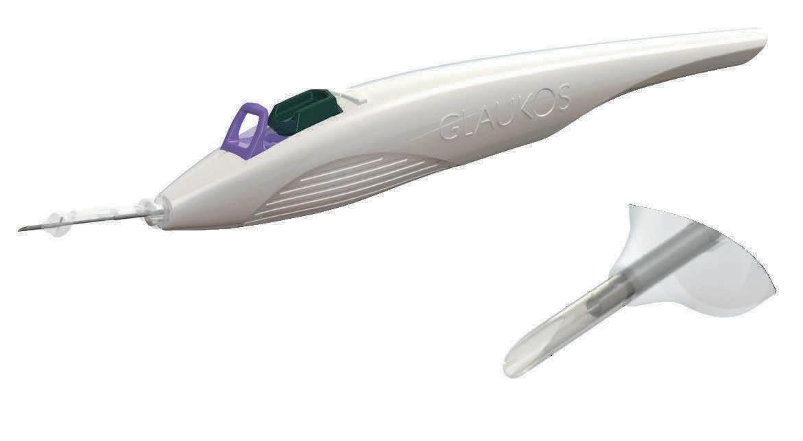
OF PATIENTS ≥20% REDUCTION IN IOP OF PATIENTS ≥30% REDUCTION IN IOP
1-Sarkisian Jr, Steven R., et al. “Effectiveness and safety of iStent infinite trabecular micro-bypass for uncontrolled glaucoma.” Journal of glaucoma 32.1 (2023): 9-18. iStent infinite® IMPORTANT SAFETY INFORMATION
INDICATION FOR USE: The iStent infinite System is intended to reduce intraocular pressure safely and effectively in adult patients diagnosed with primary open-angle glaucoma, pseudo-exfoliative glaucoma or pigmentary glaucoma. The device is safe and effective when implanted in combination with or without cataract surgery in those subjects who require intraocular pressure reduction and/or would benefit from glaucoma medication reduction. The device may also be implanted in patients who continue to have elevated intraocular pressure despite prior treatment with glaucoma medications and/or conventional glaucoma surgery. CONTRAINDICATIONS: The iStent infinite System is contraindicated under the following circumstances or conditions: •In eyes with primary angle closure glaucoma, or secondary angle-closure glaucoma, including neovascular glaucoma, because the device would not be expected to work in such situations.• In patients with retrobulbar tumor, thyroid eye disease, Sturge-Weber Syndrome or any other type of condition that may cause elevated episcleral venous pressure. WARNINGS/PRECAUTIONS: • For prescription use only. • Intended users are trained ophthalmologists only. • This device has not been studied in patients with uveitic glaucoma. • Do not use the device if the Tyvek® lid has been opened or the packaging appears damaged. In such cases, the sterility of the device may be compromised. • Due to the sharpness of certain injector components (i.e., the insertion sleeve and trocar), care should be exercised to grasp the injector body. Dispose of device in a sharps container. • iStent infinite is MR-Conditional • Physician training is required prior to use of the iStent infinite System. • Do not re-use the stent(s) or injector, as this may result in infection and/or intraocular inflammation, as well as occurrence of potential postoperative adverse events • There are no known compatibility issues with theiStent infinite and other intraoperative devices (e.g., viscoelastics) or glaucoma medications. • Unused product & packaging may be disposed of in accordance with facility procedures. Implanted medical devices and contaminated products must be disposed of as medical waste. • The surgeon should monitor the patient postoperatively for proper maintenance of intraocular pressure. If intraocular pressure is not adequately maintained after surgery, the surgeon should consider an appropriate treatment regimen to reduce intraocular pressure.
• Patients should be informed that placement of the stents, without concomitant cataract surgery in phakic patients can enhance the formation or progression of cataract. ADVERSE EVENTS: The most common postoperative adverse events reported in the iStent infinite pivotal trial included IOP increase ≥ 10 mmHg vs. baseline IOP (8.2%), loss of BSCVA ≥ 2 lines (11.5%), ocular surface disease (11.5%), perioperative inflammation (6.6%) and visual field loss ≥ 2.5 dB (6.6%). CAUTION: Please see DFU for a complete list of contraindications, warnings, precautions, and adverse events.
Glaukos®, iStent®, iStent infinite® and TMB® are registered trademarks of Glaukos Corporation.
©2026. PM-EU-0351