Integrating research, reusable practices, and industry collaboration in ophthalmology.
ALSO IN THIS ISSUE
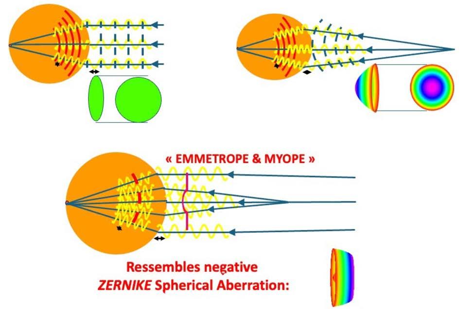
Rethinking Aberrations
Soosan Jacob and Damien Gatinel discuss a new way of applying aberrometry in clinical practice.
Developing Interventions to Reduce Physician Suicide
Addressing stigma and fear about the professional consequences of mental illness are key to helping colleagues, experts say.
Ukraine’s Ophthalmic Surgeons Hold Congress Amidst Conflict Ukrainian Congress proceeds as war continues, highlighting equipment shortages.
We’re willing to bet most eye care professionals don’t realise just how prevalent Demodex blepharitis is.
In fact, ~54% of eye care patients in Europe may have Demodex blepharitis (DB).1*
*Data from an eva lu ation of 804 patients from 6 countr ie s in Europe, including 15 c li nicians each with ~50 consecutive patients
WHAT ABOUT YOUR PATIENTS?
LEARN HOW DB CAN FLY UNDER THE RADAR AT
Reference: 1. Data on fi le, Tarsus Pha rma c eu ticals, Inc. Find us
Sponsored by
JEANETTE, real DB patient.
Set Yourself Apart
Demonstrate your knowledge and leadership by taking the FEBOS-CR exam.
The FEBOS-CR Subspecialty Exam has been developed by ESCRS and the European Board of Ophthalmology (EBO) to certify the expertise and advanced knowledge of experienced cataract and refractive surgeons. Successful candidates earn the right to use the post-nominal title FEBOS-CR to show they—
• hold a superior theoretical and practical knowledge;
• can deal with more challenging cases and a complex case mix; and
• appreciate the importance of evidence-based medicine and its purpose in developing scientific knowledge and clinical practice of the subspeciality.
Candidates must be independent surgeons with a varied case mix who regularly deal with complex situations and who are willing to have their expertise and theoretical knowledge tested by a rigorous theoretical examination and interviews with opinion leaders in European and worldwide ophthalmology.
Please note: the FEBOS-CR is open to cataract and refractive subspecialists who hold a medical diploma awarded in one of the European Union of Medical Specialists (UEMS) member countries, licensing them to practice medicine in that country. ESCRS membership is not required.
Applications close 30 March. Submit your application now!
12 Cover
Rethink. Reuse. Reimagine Eye Care. Integrating research, reusable practices, and industry collaboration in ophthalmology.
THEME
16 Instrument Lifespan Metrics Power Sustainability
Update: In Memoriam: Professor José Cunha-Vaz, Former ESCRS President; Applications Being Accepted for FEBOS-CR Subspecialty Exam; New Award to Honour Efforts to Expand Access to
Johnson Yan Ning Neo FHEA, FRCOphth, FEBO, CertLRS
17 AI Telephone Call to Reduce Carbon Footprint
Aisling Higham MSc, FRCOphth
18 Small Changes for a Necessary Shift
Pei-Fen Lin MD
20 Refractive Surgery Grey Zones
Béatrice Cochener-Lamard MD, PhD
21 EUREQUO Evolves
Volodymyr Melnyk MD; Gauti Jóhannesson MD, PhD; Thiemo Rudolph MD
Maartje Segers MD
23 The European Experience with KLEx
Thomas Kohnen MD, PhD, FEBO
24 Managing Femto Flap Complications
Namrata Sharma FRCOphth, FRCSEd
25 Defining the Costs of ISBCS
Steve A Arshinoff OC, MD, FRCSC
CORNEA
26 Expanding Options for Stromal Supplementation
Sayan Basu MBBS, MS; Aylin Kılıç MD
28 How to Avoid Refractive Surgery Complications
Sotiria Palioura MSc, PhD, CEBT, FEBO, FEBOS-CR
29 Handling KLEx Complications
Soosan Jacob MS, FRCS, DNB
31 Rethinking Aberrations: the Low-Degree/High-Degree (LD/HD) Scheme
Soosan Jacob MS, FRCS, DNB
34 DSAEK or DMEK?
Massimo Busin MD
35 Expanding Options for Rho-Associated Protein Kinase Inhibitors
Friedrich E Kruse MD
RETINA
36 First-Line Therapy for Non-Infectious Uveitis
Sapna Gangaputra MD, MPH; Arthi Venkat MD, MS
38 Cataract Surgery in Diabetic Patients
Toke Bek DMSci, MBA
39 Maculopathy in Pentosan Polysulfate Sodium Users
Brendan Tao MD
Arzu Seyhan Karatepe Haşhaş
Paolo Cecchini MD, PhD; Gerardo López Oseguera MD, MBA, IB; Alessandro Pozzato MSc, MBA
Publisher
Tom Ogilvie-Graham
Executive Editor
Stuart Hales
Editor-In-Chief
Sean Henahan
Senior Content Editor
Kelsey Ingram
Creative Director
Kelsy McCarthy
Graphic Designer
Emily Christenson
Circulation Manager
Lucy Matthews
Contributing Editors
Cheryl Guttman Krader
Howard Larkin
Roibeárd O’hÉineacháin
Contributors
Laura Gaspari
Soosan Jacob
Timothy Norris
Andrew Sweeney
Colour and Print CitiPost
Advertising Sales
Roo Khan
MCI UK
Tel: +44 203 530 0100 | roo.khan@wearemci.com
EuroTimes® is registered with the European Union Intellectual Property Office and the US Patent and Trademark Office.
Published by the European Society of Cataract and Refractive Surgeons, Suite 7–9 The Hop Exchange, 24 Southwark Street, London, SE1 1TY, UK. No part of this publication may be reproduced without the permission of the executive editor. Letters to the editor and other unsolicited contributions are assumed intended for this publication and are subject to editorial review and acceptance.
Michael Levitt
ESCRS EuroTimes is not responsible for statements made by any contributor. These contributions are presented for review and comment and not as a statement on the standard of care. Although all advertising material is expected to conform to ethical medical standards, acceptance does not imply endorsement by ESCRS EuroTimes. ISSN 1393-8983
Learn more about EuroTimes or connect with ESCRS at ESCRS.org
In Memoriam: Professor José Cunha-Vaz (1934–2026)
It is with profound sadness that we record the passing of José CunhaVaz, a trailblazing scientist, an outstanding educator, and a past president of the ESCRS, serving from 2000 to 2001.
Over two decades, I had the privilege of knowing him both as a scientist and as a friend. He was a man of outstanding clarity, humility, and gentleness of heart. José’s impact can also be seen in modern ophthalmology and its relevance today.
He was a giant in retinal research and completely transformed the field of diabetic retinopathy and blood-retinal barrier research. Some of his early work defined novel approaches to non-invasive assessment of retinal permeability, and he conducted several studies that established new paradigms in clinical research.
José founded AIBILI and EVICR. net and was a strong proponent of translational medicine even before it became fashionable to talk about it. This is what made him a giant in European ophthalmology.
His tenure as president of the ESCRS at the turn of the millenni-
EDITORIAL BOARD
Adi Abulafia (Israel)
Bruce Allan (UK)
Noel Alpins (Australia)
Juan Alvarez de Toledo (Spain)
Gerd Auffarth (Germany)
Başak Bostanci (Türkiye)
John Chang (Hong Kong SAR, China)
Béatrice Cochener-Lamard (France)
Burkhard Dick (Germany)
Mor Dickman (The Netherlands)
um was typical of his unflappable, thought-provoking style. He was the retina colleague among the innovators and inventors of refractive surgery and intraocular lenses, and he reminded us that true progress comes from appreciating the entire visual system. His presidency helped expand the scientific scope of ESCRS without losing sight of the patient behind every procedure.
But what I think I shall miss the most is the man behind the title. He listened intently to what was being said, spoke thoughtfully, and never needed to raise his voice to make a point. He was open-minded, unstinting with his time, and possessed a wonderful capacity to soothe even the most heated scientific debate. He was a man who embodied quiet speed.
Over the years, our lives have crossed paths in various ways: at ESCRS congresses, in scientific networks such as EVICR.net, on joint panels, and in Europe-wide projects. Although our scientific fields were different, we shared a common commitment to collaboration in advancing the field and mentoring the next generation of eye care profes-
sionals. He was a steady presence in all these environments.
José leaves a legacy that extends far beyond his publications and accolades. He leaves behind generations of students, colleagues, and friends who are better, both professionally and personally, for having known him.
We at the ESCRS mourn the passing of a great mind and a gracious soul. I, personally, mourn the passing of a friend whose gentle wisdom will remain with me forever.
H Burkhard Dick MD, PhD, FEBOS-CR is Chair of Ophthalmology, Ruhr University Bochum, and President of the ESCRS.
Joaquín Fernández (Spain)
Oliver Findl (Austria)
Nicole Fram (US)
Sri Ganesh (India)
Farhad Hafezi (Switzerland)
Nino Hirnschall (Austria)
Soosan Jacob (India)
Jack Kane (Australia)
Yao Ke (China)
Mika Kotimäki (Finland)
David Lockington (UK)
Artemis Matsou (Greece)
Cyres Mehta (India)
Jod Mehta (Singapore)
Sorcha Ní Dhubhghaill (Belgium)
Rudy Nuijts (The Netherlands)
Catarina Pedrosa (Portugal)
Konrad Pesudovs (Australia)
Nic Reus (The Netherlands)
Filomena Ribeiro (Portugal)
Andreia Rosa (Portugal)
Giacomo Savini (Italy)
Julie Schallhorn (US)
Sathish Srinivasan (UK)
Paola Vinciguerra (Italy)
Shin Yamane (Japan)
Ron Yeoh (Singapore)
Mihail Zemba (Romania)
Thomas Kohnen
José Güell
Paul Rosen
ESCRS Update
In Memoriam: José Cunha-Vaz, Former ESCRS President
ESCRS regrets to announce the passing of José Cunha-Vaz, a visionary scientist and influential educator who served as president of the Society in 2000–2001.
Professor Cunha-Vaz was chair and director of the Department of Ophthalmology at the University of Coimbra Hospital (Portugal) from 1986 to 2008. He previously spent two periods (1979–1981 and 1984–1986) at the University of Illinois (US), where he was appointed professor and director of the university’s Retina Service. Following his return to Portugal, he created two new institutes dedicated to vision research: the Institute for Biomedical Research on Light and Image and the Association for Innovation and Biomedical Research on Light and Image.
Prof Cunha-Vaz was elected to the Academia Ophthalmologica Internationalis (1994) and the European Academy of Ophthalmology (2004) and chaired the Portuguese Society of Ophthalmology for two terms (1985–1986 and 1991–1992). The author of more than 550 peer-reviewed papers and books,
he left a remarkable legacy of vision, scientific rigour, and commitment to the sustained development of ophthalmology in Portugal and throughout the world.
“He was a giant in retinal research and completely transformed the field of diabetic retinopathy and blood-retinal barrier research,” writes current ESCRS President Burkhard Dick in this issue’s editorial. “Some of his early work defined novel approaches to non-invasive assessment of retinal permeability, and he conducted several studies that established new paradigms in clinical research. He … was a strong proponent of translational medicine even before it became fashionable to talk about it. This is what made him a giant in European ophthalmology.”
Applications Being Accepted for FEBOS-CR Subspecialty Exam
ESCRS is inviting applications for the FEBOS-CR Subspecialty Exam, which recognises cataract and refractive surgeons who hold a superior theoretical and practical knowledge, can deal with more challenging cases and a complex case mix, and
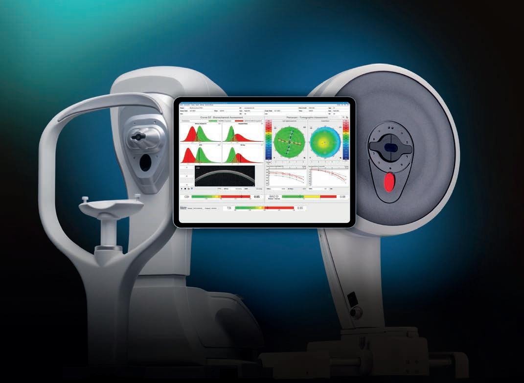
Double Down On Your Decision
Screening for ectasia with double safety
The Tomographic Biomechanical Index, or TBI, provides a unique combined expression of Corvis® ST und Pentacam® measurement data. It allows the risk of corneal ectasia to be assessed with greater reliability than ever before. The TBI assists you in selecting the optimal treatment based on sound reasoning. More safety for you and your patients!
appreciate the importance of evidence-based medicine and its purpose in developing scientific knowledge and driving excellence in clinical practice.
The exam, which is open to cataract and refractive subspecialists who hold a medical diploma awarded in one of the UEMS member countries, was developed by ESCRS and the European Board of Ophthalmology (EBO) to certify the expertise and advanced knowledge of experienced cataract and refractive surgeons. Candidates should be independent surgeons with a varied case mix who regularly deal with complex situations and are willing to have their expertise and theoretical knowledge tested by a rigorous examination and interviews with opinion leaders in European and worldwide ophthalmology.
Application materials, including a letter of recommendation, a video of a complex case, and a completed CV that lists surgical, teaching, and publication experience, must be submitted by 30 March. Candidates whose applications are accepted will sit for the exam on 10 September, just prior to the opening of the ESCRS Annual Congress in London. Successful candidates will be added to the FEBOS-CR Wall of Honour on the ESCRS website.
New Award to Honour Efforts to Expand Access to Eye Care
ESCRS has created a new award to honour individuals who have engaged in sustained, high-impact initiatives in cataract and refractive surgery that have transformed lives through service delivery, clinician training, infrastructure development, community education, and innovative outreach.
The recipient of the annual Humanitarian Service Award will receive a charitable grant of up to €100,000 to support the ongoing work of a charitable organisation of their choice. The recipient will also be invited to deliver the Humanitarian Lecture and will be formally recognised at the ESCRS Annual Congress.
Priority in bestowing the award will be given to projects that deliver measurable benefits to low-income or underserved populations, with particular consideration for initiatives in sub-Saharan Africa.
For more information about the exam, scan the QR code.
Nominations for the award opened in January and closed on 20 February. The award recipient, to be known as the ESCRS Humanitarian Laureate, will be announced in spring/ summer and honoured at the Annual Congress in London in September.
Support from ESCRS Boosts Cataract Care
Charity Committee initiative trains surgeons in MSICS technique.
In November 2025, I participated, as an active member of ESCRS, in a vital educational initiative focused on teaching manual small-incision cataract surgery (MSICS). The mission took me to the University of Cape Town’s Community Eye Health Institute (UCT-CEHI) in South Africa, followed by clinical work in Eswatini at the Good Shepherd Hospital and the Grace Vision Clinic in Siteki.
The primary objectives of my mission were clear: to instruct young African doctors in this highly effective surgical method and to immerse myself in the clinical environment of Siteki, Eswatini. The instructional goal was successfully achieved through an intensive programme led by four expert tutors who provided three full days of hands-on wet lab training in MSICS. This effort culminated in the successful certification of six young surgeons, significantly boosting regional capacity.
I discovered the call for interest for this training programme via the ESCRS website. The programme follows a well-established curriculum, with the initial two days providing trainees with a comprehensive theoretical and practical introduction to every step of the MSICS procedure. Educational support materials included a specialised booklet and detailed PowerPoint presentations. This was followed by extensive hands-on practice of all surgical steps using sophisticated cataract eye models.
The course concluded on the third day with advanced training modules focused specifically on complication management, using both eye models and sessions on the advanced EyeSi Simulator. A key component of the assessment was a self-evaluation performed by trainees using the Sim-OSSCAR (SICS Ophthalmic Simulated Surgical Competency Assessment Rubric), a tool validated against the rigorous International Council of Ophthalmology (ICO) standards for evaluating competency.
During the second week of my trip, I took a two-hour flight to the Kingdom of Eswatini, an independent nation bordered mostly by South Africa. During my time there, I performed several surgical operations at the Good Shepherd Catholic Hospital in Siteki. I also actively participated in numerous clinical consultations at the Grace Vision Clinic, a specialised eye care centre situated in the same region.
In South Africa and Eswatini, the treatment of cataracts— the single leading cause of blindness—is severely hindered by systemic issues, including a critical shortage of surgeons and essential hospital resources. This results in multi-year waiting lists for necessary surgeries and significant geographic disparities in care access. To address these systemic gaps and
alleviate the frustration of junior doctors lacking sufficient hands-on experience, the UCT Community Eye Health Institute (CEHI), with vital financial and material support from the ESCRS, organises these annual MSICS courses.
This highly-rated programme provides essential surgical training in a cost-effective, high-quality technique that is ideal for treating hard cataracts. It simultaneously fosters a valuable and reciprocal exchange of knowledge between local practitioners and international surgeons.
Special thanks are due to Dr Mark Wevill, a surgical trainer at the UCT-CEHI and a member of the ESCRS Charity Committee, who provides financial and material support to UCT-CEHI. Additionally, immense gratitude is extended to Dr Jonathan Pons for his role as a surgical trainer at the UCT-CEHI and for his outstanding hospitality in Siteki, Eswatini. I was profoundly impressed by the great clinical and teaching work Dr Pons has provided consistently for many years.
In conclusion, I highly recommend this MSICS course to all ESCRS members who are cataract surgeons, as it is beneficial for those both with and without previous experience in the technique.
Vasileios Petousis is a consultant ophthalmologist in vitreoretinal and cataract surgery at the Centre Ophtalmologique Luxembourg. vpetousis@outlook.com
VASILEIOS PETOUSIS REPORTS
Preventing Physician Suicide
Efforts exist on multiple fronts, but more work needs to be done.
CHERYL GUTTMAN KRADER REPORTS
Stigma around mental health is ubiquitous in the medical establishment, and preventing sufferers from seeking help only makes matters worse, according to Michael F Myers MD.
“In conducting research for [my] book, I found that at least 10% to 15% of doctors who took their lives had received absolutely no treatment for the illness that drove them to [the] act because they were terrified to talk about their situation,” said Dr Myers.
“My colleagues and I, whose work focuses on physician health, are doing what we can to eliminate that hesitancy. I feel confident that we will make the study and practice of medicine healthy, fulfilling, and creative.”
Removing the stigma
Fearing consequences that will affect their training and career opportunities, including the ability to obtain licensure and credentialling, medical school graduates and practicing physicians can feel pressured to hide struggles with mental health. Dr Myers noted that in the United States, three independent groups are working to implement changes in questions asked on medical license applications to bring them into compliance with the Americans with Disabilities Act.
“Now there are laws in multiple US states disallowing questions about psychiatric history in applications for a new or renewed medical license,” Dr Myers said.
Citing other efforts to eliminate the stigma of mental health among physicians, Dr Myers mentioned that the family of Lorna Breen MD—an emergency medicine physician who died from suicide while recovering from COVID-19 in the early days of the pandemic—established the Lorna Breen Heroes’ Foundation, which advocates for the professional well-being of health workers.
“The foundation’s mission is to help advocate for a world where seeking mental health services is universally viewed as a sign of strength—and one of its pillars is to make access to care safe and easy,” Dr Myers said.
Strategies for preventing suicide fall into primary, secondary, and tertiary domains. Primary interventions aim to mitigate stress in training and work environments. They include programmes intending to prepare potential medical school students for the realities of training and practice and initiatives promoting healthy lifestyles and targeting sources of stress, such as extended duty hours.
Secondary prevention strategies focus on identifying atrisk individuals and providing early intervention to normalise stress and minimise vulnerabilities as well as to encourage care seeking and enable access. As an example, Dr Myers mentioned the New York State Medical Society established a peer counselling programme.
“Unfortunately, it tends to be underutilised, and we are not sure why because there are a lot of first-person accounts about its effectiveness,” he said.
Other targets include ensuring that counselling with confidential firewalls is available at no cost or fully covered by insurance and accessible during a protected time for care. A support line for medical students and physicians in the US fits this model. Free, confidential, and anonymous, the toll-free number (1-888-409-0141) is served by more than 800 psychiatrists daily.
Tertiary prevention pertains to the provision of mental health treatment by professionals from various branches of healthcare, along with other supportive strategies. The latter includes support groups for physicians with psychiatric disorders and workplace policies allowing recovering physicians to return to work gradually or part-time.
Dr Myers pointed to a New York Times article in which columnist David Brooks reflected on the suicide of his lifelong friend, American ophthalmologist Dr Peter Marks. It was noted that Dr Marks found talking to his wife more helpful than talking to any of the experts.
“That gave me pause and was very hurtful given that psychotherapy, which is talk therapy, is the bedrock of the mental health field,” Dr Myers said. “I came away thinking how let down Dr Marks felt. We must redouble our efforts to make intimate (and lifesaving) connections with physicians feeling so desperate.”
Dr Myers spoke on this topic at AAO 2025 in Orlando, US.
Michael F Myers MD is Professor of Clinical Psychiatry, SUNY Downstate Heath Sciences University, Brooklyn, New York, US. His latest book is Physicians With Lived Experience: How Their Stories Offer Clinical Guidance and his email is michael.myers@downstate.edu.
Ukraine’s Ophthalmic Surgeons Hold Congress Amidst Conflict
An ophthalmic light still shines in Kyiv despite the challenges posed by Russia’s brutal invasion.
ANDREW SWEENEY REPORTS
Our train pulled in just after 5:00 a.m., right on time. We emerged from our compartment as the carriage screeched to a halt and climbed down the stairs into Kyiv’s morning mist. The curfew had just been lifted across the city as we arrived at our hotel, still in the dark of night.
Many of us have made long journeys to attend ophthalmology conferences, but few will experience travelling across Ukraine at night to reach the nation’s capital. From the Polish border town of Chełm, it took about 12 hours in all—half a day but a whole different reality as Russia continues its merciless invasion, the skies closed to civilian aircraft.
The 6th Congress of the Union of Ukrainian Ophthalmic Surgeons (SUO), named ‘Ophthalmic Light,’ had all the hallmarks of an ophthalmology congress: guest speakers (including members of the ESCRS), presentations, company exhibits, and the occasional free giveaway. But the bomb shelter may have caught many international guests off guard, as might the presence of multiple military doctors in their fatigues. For as much as ophthalmology in Ukraine tries to persist as it did before the full-scale invasion in 2022, the reality is that the work of doctors and their colleagues is dominated by war.
“I am very, very proud that our society, that our Ukrainian ophthalmologists and ophthalmic surgeons, can do their surgery at this time, improve their skills, and develop their knowledge,” said Volodymyr Melnyk MD, the organiser of the conference and one of Ukraine’s leading ophthalmologists.
There are never enough Ahmed valves
Like many of his colleagues, Dr Melnyk’s usual practice has been significantly disrupted by Russia’s invasion. Disruption of transport services and regular blackouts make it difficult for many patients to travel to his clinic, and others are trapped in frontline cities or behind enemy lines, preventing them from continuing treatment.
Something that Dr Melnyk urgently requires, as do many of his colleagues, is Ahmed valves. This is an ongoing need, but some respite came in the form of Gauti Jóhannesson MD, PhD and Thiemo Rudolf MD, the only European ophthalmologists to make it to the congress, who brought some of the valves with them on the train from Poland.
“I think it’s extraordinary how resilient they are, despite the war and all the difficulties they face. They’re still managing to host a congress, which is extraordinary. So, I‘m very impressed,” Dr Rudolf said.
“They face incredible problems getting tools and equipment,” Dr Jóhannesson said. “It’s really a hard struggle for them to keep business and healthcare running, especially from what we’ve heard when you have to deal with air raid alarms regularly during the night, and then you’re supposed to work 8–12 hours.”
While the war continues, and after it finally stops, Ukraine’s ophthalmologists will need help from their European counterparts.
The dangers of drones to eyes
Ocular trauma dominated much of the discussion at Ophthalmic Light. A common injury in wartime, ocular trauma is significantly more prevalent in the Ukraine-Russia War than in other recent conflicts, including the Iraq War and the war in Afghanistan.
There are a variety of reasons for this, with the unprecedented presence of drones being foremost among them. First-person viewer (FPV) drones are small, but they can carry fragmentary explosives like grenades and fly directly into small places that maximise the damage caused by such blasts.
It made for an unusual scene, perusing the stalls organised by familiar pharmaceutical and technology companies such as Alcon, Bausch + Lomb, and World Medicine, then entering a conference room where some of the most horrific ocular injuries this reporter has ever seen were being discussed. Notwithstanding the fleeting moments of normality I experienced, the war raging in Ukraine’s east and south was ever present.
It was remarkable that Dr Melnyk and his colleagues were able to organise a conference under these conditions. While the war continues, and after it finally stops, Ukraine’s ophthalmologists will need help from their European counterparts. Dr Melnyk is already grateful for the support of the ESCRS and its members.
“Thanks to the efforts of ESCRS, we can send our young Ukrainian ophthalmologists to Poland for phacoemulsification education. We can send them abroad to learn from the best ophthalmologists in Europe today,” Dr Melnyk said.
“We plan to share our experience with ocular trauma at the next ESCRS Congress in London, 2026. We are very thankful for this; unfortunately, our experience in this area is unique. We don’t know what can happen tomorrow or the day after tomorrow; learning from us is better than having to go through what we did.”
Volodymyr Melnyk MD, PhD is Head of the Society of Ukrainian Ophthalmic Surgeons. suo.org.ua@gmail.com
Gauti Jóhannesson MD, PhD is an associate professor and senior consultant physician at Umeå University, Umeå, Sweden. gauti.johannesson@umu.se
Thiemo Rudolph MD, FEBO is an ophthalmologist consultant at Sahlgrenska University Hospital, Gothenburg, Sweden. thiemo.rudolph@gu.se
Do Your Patients Know What to Expect?
Helping your patients understand what to expect from their cataract or refractive surgery is critical to maximizing their satisfaction. ESCRS has developed a Patient Portal to educate patients about their conditions relating to upcoming or recent cataract or refractive surgery.
The Patient Portal is split into two sections: Cataract and Refractive. Each section provides an easy-to-understand summary and clear diagrams of the different types of conditions, including the benefits, risks, procedures, and aftercare of common conditions.
Posters are available to download, and we encourage you to print them and place in your clinic waiting rooms or present them on screens as appropriate.
Poster languages: English / French / Italian
Reimagine Eye Care. Rethink. Reuse.
Integrating research, reusable practices, and industry collaboration in ophthalmology.
BY LAURA GASPARI
Almost 40 years ago, in 1987, former Norwegian Prime Minister Gro Harlem Brundtland issued a report with an unmistakable title: “Our Common Future”. Brundtland wrote that sustainable development “meets the needs of the present without compromising the ability of future generations to meet their own needs.” Sustainable development is therefore part of the broader concept of sustainability, an increasingly familiar word that reflects the long-term necessity for continued existence.
As the Latin etymology sustinere suggests, sustain means to hold, to resist, to endure. In essence, to survive. Alarming reports about the impact of pollution, waste, and climate change on global health have revealed threats to our ability to survive. This has inspired ophthalmologists and their scientific societies to start raising awareness and finding solutions. In this regard, the ESCRS has been at the forefront, co-initiating the EyeSustain project and implementing strategies with solid scientific evidence.
Implementation lags behind
Over the last three years, there has been a significant increase in awareness of this topic, as Diana Silva MD, member of the Young Ophthalmologists for Sustainability (YOFS) of the ESCRS, observed. A 2023 survey of ESCRS members showed that 99% were concerned about global warming and climate change, and 92% felt that operating room (OR) waste is excessive and should be reduced.1 An earlier survey conducted by David Chang MD in North America showed many similar results.2
In light of these findings, ophthalmology societies are now taking their commitment to sustainability more seriously, with 55 of them joining ESCRS in backing the EyeSustain platform as active partners.
Awareness of and education about sustainability are growing, with the ESCRS actively reducing waste, cutting plastic use, promoting public transportation, and pushing for the use of the SIDICS tool to evaluate the sustainability of cataract packs used by hospitals and surgical centres.
Yet, fully integrating sustainability into ophthalmic practice remains a challenge. “It is quite difficult to implement something that is very disruptive in comparison to what has been done,” Dr Silva said. “It is a big challenge, and I think we are still far from attaining what we need, at least from a global standpoint, in order to decrease the carbon footprint of cataract surgery and ophthalmology in general.”
Some hesitations in implementing more sustainable practices stem from concerns about their safety. In this regard, research is fundamental to convince the ophthalmic community to act.
“We can convince our colleagues by telling them what is happening worldwide with data which have been published over the last few years,” said Oliver Findl MD.
Research on sustainability has grown rapidly, as revealed at the 2025 ESCRS Annual Congress in Copenhagen. From Professor Chang’s initial research at the Aravind Eye Centre in India to recent studies on the safe reuse of surgical materials and hospital waste management, there is evidence suggesting benefits from sustainability in environmental, safety, and cost terms.3 Optimizing patient workflows through telemedicine also helps reduce the carbon footprint, since patients’ travel to clinics and hospitals accounts for a large amount of the carbon footprint in cataract surgery.4
Yet progress remains slow, particularly when it comes to reusing ophthalmic surgical materials, where the challenges lie partly in regulation and partly in market and industry demands.
Single use versus multi-use
A key step towards sustainable ophthalmology is safely reusing sterilized surgical instruments. Studies comparing outcomes in Aravind and AAO IRIS Registry data show no increase in endophthalmitis, even with phacoemulsification components like cassettes, tubing, and I/A handpieces.
Leading ophthalmology societies, including the ESCRS, recently issued a joint statement urging the development and approval of multi-use phacoemulsification supplies, noting that single-use mandates create unnecessary plastic and energy waste without proven safety benefits over reusable systems.
“In the ’90s in Europe, we used to use a phaco cassette for many procedures, but then we were told that to use a new cassette every single surgery gives you better safety,” Professor Findl, one of the authors of the position paper, said. The data collected in the paper show cataract surgeons strongly support reusable products and call for greater flexibility from regulatory agencies.
As Prof Chang noted in Copenhagen, both the FDA and EU MDR require manufacturers to validate the safety and efficacy of a device for a set number of reuses; without such validation, products are automatically labelled single use. Regulations are, in fact, a barrier to reuse because manufacturers are not prone to invest money and time in studies proving the safety of multi-use products. The current regulatory structure in Europe, Dr Silva explained, allows member states to determine whether single-use devices can be reprocessed in some ways, creating an ambiguous interpretation.
“This is something that not a lot of ophthalmologists are aware of because it is different from country to country— there is no uniform legislation for the entirety of Europe,” she commented. However, there is still no push towards a unified approach, Dr Silva said. “This emphasizes the need for ophthalmologists to become more educated around this topic, to take part in discussions with regulatory agencies and policymakers, and to be involved in the change.”
Concerning the European environment, a BMJ study on 1,000 surgeries in Belgium showed reusable phaco cassettes cut 75% of plastic, saved storage space, reduced costs (€54 per 10 procedures), and sped up priming without compromising safety, confirming the Aravind model is safe, possible, and efficient.5
As a matter of fact, some machines in Europe use day cassettes, which are approved and safe, and this can inspire new business models for future ophthalmology, as Prof Findl noted. “The hope is that this will also ignite some innovation in industries—because we need to do this together, surgeons and manufacturers,” he affirmed.
Of course, industry follows the laws of the market, so change is proving difficult despite advancements such as the introduction of electronic instructions for IOLs and OVDs, simplified packaging, recycling products, and rethinking life cycle analysis in favouring more sustainable choices like transportation by boat or offsetting of plastic waste. The path towards multi-use products, however, is still slow and difficult, especially concerning costs in the short term. In this, continuous collaboration is crucial.
The health sector paradox
WHO data demonstrates the health sector is responsible for 4–5% of global greenhouse emissions, creating a paradox: in caring for their patients, doctors may inadvertently harm the environment, which in turn generates further health challenges and burdens for the healthcare system. And for this reason, sustainability is not a trend or a luxury: it is a vital necessity. Such sustainability means optimizing the resources we have,
standardizing more sustainable practices, and reducing costs and waste, especially given the expected growth in ophthalmic patients, which will pose even greater challenges in the future. Change must start at the individual level and then expand to the hospital, clinic, staff, and colleagues.
“There are so many ways of doing this. Some of them will have more impact than others, but just look at your processes—the things you do inside the OR, or the clinic, the staff room—and think about how you could do it better,”
Prof Findl suggested.
Doctors, their knowledge and their instruments, are pivotal in a change of mindset. They are the driving force behind health and sustainability advocacy, through collaboration beyond frontiers.
“It is about caring for our patients and thinking with our heads, rethinking how we practise our profession and be more efficient, use more technology to help us streamline workflow, reuse more, use different kinds of materials,” Dr Silva said.
Just look at your processes—the things you do inside the OR, or the clinic, the staff room—and think about how you could do it better.
For citation notes, see page 48.
Oliver Findl MD, MBA, FEBO is a past president of the ESCRS, Chief of the Department of Ophthalmology, Hanusch Hospital, Vienna, Austria, and Co-Chair of the EyeSustain Global Council. ofindl@googlemail.com
Diana Silva MD, FEBO is an ophthalmologist at Fernando Fonseca Hospital, Amadora, and Hospital da Luz, Lisbon, Portugal, and Co-Chair of the EyeSustain Global Council. diana_silva1@hotmail.com
SCHWIND has been driving the development of corneal surgery for over 30 years Today the company offers doctors and their patients a comprehensive portfolio of eye laser correction solutions, with the SCHWIND ATOS femtosecond laser and the SCHWIND AMARIS excimer laser – including SmartSight lenticular extraction, touchless SmartSurf ACE treatment, intrastomal SmartLASIK and PresbyMAX.
The seamlessly linked high-performance SCHWIND MS-39, SCHWIND SIRIUS+ and SCHWIND PERAMIS diagnostic systems provide impressive depth of detail for treatment decisions, whether with corneal and ocular wavefront data, pachymetry or OCT-based epithelium data.
www.eye-tech-solutions.de
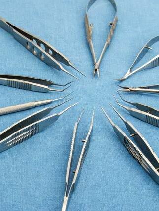
Instrument Lifespan Metrics Power Sustainability
YOFS study calculations demonstrate potential cost and environmental savings of reusables.
LAURA
GASPARI REPORTS
Amedian lifespan of reusable instruments (RUIs) for cataract surgery can be evaluated according to a study by Johnson Yan Ning Neo MD.
“We know there is a strong desire [among] surgeons across Europe and the US for more reusable options, [particularly] for instruments,” he said. However, controversy surrounds the environmental benefits of reusable surgical instruments because of a lack of data on their usage duration and frequency.
Single-use instruments have a higher carbon footprint due to manufacturing and waste disposal processes,1,2 Dr Neo emphasised. Yet RUIs risk potential fatigue and failure after repeated sterilisation cycles, which may not meet manufacturer specifications. Also, while some studies show lower carbon
While some studies show lower carbon footprints for reusable instruments, the single-use variety may be more cost effective.
footprints for reusable instruments, the single-use variety may be more cost effective in high-volume sterilisation centres. Previous cost calculations were based on low RUI reuse rates, which may not reflect real-life scenarios, he noted.
To fill this gap in the literature, the ESCRS Young Ophthalmologists for Sustainability (YOFS) conducted a simple survey in 16 European centres examining annual cataract surgery volumes, reusable cataract instrument usage and replacement frequency, and single-use tool use. The evaluation assessed the instruments for median lifespan in years and median lifespan in surgeries.
The study covered a median of 3,000 cataract surgeries, showing that only about 5% of centres avoid single-use instruments while more than 95% use a combination of RUIs and single-use tools. The 5 instruments used most often were the IOL injector, adjustable lid speculum, notched forceps, nucleus chopper, and, surprisingly, capsulorhexis forceps. As Dr Neo highlighted, even the most fragile capsulorhexis forceps last nearly 3 years and undergo approximately 340 surgeries before replacement.
These data are significant, as they show that using reusable forceps at least 20 times instead of single-use equivalents achieves carbon neutrality. The study also indicates RUIs were used far more frequently than expected before getting replaced, demonstrating additional carbon savings. While this information is already helpful, Dr Neo concluded more research is needed to fully optimise RUIs and reduce waste.
“Median lifespan for reusable instruments was potentially underestimated because a lot of these instruments [were] bulk purchased before the start of the calculated lifespan,” Dr Neo said.
Founded in 2022, the Young Ophthalmologists for Sustainability (YOFS) group strives to enhance sustainability within the field of ophthalmology. YOFS initiatives focus on waste reduction, research, education, advocacy, and fostering collaboration. For more information on current projects, please visit https://www.escrs.org/ special-interest-groups/yos/yofs.
Dr Neo spoke at the 2025 ESCRS Annual Congress in Copenhagen. For citation notes, see page 48.
Johnson Yan Ning Neo FHEA, FRCOphth, FEBO, CertLRS is a consultant ophthalmic surgeon at Barts Health NHS Trust, London, UK.
AI Telephone Call to Reduce Carbon Footprint
Fewer clinic visits, lower emissions in cataract care.
LAURA GASPARI REPORTS
Especially in cataract and refractive surgery, the environmental impact of ophthalmology is an ever-growing concern. According to Aisling Higham MSc, one of the driving changes that can alleviate the carbon footprint of health services can be found in the use of AI-based telephone assistance.
“In the British National Health Service, 25 million tonnes of CO2e are emitted every year, and we are driven to reduce that to a net zero by 2045,” Dr Higham said. “However, this is all on the backdrop of an ever-increasing number of cataract surgery operations.”
With this purpose in mind, she noted recent papers suggest how AI can really be a potential solution to reduce the environmental burden.1,2 She explained that the NHS is already using AI as a conversational assistant—a regulated medical device called Dora (Ufonia). It is a telephone assistant program that automates conversations between the patient and the clinic or hospital, an interaction previously held solely between the patient and the clinician, Dr Higham noted. This, for example, reduces the need for face-to-face visits in the earliest parts of the patient’s clinical pathway, or at the pre-assessment.
Dr Higham, the Medical Director at Ufonia, says the AI assistant identifies patients who are interested in surgery and saves them from having to visit the clinic for cataract assessment. Moreover, this system avoids repetitive visits by enhancing the referral phase. In the following phase, Dora can call to confirm patients are able to come on the day of surgery.
The AI-based telephone assistant comes in handy again during follow-up. Rather than having every patient attend a
face-to-face visit, an automated telephone call can help identify those patients who really have an issue, filtering those who need a follow-up visit in person while managing the others through a virtual pathway. This can help reduce the carbon footprint, she explained.
A study using the Dora pathway as a standard of care reported that each conventional outpatient appointment produced a carbon footprint of 12.78 kg of CO2e, equivalent to 91 km of driving.1 Comparatively, the carbon footprint for using Dora is 0.10 to 0.14 kg of CO2e per call, equivalent to 0.8 km of driving. Dr Higham explained that the emissions are based on the server utilisation, so the relative cost per call decreases as the volume of calls managed by AI increases. Of the 14,000 operations performed in the study, the aggregate carbon footprint was 16,037 kg of CO2e saved per year.
“AI can really be used to support that pathway transformation, and we can see a real difference in carbon footprint doing that,” she said. “Higher use of these systems can paradoxically reduce the carbon footprint per patient.”
Dr Higham spoke at the 2025 ESCRS Annual Congress in Copenhagen.
For citation notes, see page 48.
Aisling Higham MSc, FRCOphth is a consultant ophthalmologist at Oxford University Hospitals NHS Foundation Trust and Medical Director of Ufonia, both of the UK. aisling.higham@nhs.net
Small Changes for a Necessary Shift
Simple solutions can help increase sustainability in ophthalmology.
LAURA GASPARI REPORTS
The paradigm shift towards a sustainable practice can happen incidentally just by optimizing resources with small, thoughtful adjustments, according to Pei-Fen Lin MD.
Sustainability in healthcare has become an increasingly important concept, particularly in cataract surgery. Many practical ideas to reduce waste and costs have been designed and put in place to make ophthalmic practice greener. However, implementing sustainable practices has financial and human costs, including changes in management and research efforts.
According to Dr Lin, it is a matter of balancing sustainability with affordability, and sometimes this can happen by accident.
“We became sustainable warriors just by implementing a digital solution to our practice,” Dr Lin said.
Change began with the COVID-19 pandemic and the challenges it posed to ophthalmic practice, she explained. Cataract services were the first to be shut down and the last to be restarted. Especially in densely populated areas like Croydon in South London, it was particularly difficult to provide cataract services.
In the absence of sufficient funds and by using existing resources and digital technologies for consultation and imaging, her team reduced patient visits from five to one, especially for follow-up visits. This significantly reduced travel, face-to-face appointments, and carbon emissions. They calculated that by switching to a more extensive use of a digital solution, they saved more than 6,000 trees over five years.
Sometimes sustainability is not very high on the agenda of hospital management, so part of the challenge is to convince them that sustainable solutions are efficient, particularly from a financial standpoint, Dr Lin emphasized.
The period immediately after the pandemic saw a steady increase in cataract referrals and attendance. Venues such as public hospitals shifted to using digital technology in the cataract pathway to help process more patients without building new rooms or hiring new staff to accommodate increases in in-person appointments, leading to greater time and cost savings.
Digital tools and strategies are therefore extremely useful and cost effective because they help practitioners streamline existing resources to reduce environmental impact without incurring additional costs. For example, a clinic could stop printing patient leaflets that often get thrown away and switch to electronic instructions, an app, or short videos.
Finally, Dr Lin said the sustainability shift is not exclusive to patient resources, as surgeon training appears to have an environmental impact. A trainee surgeon generates a greater carbon footprint (almost 60% more) than a senior surgeon for cataract surgery since they take more time, are more likely to break equipment, and require extra material. Reviewing existing digital simulation programmes for training can help tackle such carbon waste.
Even the smallest change is useful, Dr Lin added, as it helps shift the existing paradigm, finding the minimum viable product to step into sustainability.
“What can you do to start reducing carbon footprint? Just finish work on time! You switch the lights of your office off, and you have started reducing waste already,” she concluded.
Dr Lin spoke at the 2025 ESCRS Annual Congress in Copenhagen.
Pei-Fen Lin MD is a leading consultant ophthalmic surgeon at Moorfields Eye Hospital, London, UK. p.lin@nhs.net
Drive Your Career Forward
Want to improve your clinical skills, stay on top of developments in the field, and have an active voice in the evolution of cataract and refractive surgery?
As an ophthalmologist in training, all these things (and much more) are within your grasp—and at no cost to you— by joining ESCRS!
Since 1991, ESCRS has promoted education in the field of implant and refractive surgery and supported research into the practice of intraocular lens implantation and refractive surgery. With more than 7,000 members across Europe and the world, ESCRS wields a powerful voice in the global discourse about ophthalmology and provides members with a rich and diverse network of professional connections.
Drive ophthalmology—and your career—forward. Take advantage of free ESCRS trainee membership today!
ESCRS YO membership has given me a strong international community, real integration across cultures and languages, and meaningful connections that extend far beyond meetings. It has opened opportunities to present on stage, build confidence, and grow as a leader within ESCRS and the wider ophthalmology community.
—LAURA MAUBON
As a young ophthalmologist, ESCRS membership opened doors to high-quality education, mentorship, and opportunities that have shaped my early career. Being an ESCRS YO member has connected me with an inspiring global community and given me access to resources that continually improve my clinical practice.
—VALENTIN HOOIJER
Refractive Surgery Grey Zones
Despite their comprehensive nature, the preliminary ESCRS refractive surgery guidelines leave some questions unanswered.
ROIBEARD O’HÉINEACHÁIN REPORTS
The ESCRS Guidelines for Cataract and Refractive Surgery reflect the current consensus on the numerous advancements and innovations. However, newer or less common refractive procedures, as well as borderline cases, still fall into uncertain areas where optimal care demands a highly personalised approach, said Béatrice Cochener-Lamard MD PhD.
“The grey zones persist despite many advances; therefore, shared decision-making [remains] vital in borderline indications [where] procedure choice must consider age, morphology, lifestyle, and a safety-first approach,” she said.
Cross-linking controversies
The use of corneal cross-linking (CXL) in corneal refractive surgery is still debated, she noted. Further studies are required to determine its long-term effectiveness and safety in improving corneal biomechanical stability for eyes undergoing lenticular surgery, surface ablations, or LASIK. Progressive flattening after cornea cross-linking might affect the long-term refractive stability, and the treatment’s impact on infection risk and fibrosis is unclear.
Additionally, stronger evidence is needed to determine the safety and long-term efficacy of CXL combined with transPRK in cases of subclinical ectasia or irregular topography, as well as pairing LASIK with CXL in borderline or highrisk cases. PRK and trans-PRK seem to carry a lower risk of ectasia than LASIK, although this too requires more extensive research, as do comparative ectasia rates.
Other areas of uncertainty in LASIK include the comparative benefits and limitations of wavefront-guided, topography-guided, and conventional ablation techniques. In addition, hyperopia correction with LASIK remains challenging, especially in cases of higher hyperopia, and questions persist regarding complication rates, refractive stability, and overall postoperative quality of vision.
PresbyLASIK shows promise as a means of providing presbyopic patients with an extended depth of focus. The main indication would be younger presbyopes—those younger than 55 years old with clear lenses—and hyperopes, who seem to benefit most from the procedure. However, there is a lack of long-term follow-up data, and these procedures inevitably come with some loss of visual quality.
For the complete ESCRS Clinical Practice Guidelines, scan the QR code.
KLEx advantages under scrutiny
Keratorefractive lenticule extraction (KLEx) offers several theoretical safety benefits compared to LASIK, though these advantages may only make a minimal difference in the long run, Professor Cochener-Lamard said. Since KLEx cuts fewer corneal nerves, it lowers the risk of dry eye and neuropathic pain, particularly within the first year after surgery. Beyond that period, however, there appears to be no distinct difference between the two procedures. Moreover, there is still no clear evidence that corneas have improved postoperative biomechanical stability with KLEx compared to LASIK. There is currently no consensus regarding retreatment protocols following KLEx, and limited data exist on interventions such as PRK (with or without mitomycin C) or conversion to LASIK. Furthermore, there are presently no methods available for performing customised topography-guided ablations with KLEx for managing pre- or postoperative corneal irregularities.
The long-term safety and stability of phakic intraocular lenses (IOLs) require continuing vigilance. Because of their potential risk to the crystalline lens and cornea, each new class of lens design requires at least 10 years of follow-up to adequately assess its safety profile. The optimal method for phakic IOL sizing also has yet to be validated, although the use of AI for that purpose is showing promise, she said.
Furthermore, the risk of cataract induction in hyperopic and presbyopic patients remains insufficiently investigated. Clearly defined thresholds for lens model selection and patient age are still needed. There is also a lack of evidence regarding the use of these lenses for specialised indications. While younger high myopes undergoing refractive lens exchange (RLE) have an increased risk of retinal detachment, the exact age and axial length thresholds remain uncertain. The effects of RLE on ageing eyes, such as changes to the capsular bag and risk of age-related macular degeneration, also require further investigation.
“It is essential to provide detailed and individualised informed consent to your patients, and to encourage the undertaking of standardised studies that review registry and post-marketing data,” she concluded.
Prof Cochener-Lamard made her presentation at the 2025 ESCRS Annual Congress in Copenhagen.
Béatrice Cochener-Lamard MD, PhD is Professor and Head of the Department of Ophthalmology, CHU Morvan Brest – UBO University, Brest, France. beatrice.cochener-lamard@chu-brest.fr. She is coordinator of the ESCRS refractive guidelines, along with Prof Thomas Kohnen.
EUREQUO Evolves
Real-world wins and challenges prompt ongoing development.
HOWARD LARKIN REPORTS
European guidelines for cataract surgery, benchmarking, and pioneering outcomes research are just some of the proven benefits of the European Registry of Quality Outcomes for Cataract and Refractive Surgery (EUREQUO), said Maartje Segers MD. Established in 2007 and including data on nearly 5 million cataract and nearly 250,000 refractive surgeries, EUREQUO has grown into the world’s largest multinational registry of cataract and refractive surgery.
And it is poised to help improve clinical practice outcomes even further, Dr Segers added.1 EUREQUO “not only stores information, but it also organises it in a structured way, [helping us] track outcomes and compare them with colleagues,” she observed. The registry is continuously improving to provide up-to-date information, guide practice, and improve outcomes.
Guidelines and practice impact
Creating Europe’s first continent-wide evidence-based cataract surgery guidelines was among the biggest wins for EUREQUO. First published in 2012, the guidelines are based on data collected from more than half a million surgeries between 2009 and 2011.2 They are under ongoing review for updates as the database grows and includes more complete and structured information about surgeries, Dr Segers noted.
Widespread adoption of intracameral antibiotics is one practice change encouraged in part by EUREQUO-enabled guideline adherence monitoring, Dr Segers said, which is reflected in the registry’s documentation of a dramatic decrease in endophthalmitis rates.
Similarly, EUREQUO outcomes data suggested no benefit from using femtosecond (FS) lasers in cataract surgery.3 This also appears to have changed practice, as evidenced by an initial spike in FS laser-assisted surgery followed by a sharp decline and gradual adoption, Dr Segers said.
Other EUREQUO studies have examined topics ranging from risk factors for refractive errors after surgery to the likelihood of cataract surgery after vitrectomy to complications related to posterior capsule rupture and anaesthesia techniques. A prospective study of bilateral toric IOL implantation is currently underway.
Additional research questions include practice variation and post-marketing surveillance, Dr Segers said. These could help identify promising technologies and clinical best practices, potentially improving cataract surgery outcomes across the continent.
Strengthening the database
Improving data collection and interoperability is a complex but necessary challenge to make registries more useful. EUREQUO is responding by adopting structured data entry forms and collaborating with national registries, in part through the International Consortium for Health Outcomes Measurements (ICHOM), which helps standardise data structure, Dr Segers said. “Data entry needs to be as easy as possible.”
She added that the next step is automating data collection through integration with practice electronic records, which requires improving interoperability among data systems, including standardising digital imaging in collaboration with
EUREQUO outcomes data suggested no benefit from using femtosecond (FS) lasers in cataract surgery. This appears to have changed practice.
The European Experience with KLEx
Flapless procedure offers a safe and effective option, with advantages for select patients.
CHERYL
Keratorefractive lenticule extraction (KLEx) for correcting myopic astigmatism is a safe, effective, and precise procedure that provides outcomes comparable to those associated with LASIK and PRK, said Thomas Kohnen MD, PhD.
“As a mature, flapless procedure involving a very small incision, I think that KLEx is a very intriguing option for patients seeking refractive surgery,” he commented.
Sharing an update on the European experience with KLEx, Professor Dr Kohnen said it began in 2006 with the VisuMax femtosecond laser (Carl Zeiss Meditec). SMILE using the VisuMax laser received the CE mark in 2011, and between 2020 and 2023, the CE mark was given to four additional KLEx procedures/laser platforms: CLEAR/FEMTO LDV (Ziemer), SmartSight/ATOS (Schwind), SMILE PRO/VisuMax 800 (Carl Zeiss Meditec), and SILK/ELITA (Johnson & Johnson).
Prof Dr Kohnen reviewed published literature reporting on these procedures, including a meta-analysis by German authors.1 The investigators reviewed studies with minimum follow-up of 10 years, concluding that the results of KLEx (SMILE) are at least equivalent to PRK and LASIK, with none of the three procedures demonstrating superiority.
Prof Dr Kohnen said he performs KLEx with the ATOS laser. A preclinical study investigating its use for lenticule creation in fresh porcine eyes provided evidence that its accuracy and repeatability were comparable to already established systems.2
“This solid-state laser has a high repetition rate, low pulse energy, integrated eye tracking, cyclotorsion correction, pupil detection, and astigmatic correction,” he said. “Using it at our centre, we have found very good outcomes in the first 50 KLEx cases.”
KLEx candidates
In 2023, the German Society of Ophthalmology (DOG) Committee of Refractive Surgery published its recommendation on refractive surgical interventions that stated KLEx was appropriate for myopia correction from -1 to -8 D and astigmatism correction up to 5 D. The recommendations stated KLEx had limited application for correcting -8 to -10 D of myopia. Guidelines from the ESCRS are undergoing final revision and are expected to be published early in 2026. In agreement with the DOG recommendations, the ESCRS guidelines will recommend KLEx for treating up to -8 D myopia and 5 D astigmatism and specify that application over this limit should only be done with a regular cornea, no risk factors, and sufficient corneal thickness.
“In developing the ESCRS guidelines, we referenced a huge amount of data from the European experience, [particularly] refractive outcomes,” Prof Dr Kohnen said.
He also identified clinical scenarios that represent “special indications” for choosing KLEx when patients seek refractive surgery. Prof Dr Kohnen said KLEx is ideal for patients with thin corneas because it has minimal impact on corneal biomechanics. In addition, it could be preferred over LASIK for people who engage in contact sports for whom there is concern about trauma causing flap dislodgement. KLEx might also be a better choice than LASIK for patients with dry eye because KLEx may cause less disruption of corneal nerves. Since KLEx is performed with a large optical zone, it can also be a good option for individuals who are prone to halos and visual symptoms from spherical aberrations.
Prof Dr Kohnen spoke at AAO 2025 Refractive Surgery Subspecialty Day in Orlando, US.
For citation notes, see page 48.
Thomas Kohnen MD, PhD, FEBO is Professor and Chair of the Department of Ophthalmology, Goethe University, Frankfurt, Germany. kohnen@em.uni-frankfurt.de
GUTTMAN KRADER REPORTS
Managing Femto Flap Complications
Opaque bubble layer and gas breakthrough continue to challenge surgeons.
ROIBEARD O’HÉINEACHÁIN REPORTS
Complications associated with femtosecond laser flap creation in LASIK procedures may impede successful flap formation. However, implementing appropriate preoperative measures can minimise the risk of these complications, and a timely, effective response can help mitigate their impact if they arise, explained Namrata Sharma MD.
She noted that such flap complications can result from factors such as opaque bubble layer, anterior chamber air bubbles, vertical gas breakthrough, pseudosuction, suction loss or break, micro- or macro-adhesions, and flap holes. Proper docking helps reduce these risks, and Dr Sharma recommended preoperative counselling to promote patient cooperation during the procedure.
The incidence of the opaque bubble layer is between 5.0% and 7.6% and occurs less often with newer machines. It is caused by gas bubbles collecting in the cornea’s interlamellar spaces and can last several minutes after forming the lamellar bed, interfering with the surgeon’s view and impeding laser tracking devices. When air bubbles appear in the anterior chamber, the surgeon can wait for them to disappear before proceeding with the excimer laser ablation. However, Dr Sharma said that, in her experience, the bubbles do not compromise the procedure.
Vertical gas breakthrough occurs in 0.03% to 0.13% of cases—the result of a laser-induced optical breakdown in the interface, allowing gas to dissect along the plane of least resistance along the collagen lamellae. If there is an anterior corneal abnormality, gas migrates vertically, forming a visible black island that can be difficult to dissect. When vertical gas breakthrough occurs, the surgeon should stop the procedure by releasing the foot pedal. If the breakthrough occurs before the side cut, one option is to repeat the flap creation at a depth 50 microns greater than the original. Another option is to perform a surface ablation or LASIK at another time.
Dr Sharma stressed the importance of preventing suction loss, identifying pseudosuction and actual suction loss, and responding promptly. While modern machines have reduced the incidence, suction loss may lead to an incomplete flap, which typically occurs during the raster stage or side cut, primarily due to improper docking, entrapment of eyelashes, or oedematous conjunctiva.
In the event of suction loss, Dr Sharma advised completing the procedure immediately if the lamellar bed has been treated and there is no side cut. But if it occurs during the side cut, surgeons should avoid lifting the flap and instead create a new flap that is 0.5 mm smaller and 40 microns deeper. In rare
cases, if the refractive error allows, a surface ablation may be performed.
Micro-adhesions primarily appear because gas bubbles do not always pass uniformly through the intended flap area. Minor adhesions can usually be carefully separated, while attempts to separate larger or more coalescent adhesions may increase the risk of flap tears or super holes. Proper LASIK flap hydration is important both during its creation and elevation.
Prof Sharma made her presentation at the 2025 ESCRS Annual Congress in Copenhagen.
Namrata Sharma FRCOphth, FRCSEd is Professor of Ophthalmology at the Cornea and Refractive Surgery Services, Dr Rajendra Prasad Centre for Ophthalmic Sciences, All India Institute of Medical Sciences, N ew Delhi, India. namrata.sharma@gmail.com
Defining the Costs of ISBCS
Overcoming present issues with the procedure remains crucial to addressing future challenges.
Immediate sequential bilateral cataract surgery (ISBCS) has the potential to reduce costs, lessen environmental impact, and improve healthcare efficiency, according to Steve A Arshinoff MD.
In his clinical practice in Canada, 90% of his cataract patients undergo bilateral surgery.
“I think that, historically, cataract surgery has inevitably evolved to become immediate, sequential, and bilateral,” Dr Arshinoff said. “It turns out that up until recently, cataract surgery before 1930 was done everywhere as a bilateral procedure.”
The trend changed owing to fear of infection and the absence of any infection prophylaxis, such as intracameral antibiotics. As a result, surgeons became reluctant to take risks, giving bilateral cataract surgery, and any bilateral surgery, a reputation for being too dangerous. The subsequent advent of effective intracameral antibiotics reduced the dreaded risk of bilateral simultaneous postoperative endophthalmitis (BSPOE) to about one in one hundred million. This risk is the same as getting instantly killed while driving one kilometre on an empty road in Europe, he pointed out.
ISBCS concerns include interprocedural IOL power adjustments for the second eye. However, the literature does not show any benefit for the refractive outcomes, which he said makes no difference with modern biometry.
There is also fear of litigation, but, as he observed, the bilateral procedure has never been the main culprit. The real outstanding problem is money.
It is possible to consistently save up to 33% of the cost of two separate procedures by doing ISBCS, Dr Arshinoff said. This is supported by numerous studies from the US, Finland, Canada, Sweden, and the UK, conducted in hospitals and private surgical centres, showing cost savings for the patients and their families, the surgical centres, and the payer.1,2 Savings come from registering the patient only once, reducing office visits, and achieving a 10% improvement in patient turnover in the operating room. In addition, the second eye is always easier and a bit quicker, he observed.
The economic advantage for doctors heavily depends on the healthcare system. The Canadian province of Québec, for
example, pays the same amount for both eyes, whereas the province of British Columbia applies a 50% fee reduction for the second eye when operated on the same day, Dr Arshinoff explained.
Current global trends show increasing cost per cataract procedure and in the number of surgeries performed annually. This growth is largely driven by the introduction of modern techniques and intraocular lenses that produce better surgical results.
Payers would like to have some control over this increase in expenses, and ISBCS could be a way to save money without limiting innovation, he noted. However, payers want surgeons to be responsible for the successful outcomes in every case, which Dr Arshinoff said has developed into a severe accountability obstacle, creating mistrust and limiting surgeons from taking on difficult cases.
Despite the current financial constraints, ISBCS is gaining acceptance as the preferred solution in many parts of the world.
“ISBCS has proven in many ways better than delayed sequential bilateral cataract surgery (DSBCS), and it is increasing globally. Patients love it and doctors also like it,” Dr Arshinoff said.
“We must try hard to convince the payers and the other groups we work with to cooperate with each other as much as possible. We should not wait to be told by government to do ISBCS so that government can save money. We should work on doing things well ourselves and try to develop systems that work. Surgeons should be the leaders.”
Dr Arshinoff spoke at the 2025 ESCRS Annual Congress in Copenhagen.
For citation notes, see page 48.
Steve A Arshinoff OC (Officer of the Order of Canada), MD, FRCSC is Professor of the Department of Ophthalmology and Vision Sciences, University of Toronto, Canada.
TIMOTHY NORRIS REPORTS
Expanding Options for Stromal Supplementation
Bioengineering and lenticular implantation offer new opportunities.
ANDREW SWEENEY REPORTS
There’s never enough supply for the growing demand for corneal tissue around the world, making bioengineering and innovative intrastromal lenticular implantation enticing solutions.
This two-pronged approach to one of ophthalmology’s most pressing issues—supplementing the stroma via recreation or enhancement—is full of fascinating opportunities.
According to Sayan Basu MBBS, the bioengineering approach has three potential directions to explore.
“The first is with keratocytes (mesenchymal cells), which can be targeted towards regenerating optically transparent corneal tissue through remodelling of opacity,” Dr Basu said.
“The second is to examine the decellularised extracellular matrix (dECM), repairing corneal tissue via scaffolding and scarless wound healing. The third is to put these two together. This involves replacing corneal opacity with bioengineered, transparent and tectonically stable tissue.”
While bioengineering is still relatively novel in ophthalmology (from a historical perspective), lenticular implantation or epikeratophakia is not. According to Aylin Kılıç MD, while this technique is not new, doctors can unlock its full potential by using corneal allograft tissue as a novel material.
“Using corneal allograft tissue has the advantage of being fully biocompatible without foreign body reaction. It’s also shapeable, and can be prepared as rings, segments, lenticules, or inlays,” Dr Kılıç said.
“We can shape lenticular tissue precisely by using an excimer laser to create inlays and put the stroma under a flap.
Using corneal allograft tissue has the advantage of being fully biocompatible without foreign body reaction.
We can create intracorneal rings and change the shape of the cornea for keratoconus patients too.”
Engineering good patient outcomes
Human limbus-derived mesenchymal cells (hLMCs), taken from cadaveric corneo-scleral rims, are particularly advantageous, according to Dr Basu. That’s because they can create new matrices in the cornea, remove any present opacity, and produce a clear stroma.
“We examined one case involving a patient with a Hansen’s disease-derived persistent epithelial defect who had not responded to treatment with amniotic membrane grafts,” Dr Basu said. “The effect of the hLMCs treatment was so profound that there was an amount of nerve regeneration that occurred, along with stromal regeneration.”
Dr Basu said that the dECM method is even more innovative because it uses corneo-scleral rims that are “discarded from the eye bank as they are not of high optical quality.” These can then be processed and converted into dECMderived treatments such as hydrogels.
Dr Basu added that this process is extremely efficient since multiple samples can be taken from the same donor. The resulting dECM hydrogel can then be applied rapidly to a large number of patients affected by a wide range of conditions.
The same level of efficiency can be achieved by using corneal allograft tissues too, according to Dr Kılıç. That’s because it’s reversible and can be removed or exchanged if needed, so the operating doctor can create a thinner lenticule if needed, then re-implant the lens based on the cornea’s behaviour.
“This means there are multiple clinical applications,” Dr Kılıç said. “These include cone flattening and astigmatic reduction for keratoconus, and myopia, hyperopia, and presbyopia correction with lenticules and inlays for refractive purposes.”
Life-changing 3D printing
Dr Kılıç believes surgeons can “change a patient’s life within minutes” by using novel materials such as allograft corneal tissue in intrastromal lenticular implantation. The ability to reverse implantation is valuable, as is the fact that it is flexible, allowing the shape, size, and thickness of the lens to be customised according to each patient’s needs.
“This form of biological integration with the cornea is safe, and it can use sterilised cornea tissue that’s already available today,” Dr Kılıç explained. “It can be tailored for each patient and has been clinically proven with clear long-term safety and efficiency.”
As for Dr Basu, something with less clinical provenance (given its novelty) but full of potential is 3D bioprinting using dECM hydrogel as a type of ‘ink’ to create 3D-generated constructs. Studies into 3D bioprinting are at an elementary stage, but he’s adamant that the technology shows considerable promise.
“You scan the patient’s cornea and you get your 3D model. It will then print out the substrate of the cornea, the engineered corneal substitute, and it can also carry out photo cross-linking,” Dr Basu said.
“3D bioprinting allows for the customised integration of cells in the corneal stromal scaffold. It’s affordable, highly customisable, and the human tissue alternatives it produces are optically clear and mechanically strong.”
Drs Kılıç and Basu presented at the 2025 ESCRS Annual Congress in Copenhagen.
Sayan Basu MBBS, MS is the Head of Research at the L V Prasad Eye Institute, Hyderabad, India. sayanbasu@lvpei.org
Aylin Kılıç MD is an associate professor at Biruni University and the Medical Director of the Swiss Vision Group Istanbul, Türkiye. aylinkilicdr@gmail.com
Need a quick introduction or refresher about a surgical procedure? Have a tip to share about a technique or approach you use that makes surgery easier?
The ESCRS 100 is the place to go. It’s a library of short (roughly 100 seconds), high-quality instructional videos about all fields of cataract and refractive surgery. More than a dozen videos have already been created, and additional videos are being uploaded each month. Current videos include the following topics:
• Phaco chop
• CTR implantation
• Phacoemulsion, IOL implantation, and DSO on a central guttae Fuchs patient
MAKE EVERY SECOND COUNT
PUT THE ESCRS 100 VIDEO SERIES ON YOUR LIST
How to Avoid Refractive Surgery Complications
Optimisation, protection, and monitoring should be the rule in laser refractive surgery.
LAURA GASPARI REPORTS
Maintaining a healthy ocular surface is critical to surgical success, and complications can be avoided thanks to good optimisation, protection, and monitoring at every stage, according to Sotiria Palioura PhD. She highlighted four ocular surface complications to consider: dry eye disease (DED), neuropathic corneal pain, persistent epithelial defects (PEDs), and Salzmann nodular degeneration.
Almost every patient has dry eye disease after corneal laser refractive surgery, which should improve after six months. If the dry eye is still present after nine months, it can be quite severe.
“Dry eye is the number one complaint, responsible for about 20% of post-LASIK dissatisfaction referrals,” she said.
Dry eye disease causes significant visual fluctuations and decreases contrast sensitivity, undermining the primary benefits of refractive surgery. Contributing factors in LASIK procedures are corneal nerve damage, suction ring and conjunctival goblet cell damage, and the use of topical drops with preservatives, or NSAIDs, which can be toxic to the corneal epithelium. KLEx and PRK procedures can also lead to postoperative dry eye, with some differences.
The main risk factor for postoperatively developing DED is preexisting dry eye, which Professor Palioura said should be treated more aggressively before the surgery—especially in female patients, those older than 40 years of age, or in cases of contact lens intolerance, blepharitis, and meibomian gland dysfunction (MGD). Frequently lubricating the cornea using a longer steroid taper and long-term immunomodulators such as cyclosporine are effective treatments.
Neuropathic corneal pain is characterised by hypersensitivity and pain without obvious injury that affects LASIK, PRK, and SMILE patients. Distinguishing between peripheral and central pain components is crucial for treatment, Prof Palioura pointed out. Peripheral pain can be managed with surface optimisation using serum tears or plasma rich in growth factors (PRGF). Central pain may require a pain specialist and systemic medications, such as gabapentin, pregabalin, and duloxetine. Recent case reports suggest the use of intranasal neurostimulation or corneal neurotisation in severe cases and in specialised centres.
PEDs occur when the epithelium fails to close after surgery, especially in PRK and LASIK surgeries.
“During LASIK flap creation, we see epithelial defects in 1.6% to 5.0% of cases, and most tend to heal [quickly],” she said. “If they do not heal, it may be because the patient had EBMD, age over 40, diabetes, dry eye, or intraoperative trauma. Early consequences include delayed healing, discom-
fort, and higher risk of microbial keratitis and diffuse lamellar keratitis. Late consequences include epithelial ingrowth, flap melt, and corneal haze in PRK cases.”
Initial treatment should include bandage contact lenses, intensive lubrication, and broad-spectrum antibiotics. NSAIDs and preservatives should be avoided to minimise epithelial toxicity. If the PED persists, amniotic membrane, autologous serum, PRGF, and topical insulin or NGF drops may be used. Close monitoring and restarting topical steroids after closure are necessary to prevent haze, she added.
While Salzmann nodular degeneration is a rare condition, its nodules are grey or white, subepithelial, and linked to chronic irritation or prior corneal injury. They appear years or decades after refractive surgery and can cause significant irregular astigmatism if central and large. Small, peripheral nodules can be managed with lubrication and surface inflammation control. Central nodules require surgical removal using diluted alcohol and a crescent blade, as well as caution to avoid disturbing the flap.
“You really have to trim the nodule and be more tolerant of leaving some scarring behind,” she advised. “And if that scarring is still visually significant, you can consider PTK afterwards, but you do not have to remove everything if the nodule is deep at the flap edge.”
Prof Palioura spoke at the 2025 ESCRS Annual Congress in Copenhagen.
Sotiria Palioura MSc, PhD, CEBT, FEBO, FEBOS-CR is Professor of Clinical Ophthalmology at the Bascom Palmer Eye Institute, University of Miami, US.
Handling KLEx Complications
Expert offers tips for minimising risk and managing untoward events .
CHERYL GUTTMAN KRADER REPORTS
As a treatment for myopia and myopic astigmatism, keratorefractive lenticule extraction (KLEx) offers several advantages compared with LASIK and PRK. But like all surgical procedures, it carries risks for intraoperative and postoperative complications. Awareness of these events, along with strategies for their prevention and management, is essential for achieving surgical success, according to Soosan Jacob MD.
Among intraoperative events, the most important are those related to lenticule creation, dissection, or extraction. Possible complications during lenticule creation include suction loss, opaque bubble layer (OBL) formation, black spot development, incisional bleeding, and subconjunctival haemorrhage.
Managing cases of suction loss depends on the timing of the loss—however, it is generally possible to proceed with KLEx unless the suction loss occurs after more than 10% of the lenticular cut has been completed.
“In such cases of suction loss, the procedure must be converted to femto-LASIK, and the treatment wizard will automatically convert to flap creation,” Dr Jacob said.
If suction is lost before 10% of the lenticule cut, surgeons can simply redock and restart the laser. If the loss happens while making the lenticule side cut, the procedure can be repeated, but the lenticule diameter should be decreased by 0.2 to 0.4 mm. Similarly, if the loss occurs during the cap side cut, the side cut can be repeated after reducing the cap diameter by 0.2 to 0.4 mm. In cases of suction loss during the cap cut, the surgeon can repeat the cut after recentring the procedure and clearing any central bubble.
Black spots are the result of inadequate laser energy delivery and typically occur due to interference from debris or secretions at the laser-cornea interface, which can lead to incomplete cuts with tissue bridges and difficult dissection.
“If the spots are small and in the periphery, they may not cause any problem. In more severe cases, it may be necessary to convert to surface ablation or LASIK. To avoid black spots, be certain the surface is completely clear before starting the procedure,” Dr Jacob advised.
The presence of an OBL, which forms from entrapment of cavitation bubbles, is a problem that interferes with lenticu-
lar dissection plane identification. Dissection in false planes can lead to severe irregular astigmatism, to which Dr Jacob suggested it is better to abort the procedure than make brave attempts to dissect in difficult scenarios.
Lenticule dissection complications
Complications relating to lenticule dissection include incisional epithelial loss, difficulty with dissection, cap tears, buttonholes, and lenticule adherence to the cap. Dr Jacob said incisional epithelial loss can occur due to excessive use of topical anaesthetic, a dry ocular surface, rough handling, or the presence of epithelial basement membrane dystrophy. She urged surgeons to be cautious in cases of incisional epithelial loss because it can lead to epithelial ingrowth or diffuse lamellar keratitis.
To enable smooth lenticule extraction and minimise the risk of cap tears, partial lenticule dissection, and torn lenticules, Dr Jacob emphasised the importance of dissecting the anterior plane first, followed by the posterior plane.
Surgeons can use what Dr Jacob calls the “white ring sign” as a valuable guide to differentiate between the anterior and posterior lenticular surfaces.1 The white ring represents the circular white light reflected from the lenticular side cut, and it will lie below the dissector when the instrument is at the anterior lenticular plane and above the dissector when the instrument is at the level of the posterior plane, she explained.
To facilitate dissection in cases where there is a thin lenticule, Dr Jacob recommends the sequential segmental lenticu-
lar side cut dissection.1 In this technique, the anterior lenticular plane is dissected first, followed by the central posterior lenticular plane. This leaves a thin, undissected area attached to the side cut, anchoring the lenticule and preventing it from moving or folding on itself during the remainder of the procedure. The sequential side cut dissection is performed by making short, sequential sweeps to separate the lenticule, leaving anchored points on either side for easy and complete lenticular dissection. Dr Jacob urges care when inserting instruments to avoid snagging the incision and creating tears.
Epithelial ingrowth risk
Dr Jacob said most of the possible postoperative complications associated with KLEx are like those seen after LASIK, except for epithelial implantation and epithelial ingrowth, which can lead to irregular astigmatism. Epithelial implantation occurs when epithelial cells are inadvertently pushed into the pocket intraoperatively, forming epithelial nests, whereas epithelial ingrowth refers to the postoperative migration of epithelial cells into the interface.
Although epithelial ingrowth is less common after SMILE compared to after LASIK, it can occur through the cap side cut, especially if there is a tear from excessive manipulation or trauma.
Dr Jacob said epithelial ingrowth after KLEx is best addressed by using CIRCLE software to convert the area of the cap into a flap, allowing for interface cleaning.
“Be sure to also scrape off the underside of the cap and then apply alcohol so no residual live cells remain,” she added.
Dr Jacob spoke at AAO 2025 in Orlando, US.
For citation notes, see page 48.
Soosan Jacob MS, FRCS, DNB is Director and Chief, Dr Agarwal’s Refractive and Cornea Foundation, Dr Agarwal’s Group of Eye Hospitals, Chenai, India. dr_soosanj@hotmail.com
Dr Jacob recommends viewing the following videos.
Learning SMILE— White Ring Sign for SMILE
Sequential segmental terminal Lenticular Side Cut dissection in SMILE
Rethinking Aberrations: the Low-Degree/High-Degree (LD/HD) Scheme
In this first part of a three-part series, surgeons will gain a renewed clinical understanding of aberrations
SOOSAN JACOB MS, FRCS, DNB IN CONVERSATION WITH DAMIEN GATINEL MD, PHD
Ophthalmologists now receive wavefront information in daily practice, whether from aberrometers, tomographers, or laser platforms. Nearly all these devices describe the ocular wavefront using Zernike polynomials, which have become the default ‘language’ of optical aberrations. However, the way Zernike polynomials are constructed can quietly distort what matters most clinically: the subjective sphero-cylindrical refraction and the quality of the retinal image. Some higher-order Zernike modes contain small but meaningful amounts of low-degree terms such as tilt and defocus. When higher-order aberrations (HOA) are large, this mixing can create confusing results: e.g., predict spurious myopia after refractive surgery and retinal image simulations that look worse than what the patient really sees.
The Gatinel–Malet (GM) decomposition, also called the Low-Degree/ High-Degree (LD/HD) scheme, was developed to solve this problem. Rather than abandoning Zernike polynomials, it reorganises the terms of the wavefront expansion to enforce a strict separation:
• a low-degree (LD) component that contains all piston, tilt, defocus, and primary astigmatism and is sealed or ‘watertight’ and corresponds more faithfully to the subjective sphero-cylindrical refraction;
• a high-degree (HD) component completely free of any low-degree terms and which provides a more accurate prediction of the retinal image and visual quality derived from higher-order aberrations.
With this rearrangement, the functional relationship between the two domains
becomes clearer: the LD part of the wavefront aligns better with subjective refraction, and the HD part predicts the retinal point spread function and visual quality more faithfully. The measured wavefront remains unchanged, but its contents are redistributed to better interpret aberrometry and match how clinicians think about refraction versus HOAs.
In this first part of a three-part series, Soosan Jacob speaks with Damien Gatinel MD, PhD, head of the Anterior and Refractive Surgery Department, Rothschild Foundation, Paris, France, to get a renewed understanding of wavefronts in clinical terms and discover why the way ophthalmologists usually describe ocular aberrations may actually be misleading—especially in complex corneas and post-refractive surgery eyes.
The way ophthalmologists usually describe ocular aberrations may actually be misleading— especially in complex corneas and post-refractive surgery eyes.
SJ: Professor Gatinel, most of us see Zernike polynomials every day on our aberrometers, but we don’t really think about the mathematics behind them. To set the stage, could you briefly remind us what a ‘wavefront’ is in clinical terms?
DG: Of course. In clinical optics, the wavefront describes the surface of an equal optical phase emerging from the eye. If that surface is perfectly flat across the pupil, all the emerging rays are parallel and focus on the retina—that’s emmetropia. If, instead, the wavefront assumes the form of a simple paraboloid of revolution—mathematically represented as a pure r² term—the eye is said to be defocused. A myopic wavefront is a convex (forward-bowed) paraboloid, whereas a hyperopic eye has a concave (backward-bowed) paraboloid. This type of defocus is classified as a low-degree aberration and can be completely corrected using ordinary spectacles or contact lenses.
Could you walk us through in some more detail about wavefronts and rays and why they matter clinically?
First, we often use light rays as a convenient way to visualise the local direction of light propagation. They are an abstraction, but a very useful one in clinical practice. In contrast, the wavefront represents the surface on which the optical phase is identical everywhere. So, while rays tell us which way the light is going, the wavefront tells us where the light is in its phase.
For clinicians, the most intuitive representation is often a map of vergence, like the one shown in the figure. This
type of map tends to be the easiest to interpret clinically, as it directly relates to the convergence or divergence of the light at each point. Geometrically, the important point is that rays are always perpendicular to the wavefront at every location. You can think of rays as the trajectories photons would follow, orthogonal to that phase surface.
Looking at the figure reminds us how optical systems form images. They do this by refracting light as it passes through media with different refractive indices. When light enters a denser medium, its wavelength becomes shorter, but the frequency stays the same. If you count how many wavelengths fit along the path from the source to the image plane, you obtain the optical path length, which is exactly the total number of wavelengths between those two points. When this optical distance is identical for every ray travelling through the system, the resulting image is optically ‘perfect’: all contributions arrive in phase, and you get maximal constructive interference on the image plane.
In an emmetropic eye, an incident plane wavefront (light coming from infinity, for example) is transformed by the eye’s optics into a spherical wavefront that comes to focus on the fovea. In a myopic eye, the geometry is different. The wavefront that emerges from the retina is still spherical, but its centre of curvature lies at a finite distance in space; this point is the punctum remotum, or far point, of the myopic eye.
Finally, the bottom schematic in the figure shows how a negative Zernike spherical aberration behaves. Its shape mimics a multifocal wavefront. The central part of the wavefront is steeper and provides a focus for near vision, while
Figure courtesy of Professor Damien Gatinel.
the peripheral part is flatter, favouring distance vision. This multifocal-like profile is exactly why negative spherical aberration has been exploited in various presbyopia-correcting approaches, and the figure gives a visual intuition for that behaviour.
We’ve talked about defocus as a low-degree aberration that we can correct with glasses or contact lenses. What happens when we move to higher-degree terms—those we usually call higher-order aberrations?
That’s where things get interesting. Once the radial exponent of the wavefront term reaches 3 or higher, we are dealing with higher-degree aberrations, and these cannot be corrected through ordinary lenses. In practice, the most clinically relevant of these are the third-degree terms, which correspond to coma, and the fourth-degree terms, where spherical aberration is the key player. Ideally, when we talk about the ‘high-degree’ group, it should contain only genuine higher-degree terms—things that really start at r³, r⁴, and so on, without any hidden low-order content. However, some of the Zernike modes we routinely use violate this principle. A classic example is the Zernike spherical aberration mode (Z₀⁴). Mathematically, it is not a ‘pure’ r⁴ term; it actually contains a hidden quadratic (r²) component of defocus.
This shows up very clearly if you look at its shape. Instead of being a simple, uniformly concave or convex bowl with a relatively flat central portion—what you would expect from a pure r⁴ profile—the Zernike spherical aberration mode has a ‘sombrero-like’ appearance with a central inflection. That central steepening is exactly the signature of the embedded r² content. Visually, this morphology is reminiscent of a schematic multifocal wavefront: it is steeper and more parabolic in the centre, which favours near vision, and becomes flatter toward the periphery, which favours distance vision. This resemblance is not just an academic curiosity; it helps explain why the negative spherical aberration profile of the Zernike mode has been historically popular in presbyopia-correcting strategies. Both corneal photoablations and refractive simultaneous vision intraocular lenses have often been designed around this type of profile, precisely because it mimics a multifocal behaviour at the level of the wavefront.
And that’s where Zernike polynomials come in?
Absolutely! Zernike can mislead when HOAs are large. Zernike polynomials (Zm) are mathematically sophisticated functions that constitute a complete, orthogonal set over a circular pupil. In practical terms, this implies that, from a purely mathematical point of view, each mode is independent of all the others, and the total wavefront error can be written as the quadratic sum of the individual coefficients, in accordance with the Pythagorean principle. Because of this orthogonality, we can calculate a single global root mean square (RMS) value and combine the contributions of all low- and high-degree modes without any overlap between them.
In the physical sciences—where Zernike polynomials were originally introduced—this kind of global orthogonality is crucial. It guarantees that each mode has a unique contribution to the overall optical error, and there is no distinction
between aberrations that can be corrected with spectacles and those that cannot. In ophthalmology, however, this same feature leads to a subtle paradox. To preserve strict orthogonality across the full set of polynomials, certain higher-order Zernike modes are forced to contain small amounts of lower-order terms. For example, the Zernike coma modes (Z3±1) include a linear component (r1, corresponding to tilt), and the spherical aberration modes (Z40,Z60 ) incorporate a quadratic component (r2, corresponding to defocus). These embedded lower-order pieces are what keep the basis rigorously orthogonal, but they do so at the expense of clinical interpretability.
You’ve explained that some Zernike modes ‘mix’ true higher-order content with hidden low-order components like defocus or astigmatism. Clinically, what problems does this mixing create, and how does the LD/HD approach help us get around them?
In practical terms, this structural mixing has at least three important consequences for us as clinicians. First, a portion of the low-degree information—defocus or regular astigmatism—can be buried inside the higher-order modes. As a result, the low-degree Zernike coefficients that we usually rely on become unreliable predictors of the true sphero-cylindrical refraction. Second, because some low-degree curvature is embedded within the high-degree modes, retinal image simulations based on the Zernike expansion can look worse than the patient’s real-life vision. This discrepancy is especially striking when there are large higher-order aberrations: the simulated point-spread function or retinal image may appear excessively degraded, even though the patient’s subjective experience is better once refraction is optimally corrected. Third, there is a reciprocal effect: the numerical amplitudes of the high-degree Zernike coefficients are artificially lowered. Their coefficients appear deceptively small, even though their true contribution to wavefront deformation remains substantial.
From an ophthalmic standpoint, we really need a different kind of separation. Ideally, the low-degree modes should describe the curvature of the wavefront near the centre of the pupil—the region that determines the sphero-cylindrical refraction. The high-degree modes, by contrast, should capture the deformations that develop toward the pupil periphery, while staying as flat and neutral as possible in the central zone.
This is exactly the type of strict separation that the Gatinel–Malet (LD/HD) decomposition provides. In the accompanying figure, we illustrate this by converting the Zernike spherical aberration mode (Z₄⁰) with a negative coefficient into dioptres. The result shows that this mode is far from refractively neutral: its central curvature behaves as if it contains a significant amount of defocus, even though we tend to think of spherical aberration as a purely higher-order effect.
This conversation will be continued in the next issue of EuroTimes
Dr Soosan Jacob is Director and Chief of Dr Agarwal’s Refractive and Cornea Foundation at Dr Agarwal’s Eye Hospital, Chennai, India, and can be reached at dr_soosanj@hotmail.com.
DSAEK or DMEK?
Old school still beats the new, according to one leading Italian ophthalmologist.
ANDREW SWEENEY REPORTS
There are those who prefer all things new and shiny, and others who prefer to keep it old school.
Massimo Busin MD made it clear that there’s still much ado about Descemet stripping automated endothelial keratoplasty (DSAEK), despite the rising prevalence and popularity of Descemet membrane endothelial keratoplasty (DMEK).
“In 2013, the vast majority of endothelial keratoplasties in the United States were DSAEKs, but last year, a slight majority were DMEKs,” Professor Busin said.
“When choosing between the two techniques, there are three things to [consider]: the visualisation of the anterior chamber structures, the anatomy of the eye, and visual acuity potential. I’m still an advocate of thin DSAEK; what’s thin is almost a personal option, but it could be considered slightly thicker than a nanograft, which is supposed to be in the 50–60 μm range.”
Prof Busin pointed to the example of a 72-year-old patient with right eye vision only after suffering severe bilateral trauma following a car crash. This patient was aphakic and aniridic in both eyes, thus creating a large gap between the anterior chamber and vitreous cavity. Anchoring to the recipient cornea with a suture enabled a successful DSAEK, and the patient recovered 20/30 vision.
In another case, a 62-year-old patient, similarly with right eye vision only and aphakia, had undergone four glaucoma surgeries and seven retinal procedures, resulting in an extremely low visual potential. After being treated with DSAEK, the patient’s vision improved to the point of being able to
count fingers. “[It was] perhaps the best he could see after the trauma,” Prof Busin said.
“There’s very little sense in striving for DMEK in an eye with a very low visual potential. Instead, DSAEK can be safely performed in these complicated eyes, avoiding the risks of ‘open sky’ surgery, [that is] of conventional penetrating keratoplasty.”
Prof Busin did note DMEK does appear to yield a faster visual rehabilitation, which has its own advantages. However, he emphasised that the scientific literature proving a decisive advantage is lacking. In his view, ophthalmology needs “a larger, bigger study to be able to say a final word about this.”
The ‘pull-through technique’ for both DMEK and DSAEK is Prof Busin’s preferred method for now, as it allows for rapid graft placement and minimal manipulation. While this method does allow for good DMEK outcomes, he remains unconvinced of its overall superiority over DSAEK.
“There is no compelling evidence that DMEK, especially in complicated eyes, is much better than a thin DSAEK. Call it ultrathin or nano; it doesn’t make any difference,” Prof Busin concluded.
Prof Busin spoke at the 2025 ESCRS Annual Congress in Copenhagen.
Massimo Busin MD is Professor of Ophthalmology at the University of Ferrara and Head of Ophthalmology at Villa Igea Hospital in Forlì, both of Italy. massimo.busin@unife.it
Expanding Options for Rho-Associated Protein Kinase Inhibitors
ROCK inhibitors are proving highly effective in endothelial treatment, among other fields.
ANDREW SWEENEY REPORTS
Most people are focused on finding new and better sources of protein, but what about finding something that activates protein instead? How can ophthalmology use protein to activate better patient outcomes?
The good news is that Rho inhibitors are showing a strong capability of revealing the workings of protein activation. Friedrich Kruse MD provided a comprehensive overview of the topic at a recent conference.
Dr Kruse first described Rho-associated protein kinase inhibitors (ROCKis) as serine-threonine kinases, i.e., downstream effectors of Rho-GTPase, which activate in response to growth factors such as hormones, cytokines, integrins, cell stress, and wound healing.
The ROCK 1/2 variant activates approximately 40 proteins through adenosine triphosphate (ATP)-dependent phosphorylation. According to Dr Kruse, these play a critical role in cell adhesion, migration, proliferation, contraction, and apoptosis via the modulation of the cytoskeleton.
ROCKis have enjoyed considerable utility in ophthalmology, with ripasudil and netarsudil as the most widely known. The former is a non-selective ROCK 1/2 applied as topical eye drops for glaucoma and intraocular hypertension (IOP).
Netarsudil, on the other hand, is a norepinephrine transporter (NET) ROCK 1/2 that’s also applied as an eye drop, primarily used to treat open-angle glaucoma and lower IOP.
“IOP in the patient is lowered by ROCKis via the enhancement of the trabecular meshwork and by Schlemm’s canal outflow,” Dr Kruse explained. “In experimental use, they can reduce subconjunctival fibrosis after filtration surgery and could provide neuroprotection in retinal and optic nerve ischaemia by enhancing blood flow.”
There are many possibilities when it comes to applying ROCKis in endothelial disease, including endothelial protection during surgery, corneal oedema (as an adjunct to Descemet stripping only [DSO]), corneal endothelial cell (CEC) injections, and Fuchs’ endothelial corneal dystrophy (FECD).
For example, ripasudil’s efficacy in treating FECD is considerable thanks to its ability to interact with cell matrices. Dr Kruse said the drug achieves this by causing “significant downregulation of FECD-related matrix components and concomitant upregulation of matrix metalloproteinases (MMPs).”
He also highlighted applications of ROCKis as adjuncts in DSO, describing how drugs like ripasudil and netarsudil
“support endothelial wound healing and regeneration by promoting endothelial cell migration, proliferation, and healing.”
He added that clinical studies have shown that using ROCKis is associated with faster visual recovery and higher success rates than with DSO alone.
In terms of what to expect from ROCKis in the future, Dr Kruse said there’s potential for further advancements in corneal oedema treatment, as well as for graft survival enhancement after Descemet membrane endothelial keratoplasty.
“We’re now at a point where we can think about pharmacologic regeneration, helping the cornea restore itself instead of replacing it,” Dr Kruse said. “This could fundamentally change how we treat endothelial disease—with less reliance on transplants and more focus on biology.”
Dr Kruse spoke at the 2025 ESCRS Annual Congress in Copenhagen.
Friedrich E Kruse MD is Chairman of the Department of Ophthalmology at University Hospital Erlangen, Germany. friedrich.kruse@uk-erlangen.de
First-Line Therapy for Non-Infectious Uveitis
Experts offer differing perspectives on the relative benefits and drawbacks of biologics versus traditional IMT.
CHERYL GUTTMAN KRADER REPORTS
Uveitis experts agree that biologics are effective in treating non-infectious uveitis (NIU), but opinions differ regarding their use as a first-line therapy.
In a point-counterpoint discussion, Sapna Gangaputra MD took the pro position. She emphasised that biologic treatment has a faster onset of efficacy, translating into earlier vision improvement and quicker attainment of corticosteroid sparing or discontinuation compared with traditional immunomodulatory therapy (IMT). She also reviewed evidence addressing safety concerns and noted patients on biologics require less frequent laboratory monitoring than those on IMT.
“Traditional IMT is great, but biologics are better,” Dr Gangaputra said.
Arthi Venkat MD conceded that patients started on biologics are slightly more likely to achieve corticosteroid sparing but said the difference compared with IMT is small and offset by the downsides of biologics, which include their higher cost burden and risk of adverse events.
“Traditional IMT for treatment of NIU has been around a long time and is perfectly reasonable to be first-line therapy for NIU because it is tried and true, affordable, and safe,” she said.
The case for biologics
Dr Gangaputra noted that in 2014, an expert panel recommended considering the TNF inhibitors infliximab and adalimumab as first-line immunomodulatory agents for treating ocular manifestations of Behçet’s disease and as second-line therapy for uveitis associated with juvenile idiopathic arthritis (JIA) or for the treatment of severe cases where traditional IMT failed or was unsuitable.1 The recommendations considered safety concerns associated with the biologics,
including risks for tuberculosis (TB) reactivation, opportunistic infections, demyelinating disease, paradoxical autoimmune adverse events, antiidiotypic antibody development, and lymphoma.
“A huge volume of data has emerged since then, mostly from the SITE cohort study that found that the risk of overall mortality and of cancer-related mortality in patients being treated for eye disease was not much different in those receiving biologics compared with traditional IMT,2” Dr Gangaputra said.
Reviewing the literature, she also concluded that there is solid evidence supporting the effectiveness of biolog-
ics, citing results from the retrospective SITE study showing treatment with a TNF inhibitor—alone or combined with a traditional IMT agent—was more effective than conventional IMT for achieving disease quiescence and corticosteroid sparing among patients with active NIU.2 Similarly, the proportion of patients achieving corticosteroid sparing at 6 months and corticosteroid discontinuation at 12 months was higher in the group treated with adalimumab versus the IMT group in the prospective, randomised ADVISE trial.3
Dr Gangaputra also mentioned the ADJUST study, which found that among patients whose disease was
controlled on adalimumab, adding a traditional disease modifying antirheumatic drug did not protect against treatment failure (disease recurrence) after adalimumab was discontinued. Importantly, all patients who restarted adalimumab because of treatment failure regained disease control.
“We repeatedly say that we don’t treat uveitis but that we immunosuppress patients to control the disease,” she said. “The biologic was the only thing allowing these children to regain good control.”
Justification for traditional IMT
“We are not arguing that traditional IMT is better than biologic therapy in all scenarios and for all NIU. And I am not arguing against the use of biologics altogether or that biologics do not work,” Dr Venkat said. “Rather, the issue we are debating is that traditional IMT with an antimetabolite or calcineurin inhibitor is an appropriate and better first-line agent than biologic therapy for NIU.”
Reviewing IMT efficacy, Dr Venkat also mentioned the SITE study, noting it showed most NIU patients (80% to 85%) were managed on a single nonbiologic immunosuppressive. She added data from head-tohead comparative trials showed that the difference between adalimumab and IMT in percentage of patients achieving corticosteroid sparing is only 5% to 10%.
Putting the difference in perspective, Dr Venkat offered an analogy comparing a luxury brand and a highquality duplicate.
“The duplicate does the same thing but for a lot less money,” she said.
Dr Venkat cited several studies documenting the overall tolerability and safety of traditional IMT and stated that, compared with IMT, biologics are associated with higher rates of infections and TB reactivation.
“Those of us who routinely practice uveitis care know there is a lot of latent TB out there,” she commented.
Further arguing against first-line use of biologics, Dr Venkat emphasised the risks of demyelinating disease and formation of anti-idiotypic antibodies that render biologics ineffective. Discussing cost, she reviewed research showing that, in general, the incremental cost-effectiveness ratio is high for biologic therapy and cautioned that biologic treatment for NIU is often not completely covered by insurance.
The debate took place during AAO 2025 in Orlando, US.
For citation notes, see page 48.
Sapna Gangaputra MD, MPH is Associate Professor, Vanderbilt Eye Institute, Nashville, Tennessee, US. sapna.gangaputra@vumc.org
Arthi Venkat MD, MS is Associate Professor of Ophthalmology, University of Virginia School of Medicine, Charlottesville, Virginia, US.
Research Education Innovation
ESCRS’s vision is to educate and help our peers excel in our field. Together, we are driving the field of ophthalmology forward.
Cataract Surgery in Diabetic Patients
Information on retinopathy-related risks provides foundation for decisions on procedure timing and follow-up.
CHERYL GUTTMAN KRADER REPORTS
Nearly all adults with diabetes who live long enough will develop vision-impairing cataracts, but decisions on surgical timing must consider the procedure’s benefits and risks in this patient population, according to Toke Bek DMSci.
“Cataract can obstruct the view to the retina, which argues for performing cataract surgery early to enable fundus examination for monitoring of diabetic retinopathy (DR),” Dr Bek explained. “On the other hand, cataract surgery leads to retinal swelling that may accelerate the development of maculopathy, and there is also evidence suggesting that cataract surgery can accelerate the development of proliferative diabetic retinopathy (PDR). These risks argue for postponing cataract surgery.”
To gain further insight to guide optimal timing of cataract surgery and postoperative follow-up duration in patients with diabetes, Dr Bek and colleagues undertook a study to identify how known risk factors for progression of DR affect postoperative risk for developing diabetic macular oedema (DMO) or PDR.1
The research used clinical data from diabetic patients seen at the Aarhus University Hospital ophthalmology department over a 25-year period and survival analysis methodology that accounted for death as a competing risk for developing the vision-threatening complications postoperatively. Statistical analyses used to characterise risk for the two complications over time and the impact of different risk factors accounted for the finding that the hazards for both DMO and PDR were not proportional over time, Dr Bek explained.
Results showed that almost all patients (98.8%) who reached age 90 years underwent cataract surgery, which
was associated with increased risk for developing both DMO and PDR. Cataract surgery increased the risk for DMO threefold overall (P < 0.0001). The risk varied over time—reaching its highest in the first 5 years postoperatively, falling between 5 and 20 years, and increasing thereafter—but was statistically significant during all three postoperative observation intervals.
Investigation of DMO risk factors confirmed a role for known predictors but also revealed that some factors had an impact immediately after surgery whereas others contributed to later development. The analysis of risk factors for PDR, which used different methodology, likewise revealed different factors impacted risk at different follow-up times after surgery.
Commenting on the clinical implications of the findings, Dr Bek said they make it very difficult to explain to patients their risk of developing DMO or PDR after cataract surgery.
“So, the take-home message might be that the timing of cataract surgery in patients with diabetes cannot be calcu-
lated in advance because the risks are difficult to estimate,” he said. “Therefore, the determination needs to be a clinical decision based on the patient’s subjective symptoms weighed against the clinician’s need for examining the fundus.”
Addressing the issue of how long to continue postoperative follow-up, Dr Bek said, “The fact that the risk of both DMO and PDR persisted for more than 20 years indicates that the recommendation to maintain vigilant control after cataract surgery in patients with diabetes may be relevant for a long time after the procedure.”
Dr Bek presented at EURETINA 2025 in Paris.
For citation notes, see page 48.
Toke Bek DMSci, MBA is Professor, Head Consultant, and Chair of the Department of Ophthalmology, Aarhus University Hospital, Aarhus, Denmark. toke.bek@mail.tele.dk
Maculopathy in Pentosan Polysulfate Sodium Users
Meta-analysis identifies relationship to cumulative dosage that supports causality.
CHERYL GUTTMAN KRADER REPORTS
There is moderate certainty evidence supporting a dose-dependent relationship between pentosan polysulfate sodium (PPS) exposure and risk of maculopathy, concluded researchers who conducted a systematic review and meta-analysis focusing on this topic.
“We decided to conduct this study because, in our experience, we noticed a trend where patients who had been taking PPS for many years (and at a high dose) were often the same patients who developed an associated maculopathy. This condition is only recently recognised, and while several observational studies suggest there is a dose-response relationship between PPS and maculopathy, there has not been a literature-pooled synthesis on risk across strata of cumulative dose. Furthermore, a competing hypothesis posits that maculopathy in PPS users is a result of systemic inflammation rather than being PPS-induced,” said first author, Brendan Tao MD.
“We felt that if a meta-analysis showed a dose-response relationship, which is a criterion for establishing causality, it might be helpful to better understand the pathophysiology of the maculopathy in patients treated with PPS. Based on our findings, we recommend considering tapering PPS to an effective minimum dose, even discontinuing it altogether if possible, and screening patient users for maculopathy.”
The meta-analysis included five studies containing 141,785 patients and 6,432 cases of PPS maculopathy. The studies were identified by searching three databases from the date of their respective inception to 15 September 2024. To be included in the meta-analysis, studies had to report the incidence of PPS-associated maculopathy and cumulative PPS dose information. The published papers were also reviewed for risk of bias, and researchers assessed the included studies for certainty of evidence using the GRADE method.
A linear dose-response regression model determined the relative risk of maculopathy among patients exposed to PPS stratified by PPS dose. The model estimated a 0.1% increase in relative risk for every 1 g increase in cumulative dose of PPS (P < 0.0001). With individuals not exposed to PPS serving as the reference group, patients who received a cumulative PPS dose ≥ 2,000 g had a nearly 7.5-fold increased risk of maculopathy. The relative risk of maculopathy increased 1.65-fold within the group of individuals with a PPS cumulative dose ranging from 1 to 500 g.
“Patients who are receiving the standard PPS dosing schedule of 300 mg/day—as per the product labelling approved by the US Food and Drug Administration—would reach a cumulative dosage of 500 g in 4.6 years and 2,000 g in 18.3 years,” Dr Tao observed.
Commenting on the strengths and limitations of the study, Dr Tao told EuroTimes, “The strength of our research for evaluating the dose-response relationship between PPS and maculopathy is that we were able to identify a considerable number of patients.
“Of course, we recognise this is a meta-analysis of observational studies. Therefore, it lacks ability to leverage individual patient-level data for meta-analysis. This limitation precludes further control for unmeasured confounding and should be addressed in further research.”
PPS is the only US FDA-approved treatment for interstitial cystitis. Symptoms of maculopathy were observed in patients on long-term therapy in 2018. The FDA required a label change in 2020 to warn of this potentially sight-threatening side effect.
Dr Tao presented at EURETINA 2025 in Paris.
Brendan Tao MD is a resident in the Department of Ophthalmology & Vision Sciences, University of Toronto, Canada. brendantao@gmail.com
IOL Use Could Expand to Younger Infants
Swedish experience points to the efficacy of the one-piece acrylic hydrophilic bag-in-the-lens.
ANDREW SWEENEY REPORTS
Congenital cataract is one of the leading causes of childhood blindness, and early intervention and treatment are crucial. Swedish research suggests this urgency should include a reappraisal of how doctors work with intraocular lenses (IOLs).
A recent presentation by Arzu Seyhan Karatepe Haşhaş MD highlighted the Swedish approach towards finding the right time to implant IOLs in treating this condition, which she says goes against current standard practice.
Dr Karatepe Haşhaş noted IOL implantation in patients under the age of 1 is typically rare and extremely rare in patients younger than 3 months. However, surgeons will opt to use IOLs in patients aged up to 2 years when possible, and there are usually no problems with older patients.
The Swedish approach is rather different. First, all congenital cataract patients are entered into the Paediatric Cataract Register (PECARE), a national registry for paediatric cataracts that includes diagnostic, surgical, and postoperative data.
“In Sweden, infants [younger than] 12 months often receive primary IOLs when possible. Two national centres are certified for cataract surgery in children [younger than] 3 years of age: Stockholm and Gothenburg,” Dr Karatepe Haşhaş said.
“At St Eriks Hospital in Stockholm, a one-piece acrylic hydrophobic IOL in a capsular bag is used. In Gothenburg, at Sahlgrenska University Hospital, a one-piece acrylic hydrophilic bag-in-the-lens (BIOL) is used [with] the Tassignon technique.”
Dr Karatepe Haşhaş reported that a study based on PECARE’s data found BIOLs achieved lower rates of visual axis opacification—3.8% and 8.8% unilaterally and bilaterally— compared with 23.1% and 37.3% for aphakia and 71.2% and 53.9% for in-the-bag IOLs.
Could the same positive results be visible in visual acuity outcomes? Could patients younger than the aforementioned 3-month threshold also receive BIOLs? Dr Karatepe Haşhaş believes so, based on a study involving 80 unilateral and 82 bilateral congenital cataract patients, all of whom were operated on under this age threshold.
The results demonstrated that earlier surgery produced better outcomes. In both groups, those operated on before 6 weeks of age were associated with improved visual acuity across all three methods compared to those treated after 6 weeks of age.
Bilateral cases also showed little difference between the three methods. However, in unilateral cases, Dr Karatepe Haşhaş reported that visual acuity in the BIOL group was 0.5 logMAR five years postoperatively, whereas aphakia
and the IOL-in-the-bag technique scored 1.3 and 0.9 logMAR, respectively.
This data, she added, demonstrates BIOLs as a viable option for congenital cataract treatment. Not only that, but she also believes this approach could and should become standard practice.
“If you operate on an infant, then you should implant an IOL if you can, especially if you have access to BIOLs,” Dr Karatepe Haşhaş said. “It may offer the best patient outcomes, especially in unilateral cases.”
Dr Karatepe Haşhaş presented at the 2025 ESCRS Annual Congress in Copenhagen.
Arzu Seyhan Karatepe Haşhaş MD is working as a consultant ophthalmologist at Sahlgrenska University Hospital, Sweden, specialising in paediatric cataract and glaucoma surgery as well as vitreoretinal surgery. She is a Fellow of the European Board of Ophthalmology and continues clinical research as a PhD student at the University of Gothenburg, Sweden. arzu.karatepe.hashas@vgregion.se
Incision-Free Interventional Glaucoma
Femtosecond laser treatment does away with need for OR.
HOWARD LARKIN REPORTS
Most minimally invasive glaucoma surgery (MIGS) is still done with cataract surgery. Even when performed alone, today’s MIGS procedures require an entry incision and a trip to the OR. Such trips are considered a big reason why interventional glaucoma remains more a dream than a reality.
The ViaLase Laser System could change that with its non-invasive femtosecond laser, image-guided, high-precision trabeculotomy (FLigHT) treatment, company CEO Shawn O’Neil said. The device, which uses a femtosecond laser to cut precise apertures in the trabecular meshwork, received the CE mark in July 2024.
“At ViaLase, we believe we’re going to bring interventional glaucoma to reality by going outside of the cataract theatre,” O’Neil said.
In addition to eliminating the need for an incision, FLigHT offers micron-level precision guided by real-time, ultra-high-definition gonioscopic and optical coherence tomography (OCT) imaging. This visualisation allows surgeons to clearly identify critical physiologic landmarks at the micron level, enabling the creation of precise apertures of the right size at the right place to enhance aqueous outflow for every patient, O’Neil added.
Moreover, this is accomplished without collateral damage to surrounding tissues, which is a trademark of the femtosecond laser, O’Neil said. And repeat treatments can be performed as needed in any quadrant to enhance or maintain results.
Precise and durable
The FLigHT procedure starts with docking the laser to the eye with suction. High-definition cameras give a clear and steady view of the angle and trabecular meshwork. “We know [this]
is so hard to see in intraoperative gonioscopy in your non-dominant hand,” O’Neil noted.
As the laser creates a precise connection between the anterior chamber and Schlemm’s canal, the results are clearly visible on both a live video feed and real-time OCT. The distinct opening remains clearly visible 1 day after surgery.
Intraocular pressure-lowering results are significant. In a first-in-human study, 18 eyes with open-angle glaucoma received a single-aperture 500 x 200-micron treatment. At 24 months, mean IOP was reduced by nearly 35% with no serious adverse events reported. More than 80% of eyes achieved a >20% reduction in IOP, and 53% achieved a >25% reduction. Only 2 patients lost 2 or more lines of visual acuity, with mean VA virtually unchanged for 2 years.1
“FLigHT achieves what clinicians want in a glaucoma treatment, which is effective IOP lowering in a safe and durable manner,” O’Neil said. “ViaLase is poised to fuel the practice of incision-free interventional glaucoma by bringing the first and only femtosecond laser to this market.”
O’Neil estimates the standalone interventional glaucoma market at $6 billion annually, making it the largest unaddressed glaucoma segment.
O’Neil spoke during iNovation Day at the 2025 ESCRS Annual Congress in Copenhagen.
For citation notes, see page 48.
Shawn O’Neil is chief executive officer at ViaLase Inc, Aliso Viejo, California, US.
Meeting Challenges Through Innovation
Innovators Den finalists pitch solutions to knotty ophthalmic problems.
HOWARD LARKIN REPORTS
ESCRS iNovation Day 2025 featured another exciting Innovators Den, a competition for ophthalmic entrepreneurs developing new devices and treatments to improve eye care. Out of dozens of competitors, three finalists selected by a panel of judges made pitches to an international audience of ophthalmologists, investors, and industry leaders in bids for partnerships to help bring new products to market.
One would be chosen the winner of the 2025 Innovators Den medal. Should they complete the arduous path to market, their innovations could significantly contribute to patient care.
Titratable filtration
Treating glaucoma with tubes is challenging in part because post-surgery aqueous outflow rates are not entirely predictable. Existing devices aren’t adjustable after surgery, which can cause problems—including an immediate excessive outflow after surgery and a later under-filtration. Titran, a glaucoma titratable device in development by Bellagio Medical, will help meet the challenge, Paolo Cecchini MD, PhD said. Instead of relying on a fixed lumen diameter and length to control flow rates, Titran uses multiple filtration holes in its tube. Filtration is set at a zero or minimal rate at implantation, and a YAG or thermal laser can be used at the slit lamp to increase outflow by opening new holes of different diameters.
Sequential activation allows the conjunctiva to heal, reducing the risk of aqueous leaks, hypotony, athalamia, and choroidal detachment, Dr Cecchini said. The new titratable filtering system allows for increasing the outflow months or years later if IOP rises.
“This allows early and late titration for each and every patient,” he said. “The surgeon decides when to open new filtration holes, where to open them, and how much filtration is needed for that specific patient.”
With global glaucoma device sales approaching $1.8 billion in 2024 and expected to hit $2.8 billion by 2034, Dr Cecchini envisions a big market for Titran. Bellagio Medical seeks $2 million in seed funding to develop the existing prototype and conduct initial human trials.
Dislocated IOL rescue
With populations ageing worldwide, dislocated and subluxated intraocular lenses (IOLs) are becoming more common, particularly in post-trauma cases or after complicated cataract surgeries. The Oseguera Ring can help rescue virtually any type of dislocated lens, including simultaneous vision lenses and other premium IOLs, in just a few minutes—without unnecessary explants.
With populations ageing worldwide, dislocated and subluxated IOLs are becoming more common, particularly in post-trauma cases or after complicated cataract surgeries.
In many cases, this can reduce the cost, complexity, and risk of existing rescue techniques, which may require challenging special sutures, backup lenses, or even a second surgery, said inventor Gerardo López Oseguera MD. “It complements rather than replaces the other techniques.”
Developed by Eyedictive, the Oseguera Ring is a universal, sutureless, scleral fixation device designed for affordable and accessible lens rescue. It offers secure, centred IOL fixation and can be readily kept on hand even in resource-challenged sites, Dr Oseguera said.
Compatible with most IOLs, including one-piece and threepiece lenses, the device features two anchors that are inserted into scleral incisions supporting a central double ring. The rescued lens is inserted between the rings, which centre the optic. The off-axis anchors counter-tilt and improve stability, Dr Oseguera said.
Dr Oseguera estimates that secondary fixation is required in about 1–2% of 28 million cataract cases per year globally, or about 500,000 cases. Eyedictive seeks €500,000 to develop industrial tooling to produce the ring and support regulatory certification, an initial production run, and pilot clinical.
“Let’s make IOL rescue accessible to every surgeon, everywhere,” Dr Oseguera said.
Regenerating retinas
Age-related macular degeneration (AMD) is a progressive disease that can be slowed, but not reversed, with existing injection and photobiomodulation (PBM) therapies. Adding quantum molecular resonance (QMR) technology may have a regenerative effect. Resono Ophthalmic is now developing a combined QMR and PBM treatment for dry AMD, said Alessandro Pozzato MSc.
Indeed, results from a pilot study show QMR alone improved visual acuity and microperimetry sensitivity and slowed progression to wet AMD in patients with dry AMD.1
The treatment activates the body’s regenerative capacity by applying low-intensity, high-frequency electric fields over several sessions, Pozzato said. This technology interacts with the biological tissue to reduce inflammatory cytokines and release anti-inflammatory cytokines, stimulating stem cells to regenerate tissue, he explained. QMR already is available as a dry eye treatment.
Combining QMR with PBM has a synergistic effect that could make it an attractive option for many of the more than 200 million AMD patients worldwide. Resono has developed a device and mask with single-use pads that integrate the two technologies for in-office treatment. The firm is now moving to conduct a pivotal study, marketing and consultancy, certifications, and industrialisation.
“The future of medicine is regeneration,” Pozzato said.
And the winner is…
After careful deliberation, the judges named Dr Cecchini’s Titran device the winner. But all Innovators Den participants benefited from expert mentoring and from developing valuable financing and industry contacts that will help transform insightful ophthalmology therapy ideas into commercial products that can improve patients’ lives.
All comments were made during iNovation Day at the 2025 ESCRS Annual Congress in Copenhagen.
All Innovators Den participants benefited from expert mentoring and from developing valuable financing and industry contacts.
For citation notes, see page 48.
Paolo Cecchini MD, PhD is medical director at Clinica Baviera, Milan, Italy, and co-founder of Bellagio Medical, Titran, a new titratable glaucoma device. paolo_cecchini@yahoo.com
Gerardo López Oseguera MD, MBA, IB is a certified ophthalmologist and vitreoretinal fellow at IMO Groupo Miranza, and a biomedical engineer and founder of Eyedictive, a medtech company, both in Barcelona, Spain. drlopezoseguera@gmail.com
Alessandro Pozzato MSc, MBA is an engineer who is director of research and development at Telea Medical, and executive board member of Resono Ophthalmic SRL, both in Sandrigo, Italy. contact@resono.it, alessandro.pozzato@resono.it
Optimizing Innovation Through AI Collaboration
Leveraging design thinking and prompt selection to enhance AI-generated solutions.
CHERYL GUTTMAN KRADER REPORTS
Co-creating ideas by integrating artificial intelligence (AI) with design thinking can accelerate the capacity to innovate, but getting optimal value out of this interaction depends on the effectiveness of user prompts.
“If you simply ask AI for ways to motivate ophthalmologists for grassroots advocacy, you will get generic answers,” data scientist Michael Levitt said to illustrate this point. “However, pairing specialized AI prompt patterns with design thinking can create a problem-solving framework that sparks AI to supply tailored ideas.”
He provided an in-depth discussion of integrating the design thinking process and specialized prompt patterns to enlist AI’s assistance with the above query. Mr Levitt also offered ideas of how ophthalmologists could use the specialized AI prompt patterns to brainstorm for help preparing for a job interview or addressing various practice management issues.
Mr Levitt explained that design thinking consists of a series of steps, each with a distinct purpose and subject to different AI prompt patterns. ‘Empathize’, the first step in the process, seeks to understand the user’s need. ‘Define’ comes next and serves to craft a problem statement, followed by ‘ideate’ where ideas are generated. The ideas are then refined and improved in the ‘iterate’ step and subsequently (or simultaneously) assessed in the ‘evaluate’ step. Next comes ‘prototyping’, where models or representations are created, and finally the feedback-gathering ‘validate’ step.
Showcasing AI prompt patterns that can be used in these steps, Mr Levitt said that a ReAct (Reason and Act) loop pattern in the empathize step will prompt AI to conduct an open-ended interview. After the user inputs a thought, AI searches for key points, themes, and/or gaps and reacts with clarifications, reflections, and/or questions.
“Based on what it learns, AI adapts follow-up questions for deeper insights,” Mr Levitt said.
A flipped interaction pattern can be applied in the define step, in which the human co-creator prompts AI to ask context-building questions. In the ideate step, a persona pattern is applied to create an ideation team and shape the tone, style, and output from AI. Each persona brings a different perspective by virtue of having unique traits, motivations, and approaches, and the team works with AI in a roundrobin exercise where each participant seeds an idea and the others build on it sequentially using a chain-of-thought prompt pattern.
The iterate and evaluate steps work in tandem, Mr Levitt said, and are implemented with a tree-of-thought pattern
where the user prompts AI to branch and compare multiple reasoning paths. As it evaluates the various ideas, AI selects the most promising one(s), generates new iterations, and then evaluates and selects the best options until the co-creator selects the ‘winner’.
“AI can be prompted for ways to improve prompts at each node of the branch and compare process, and this can sharpen the output from the iteration and evaluation steps,” he said. “In the iterate and evaluate steps, AI can also describe the potential weaknesses of the generated variants and develop targeted solutions.”
In the prototype step, AI can be asked to generate visual models in the aptly named ‘AI Generated images’ prompt pattern. The AI-created conceptualizations emphasize key interactions, processes, user journeys, and system cohesion using the most suitable visualization method.
Finally, the multi-agent prompting pattern is applied in the validation step: the co-creator’s prompt asks AI to run a focus
Using AI prototyping, ophthalmologists could visualize changes in clinic layout to enable improved patient flow, while the persona pattern allows ophthalmologists or their staff to appreciate how insurance claims representatives might respond to a payor’s complaint.
group to gather feedback on an idea in a simulated structured discussion among AI agents representing key stakeholders.
More real-world examples
Discussing other areas where ophthalmologists could use the AI prompt patterns, Mr Levitt noted that the ReAct Loop pattern could be applied when turning to AI for help in preparing for a job interview, while the chain-of-thought pattern could be helpful for drafting step-by-step training modules for office hires.
He also suggested that ophthalmologists seeking AIgenerated ideas for easing clinic bottlenecks could use the flipped interaction pattern to help define the existing problems, or the tree-of-thought pattern to assess trade-offs in choosing different clinical scheduling plans, such as for comparing a shift to lengthening visit time slots, introducing double-booking, or initiating weekend clinics. Using AI prototyping, ophthalmologists could visualize changes in clinic
layout to enable improved patient flow, while the persona pattern allows ophthalmologists or their staff to appreciate how insurance claims representatives might respond to a payor’s complaint.
As another suggestion, Mr Levitt proposed using the multi-agent prompt to gain insights into the pros and cons of moving forward with a significant capital investment, such as acquiring a new femtosecond or excimer laser. In this exercise, the agents could include the practice’s operations manager, finance officer, compliance agent, strategic analyst, and patient experience representative.
Mr Levitt spoke on this topic at AAO 2025 in Orlando, US.
Michael Levitt is Head of Data Analytics for State and Federal Affairs at the American Academy of Ophthalmology. mlevitt@aao.org
Rayner’s Sophi phaco system gets FDA clearance
Rayner has secured US Food and Drug Administration (FDA) 510(k) clearance for Sophi, the Swiss-engineered phacoemulsification system. Featuring triple pump fluidics (both peristaltic and venturi), an intuitive user interface, and wireless power concept, the system is designed for faster set-up, quicker patient turnover, and greater mobility, without sacrificing patient safety. rayner.com
FDA approval for daily disposable SVL
Cataltheia Group and its US subsidiary, Bruno Vision Care, announced FDA approval for Deseyne, the first daily disposable simultaneous vision lens for presbyopia. Made from vifilcon C hydrogel with natural copolymers for improved comfort and performance, Deseyne has more than a decade of clinical use in Europe under the Safilens brand.
brunovision.com
Breakthrough status granted to Oculis’ Privosegtor
Oculis has received Breakthrough Therapy designation from the US FDA for Privosegtor, a neuroprotective agent for optic neuritis. Privosegtor is a peptoid molecule designed to cross the blood–brain and retinal barriers. In Oculis’ phase 2 ACUITY trial, Privosegtor led to an average 18-letter improvement on the LCVA chart at three months and helped preserve retinal and optic nerve structures affected by acute optic neuritis. oculis.com
Fast track designation for geographic atrophy gene therapy
Complement Therapeutics announced that the FDA has granted Fast Track designation for CTx001, the company’s lead gene therapy candidate for the treatment of geographic atrophy (GA) secondary to age-related macular degeneration (AMD). The designation follows the FDA’s clearance of the investigational new drug (IND) application for CTx001, allowing the programme to advance into clinical development. complementtx.com
Allotex receives FDA IDE approval for US presbyopia clinical trial
Allotex has announced that the FDA has granted conditional approval for its Investigational Device Exemption (IDE), permitting the commencement of a US clinical trial for Allo-1, the company’s proprietary tissue-based solution for presbyopia. This conditional IDE approval allows Allotex to shift from international commercialisation in Europe, Canada, and additional regions to conducting FDA-regulated clinical studies within the United States. us.allotex.com
STAAR shareholders reject Alcon Merger
STAAR Surgical (NASDAQ: STAA) announced it did not secure enough shareholder votes to approve its merger with Alcon at the 6 January 2026, meeting. Alcon has terminated the $1.5 billion merger agreement originally announced in August 2025. Broadwood Partners, holding a 30.2% stake in STAAR, opposed the deal, claiming it undervalued the company. STAAR will continue as an independent, publicly traded company under the symbol ‘STAA.’ staar.com
INTRACAMERAL MOXIFLOXACIN SHOWS EFFICACY IN ENDOPHTHALMITIS PROPHYLAXIS IN A LARGE RANDOMISED CONTROLLED TRIAL
The findings of a large randomised controlled trial appear to confirm the efficacy of intracameral moxifloxacin in the prevention of endophthalmitis following cataract surgery. The study was conducted across three tertiary eye centres in northern and central India and included 60,000 eyes that underwent phacoemulsification and intraocular lens (IOL) implantation. Both patient groups received intracameral moxifloxacin in combination with pre- and postoperative moxifloxacin drops, while the control group received only moxifloxacin drops. The incidence of postoperative endophthalmitis was significantly lower in the group receiving intracameral moxifloxacin, with only 6 cases (0.02%) reported, compared to 16 cases (0.05%) in the control group. The odds of developing endophthalmitis were found to be 2.5 times lower when intracameral moxifloxacin was administered. The mean time to presentation of endophthalmitis was 32.26 days in the intracameral moxifloxacin group, compared to 23.5 days in the control group. The incidence of posterior capsule rupture was much more common in the eyes with endophthalmitis, having occurred in 3 of the 6 cases in the active treatment group and 6 of 16 in the control group, compared to less than 2% in both groups overall. There were no significant differences between the treatment groups in age, sex, or percentage of wounds sutured versus only hydrated (<4%). Eight of the 22 cases of endophthalmitis (36.36%) were culture positive. Coagulase-negative Staphylococcus was the most commonly isolated microorganism. The other isolated microorganisms were Pseudomonas aeruginosa and Staphylococcus aureus N Sharma, et al. “Efficacy of intracameral moxifloxacin in prevention of post-cataract surgery endophthalmitis: randomized control trial,” 52(1): 67–73.
STUDY’S SCALE AND DESIGN STRENGTHEN ITS FINDINGS
Dr William J Dupps Jr noted in a recent JCRS editorial that the results from the study mentioned above provide robust evidence for a greater protective effect of intracameral moxifloxacin against post-cataract surgery endophthalmitis compared to moxifloxacin drops alone. He added the study stands out for its scale and prospective design, featuring 30,000 eyes per group in the largest randomised trial on post-cataract surgery endophthalmitis prophylaxis. And although it does not isolate the protective effects of intracameral administration, the study demonstrates a benefit of the intervention beyond antibiotic drops alone. He also speculated that the unusually long time between surgery and endophthalmitis presentation may have been influenced by the patients’ extended regimen of preoperative and postoperative topical moxifloxacin and concurrent six-week topical steroid regimen. Noting non-preserved moxifloxacin 0.5% has the practical advantage of being widely commercially available, he added that the use of a single-use moxifloxacin bottle for both injection and postoperative drops reduces costs and ensures patients have the necessary medication upon discharge.
WJ Dupps. “New evidence for effectiveness of intracameral moxifloxacin for post-cataract surgery endophthalmitis prophylaxis,” 52(1): 1–2.
JOIN the leading community and trusted source for SCIENCE, EDUCATION & PROFESSIONAL DEVELOPMENT in the fields of cataract and refractive surgery.
1. Taneri S, et al. Ophthalmologie, 2022; 119(2): 163–169.
2. Lwowski C, et al. Transl Vis Sci Technol, 2022; 11(6): 20.
3. Berufsverband der Augenärzte Deutschlands e. V. (BVA); Deutsche Ophthalmologische Gesellschaft (DOG). Ophthalmologie, 2023; 120(6): 633–644.
Defining the Costs of ISBCS
Page 25
1. Lee F, Arshinoff SA, et al. Can J Ophthalmol, 2025 Jun 5: S0008-4182(25)00159-0. doi:10.1016/j.jcjo.2025.04.010.
2. Arshinoff SA, ed. “Immediately Sequential Bilateral Cataract Surgery (ISBCS),” Academic Press, 2022.
Handling KLEx Complications
Page 29
1. Jacob S, Agarwal A. J Refract Surg, 2018; 34(2): 140–141.
First-Line Therapy for Non-Infectious Uveitis
Page 36
1. Levy-Clarke G, et al. Ophthalmology, 2014; 121(3): 785–796. e3.
2. Kempen JH, et al; Systemic Immunosuppressive Therapy for Eye Diseases Cohort Study Research Group. Ophthalmology, 2023; 130(12): 1258–1268.
3. Jabs DA, et al. Ophthalmology, 2025 Oct 10: S01616420(25)00639-6. Epub ahead of print.
4. Acharya NR, et al. Lancet, 2025; 405(10475): 303–313.
Cataract Surgery in Diabetic Patients
Page 38
1. Bek T, et al. Acta Ophthalmol, 2022; 100(3): e719–e725.
Incision-Free Interventional Glaucoma
Page 41
1. Nagy ZZ, et al. Ophthalmol Sci, 2023 Apr 17; 3(4): 100313.
Meeting Challenges Through Innovation
Page 42
1. Scalini SZ. Invest Ophthalmol Vis Sci, 2025; 66(8): 691.
Upcoming Events
March 6–8
ESCRS Winter Meeting 2026
Helsinki, Finland
April 10–13
ASCRS
Washington, DC, US
May 4–8
ARVO
Salt Lake City, Utah, US
May 9–11
French Society of Ophthalmology (SFO)
Paris, France
May 22–24
EuCornea
Porto, Portugal
June 4–6
APACRS
Pataya, Thailand
June 26–29
World Ophthalmology Congress Prague, Czech Republic
September 11–15
ESCRS Annual Congress London, UK
November 12–14
UKISCRS Annual Conference London, UK
April 10
March 6
May 4
References: 1. Sarkisian Jr, Steven R., et al. “Effectiveness and safety of iStent infinite trabecular micro-bypass for uncontrolled glaucoma.” Journal of glaucoma 32.1 (2023): 9-18. 2. Paletta Guedes, Ricardo Augusto, et al. “Standalone Implantation of 2–3 trabecular micro- bypass stents (iStent inject±iStent) as an alternative to trabeculectomy for moderate-to-severe glaucoma.” Ophthalmology and Therapy 11.1 (2022): 271-292. 3. Ahmed, Iqbal Ike K., et al. “Six-Month Outcomes from a Prospective, Randomized Study of iStent infinite Versus Hydrus in Open-Angle Glaucoma: The INTEGRITY Study.” Ophthalmology and Therapy 14.5 (2025): 1005-1024. 4. Paletta Guedes, Ricardo Augusto, et al. “Two-year comparative outcomes of first-and second-generation trabecular micro-bypass stents with cataract surgery.” Clinical Ophthalmology (2021): 1861-1873. 5. Shultz, Mitchell, et al. “Third-Generation Trabecular Micro-Bypass Implantation and Phacoemulsification in Patients with Glaucoma: A Multicenter Study.” Vision 9.3 (2025): 61. 6. iStent infinite® Instructions for Use. Glaukos Corporation; 2025. 7. Vest, Zachary, Nadia Alinaghizadeh, and Connor Prendergast. “Third-Generation Trabecular Micro-Bypass Implantation with Phacoemulsification for Glaucoma.” Ophthalmology and Therapy 14.3 (2025): 529-539.
iStent infinite® IMPORTANT SAFETY INFORMATION
INDICATION FOR USE. The iStent infinite System is intended to reduce intraocular pressure safely and effectively in adult patients diagnosed with primary open-angle glaucoma, pseudo- exfoliative glaucoma or pigmentary glaucoma. The device is safe and effective when implanted in combination with or without cataract surgery in those subjects who require intraocular pressure reduction and/or would benefit from glaucoma medication reduction. The device may also be implanted in patients who continue to have elevated intraocular pressure despite prior treatment with glaucoma medications and/or conventional glaucoma surgery. CONTRAINDICATIONS: The iStent infinite System is contraindicated under the following circumstances or conditions: •In eyes with primary angle closure glaucoma, or secondary angle-closure glaucoma, including neovascular glaucoma, because the device would not be expected to work in such situations.• In patients with retrobulbar tumor, thyroid eye disease, Sturge-Weber Syndrome or any other type of condition that may cause elevated episcleral venous pressure WARNINGS/PRECAUTIONS:
• For prescription use only. • Intended users are trained ophthalmologists only. • This device has not been studied in patients with uveitic glaucoma. • Do not use the device if the Tyvek® lid has been opened or the packaging appears damaged. In such cases, the sterility of the device may be compromised. • Due to the sharpness of certain injector components (i.e., the insertion sleeve and trocar), care should be exercised to grasp the injector body. Dispose of device in a sharps container. • iStent infinite is MR-Conditional
• Physician training is required prior to use of the iStent infinite System. • Do not re-use the stent(s) or injector, as this may result in infection and/ or intraocular inflammation, as well as occurrence of potential postoperative adverse events • There are no known compatibility issues with theiStent infinite and other intraoperative devices (e.g., viscoelastics) or glaucoma medications. • Unused product & packaging may be disposed of in accordance with facility procedures. Implanted medical devices and contaminated products must be disposed of as medical waste. • The surgeon should monitor the patient postoperatively for proper maintenance of intraocular pressure. If intraocular pressure is not adequately maintained after surgery, the surgeon should consider an appropriate treatment regimen to reduce intraocular pressure. •