Helping the Unhappy Simultaneous Vision Lens Patient
Evaluating aetiology of ametropia and dysphotopsia are key steps to finding solutions.
The quest for the perfect presbyopic IOL continues. Will we ever see a true accommodating IOL?
Gatinel–Malet Decomposition
Novel formula addresses Zernike mixing and low-degree/high-degree separation in wavefront analysis.
Authors beware! Think Twice Before Using AI in Manuscript Preparation AI assistance degrades intellectual virtues and raises ethical concerns.
https://congress.escrs.org/
06 Cover
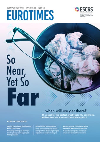
So Near, Yet So Far
The quest for the perfect presbyopic IOL continues. Will we ever see a true accommodating IOL?
Publisher Tom Ogilvie-Graham
Executive Editor
Stuart Hales
Editor-In-Chief
Sean Henahan
Senior Content Editor
Kelsey Ingram
Creative Director
Kelsy McCarthy
Graphic Designer
Jennifer Lacey
Circulation Manager
Lucy Matthews
Contributing Editors
Cheryl Guttman Krader Roibeárd O’hÉineacháin
Contributors
Laura Gaspari
Soosan Jacob
Priscilla Lynch
Timothy Norris
Clare Quigley
Andrew Sweeney
Colour and Print
CitiPost
Advertising Sales
Roo Khan
MCI UK
Tel: +44 203 530 0100 | roo.khan@wearemci.com
EuroTimes® is registered with the European Union Intellectual Property Office and the US Patent and Trademark Office.
Published by the European Society of Cataract and Refractive Surgeons, Building 4000, Langstone Park, Langstone Road, Havant, PO9 1SA, UK. No part of this publication may be reproduced without the permission of the executive editor. Letters to the editor and other unsolicited contributions are assumed intended for this publication and are subject to editorial review and acceptance.
ESCRS EuroTimes is not responsible for statements made by any contributor. These contributions are presented for review and comment and not as a statement on the standard of care. Although all advertising material is expected to conform to ethical medical standards, acceptance does not imply endorsement by ESCRS EuroTimes. ISSN 1393-8983
The Near Future
Simultaneous vision lenses (SVLs) have emerged at the forefront of ophthalmology’s near future, representing the next step towards the perfect presbyopic IOL. In this issue, EuroTimes examines the lenticular solutions now available for presbyopia and the ongoing search for a truly accommodating IOL.
When monocular IOLs were the only type of lenses available, patients could expect good distance vision but would ordinarily need reading glasses to see up close.
Micromonovision strategies offered the possibility of good distance and near vision for patients who could tolerate that approach. The quest for a lenticular treatment for presbyopia saw the development of a vast number of refractive and diffractive SVLs—trifocals, extended depth of focus, lightadjustable, enhanced monofocals, and phakic IOLs.
SVL designs continue to evolve. None of the simultaneous vision lenses is perfect; all have compromises, but they do offer good options for carefully selected presbyopic patients. Indeed, with more than a dozen SVL platforms available, choosing an IOL can be confusing for both doctors and patients, and marketing messages only add to the confusion. Several articles in this issue offer ways to enhance communication with presbyopic patients before and after surgery.
The ESCRS has provided a valuable service to the ophthalmic surgery community with the publication of an evidence-based functional classification of simultaneous vision lens types.1 The issue is also addressed in the new ESCRS Cataract Surgery Guidelines.2
Deciding which lens is best for each patient involves discussing their lifestyle needs and preferences and then selecting the lens that matches those requirements.
The ultimate goal, the holy grail, has always been to have an IOL that does what the natural lens can do—adjust vision at all distances. Unfortunately, what nature does so effortlessly has proven to be a complex challenge for IOL designers. Recent developments suggest we are getting closer to the goal.
Jorge Alió and Ioannis Pallikaris share the latest in accommodating IOL clinical research with Tim Norris in this issue’s cover story. The article reviews several accommodating IOLs nearing market release, such as the Juvene (LensGen) and FluidVision (Alcon) lenses.
An article by Laura Gaspari suggests an entirely different approach that could radically change cataract surgery—using an eye drop containing lipoic acid to prevent the progressive stiffening of the natural lens with age and restore its elasticity. Clinical studies are underway.
While the restoration of natural accommodation has not yet been achieved, we can now offer our presbyopic patients more vision correction options than ever before, providing excellent vision with or without glasses.
Thomas Kohnen
Editor of the Journal of Cataract & Refractive Surgery and Executive Board Member of ESCRS
For citation notes, see page 38.
EDITORIAL BOARD
Adi Abulafia (Israel)
Bruce Allan (UK)
Noel Alpins (Australia)
Juan Alvarez de Toledo (Spain)
Gerd Auffarth (Germany)
Başak Bostanci (Türkiye)
John Chang (Hong Kong SAR, China)
Béatrice Cochener-Lamard (France)
Burkhard Dick (Germany)
Mor Dickman (The Netherlands)
Joaquín Fernández (Spain)
Oliver Findl (Austria)
Sri Ganesh (India)
Christina Grupcheva (Bulgaria)
Farhad Hafezi (Switzerland)
Nino Hirnschall (Austria)
Soosan Jacob (India)
Jack Kane (Australia)
Yao Ke (China)
Georgios Kymionis (Greece)
David Lockington (UK)
Artemis Matsou (Greece)
Cyres Mehta (India)
Jod Mehta (Singapore)
Sorcha Ní Dhubhghaill (Belgium)
Rudy Nuijts (The Netherlands)
Catarina Pedrosa (Portugal)
Konrad Pesudovs (Australia)
Nic Reus (The Netherlands)
Filomena Ribeiro (Portugal)
Andreia Rosa (Portugal)
Giacomo Savini (Italy)
Julie Schallhorn (US)
Sathish Srinivasan (UK)
Paola Vinciguerra (Italy)
Shin Yamane (Japan)
Ron Yeoh (Singapore)
Mihail Zemba (Romania)
Thomas Kohnen
José Güell
Paul Rosen
ESCRS UPDATE
2025 Annual Report Now Available
ESCRS has released its 2025 Annual Report, highlighting the people, programmes, and initiatives that have made the Society a driving force in advancing ophthalmic training and research in Europe and around the world.
The 58-page report, available on the Society’s website, touches on all aspects of ESCRS programming and especially the educational, research, and charitable projects that lie at the heart of the Society’s mission and vision. It also focuses on ESCRS’s collaborations with other ophthalmic societies, including new partnerships with national societies in Brazil, Egypt, and Taiwan. These collaborations provide a structured framework for alignment and shared progress across Europe as well as in Asia, Africa, and the Americas.
One of the most significant achievements of the year was the international consensus reached at the 2025 Annual Congress in Copenhagen on the ESCRS classification of simultaneous vision intraocular lenses. By bringing together leading experts in cataract and refractive surgery, the consensus aimed to establish a shared framework and terminology for the classification of IOLs, helping surgeons, researchers, industry, and patients better understand the optical characteristics and intended outcomes of different lens designs.
The report also highlights ESCRS charitable initiatives in Ukraine, Nepal, the Middle East, and sub-Saharan Africa, including funding for the Surgical Training Unit at the University of Cape Town that has helped strengthen surgical capacity across Africa, with more than 100 surgeons benefiting from advanced training programmes.
Among the many educational and research initiatives featured in the report are the following:
• Masterclasses in minimally invasive glaucoma surgery and complex cataract;
• the €60,000 Peter Barry Fellowship Award;
• the IOL calculator, which made nearly 4.6 million calculations in 2025;
• weekend programmes and meeting presentations to support leadership development among ophthalmologists working in public practice settings, in hybrid public–private roles, and those in the final years of specialist training;
• research awards and new research working groups; and
• the continuing growth of the ESCRS Registries programme, whose three databases now contain data on more than 4 million surgical procedures.
Scan the QR code to read the report.
Annual Report 2025
Latest IME Video Explores Transition to Digital OR In episode #3 of the ESCRS Independent Medical Education (IME) video interview series, Drs Gerd Auffarth and Amir Hamid discuss how adopting certain practices can help ophthalmologists overcome cost barriers and successfully transition to a digital operating room. The video, “Cost, Adoption, and Implementation,” follows on previous videos in this series titled “The Three-Phase Workflow” and “Ergonomics and 3D Visualisation.”
All three videos are available through the ESCRS Educational Forum, which is supported by multiple industry partners to provide independent didactic education on selected therapeutic areas. The platform combines presentations from ESCRS Winter and Annual Congresses, selected EuroTimes articles, videos, and webinars to provide an in-depth overview of current clinical outlooks.
Scan the QR code to watch the video.
BY TIMOTHY NORRIS
Another successful idea
Pioneering surgeon Ioannis Pallikaris MD, PhD, best known for performing the first LASIK surgery, has also maintained a strong interest in IOL development.
He recalled the anticipation from two decades ago, when the field was excited to know which accommodative lens would next reach the market; by 2007, at least 10 distinctive designs were waiting for regulatory approval. Now, patients and surgeons are happier with available pseudophakic technologies, making it harder for new accommodation designs to find a spot in the same market, Prof Pallikaris said.
The first product labelled as an accommodative IOL was the Crystalens (Bausch + Lomb), which received FDA approval in 2003 and updates in 2008. The Trulign toric IOL (Bausch + Lomb), which was based on the Crystalens design, received approval in 2013.
Although the Crystalens initially generated a lot of excitement, enthusiasm waned once it became clear the lens did not provide complete accommodation, Prof Pallikaris recalled. This kind of lens had a problem with tilt and capsular fibrosis, eventually becoming ineffective, he said.
Agreeing with Prof Alió that fibrosis of the capsule is unavoidable, Prof Pallikaris said overcoming this limitation is essential for the evolution of accommodating IOLs.
“We are not so far from a functional accommodative IOL,” Prof Pallikaris said. “There are other problems, starting from capsulorhexis, lens design, binocular summation, and the lack of a centration system, but solving fibrosis is essential for obtaining a functional accommodation.”
One of the solutions he proposed for addressing the fibrosis issue is fixOflex™ (EYE PCR), an endocapsular device specifically designed to avoid posterior capsule opacification (PCO) formation. The device is implanted in the sulcus before phacoemulsification protects the IOL from fibrosis, potentially providing some accommodative power.
“When dealing with accommodative IOLs, reconstructing a normal capsule shape makes sense,” Prof Pallikaris explained. “FixOflex has some degree of elasticity and, with some small design changes on the way, may adapt a certain design with accommodation.”
In a prospective study involving 121 patients, those receiving the fixOflex device had a PCO rate of 0.83% at one year compared with 13.0% in controls, with no patients requiring Nd:YAG capsulotomies.3 Another study suggested the device had the potential to all but eliminate dysphotopsia.4
As Gerd Auffarth MD famously titled his 2021 Binkhorst Lecture, there is no free lunch in optics5—especially for accommodative lenses. According to Prof Alió, the final
IOL Dual optic, fluid-filled system in which ciliary body contraction reshapes the optics
IOL Uses a biomimetic gel replicating the paediatric crystalline lens
OmniVu IOL Continuously variable power via a fluid-based element driven by ciliary contraction
Lumina IOL Sulcus based, relies on two sliding plates that move with ciliary muscle contraction
CE Mark 2026; Available in EU AkkoLens Akkolens.com
limitation in restoring physiological capability in lieu of the natural lens is variability among patients’ eyes. Additional challenges include improving accommodation measurement, increasing dioptric change, and maintaining optical quality.
We still have better systems today, and we are working on the potential of binocular summation.
“We still have better systems today, and we are working on the potential of binocular summation. I do not think we have to stick to a very precise accommodative system, because our brains are very forgiving and can accept a high degree of compromise on the precision of the optics,” Prof Pallikaris concluded.
For citation notes, see page 38.
Jorge L Alió MD, PhD is Professor of Ophthalmology at the Miguel Hernández University in Alicante, Spain, and founder of Vissum Miranza, Alicante, Spain. Jlalio@vissum.com
Ioannis Pallikaris MD, PhD is Professor of Ophthalmology, Rector University of Crete, Greece; Founder and Director of the Institute of Vision and Optics, University of Crete, Greece; and Founder of EYE PCR, Greece. pallikar@gmail.com
Seeking Personalised Vision
Adjustable IOL technology may help in finding postoperative surgical fine-tuning.
LAURA GASPARI REPORTS
Adjustable IOL technology may offer a shift towards personalised medicine, especially in avoiding refractive surprise, according to H Burkhard Dick MD, PhD.
Despite the optimisation of biometry across formulas and AI support, residual refractive error remains a problem for many patients. According to Professor Dick, the predictive models are excellent, but many other aspects come into play, such as technical, biological, procedural, and mechanical issues. Adjustable IOL technology is a response to the increasing number of post-keratorefractive eyes seeking freedom from spectacles and to the low tolerability of postoperative refractive errors.
“I think we are about to have a paradigm shift,” Prof Dick said. “The old model was measure, calculate, implant, and hope. Over the next few years, the new model will be measure, implant, heal, adjust, and personalise.”
There are different modalities: IOLs requiring secondary procedures for power adjustment, those that do not require it, and, finally, noninvasive IOL power adjustment.
The multicomponent approach includes platform support, making the IOL easily exchangeable. Early results from 12-month follow-up data are promising, and, as Prof Dick noted, these lenses are useful when secondary IOL intervention is more likely, such as in paediatric patients, progressive corneal pathologies, toric IOLs in high myopes, and in eyes requiring tamponade agents during vitreoretinal surgery.
The mechanically adjustable lenses contain springs activated by heating with a green continuous laser (e.g., argon), enhancing the anterior and posterior movement of the optic or facilitating rotation. Human trials have just started for this technology.
I think we are about to have a paradigm shift.
Noninvasive IOL power adjustment approaches, such as la ser-induced refractive index change (LIRIC) with a femtosec ond laser, are driven by multiphoton absorption of ultrashort laser pulses and can be performed intracorneally and in the lens material. Multiphoton absorption enables local modification of the refractive index. In RIS (Perfect Lens), light (520 nm low-energy laser) breaks ester molecules within the polymeric material and creates polar hydrophilic molecules within the IOL. Another project by the University Eye Hospital of Bochum and Schwind Eye Tech Solutions, supported by the German Ministry of Education and Research, demonstrated good first results in the cornea.
As for light-adjustable lens technology, Prof Dick has 17 years of experience, with 3,000 light-adjustable lenses implanted in various conditions, demonstrating longterm stability. What patients really appreciate about this lens, according to Prof Dick, is the ability to adjust their vision postoperatively with a precision of 0.25 D, ideally for minimonovision. These lenses offer new presbyopia solutions, including individually adjustable monovision and simultaneous vision profiles, as well as negative asphericity.
Light Adjustable Lens 2.0 (RxSight) has good results, with intermediate vision of 20/30 or better in more than 90% of patients and particularly good uncorrected near vision acuity. They are safe and of high quality, offer a wide range of vision solutions, and have no photic phenomena. Gaining momentum in Europe, the Light Adjustable Lens 2.0 is already successful in the US and Canada.
“I think the future lies in personalised medicine,” Prof Dick concluded.
Prof Dick spoke at the 2026 ESCRS Winter Meeting in Helsinki.
H Burkhard Dick MD, PhD, FEBOS-CR is professor and chairman of the Ruhr University Eye Hospital in Bochum, Germany, and ESCRS president. dickburkhard@aol.com
Addressing Dissatisfaction with Dysphotopsia
Managing photic phenomena is key for lowering patient dissatisfaction.
TIMOTHY NORRIS REPORTS
With up to 20% of patients implanted with either partial range of focus enhanced IOLs or full range of focus smooth transition IOLs reporting some level of dissatisfaction with their postoperative results, good preoperative counselling is essential, stresses Lars Morten Holm MD, PhD.
“We did a study in my clinic on the satisfaction rates of cataract patients, delivering patient-reported outcome (PRO) questionnaires,” Dr Holm said. “Surprisingly, there was no significant difference between extended depth of focus (EDOF) and monofocal IOLs.”
The literature showed blurred vision as the main complaint with full range of focus smooth transition (FULL-RoFSmooth-Transition) IOLs as well as partial range of focus extend (PARTIAL-RoF-Extend) IOLs, with 13% of patients reporting it in both groups. Positive dysphotopsia (or, more precisely, photic phenomena) was present in 39% to 80% of FULL-RoF-Smooth-Transition IOL patients, with halos as the most commonly noted phenomenon. However, the majority were not bothered by these effects. The frequency of glare, halo, and starbursts in the PARTIAL-RoF-Extend IOL group from Dr Holm’s clinic was around 4%—not significantly different from the monofocal results.
Among dissatisfied referred patients, the dominant complaint was blurred vision, as evidenced by 75% of patients in the PARTIAL-RoF-Extend lens group versus 62% of patients in the FULL-RoF-Smooth-Transition lens group, Dr Holm said. Ametropia emerged as the main reason for these high
percentages (55–65%), followed by dry eye disease (4–26%), posterior capsular opacification (PCO, 12–16%), and large pupil (15%). Other issues included maculopathy (such as cystoid macular oedema), epithelial basement membrane dystrophy, Fuchs’ dystrophy, floaters, diabetic macular oedema, minimonovision intolerance, and epiretinal membrane, Dr Holm noted.
Evaluating the unhappy patient
When evaluating an unhappy patient at the clinic, Dr Holm suggested combining good counselling with a review of the patient’s ocular history. The investigation should include whether the patient was a good candidate from the start and if there were signs of amblyopia from previous strabismus surgery (or any other ocular surgery), a challenged retina from a retinal detachment surgery, or a challenged cornea from a laser surgery. Surgeons should note the preoperative refraction since previous myopic patients have high demands for near visual acuity. Moreover, he suggested paying attention to whether the dissatisfaction occurred immediately after implantation or later, as this may indicate the reason for dissatisfaction.
After considering these causes of dissatisfaction, surgeons should evaluate whether the patient has PCO, macular oedema, or floaters that developed in the weeks or months after implantation, or PCO or a calcified lens that developed years later.
The eye examination needs to be thorough, Dr Holm said, noting the refraction must be subjective because autorefraction can be misleading. Pupil size greater than 5.0 mm mesopic or 6.0 mm scotopic can impact night driving due to the exposure of more optical zones.
Before considering a surgical approach with these dissatisfied patients, Dr Holm recommended first prescribing spectacles or contact lenses as an ametropia treatment. Moreover, the surgeon should be able to explain the reason for dysphotopsia and negotiate a viable solution. According to Dr Holm, only 2% of patients will fail to neuroadapt. Of these, 4% will continue to be intolerant of dysphotopsia and should be counselled for IOL exchange.
Dr Holm spoke at the 2026 ESCRS Winter Meeting in Helsinki.
Lars Morten Holm MD, PhD is Clinical Associate Professor, University of Copenhagen, Denmark. lars.morten.holm.01@regionh.dk
Maximising Success While Minimising Dissatisfaction
Choosing the right IOL strategy for each patient.
TIMOTHY NORRIS REPORTS
Presbyopia is a common life experience. What changes are patient needs and expectations when they ask for presbyopia correction, according to Hrvoje Kovacic MD.
“Historically, the majority of daily tasks were distanceoriented,” Dr Kovacic explained. “Now it has changed to near work with laptops, tablets, and smartphones. We went from restoring vision to optimising it, where spectacle independence is increasingly expected.”
This evolution makes the life of an ophthalmologist a little more difficult, but luckily technology is answering the call, Dr Kovacic said.
Monovision with monofocal-plus IOLs is one option. The dominant eye is targeted for emmetropia and the non-dominant eye for myopia (-0.75 D to -2.00 D) to provide a functional range of vision without optical disturbances. These lenses show a good spectacle independence rate (90%) and high patient satisfaction. Suitable patients are those who had contact lens monovision and those who are intolerant of glares and halos.
Monofocal-plus IOLs have been shown to improve in-
Careful patient selection and appropriate IOL matching are crucial to successful outcomes.
termediate visual acuity compared to standard monofocals without the trade-off of simultaneous vision IOLs. Eligible patients prioritise distance clarity, with a small bonus at intermediate distances. Candidates for this approach include those not suitable for extended depth of focus or multifocal IOLs
and patients seeking monofocal-like vision with high contrast sensitivity and minimal photic phenomena.
Extended depth of focus IOLs create a continuous range of vision through an elongated focal zone, with the advantage of excellent distance vision, good intermediate vision, fewer halos and glares, and higher contrast sensitivity compared to multifocal IOLs. However, near vision remains limited, typically requiring glasses, and some photic phenomena may occur.
Multifocal lenses are another option, and many improvements have been made since the US FDA first approved them in 1997. Dr Kovacic highlighted the ReSTOR (AcrySof) from 2005 as an example, with its apodised diffractive design. It showed good results, but with a major downside related to dysphotopic phenomena. Trifocal IOLs rose as a popular option in the 2010s. A study comparing the PanOptix (Clareon) and Synergy (TECNIS) intraocular lenses showed good distance and intermediate uncorrected visual acuity, with slightly less near vision and fewer dysphotopsia.1 Patients reported no need for spectacles in 80% of cases, with 90% never or rarely needing them.
Suitable candidates for trifocal IOLs include patients with multiple visual demands, especially for near vision, and a strong desire for spectacle independence. Dr Kovacic said these patients should have realistic expectations, including the possibility of halos and glares. Trifocal IOLs are not suitable in patients with severe dry eye, retinal and corneal pathologies, and glaucoma.
Dr Kovacic also suggested different IOL combinations, including mix-and-match strategies or monovision, as options for reducing spectacle dependence while minimising visual disturbances from photic phenomena. He further emphasised the importance of carefully evaluating the type of patients sitting in the ophthalmologist’s waiting room.
“Careful patient selection and appropriate IOL matching are crucial to successful outcomes,” Dr Kovacic said. “[It’s vital to] manage expectations to minimise postoperative dissatisfaction.”
Dr Kovacic spoke at the 2026 ESCRS Winter Meeting in Helsinki.
For citation notes, see page 38.
Hrvoje Kovacic MD is an ophthalmologist at Het Oogziekenhuis Rotterdam and Franciscus Gasthuis and Vlietland, Netherlands. h.kovacic@oogziekenhuis.nl
Phakic IOLs Expand Presbyopic Treatment Options
Strict implantation criteria must be considered when dealing with presbyopic phakic IOLs.
LAURA GASPARI REPORTS
Presbyopic phakic IOLs are a good surgical solution to presbyopia, but strict criteria for patient selection is essential, stresses António Marinho MD, PhD.
Phakic IOLs are considered to be a valuable instrument for refractive correction because of their noninvasive nature, as they do not change the natural refractive structures of the eye.
“They are very good in high ametropias and also in lower ametropias, as they are predictable and give high stability of refraction from day one,” Dr Marinho said. “Phakic IOLs preserve accommodation and, most importantly, they give us a very good quality of vision with gains of lines.”
But how do these technologies translate to presbyopia?
Phakic IOLs in young patients and in presbyopes share the same concept, but differ in optic design. Three types of optics are available for presbyopic phakic IOLs: the diffractive, the extended depth of focus (EDOF), and the multi-zone refractive optics.
The oldest phakic IOL is the presbyopic implantable phakic contact lens (IPCL), a posterior chamber IOL of hydrophilic hybrid acrylic material, available in different sizes, with a central hole, diffractive optic, and various additions (from +1.5 to +4.0 D). An example of the second type, the EVO Viva ICL (STAAR Surgical) is a posterior chamber IOL made of collamer, available only for myopia (-0.50 to -18.00), with a central hole and an EDOF optic without diffractive rings. The Artiplus (Ophtec), an example of the third type, features a hybrid IOL with a fixed addition of 2.5 D and an aspheric segmented optic with multiple elongated focal points. However, despite the good performance of these lenses, not all patients are suitable candidates for implantation. Inclusion criteria are a healthy eye, a normal retina, no glaucoma, stable refraction, and specific anatomical characteristics
including anterior chamber depth (at least 2.8 mm from the endothelium for posterior chamber IOLs and 3.2 mm for Artiplus), crystalline lens rise (less than 600 microns), and good angle and endothelium.
These implantation criteria should be considered carefully when selecting a patient for a presbyopic phakic IOL, said Dr Marinho. He added medium myopes are the ideal candidates, with the possibility of a slight monovision if needed, whereas high myopes potentially present retinal pathologies and low myopes tend to adapt poorly to multifocality. Contact lens users are also ideal candidates, as they are accustomed to good vision at different distances.
Concerning hyperopia, theoretical hyperopes between 45 and 55 years are considered ideal due to their easy adaptation to multifocality. However, the anatomical criteria can be challenging, as hyperopes sometimes have a low anterior chamber depth. Unstable refraction in hyperopic patients can be a challenge, but clear lens extraction can help solve this issue.
In general, presbyopic phakic IOLs are a good and available solution, Dr Marinho said. “We don’t know exactly about the quality of vision and patients’ satisfaction in the real world, but I think we must still have some strict criteria for selection.”
Dr Marinho spoke at the 2026 ESCRS Winter Meeting in Helsinki.
António Marinho MD, PhD, FEBOS-CR (Hon) is an ophthalmologist specialist in refractive, cornea, and cataract surgery at the Hospital da Luz Arrábida, Universidade do Porto, Porto, Portugal.
Weighing LASIK Ablation Profile Options
Four options exist, but is any one the best?
CHERYL GUTTMAN KRADER REPORTS
Leading refractive surgeons agree that when performing LASIK, excellent results can be achieved whether one chooses to use the now standard wavefront-optimised (WFO) technique or any of the customised options: wavefront-guided (WFG), topography-guided (TPG), or raytracing-guided (RTG).
Available evidence from comparative studies, however, indicates that between WFO-, WFG-, and TPG-LASIK, the WFG technique delivers the highest levels of 20/16 and 20/12.5 uncorrected distance visual acuity (UCDVA), better low contrast VA, and greater gains in corrected distance visual acuity (CDVA), said Edward E Manche MD.
Raytracing-guided LASIK represents the newest technique, with the platform used for this procedure only receiving the CE mark in 2019 and FDA approval in 2025. Results of a headto-head study being conducted by Dr Manche will provide insight as to how its outcomes compare to those of a WFG procedure (study NCT07078799).
John Kanellopoulos MD agrees that more information is needed to determine how RTG-LASIK performs relative to WFG-LASIK. However, describing RTG-LASIK as a “WFG+” procedure, he suggested RTG-LASIK will give WFG-LASIK “a run for its money”, outperforming WFG-LASIK for improving visual outcomes beyond 20/20 because it provides a better view of the whole eye.
Conclusions grounded in scientific evidence
Dr Manche said there is an abundance of head-to-head studies comparing WFG-LASIK with WFO-LASIK. A meta-analysis including early studies evaluating the two techniques found their outcomes were comparable.1 However, Dr Manche reviewed subsequently published investigations, including several he conducted, that showed superior results across multiple endpoints using WFG-LASIK.2–4
Other published articles also describe studies that directly compared TPG-LASIK and WFO-LASIK, and these trials have generated conflicting results. Some studies showed the two techniques delivered similar outcomes, although a more recent randomised clinical
trial reported superior outcomes with WFO-LASIK compared to TPG-LASIK.5 Stating that he found only three published papers comparing WFG- and TPG-LASIK,6–8 Dr Manche said the results of these trials were more consistent in showing some advantages for WFG-LASIK.
Understanding a role for RTG-LASIK
Dr Kanellopoulos authored the first published paper on RTGLASIK, followed by subsequent studies, including a multinational trial.9–11 Prefacing a discussion of this technique, he reiterated that WFG-, WFO-, and TPG-LASIK all perform well in delivering 20/20 UCVA.
“While wavefront guidance was a major advance in LASIK, raytracing is the next optical model, and achieving better quality of vision is where raytracing matters,” Dr Kanellopoulos said.
“It only makes sense to consider RTG-LASIK if the goal is to improve the optics of the entire eye, not just the cornea, and to achieve better retinal image quality, not just emmetropia and spectacle independence.”
Providing an overview of RTG-LASIK, Dr Kanellopoulos said it uses a better planning engine than WFG-LASIK because it personalises the eye rather than just the wavefront. He explained that, while the WFG technique treats measured wavefront aberrations and low-order aberrations based on the Gullstrand 2D eye model, the raytracing approach builds a personalised 3D model of the whole eye and optimises treatment through that model using wavefront and corneal tomographic data. In addition, planning for RTG-LASIK incorporates data for expected biomechanical changes and epithelial remodelling, calculates multiple treatment patterns, and chooses the optimal pattern for correcting both low- and high-order aberrations.
“Furthermore, by reshaping the cornea along the visual path and improving corneal symmetry in relation to the visual path, RTG-LASIK may facilitate a more favourable optical system for future lens-based procedures, such as cataract surgery,” Dr Kanellopoulos said.
In a study presented at the 2026 ASCRS meeting, Dr Kanellopoulos compared RTG-LASIK with keratorefractive lenticule exchange (KLEx) performed using SMILE pro. He said the results showed subtle differences overall, but RTGLASIK demonstrated its advantages when comparing the percentages of eyes with gains in CDVA and contrast sensitivity outcomes. However, Dr Kanellopoulos noted the results need to be considered in light of the study’s methods, which relied on RT measurements to determine the amount of myopia and astigmatism correction in the KLEx procedures.
“In addition, the elephant in the room is that none of the customised LASIK treatments take into account the contribution of the human lens. In younger patients, accommodation can bias lower- and higher-order aberrations, affecting accuracy of the treatments, including RTG-LASIK,” he said. “Managing this variability is an essential component of the learning curve when adopting raytracing technology. We routinely obtain measurements under cycloplegic conditions, using two drops of 0.5% tropicamide administered approximately 15 minutes apart. Measurements are done approximately 30 minutes later and used for planning without modification or nomogram intervention.”
I still believe that the best customisation tool for LASIK is the experienced refractive surgeon.
Practical
advice for practising surgeons
Dr Kanellopoulos emphasised the difficulty in debating which of the four LASIK techniques is the best, but he offered some personal recommendations. He suggested that surgeons who do not have a comprehensive understanding of the scientific principles underlying WFG-LASIK, TPG-LASIK, or RTG-LASIK can do well performing WFO-LASIK when the ablation plan is based on population averages. Surgeons who are comfortable with the more complex planning involved when performing WFG-LASIK or TPG-LASIK can expect to achieve good outcomes by choosing either of the two more customised procedures.
The bottom line for successful LASIK, however, is not how the procedure is performed, but by whom.
“I still believe that the best customisation tool for LASIK is the experienced refractive surgeon,” Dr Kanellopoulos said.
Drs Manche and Kanellopoulos debated the relative merits of LASIK planning at a session of the 2026 ASCRS annual meeting in Washington, DC.
For citation notes, see page 38.
Edward E Manche MD is Professor of Ophthalmology, Byers Eye Institute, Stanford University School of Medicine, Palo Alto, California, US. edward.manche@stanford.edu; lasik.manche@stanford.edu
A John Kanellopoulos MD is Medical Director, Laservision Ambulatory Eye Surgery Unit, Athens, Greece, and Clinical Professor of Ophthalmology, NYU Grossman Medical School, New York, US. ajkmd@mac.com
Premium IOLs start with premium
measurements
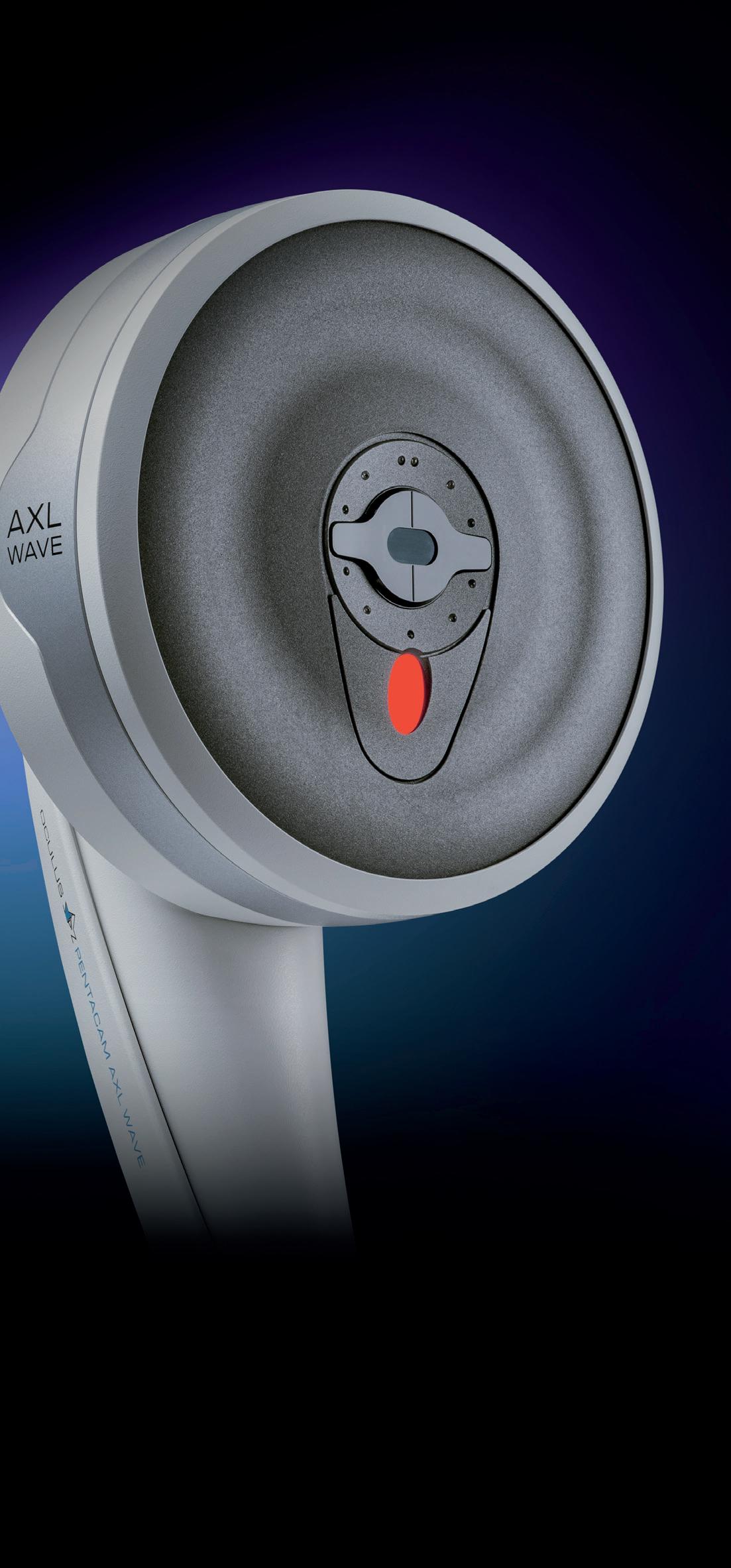
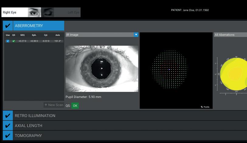
With the Pentacam ® AXL Wave, you consolidate all key measurement data into a single workflow.
Maximize Premium IOL success –for you and your patients. oculus.de
Phakic IOLs Step into the Presbyopia Spotlight
Evidence points to safe, effective outcomes despite minor adaptation hurdles.
LAURA GASPARI REPORTS
Phakic IOLs are a safe and effective approach for myopia and presbyopia correction in patients with early and advanced disease stages, although some adaptation issues may occur, according to Erik L Mertens MD.
“The advantages of phakic IOLs are many,” Dr Mertens explained. “They preserve the natural crystalline lens and any possible residual accommodation. Retinal detachment rates for IOL exchange are much higher than after phakic IOL implantation, and looking at the literature, the latter has a lower risk of endophthalmitis. Finally, phakic IOLs are reversible implants and easier to remove than in-the-bag IOL exchanges.”
Regarding accommodation for those between the ages of 45 and 50 years old, there is a drop from approximately 4.00 D to 2.00 D, but some residual accommodation could be helpful when implanting a phakic IOL. Despite the prevailing view that these kinds of lenses should not be implanted in this population, two peer-reviewed studies showed good results, demonstrating a safety index of 1.00 and an efficacy index of 0.95 and 1.00 in patients between 40 and 56.1 Dr Mertens said with such results, phakic IOLs should be considered for correcting myopia and myopic astigmatism in older patients with a clear lens.
Presbyopia-correcting implantable collamer lenses (ICLs) are available on the market, including the EVO Viva™ by STAAR Surgical, an aspheric refractive collamer lens. Its powers are in the myopic range (-0.50 D to 18.00 D in 0.50 D steps) and the lengths are from 12.1 mm to 13.7 mm. It is a single piece, CEmarked lens with concave/convex optics, and it features a 360µm hole in the middle to avoid iridotomies and blockage after surgery. The indications for the lens include the correction of myopia with mild presbyopia in the range of -0.50 D to -20.00 D. The refractive optic of the lens works in conjunction with the
crystalline lens and residual accommodation. It induces spherical aberration, but with a downside: it can cause coma when not well centred, which can impact near and distance vision, leading to potential patient dissatisfaction, he noted.
A prospective multicentre open-label clinical study involving five centres in Europe evaluated the safety and efficacy of this lens in phakic subjects with presbyopia.2 The indication for the EVO Viva lens was the correction of myopia with presbyopia between -0.50 D and -20.00 D at the spectacle plane. The primary efficacy endpoint of this study was met: 98% of eyes achieved monocular uncorrected near visual acuity of 20/40 or better at six months postoperatively. There was a significant improvement in distance vision and especially in intermediate and near vision, which were the main objectives of the study. Improvements reached 8 lines for intermediate vision and 6.5 lines for near vision, demonstrating significant gains across all distances.
“There was a transient decrease in corrected distance visual acuity due to induction of coma and spherical aberration. At six months postoperatively, 91% of patients were satisfied,” Dr Mertens concluded.
Dr Mertens spoke during the 2026 ESCRS Winter Meeting in Helsinki.
For citation notes, see page 38.
Erik
L Mertens MD, FEBO, PCEO, FWCRS, FEBOS-CR is Founder and Medical Director of Medipolis Eye Centre in Antwerp (Wilrijk), Belgium. e.mertens@medipolis.be
Exclusive learning. Unlimited access for ESCRS members.
Artiplus Phakic IOL Expands Treatment Options
Multicentre trial confirms safety and efficacy of the iris-fixated multifocal phakic IOL.
LAURA GASPARI REPORTS
The Artiplus phakic IOL (Ophtec) yielded good refractive and visual acuity outcomes at the two-year mark in presbyopic patients, an international multicentre study suggests.
The lens is the multifocal successor to the Artiflex, with a multisegmented refractive optic on the posterior surface to minimise dysphotopsias and a reading add power of +2.50 D.
According to Robin Ollivier MD, iris fixation prevents the biggest challenge facing phakic multifocal IOLs, especially those in the posterior chamber: centration.
“As the lens is fixated on the iris, it grants maximum stability,” she clarified.
The optical design includes continuous transitional focus technology, providing a full range of vision with a smooth transition, and is available in both toric and non-toric versions for myopia and hyperopia, which is quite unique.
The lens needs a pre- or intraoperative peripheral iridectomy to avoid any intraocular pressure (IOP) rise, and the surgical procedure for the implantation is straightforward, requiring only 5 to 15 minutes depending on the peripheral iridectomy.
The international multicentred study, led by Professor José Luis Güell and conducted in nine centres across Europe and South Korea, involved 49 patients implanted with Artiplus in both eyes (98 eyes). It was an open label, prospective and non-controlled study with a follow-up period of three years.
Patients selected were presbyopic adults requiring at least +1.00 D reading glasses and with good ocular health. The study was performed on the non-toric model, so the expected postoperative refractive cylinder would be ≤ 0.75 D. The minimal anterior chamber depth was 2.8 mm from the corneal endothelium, and other implantation criteria, like the endothelial cell count, were in line with those of iris-fixated phakic IOLs.
The refractive result remained stable after two years, with a mean of -0.25 D. Visual acuity in logMAR was 0.04 for far, near, and intermediate vision, both monocularly and binocularly.
Around 90% of patients achieved uncorrected visual acuity of 20/25 for distance, intermediate, and near vision, with potential correction up to 20/20 vision. The defocus curve showed a smooth transition from far to near vision, and contrast sensitivity remained unchanged pre- and postoperatively.
After surgery, 91% of patients no longer needed glasses while 9% required glasses occasionally for distance, intermediate, or near vision. The mean endothelial cell count loss was -0.6%, aligning with expected annual levels. Patient satisfaction questionnaires returned ‘very’ to ‘quite’ positive results, with minimal reports of glare, halos, and starbursts, which were reported as not bothersome. Adverse events included dry eye, two cases of transient high IOP, one case of mild viral conjunctivitis, one case of non-pathological guttata, one case of an optic neuritis unrelated to the Artiplus, and three cases of mild inflammatory deposits on the IOL treated with topical steroids.
The two-year interim results were excellent, with investigators highly satisfied with the lens’s safety and efficacy for presbyopia correction.
“It is a reversible procedure. Patient- and surgeon-reported outcomes are very positive,” Dr Ollivier concluded.
Dr Ollivier spoke at the 2026 ESCRS Winter Meeting in Helsinki.
Robin Ollivier MD is a cataract and refractive surgeon at the University of Maastricht, Netherlands.
Is Presbyopia Reversible with a Drop?
New research reopens the debate on pharmacological presbyopia treatment.
LAURA GASPARI REPORTS
Presbyopia is a nearly universal age-related visual disorder, typically driven by progressive stiffening of the crystalline lens. According to Aadithreya Varman, this process results from biochemical changes, particularly cross-linking and the formation of disulphide bonds.
“The progressive stiffening leads to a lack of accommodation with near vision difficulties,” he said. “Preventing these disulphide bonds from occurring may prevent the progressive stiffening and return the lens to its more elastic nature.”
Now, lipoic acid-based therapies are the leading candidates in the pharmacological lens softening concept, Dr Varman explained. The goal is to reduce the disulphide cross-linking naturally occurring in an ageing eye by using lipoic acid to function as a redox-active antioxidant.
Preventing these disulphide bonds from occurring may prevent the progressive stiffening and return the lens to its more elastic nature.
This treatment approach demonstrated encouraging results during preclinical phase one trials but did not meet statistical relevance in phase two trials. Phase three data, on the other hand, provided more advances in a multi-approach strategy that considered a topical delivery system.
In the preclinical phase, eight-month-old mice with biologically aged lenses were incubated with lipoic acid. The results showed a promising dose-dependent reduction in protein disulphide levels, with a corresponding increase in lens elasticity.1 The challenge is getting the lipoic acid to adequately penetrate further. For this reason, additional research was conducted to develop a prodrug that can be released into the aqueous humour. This led to the formulation of lipoic acid choline ester, which Dr Varman said can penetrate the cornea and release the drug into the aqueous humour.
Topical eye drops were eventually assessed in aged mice, showing a significant increase in elasticity in comparison to untreated eyes—providing the first real proof that age-related stiffening is reversible in vivo, Dr Varman said.
The following randomised, double-masked, placebocontrolled phase one/two study showed a greater improvement in distance-corrected near visual acuity (DCNVA) than placebo. In this study, 53% of treated patients gained more than 10 letters compared to the 21.7%
of the placebo group. The promising part, Dr Varman said, is these effects lasted five to seven months after treatment, suggesting some true disease modification.2 The phase two study, however, failed to show statistical improvement over placebo on a larger group at the three-month endpoint in the thrice-aday application protocol. Despite the high tolerability, the clinical programme was discontinued, he reported.
Dr Varman believes there is some rationale to suggest the strategy can really work. Evidence of an elasticity increase was demonstrated in previous studies, with patients gaining more DCNVA. The challenge is permeability, he pointed out, observing how formulation issues could have been the cause for failure in the phase two study.
Renewed interest led to the development of CLX-162, a new compound combining lipoic acid and pilocarpine to the formulation for an immediate meiotic effect. This new solution already showed promising results in preclinical studies.3
“The first-generation challenges were not a failure,” he emphasised. “The next steps of this compound would be to separate the symptomatic effect from true disease modifications. This requires a thoughtful decalibration and not a total abandonment of the therapy.”
The FDA has approved three prescription eye drops for presbyopia treatment: Vuity (Allergan) and Qlosi (Orasis Pharmaceuticals) use pilocarpine (1.25% and 0.40%, respectively) and VIZZ (LENZ Therapeutics) uses aceclidine hydrochloride 1.44%.
There are currently no FDA-approved eye drops to prevent or treat cataracts. Experimental agents under evaluation include lanosterol, C-KAD (Livionex), ethylenediaminetetraacetic acid, and methylsulfonylmethane.
Dr Varman spoke at the 2026 ESCRS Winter Meeting in Helsinki.
For citation notes, see page 38.
Aadithreya Varman MS, FWCRS, FAICO(Cataract/Phaco) is a cataract and refractive surgeon at the Uma Eye Clinic in Chennai, India. aadithreyavarman@gmail.com
SVIOLs and Comorbidities?
Overcoming lack of literature with evidence.
TIMOTHY
SNORRIS REPORTS
hould a patient with comorbidities be considered for simultaneous vision intraocular lenses (SVIOLs)?
According to Joaquín Fernández MD, PhD, the answer to this question followed the global consensus on the new classification of intraocular lenses proposed by ESCRS’s Functional Vision Working Group.
“Patients should not be excluded from SVIOL implantation solely due to the presence of comorbidities,” he said. “Exclusion should instead be considered only in cases where the anticipated visual performance and level of spectacle independence reported in comparable populations are unlikely to align with patient expectations, or when the balance between risks and benefits is unfavourable in the long term.”
Citing “Navigating the Lens Labyrinth: A Practical Approach to Intraocular Lens Classification and Selection,”
Professor Fernández noted that 52% of patients with and 12% of patients without comorbidities obtained spectacle independence, at intermediate and near respectively, with partial-depth-of-field-narrowed lenses (conventional monofocals). In the same analysis on rates of spectacle independence in eyes without comorbidities, patient rates were 77% at intermediate and 30% at near for the enhanced, 90% intermediate and 65% near for extended, 85% intermediate and 81% near for steep, 95% intermediate and 89% near for smooth, and 96% intermediate and 91% near for continuous.1
Using the IOLEvidence app, Prof Fernández provided some examples of enhanced and narrowed partial depth of focus lenses in eyes with glaucoma.2 In the moderate visual field loss group, outcomes with enhanced and narrowed lenses share similarities with some of the data available in literature regarding eyes with no comorbidities, he said. Patients with only a moderate affection of the visual field can be considered for the implantation of an enhanced lens.
Considering the balance between benefits and risks, Prof Fernández mentioned a study reporting that, among patients
with postoperative complaints, 28.9% of cases were associated with ocular disease.3 Therefore, comorbidities should not always be considered an exclusion criterion; rather, they require careful review of the literature involving patients with similar characteristics to provide the best possible evidence-based information and expected long-term visual outcomes.
Another study also showed dissatisfaction can be predicted through patients’ preoperative issues.4 Dry eye and residual refractive errors can be avoided in this phase, Prof Fernández observed. For SVIOLs in patients with amblyopia, it is important to understand distant visual acuity expectations and ensure that visual acuity at the desired distance exceeds the threshold (generally the 0.2 logMAR or 0.3 logMAR cut-off point), as poorer visual acuities are unlikely to provide comfortable functional vision for patients without spectacles, Prof Fernández said.5
The findings related to SVIOLs and retinal disease are quite interesting, he observed. The lack of evidence in literature suggested these kind of lenses should be discouraged in patients with a retinal disease. However, Prof Fernandez noted a study published in 2020 examined whether these lenses should be discouraged in diabetic patients (considering the risk of diabetic macular oedema), if they should be discouraged only in patients with existing macular oedema, or even in all patients with diabetes.6
Prof Fernández spoke at the 2026 ESCRS Winter Meeting in Helsinki.
For citation notes, see page 38.
Joaquín Fernández Pérez MD, PhD is the CEO and Medical Director in the Ophthalmology Department at Qvisión in Vithas Virgen del Mar Hospital, Almería, Spain. He is the secretary of the ESCRS. joaquinfernandezoft@qvision.es
The Evolving Role of Mitomycin C in Surface Ablation
Similar outcomes may carry different biological costs.
BY SOHEIL ADIB-MOGHADDAM MD
Few adjunctive treatments in refractive surgery have become as routine—and as rarely questioned—as mitomycin C (MMC). For years, its use during surface ablation has been widely accepted as an effective strategy for reducing postoperative corneal haze, particularly in higher corrections and deeper stromal ablations. Yet with the evolution of refined single-step transepithelial photorefractive keratectomy (trans-PRK), an important question deserves renewed attention: should MMC still be used routinely in every case?
Refined trans-PRK may have fundamentally changed the biological equation. Compared with conventional PRK, modern transepithelial approaches appear to produce smoother stromal surfaces, reduced tissue dehydration, and decreased keratocyte activation. These changes may contribute to a lower inflammatory response and, consequently, a lower intrinsic tendency towards postoperative haze formation.
This evolving perspective was reflected in a study my team and I previously published in the Journal of Refractive Surgery evaluating single-step transepithelial PRK performed with and without intraoperative MMC in eyes with mild-to-moderate myopia and total ablation depths of 160 µm or less. Visual, refractive, contrast sensitivity, and higher-order aberration outcomes were comparable between the two groups. Most importantly, postoperative haze remained similarly low regardless of MMC application.1
At the same time, however, MMC-treated eyes demonstrated less favourable endothelial findings, including greater endothelial cell loss and increased variation in cell size. Although these changes were not clinically dramatic, they raised an important concern: if refractive and haze outcomes are already comparable, even subtle biological costs deserve careful consideration.
The conclusion of the study reflected this concern directly: routine MMC application in this subgroup of patients ‘may be reconsidered.’ Importantly, the low haze incidence
observed in both groups was unlikely to result from a single factor. In addition to the smoother biological profile associated with transepithelial ablation, several adjunctive measures were consistently incorporated into perioperative management, including prolonged low-dose topical corticosteroids, intraoperative use of a chilled balanced salt solution, and rigorous postoperative ultraviolet protection.
Rather than a rigid protocol, this approach represented a practical strategy aimed at minimising unnecessary biological stress while preserving excellent refractive outcomes. In this context, refined trans-PRK became not merely a technical modification but part of a broader effort to improve the biological behaviour of surface ablation.
Perhaps the most important shift arising from this experience was conceptual rather than technical. Historically, refractive surgery has focused primarily on maximising visual outcomes and minimising haze incidence. However, when different approaches achieve similar refractive results, the biological price of adjunctive treatments becomes increasingly relevant.
This is particularly true for MMC. Its antifibrotic efficacy is undeniable in selected cases, and it remains valuable in eyes at elevated risk for haze formation, including higher myopia, deeper ablations, retreatments, and irregular corneas. Yet MMC is also a potent cytotoxic agent. Concerns regarding endothelial safety have already been raised, and potential effects on the ocular surface environment—including limbal stem cell integrity—should encourage thoughtful and selective use rather than automatic routine application.
The issue, therefore, is not whether MMC works. Clearly, it does. The more relevant question is whether advances in surface ablation technology now allow refractive surgeons to reserve MMC for situations in which its benefits clearly outweigh its biological cost.
In refractive surgery, progress is often associated with new lasers, algorithms, or platforms. Sometimes, however, progress comes from re-examining established habits. The future of surface ablation may depend not only on achieving excellent visual outcomes but also on doing so with the least possible biological compromise.
For citation notes, see page 38.
Soheil Adib-Moghaddam MD is an ophthalmologist at Bina Eye Hospital, Tehran, Iran. 2dr.soheil@gmail.com
The Quest for True Accommodation
What is the state of the art of injectable accommodative IOLs?
LAURA GASPARI REPORTS
The technology needed to build a true accommodative intraocular lens (IOL) is not yet ready, but there is reason to remain optimistic about seeing them in the not too distant future, according to Mayank Nanavaty MBBS, PhD.
“Traditional IOLs only give pseudoaccommodation, and multifocal and EDOF lenses have their trade-offs with dysphotopsia. There is an unmet need to correct accommodation in pseudophakic eyes,” he emphasised.
Injectable accommodative IOLs have been in development for at least a decade. The common mechanisms companies use to improve accommodation in these kinds of lenses include capsular bag expansion, anterior optic displacements, anterior surface curvature changes, and gradient refractive index modulation.
Over the years, many models and designs have been developed and remain under investigation, but as Professor Nanavaty explained, the literature is scant for most of them.
Of the accommodating IOL platforms in development, each takes a distinct approach.
The FluidVision IOL (Alcon) is a dual-optic, fluid-filled system in which ciliary body contraction reshapes the optics to deliver up to 5.0 D of accommodation through a standard phaco incision. It currently shows the highest clinically demonstrated accommodative amplitude among existing platforms.
The JelliSee IOL (JelliSee Ophthalmics Inc.) uses a biomimetic gel replicating the paediatric crystalline lens, demonstrating good rotational stability in human trials, with a bilateral study initiated in Q1 2025. The Juvene IOL (LensGen) has two plates, with the anterior element shifting forward for near and intermediate vision, and its 36-month data represents the strongest long-term evidence among injectable platforms, despite not yet being commercially available.
The OmniVu IOL (Atia Vision) offers continuously variable power via a fluid-based element driven by ciliary contraction and is currently in human trials. Finally, the Lumina IOL (AkkoLens International) relies on two sliding plates that move with muscle contraction, achieving up to 2.5 D in phase 3 trials, while sulcus fixation entirely avoids capsular bag complications. The cumulative amplitude of these technologies ranges from 1.5 D to 5.0 D, Prof Nanavaty reported.
Reported clinical outcomes include an uncorrected visual acuity of 20/25 or better in most patients, intermediate acuity of 20/32 or better at 60–80 cm, and near acuity of 20/40 or better in 70–85% of cases without distance compromise. Spectacle independence ranges from 85% to 92%, although this falls short of existing simultaneous vision lenses. Dysphotopsia rates have been reported as low.
However, Prof Nanavaty said the major challenge is posterior capsule opacification (PCO), when lens epithelial cells migrate and compromise the optics and accommodation over time, followed by capsular fibrosis and bag contraction.
Material science requirements include biocompatibility, long-term stability, optical clarity, viscoelasticity, and molecular weight. Other challenges include the unproven role of capsulorhexis and the shrink-wrap effect in PCO prevention, neuromuscular adaptation, and the difficulty of delivering complex devices through small incisions without deforming the capsular bag. Each model also requires tailored surgical techniques, and none have yet proven compatible with the available phacoemulsification technology.
Prof Nanavaty anticipates a future with the development of smart polymers, microincision delivery systems, modular upgrade platforms for accommodative lenses, and the integration of new IOL power calculations.
“Collaboration across material science, bioengineering, physiology, and clinical surgery is essential for successful future clinical translation,” he concluded.
Prof Nanavaty spoke at the 2026 ESCRS Winter Meeting in Helsinki.
Mayank Nanavaty MBBS, DO, FRCOphth, PhD is Consultant Ophthalmologist and surgeon at the University Hospitals Sussex NHS Foundation Trust, UK. mayank.nanavaty@nhs.net
Is Ab Interno Canaloplasty the Best Choice in Mild Glaucoma?
Real-world outcomes from the iTrack Global Data Registry of ab interno canaloplasty combined with cataract surgery.
PRISCILLA LYNCH REPORTS
Canaloplasty performed via an ab interno approach significantly reduces intraocular pressure (IOP) and medication burden safely in patients with primary and secondary open-angle glaucoma, according to data from the iTrack Global Data Registry (iTGDR) of glaucoma surgery and one of its founders, Keith Barton MD.
The iTGDR is a prospective, multicentre study established to collect comprehensive real-world clinical outcomes on the efficacy and safety of canaloplasty using the iTrack and iTrack Advance microcatheter devices for treating glaucoma. The cloudbased data set collects surgeon data across approximately 20 sites in the United States, Canada, Europe, Asia, and Australia.
“The whole aim of the registry was to provide a bigger prospective picture of the glaucoma landscape, as there are so many procedures,” Dr Barton explained. “A lot of them are doing the same thing in slightly different ways, making it much more difficult to get a perspective on what is working better. So we hoped, by having large data on many procedures, we would get a feel for what the reality is.”
Real-world data
Dr Barton presented real-world outcome data on eyes with primary or secondary open-angle glaucoma that underwent ab interno canaloplasty using the iTrack or iTrack Advance in combination with phacoemulsification and had at least 12 months of follow-up. This study involved 334 eyes of 250 patients up to November 2025.
Outcomes were assessed at baseline and at the last available follow-up. Success was defined using American Academy of Ophthalmology criteria.
Mean baseline IOP and medication use were 17.0±5.1 mmHg and 2.05±1.1, respectively, and were significantly reduced to 14.1±3.8 mmHg and 1.2±1.4 medications (P < 0.001) at the last available follow-up (mean 20.2±7.8 months).
Medication-free eyes increased from 6.6% (n=22) at baseline to 42.5% (n=142) postoperatively, Dr Barton reported. Success was achieved in 62.0% of eyes (n=207) postoperatively.
Intraoperative complications occurred in 0.3% of cases (n=1), postoperative complications in 2.7% of cases (n=9), and 4.2% of eyes (n=14) required additional glaucoma surgery.
Strong evidence base
The data shows the iTGDR provides valuable real-world evidence on the clinical effectiveness of canaloplasty in mild glaucoma and confirms why it makes sense to choose this minimally invasive option where possible, he commented.
“There isn’t much evidence that any of the canal procedures are better than any other,” he told EuroTimes. “So, what you want to do is the least invasive procedure first, which is canaloplasty. You are not leaving a device behind, you are not tearing the mesh or stripping it or anything. It does make a lot of sense to use it in mild glaucoma, especially in cataract surgery as the complication rate is very low.”
The registry continues to collect data to further strengthen its findings and should be viewed by all glaucoma surgeons to support evidence-based decision making, Dr Barton said.
Dr Barton presented the iTGDR data at the Irish College of Ophthalmologists 2026 annual conference in Galway.
Keith Barton MD, FRCP, FRCS, FRCOphth is a consultant ophthalmic surgeon at Moorfields Eye Hospital, London, UK, and Professor of Ophthalmology, University College London. keith@keithbarton.co.uk
Drive Your Career Forward
Want to improve your clinical skills, stay on top of developments in the field, and have an active voice in the evolution of cataract and refractive surgery?
As an ophthalmologist in training, all these things (and much more) are within your grasp—and at no cost to you— by joining ESCRS!
Since 1991, ESCRS has promoted education in the field of implant and refractive surgery and supported research into the practice of intraocular lens implantation and refractive surgery. With more than 7,000 members across Europe and the world, ESCRS wields a powerful voice in the global discourse about ophthalmology and provides members with a rich and diverse network of professional connections.
Drive ophthalmology—and your career—forward. Take advantage of free ESCRS trainee membership today!
ESCRS YO membership has given me a strong international community, real integration across cultures and languages, and meaningful connections that extend far beyond meetings. It has opened opportunities to present on stage, build confidence, and grow as a leader within ESCRS and the wider ophthalmology community.
—LAURA MAUBON
As a young ophthalmologist, ESCRS membership opened doors to high-quality education, mentorship, and opportunities that have shaped my early career. Being an ESCRS YO member has connected me with an inspiring global community and given me access to resources that continually improve my clinical practice.
—VALENTIN HOOIJER
Results of Paul Glaucoma Implant Surgery in Ireland
The study is the largest analysis of glaucoma drainage devices in Ireland to date.
PRISCILLA LYNCH REPORTS
The Paul Glaucoma Implant (PGI) is an effective and safe surgical intervention for the management of medically refractive glaucoma compared to its predecessors. That is the key conclusion of a multicentre retrospective analysis on the efficacy and safety of glaucoma drainage devices in Ireland.
Caoilfhinn Tan MD reported on the results of a study on PGI usage in Ireland from 2020 to 2024.
The usage of other drainage devices in the country, such as the Baerveldt glaucoma implant (BGI) and Ahmed Glaucoma Valve (AGV), was studied separately. These results were compared to the PGI usage study. The total study represents the largest analysis of glaucoma drainage devices in Ireland.
PGI study details
Dr Tan discussed the findings of a retrospective comparative chart review of the first 130 cases of glaucoma surgery using the PGI across three Irish tertiary centres—the Mater Misericordiae University Hospital and Royal Victoria Eye and Ear Hospital of Dublin and University Hospital Galway.
The patient cohort was complex, reflecting the typical glaucoma patient undergoing such surgery, with more than 71% having undergone previous eye surgery, primarily phacoemulsification. A smaller percentage had also undergone trabeculectomy, among other procedures.
The primary outcome of this study was surgical success based on predefined intraocular pressure (IOP) criteria (A ≤ 21, B ≤ 18, C ≤ 15, and D ≤ 12 mmHg). Secondary outcomes included reduction in IOP-lowering medications, best-corrected visual acuity maintenance, and visual field progression over an extended follow-up of five years (minimum follow-up of 12 months).
Positive findings
Across the three centres, a total of 130 cases of PGI surgery were reviewed, of which 27 were excluded and 103 included for analysis. Initial results demonstrated a marked reduction in mean IOP at 12 months (P < 0.0001) and 12±4 mmHg. The mean reduction in drops was significant (-2.06) and similar when compared with the other implant groups, Dr Tan reported. The complication rate was also low, and lowest in the PGI group compared to AGV and BGI, she said.
Dr Tan said the study findings showed PGI surgery for glaucoma had a high success rate, leading to a meaningful IOP reduction and drop in medication burden, which is particularly valuable in older patients.
Speaking to EuroTimes, Dr Tan said the patients who received the implants were quite a complex cohort, with refractory disease not adequately controlled by medication, a history of previous surgery, and a lack of suitability for a trabeculectomy. Further work will now take place on longer-term follow-up and subset analysis of patient selection.
Noting that glaucoma remains the leading cause of irreversible blindness worldwide, Dr Tan said this study is a valuable contribution to advancing the evidence base for complicated cases of glaucoma and optimum patient selection for drainage device surgery. The findings also highlight the need for national data sets on glaucoma implant usage.
The PGI is a non-valved glaucoma drainage device with an internal tube diameter of 0.127 mm and a flexible silicone endplate.
Dr Tan presented the PGI data at the Irish College of Ophthalmologists 2026 annual conference in Galway.
Caoilfhinn Tan MD serves in the Ophthalmology Department, Mater Misericordiae Hospital, Dublin, Ireland. caoilfhinntan@outlook.ie
Be a Pioneer in Clinical Research
A pp l y n ow for the E SCR S Pioneer R esea r ch Awa r d
The ESCRS Pioneer Research Award (PRA) aims to suppor t and encourage independent
The award competition is open to ophthalmologists up to the age of 45 (at the application deadline). Eligible par ticipants must hold a full-time clinical or research position at a clinical or academic centre within the European region.
Purpose of the Award
The Pioneer Award aims to fund various new initiatives, which may include:
• a novel research idea for the development of clinical trial studies;
• a non-inter ventional or obser vational study ;
• a natural histor y/epidemiological study ;
• a comprehensive series of retrospec tive case - control studies; or
• a patient or disease registr y
Successful applicants may receive up to €50,000 for a single projec t with a duration no longer than two years
A PRA application requires clear reference to a current systematic review and meta-analysis, if available, published on the topic of which the applicant wishes to apply The super vision of an established researcher is required by ESCRS.
Only one application per lead investigator will be accepted. Should more than one which to keep in peer review.
Application Deadline: 24 September 2026
Mpox Represents a Serious Threat to Sight
Though rare, Mpox can cause corneal infections that are hard to diagnose.
ANDREW SWEENEY REPORTS
When a patient presented to Rubén Delgado Weingartshofer MD complaining of red eye, pain, and photophobia in the right eye, he knew something was off.
“He told me that one month before the red eye, he and his son had started suffering from fever and skin lesions that a paediatrician told them might be [similar to] hand, foot, and mouth disease. I asked him for a photo of those lesions; they were complicated pustules, they didn’t look like hand, foot, and mouth,” Dr Delgado Weingartshofer said.
A diagnosis of unspecific keratoconjunctivitis was made, but the prescribed ganciclovir gel five times per day ended up worsening the patient’s condition. Broad-spectrum topical and oral antibiotics had no effect either, though prednisolone drops (q12h) did achieve partial improvement.
Unsatisfied with these treatment results, Dr Delgado Weingartshofer ordered corneal swabs and scraping as part of a polymerase chain reaction test for herpes simplex virus and varicella-zoster virus. The results came back negative.
Finally, he performed two more tests: antibody serology for Coxsackie B virus and corneal and conjunctival swabs/posterior capsule rupture (PCR) for the monkeypox (Mpox) virus. The latter came back positive, leading to a final diagnosis of late-onset necrotising stromal keratitis due to Mpox.
“The patient was referred to hospital and placed under isolation to receive the proper treatment, which was tecovirimat (600 mg every 12 hours for 14 days) and trifluridine 1% eye drops. A supportive treatment with prophylactic topical antibiotics, topical steroids, and cycloplegia was also provided,” Dr Delgado Weingartshofer said.
Mpox is an infectious disease caused by a virus in the same family as smallpox, usually causing a painful or itchy rash, swollen lymph nodes, and flu-like symptoms. Most patients recover from the condition, but severe cases can result in pneumonia and corneal infections resulting in vision loss.
“One month after treatment, the patient’s ulcer was cured, though leucoma was present in the middle of the cornea,” Dr Delgado Weingartshofer said. “The patient’s pain was gone. However, his vision did not recover.”
Based on his experience, Dr Delgado Weingartshofer emphasised that Mpox ocular disease is uncommon but can threaten sight. Most cases are self-limiting blepharoconjunctivitis, but more severe cases can involve peripheral ulcerative keratitis, immune stromal keratitis, and severe chronic progressive keratouveitis.
“Late-onset ocular disease can be possible even after apparent resolution of the systemic infection. An early PCR
confirmation and proper treatment with oral tecovirimat and topical 1% trifluridine are essential to reduce the risk of corneal melting and prevent vision loss,” Dr Delgado Weingartshofer said.
Late-onset ocular disease can be possible even after apparent resolution of the systemic infection.
“If you have a patient with peripheral ulcerative keratitis refractory to corticosteroids or HSV keratitis with poor response to standard therapy, then you should suspect Mpox ocular disease upfront. This also applies to an ulcer that fails to respond to standard therapy.”
Dr Delgado Weingartshofer presented at the 16th EuCornea annual congress in Porto, Portugal.
Rubén Delgado Weingartshofer MD is an ophthalmologist specialising in cornea and ocular surface at the Barraquer Ophthalmology Centre in Barcelona, Spain. atenciousuari@santpau.cat
Corneal Transplantation in the Republic of Ireland
Ongoing Creutzfeldt-Jakob disease fears limit access to grafts.
PRISCILLA LYNCH REPORTS
In line with international trends, there has been a decline in full thickness keratoplasty and a corresponding shift towards lamellar techniques in corneal transplantation practices in the Republic of Ireland.
That is the key finding of the first study to report 14 years of data from the National Corneal Transplant Registry in the Republic of Ireland.
Emmanuel Neves MD and colleagues conducted a retrospective review of keratoplasties performed in Ireland between 2012 and 2025 and charted the changes in corneal graft surgical techniques since the inception of the registry.
Key findings
A total of 2,376 keratoplasties were performed during the study period. Of these, 944 (39.7%) were penetrating keratoplasties (PK), 781 were Descemet stripping automated endothelial keratoplasties (DSAEK), 448 were Descemet membrane endothelial keratoplasties (DMEK), 137 were deep anterior lamellar keratoplasties (DALK), 49 were Boston Type 1 Keratoprostheses, and 17 were superficial anterior lamellar keratoplasties (SALK).
The leading indication for corneal surgery was Fuchs’ endothelial dystrophy (FED) at almost 25% of cases, followed by keratoconus (16.5%), and pseudophakic corneal oedema/ bullous keratopathy (PBK) at just under 15% of cases.
Keratoconus was the top reason for both PK (33.3%) and DALK (55.5%), while FED and PBK accounted for 66.3% and 84.5% of DSAEK and DMEK procedures, respectively.
Over the study period, there was a significant decrease in the annual number of PK procedures and a significant increase in both DALK and DMEK procedures. The annual number of DSAEK procedures remained stable.
Comparisons to other countries
“The study allowed us to really compare what is going on in Ireland to registries in other countries, and to see if we are following the same trends,” Dr Neves told EuroTimes
Compared to other countries, corneal grafts in Ireland are also now primarily endothelial grafts rather than full thickness grafts, he noted. “So, I think we are doing things the right way. It mirrors what is happening in Australia, the US, and the UK, and we have the data now in Ireland to show that.”
The study also shows the amount of corneal transplants being carried out in Ireland is lower than in other countries. “We are doing about 200 grafts a year, which, when adjusted for the size of the population, is still not as much [as other countries] and what we could do,” Dr Neves said.
The reasons for this will be explored further but likely reflect, in part, the need to import corneas from the US for corneal surgery in Ireland, as well as more limited surgical experience with the different techniques, Dr Neves said.
The Irish Eye Bank ceased accepting ocular donations in January 2004 due to concerns surrounding variant CreutzfeldtJakob Disease (vCJD). As corneas are thought to carry a higher theoretical risk of transmission of vCJD than blood, the eye bank decided to import all ocular tissue from Rocky Mountain Lions Eye Bank in Denver, Colorado, US—a vCJD-free zone. However, there is hope the Irish Eye Bank will accept local corneal donations again in the coming months.
Dr Neves reported the study data at the Irish College of Ophthalmologists 2026 annual conference in Galway.
Emmanuel Neves MD, MSc, PgDip, FEBO is an ophthalmologist completing a cornea and uveitis fellowship at the Royal Victoria Eye and Ear Hospital Dublin, Ireland. e.rebeloneves@gmail.com
In this second part of a three-part series, two experts delve into Gatinel–Malet decomposition.
SOOSAN JACOB MS, FRCS, DNB IN CONVERSATION WITH DAMIEN GATINEL MD, PHD
SJ: In part one, we discussed that some higher orders ‘borrow’ lower shapes. Why does this happen?
DG: Maintaining orthogonality means ensuring two distinct Zernike polynomials have a net product of zero when integrated over a circular pupil. Therefore, each basis mode is mathematically independent of the others, and changing the coefficient of one mode does not alter others.
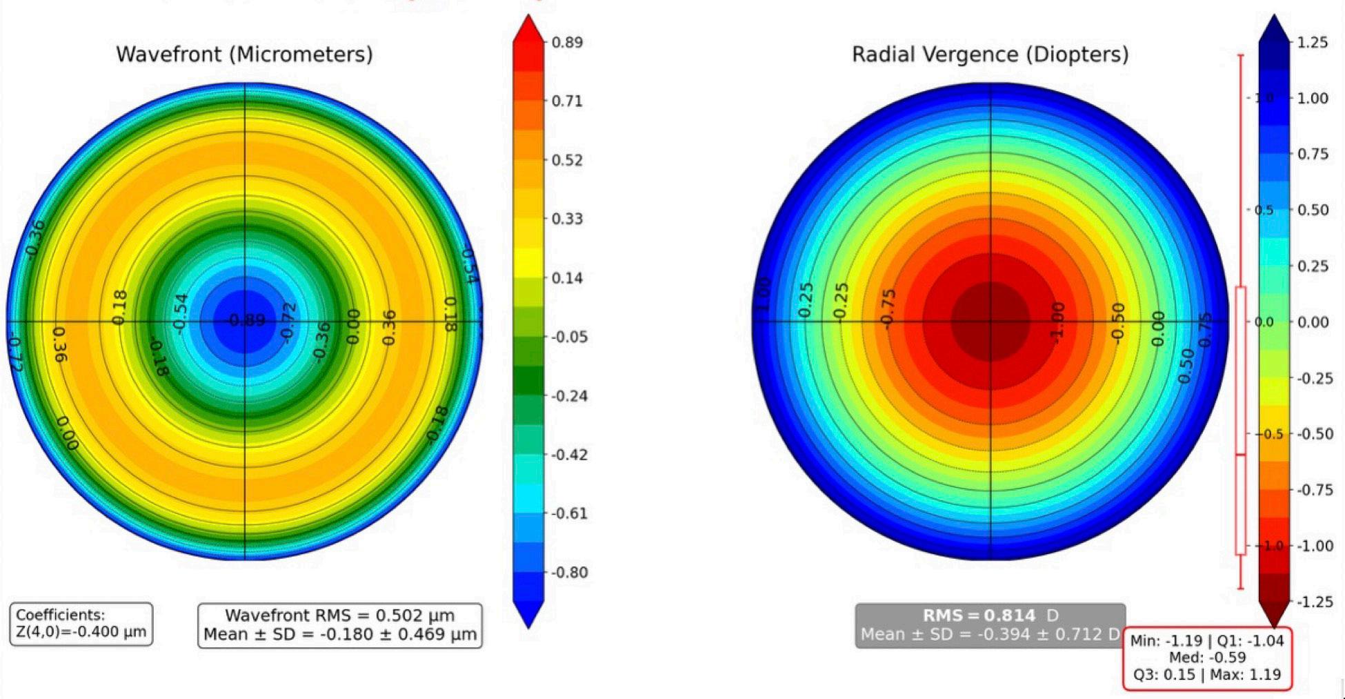
A pure higher-order shape (like a sharp outer edge) naturally introduces central slopes. These central slopes mathematically mimic lower-order shapes, such as defocus or tilt. To keep the net product at zero against those lower modes, the higher-order polynomial must include a ‘counterweight.’ It adds a negative version of the lower-order shape into its own formula to balance the equation to zero. Thus, though the Zernike fit is mathematically correct, the higher-order coefficients appear deceptively small as descriptors of pure higher-order aberration (HOA) content.
WAVEFRONT (microns)
Z40 = -0.4 microns (@ 6 mm)
When we actually examine the structure of these modes, what do we find?
We see the problem is visible in the modes. Because classic Zernike spherical aberration (Z40) is labelled as a fourth-order aberration, one might expect it to behave like a clean quartic profile, a pure (r4) shape that is relatively neutral centrally. However, in its central part, it behaves more like defocus, carrying a distinctly low-order signature near the pupil centre. The embedded quadratic defocus-like component at the centre is far from negligible. From a refractive point of view, it can outweigh genuine fourth-order part. Thus, what we call a ‘higher-order mode’ may exert much stronger low-order influence than its name suggests. Similarly, secondary astigmatism, an HOA, contains a cylindrical component resembling ordinary astigmatism and coma includes the linear component, tilt.
So clinically, these are not optically pure. Although mathematically assigned as HOA, structurally they contain hidden
CPRRESPONDING VERGENCE (diopters)
Figure: A coefficient of about -0.40 microns over a 6-mm pupil produces a vergence gradient of roughly 2.4 D from centre to edge, with relative central myopia of about -1.2 D and peripheral hyperopia of about +1.2 D. Optically, this behaves like a multifocal profile, with the centre favouring near focus and the periphery favouring distance focus. This is why negative spherical aberration has been used in presbyopia-correcting strategies, including corneal photoablation and multifocal or extended depth of focus IOLs, to extend the range of focus through a controlled centre-to-periphery change in vergence. Image courtesy of Professor Damien Gatinel.
low-degree behaviour. Therefore, its coefficient no longer represents purely higher-order optical deformation but instead a mixture, making interpretation much less intuitive.
Is what you call the ‘true’ quartic part essentially the old Seidel representation? Is the Gatinel–Malet approach simply returning to Seidel aberrations? Not exactly, but there is a meaningful connection. The classical Seidel spherical aberration is a pure quartic term, proportional to r4, whereas the usual Zernike spherical aberration mode is a balanced polynomial that contains not only an r4 term but also a quadratic r2 defocus component. That defocus is introduced so the Zernike mode remains orthogonal to the defocus mode over the pupil. From a mathematical point of view, it is elegant. From a clinical point of view, it can be misleading because a mode that is labelled ‘higher order’ is no longer refractively neutral.
The low-degree/high-degree (LD/HD) approach is not simply Seidel theory revived. Its purpose is different. Seidel aberrations belong to classical optical aberration theory, whereas the LD/HD decomposition is a new polynomial basis designed for clinical wavefront interpretation. But the LD/ HD philosophy does move closer to the Seidel intuition in one important respect: it tries to ensure that a higher-order term behaves like a genuinely higher-order term, without hidden low-degree content. In that sense, it restores a kind of optical purity that is lost in the standard Zernike representation.
Is that the main reason the wavefront needed to be reorganised?
Precisely. Zernike polynomials are not mathematically wrong—they are elegant, powerful, and very useful. But the problem is their orthogonality does not provide the separation clinicians need. In ophthalmology, a low-degree component should represent wavefront curvature that determines sphero-cylindrical refraction, while a high-degree component should represent true residual deformations that affect image quality beyond ordinary refraction. In the standard Zernike system, that boundary is blurred: refraction leaks into HOA side, and HOA content in turn alters the apparent refractive side. That is why a different organisation is needed.
So how does the Gatinel–Malet LD/HD decomposition deal with this?
It is straightforward. The wavefront is reorganised into two deliberately defined components: (a) the low-degree component that determines ordinary refraction, which includes everything up to degree two (piston, tilt, defocus, and primary astigmatism) and (b) the high-degree component, which includes only terms of degree three and above (coma, trefoil, spherical aberration, secondary astigmatism, and the other genuine HOAs).
This is not just relabelling. It is designed to keep all quadratic behaviour in LD and therefore to correspond to true spectacle-correctable refractive component. Similarly, the high-degree side is purified of hidden low-order contamination.
But mathematically, there is a trade-off?
Yes, and that is the key trade-off. To achieve a clinically clean
separation, we deliberately give up global orthogonality between LD and HD. In return, we gain a more useful clinical orthogonality: LD becomes a watertight representation of refraction while HD is purified of hidden low-order terms. Importantly, orthogonality is still preserved within each group. Low-degree modes remain orthogonal to one another, and high-degree modes remain mutually orthogonal, so HOA root mean square (RMS) can still be calculated reliably to compare aberrations.
What difference does this make when we interpret the coefficients?
It makes an important difference. In the classical Zernike basis, higher-order coefficients can appear deceptively small because part of their optical effect is represented by loworder coefficients to preserve global orthogonality. In the LD/HD framework, that hidden mixing is removed, so the HD coefficients more faithfully reflect the true contribution of each HOA to the wavefront. That, in turn, makes derived measures of image quality such as the point spread function, convolution-based simulated retinal images, and modulation transfer function curves more clinically meaningful and easier to reconcile with the patient’s visual experience.
How should we picture the LD/HD basis in practical terms?
A useful way to think of it is as two distinct spaces. The low-degree space contains the spectacle-correctable terms— piston, tilt, defocus, and primary astigmatism—that define the eye’s overall refractive behaviour. The high-degree space, beginning from degree three, contains purified higher-order modes that no longer carry hidden tilt or defocus. They are built to remain as neutral as possible near the centre and therefore better represent the true residual optical imperfections affecting image quality that cannot be neutralised by ordinary sphero-cylindrical correction. This is done while keeping the HD modes mutually orthogonal.
The practical rule is simple: Keep all refraction-defining quadratic content in LD, with no r2 leakage into HD. Then read sphere and astigmatism from LD, and reserve HD for genuine higher-order aberrations.
That is the essence of the Gatinel–Malet separation: low-order modes are normalised and orthogonal as are the now pure high-order modes. However, there is no orthogonality between low-order and high-order modes. Therefore, refractive side and image quality-degrading side are no longer contaminating each other.
Part one of this interview appeared in the March/April 2026 edition of EuroTimes
Soosan Jacob MS, FRCS, DNB is Director and Chief of Dr Agarwal’s Refractive and Cornea Foundation at Dr Agarwal’s Eye Hospital, Chennai, India, and can be reached at dr_soosanj@hotmail.com.
Damien Gatinel MD, PhD, is Head of the Anterior and Refractive Surgery Department, Rothschild Foundation, Paris, France.
Authors, Beware!
AI assistance for manuscript preparation raises ethical, intellectual, and practical concerns.
CHERYL GUTTMAN KRADER REPORTS
Researchers thinking about turning to assistance from artificial intelligence (AI) when developing a scientific manuscript should familiarise themselves with their target journal’s guidelines surrounding AI use and consider that outsourcing research and writing tasks to AI threatens essential intellectual virtues, warns Joaquín Fernández MD, PhD.
“The end or goal of academic life is flourishing through the cultivation of reason. Intellectual virtues are the means by which an academic can reach that end,” said Professor Fernández, quoting from an article in which Dr Timothy Daly, a bioethicist/philosopher of science, advocates for ‘low-tech’ academic virtue ethics.1
“A misuse of AI tools could prevent achieving this main aim. AI tools can be used to increase efficiency, but do not forget that large language models are not authors or producers of science.”
Degrading intellectual virtues
Prof Fernández explained that as a foundation for urging academics to resist delegating tasks to AI, Dr Daly detailed the negative impacts on five key intellectual virtues— scientific knowledge, intuitive reason, practical wisdom, philosophic wisdom, and technical craft/art—described in Aristotle’s Nicomachean Ethics.
“Relying on AI to perform academic tasks could weaken these intellectual virtues that are essential for academic flourishing, causing you to lose the depth of your knowledge, your ability to judge the quality of science, your ability to connect knowledge with life and society, your sense of meaningful goals and good decisions, and your ability to create work through your own reasoning,” Prof Fernández said.
Key points for authors
Turning from a philosophical discussion to practical considerations, Prof Fernández urged authors who are drafting a manuscript for publication to check the journal’s website for guidance on acceptable uses of AI and what should be avoided because different publishers have different policies. He also cautioned against asking a chatbot to create a summary of guidelines from different publishers, noting the AI tool may not present an accurate analysis of the information.
Prof Fernández reviewed the policies from select publishers on acceptable use of AI tools for writing and image creation, what uses need to be acknowledged or disclosed, and how they should be disclosed. Reinforcing the point that there are differences between publishers, Prof Fernández presented statements on these topics from Wiley, Elsevier, IEEE, Nature Portfolio, Public Library of Science, Springer, and the British Medical Journal
For example, when it comes to image development, Prof Fernández said Elsevier and Springer Nature typically have the most restrictive guidelines on AI use. Nevertheless, he advised caution, regardless of the targeted publication.
“Some publishers accept AI-generated images, but with exceptions,” he said. “In general, AI-generated scientific images are prohibited or strongly restricted, and so the safest approach is to avoid them unless the journal clearly allows them.”
Instructions on how to disclose AI use also differ among publishers. For example, in addition to requiring authors to name the specific AI tool or service used, Wiley requires the date of access.
Publishers also have instructions on AI use for reviewers. They, too, differ across publishers, but as a rule, Prof Fernández said AI use should be avoided during the reviewing process.
As AI continues to evolve, policies on its use for scientific manuscripts might also change. Therefore, Prof Fernández advised authors to periodically review journal guidelines to be sure they are following the most current information.
Prof Fernández spoke on this topic at the 2026 ASCRS annual meeting in Washington, DC.
For citation notes, see page 38.
Joaquín Fernández Pérez MD, PhD is CEO and Medical Director in the department of ophthalmology at Qvision, Vithas Hospital, Almería, Spa in. He is the Secretary of the ESCRS. joaquinfernandezoft@qvision.es
Entering the World of RoboticAssisted Cataract Surgery
An expert surgeon offers an enthusiastic and optimistic look into the future.
CHERYL GUTTMAN KRADER REPORTS
The year 1961 marked the start of the modern robotics industry. The first in-human robot-assisted surgery was performed in 1985. Although nearly 40 years passed before the first in-human robotic cataract surgery was performed from start to finish, the success of that procedure and ongoing developments make Vance Thompson MD a believer that robotics will allow cataract surgeons to do more and achieve better outcomes.
“With robotics, we can imagine a future where every outcome is optimised and new manoeuvres become possible. Because of improved ergonomics, surgeons will not be fatigued at the end of the day and so their last procedure will feel like the first,” Dr Thompson said.
“Robotics has the potential to reduce training time, lower the dexterity threshold for cataract surgery, and equalise skill level. At a time when there are not enough ophthalmologists to keep up with the volume of patients needing cataract surgery, robotics will allow us to operate on more patients every day and over the course of our careers. Therefore, robotic cataract surgery holds promise for improving global eye health and eradicating preventable cataract-related blindness.”
Over the years, as robotic platforms permeated into other surgical specialties (e.g., general surgeon, urology, gynaecology, orthopaedics), the question remained whether they could be applied to ophthalmic surgery. For many reasons, the answer seemed to be yes, but several barriers remained.
“The eye seems more amenable to robotic surgery compared with other anatomic sites where robots were already being used because its anatomy and geometry are less variable. In addition, cataract surgery is a high volume, relatively consistent and routine procedure,” Dr Thompson said. “Nevertheless, working in the extremely small intraocular surgical space requires dexterous, miniaturised instruments, sophisticated remote centre-of-motion management to minimise tissue damage, and high precision because we are operating on delicate, micron-scale ocu-
lar tissues. Thus, it seems cataract surgery is a good candidate for robotic automation given the right technology.”
Dr Thompson said he has practised robotic-assisted cataract surgery on eye bank eyes using a system from ForSight Robotics and was surprised by how comfortable he felt despite working from a console remote from the globe. Watching the video of the first in-human robotic cataract surgery performed by Robert Ang MD, who also used a ForSight Robotics platform, Dr Thompson said he was amazed by the robot’s reaction time and that the entire procedure was completed under topical anaesthesia.
Ultimately, I hope we will see improvements in outcomes and the advent of autonomous robots in the OR and our offices that will help us reach the goal of being able to do more with better outcomes.
Dr Thompson rejected the idea that robots will completely replace human surgeons, noting the technology enables ophthalmologists to be better surgeons but cannot substitute for human decision making.
“I think robots bring advantages and will expand our abilities, but they cannot replace us considering that there is always a chance for unpredictable complications where human judgement and intervention are critical for appropriate and successful management,” he said.
“With robots, I think we will see improvement in surgical technique. Ultimately, I hope we will see improvements in outcomes and the advent of autonomous robots in the OR and our offices that will help us reach the goal of being able to do more with better outcomes.”
Dr Thompson spoke on this topic at the 2026 ASCRS annual meeting in Washington, DC.
Vance Thompson MD is the director of refractive surgery at Vance Thompson Vision, Sioux Falls, South Dakota, US. vance.thompson@thompsonvision.com
GLAUCOMA LESSONS FOR THE NEXT GENERATION
Roberto Bellucci MD shares practical guidance for managing glaucoma, from early decision making to long-term surgical strategy.
SEAN HENAHAN REPORTS
Noted cataract surgeon Roberto Bellucci MD has seen glaucoma patients regularly for many years in his cataract practice. EuroTimes Editor-in-Chief Sean Henahan spoke with him about what young ophthalmologists need to know when treating these patients.
ET: What part of your practice involves glaucoma management?
RB: Every patient presenting for cataract surgery should be evaluated for dry eye, possible maculopathy, and possible glaucoma. Fortunately, only a minority of these patients are actually affected by glaucoma and require specific management. In hospital departments, the percentage can be 10–15%, but in private practices like mine, this percentage is about 5%.
When performing cataract surgery in a glaucoma patient, the purpose is to reduce the intraocular pressure (IOP) and/ or to decrease the number of the medications. Cataract surgery alone is usually enough in angle closure glaucoma when the trabeculum is functional. In open-angle glaucoma, simple lens removal is expected to get the desired result in early glaucoma, while in more advanced cases, we need to add a specific procedure to increase the aqueous outflow.
Procedure selection is driven by several factors, including the experience of the surgeon, the patient perspective, and cost. My preference goes to minimally invasive bleb surgery (MIBS), especially with the Preserflo MicroShunt (Glaukos), a device I implant under subconjunctival anaesthesia at the end of phacoemulsification by simply asking the patient to look at their
feet. Trabeculectomy combined with phacoemulsification would be my second choice when a MIBS device is not available, a frequent case in public hospitals in Italy. I have little experience with minimally invasive glaucoma surgery devices, which would be attractive for less advanced glaucoma cases. In the past, I have tried several procedures, such as laser cycloablation and ultrasound application, but without success at my hands.
What role can the cataract/refractive surgeon play in helping patients with glaucoma?
Cataract surgeons can help glaucoma patients in several ways. In angleclosure glaucoma, the removal of the natural lens frequently solves the problem, as was demonstrated by the famous EAGLE study.1 Many hyperopic patients have a shallow anterior chamber, and a simple evaluation at the slit lamp (Van Herick test) can prompt the cataract surgeon to suggest refractive cataract surgery to these patients years before closed-angle glaucoma develops. Actually, acute glaucoma attacks have almost disappeared in my area precisely because of early cataract surgery.
In the various types of open-angle glaucoma, cataract surgery can improve IOP and/or medication use—also improving our ability to grade the
glaucoma damage by removing any lens opacity that might affect visual field testing. Moreover, undetected glaucoma can be confused for a developing cataract by the patient, the optician, and sometimes the ophthalmologist if the lens is very opaque. In this regard, surgery preparation will diagnose any underlying pathology, including glaucoma.
What steps would a young ophthalmologist take to learn more about glaucoma surgery?
Compared with cataract, glaucoma is a completely different disease that requires a specific approach. Those who treat glaucoma are prepared to follow patients with great care and
Compared with cataract, glaucoma is a completely different disease that requires a specific approach.
empathy for the patient’s entire life. So, the first step for the doctors approaching glaucoma surgery is to increase their knowledge about glaucoma— how to monitor patients, when to give indications for surgery, how to select the surgery, what complications can be expected even after an uneventful surgery, and how to treat the emerging problems. My suggestion is therefore to study glaucoma, watch surgical videos available on the web, and then join a glaucoma centre for a while, because not everything can be learnt online. After starting a glaucoma surgery practice, maintain relations with mentors to ask opinions and seek help in difficult cases or complications.
Do you have any cautions or caveats about what cataract surgeons should or should not do?
I always fear for glaucoma in my highly myopic patients. Frequently, their IOP is slightly elevated, it is difficult to evaluate the visual field and the fibre
layer at optical coherence tomography, the visual field is inconclusive, and they present two to three years later with a white optic nerve. I have learnt to see them frequently, carefully recording the numerical outcome of the examinations. At the same time, surgeons should refrain from suggesting early cataract surgery to highly myopic patients asking for better vision until they are pretty sure the cataract is the problem with their vision.
The second regards the ‘wipe-out’ syndrome, or the sudden loss of the remaining central vision that can occur during surgery (cataract or glaucoma) in end-stage glaucoma. This event is very frustrating both for the patient and the surgeon and should be considered and discussed with the patient preoperatively.
A third caveat is for the postoperative period in pseudophakic, vitrectomised eyes, which sometimes develop glaucoma that goes unnoticed if the postoperative controls are diluted.
There are many others, however— surgeons learn from experience with unfavourable outcomes. Especially in glaucoma, it is easy to forget successes but keep sharp memory of failures.
It seems that cataract surgeons may shy away from selective laser trabeculoplasty (SLT) because of the gonioscopy skills required, but direct SLT (DSLT) is becoming more popular because this is not required. What do you think of some of the newer options such as FLIGHT ( ViaLase) and ELIOS (excimer)? I think the gonioscopic lens required for SLT is a problem more for the patient than for the doctor, with DSLT becoming more popular for this reason. However, SLT is an evolution of the old argon laser trabeculoplasty (as published in 1979) that used to have variable results, and this legacy may have had an impact on the procedure’s acceptance.
Newer laser techniques like FLIGHT and ELIOS are different because they try and achieve a more substantial change of the chamber angle anatomy. We can therefore expect a more reproducible IOP-lowering effect. We can also expect other machines and procedures to be developed in this area— with the common purpose to avoid the burden and the risks involved in trabeculectomy. In this perspective, the surgical management of glaucoma will probably transfer from a single high-risk surgery to a series of small, repeatable surgeries, each with effects that may only last a few years.
For citation notes, see page 38.
A former president of ESCRS, Roberto Bellucci MD is a Consultant Ophthalmologist is Salò, Italy.
Heidelberg OPERA goes global
Heidelberg Engineering is launching the global sales of Heidelberg OPERA, its fully digital surgical visualisation platform. Surgeons worldwide now have improved access to the platform’s advanced real-time imaging through a digital microscope with next-generation intraoperative OCT, offering outstanding image quality and ergonomic design. heidelbergengineering.com
New AI meibography grading tool
OCULUS has introduced Meibo Analytics, an AI-powered decision-support tool designed for the Keratograph® 5M to improve the analysis of meibography images. Using artificial intelligence, Meibo Analytics performs structure-based evaluations, allowing for direct visualisation of gland loss and examination of changes in the meibomian glands based on the Arita grading scale. The aim is to create a standardised and reproducible classification system that helps resolve diagnostic uncertainties, supports clinical classification, and ensures physicians maintain their professional judgement. oculus.de
Commercial milestone for FLIGHT procedure
ViaLase has announced the successful completion of the first commercial femtosecond laser image-guided, high-precision trabeculotomy (FLIGHT) procedures. The incision-free surgeries were carried out by Sheraz Daya MD at Centre for Sight in London and Karsten Klabe MD at Breyer, Kaymak & Klabe Augenchirurgie in Düsseldorf, Germany. Using the ViaLuxe femtosecond laser with OCT guidance, the procedure accurately creates a channel through the trabecular meshwork, allowing aqueous fluid to flow into Schlemm’s canal in patients with open-angle glaucoma or ocular hypertension. For its initial European launch, ViaLase is working with a targeted group of distributor partners in specific markets, including an exclusive distribution deal with Carleton Optical in the United Kingdom. vialase.com
Wireless upgrade for ocular pressure pump
Balance Ophthalmics has received FDA 510(k) clearance for a wireless-enabled, next-generation version of its FSYX Ocular Pressure Adjusting Pump (OPAP). This device is designed to reduce intraocular pressure during sleep in adults with open-angle glaucoma. The system combines a compact pump with pressure-modulating goggles worn overnight, applying gentle negative pressure to the eyes. According to the company, five trials involving 600 patients showed the FSYX device led to an average 39% reduction in nocturnal intraocular pressure. Balance Ophthalmics expects the new wireless FSYX OPAP to be available to eye care practices by Q4 2026. This prescription-only device will be delivered directly to patients for home use. balanceophthalmics.com
Robotic cataract surgery first
ForSight Robotics announced the successful completion of the world’s first fully robot-assisted cataract surgery performed on a human patient using its proprietary JASPER Platform. The procedure was carried out by Alexey Rapoport MD, with Robert Edward T Ang MD of the Asian Eye Institute in Manila serving as principal investigator. They completed the surgery from start to finish using robotic assistance without general anaesthesia, aligning with modern cataract surgery standards. The JASPER Platform, previously known as the ORYOM platform, integrates advanced imaging, motion scaling, and precision controls, enabling greater dexterity and stability. In addition, its ergonomic design reduces musculoskeletal strain. forsightrobotics.com
BUILDING THE BRIDGE BETWEEN THEORY AND PRACTICE IN REFRACTIVE SURGERY
Laboratory science advances refractive surgery by improving diagnostic and therapeutic methods for greater safety, effectiveness, and predictability. The Laboratory Science section of the Journal of Cataract & Refractive Surgery (JCRS) showcases research that drives these innovations into clinical practice, according to Yu Ci Liu MD and Johbir S Mehta MD in a guest editorial.
Initial studies in the 1980s for modern excimer laser refractive surgery started with ablation experiments on plastic blocks, then moved to cadaver and animal eyes, and finally included living rabbits and monkeys. This progression ultimately resulted in the development of photorefractive keratectomy (PRK) and laser in situ keratomileusis (LASIK). At the turn of the century, research using rabbit eyes revealed that a femtosecond laser could feasibly cut both a lamellar flap and an intrastromal refractive lenticule, which paved the way for the first clinical case of femtosecond keratorefractive lenticule extraction (KLEx) in 2007.
Throughout refractive surgery’s development, translational research has refined and validated surgical parameters and nomograms influencing refractive and visual outcomes. In addition, studies using animal and ex vivo models—histology, immunohistochemistry, electron microscopy, and molecular assays—have clarified the cellular and molecular mechanisms behind complications such as haze and ectasia.
Such studies have shown femtosecond lasers induce more structural changes, inflammation, and cell apoptosis compared to microkeratome. Other research has shown that compared to KLEx, LASIK upregulates immunological and wound healing and inflammatory reactions, and that stromal keratocyte activation and apoptosis are greater after high myopic treatment than low myopic treatments.
Advancements in understanding the molecular and tissue mechanisms involved in corneal wound healing have led to novel strategies aimed at improving outcomes in refractive surgery and reducing postoperative complications. These approaches include pharmacological modulation of postoperative wound-healing processes through optimised mitomycin C dosage, duration, and concentration, as well as the use of antifibrotic agents. Recently, topical losartan has emerged as an example of such therapeutic innovations.
Ex vivo and animal study models are elucidating the biomechanical changes that contribute to ectasia development. Ex vivo inflation tests and optical coherence tomography-based elastography enable quantitative assessment of corneal stiffness. Laboratory research has also clarified the safety margins, efficacy, and tissue response associated with various corneal cross-linking protocols.
Translational research is advancing the development of corneal stromal lenticule implantation as a treatment for keratoconus, corneal ectasia, presbyopia, and hyperopia. Laboratory investigations have provided valuable insights into the best preservation methods for maintaining transparency and mechanical properties of KLE-derived lenticules, along with improvements in sterilisation and decellularisation techniques. In addition, animal studies have assessed the cellular biocompatibility and structural stability of lenticules, contributing data that supports safer and more predictable procedures.
Y C Liu, et al. “Bridging discovery and practice: why translational research matters in advancing refractive surgery,” 52(5): 421–422.
GO ON THE RECORD
Ever y surger y that isn’t recorded is a missed opportunit y. A new initiative from ESCRS will help close that gap.
ESCRS is partnering with Custom Surgical, a medical technology company, to provide recording devices to help ophthalmologists record their surgeries on a secure, organised plat form This will allow mentors to review, comment on, and discuss surgical videos with trainees
The recording devices will be shared with to host and use with their members.
are urged to contact escrs@escrs.org to request the MicroRec devices.
1. Azuara-Blanco A, et al. Lancet, 2016 Oct 1; 388(10052): 1389–1397.
Drive the Research That Drives Surgery
Good surgical practice is based on solid research evidence— and there’s no better way to build research expertise than the ESCRS Peter Barry Fellowship.
The Peter Barry Fellowship is a research fellowship opportunity for young ophthalmologists who have finalised their training in ophthalmology and want to increase their knowledge and/or research skills in anterior segment surgery (cataract, cornea, or refractive). The €60,000 fellowship enables European trainees to spend a year at a global centre of excellence to build research expertise in cataract and refractive surgery.
The fellowship is named for Peter Barry (1948–2016), a founding member of ESCRS who served as president and director of the Society and helped shape modern cataract care. His leadership of the ESCRS endophthalmitis prophylaxis study—at the time the largest antibiotic study in ophthalmology—demonstrated that intracameral cefuroxime at the end of cataract surgery reduces postoperative infection five fold, changing practice across Europe and beyond. He also championed outcomes benchmarking via the EUREQUO registry and promoted education and opportunities for younger ophthalmologists.
Eligibility
Applicants must be young ophthalmologists in their second year of sub-specialty experience in anterior segment surgery and are full members of ESCRS. (This will include only ophthalmologists working in Europe and exclude trainees.) International applicants are considered but must be ESCRS members at the point of application, and their application must clearly outline a benefit to Europe.
The Peter Barry Fellowship only accepts research fellowship applications. The Fellowship is announced at each ESCRS Annual Congress, to start the following year.
Application deadline: 15 July
ESCRS
Leadership, Business & Innovation
Lead with Confidence
Catch up on the latest advances in strategic insight, operational excellence, and forward-thinking innovation from the ESCRS Leadership, Business & Innovation programme.
Enhance Your Presentation Skills
Are your presentation skills holding you back from sharing your knowledge and insights at ophthalmic conferences? Listen and learn as three experienced international speakers and educators describe their presentation tips and tricks. Join Drs Bastak Bostanci, Artemis Matsou, and David Lockington for an informative discussion on delivering engaging presentations, structuring scientific talks effectively, communicating with confidence, and maximising audience interaction in both live and virtual conference settings
Learn to Manage a Team
The medical profession has been shifting away from general practice and toward specialisation for many years, and ophthalmology is no exception. As a result, many eye care practices now employ several subspecialists offering a variety of services. Join Drs Paul Rosen, Vincent Qin, and Eric Donnenfeld as they explore the art and science of practice management in modern ophthalmology—a topic critical to the success of multisubspecialty eye care centres.