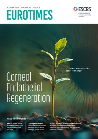
AUTUMN 2026 | VOLUME 31 | ISSUE 8
Corneal Endothelial Regeneration
Is corneal transplantation about to change?
ALSO IN THIS ISSUE New AI Column Debuts Bruce Allan aims to close the gap between code and clinic.
Learning How to Read Filomena Ribeiro discusses the importance of critical reading of journal articles.
A New Consensus for Paediatric Keratoconus European agreement emphasises early detection, regular monitoring, and appropriate treatment strategies.
Save th 17–20 March 2027
InterContinental Citystars
he Date
Cairo, Egypt
Contents Autumn 2026 | Vol 31 Issue 8
26
Is Epi-On the Way Forward? Cosimo Mazzotta MD, PhD, FWCRS
RETINA 28
Surgical Management of IOL Subluxation Francesco Boscia MD
SUSTAINABILITY 33
Cutting Waste Without Cutting Safety Cathleen McCabe MD
LEADERSHIP, BUSINESS, AND INNOVATION 34
Structuring a Multi-Physician Practice in a Changing World Paul Rosen BSc, MBChB, FRCS, FRCOphth, MBA; Vincent Qin MD; and Eric Donnenfeld MD
36
The Human Algorithm Başak Bostancı MD, FEBOphth
08 Cover
Growing a Cornea Is corneal transplantation about to change?
04
Editorial: Supply and Demand
06
First Person: Miguel Raimundo: The ESCRS IOL Calculator
CATARACT & REFRACTIVE 12
Quality of Life Through Quality of Vision Filomena Ribeiro MD, PhD, FEBO and Joaquín Fernández Pérez MD, PhD
14
PresbyLASIK Advances as Alternative to Monovision Anas-Alexis Benyoussef MD, MSc
15
Reading Between the Lines Filomena Ribeiro MD, PhD, FEBO
CORNEA 16
2
EndoArt Reaches 600 Implants with Promising Clinical Results Arie Marcovich MD, PhD
EUROTIMES | AUTUMN 2026
17
Biocompatible Scaffolding: Aiming for the Moon Sorcha Ní Dhubhghaill MBBCh, BaO, PhD, Dip(stats), FEBO, FEBOS-CR, MRCSI(Ophth)
18
A New Consensus for Paediatric Keratoconus Luca Buzzonetti MD
19
The Cretan Approach to Corneal Cross-Linking George Kymionis MD, PhD and Ingemar Gustafsson MD, PhD
22
Using DSAEK to Treat Corneal Perforations Swetha Ravichandran FRCOphth
23
Retinal Imaging Shows Promise for Earlier Alzheimer’s Detection Ahmed Abdalla MD
24
Treating Ectasia Without Keratoplasty Farhad Hafezi MD, PhD, FARVO
INSIGHTS 20
PDEK: A Unique Mix of Anatomy and Technique Soosan Jacob MS, FRCS, DNB
30
Closing the Chasm Between Code and Clinic Bruce Allan MD, FRCS
32
A Case of Inturning Lashes Clare Quigley MD
12
18 Publisher Tom Ogilvie-Graham Executive Editor Stuart Hales Editor-In-Chief Sean Henahan Senior Content Editor Kelsey Ingram Creative Director Kelsy McCarthy Graphic Designer Jennifer Lacey Circulation Manager Lucy Matthews
28
33
34
Contributing Editors Cheryl Guttman Krader Roibeárd O’hÉineacháin Contributors Laura Gaspari Soosan Jacob Timothy Norris Clare Quigley Andrew Sweeney Colour and Print CitiPost Advertising Sales Roo Khan MCI UK Tel: +44 203 530 0100 | roo.khan@wearemci.com
ALSO ALSOIN INTHIS THISISSUE ISSUE 38
Industry News
39
JCRS Highlights
40
Citation Index
41
Upcoming Events
EuroTimes® is registered with the European Union Intellectual Property Office and the US Patent and Trademark Office. Published by the European Society of Cataract and Refractive Surgeons, Building 4000, Langstone Park, Langstone Road, Havant, PO9 1SA, UK. No part of this publication may be reproduced without the permission of the executive editor. Letters to the editor and other unsolicited contributions are assumed intended for this publication and are subject to editorial review and acceptance. ESCRS EuroTimes is not responsible for statements made by any contributor. These contributions are presented for review and comment and not as a statement on the standard of care. Although all advertising material is expected to conform to ethical medical standards, acceptance does not imply endorsement by ESCRS EuroTimes. ISSN 1393-8983
Learn more about EuroTimes or connect with ESCRS at ESCRS.org 2026 AUTUMN | EUROTIMES
3
EDITORIAL
Supply and Demand
W
hen Austrian ophthalmologist Eduard Zirm performed the first successful penetrating keratoplasty in 1905, the arithmetic of corneal transplantation was remarkably simple. His patient, blinded by bilateral alkali burns, needed sight restored. Two donor corneas taken from the enucleated eye of a young trauma victim made the operation possible. For one historic moment, supply met demand. More than a century later, the equation is far more difficult. Penetrating keratoplasty (PK) remains one of the most successful forms of human tissue transplantation. It is still used to treat severe corneal disease caused by trauma, infection, keratoconus, scarring, dystrophy, graft failure, and other causes of corneal blindness. Yet despite the work of eye banks and transplant surgeons, global demand continues to exceed supply. By some estimates, there is only one donor cornea available for every 70 patients who might benefit from corneal transplantation. That shortage has increased the urgency of finding alternatives—not only by expanding donor tissue availability, but also by reducing the number of patients who ultimately require full-thickness transplantation. In this issue, EuroTimes looks at both sides of that problem: efforts to regenerate or replace damaged corneal tissue and strategies to intervene earlier in diseases that once led almost inevitably to PK. The cover story by Professor Soosan Jacob provides a comprehensive review of new approaches to corneal endothelial regeneration. Rather than a single technique, it is a broad field that includes stimulating remaining endothelial cells to enlarge, migrate, and possibly proliferate; injecting human corneal endothelial cells or scaffold-based endothelial constructs into the anterior chamber; using stem cell-derived or alternative cell sources; and developing bioengineered or artificial endothelial substitutes.
Keratoconus offers another example of how the demand side of the equation may be changing. Historically, it has been one of the leading indications for PK in Europe. Earlier diagnosis and earlier intervention with corneal collagen cross-linking (CXL), phototherapeutic keratectomy (PTK)-assisted procedures, intrastromal implants, and related approaches are increasingly helping surgeons stabilise disease, regularise the cornea, and avoid or delay transplantation. One example highlighted in this issue is Professor Cosimo Mazzotta’s update on promising customised transepithelial procedures. Epi-off cross-linking remains the reference standard for halting keratoconus progression, but newer epi-on and customised approaches continue to evolve. Professor Farhad Hafezi discusses another strategy: epithelial map-guided PTK customised epi-on CXL, or ELZA-PACE. In related articles, Professor Sorcha Ní Dhubhghaill discusses scaffold-based tissue engineering and Andrew Sweeney reports on EndoArt, a synthetic artificial endothelial layer that may act as a functional posterior barrier in eyes unsuitable for other therapies. The shortage of donor tissue also reflects the need to strengthen eye bank infrastructure worldwide. While countries such as the United States and Sri Lanka can export donor corneas, much of the world still has little or no practical access to corneal transplantation. Eduard Zirm showed corneal blindness could be reversed with human donor tissue. The challenge now is to make that possibility more widely available—and to develop new therapies that reduce the need for transplantation in the first place. That is the new supply and demand equation in corneal surgery. Sean Henahan, Editor-in-Chief
EDITORIAL BOARD
4
Thomas Kohnen
José Güell
Adi Abulafia (Israel) Bruce Allan (UK) Noel Alpins (Australia) Juan Alvarez de Toledo (Spain) Gerd Auffarth (Germany) Başak Bostanci (Türkiye) John Chang (Hong Kong SAR, China) Béatrice Cochener-Lamard (France) Burkhard Dick (Germany) Mor Dickman (The Netherlands)
Joaquín Fernández (Spain) Oliver Findl (Austria) Sri Ganesh (India) Christina Grupcheva (Bulgaria) Farhad Hafezi (Switzerland) Nino Hirnschall (Austria) Soosan Jacob (India) Jack Kane (Australia) Yao Ke (China) Georgios Kymionis (Greece)
EUROTIMES | AUTUMN 2026
Paul Rosen
David Lockington (UK) Artemis Matsou (Greece) Cyres Mehta (India) Jod Mehta (Singapore) Sorcha Ní Dhubhghaill (Belgium) Rudy Nuijts (The Netherlands) Catarina Pedrosa (Portugal) Konrad Pesudovs (Australia) Nic Reus (The Netherlands) Filomena Ribeiro (Portugal)
Andreia Rosa (Portugal) Giacomo Savini (Italy) Julie Schallhorn (US) Sathish Srinivasan (UK) Paola Vinciguerra (Italy) Shin Yamane (Japan) Ron Yeoh (Singapore) Mihail Zemba (Romania)
FIRST PERSON
Miguel Raimundo: The ESCRS IOL Calculator
S
ince its launch in 2022, the ESCRS IOL Calculator has become a go-to tool for cataract and refractive surgeons, performing thousands of calculations each month. The calculator recently received some upgrades courtesy of Miguel Raimundo, an ophthalmologist and ESCRS member who also has experience in the field of computer programming. EuroTimes sat down with Dr Raimundo at the ESCRS Annual Congress in London to discuss his work on the calculator and what the future holds for its users.
How and why did you get involved with the ESCRS IOL Calculator?
I’m an anterior segment and cataract surgeon, but I also have a very strong computer science background. I’ve been programming since my early adolescence. When ESCRS was starting to develop the IOL calculator, I kind of approached them spontaneously, offering help in bridging the ophthalmic side of it with the more technical side of it. And I’ve been working on this ever since. I really like to create digital solutions to solve workflow problems, and I publish and present regularly on the topic of IOL calculations. That’s why the project was a great fit for me.
The ESCRS IOL Calculator is essentially a centralised tool for different IOL power formulas. Is that a reasonable description, or is it too simple? It’s important to clarify something that people often do not fully understand: We don’t calculate formulas within our application itself. We contact each formula author’s external servers and get the calculations from their end, then compile the results in the ESCRS IOL Calculator. We also work with IOLCon, an external database that has IOL reference data for lens constants, including optimised ones.
6
EUROTIMES | AUTUMN 2026
We make it available all in a single place, with a single data input. The calculator brings together the results of all those different formulas and shows them side by side. That’s the main value proposition of the ESCRS IOL Calculator.
Is there, or has there been, another tool out there doing this sort of thing?
To the best of our knowledge, not really, because the calculator is both a technical and also kind of a political challenge. The ESCRS leadership had to get all the formula authors to agree to collectively share, for the common good, the products of their own intellectual property. So, it was both a technological solution and a political agreement of sorts that led to the calculator.
From time to time, upgrades are made to the calculator. Who makes decisions about the need for upgrades and the kinds of upgrades to be made? We have a very collegial system. The ESCRS IOL Calculator Team meets online every two months or so. The members are myself, Adi Abulafia, Dante Buonsanti, Oliver Findl, Filomena Ribeiro, and Giacomo Savini, plus Damian Clarke, the IT staff lead.
As clinicians, we feel the friction every day. Someone will say, ‘I would like the computer to do this,’ or, ‘My colleague thinks it would be really cool to have that feature.’ We are gradually trying to fix every pain point that people keep reporting. And that’s a really useful iteration cycle, particularly for me on the IT side, because I immediately benefit from the improvements as a cataract surgeon. I’m simultaneously developing and using the tool, with the steering of the IOL Calculator Working Group. It has been working nicely so far because I think we have really answered surgeons’ needs. When we started, we only did spherical IOL calculations, and people were saying, ‘Well, it would be nice to have toric IOLs,’ so we added that. And then it was post-LASIK, and we added that. It’s a powerful way to guide development. In the last year or so, we’ve mainly been working on making it easier to use the calculator, because so much of our life nowadays with AI is becoming increasingly automatic. With the calculator, you were still repeating data inputs and re-entering settings and preferences. The last year has been all about reducing that friction and repetition in performing online calculations.
You mentioned AI. Does the ESCRS IOL Calculator use artificial intelligence?
That’s a loaded question. Some of the formulas we are using already leverage AI, like the Hill-RBF and the Kane. It’s probably better called machine learning in that setting, but it’s still a form of artificial intelligence. But now we’ve brought AI into the calculator as well, through a service I developed and freely licensed to ESCRS called BiomAPI. It has a module called BiomAI, which uses a large language model with vision capabilities to extract structured information from biometry printouts. Basically, that allows a user of the calculator to just take a photo of a biometry printout or upload a PDF, and then BiomAI will read it and extract the relevant information with extremely high accuracy. You still must double-check because errors are possible, but based on our extensive testing over thousands and thousands of printouts, they’re exceedingly rare. So, in a way, we’ve already incorporated AI to really decrease the friction of data entry. The most common reason for not doing online calculations is having to manually enter every single value. That takes a while, particularly for long surgical lists. And that really was the priority behind the BiomAPI project. We also support other kinds of integrations. For example, BiomAPI can also work without AI, creating direct connections between devices and web applications. Anyone can use it to build integrations. It’s an open, interoperable layer of biometry data, optimised for the web. Another addition worth mentioning, although it’s not AI, is the settings interface in the ESCRS IOL Calculator. In the past, every time someone wanted to perform a calculation, they had to re-enter their name, targets, and preferred IOLs and decide which sources of constants they were using. So, we created a focused ‘Settings’ interface that stores preferences in the user’s browser, with no account or login needed. Com-
bining BiomAPI and the Settings interface, you can now take a photo of your biometry printout and press ‘Calculate.’ The friction has gone down immensely over the past year.
I haven’t seen an exact number, but I’m told the ESCRS IOL Calculator has had millions of uses.
The latest data I have are from six months ago, and I have every reason to believe these numbers have been increasing. We perform over 90,000 calculations every month, which is more than 1,000,000 per year. It’s a massive number of calculations. The interesting part of it is that the most frequently implanted IOL is still the standard monofocal. But on the ESCRS calculator, half are presbyopia-correcting IOLs of some kind, including advanced monofocals. It’s clearly a tool that people are using for their most demanding cataract and refractive cases. It has really become part of the premium workflow.
As you look back at the time you’ve spent working on the calculator, are you happy with it? Do you feel like there are things you still want to do, or anything you wish you had done differently? We still have a lot of work to do on data interoperability to make it easier for devices to talk with each other. That has never been easier than it is today, thanks to all the AI tools we have. We already have small prototypes that you can use to talk to an AI agent like ChatGPT, Claude, or Gemini, and ask it to use BiomAPI and the ESCRS calculator to do a calculation. I think we can really work towards natural language workflows, electronic health record integrations, and so on. Eventually we could move beyond being a website you visit and become a platform that everyone can connect to and use. There are regulatory and legal hurdles we may have to work through, but that’s the direction I’d like to take. The platform would be tightly integrated into daily workflows, using all the technology that people are waking up to like AI assistants. That’s definitely within reach for the next year or so.
2026 AUTUMN | EUROTIMES
7
COVER ARTICLE
Corneal Endothelial Regeneration
8
EUROTIMES | AUTUMN 2026
Is corneal transplantation about to change? BY SOOSAN JACOB MS, FRCS, DNB
T
he World Health Organization estimates corneal blindness affects 20–30 million people worldwide, with roughly 2 million new cases diagnosed each year, but the supply of donor tissue has never matched the growing demand. This disparity has inspired the search for alternative approaches, some of which have now completed the arduous journey from the laboratory to the clinic. The pump-barrier function of the single posterior monolayer of corneal endothelial cells (CECs) helps maintain corneal function and transparency. A decrease in the number or functional capacity of these cells leads to corneal oedema and reduced vision. Human corneal endothelial cells (HCECs) do not have functional proliferative capacity in vivo, as they are arrested in the G1 phase of the cell cycle. Regardless of age, the standard response to both physiological and pathological cell loss is through polymegathism and cell migration. Endothelial keratoplasty has transformed management by offering faster rehabilitation and better optical outcomes, particularly with new alternatives such as pre-Descemet’s endothelial keratoplasty (PEDK), which is feasible even in complex anterior segments, and Descemet’s membrane endothelial keratoplasty (DMEK). However, these procedures still depend on donor availability, eye bank infrastructure, surgeon factors, and long-term graft survival. A global shortage of high-quality transplantable corneas remains a barrier to providing the benefit of surgery to everyone who needs it.
Many strategies
Endothelial regeneration includes several different strategies, such as stimulating the remaining endothelial cells to enlarge, migrate, and possibly proliferate; injecting HCECs or scaffold-based endothelial constructs into the anterior chamber (AC); using stem cell-derived or alternate cell sources; and creating bioengineered and artificial endothelial substitutes. The aim is to restore a functional pump using biological, pharmacological, or engineered means. One leading strategy involves using pharmacology to increase cell migration, adhesion, proliferation, and survival. Rho kinase (ROCK) inhibitors are currently the most promising pharmacological tool available for endothelial regeneration. Pharmacologically stimulating existing endothelial cells provides functional recovery without the risk of rejection or the need for rigorous surgical skills. The ROCK signalling pathway regulates several key cellular processes, including cell adhesion and migration, stress fibre formation, modulation of actomyosin contraction, cell proliferation, and apoptosis. It accelerates centripetal spreading and cell cycle progression from G1 to S phase. Mild vasodilator activity may cause ocular congestion and inflammation. Another approach, fibroblast growth factors (FGF), enhances CECs through protective and mitogenic effects. They are a standard component of CEC culture media. Engineered FGF can stimulate CEC proliferation while offering greater stability and improved tissue binding, even in dystrophic corneas. They may be used in isolation or to enhance recovery following Descemet stripping only (DSO). Absence of αSMA (alpha-smooth muscle actin) staining in studies indicates that endothelial–mesenchymal transition and scarring are not
2026 AUTUMN | EUROTIMES
9
COVER ARTICLE
induced. Angiogenesis and trabecular meshwork blockage are potential problems. Other traditional mitogens such as epidermal growth factor and platelet-derived growth factor stimulate replication when combined in specialised media. Hepatocyte growth factor has also been shown to enhance CEC viability, migration, and proliferation without triggering fibrotic transitions. Sulforaphane, found in cruciferous vegetables, is thought to activate endogenous antioxidant pathways. It exerts cytoprotective effects and promotes cell survival, potentially preventing degeneration in Fuchs’ endothelial corneal dystrophy (FECD). Ex vivo studies showed a decrease in CEC apoptosis in both stressed and unstressed FECD corneas. RTA408, a synthetic compound, activates cytoprotective factors that are downregulated in Fuchs’ dystrophy and is being investigated as a topical treatment for patients. Wnt/β-catenin pathway modulators (e.g., TWS119) overcome contact inhibition, triggering localised cell proliferation and lateral migration. Niacinamide (vitamin B3) is an NAD+ precursor that increases mitochondrial metabolism and protects CECs from ultraviolet and age-related apoptosis. Other substances under investigation include vasoactive intestinal peptide and ciliary neurotrophic factor, which protect CECs from oxidative stress, and connexin hemichannel blockers, which protect CECs from injury.
Leveraging the eye’s capacity to regenerate
DSO, also known as Descemetorhexis without endothelial keratoplasty (DWEK), is used in cases with localised FECD, confined mostly to central cornea. The central 4.0 mm of the abnormal host Descemet’s membrane with guttae is stripped. Loss of contact inhibition allows relatively healthier peripheral endothelial cells to undergo localised expansion and spreading. Slow centripetal migration and polymegathism allow the central cornea to clear over the ensuing one to two months. Advantages of this approach include avoiding donor tissue and long-term steroid use and their associated risks. Disadvantages include its limited applicability in advanced or diffuse endothelial disease and in eyes with poor peripheral endothelial reserve. Additionally, the stripped zone must be kept small and the margins of the Descemetorhexis smooth to minimise the failure rate. ROCK inhibitors may be administered in conjunction with DSO to increase the rate of deturgescence and hasten recovery. Sulforaphane may be added to protect the peripheral CEC from the oxidative and inflammatory stress of surgery.
Endothelial cell therapy
Cultured endothelial cell injection therapy is the most visible leap beyond keratoplasty toward cell-based therapies. Pioneered by Shigeru Kinoshita MD, PhD, HCECs can be expanded ex vivo from a single donor cornea to treat up to 100 eyes. Donor tissue selection, cell isolation by enzymatic or non-enzymatic digestion, and cell suspension seeding with culture media and growth factors are key steps. The cell suspension is injected into the anterior chamber, and face-down
10 EUROTIMES | AUTUMN 2026
positioning helps to ensure the injected cells settle onto the posterior stroma. One approach uses supermagnetic iron oxide nanoparticle (SPION) cell labelling followed by an external magnet to draw the cells towards the posterior stroma. ROCK inhibitors may improve cell adhesion and survival, while agents such as sulforaphane or niacinamide help protect the injected cells from metabolic and inflammatory stress. Advantages of cell injection include ease of therapy and improved availability, as one donor cornea can be used to treat multiple recipients after cell expansion. In addition, unlike DSO, it is not dependent on a reserve of healthy peripheral cells and can therefore be used for more diffuse disease, such as bullous keratopathy. Host-related challenges include the presence of guttae and abnormal extracellular matrix if the host Descemet’s membrane (DM) is not removed. The fate of unattached cells remains unknown, as does whether transplanted cells will adhere to intraocular structures or even enter the blood stream. Other considerations include the need to inject a much larger number of transplanted cells than may be needed, uncertainty surrounding optimal dosing and delivery systems, patient selection, and postoperative positioning. Manufacturing challenges include the difficulty in reliably expanding adult HCECs and an undesirable occurrence of endothelial-to-mesenchymal transition. A recent study by Aouimeur (et al.) defined culture success as obtaining
Cultured endothelial cell injection therapy is the most visible leap beyond keratoplasty toward cell-based therapies.
typical endothelial morphology and cell density of at least 1,500 cells/mm2 at passage three.1 Success was determined to be dependent on donor age, with 92% in young donors ≤29 years, 42% in intermediate age (30–49 years), and only 12% in donors aged 50 years or older. For older donors, higher initial endothelial cell density (≥1,700 cells/mm2) and shorter organ culture duration (≤15 days) increased the success rate to 25%. Hypertrophic endothelial cells and mesenchymal-like cells with reduced functional phenotype may be seen and therefore quality checks like identity, purity, morphology, density, viability, sterility, potency, and nuclear markers are needed. Other approaches include using human adipose-derived mesenchymal stem cells, which are very versatile and can be reprogrammed into an endothelial lineage to reduce reliance on donor eye banks. Human embryonic or induced pluripotent stem cells are induced to differentiate into functional, hexagonal endothelial layers. Alternative cells or precursors that can adapt to perform the pump and barrier functions of native endothelium are also being studied.
the DM. However, challenges include creating a scaffold that is thin, transparent, permeable, biocompatible, and appropriately curved, while also providing flexibility and mechanical strength for surgical handling. At the same time, it must allow a high-density endothelial monolayer and support cellular adhesion. Natural materials such as ultrathin decellularised porcine corneal stroma, thin human corneal stroma, collagen sheets, cellulose, gelatine, chitosan, cross-linked amniotic membrane, and denuded Descemet’s membrane are under investigation. Synthetic material scaffolds, such as polycaprolactone, polylactic-co-glycolic acid (PLGA), polyethylene glycol (PEG), and biohybrids, are also being studied. Advantages of scaffold-based therapy include uniformity, ease and rapidity of production, easy donor availability, decreased need for transplanted cell numbers, good cellular adhesion on carrier, better systemic safety profile, and known surgeon comfort. Manufacturing and assessment challenges exist, however. PVEK (PreciseBio Vision endothelial keratoplasty) is a 3D-bioprinted, cell-scaffold transplant.
Mind the scaffolding
Artificial endothelial substitutes
Scaffold-based tissue engineering is another fascinating approach. Here, endothelial cells are not simply injected but cultured on a carrier and transplanted, much like a traditional endothelial keratoplasty. This tissue-engineered posterior lamella replaces both the endothelial cells and
The use of artificial tissue substitutes is not true regenerative therapy because it does not create an endothelial layer. However, it acts as a functional posterior barrier, particularly in eyes that are unsuitable for other forms of therapy, such as eyes with multiple rejections or uveitis. EndoArt (EyeYon Medical) is a 50-micron artificial lamella positioned similarly to endothelial keratoplasty that deturgesces the cornea by acting as a fluid barrier. Long-term steroids and rejection are avoided. Other materials are also being evaluated. Biomarkers are being identified that may serve as potential therapeutic targets for FECD treatment. Gene therapy has been used in mouse models to treat early-onset FECD. Viral and nonviral vectors and techniques to introduce genes into the cornea are being researched. Gene editing and CRISPR technologies may give breakthroughs in FECD.
Ethical and regulatory concerns
Embryonic stem cell therapy raises several ethical issues, so adult stem cells are more commonly used. The efficacy and safety seen in animal studies needs to be validated by human clinical trials, which are subject to further safety standards. Biomimetics also face many regulatory and translational challenges before they can come into regular clinical practice. Manufacturers must meet quality assurance and safety guidelines, obtain manufacturing licences, and adhere to Good Manufacturing Practice standards. For citation notes, see page 40.
Dr Soosan Jacob is Director and Chief of Dr Agarwal’s Refractive and Cornea Foundation at Dr Agarwal’s Eye Hospital, Chennai, India, and can be reached at dr_soosanj@hotmail.com.
2026 AUTUMN | EUROTIMES 11
CATARACT & REFRACTIVE
Quality of Life Through Quality of Vision Overcoming the personal and socioeconomic impact of presbyopia in an ageing population. TIMOTHY NORRIS REPORTS
O
phthalmologists should weigh the impact of presbyopia-correcting surgery on patients’ quality of vision alongside the likelihood of spectacle independence, ensuring patients have realistic expectations for both. Filomena Ribeiro MD, PhD, FEBO and Joaquín Fernández MD, PhD agreed on this point in a recent debate. The huge demographic shift happening today, particularly in developed countries, is transforming presbyopia from a natural occurrence into a pressing matter for the ophthalmic community. Rising life expectancy should be accompanied by a high quality of life. According to a position paper from the European Parliament, people older than 55 play a significant role in society, in family life, in the community, and in political and sociopolitical decision making.1
12 EUROTIMES | AUTUMN 2026
“We need to consider that our goal should be preserving the ability of our patients to perform all their daily tasks,” Professor Ribeiro said. “Presbyopia is not only a growing phenomenon, but something that can have a huge impact on all these day-to-day activities.” Currently, the best option is a device that supports patients in performing tasks requiring intermediate and near vision. However, Prof Ribeiro stressed that there is no solution without drawbacks. Even spectacles or contact lenses can have some trade-offs, such as halos or glare, as well as an additional economic burden on the patient. Presbyopia is a near-universal condition for these patients, she added, and its impact on visual function and quality of life warrants greater attention from the ophthalmological community.
Prof Ribeiro observed that, despite the significant public health implications, there are few assessments on this issue thus far. Citing one study from the British Journal of Ophthalmology, she suggested evaluating its impact across four domains: physical, physiological, social, and environmental. According to the study, the combined effect of these domains was 3.92% greater for near vision than for distant vision in overall quality of life—a meaningful impact Prof Ribeiro said remains underappreciated.2 Due to the global retirement age gradually getting pushed higher, providing the patient with the ability to perform vision-related daily activities at all distances has become all the more pressing. Functional vision and spectacle independence are not a cosmetic issue, Prof Ribeiro emphasised; they are a matter of personal ability and disability, with significant consequences across all social domains. She also outlined the importance of having more objective real-world measurements: Patients develop presbyopia with different needs and face presbyopia correction with different expectations, and many are not willing to accept spectacles as an adjustment.
Patient-centred outcomes
Prof Fernández noted that binocular uncorrected visual acuity can be translated into a more patient-centred outcome: the likelihood of spectacle independence. Studies reporting a mean binocular uncorrected visual acuity of approximately 0.2 logMAR generally demonstrate spectacle independence rates of about 50%. Therefore, he said, if spectacle independence is considered the clinically relevant outcome, it is reasonable to define 0.2 logMAR as a clinically meaningful cut-off, in line with current international standards.3 Prof Fernández and colleagues compared IOL categories using the evidence-based functional classification system developed by the ESCRS Functional Vision Working Group and the spectacle independence rates reported in the literature. They sought to identify which lens categories provide the most reliable outcomes for patients who wish to reduce their dependence on spectacles for intermediate and near vision. According to the pooled estimates reported in their review, patients implanted with partial-DOFi (depth of field) narrowed lenses achieve spectacle independence at intermediate distance in 52% of cases, but only 12% achieve independence at near distance. Moving from the narrowed to the enhanced category increases intermediate spectacle independence from 52% to 77%, although near spectacle independence remains limited at 30%. Partial-DOFi extended lenses provide a further improvement, with 90% of patients achieving spectacle independence at intermediate distance and 65% at near distance.4 Full DOFi lenses provide the highest rates of spectacle independence across both intermediate and near distances. Full-DOFi continuous lenses achieve spectacle independence in 96% of patients at intermediate distance and 91% at near distance, while full-DOFi smooth lenses show similarly high rates of 95% and 89%, respectively. Full-DOFi steep lenses, which are typically associated with a more bifocal profile, show lower but still substantial rates of 85% at intermediate distance and 81% at near distance.4
These results represent the type of information that should be clearly communicated to the patient to determine which lens category best suits their needs, Prof Fernández emphasised. “This is useful information to assess whether a patient can become independent from glasses or not,” he said. “We need to translate visual acuity in terms of logMAR into information to our patients to tell them what their objective possibility of this outcome is.” Choosing the proper IOL category based on the potential best quality of life for the patient, with clear information on its impact, is therefore extremely important. Prof Ribeiro concurred that clinical decisions should be supported by a patient-centred approach with evidence based on functional classification. This process can be achieved by choosing the IOL category most likely to provide the highest quality of life, matching functional performance with the patients’ expectations. “We need to match the patient with the right choice and mitigate dissatisfaction,” she concluded. “The work of the Functional Vision Group says that, right now, the partial group, subtype enhanced, can be considered a standard of care.” Profs Ribeiro and Fernández spoke at the 2026 ESCRS Winter Meeting in Helsinki. For citation notes, see page 40.
Filomena Ribeiro MD, PhD, FEBO is immediate past president of ESCRS and Head of Department at Hospital da Luz, Lisbon, Portugal. filomenajribeiro@gmail.com Joaquín Fernández Pérez MD, PhD is the CEO and Medical Director in the Ophthalmology Department at Qvisión in Vithas Virgen del Mar Hospital (Almería), Spain. joaquinfernandezoft@qvision.es
2026 AUTUMN | EUROTIMES 13
CATARACT & REFRACTIVE
PresbyLASIK Advances as Alternative to Monovision Expanding technologies improve outcomes for presbyopia, but careful patient selection and neuroadaptation remain critical. LAURA GASPARI REPORTS
P
resbyLASIK is a sophisticated alternative to monovision for ageing eyes, but patient selection, biometric safety, and the need for neuroadaptation are still challenges, according to Anas-Alexis Benyoussef MD. The surgical options for presbyopia are continuing to expand. PresbyLASIK is a cornea-based approach designed to address the limitations of monovision LASIK, such as the loss of stereopsis when anisometropia exceeds 1.50 D. “The arsenal of technologies that we have today for presbyLASIK is based on multifocal approaches with distinct refraction zones, with a priority on strong near vision but with some photic phenomena,” Dr Benyoussef explained. “But we also have extended depth of focus or hybrid approaches, where we try to have an aberration control to elongate focality.” Among the multifocal approaches are technologies such as the Supracor (Technolas), which creates a hyperpositive dome (+1.00 D to +3.00 D) targeted for moderate hyperopes, and the PresbyMAX (Schwind), a bispheric profile with options for symmetric treatment or monovision. Another approach, called laser-blended vision (LBV), works by adjusting spherical aberrations to extend the depth of focus, allowing clearer vision over a range of distances. Presbyond (Zeiss), for example, combines monovision with spherical aberration to create a blend zone for better results, with myopes and emmetropes among the more ideal candidates. PresbyLASIK success depends on selecting the right patient. Ideal candidates are 45–55 years old and are especially moderate hyperopes who show good neuroadaptation and satisfaction. Emmetropes are more challenging due to potential loss of distance vision. Proper selection also requires good ocular parameters and patient acceptance of visual compromises. Exclusion criteria include dysfunctional lens syndrome, small pupils, preexisting contrast loss, binocular instability, and ocular surface disease. In such cases, lens-based procedures are preferable. Overall, success depends not only on treating the cornea but also on the patient’s ability to adapt neurologically. Effectively managing both the patient and the brain is important, as presbyLASIK is a static treatment while presbyopia worsens over time. Tolerance should be assessed preoperatively, for example with contact lens trials. Patients must also be informed that results are not immediate, as neural adaptation and binocular fusion can take up to three months. Evaluating presbyLASIK efficacy is challenging due to varying study designs and techniques. However, many patients achieve good distance and near vision shortly after surgery. Limitations include a small risk of reduced best-cor-
14 EUROTIMES | AUTUMN 2026
rected distance vision, decreased contrast sensitivity, dysphotopsia, and potential complications for future IOL calculation. For the future, presbyLASIK may further expand with a comeback of other corneal solutions, such as allogenic inlays. “They are on the market—not in [every] country, but [this option] is developing very fast,” Dr Benyoussef said. “The technology is based on an allograft inlay that creates a multifocal corneal profile, leveraging fourth- and sixth-order spherical aberrations to effectively extend the depth of focus.” Dr Benyoussef spoke at the 2026 ESCRS Winter Meeting in Helsinki.
Anas-Alexis Benyoussef MD, MSc is an ophthalmologist at the University Hospital of Brest, France. anas-alexis.benyoussef@chu-brest.fr
Reading Between the Lines Why critically reading the SVIOL literature matters for everyone. LAURA GASPARI REPORTS
A
n accurate and critical appraisal of scientific studies on simultaneous vision IOLs (SVIOLs) contributes to the best possible outcome for cataract and refractive surgery patients, stresses Filomena Ribeiro MD, PhD. In recent years, simultaneous vision IOL technology has been evolving rapidly, resulting in an increase in published research and clinical studies. However, the quality of evidence is not always consistent, Professor Ribeiro observed. “Rather than simply looking at the conclusions, we need to understand how the study was designed, how outcomes were measured and reported, and how much confidence we can place in the evidence,” she stressed, as unfortunately some studies may be affected by bias or poor methodology. These are not marginal concerns. A study with a small sample size, a short follow-up period, a flawed or inaccurate methodology, or an inconsistent definition of outcomes can lead to conclusions that, while appearing promising on paper, do not necessarily stand up in daily practice. The ability to read studies critically should not be the exclusive preserve of clinical researchers, Prof Ribeiro said. Cataract and refractive surgeons, fellows, and any others involved in patient care should have the practical tools to judge whether published results are reliable enough to influence everyday clinical practice. The ability to identify good findings and trustworthy evidence leads to better treatment for patients, especially when choosing the best lens for them.
Ultimately, better critical appraisal leads to better clinical decisions, and better clinical decisions lead to better outcomes for our patients. There is also a need for a better understanding of standardised classifications to translate research into clinical decision making. The increasing number of studies on SVIOLs introduces a large number of new technologies and terminologies that may be confusing. Prof Ribeiro noted there has been a definite improvement in the quality and consistency of IOL research, but there is still room for further progress. One of the Society’s key contributions in this regard is the ESCRS Simultaneous Vision IOL Classification system, an important milestone that provides ophthalmologists with a scientifically based framework of standardised terminology that also supports more consistent reporting across future studies.
Scientific research is conducted primarily to provide patients with an ever-increasing range of therapeutic and surgical options. Failing to read it critically, or stopping merely at the abstract and conclusions, risks negative consequences for patient care. Congresses, meetings, and scientific societies’ guidelines, Prof Ribeiro said, are there to help people learn these skills and to know which instruments and tools are in place to support them. Research on SVIOLs will continue to grow. What also needs to grow is the responsibility of clinicians to keep refining their abilities to select the most appropriate lens and give their patients the best possible outcome, Prof Ribeiro concluded. “Ultimately, better critical appraisal leads to better clinical decisions, and better clinical decisions lead to better outcomes for our patients.” Prof Ribeiro presented at the 2026 ESCRS Annual Congress in London.
Filomena Ribeiro MD, PhD, FEBO is head of ophthalmology at Hospital da Luz, Lisbon, Portugal, and the immediate past president of ESCRS.
2026 AUTUMN | EUROTIMES 15
CORNEA
EndoArt Reaches 600 Implants with Promising Clinical Results Improvements in central corneal thickness and best-corrected visual acuity highlight the potential and efficacy of this artificial graft. ANDREW SWEENEY REPORTS
W
ith 600 cases and counting, the EndoArt (EyeYon) is starting to find its place in the armamentarium of treatments for severe corneal disease, according to Arie Marcovich MD, PhD. Essentially a synthetic replacement for the corneal endothelium, EndoArt substitutes the innermost layer of the cornea, which fails in conditions such as bullous keratopathy and chronic corneal oedema and is typically treated with donor endothelial tissue (e.g., Descemet stripping automated endothelial keratoplasty/Descemet membrane endothelial keratoplasty), he explained. “The EndoArt is an impermeable barrier to fluid and decreases passive movement of aqueous humour into the corneal stroma,” Dr Marcovich explained. “It also allows nutrition to come in from the sides and does not interfere with the health of the cornea. Eyes filled with silicone are dehydrated, and when it’s removed, we get oedema. The EndoArt is an artificial membrane; it’s available, and it’s not subject to rejection.” Dr Marcovich said EndoArt is also indicated in patients with failed keratoplasties and glaucoma drainage devices and when tissue is not available. It is contraindicated in post-refractive surgery cases, patients with naturally thin corneas, and corneal spontaneous perforation. Benefits are not limited to just regraft patients and those with a ‘tubed eye’ due to glaucoma drainage devices, he said, stressing that complex eye patients and those with low visual potential are good candidates for treatment. The artificial device can also plug the gap when donor tissue is in short supply and can circumvent objections to treatment based on religious or cultural restrictions. Dr Marcovich pointed to several case studies to highlight the efficacy of the EndoArt, starting with a study of 10 eyes with a
16 EUROTIMES | AUTUMN 2026
4.5-year follow-up after the device was grafted. In 7 out of 10 patients, best-corrected visual acuity (BCVA) improved from 1.94+0.63 logMAR to 0.80+0.64 logMAR, and central corneal thickness (CCT) decreased from 927+241 µm to 621+140 µm. In another study, 24 pseudophakic patients were examined, of which 33% had failed endothelial keratoplasty, 21% had glaucoma filtering surgery, and 21% had undergone posterior vitrectomy. All patients showed significant improvement in CCT after one month and had stabilised after three months, with mean CCT improving from 793+104 µm postoperatively to 583+106 µm after three years. Other studies emphasised the consistency of CCT improvement along with significant improvements in BCVA and distance-corrected visual acuity. For Dr Marcovich, these results show the current efficacy and future potential of EndoArt. “We have performed more than 600 implants with over six years of follow-up [that show a] 60% improvement in vision and 90% reduction in pain. The EndoArt is a revolutionary concept,” Dr Marcovich said. “The graft maintains a clear and viable cornea over the long term without major complications. It expands our treatment for patients that don’t have another solution and, of course, in countries without corneal availability, it’s a viable option too.” Dr Marcovich presented at the 2025 ESCRS Annual Congress in Copenhagen.
Arie Marcovich MD, PhD is the co-founder of EyeYon Medical and Head of the Department of Ophthalmology at the Kaplan Medical Center, Rehovot, Israel. arie.marcovich@gmail.com
Biocompatible Scaffolding: Aiming for the Moon Scaffolding holds considerable promise in the drive for endothelial therapy cells. ANDREW SWEENEY REPORTS
I
n a survey of more than 100 cornea specialists, 55% had no preference regarding the source of cells required for endothelial therapy. Could biocompatible scaffolds gain traction as a result? “When we look at the scaffold approaches, it’s the interaction between the cells and the underlying Descemet’s membrane that gives us inspiration. We want to recreate this in the lab,” said Sorcha Ní Dhubhghaill MD, PhD. “What we want is an endothelial cell that attaches nicely and firmly to our scaffold. We want the scaffolds because they are a targeted therapy, can be affixed to the site, and may perhaps reduce cell spread.” Professor Ní Dhubhghaill strongly believes this therapy has considerable potential. Based on a 3D structure made of natural or synthetic materials, biocompatible scaffolds can mimic the extracellular matrix (ECM) of native eye tissue. Amniotic membrane and lens capsules can be used to create biocompatible scaffolds, but there are more options when it comes to synthetics. Prof Ní Dhubhghaill said that natural polymers like collagen can be used, as can a host of semi- and fully synthetic materials, such as polycaprolactone. The natural model for biocompatible scaffold design is the Descemet’s membrane, and the goal is to be able to recreate its key properties, Prof Ní Dhubhghaill said. Endothelial cells— either from donor tissue, cultured primary cells, or induced pluripotent stem cells (iPSCs)—are seeded onto the scaffold in vitro as part of this process, but it does pose a challenge. “We’re calling this the moonshot project because that is what this approach is. It’s a moonshot; it’s quite an ambitious way to go,” Prof Ní Dhubhghaill said.
“I have used scaffolds that have been beautifully cellularised, and at the last moment, when we were introducing them into a surgical model, the cells fell off. That is absolutely not what we want to do when developing a scaffold.” Other difficulties reported by Prof Ní Dhubhghaill included problems with mimicking the Descemet’s membrane, material limitations that could cause the scaffolds to become rigid or slippery, and regulatory and cost barriers. All of these would need to be addressed before widespread adoption. However, thanks to the familiarity of a surgical graft approach for biocompatible scaffolds, along with their consistency and scalability (potentially based on a ‘cookie-cutter’ approach), Prof Ní Dhubhghaill’s moonshot may not be so far out of reach. It is an ambitious target, but one she feels can be achieved. “It’s not clear yet whether scaffolds are the future. In a world where we can get what we want with iPSCs, I think they could really take off,” Prof Ní Dhubhghaill said. “Biocompatible scaffolds could also limit spread of cells in the anterior chamber, which I think would be of particular interest. However, they may also require more complex regulation than pure cells, and that will translate into an addition in price.” Prof Ní Dhubhghaill presented at the 2025 ESCRS Annual Congress in Copenhagen.
Sorcha Ní Dhubhghaill MBBCh, BaO, PhD, Dip(stats), FEBO, FEBOS-CR, MRCSI(Ophth) is Head of Department, Ophthalmology at Brussels University Hospital, Belgium. sorcha.ni.dhubhghaill@uzbrussel.be
2026 AUTUMN | EUROTIMES 17
CORNEA
A New Consensus for Paediatric Keratoconus Early diagnosis, screening, and prompt treatment at the core of new European recommendations. LAURA GASPARI REPORTS
C
onstant cooperation to support early diagnosis, good screening programmes, and immediate treatment are among the new European Reference Network (ERN) consensus recommendations for paediatric keratoconus, developed under the auspices of the European Commission. Established by the European Commission seven years ago, the ERN virtually connects healthcare providers across Europe, particularly for the management of rare diseases. ERN-EYE focuses specifically on rare eye conditions. “It is composed of 52 full members, covering more than 900 rare eye diseases in six main areas of interest: paediatric ophthalmology, retina, neuro-ophthalmology, anterior segment, genetic testing, and low vision,” Luca Buzzonetti MD explained. In addition to connecting specialists, ERN-EYE supports clinical research, educational tools, multidisciplinary care, and partnerships with patient associations while helping to improve access to innovation, strengthen national integration, and develop guidelines. For paediatric keratoconus, a clinical consensus statement was prepared by a team of 15 authors from eight European countries. Professor Buzzonetti served as team coordinator, working alongside three working groups, a medical writer, and a patient representative. The contributors represented the Czech Republic, Estonia, France, Germany, Italy, Lithuania, Slovenia, and Spain. Prof Buzzonetti described the process as complex from the start, as the project involved several steps before completion. These included defining the target population, establishing a list of keywords, ranking the research questions by importance, and conducting the search, selection, and appraisal of scientific evidence. This was followed by drafting the consensus statements and analysing the Delphi process, which assessed the level of agreement with each recommendation using a five-point scale (0–100%). Statements with more than 75% average agreement were accepted, those with 25–74% agreement were revised, and statements with less than 25% agreement were excluded. The draft was then finalised and approved, leading to the release of the guidelines. The consensus statement provides an overview of paediatric keratoconus, including its pathophysiology, diagnosis, risk factors, and classification. It emphasises early detection, regular monitoring, patient and family education, and appropriate treatment strategies. The goal is to help healthcare professionals deliver timely care and improve outcomes for affected children. The recommendations are based on a literature review of studies published between 2000 and 2024, using
18 EUROTIMES | AUTUMN 2026
MEDLINE and EMBASE databases with specific keywords for each working group. The key recommendations include close collaboration between paediatricians and ophthalmologists for early diagnosis; screening at-risk children using tools such as Scheimpflug imaging or retinoscopy; frequent patient monitoring every three to four months initially and then annually until the age of 18; immediate treatment upon early diagnosis; and using epi-off cross-linking as first-line treatment and deep anterior lamellar keratoplasty for advanced cases. The guideline also proposes a management algorithm covering prevention, disease progression, and visual rehabilitation. International collaboration is still a crucial factor. “Global initiatives could promote data sharing and the creation of common registries in the attempt to deepen our understanding of paediatric keratoconus and optimise patients’ outcomes,” Prof Buzzonetti said. “In our experience, a significant number of patients still present with a delayed diagnosis, thereby losing the potential benefits of early cross-linking treatment.” Prof Luca Buzzonetti spoke at the 3rd World Keratoconus Congress 2026 in Florence, Italy.
Luca Buzzonetti MD is Head of the Ophthalmology Department at Bambino Gesù IRCCS Children’s Hospital, Rome, Italy.
The Cretan Approach to Corneal Cross-Linking Adding a refractive element could help improve outcomes for the contact lens intolerant. ANDREW SWEENEY REPORTS
B
eing a doctor is as much an art of managing expectations as it is of achieving good outcomes, as one of the earliest corneal cross-linking (CXL) patients could have attested in 2005. “This was a patient with contact lens intolerance. The treatment was successful with an impressive inferior-superior steepening, and the patient gained a bit of improvement,” recounted Ingemar Gustafsson MD, PhD. “However, with a 20 D difference between the superior and inferior part of the cornea, there was insufficient recovery for the patient to be satisfied. It was ‘I’ve been cured (stabilised), but I’m not satisfied.’” In 2026, with 20% of cornea patients reportedly suffering from contact lens intolerance, “something refractive” needs to be added, said Dr Gustafsson. For that role, he turned to George Kymionis MD, PhD, a pioneer in excimer laser treatment since 2009. The limitations of using custom, topography-guided ablations have been clear for some time, Dr Gustafsson said. They focus primarily on two areas: deep corneal scarring and progressive corneal flattening. The more tissue ablated (50 μm), the more side effects can occur due to the synergic effect of laser and CXL. Ablation creates a deeper cross-linking effect and a higher oxygen availability. Unsatisfied with this outcome, Dr Kymionis considered how to reduce the ablation and came across transepithelial phototherapeutic keratectomy (PTK) instead, in combination with corneal cross-linking. “One can see from the epithelial map that it works as a masking agent. We all know by now that when the cone is increasing its curvature, the overlying epithelium is reducing everything just to make the cornea be more normal,” Dr Gustafsson said. “When you ablate with PTK, you have the epithelium as the masking agent. This allows you to just ablate directly over
the cone and less in the periphery, causing less tissue damage. [Dr Kymionis] describes this as the ‘Cretan Protocol.’” The combination of PTK and cross-linking demonstrated significant superiority compared with manual laser debridement and cross-linking, as it allows the surgeon to ablate over the cone. As a result, the patient has a higher riboflavin concentration at that spot, lower riboflavin concentration in the periphery, and greater oxygen availability. The Cretan Protocol can be applied to all keratoconus patients, as it supports the corneal tissue in achieving regularisation and has a lower ablation rate with fewer overall side effects, Dr Gustafsson said. “The Cretan Protocol is an easy procedure with no complicated calculations or fancy devices needed to get topographies in highly irregular corneas,” he said. It’s simple: 45 μm of transepithelial PTK at 8.5 mm (according to epithelial map thickness). “PTK combined with CXL is not only better than just CXL or topography-guided photorefractive keratectomy, but it is the best treatment for keratoconic patients suitable for CXL. It also gets around the selection oasis of refractive treatment due to ablation being able to treat those ineligible for topography-guided treatment.” Dr Gustafsson presented at the 2026 EuCornea Annual Congress in Porto, Portugal, on behalf of Dr Kymionis.
George Kymionis MD, PhD is Professor of Ophthalmology at the University of Athens, Greece, and a former president of EuCornea. gkymionis@gmail.com Ingemar Gustafsson MD, PhD is a consultant and medical coordinator of the cornea service at Skåne University Hospital, Lund, Sweden. ingemar.gustafsson@med.lu.se
2026 AUTUMN | EUROTIMES 19
CORNEA
PDEK: A Unique Mix of Anatomy and Technique The first in a new series on pre-Descemet endothelial keratoplasty examines the anatomical and surgical factors behind its low rebubbling rates. BY SOOSAN JACOB MS, FRCS, DNB
E
ndothelial keratoplasty (EK) has evolved into three major surgical options: Descemet stripping automated endothelial keratoplasty (DSAEK), Descemet membrane endothelial keratoplasty (DMEK), and pre-Descemet endothelial keratoplasty (PDEK). Each procedure replaces dysfunctional corneal endothelium, but the transplanted tissue layers, surgical handling, and postoperative graft behaviour differ significantly. To understand these differences, it is necessary to revisit corneal anatomy. In 2013, Professor Harminder Dua described an additional layer between the posterior stroma and Descemet’s membrane (DM), now called the pre-Descemet’s layer (PDL), or Dua’s layer. This layer is central to understanding PDEK surgery.
Comparing different EK procedures
In DSAEK, transplanted tissue includes endothelium, DM, PDL, and a variable thickness of posterior stroma. The stromal component varies from approximately 150 microns to 80 or even 50 microns, depending on whether standard DSAEK, ultrathin DSAEK, or nano-DSAEK is being performed. Though posterior stroma makes the graft relatively easier to handle, it also results in transplantation of a thicker graft. It therefore increases effective postoperative corneal thickness, requires the endothelium to pump fluid across a thicker corneal complex, and affects refraction, visual quality, and rejection rates. DMEK was therefore a major refinement because only the DM and endothelium were transplanted. However, it brought its own challenges. At only approximately 15 microns thick, DMEK produces excellent optical results. Its extremely thin, delicate nature and tendency to scroll tightly, however, make surgery difficult, especially in challenging anterior segments. DMEK graft preparation is also more difficult with young donors, particularly those younger than 40 to 50 years of age, as the graft scrolls even tighter. As a result, DMEK grafts are often preferred from older donors, although they generally have lower endothelial cell counts than younger ones. Additionally, in diabetic donors, the risk of tissue preparation failure is higher. Thus, while DMEK is anatomically superior, it may not always allow the use of the youngest and potentially healthiest endothelial tissue. PDEK occupies a middle ground. The PDEK graft includes the PDL, DM, and endothelium. It is slightly thicker than DMEK, since it is approximately 25 microns rather than 15 microns, but still far thinner than DSAEK. Its major advantage
20 EUROTIMES | AUTUMN 2026
is that it can be prepared from donors of any age. In practice, PDEK grafts are often taken from donors younger than 40 years old and frequently from those younger than 20 or even 10 years, because younger tissue generally has healthier endothelium and higher endothelial cell counts. The PDL gives PDEK distinctive surgical characteristics. It is an acellular, tough layer, about 10 to 15 microns thick, made of compact type I collagen lamellae arranged in longitudinal, transverse, and oblique directions. While its elastin content gives the graft elasticity, the PDL gives it a splinting effect, and the collagen lamellae make it resistant to tearing. Therefore, although PDEK grafts may still scroll like DMEK grafts, the PDL supports the DM and endothelium, making the graft easier to manipulate within the anterior chamber (AC). In this sense, PDEK offers the ease of handling associated with DSAEK while retaining many optical advantages of DMEK.
Rebubbling
A crucial advantage of PDEK is its low detachment rate of about 3%. DMEK has a reported graft detachment rate ranging from 2% to 82%, with a rough average of 33%. Thus, the need for postoperative rebubbling is significantly lower with PDEK. Rebubbling, whether performed in the operating room or clinic, exposes the patient to a second intervention and additional risks, including infection, excess air fill, pupillary block, Urrets-Zavalia syndrome, anterior capsular cataract, repeat detachment, underfill, and hypotony. Higher rebubbling rates also add significant burden in terms of cost, delayed visual recovery, and lost time from work. PDEK, in the author’s own experience, has a lower detachment rate even in difficult eyes and complex anterior chambers, which removes postoperative uncertainty for both patient and surgeon.
Why should PDEK detach less often?
The answer to this key question has a few proposed hypotheses. One proposed explanation again returns to the PDL. DM is composed mainly of type IV and type VIII collagen. Type IV collagen forms a loose, two-dimensional meshwork or ‘chicken-wire’ network, specialised for basement membrane function, endothelial support, selective permeability, cellular attachment, and migration. Type VIII collagen forms a short, non-fibrillar hexagonal network that contributes to DM structure. In contrast, type I collagen, which forms the PDL, is a fibrillar collagen that provides tensile strength and structural toughness.
graft in an ideal anatomical location that promotes attachment. Fourth, the small period of higher intraoperative pressure eliminates the need to close the eye with a full air bubble followed by later burping to avoid pupillary block. With air pump-assisted PDEK, the graft is already well attached by the time the eye is closed, and an approximate 80% air bubble is generally sufficient. This has also completely negated the need for long-acting gas.
PDEK offers the ease of handling associated with DSAEK while retaining many optical advantages of DMEK.
Figure: A) DM of DMEK graft apposed against PDL (smooth on rough); B) PDL of PDEK graft apposed against PDL (rough on rough); C) Air pump-assisted PDEK acts as a third hand.
In DMEK, the donor DM and endothelium are placed against the recipient’s posterior corneal surface after removal of host DM and endothelium. Thus, the donor type IV and type VIII collagen complex must adhere to the host posterior stromal or PDL surface. This may create a potential plane or space at the interface, contributing to detachment. In PDEK, especially when prepared from young donors, the PDL, DM, and endothelium remain tightly attached as one complex. In younger donors, the bond between PDL and DM is stronger, making spontaneous separation less likely. During surgery, the host DM and endothelium are removed, and the donor PDL–DM–endothelium complex is placed against the host. This results in donor PDL apposing the host PDL. Since both are similar tissues and have relatively rough surfaces, this ‘rough to rough’ apposition may provide more secure adherence than the smoother DM to PDL interface in DMEK. It may explain the lower rebubbling rate observed with PDEK. The air pump-assisted PDEK technique, as previously described, may further reduce detachment rates. During surgery, pressurised air infusion maintains the AC and acts like a third hand. It helps displace fluid trapped between the graft and the host surface, even without venting incisions. Second, it allows the surgeon to identify and remove even small edge folds, which are common starting points for graft detachment. Third, it improves graft centration, placing the
The air pump-assisted technique is also particularly helpful in complex anterior chambers. For instance, in eyes with intracameral bleeding, the pressurised air can tamponade bleeding, float the graft quickly, and still allow blood washout, even after graft floatation. This reduces postoperative fibrin, which could otherwise contract and contribute to graft detachment. Similarly, the air pump can support the graft while additional manoeuvres are performed, such as synechiolysis, pupilloplasty, vitrectomy, or other intraocular steps. It also allows rapid unfolding in complex eyes even with peripheral edge folds present, which can then be unfolded using the air pump. Thus, the low rebubbling rate in PDEK is explained by a combination of graft anatomy, donor selection, interface behaviour, and surgical technique. The PDEK graft is thin enough to offer DMEK-like optical advantages, strong enough to provide DSAEK-like handling, biologically suited to using younger donor tissue and has anatomy that promotes attachment. When combined with the air pump-assisted technique, it provides stable intraoperative apposition and lower postoperative detachment.
Dr Soosan Jacob is Director and Chief of Dr Agarwal’s Refractive and Cornea Foundation at Dr Agarwal’s Eye Hospital, Chennai, India, and can be reached at dr_soosanj@hotmail.com.
2026 AUTUMN | EUROTIMES 21
CORNEA
Using DSAEK to Treat Corneal Perforations New treatment could significantly reduce the risk of negative outcomes from corneal perforation treatment. ANDREW SWEENEY REPORTS
E
very procedure comes with a cost, and corneal perforation management is a perfect example, with each treatment option carrying its shortcomings. “Traditional measures used in the management of corneal perforation like glue, amniotic membrane transplantation, conjunctival flaps, and therapeutic penetrating keratoplasty (PK) have significant disadvantages. These include high astigmatism, poor visual outcomes, and the risk of operating on a hot eye,” said Swetha Ravichandran MD. Dr Ravichandran believes there is a better alternative to these procedures: the mini Descemet stripping automated endothelial keratoplasty (DSAEK), a technique developed by James Myerscough MB, a consultant ophthalmologist and clinical lead at the Mid and South Essex Hospital Trust. According to Dr Ravichandran, using this procedure can help reduce suture-related complications, shorten surgery time, and spare the visual axis. In some cases, it could also reduce the need for further surgery. “We pull a mini-DSAEK graft into the eye from a Busin glide via a limbal wound and place it just under the perforation, with subsequent air tamponade,” she explained. “We intentionally microkeratome a thicker graft than in conventional DSAEK to increase the stromal rigidity of the graft and reduce the risk of postoperative prolapse through the perforation.” To confirm her position, Dr Ravichandran presented a review of five-year outcomes in the first 20 eyes treated using
the mini DSAEK technique. Some of the patients, treated across four National Health Service (NHS) sites, had a maximum follow-up of five years; five had undergone a primary DSAEK, while 15 initially received glue before subsequently requiring a tectonic DSAEK. Perforation size ranged from 0.5 mm to 5.0 mm, graft thickness from 130 μm to 500 μm, and graft diameter from 4.0 mm to 9.0 mm. The surgical procedure was as follows: automated microkeratome (approximately 300 μm), graft trephination (4.0–6.0 mm), tangential trimming to maintain a clear visual axis if required, pull through from Busin glide, anterior chamber air tamponade, and sutureless closure. This contrasts with other reported tectonic DSAEK techniques, in which the graft is delivered directly through the corneal perforation into the anterior chamber. Such a manoeuvre may enlarge the perforation, requiring subsequent sutures, thus defeating the very premise of using an internal approach. “The thickness of the graft and its shape, which includes placing a crescentic graft if it is occluding the visual axis, helps to customise it. Regarding tissue conservation, harvest the graft from unused donor material,” Dr Ravichandran said. She reported that 19 of 20 eyes surveyed in the review had achieved tectonic closure, 16 of which did so without the need for revision. Five patients subsequently underwent mushroom penetrating keratoplasty (PK) to improve their visual outcomes, and 75% of those involved in the review achieved “preoperative or better best-corrected visual acuity.” “Mini/tectonic DSAEK is a safe, effective, and sutureless option for corneal perforation originating from a range of aetiologies, and the technique is reproducible across independent NHS sites. It spares the visual axis and enables later optical rehabilitation via mushroom PK or deep anterior lamellar keratoplasty on DSAEK,” Dr Ravichandran said. “Tissue can be harvested pragmatically from unused donor material such as the anterior lamella remaining after a DMEK peel, which is important given national shortages. Prospective multicentre studies are warranted, and microkeratome availability does remain a concern to be addressed.” Dr Ravichandran presented at the 2026 EuCornea Annual Congress in Porto, Portugal.
Swetha Ravichandran FRCOphth is Locum Consultant Ophthalmologist Corneal at the Mid and South Essex Hospital Trust, UK. swetha.ravichandran@nhs.net
22 EUROTIMES | AUTUMN 2026
Retinal Imaging Shows Promise for Earlier Alzheimer’s Detection OCT may also aid risk stratification and disease progression. PRISCILLA LYNCH REPORTS
R
etinal structural and microvascular abnormalities are promising, non-invasive biomarkers for early detection and monitoring of Alzheimer’s disease, according to Ahmed Abdalla MD. A systematic review carried out by Dr Abdalla and colleagues found consistent associations between retinal changes and both mild cognitive impairment (MCI) and Alzheimer’s disease (AD). Using retinal imaging modalities to detect these abnormalities could help identify Alzheimer’s disease at an earlier stage. The findings are particularly relevant given the growing global burden of AD and the challenges associated with early diagnosis, Dr Abdalla said. Alzheimer’s disease is often diagnosed relatively late in its course, after significant neurodegeneration has already occurred. Current diagnostic biomarkers, including cerebrospinal fluid analysis and advanced neuroimaging, can be invasive and costly or have limited availability.
Retina potential
Dr Abdalla noted that the retina is an embryological extension of the central nervous system, making it an attractive target for non-invasive assessment of neurodegenerative disease. Dr Abdalla and colleagues conducted a narrow systematic review to evaluate whether structural and microvascular retinal changes could serve as biomarkers for Alzheimer’s disease and MCI. The review identified studies published between 2016 and 2026 that assessed the diagnostic and prognostic value of different retinal imaging modalities, optical coherence tomography (OCT) and OCT angiography (OCTA), and measurement techniques on cognitive outcomes. Twenty studies from Europe, Asia, and North America met the inclusion criteria.
Key findings
Across the studies, significant thinning of the macular ganglion cell-inner plexiform layer (GC-IPL) and retinal nerve fibre layer (RNFL) was consistently observed in patients with MCI and Alzheimer’s disease compared with cognitively normal controls. Reduced retinal microvascular density was also reported. Importantly, these associations remained significant after adjustment for age, sex, APOE genotype, and vascular comorbidities, Dr Abdalla reported. Greater retinal thinning was also associated with increasing disease severity and established Alzheimer’s biomarkers, including amyloid and tau pathology.
The review found that combining structural OCT measures with OCTA vascular metrics improved diagnostic performance compared with individual measurements alone. Variations in imaging protocols, study populations, and analytical methods contributed to moderate heterogeneity across studies, he noted.
Scalable technology
Dr Abdalla said the findings support the potential role of retinal imaging as a non-invasive, accessible biomarker for early detection and monitoring of Alzheimer’s disease. Such approaches could also potentially aid risk stratification and disease progression and facilitate earlier intervention in patients at risk of cognitive decline. “OCT is a very scalable technology and is more available in a primary care setting compared to other diagnostic modalities for Alzheimer’s disease, which are quite expensive and invasive,” Dr Abdalla told EuroTimes. However, he cautioned that further work is needed before retinal biomarkers can be incorporated into routine clinical practice. The review concluded that standardised retinal imaging protocols and large-scale longitudinal studies will be required to determine whether OCT and OCTA can be integrated into future population screening and diagnostic pathways for Alzheimer’s disease. Dr Abdalla spoke at the Irish College of Ophthalmologists 2026 Annual Conference in Galway.
Ahmed Abdalla MD practices at Cavan General Hospital, Ireland. Ahmedhsn9696@gmail.com
2026 AUTUMN | EUROTIMES 23
CORNEA
Treating Ectasia Without Keratoplasty Early results suggest a new procedure could replace penetrating keratoplasty for keratoconus. SEAN HENAHAN REPORTS
C
an ophthalmic innovation make penetrating keratoplasty for keratoconus obsolete? A Clinical Research Symposium at the 2026 ESCRS Annual Congress examined several promising techniques that could make it possible. Farhad Hafezi MD, PhD discussed a technique he has developed known as epithelial map-guided phototherapeutic keratectomy (PTK) combined with customised epi-on cross-linking, or ELZA-PACE. ELZA-PACE for treating keratoconus or post-refractive ectasia begins with anterior segment OCT epithelial mapping and topography/tomography. The surgeon then performs a phototherapeutic keratectomy to remove a limited epithelial area over the cone, followed by collagen cross-linking (CXL) with riboflavin and customised UV-A irradiation.
Understanding the early clinical results
“To date, we have performed ELZA-PACE in almost 300 eyes at the ELZA Institute in Zurich, Switzerland, of which more than 100 have now completed one-year follow-up,” Professor Hafezi reported. “A consistent pattern has emerged: cone-centred flattening is apparent immediately after the procedure (the photochemical effect is essentially immediate)
24 EUROTIMES | AUTUMN 2026
and continues as the epithelium remodels, with topographic stability typically reached by month six.” A flattening of 4–5 D within the first month is common and, because central flattening is accompanied by compensatory mid-peripheral steepening (the ‘coupling’ effect), the overall reduction in corneal asymmetry is often even greater, typically 8–12 D and up to 20 D in selected cases, he noted. Functionally, this regularisation translates into a reduction of vertical coma and gains in corrected distance visual acuity (CDVA) over 6–12 months. In one representative case, CDVA improved from 0.4 to 1.0 logMAR with glasses within a week of treatment. Crucially, this was achieved without removing any stromal tissue, preserving the corneal thickness profile. ELZA-PACE therefore regularises the cornea and improves vision rather than merely stabilising the disease, he explained.
Noting the procedure’s progression
ELZA-PACE is the result of more than two decades of CXL research. The original Dresden protocol (epi-off, 3.0 mW/cm² for 30 minutes, 5.4 J/cm²) normally halts ectasia progression and typically does little to correct the irregularities on the corneal surface. Epi-on approaches initially underperformed because
the epithelium absorbs riboflavin and UV light while consuming oxygen—the rate-limiting component of the photochemical reaction. Higher fluences, pulsed UV delivery, and penetration enhancers have since closed the efficacy gap between epi-on and epi-off CXL. The first generation of customised CXL used a topography-guided, eye-tracking UV source to deliver more energy over the cone apex; this improved flattening to roughly 3–4 D but remained moderately predictable. ELZA-PACE is the second generation: instead of shaping only the UV beam, it also models oxygen availability and riboflavin distribution. An epithelial map-guided PTK removes the epithelial cells over the cone, so the three overlapping gradients—riboflavin concentration, oxygen availability, and UV fluence—all reinforce each other at the cone apex. This also distinguishes ELZA-PACE from excimer-plus CXL protocols (Athens, Cretan, STARE-X, TREK) that achieve regularisation through stromal ablation. In ELZA-PACE, the stroma remains untouched, with the entire effect coming from cross-linking. That tissue preservation matters, particularly in younger patients.
Establishing the CXL part of the treatment
In contrast to cross-linking, the PTK step is deliberately minimal. A custom-offset, custom-sized ablation zone, typically oval and a few millimetres across, is placed over the cone tip region and removes only the local epithelial thickness, without any stromal tissue removal. “We then saturate the cornea with a penetration-enhancer-assisted, hypo-osmolar 0.1% riboflavin solution,” Prof Hafezi explained. “Over the denuded cone, the stroma undergoes rapid saturation. Under the intact peripheral epithelium, penetration is slower, and this creates a riboflavin gradient. Because the epithelium also consumes oxygen and absorbs UV light, oxygen and UV gradients form in parallel, all centred on the epi-off window over the cone.” UV-A delivery uses the C-eye device (EMAGine AG, Switzerland) through an accelerated, pulsed protocol at 18 mW/cm² across the whole treatment zone. The protocol is followed by an additional fluence boost confined to the central cone region, resulting in a considerably higher cumulative fluence at the cone tip than in the periphery. Cross-linking is therefore strongest where the cornea is weakest: the cone flattens, the mid-periphery steepens in compensation, and the cornea becomes more regular—all photochemically, without ablating stroma, he said. Prof Hafezi presented at the 2026 ESCRS Annual Congress in London.
GO ON THE RECORD Every surgery that isn’t recorded is a missed opportunity. A new initiative from ESCRS will help close that gap. ESCRS is partnering with Custom Surgical, a medical technology company, to provide recording devices to help ophthalmologists record their surgeries on a secure, organised platform. This will allow mentors to review, comment on, and discuss surgical videos with trainees. The recording devices will be shared with affiliated national ophthalmology societies to host and use with their members. Leaders of affiliated national societies are urged to contact escrs@escrs.org to request the MicroRec devices.
Farhad Hafezi MD, PhD, FARVO is Medical Director at the ELZA Institute of Zurich, Switzerland, and holds professorships in Switzerland, the United States, and China. farhad@hafezi.ch.
MicroRec-Devices_HPV.indd 1
8/19/26 9:18 AM 2026 AUTUMN | EUROTIMES 25
CORNEA
Is Epi-On the Way Forward? Customised transepithelial procedures promising, but still far from becoming the new gold standard. TIMOTHY NORRIS REPORTS
E
pi-on corneal cross-linking (CXL) may not yet be the gold standard for the treatment of keratoconus, but it is heading in that direction, according to Cosimo Mazzotta MD, PhD. “Epi-on is still struggling to win trust amongst ophthalmologists around the world, and many of the protocols that are available today are still broadly unfamiliar,” he said. Epi-off protocols remain the gold standard for keratoconus treatment, a position reinforced by the Second Global Consensus on Keratoconus and Ectatic Diseases of 2026, in which Professor Mazzotta participated as Group 3 coordinator with Prof Michael Belin and Dr Emilio Torres. The consensus helped define new best practices and a new understanding of keratoconus as a multifactorial disease, while paving the way to a more multimodal diagnostic approach. At the same time, Prof Mazzotta observed that accelerated epi-off is likely to remain the dominant procedure, although there is still no majority agreement on the optimal protocol. However, Prof Mazzotta believes recent advances in epi-on protocols could change this balance. The first step, he said, is to rebuild confidence in the technique by making its mechanisms and potential benefits better understood. One reason for the lack of popularity of epi-on can be traced back to its early development. Original protocols attempted to reproduce epi-off conditions without adequately compensating for the presence of the epithelium. Low oxygen
26 EUROTIMES | AUTUMN 2026
availability, insufficient stromal riboflavin penetration, and inadequate photochemical energy resulted in disappointing outcomes and, in some cases, retreatment. Translational research paved the way to a different approach, Prof Mazzotta explained. The introduction of Iontophoresis Plus, for example, combined enhanced riboflavin delivery with higher fluence and pulsed light. A study Prof Mazzotta and colleagues conducted, published in the Journal of Refractive Surgery, demonstrated a consistent demarcation line on optical coherence tomography (OCT) at a meaningful stromal depth, Kmax flattening close to epi-off, and stable keratoconus over time. The findings suggested that epi-on required a different energy balance from epi-off. Higher fluence, pulsed illumination, and active riboflavin delivery appeared to address some of the limitations of the earlier protocols. These results were a huge step forward, Prof Mazzotta said. Findings demonstrated that the limitations of epi-on were not necessarily intrinsic to the technique but could be addressed by understanding and modifying its underlying photochemical and pharmacokinetic mechanisms. Oxygen then became another important piece of the puzzle. In a prospective clinical study published in the Journal of Cataract and Refractive Surgery, Prof Mazzotta and colleagues evaluated customised transepithelial CXL with supplemental oxygen, reporting clinical and OCT outcomes with an epi-on approach.
Oxygen is not simply an accessory to the treatment but a fundamental component of the photochemical reaction, particularly when the epithelium remains intact. This research has gained new relevance following the FDA approval of Epioxa (Glaukos) in the United States in October 2025. Epioxa is approved for epithelium-on corneal collagen cross-linking in adults and paediatric patients aged 13 years and older, in conjunction with the O2n System and Boost Goggles. The treatment combines riboflavin photoenhancers with an oxygen-enriched environment. For Prof Mazzotta, the development represents an important validation of the scientific rationale behind oxygen-enriched epi-on CXL. “I am honoured to have been among the pioneers exploring oxygen supplementation in transepithelial cross-linking,” he said. “Our early clinical work helped demonstrate the importance of controlling oxygen availability during the photochemical reaction.” His more recent work has moved towards high-fluence, pulsed-light epi-on protocols at around 10 joules, using either iontophoresis or new-generation riboflavin formulations. The aim is to narrow the remaining efficacy gap with epi-off while preserving the potential advantages of leaving the epithelium intact. The challenge now is to make these advances better known among ophthalmologists, explain the mechanisms behind them, and establish which patients may benefit most. Prof Mazzotta’s involvement in keratoconus extends beyond CXL research. In 2026, he served as president of the third World Keratoconus Congress in Florence. He was also among the authors of the first European Reference Network for Rare Eye Diseases (ERN-EYE) clinical consensus statement on paediatric keratoconus, providing recommendations for diagnosis, monitoring, and management within the European ERN-EYE. The next frontier may be paediatric treatment. “It’s important to be bold but cautious,” he said. In his clinical practice, Prof Mazzotta already uses epi-on in selected paediatric patients, particularly in the better eye, where a less aggressive approach may be desirable. Prof Mazzotta believes the transition towards epi-on can ultimately succeed, provided it never comes at the expense of early diagnosis, eliminating eye-rubbing, timely treatment, and safe retreatment when necessary. “Working together, we can make epi-on the future of corneal cross-linking,” he said. “The goal is not simply to replace epi-off, but to develop a safer, repeatable, and increasingly effective treatment strategy that can be adapted to the patient and to the stage of disease.”
Do Your Patients Know What to Expect? Helping your patients understand
what to expect from their cataract or refractive surgery is critical to
maximising their satisfaction. ESCRS has developed a Patient Portal to educate
patients about their conditions relating to upcoming or recent cataract or refractive surgery.
The Patient Portal is split into two
sections: Cataract and Refractive. Each
section provides an easy-to-understand summary and clear diagrams of the
different types of conditions, including the benefits, risks, procedures, and aftercare of common conditions.
Posters are available to download, and we encourage you to print them and place in your clinic waiting rooms or
present them on screens as appropriate. Poster languages:
English / French / Italian
For citation notes, see page 40.
Cosimo Mazzotta MD, PhD, FWCRS is Associate Professor of Ophthalmology at University Kore of Enna, Italy.
2026 AUTUMN | EUROTIMES 27
RETINA
Surgical Management of IOL Subluxation Patient-centric approach tailors care to clinical presentation. CHERYL GUTTMAN KRADER REPORTS
T
he debate over which subspecialist—an anterior segment surgeon or a vitreoretinal surgeon—should operate in cases of intraocular lens (IOL) subluxation represents an outdated model, asserted Francesco Boscia MD at a recent conference. He emphasised that, instead of being a competition, emerging best practice should reflect a collaborative model that leverages the unique and indispensable skill set of each subspecialist. Presenting a patient-centric algorithm, Professor Boscia said, “This type of unified algorithm of care is the hallmark of a mature surgical community that prioritises patient-centred care above subspecialty silos.”
Surgical decisions
In cases of IOL subluxation, the underlying cause of the dislocation is fundamental to determining surgical management. Other major issues to consider include ensuring retinal safety, determining the presence of concurrent retina pathology, and evaluating whether the risks associated with an anterior versus posterior approach are more acceptable for a given patient’s ocular history. “An anterior segment procedure has the advantage of being less invasive and likely having a shorter operative time, and it avoids the risks associated with pars plana vitrectomy (PPV),” Prof Boscia said. “An anterior approach, however, can lead to corneal endothelial damage, pupil ovalisation, uveitis/glaucoma/hyphaema syndrome, and haemorrhages. As for its most critical limitation, it does not permit comprehensive examination of the posterior segment or PPV when it is indicated.” The patient-centric algorithm divides cases of IOL subluxation into four groups described by clinical presentation and provides recommendations on the primary surgeon and surgical options for each group, taking the above-mentioned concerns into account. The algorithm recommends that cases of mild IOL subluxation in which the anterior chamber is the site of IOL subluxation and vitreous-free should be managed by an anterior segment surgeon using either an iris-fixated or anterior scleral-fixated technique for repositioning or exchanging the IOL. According to the algorithm, a vitreoretinal surgeon should be the primary surgeon in cases of complete posterior IOL luxation (a ‘dropped’ IOL) where PPV is required for safe retrieval of the implant and assessment of the retina. In such cases, the algorithm suggests performing PPV and then implanting a secondary IOL with a Yamane or scleral-sutured fixation technique.
28 EUROTIMES | AUTUMN 2026
“Even if an eye had previous vitrectomy, the surgeon may not have completely cleaned the vitreous base,” Prof Boscia said. “A meticulous peripheral PPV will eliminate all vitreous traction on the IOL and retina and thereby minimise the risk of iatrogenic peripheral retinal tears during IOL manipulation.” A comprehensive approach addressing both the IOL and retinal pathology with combined PPV and IOL fixation/ exchange is also needed in eyes with retinal disease, such as diabetic retinopathy, epiretinal membrane, or history of retinal detachment. Here again the algorithm identifies the posterior segment surgeon as the preferred operator. The fourth group of eyes described in the algorithm is defined by those with a complex anterior presentation in which there is extensive vitreous in the anterior chamber. In this situation, definitive vitreous management with PPV is needed to prevent future complications. A posterior segment surgeon working alone or collaboratively with an anterior segment surgeon are the primary surgeon recommendations. Prof Boscia spoke at an EURETINA/ESCRS joint symposium at Euretina 2025 in Paris, France.
Francesco Boscia MD is a professor at the Ophthalmology Institute of the University of Bari, Italy. francesco.boscia@uniba.it
Exclusive learning. Unlimited access for ESCRS members.
DIGITAL OPHTHALMOLOGY
Closing the Chasm Between Code and Clinic The first in a monthly series on AI and ophthalmology. BY BRUCE ALLAN MD, FRCS
M
ost of you reading this will be excited by AI or afraid of it, or both. Of the two broad AI categories, generative AI and discriminative AI, generative AI is already well established. The nature of search, for example, has changed thanks to generative AI. Where previously I might have looked up where I could find out how to do a power analysis for a clinical trial, a large language model (LLM) using a suitable prompt can now perform the power analysis for me, explain how to do it, and supply source references in seconds. Generative AI is all around us, and it is a fabulous tool for evidence summary, preparing patient materials, and consultation summaries. It’s a first draft superpower. But when it comes to making decisions that directly affect patient care, we probably would want to check those source references before we act. And rightly so. Generative AI models are, after all, simply making a best guess as to the next ‘token’ word or image element to build a response to a prompt. It’s an increasingly good guess, but the risk of failure or ‘hallucination’ is baked in. Discriminative AI has greater potential for guiding clinical decisions. Discriminative AI bases classification or prediction, the essence of most clinical decision-making tasks, on feature recognition in a training data set. In contrast to the vast banks of unstructured data used to train LLMs, data sets used to train discriminative models are based on labelled data—
30 EUROTIMES | AUTUMN 2026
example data with a description of what it represents. With enough accurately labelled data, discriminative models can be trained with a tuning algorithm to home in on the features that are clinically important. Because there are no preconceptions about what these features are, AI models can spot important signals, or ‘biomarkers’, that are previously unrecognised, giving discriminative AI models a massive advantage. This is particularly true for so-called deep learning models that are designed to spot patterns in raw data rather than processed tabular data outputs. Multimodal models combining different types of imaging or combinations of imaging with other biometric data do even better. The net result is that discriminative AI models can match or outperform expert clinicians in many common clinical decision-making tasks. In ophthalmology, applications range from quantifying keratoconus or glaucoma progression risk to screening and classification for retinal disease. Beyond this, the new field of oculomics, in which routine eye imaging is used to screen for general health problems, is making astonishing progress. You will learn more about all these exciting possibilities in a series of editorials from the ESCRS Digital Health Special Interest Group in upcoming issues. But, if discriminative AI is so good, why aren’t we using it already? First, we need the labelled data—lots of it. Healthcare data is often siloed in vendor-specific archive systems or is
inaccessible because of legal constraints, perceived or real. Interoperable systems are needed to store labelled data in a tractable form, ready for model training, in the cloud. This healthcare data needs to be safely de-identified in most jurisdictions, including Europe, before it can be used for research, while interoperability standards such as DICOM (Digital Imaging and Communications in Medicine), FHIR (Fast Healthcare Interoperability Resources), and SNOMED CT (Systematized Nomenclature of Medicine Clinical Terms) need to be fully developed for ophthalmology for data to flow freely. Navigating these constraints takes time, money, and a lot of effort. Developing high-quality research data sets is doable, but difficult.
It’s a first draft superpower. The second barrier to wider adoption is generalisability. AI models can only work from the data they are trained on. Stellar performance for a model trained on state-of-the-art imaging data collected in an academic centre may not hold up when the same model is applied in an underserved population screened with older tech. To get around this we need, you guessed it, more training data collected in a wider range of settings. Again, not trivial. Proving that AI models generalise well with performance evaluation in a range of devices and settings is just one step in the long journey to regulatory approval for new AI as a medical device. The whole product lifecycle must be developed with reference to a suite of quality and safety standards, and the intended use needs clear definition. This requires significant additional funding and know-how. To date, only three autonomous or semi-autonomous ophthalmic AI decision tools have been approved in the US, all designed to automate diabetic retinal health screening. Despite an equally stringent regulatory environment, around 10 times that number of AI decision tools have approval in Europe, reaching into a wider range of use cases. What started with a trickle will become a flood as the pathways from clinical data curation to modelling to clinic-ready discriminative AI tools become better understood. Other challenges remain. The more powerful AI models are complex, and their decisions are not easily explainable. But their predictive value can be measured. Based on this, input from everyone with an interest, including target patient groups, is needed to design classification thresholds and prediction tolerances to build public trust in the tools we are using. Want to understand AI decision tools better yourself? Start by asking your favourite LLM.
Help Shape the Future of ESCRS Membership Help us ensure ESCRS remains a trusted, member-driven society that continues to evolve and meet the needs of ophthalmologists worldwide.
We invite ESCRS members to share your
thoughts, experiences and feedback through our ESCRS Member Survey. Your responses will help us better understand how we
can continue to support your professional
development and deliver value to our global membership community.
The survey takes approximately 10 minutes to complete and covers your perceptions, satisfaction, and engagement with ESCRS Membership. Your feedback plays a vital
role in shaping future initiatives, services, benefits, and communications.
For your convenience, the survey is available
in multiple languages, which can be selected using the drop-down menu in the top righthand corner.
Complete the Survey
Bruce Allan MD, FRCS is a Professor of Ophthalmology and Consultant Ophthalmic Surgeon at Moorfields Eye Hospital, London, UK.
2026 AUTUMN | EUROTIMES 31
OCULAR UPDATE
A Case of Inturning Lashes Clinical assessment guides choice of repair procedure. BY CLARE QUIGLEY MD
H
“
er eye is very scratchy and sore,” the daughter of my elderly patient says. “It’s been going on for close to six months. She is suffering with it.” Where I should see an arc of lower lid lashes, I see a rolled skin edge, a soft line abutting a red irritated eye. “Yes, I can see that,” I say before turning to my patient from outside the slit lamp. “I’m going to gently touch your eyelid.” My patient nods at me. I place my fingertip below her eye, pressing down on the skin of her upper cheek. The lower lid distracts easily, pulling down away from the eye. Lashes unscroll from where they are curled, buried in against the eye. The inner surface of her eyelid shows an inflamed ruby-red conjunctiva, but thankfully no bands of symblepharon. I move my finger beside her eye, pressing next to her lateral canthus, distracting to the side. Her eyelid pulls sideways over her eye, moving more easily than it should. When I take my hand away, her lower lid sits normally, lashes curving down, no longer scratching. Where has her entropion gone? “Blink please,” I ask. On blinking, the lower lid lashes disappear again, tucking in against her eye, rubbing and abrading with every movement. With a drop of fluorescein, her lower cornea lights up in a spread of confluent speckles, green-yellow dots shining bright when I dial in the cobalt blue filter. “You have an entropion,” I say. “Surgical repair is usually successful, and we can plan that within the next few weeks.” Entropion repair is a procedure that effectively removes pain and discomfort for these patients. Often, as the appearance of an entropion can be relatively subtle, their complaints of soreness and irritation can grumble on for a protracted period. Occasionally, the underlying cause of their discomfort remains unknown for years. Trauma from the lashes causes corneal irritation and ocular surface disease. A frank corneal abrasion can develop, risking microbial keratitis. Of the different presentations in the oculoplastic clinic, I aim to provide entropion repair within a short time frame. Early detection and repair of entropion reduces the risk of vision loss. Assessment in the clinic guides the choice of type of repair procedure. The most common type of entropion is caused by age-related disinsertion of the lower lid retractors and horizontal laxity of the canthal tendons. The portion of the orbicularis muscle that overlies the orbital septum, the preseptal orbit, can spasm and override the pretarsal portion of the muscle, further curling in the lower lid. In the clinic, looking inside the lower eyelid is mandatory. This is an essential step in ruling out other less common causes of entropion. Cicatricial eyelid disease features abnormal bands of connection between the bulbar and palpebral conjunctiva. Symblepharon develop for a wide variety of reasons
32 EUROTIMES | AUTUMN 2026
including trauma, but autoimmune causes are important to detect. Inflammatory cicatrising disease can be acute or chronic and requires systemic immunosuppression to reduce the risk of progression and blindness.
Trichiasis or entropion
Confusion can arise, particularly for non-ophthalmologists, in distinguishing trichiasis from entropion. Referrals may label trichiasis as entropion and vice versa. Having an open mind when seeing the patient is helpful. Trichiasis can be difficult to detect, as the ingrowing hairs are usually tiny and barely visible. Despite their relatively inconsequential appearance, they cause sharp pain. Definitive management can be challenging, and often these patients have been getting by with frequent epilation performed by a sharp-eyed relative with a tweezer, or by regularly seeing an obliging optometrist. Long-term treatment involves ablation of the abnormal lashes, and recurrence rates can be disappointing. Techniques include electrolysis, cryotherapy, and argon thermal laser. Direct excision of the trichiatic lash roots can also be performed. Entropion repair is an elegant procedure that involves correction of the underlying anatomic abnormalities. There are a variety of procedures described, and temporising methods can be used to treat the entropion during the wait before surgery. Taping the lower lid down is a simple method that works surprisingly well. Lower lid everting sutures can be placed prior to definitive repair also. Ultimately, repair will be required because the laxity persists and needs correction. Horizontal shortening of the lower lid and plication of the lower lid retractors, combined with orbicular stripping, are one good approach. Horizontal shortening can be achieved with a lateral wedge or lateral tarsal strip-type procedure. I usually plicate the retractors from a transcutaneous approach, and that helps to flip the lashes outwards. Finally, orbicularis can be trimmed out with Weskott scissors, reducing the spasm-related closure. Like cataract surgery, entropion repair is a satisfying procedure to perform, as it provides a step change in quality of life for patients. If cataract coexists, manage the entropion first and wait until healing has taken place to allow an optimal ocular surface for the cataract procedure. This is the fourth in a series of columns discussing conditions of the eyelids.
Clare Quigley MD is a Consultant Eye Surgeon in private practice in Progressive Vision, and in public practice in the Royal Victoria Eye and Ear Hospital and St James’s Hospital, Dublin.
SUSTAINABILITY
Cutting Waste Without Cutting Safety Outlining the practical and policy-level opportunities to reduce the environmental impact of cataract surgery. CHERYL GUTTMAN KRADER REPORTS
A
lmost 10% of greenhouse gas emissions in developing countries come from medical waste, with surgeryrelated waste being a major contributor. Cataract surgery, in particular, is a significant source because it is the most common surgical procedure performed in the world. “Because of our role in creating greenhouse gas emissions, we as ophthalmologists carry a great responsibility to care about this problem. At the same time, we have a great opportunity to impact it in a positive way,” said Cathleen McCabe MD, speaking about developing sustainable clinical guidelines and changing pathways at a symposium dedicated to sustainability. “Fortunately, we know from large surveys conducted in the United States, Europe, and Asia that there is a strong commitment within the ophthalmology community to advocate for institutional sustainability and to partner with industry to develop ecologically responsible, clinically safe alternatives.” Based on the results of multiple studies, there is no doubt that limiting cataract surgery-related greenhouse gas emissions can be achieved without sacrificing patient safety. “Data show that, on average, the CO2-equivalent produced by a single cataract surgery in the US is like that associated
with driving about 300 miles. In contrast, at the Aravind Eye Care System in India—where there is reliance on optimised workflows, reusable instruments, and multidose medications—the CO2-equivalent emission from a case is the same as driving 15 miles. Importantly, the difference is achieved without compromising safety as measured by rates of endophthalmitis and other complications,” Dr McCabe said. Outlining measures for addressing sustainability by reducing waste, Dr McCabe highlighted opportunities to modify the content of custom surgical packs, switch to electronic instructions for use for intraocular lenses, and increase the use of reusable instruments. She also discussed developing reusable one-day cassettes and tubing for phacoemulsification machines, creating pathways for removing the arbitrary 28-day expiration policy for multidose perioperative medications, allowing patients to take home opened bottles of medications needed for postoperative care, and establishing a new reimbursement model in the United States that would remove the existing financial disincentive to performing immediate sequential bilateral cataract surgery. While there is a considerable amount of advocacy activity at the professional society level, Dr McCabe urged individuals to take action as well. She said a good place to start is by visiting the EyeSustain website, eyesustain.org, where there is a wealth of information on reducing surgical waste, becoming involved in advocacy efforts, and decreasing one’s personal carbon footprint. She also highlighted online tools available on the ESCRS (SIDICS Calculator) and EyeSustain (Zasti Carbon Footprint Calculator) websites that allow surgeons to analyse their carbon footprint and the sustainability impact of introducing various modifications. Providing take-home messages, Dr McCabe said, “There is robust scientific evidence from centres in India, Brazil, and elsewhere outside the United States establishing the safety and effectiveness of practices designed to decrease ophthalmology’s impact on greenhouse gas emissions and our carbon footprint. Equipped with this evidence, we should aim to reform current regulatory frameworks acting as barriers in the United States so we can integrate proven strategies for increasing sustainability.” Dr McCabe presented during the 2026 ESCRS Annual Congress in London.
Cathleen McCabe MD is on the EyeSustain medical advisory board, holds a director position for sustainability on the Outpatient Ophthalmic Surgery Society board, and practises at The Eye Associates in Bradenton and Sarasota, Florida, US.
2026 AUTUMN | EUROTIMES 33
LEADERSHIP, BUSINESS, AND INNOVATION
HERE IS A HEADLINE – THE DESIGN OF THIS PAGE IS FLEXIBLE Here is the deck BY AUTHOR
Structuring a Multi-Physician Practice in a Changing World An ESCRS Leadership, Business, and Innovation podcast explores the opportunities and challenges of expanding a practice. ROIBEARD O’HÉINEACHÁIN REPORTS
I
n a recent episode of the ESCRS Leadership, Business, and Innovation (LBI) podcast series, LBI committee members Paul Rosen FRCS and Vincent Qin MD spoke with Eric Donnenfeld MD about strategies for structuring a multi-physician practice. Dr Donnenfeld, past president of ASCRS and the International Intra-Ocular Implant Club, is one of the founders of OCLI, Ophthalmic Consultants of Long Island, now one of the largest ophthalmology practices in the United States. The LBI programme is designed for highly skilled surgeons seeking to develop the business and leadership skills needed to become surgeon CEOs. “I think everyone now realises that the single-handed practice is finished and we all have to combine together to grow together and create value within our medical practice,” Dr Rosen said.
Scaling for success
Turning from theory to experience, Dr Rosen then asked Dr Donnenfeld how he and his partners grew the practice from small beginnings into today’s organisation. Dr Donnenfeld noted that OCLI began as a small partnership of highly respected ophthalmic surgeons focused primarily on cataract and refractive surgery. As the clinic expanded, it broadened its clinical offering to include retinal pathology, glaucoma, and oculoplastics. Over 20 years, the practice grew steadily to around 25 doctors. At that point, the partners recognised the external environment was changing. Hospitals and insurance companies were
34 EUROTIMES | AUTUMN 2026
increasingly shaping access to patients, and smaller independent practices risked being excluded from key panels and negotiations. To remain relevant, protect patient access, and preserve clinical autonomy, OCLI needed to grow. The practice therefore chose to partner with private equity, which provided the resources needed to accelerate their expansion. After seven years, OCLI grew from its 25 doctors to around 170 ophthalmologists. Its footprint expanded beyond New York into four states in the wider New York region, and its surgical infrastructure grew from one operating room to five operating rooms. “We’ve grown very nicely, and now we’re at the stage where we actually are at the table in conversations where we can speak directly to insurance companies, to third-party providers, and I think that allows us to have a level of autonomy that we feel very comfortable with,” Dr Donnenfeld said.
Navigating potential conflicts of interest
“There’s been a lot of discussion in Europe about private equity involvement in healthcare, [especially] in terms of control and how independent people can be. What are the pluses and minuses of working with private equity?” Dr Rosen asked. “I think the most important concept is that, for a successful multi-specialty ophthalmology practice in private equity, there has to be a physician-centric model in which the doctor is basically in charge, and they work together with the private equity for a common goal,” Dr Donnenfeld said.
“The problem that can occur in private equity is that doctors sometimes give up their involvement in their practice and their decision making, and they let bureaucrats do the work. To be really successful, [it’s important] to have a great chief medical officer who is willing to stand up and say, this is the right thing to do, and make certain that the private equity people listen to it.”
Growth strategy before private equity
“Before bringing on private equity, how did you approach growth as the practice expanded from its second, third, and fourth locations to around 25 ophthalmologists and collaborators?” Dr Qin asked. Dr Donnenfeld said that in the early stages, they used a hub-and-spoke model: a central centre of excellence—usually a surgery centre and sometimes a clinic—supported by nearby satellite locations. These satellites channelled patients from the wider area to the surgery centre and main offices, which was critical to growth. Before partnering with private equity, the partners committed significant resources to expanding the practice so they would be well-positioned to control their future when the opportunity arose. First, OCLI built a large surgery centre with six operating suites and two laser rooms, giving the team capacity to recruit and support more doctors. The practice also implemented a shared computer and EMR system across 15 offices, moving
new practices onto the same platform early so the group could communicate, share information, assess best practices, and operate consistently. Since one surgery centre was not enough, they targeted practices with similar hub-and-spoke structures, surgery centres, values, and market profiles. “How often do you take on new ophthalmologists, people who’ve just qualified and haven’t really got much of a practice, as opposed to buying an established practice?” Dr Rosen asked. “The best way to grow a practice is to bring in young doctors who are extraordinarily capable,” Dr Donnenfeld said. “It’s one of the aspects about my practice that makes me extremely proud. We are getting the best, highest quality young doctors coming out of residencies and fellowships to join us.”
Paul Rosen BSc, MBChB, FRCS, FRCOphth, MBA is Consultant Ophthalmic Surgeon at the Oxford Eye Hospital, Oxford University Hospital Foundation Trust, UK. phrosen@rocketmail.com Vincent Qin MD is based at Centre Médical Du Val – Stockel in Woluwe-SaintPierre, Belgium. Eric Donnenfeld MD is Clinical Professor of Ophthalmology at New York University and Trustee, Dartmouth Medical School, US. ericdonnenfeld@gmail.com
Need a quick introduction or refresher about a surgical procedure? Have a tip to share about a technique or approach you use that makes surgery easier? The ESCRS 100 is the place to go. It’s a library of short (roughly 100 seconds), high-quality instructional videos about all fields of cataract and refractive surgery.More than 50 videos have already been created, and additional videos are being uploaded each month. • Continuous curvilinear capsulorhexis (CCC) with forceps • Toric partial depth of field extended IOL • Epithelial ingrowth removal after LASIK
MAKE EVERY SECOND COUNT PUT THE ESCRS 100 VIDEO SERIES ON YOUR L I S T O F M U S T- W A T C H E D U C A T I O N A L R E S O U R C E S !
ESCRS 100
H T T P S : // W W W . E S C R S . O R G / E D U C A T I O N / E D U C A T I O N A L - R E S O U R C E S / E S C R S - 1 0 0 /
2026 AUTUMN | EUROTIMES 35
LEADERSHIP, BUSINESS, AND INNOVATION
The Human Algorithm A new way of thinking about refractive decision making. BAŞAK BOSTANCI MD, FEBOPHTH
E
very refractive surgeon remembers at least one patient whose surgery was technically perfect but who still left disappointed. The refraction was close to plano. Visual acuity was excellent. The surgery itself was uncomplicated. Yet sitting across the desk at the postoperative visit, the patient quietly says, “Doctor, I thought I would see better.” I have had those conversations, and they never stop being uncomfortable—because they challenge the way we define success. If the surgery was successful, why isn’t the patient happy? The answer, I believe, is that refractive surgery has evolved faster than the way we make decisions about it. Success is no longer measured solely by visual acuity or refractive accuracy. It is measured by how closely the postoperative result aligns with the patient’s expectations, lifestyle, and visual priorities. At the same time, our specialty has entered an extraordinary era of innovation. Sophisticated biometry, premium intraocular lenses, advanced laser platforms, and, increasingly, artificial intelligence allow us to plan surgery with remarkable precision. We can measure almost every aspect of the eye and predict outcomes better than ever before. Yet one of the most important questions remains exactly the same: which procedure is truly the right one for this patient? Technology tells us what we can do. Clinical judgement tells us what we should do. The longer I have practised refractive surgery, the more I have realised that the most challenging decisions rarely depend on keratometry, axial length, or corneal tomography alone. They depend on understanding the person sitting in front of me. That realisation gradually changed the way I approach every consultation: instead of beginning with the eye, I found myself beginning with the patient. Over time, these reflections evolved into a simple framework that now guides every refractive consultation in my practice—a concept I recently had the opportunity to present during the ESCRS Refractive Surgery Masterclass in a session on patient expectations and the final surgical decision. I now think of it as ‘the Human Algorithm’. Unlike biometric algorithms, the Human Algorithm does not calculate lens power or recommend a specific procedure. Instead, it structures the clinical thinking that takes place before any recommendation is made. It begins with five simple questions.
WHO is this patient?
Every consultation begins with a person, not an eye. Age, refraction, and corneal measurements are essential, but they rarely tell the whole story. Patients arrive with different personalities, different motivations, and very different levels of tolerance for visual compromise.
36 EUROTIMES | AUTUMN 2026
Some readily accept trade-offs if it means spectacle independence. Others notice the smallest imperfection and may never fully adapt to dysphotopsia or reduced contrast sensitivity. Some seek reassurance as much as surgery, while others arrive having already decided what they believe they need. Understanding the patient behind the measurements is the first step toward an appropriate recommendation.
WHY are they seeking surgery now?
The second question often changes the consultation completely. Patients rarely come asking for LASIK, a phakic IOL, or refractive lens exchange. They come because they want something in their lives to be different. Perhaps presbyopia has started affecting their work. Perhaps contact lenses have become uncomfortable after years of successful wear. Perhaps they simply want the freedom to travel, exercise, or enjoy everyday life without depending on glasses. When we understand the motivation behind the visit, we are better able to determine whether surgery is truly the right solution—or whether a different recommendation may better serve their long-term interests.
HOW do they use their vision?
Visual acuity is measured in the clinic. Vision is experienced everywhere else. A surgeon, a professional driver, a graphic designer, and an avid golfer may all have healthy eyes, yet they rely on their vision in completely different ways. Lifestyle is therefore not a secondary consideration; it is central to refractive decision making. Understanding how patients actually use their vision allows us to move beyond correcting refractive error and begin addressing functional vision. In my experience, these conversations also build trust. Patients quickly recognise that the consultation is not about fitting them into a procedure but about finding the procedure that genuinely fits them.
WHAT outcome are they truly expecting?
This may be the most important question of all. Patients do not simply expect better vision. They expect a better life. Some imagine never wearing glasses again. Others expect vision that feels younger than it has for decades. Many arrive influenced by friends, social media, or advertising, with expectations that are optimistic but not always realistic. Managing expectations has long been considered essential in refractive surgery, but I believe a more accurate goal is to align expectations rather than simply manage them. The consultation should not lower expectations unnecessarily nor promise outcomes that current technology cannot consistently deliver. It should create a shared understanding
of what success will look like for that individual patient. When surgeon and patient define success differently, disappointment becomes almost inevitable.
expectations and ocular reality. Neither should dominate the conversation; both deserve equal respect.
CAN today’s technology realistically deliver it?
Refractive surgery has never been more sophisticated. Artificial intelligence will continue to improve diagnostics, imaging will become even more precise, and new lenses and laser platforms will expand our options in ways we can only imagine today. But no technological advance will replace clinical judgement. Every refractive consultation is ultimately a human conversation before it becomes a surgical decision. For me, that conversation begins with five simple questions. Who is this patient? Why are they here? How do they use their vision? What outcome are they expecting? Can I realistically deliver it? Only then do I ask the final question: What is the right procedure? Because patients do not come to us asking for the newest technology. They come to us asking for the best decision. And in the end, the best refractive surgeons are not those who perform the most advanced procedures but those who know when, and for whom, to perform them.
This is the point at which objective clinical findings finally take centre stage. Corneal tomography, ocular surface health, pupil size, lens status, retinal pathology, and countless other factors all contribute to the final recommendation. Not every patient is an ideal candidate for every procedure, and not every visual goal can be achieved without compromise. Technology is essential. But technology should confirm the decision—not make it. One of the most valuable lessons I have learned is that saying ‘no’ can sometimes be the best refractive procedure we offer. Choosing not to operate, delaying surgery, or recommending a more conservative option may ultimately lead to the highest level of patient satisfaction, because every recommendation must satisfy two decision makers: the patient and the eye.
The eye has a vote
Throughout my career, one simple principle has remained constant: the eye has a vote. Patients may desire complete spectacle independence. Surgeons may be enthusiastic about the latest technology. Industry continues to deliver remarkable innovations. Yet biology still defines the boundaries of what is possible. The best refractive decisions lie at the intersection of patient
The final question
Başak Bostancı MD, FEBOphth is an Associate Professor of Ophthalmology specialising in cataract and refractive surgery at Dünya Göz Hospital, Istanbul, Türkiye. She presented this framework at the ESCRS Refractive Surgery Masterclass.
2026 AUTUMN | EUROTIMES 37
INDUSTRY NEWS
Topcon’s ultra widefield retinal New confocal imaging device gets CE mark microscopy device
Topcon Healthcare has announced European Union (EU) CE mark approval for GAIA, a fully automated, non-contact retinal slit-scanner that captures a 209° ultra-widefield field of view in a single image. The compact, user-friendly platform provides true-colour ultra-widefield fundus imaging with automated alignment and non-mydriatic operation. topconhealthcare.com
OMNI canaloplasty trabeculotomy platform receives FDA clearance
Sight Sciences announced US FDA 510(k) clearance of the OMNI Ultra Surgical System (OMNI Ultra) for canaloplasty followed by trabeculotomy for the reduction of IOP in adult patients with primary open-angle glaucoma. OMNI Ultra is the latest iteration of the OMNI platform and includes TruSync Plus technology, which provides enhanced control, precision, and surgeon confidence of predictable viscodilation on advancement and retraction throughout the canaloplasty procedure. sightsciences.com
FDA approves ophthalmic bevacizumab formulation
The FDA has approved Outlook Therapeutics’ Lytenava, the first approved ophthalmic formulation of bevacizumab for the treatment of neovascular age-related macular degeneration (AMD). Currently, bevacizumab, a cancer drug, must be repackaged by compounding pharmacies for ophthalmic use. outlooktherapeutics.com
Retinal implant comes to market
Science Corp. announced the European commercial launch of the PRIMA subretinal photovoltaic implant designed to restore functional central vision to patients with geographic atrophy caused by AMD. It consists of a wireless retinal implant, a pair of glasses with a camera and digital projector, and a pocket processor. PRIMA received a European Union CE mark authorising its commercial availability across 30 European countries and has received FDA breakthrough device and humanitarian use device designations, although it is not yet commercially available in the United States. science.xyz 38 EUROTIMES | AUTUMN 2026
Heidelberg Engineering has expanded its anterior segment imaging portfolio by launching Heidelberg RCM, marking its return to in vivo corneal confocal microscopy. This device offers high-resolution, cellular-level imaging of the cornea, supporting both clinical practice and research. Developed in collaboration with Gaush Meditech, Heidelberg RCM will be exclusively distributed worldwide outside China through Heidelberg Engineering’s established network. The device debuted at the 2026 ESCRS Annual Congress in London. heidelbergengineering.com
FDA clears for comprehensive medical data browser
ZEISS Medical Technology announced US FDA 510(k) clearance for ZEISS CLINIC 360, an all-inone cloud- and browser-based software platform that brings diagnostic and medical data together in a comprehensive patient record. By making relevant information easier to access and review, ZEISS CLINIC 360 helps clinicians assess disease progression, inform treatment decisions, and improve workflow efficiency. Part of the Zeiss ophthalmic workflow portfolio, ZEISS CLINIC 360 is expected to be available in the US in early fall 2026. zeiss.com
EMA recommends approval for Susvimo
Roche reported the European Medicines Agency’s Committee for Medicinal Products for Human Use has recommended approval of Susvimo (ranibizumab injection) 100 mg/mL for neovascular AMD. The treatment delivers ranibizumab continuously into the vitreous through the Contivue implant. A European Commission decision is expected soon. In 2025, Roche received a CE mark for Contivue, its EU port delivery platform, which includes the implant and the devices used to fill, insert, refill, and, if needed, remove it. roche.com
JCRS HIGHLIGHTS ADVANCEMENTS IN INTRASCLERAL IOL FIXATION In an editorial, Professor Thomas Kohnen noted that despite advances in techniques, technology, and calculation formulas, some patients still require remedial procedures such as IOL exchange or repositioning, the focus of the JCRS August issue. “Dislocation of IOLs will become an increasing problem for ophthalmic surgeons. Use of the correct IOLs, in particular the haptic materials, a standard surgical technique, and the use of flanged techniques will become a part of modern IOL surgery,” he said. T Kohnen. “Keeping up the pace: advancements in intrascleral intraocular lens fixation,” 52(8): 735–736.
PMMA AND PVDF HAPTICS COMPARED FOR SCLERAL IOL FIXATION A retrospective study found that polymethylmethacrylate (PMMA) haptic IOLs appear to offer efficacy and safety comparable to polyvinylidene fluoride (PVDF) haptic IOLs in patients undergoing the modified Yamane sutureless scleral IOL fixation procedure. The study included 60 patients: 32 received an Alcon AcrySof MA60AC IOL, and 28 received a Biotech Vision Care Eyecryl Plus IOL with PVDF haptics. Visual acuity improved significantly after surgery in both groups, with no significant between-group differences in visual acuity, refractive outcomes, or intraocular pressure measurements. Intraoperative haptic damage occurred in three cases in the PMMA haptic group and in none of the PVDF haptic group, although this difference was not statistically significant. M Icoz, et al. “Comparison of polyvinylidene fluoride and poly(methyl methacrylate) haptics used in the modified Yamane technique,” 52(8): 737–743.
FLANGED SUTURE INTRASCLERAL IOL FIXATION SAFE AND EFFECTIVE A retrospective study found flanged 6-0 polypropylene intrascleral fixation with a 30-gauge ultrathin-walled needle to be a simple, minimally invasive, and safe method for repositioning subluxated in-the-bag IOLs. In 47 eyes followed for a mean 22.36 months, corrected visual acuity improved significantly and intraocular pressure decreased. Pseudoexfoliation was present in 34 eyes and trauma in three. Complications included ocular hypertension in four eyes, cystoid macular oedema and vitreous haemorrhage in three each, IOL decentration in two, and aqueous misdirection in one. Five eyes required secondary surgery. MV de Rojas Silva, et al. “Outcomes of flanged 6-0 polypropylene intrascleral fixation for repositioning dislocated in-the-bag intraocular lenses,” 52(8): 744–750.
MULTIFOCAL IOL EXCHANGE ACHIEVES SATISFACTORY RESULTS A 15-year retrospective analysis found favourable refractive and visual outcomes after IOL exchange for dissatisfaction with multifocal lenses, with capsular bag implantation possible in most eyes. Of 10,274 eyes receiving multifocal IOLs from 2007 to 2021, 688 (0.63%) underwent exchange, mainly for poor visual quality or inadequate near vision. Afterward, 71.7% were within 0.50 D and 91.3% within 1.00 D of emmetropia; mean uncorrected distance acuity was 0.11 logMAR. Secondary IOLs were placed in the capsular bag in 86.8%, the ciliary sulcus in 12.5%, and by alternative fixation in 0.7%. D Kazakos, et al. “Incidence and outcomes of intraocular lens exchanges in patients dissatisfied with multifocal lenses: 15-year retrospective dataset analysis,” 52(8): 757–765.
ESCRS
YOUNG OPHTHALMOLOGISTS
Contact: escrs@escrs.org
2026 AUTUMN | EUROTIMES 39
CITATION INDEX
Cited in this Issue Corneal Endothelial Regeneration Page 8
1. Aouimeur I, Sagnial T, Travers G, et al. Sci Rep, 2026; 16, 21576. doi:10.1038/s41598-026-52213-3
Quality of Life Through Quality of Vision Page 12
1. Becker HK, Hughes S. “Healthcare and Active Ageing, Patient Choice in Cataract Care.” This Policy Recommendations was launched in the European Parliament on 9 July 2013. 2. Bekibele CO, Gureje O. Br J Ophthalmol, 2008 May; 92(5): 612–615. doi:10.1136/bjo.2007.124859. Epub 2008 Feb 22. PMID: 18296505; PMCID: PMC2820710. 3. Fernández J, Rodríguez-Vallejo M, Martínez-Espert A, Montagud-Martínez D, Furlan WD. Indian J Ophthalmol, 2025; 73(11): 1570–1575. 4. Fernández J, Ribeiro F, Dick HB, Rocha-de-Lossada C, Rodríguez-Vallejo M. Ophthalmol Ther, 2025; 14(9): 2313–2322.
Is Epi-On the Way Forward? Page 26
1. Mazzotta C, Bagaglia SA, Sgheri A, et al. J Refract Surg, 2020; 36(5): 286–292. 2. Mazzotta C, Sgheri A, Bagaglia SA, Rechichi M, Di Maggio A. J Cataract Refract Surg, 2020; 46(12): 1582–1587. 3. US Food and Drug Administration. EPIOXA/EPIOXA HD prescribing information and approval documentation, 2025. 4. Buzzonetti L, Mazzotta C, Iester M, et al. Ophthalmol Ther, 2026 Sept; 15(9): 2825–2861. doi:10.1007/s40123-026-01463-5. PMID: 42538519.
40 EUROTIMES | AUTUMN 2026
Upcoming Events October 30– November 1
Oct 30
Vision to Venture Weekend 1 Budapest, Hungary November 18–20
UKISCRS Annual Conference London, UK
March 17–20, 2027
2027 ESCRS Winter Meeting Cairo, Egypt
April 2–5, 2027
2027 ASCRS Meeting San Diego, US
May 14–16, 2027
18th EuCornea Congress Budapest, Hungary
Nov 18
Mar 17
2026 AUTUMN | EUROTIMES 41
Research. Education. Innovation. ESCRS’s vision is to educate and help our peers excel in our field. Together, we are driving the field of ophthalmology forward.