Urology



Interviews:
EAU Scientific Congress Committee members, Cosimo De Nunzio and Veeru Kasivisvanathan, share the breakthroughs from EAU 2026 and discuss the future of urology
Editor's Pick:
How can neurogenic bladder challenges be managed across the lifespan?

Dr Abdullah Erdem Canda
Koç University, Türkiye

Congress Features Review of the European Association of Urology (EAU) Congress 2026, 13th–16th 2026
Optimising Management of Urinary Incontinence
Bertie Pearcey
The Future of Robotic Surgery
Helena Bradbury
Factors Associated with Inferior Outcomes in Patients with Advanced Urothelial Carcinoma Treated with First-Line Enfortumab Vedotin Plus Pembrolizumab: Results from UNITE Nizam A et al.
Machine Learning Model for Predicting Sepsis Risk Following Ureteroscopy
Nedbal C et al.
The Feasibility of Treating NMIBC in an Outpatient-Based Setting with Local Anaesthetic TULA in a TURBT-Naïve Population: An EORTC Risk-Matched 1- and 5-Year Recurrence and Progression Analysis
Yamada K et al.
Metagenomic Sequencing Enables Accurate Pathogen and Antimicrobial Susceptibility Profiling in Complicated UTIs in Approximately 4 Hours
Bellankimath AB et al.
Survivorship after Curative Treatment: Results of 20 Years of Follow-Up from ERSPC Rotterdam
Remmers S et al.
Ultrasound Navigation in Robot-Assisted Percutaneous
Bazzani S et al.
Radiation Dose Reduction Through the Development of a Novel Paediatric Low-Dose CT Protocol for Urolithiasis Assessment
MacNevin W et al.
52 Comparing Performance of Automated and Semiautomated Methods for Measuring Kidney Stone Volumes: Can Machine Replace Man?
Cabo JJS et al.
54
Evaluating the Role of the 9-Valent HPV Vaccine in Preventing Recurrence of Genital Warts
Eigner E et al.

Bracing for the Flood: How Should We Manage Prostate Cancer Care by 2050?
Meenderink and Roobol
Editor's Pick: Management of the Neurogenic Bladder: Challenges Across the Lifespan
Clennon EK et al.
Natural History of a Giant Bladder Stone
Gupta A et al.










Dr Abdullah Erdem Canda
Koç University, Türkiye
Abdullah Erdem Canda is an esteemed expert in robotic urology, serving on the Board of the European Robotic Urology Society and as a trainer for leading institutions, including the Orsi Academy. With a focus on advancing robotic surgery through education, research, and international collaboration, he has played a pivotal role in shaping the field and mentoring future specialists.
Prof Riccardo Autorino
Glickman Urological Institute, Cleveland Clinic, USA


Dr Alan J Wein
University of Pennsylvania, USA
Dr Selçuk Güven
Necmettin Erbakan University, Türkiye


Prof Jørgen
Bjerggaard Jensen
Queen Ingrid’s Hospital and University of Greenland, Nuuk, Greenland
Prof Henry Woo
Blacktown and Mount Druitt Hospitals, Sydney, Australia

Prof Dr Jean de la Rosette Istanbul Medipol University, Türkiye

Dr Roberto Sanseverino
Azienda Sanitaria Locale Salerno, Italy
Mr Marius Rebek
The Princess Alexandra Hospital NHS Trust, UK
Aims and Scope
EMJ Urology is an open access, peer-reviewed eJournal committed to helping elevate the quality of healthcare in urology by publishing content on all aspects of urological function and disease.
The journal is published annually, six weeks after the European Association of Urology (EAU) Congress, and features highlights from this congress, alongside interviews with experts in the field, reviews of abstracts presented at the congress, as well as in-depth features on congress sessions. Additionally, this journal covers advances within the clinical and pharmaceutical arenas by publishing sponsored content from congress symposia, which is of high educational value for healthcare professionals. This undergoes rigorous quality control checks by independent experts and the in-house editorial team.
EMJ Urology also publishes peer-reviewed research papers, review articles, and case reports in the field. In addition, the journal welcomes the submission of features and opinion pieces intended to create a discussion around key topics in the field and broaden readers’ professional interests. The journal is managed by a dedicated editorial team that adheres to a rigorous double-blind peer-review process, maintains high standards of copy editing, and ensures timely publication.
EMJ Urology endeavours to increase knowledge, stimulate discussion, and contribute to a better understanding of practices in the field. Our focus is on research that is relevant to all healthcare professionals in this area. We do not publish veterinary science papers or laboratory studies not linked to patient outcomes. We have a particular interest in topical studies that advance knowledge and inform of coming trends affecting clinical practice in urology.
Further details on coverage can be found here: www.emjreviews.com
Editorial Expertise
EMJ is supported by various levels of expertise:
• Guidance from an Editorial Board consisting of leading authorities from a wide variety of disciplines.
• Invited contributors who are recognised authorities in their respective fields.
• Peer review, which is conducted by expert reviewers who are invited by the Editorial team and appointed based on their knowledge of a specific topic.
• An experienced team of editors and technical editors.
On submission, all articles are assessed by the editorial team to determine their suitability for the journal and appropriateness for peer review.
Editorial staff, following consultation with either a member of the Editorial Board or the author(s) if necessary, identify three appropriate reviewers, who are selected based on their specialist knowledge in the relevant area.
All peer review is double blind. Following review, papers are either accepted without modification, returned to the author(s) to incorporate required changes, or rejected.
Editorial staff have final discretion over any proposed amendments.
Submissions
We welcome contributions from professionals, consultants, academics, and industry leaders on relevant and topical subjects. We seek papers with the most current, interesting, and relevant information in each therapeutic area and accept original research, review articles, case reports, and features.
We are always keen to hear from healthcare professionals wishing to discuss potential submissions, please email: editorial.assistant@emjreviews.com
To submit a paper, use our online submission site: https://emj.kriyadocs.com/welcome
Submission details can be found through our website: www.emjreviews.com/contributors/authors
Reprints
All articles included in EMJ are available as reprints (minimum order 1,000). Please contact hello@emjreviews.com if you would like to order reprints.
Distribution and Readership
EMJ is distributed through controlled circulation to healthcare professionals in the relevant fields across Europe.
Indexing and Availability
EMJ is indexed on DOAJ, the Royal Society of Medicine, and Google Scholar®; selected articles are indexed in PubMed Central®
EMJ is available through the websites of our leading partners and collaborating societies. EMJ journals are all available via our website: www.emjreviews.com
Open Access
This is an open-access journal in accordance with the Creative Commons Attribution-Non Commercial 4.0 (CC BY-NC 4.0) license.
Congress Notice
Staff members attend medical congresses as reporters when required.
This Publication Launch Date: 2013 Frequency: Yearly Online ISSN: 2053-4213
All information obtained by EMJ and each of the contributions from various sources is as current and accurate as possible. However, due to human or mechanical errors, EMJ and the contributors cannot guarantee the accuracy, adequacy, or completeness of any information, and cannot be held responsible for any errors or omissions. EMJ is completely independent of the review event (EAU 2026) and the use of the organisations does not constitute endorsement or media partnership in any form whatsoever. The cover photo is of London, UK, the location of EAU 2026.
Front cover and contents photograph: Digitally enhanced image © alice_photo / stock.adobe.com

Editorial Director
Andrea Charles
Editor
Sean Boyle
Managing Editor
Darcy Richards
Senior Copy Editor
Noémie Fouarge
Copy Editors
Meghan Garcka, Sarah Jahncke
Editorial Leads
Helena Bradbury, Ada Enesco
Editorial Co-ordinators
Jess Nicholson, Bertie Pearcey, Alena Sofieva
Editorial Assistants
Roli Omamuli, Alexander Perkins, Katrina Thornber, Aleksandra Zurowska
Niamh Holmes
Reporter
Anna Caldwell
Head of Marketing
Stephanie Corbett
Creative Director
Tim Uden
Design Manager
Stacey White
Senior Designers
Tamara Kondolomo, Owen Silcox
Designers
Shanjok Gurung, Fabio van Paris
Junior Designers
Fraser Hoey, Helena Spicer, Caleb Wylie
Business Unit Lead
Kelly Byrne
Chief Executive Officer
Justin Levett
Chief Commercial Officer
Dan Healy
Founder and Chairman
Spencer Gore
Dear Readers,
I am thrilled to welcome you to the latest edition of EMJ Urology In this issue, our team provides extensive coverage of the 41st Annual Congress of the European Association of Urology (EAU), which welcomed a record number of delegates to London, UK, and highlighted the outstanding achievements of urologists all over the globe.
This issue includes a comprehensive review of the EAU Congress 2026, which exhibits an assortment of key abstract summaries, covering topics from robotic surgery to advanced urothelial carcinoma and complicated urinary tract infections.
This journal also showcases peer-reviewed articles that reflect the latest findings in urology, including a review of the lifelong challenges of the neurogenic bladder, a feature on the future of prostate cancer care, and a unique case report on the natural history of a giant bladder stone.
A selection of expert interviews with leaders in urology can also be found within the issue, providing exclusive insights into the latest innovations in prostate cancer diagnosis and treatment from Hashim Ahmed, Bertrand Tombal, and Veeru Kasivisvanathan. Additionally, explore breakthroughs in benign prostatic hyperplasia described by Cosimo De Nunzio, and how first-line interventional therapies are changing for this condition.
I hope that every reader finds this issue enlightening and insightful. I would also like to express my sincere gratitude to the Editorial Board, contributors, and interviewees for their valuable perspectives and their continuous dedication to the field of urology. The EMJ team looks forward to seeing everyone in 2027 at the next EAU Congress in Amsterdam, the Netherlands.
Editorial enquiries: editor@emjreviews.com
Sales opportunities: salesadmin@emjreviews.com
Permissions and copyright: accountsreceivable@emjreviews.com
Reprints: info@emjreviews.com Media enquiries: marketing@emjreviews.com



High-level perspectives. Global experts. Essential updates.
Join us for conversations with the minds shaping healthcare's future. Gain the distilled insights you need to lead in your field and make maximum impact.

Jonathan Sackier: Non Executive Director & CMO, AiM Medical Robotics, Florida, USA



Saranya Ravindran: Paediatric Emergency Medicine Registrar, Imperial College Healthcare NHS Trust

Catherine Glass: Associate NHS GP and Senior Appraiser, NHS England
Dear Colleagues,
It is a great pleasure for me to introduce you to the latest issue of EMJ Urology.
This issue includes a review of the 41st European Association of Urology (EAU) Congress that was held from 13th–16th March 2026 in London, UK. EAU is the largest urology meeting in Europe and, as an annual event, attracts a great deal of attention worldwide.
EAU 2026 offered many sessions, debates, presentations, live surgeries, and the European School of Urology (ESU) courses on urology, which discussed and presented the latest developments and cutting-edge science in the field.
In this edition, we have a number of interesting abstracts and articles, including my Editor’s Pick on neurogenic bladder dysfunction, which, in my opinion, is an underdiagnosed and important condition that has a great impact on patients.
You will also find interviews with leading experts in the field, including Hashim Ahmed from Imperial College London, UK, who discusses focal therapy, and Bertrand


EAU 2026 offered many sessions, debates, presenations, live surgeries, and the European School of Urology (ESU) courses on urology
Tombal from the Université Catholique de Louvain, Cliniques Universitaires Saint-Luc, Brussels, Belgium, who shares insights on evolving prostate cancer therapy and prostate-specific membrane antigentargeted radioligand therapy.
I would like to take this opportunity to thank everyone who helped bring this issue together.
I hope you enjoy reading and invite you all to submit your work to EMJ Urology
Abdullah Erdem Canda
Professor of Urology, Department of Urology, Koç University School of Medicine, İstanbul, Türkiye

EAU 2026 reaffirmed its position as a global hub for scientific exchange, professional development, and innovation
Location: London, UK
Date: 13th–16th March 2026
Citation: EMJ Urol. 2026;14[1]:10-23. https://doi.org/10.33590/emjurol/7LZPTZR7
THE VIBRANT city of London, UK, set the stage for Europe’s leading urological gathering this year: the 41st Annual European Association of Urology (EAU) Congress. Held from 13th–16th March 2026 at the expansive ExCeL London centre, the Congress welcomed a recordbreaking number of delegates from across the globe. Over four active days, attendees engaged with pioneering research, live surgical demonstrations, and forward-thinking discussions that highlighted the innovations continuing to redefine the field.
The Opening Ceremony captured the soul of the host city, beginning with a memorable performance by West End singers featuring the iconic chorus from Mary Poppins. This theatrical introduction set an energetic tone for the Congress, celebrating London’s rich culture while initiating a programme of scientific excellence. Following the performance, EAU Secretary General Arnulf Stenzl took to the stage to warmly welcome delegates. He reflected on the continued growth of the Congress, highlighting the remarkable number of submissions and the ever-expanding global reach of the EAU. Emphasising the importance of collaboration, Stenzl commended the collective efforts of the international urological community in advancing patient care and innovation.
A key highlight of the ceremony was the presentation of the prestigious EAU Annual Awards, honouring individuals whose
contributions have significantly shaped the field. The EAU Willy Gregoir Medal was awarded to Mark Emberton, University College London, UK; and University College London Hospitals NHS Foundation Trust, UK, in recognition of his outstanding contributions to the development of urology across Europe. The EAU Frans Debruyne Lifetime Achievement Award was presented to Jens Rassweiler, Department of Urology, SLK Kliniken Heilbronn, University of Heidelberg, Germany, celebrating a distinguished career spanning more than 4 decades, marked by leadership and enduring impact on the EAU’s growth and activities.
Emerging talent was also recognised, with Laura Mertens, Department of Urology, the Netherlands Cancer Institute (Antoni van Leeuwenhoek Hospital), Amsterdam, the Netherlands, receiving the EAU Crystal Matula Award, highlighting her exceptional promise as a young academic urologist set to shape the future of the specialty. Innovation in the field was honoured through the EAU Innovators in Urology Award, presented to Lars Dyrskjøt, Department of Molecular Medicine, Aarhus
University Hospital; and Department of Clinical Medicine, Aarhus University, Denmark, for his groundbreaking work in liquid biopsies and their transformative applications in the diagnosis and management of urological diseases. Meanwhile, Rosalind Eeles, The Institute of Cancer Research and The Royal Marsden NHS Foundation Trust, London, UK, was recognised with the EAU Prostate Cancer Research Award for her outstanding contribution to clinical and experimental prostate cancer research.
The ceremony concluded on a celebratory note, reflecting its theatrical opening and setting the scene for a congress defined by innovation and collaboration. Across the comprehensive and diverse scientific programme, delegates explored a wide array of cutting-edge topics in the field, from advances in diagnostic pathways to novel therapeutic strategies that are shaping the future of clinical practice. Hands-on training sessions and educational initiatives once again played a central role,
equipping the next generation of urologists with essential skills and knowledge and allowing them to carry forward the momentum of the present.
Beyond the lecture halls, the vast exhibition space at ExCeL London was a hive of activity, as industry leaders showcased the latest technologies set to transform urological clinical practice. EAU 2026 reaffirmed its position as a global hub for scientific exchange, professional development, and innovation.
As the Congress drew to a close, EAU 2026 stood as an outstanding success, reinforcing the organisation’s commitment to advancing urological care worldwide, beyond the walls of the ExCeL centre. With momentum continuing to build, the global urological community now looks ahead with anticipation to future congresses.

A UK-BASED randomised trial presented at the EAU Congress 2026 has found that reusing intermittent catheters, alongside single-use devices, is as safe as exclusive single-use catheterisation for urinary tract infection (UTI) risk, while significantly reducing antibiotic use.1
Intermittent catheters are widely used for long-term bladder management, but current practice in many countries, including the UK, relies on single-use devices due to concerns that reuse may increase infection risk. This has substantial financial and environmental implications, with the NHS spending approximately 200 million GBP annually on more than 100 million disposable catheters. Despite these concerns, evidence comparing infection risk between reusable and single-use catheters has remained inconclusive.
To address this, researchers conducted an open-label, randomised, non-inferiority trial involving 578 adult community-based catheter users across two UK centres, in Southampton and Glasgow. Participants were assigned to either a mixed-use approach, combining reusable and singleuse catheters, or standard single-use practice over a 12-month period. The primary outcome was the occurrence of at least one microbiologically confirmed symptomatic UTI requiring self-care or medical attention.
Results from the per-protocol analysis showed that 28.7% of participants in the mixed-use group experienced at least one UTI episode, compared with 34.4% in the single-use group. Statistical analysis confirmed that the mixed-use approach was non-inferior to single-use catheterisation in terms of infection risk.
Notably, antibiotic use was 35% lower in the mixed-use group, suggesting a potential benefit in reducing antimicrobial exposure. Other outcomes, including quality of life, catheter-related complications, and overall adverse events, were broadly similar between the two groups. However, participants using reusable catheters reported more issues with
catheter ‘sticking’, indicating a potential area for product improvement.
Participants in the mixed-use group reused catheters a median of nearly three times per day, demonstrating the practicality of this approach in real-world settings.
Overall, the findings suggest that incorporating reusable catheters into routine care is a safe alternative to exclusive single-use practice for managing long-term bladder conditions. The authors highlight that offering reusable options could reduce both healthcare costs and environmental impact while maintaining clinical safety. They also call for the development of improved reusable catheter designs to better meet patient needs.

The findings suggest that incorporating reusable catheters into routine care is a safe alternative to exclusive single-use practice for managing long-term bladder conditions
A LARGE multi-institutional study presented at the EAU Congress 2026 has found that repeated intracavitary instillations of Bacillus Calmette–Guérin (BCG) or chemotherapy following endoscopic management of upper tract urothelial carcinoma (UTUC) are associated with improved recurrence-free survival (RFS), supporting their use in clinical practice.2
Endoscopic treatment is increasingly used in selected patients with UTUC to preserve renal function while maintaining acceptable oncological outcomes. However, local recurrence remains a significant concern. Adjuvant intracavitary therapies, including BCG and chemotherapeutic agents such as mitomycin and gemcitabine, have been proposed to reduce recurrence risk, though existing evidence has been limited and inconsistent.
To address this, researchers analysed data from a multi-institutional cohort comprising 334 patients who underwent 554 endoscopic interventions for UTUC. Patients were grouped based on adjuvant treatment strategy: no instillation, a single postoperative instillation, or multiple instillations. Outcomes were assessed using Kaplan–Meier survival analysis and Cox regression models, adjusting for key clinical factors, including tumour grade, size, smoking status, and the presence of carcinoma in situ.
The findings demonstrated a clear benefit for repeated treatment. Patients receiving multiple intracavitary instillations
had significantly improved ipsilateral RFS compared with those receiving no treatment, with a 47% reduction in recurrence risk. In contrast, a single postoperative instillation did not significantly reduce recurrence.
Agent-specific analyses further reinforced these results. Multiple instillations of BCG were associated with a particularly strong reduction in recurrence risk, while repeated chemotherapy instillations also provided a significant benefit. Singledose chemotherapy, however, showed no measurable impact on outcomes.
Importantly, the route of drug delivery, whether antegrade or retrograde, did not influence RFS, suggesting flexibility in clinical practice.
Overall, the study supports the use of repeated intracavitary therapy following endoscopic UTUC management to reduce recurrence risk. The authors highlight that these findings may help inform treatment protocols, although prospective studies are needed to confirm the benefits and optimise treatment strategies.

Patients receiving multiple intracavitary instillations had significantly improved ipsilateral RFS compared with those receiving no treatment, with a 47% reduction in recurrence risk

A NEW study presented at the EAU Congress 2026 provides evidence that MRIderived biomarkers may predict sperm retrieval (SR) success in men with nonobstructive azoospermia, offering a potential non-invasive approach to guide clinical decision-making. These findings may help optimise patient selection for microsurgical testicular sperm extraction (microTESE), a procedure with variable outcomes.3
Among the 36 patients included, SR was successful in 38.9% and unsuccessful in 61.1%
Researchers analysed patients with nonobstructive azoospermia who underwent both testicular MRI and microTESE between 2022–2024, aiming to identify imaging parameters associated with successful SR. Quantitative MRI metrics reflecting testicular microstructure were assessed, including magnetisation transfer ratio, fat fraction, and multiple diffusion- and perfusionrelated parameters. Partial Least SquaresDiscriminant Analysis (PLS-DA) was used to identify variables linked to SR, with diagnostic performance evaluated using receiver operating characteristic analysis.
Among the 36 patients included, SR was successful in 38.9% and unsuccessful in 61.1%. The analysis demonstrated partial separation between groups, with higher fat fraction values associated with failed sperm retrieval. A predictive model based on the first PLS-DA component achieved an area under the curve of 0.831 (p<0.001), with 85.7% sensitivity and 77.3% specificity. The model showed a high negative predictive value of 89.5% and an overall accuracy of 80.6%.
A threshold value below −0.56759 strongly predicted successful SR, with an odds ratio of 20.4 (95% CI: 3.98–164.39; p<0.001).
These findings indicate that MRI-derived biomarkers can effectively distinguish between patients who are likely and unlikely to benefit from microTESE. Notably, the strong negative predictive value suggests a role for MRI in identifying individuals who may avoid unnecessary invasive procedures.
The study is limited by its small sample size and single-cohort design, which may restrict generalisability. Further validation in larger, multicentre studies is needed to confirm these findings and support integration into clinical practice.
This research highlights the potential of advanced imaging to transform patient selection in male infertility, supporting more personalised and less invasive treatment pathways.
RESEARCH presented at the EAU Congress 2026 suggests that stereotactic ablative body radiotherapy (SABR) provides durable tumour control and favourable safety outcomes in patients with inoperable primary renal cell carcinoma (RCC), based on long-term pooled data from the FASTRACK trials.4
In this combined analysis of FASTRACK and FASTRACK II, 107 patients with biopsy-confirmed RCC deemed medically inoperable or at high risk for surgery were included, with a median follow-up of 5 years. Treatment protocols were consistent across both trials: tumours ≤4 cm received a single 26 Gy fraction, while larger tumours were treated with 42 Gy in three fractions. The primary endpoint was local control, assessed using Response Evaluation
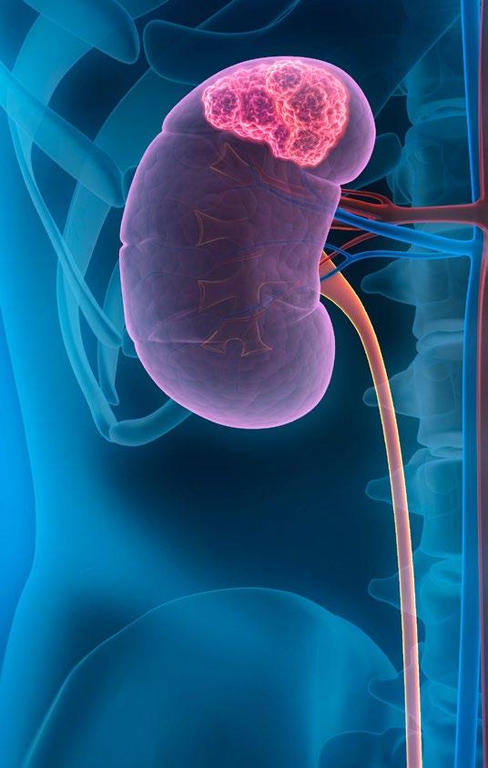
Criteria in Solid Tumors (RECIST) 1.1 criteria, with secondary endpoints including cancerspecific survival, freedom from distant progression, renal function, and toxicity.
The findings demonstrate sustained efficacy over time. Local control was 100% at 1 year and remained high at 98% at both 3 and 5 years, with only one reported local failure. Cancer-specific survival followed a similar trend, reaching 100% at 1 year and 98% at both 3 and 5 years. Freedom from distant progression declined modestly over time, from 97% at 1 year to 85% at 5 years.
Renal function declined gradually, with the estimated glomerular filtration rate decreasing by approximately 9.4 mL/min at 1 year and 15.8 mL/min at 5 years. Only one patient required dialysis, indicating a low incidence of severe renal impairment.
Safety outcomes were favourable. Grade 3 adverse events occurred in 7.8% of patients, including fatigue, vomiting, pain, and colonic obstruction, with all events reported within 2 years of treatment. No Grade 4 or 5 toxicities or late safety signals were observed.
As one of the largest prospective datasets evaluating SABR as a curative, non-invasive approach for RCC, these findings support its use in patients unsuitable for surgery. However, the absence of a comparator arm limits definitive conclusions, and randomised trials comparing SABR with surgical intervention are needed to establish its role in operable populations.
PENILE prosthesis implantation represents a treatment option for erectile dysfunction refractory to medications or less invasive therapies. This abstract, presented at the EAU Congress 2026, examined quality of life and decision regret after implantation while identifying factors associated with significant regret.5
The prospective study enrolled 86 patients (median age: 62 years; median BMI: 25.5 kg/ m2) undergoing penile prosthesis placement at a single tertiary referral centre. Participants received a comprehensive preoperative assessment, and perioperative and postoperative variables were recorded. The study employed the Quality of Life and Sexuality with Penile Prosthesis Questionnaire (QoLSPPQ), which assesses four subdomains: functional, personal, relational, and social. Decision regret was assessed using the Decision Regret Scale (DRS), with significant regret defined as >25%. Logistic regression was employed to identify predictors of significant regret.
A total of 90% of patients received a tricomponent prosthesis (77/86), with the remainder receiving a malleable prosthesis (9/86; 10%). Complications occurred in 13 patients (15%), whilst overall median decision regret was 24%, and 33% of
patients experienced significant regret. Median QoLSPPQ scores were 22 for functional, 17 for personal, 17 for relational, and 12 for social domains.
Analysis revealed that postoperative complications increased the risk of significant regret 18-fold, and lower functional and relational satisfaction were also key predictors. Personal and social subdomains did not significantly influence regret.
These findings indicate that penile prosthesis implantation achieves generally favourable outcomes with a low complication rate. However, patient counselling should emphasise the possibility of decision regret, particularly when postoperative complications occur or functional and relational expectations are unmet.
Analysis revealed that postoperative complications increased the risk of significant regret 18-fold
A MAJOR RCT, presented at the EAU Congress 2026, has found that robot-assisted laparoscopic (RAL) sacrocolpopexy offers comparable safety, effectiveness, and long-term costs to conventional laparoscopic (CL) sacrocolpopexy for pelvic organ prolapse.6
In this multicentre trial conducted across 15 centres, 366 females undergoing surgery for symptomatic pelvic organ prolapse were randomly assigned to CL or RAL sacrocolpopexy. Of these, 345 completed follow-up over 5 years. Baseline characteristics, including age, BMI, and prolapse severity, were comparable between groups.
The primary endpoint, 30-day postoperative complications, showed no significant difference between approaches, occurring in 15.3% of the CL group and 11.7% of the RAL group (p=0.3213). However, when complications were analysed by type, surgical complications were notably lower in the RAL group than the CL group (2.9% versus 7.4%). Operative times were similar overall, although robotic procedures required longer set-up times (40 minutes versus 20 minutes; p<0.0001).
Long-term outcomes further reinforced the equivalence of the two techniques. There were no significant differences in anatomical or functional results, symptom burden, or quality of life measures between groups at 5 years. Importantly, healthcare
costs, assessed using national data, were also comparable over 1-, 2-, and 5-year periods.
The findings suggest that both surgical approaches are safe and effective for the treatment of pelvic organ prolapse. However, robot-assisted sacrocolpopexy may offer a clinically relevant advantage in reducing surgical complications. This could be particularly meaningful for more complex cases, such as females with obesity or those undergoing repeat pelvic surgery.
Limitations include the potential variability in surgical expertise across centres and the extended recruitment period, which may influence outcomes. Nonetheless, the inclusion of 5-year follow-up offers valuable long-term data on the durability of both surgical approaches.
Overall, these results support the growing role of robotic assistance in pelvic reconstructive surgery, offering clinicians evidence to guide surgical decision-making in pelvic organ prolapse management.
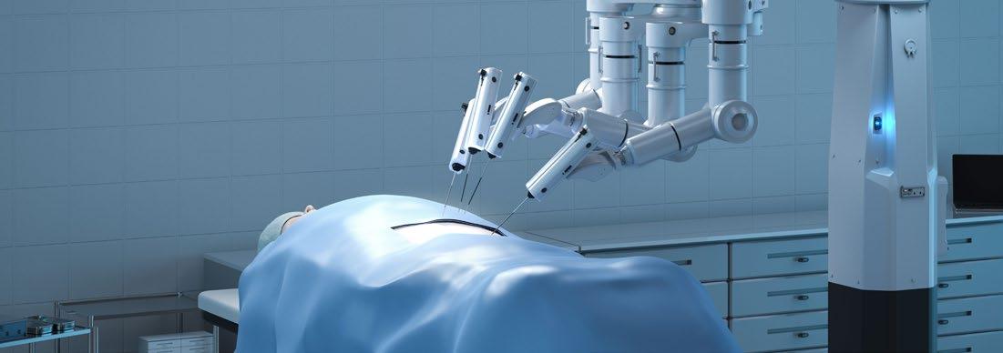
Robot-assisted sacrocolpopexy may offer a clinically relevant advantage in reducing surgical complications
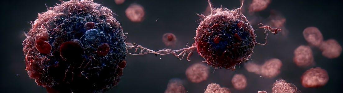
A NEW study presented at the EAU Congress 2026 reveals that cancer-associated fibroblasts (CAF) play a central role in driving tumour growth in high-risk non-muscle-invasive bladder cancer (HR-NMIBC), offering new insights into disease biology and potential therapeutic targets.
HR-NMIBC is typically treated with transurethral resection followed by Bacillus Calmette–Guérin (BCG) immunotherapy; however, outcomes vary significantly between molecular subtypes. In particular, the bombesin receptor subtype-3 (BRS3) subtype is associated with poor prognosis, epithelial-tomesenchymal transition (EMT) features, and an immunosuppressive tumour microenvironment. This study aimed to determine whether these characteristics are driven by tumour-intrinsic processes or by the surrounding stromal environment.
Researchers performed an integrative analysis using transcriptomic data from 891 patients across multiple cohorts, alongside spatial and single-cell transcriptomics, to map tumour heterogeneity. Ex vivo experiments using patient-derived tumoroids were conducted to assess the functional impact of CAF signalling on tumour growth.
Results showed that BRS3 tumours were significantly enriched for EMT, stromal, and immune-related signatures compared with other subtypes (p<0.001). However, EMT scores correlated more strongly with stromal signals (R=0.82; p<0.001) than with immune signatures (R=0.57; p<0.001), suggesting that the aggressive phenotype attributed to EMT may instead reflect stromal enrichment. Spatial transcriptomics further demonstrated that BRS3 signals were predominantly localised within stromal regions, while other subtypes were found in urothelial tumour areas. Even after excluding stromal regions, BRS3 tumour
7
cells were observed in close proximity to fibroblastrich areas.
Functional validation supported these findings. Exposure of patient-derived tumoroids to CAFconditioned medium resulted in a 21% increase in tumour cell viability compared with untreated controls (p=0.017), indicating that CAF-derived factors actively promote tumour growth.
These findings suggest that the poor prognosis associated with BRS3 is driven not by tumourintrinsic EMT programmes, but by interactions with a CAF-rich stromal niche. This highlights the tumour microenvironment, and specifically CAFs, as a key contributor to disease progression in HR-NMIBC.
The study is limited by its reliance on retrospective datasets and a relatively small number of samples for spatial and ex vivo analyses. Further research is needed to validate these findings and explore how targeting CAF-driven pathways could improve treatment outcomes.
Overall, this study provides important evidence that stromal interactions, rather than intrinsic tumour biology alone, underpin aggressive disease in HR-NMIBC, opening new avenues for therapeutic intervention.
NEW DATA presented at the EAU Congress 2026 highlight the potential of digital therapeutics in the management of premature ejaculation, a common and often stigmatising sexual dysfunction associated with significant psychological distress and reduced quality of life.8
The CLIMACS study evaluated the effectiveness of a mobile health intervention, the Melonga App® (Prognoix Health B.V., Maastricht, the Netherlands) as an alternative or adjunct to conventional treatments such as pharmacotherapy and behavioural therapy. In this RCT, 80 patients were assigned in a 1:1 ratio to either an intervention group using the app or a control group, with a partial crossover after 12 weeks.

In this RCT, 80 patients were assigned in a 1:1 ratio to either an intervention group using the app or a control group
The primary endpoint was the change in the Premature Ejaculation Profile (PEP) score at 12 weeks. Among 66 patients with complete datasets, the intervention group demonstrated a marked improvement, with a mean PEP score increase of 4.4 points compared with 0.03 in the control group (p<0.001). Overall, PEP scores improved from 5.5 to 9.4 points in the intervention arm.
Secondary outcomes also showed significant benefits. Intravaginal Ejaculation Latency Time (IELT) increased by an average of 64 seconds, from 61 to 125 seconds (p<0.001), with a substantially greater improvement in the intervention group compared with controls (75.7 seconds versus 0.5 seconds; p=0.001). Sexuality-related quality of life (SQoL-M) improved from 32.8 to 43.1 points (p<0.001), while the Premature Ejaculation Diagnostic Tool (PEDT) score decreased from 16.4 to 11.9 points (p<0.001), indicating reduced symptom severity. Notably, 22% of patients met criteria for resolution of premature ejaculation following the mobile health intervention.
These findings suggest that app-based interventions can significantly improve both clinical outcomes and patient-reported measures in premature ejaculation. As digital health solutions continue to evolve, such tools may expand the therapeutic options available in urological practice, offering accessible and non-invasive support for patients who may be reluctant to seek traditional care.
A MAJOR Phase III RCT, presented at the EAU Congress 2026, has shown that [68Ga]Ga-prostate-specific membrane antigen (PSMA)-11 PET/CT can substantially reduce unnecessary prostate biopsies while maintaining detection of clinically significant prostate cancer in men with inconclusive MRI findings.9
Multi-parametric MRI is widely used to assess males with suspected prostate cancer, yet those with Prostate Imaging Reporting and Data System (PI-RADS) 2 or 3 lesions often present a diagnostic challenge. Despite a relatively low likelihood of clinically significant prostate cancer, many still undergo biopsy due to high clinical suspicion, resulting in avoidable invasive procedures being carried out in patients with insignificant disease. Building on earlier evidence, the PRIMARY2 trial investigated whether incorporating PSMA PET/CT into the diagnostic pathway could improve patient selection for biopsy without compromising cancer detection.
This investigator-initiated, multicentre, Phase III trial enrolled 660 biopsy-naive males with suspected clinically significant prostate cancer, PI-RADS 3 or PI-RADS 2 MRI with risk factors (e.g., prostate-specific antigen [PSA] density >0.1, strong family history), PSA ≤20 ng/mL, and ≤cT2. Participants were randomised to standard systematic transperineal biopsy or a PSMA PET/CTguided pathway. In the experimental arm, only those with positive PET findings underwent targeted biopsy, while those with negative scans were monitored using PSA testing.
The results demonstrated a marked reduction in biopsy use. Nearly half of participants in the PSMA PET/CT arm avoided biopsy altogether (49%; p<0.001). Importantly, detection of clinically significant prostate cancer remained non-inferior compared with standard care (12% versus 16%; difference: –3.7%; p=0.009), meeting the trial’s predefined criteria. At the same time, the PSMA-guided approach significantly reduced the diagnosis of insignificant prostate cancer (14% versus 32%; p<0.001), indicating a meaningful decrease in overdiagnosis.
These findings suggest that PSMA PET/CT can refine diagnostic decision-making by identifying patients who are unlikely to benefit from biopsy, while still capturing those with clinically important disease. By enabling more selective, targeted biopsies and reducing unnecessary procedures, this approach may help minimise patient burden and potential complications associated with overdiagnosis.
Although longer-term outcomes and broader implementation considerations will require further evaluation, the trial provides strong evidence supporting the integration of PSMA PET/CT into diagnostic pathways for males with equivocal MRI findings.
Nearly half of participants in the PSMA PET/CT arm avoided biopsy altogether (49%; p<0.001)
EMERGENCY haematuria is associated with high morbidity, significant readmission rates, and delayed diagnosis according to findings presented at the EAU Congress 2026.10
Emergency haematuria accounts for a notable proportion of urological admissions, representing approximately 15% of cases and around four per 1,000 emergency hospital presentations. In this large, international, prospective, observational study, data were collected from 8,500 patients across 382 centres over 1 year using a collaborative research model.
The median length of stay was 4 days (interquartile range: 2–8), while 90day mortality reached 9.2% and 90-day readmission rates were 31%. At presentation, 5% of patients were haemodynamically unstable, 5% were septic, 11% required high-dependency care, and 21% required blood transfusion. Despite this clinical burden, ward-based management was successful in only 35% of cases.
The study identified considerable variation in inpatient management. Nearly half (47%) of patients did not undergo imaging during admission, and only 35% received any form of intervention. These findings suggest inconsistency in clinical pathways and potential missed opportunities for early diagnosis. Malignancy was identified as the underlying cause in 25% of cases during admission, evenly split between
pre-existing and newly diagnosed disease, with an additional 5% diagnosed during follow-up. Urothelial carcinoma accounted for 20% of malignancies detected during admission, including 18% bladder and 2% upper tract, with a further 4% identified after discharge.
Time to diagnosis differed markedly depending on when investigations occurred. During admission, the median time to diagnosis was 1 day (0–2), compared with 21 days (10–41) following discharge. This delay highlights a critical gap in post discharge pathways and raises concerns regarding deferred investigation in patients with significant underlying pathology.
These findings demonstrate that emergency haematuria carries substantial clinical risk, with outcomes comparable to outpatient haematuria cohorts in terms of malignancy prevalence. However, low intervention rates and frequent discharge without definitive diagnosis remain key challenges. The data support the need for structured early diagnostic pathways and prompt inpatient investigation. Avoiding discharge without a confirmed diagnosis, where feasible, may reduce delays and improve outcomes for patients presenting with emergency haematuria.

References
1. Fader M et al. A randomised trial comparing combined reusable and single-use catheters (mixed-use) with single-use catheters only. Abstract A0496. EAU Congress, 13-16 March, 2026.
2. Korn P et al. Impact of adjuvant intracavitary therapy on recurrence of upper tract urothelial carcinoma following endoscopic management. Abstract P0486. EAU Congress, 13-16 March, 2026.
3. Pacheco-Rendón RM et al. From PI-RADS to Te-RADS: testicular MRI biomarkers predicting sperm retrieval in non-obstructive azoospermia. Abstract A0376. EAU Congress, 13-16 March, 2026.
4. Siva S et al. 5-year outcomes of stereotactic radiotherapy for primary renal cell carcinoma from the pooled analysis of the FASTRACK trials. Abstract A0342. EAU Congress, 13-16 March, 2026.
5. Carmen G et al. Determinants of decision regret and quality of life after penile prosthesis implantation: insights from a prospective single-center study. Abstract P0034. EAU Congress, 13-16 March, 2026.
6. Kutchukian S et al. Pelvic organ prolapse: is robotic sacrocolpopexy evidence based? Abstract A0031. EAU Congress, 13-16 March, 2026.
7. Olislagers M et al. Cancer-associated fibroblast niche drives tumor growth in non-muscle-invasive bladder cancer. Abstract A0132. EAU Congress, 13-16 March, 2026.
8. Kormann F et al. A digital health application to reduce premature ejaculation symptoms: final results of the German CLIMACS study. Abstract P0503. EAU Congress, 13-16 March, 2026.
9. Buteau JP et al. Impact of [68Ga]GaPSMA-11 PET/CT in the diagnosis of prostate cancer in men with equivocal or non-suspicious findings on multiparametric MRI (PRIMARY2): a multicentre, phase III, randomised trial. Abstract GC26-006. EAU Congress, 13-16 March, 2026.
10. Bhatt NR et al. The WASHOUT study: workup and management of patients with emergency haematuria across the world. Abstract P1006. EAU Congress, 13-16 March, 2026.
Author: Bertie Pearcey, EMJ, London, UK
Citation: EMJ Urol. 2026;14[1]:24-28. https://doi.org/10.33590/emjurol/N0J5V831
AT THE European Association of Urology (EAU) Annual Congress 2026, held in London, UK, from 13th–16th March 2026, the session ‘Incontinence Uncovered: Managing Urgency Without Stress’ brought together leading clinicians to explore contemporary challenges and evolving strategies in the management of overactive bladder (OAB) and urinary incontinence. Expertly chaired by Benoit Peyronnet, University of Rennes, France; and Enrico Finazzi Agrò, University of Rome Tor Vergata, Italy, the session opened with a state-of-the-art overview spanning policy, digital health, physiotherapy, pharmacotherapy, diagnostics, and shared decision-making.
Michael R. Van Balken, Rijnstate Hospital, Arnhem, the Netherlands, opened by emphasising continence health as both a clinical and societal priority.
He presented an update on the EAU ‘Urge to Act’ campaign, launched following the 2023 EU Continence Health Summit, where stakeholders signed a joint statement calling for greater prioritisation of continence care across Europe.
A key achievement has been quantifying the burden of incontinence, which affects an estimated 55–60 million Europeans, with an economic cost approaching 70 billion EUR annually. This includes not only healthcare expenditure, but also productivity loss and environmental impact.
Van Balken stressed that clinical need alone does not drive policy change. Integrating socioeconomic and sustainability data has proven essential in engaging policymakers. A major milestone was the formation of a European Parliament Member interest group in 2025, helping elevate continence health on the political agenda. Early outcomes
include practical initiatives such as petitions for appropriate disposal facilities for continence products.
Urinary incontinence is increasingly recognised as a non-communicable disease, closely linked to conditions such as cardiovascular disease, diabetes, and stroke. With growing political engagement, Van Balken concluded optimistically: “There is a lot more to come.”
A key achievement has been quantifying the burden of incontinence, which affects an estimated 55–60 million Europeans
Laura Wiemer, Charité – Universitätsmedizin Berlin, Germany, highlighted time constraints as a major barrier to optimal OAB care. In many European settings, consultations must cover both diagnosis and education within minutes.
Wiemer elaborated that the gap between guideline recommendations and real-world practice contributes to poor adherence and outcomes, explaining that patients often find it difficult to retain complex treatment information delivered in a limited time, adding additional barriers to treatment.
App-based management offers a potential solution. Digital platforms can deliver guideline-recommended interventions, including bladder training, pelvic floor exercises, and lifestyle modification, in a structured and accessible format. They improve time efficiency, support long-term follow-up, and enable multimodal care without replacing clinicians.
Evidence that supports this is increasingly robust. The DINKS trial1 saw a 59% reduction in incontinence episodes per 24 hours with app-supported care in women with stress urinary incontinence, urgency urinary incontinence, or mixed urinary incontinence, with 92% of patients reporting improved quality of life. Similarly, the BEST study2 showed a 7-point improvement in International Prostate Symptom Score (IPSS) in men with lower urinary tract symptoms from benign prostatic hyperplasia, or OAB alone.
Benefits were consistent across severity levels and prior treatment exposure, with minimal adverse effects. While challenges remain, including patients’ digital literacy and integration into workflows, Wiemer concluded: “We might already have a contender for best first-line OAB therapy, and it’s probably already in your pocket.”
Paula Igualada-Martinez, Guy’s and St Thomas’ NHS Foundation Trust, London, UK, reframed physiotherapy as a comprehensive, patientcentred intervention rather than simply pelvic floor exercises.
While guidelines recommend conservative management as first-line therapy, Igualada-Martinez emphasised that real-world care requires individualised approaches. Physiotherapy includes bladder training, lifestyle interventions, weight management, and behavioural strategies, tailored to each patient.
Patient education is central. Many patients lack understanding of normal bladder function or fluid intake, and tools such as bladder diaries enable personalised treatment planning.
Assessment is equally important. IgualadaMartinez posed the question: “How many of you actually assess pelvic floor function before prescribing any medication?”
Supervised, intensive training requires appropriate evaluation, particularly in urgency-dominant cases.
Physiotherapy can reduce symptoms across urgency, frequency, and incontinence, while empowering patients with longterm self-management strategies. It also helps identify those requiring escalation to pharmacological or surgical treatment.

Although access may be limited and not all patients respond, Igualada-Martinez concluded: “Physiotherapy is not competing with other treatments. It is the foundation that makes all of your treatments better.”
Marie Carmela Lapitan, University of the Philippines Manila, Philippines, positioned antimuscarinics as occupying an “optimal therapeutic window” between behavioural and surgical approaches. By inhibiting muscarinic receptors, they directly target detrusor overactivity, producing reductions in urgency, frequency, and incontinence within days to weeks. They are widely accessible, scalable, and can be prescribed in primary care, with generally mild and reversible adverse effects.
Patient preference also supports their use, even when accounting for side effects, reinforcing their role alongside conservative therapy in first-line management.
Sung Yong Cho, Seoul National University Hospital, South Korea, complemented this with evidence on β3-adrenoceptor agonists. Trials such as SCORPIO,3 TAURUS,4 and BESIDE5 demonstrated significant symptom
improvements, sustained efficacy over 12 months, and favourable tolerability.
Mirabegron reduced incontinence and frequency compared with placebo, maintained long-term safety, and improved outcomes when added to antimuscarinics, with nearly half of patients achieving continence in combination therapy.
With fewer anticholinergic side effects and suitability for long-term use, Cho asked: “Is there really any reason not to use β3 agonists as a first option?”
George Bou Kheir, Ghent University Hospital, Belgium, challenged the traditional stepwise model of OAB treatment.
“OAB is not a disease, it is a symptom syndrome,” he stated, arising from diverse pathophysiological mechanisms.
He proposed that OAB represents a fluctuating homeostatic state, explaining why treatment responses vary within the same patient over time.
Examples included neurogenic-myogenic mechanisms, urethral dysfunction, and hormonal influences. In some cases, treating

one component may worsen another, highlighting the limitations of rigid algorithms.
Evidence supporting alternative approaches included improvements in both stress and urgency symptoms following midurethral sling (MUS) placement, as well as the impact of testosterone replacement on lower urinary tract symptoms.
OAB represents a fluctuating homeostatic state, explaining why treatment responses vary within the same patient over time
These findings support mechanism-based, individualised care. Bou Kheir concluded: “The real question may not be which step comes next, but which mechanism we are actually treating.”
Tufan Tarcan, Koç University, Istanbul, Türkiye, defended the continued importance of urodynamics in OAB assessment. He argued that while randomised trials are appropriate for therapies, they are not suited to evaluating diagnostics. Critiquing the FUTURE study,6 he highlighted that urodynamic findings were underutilised, limiting the validity of its conclusions.
Urodynamics reclassified a proportion of patients initially diagnosed with OAB, identifying alternative or mixed pathologies. It also enabled comparable outcomes with less invasive treatment, suggesting a role in avoiding overtreatment.
“The bladder is an unreliable witness,” Tarcan stated, emphasising that symptoms do not reliably reflect underlying mechanisms. He concluded that personalised management informed by urodynamics remains essential, particularly in complex cases.
Mohammed Belal, Queen Elizabeth Hospital, Birmingham, UK, emphasised shared decision-making as a central part of modern OAB care.
Shared decision-making integrates clinical evidence with patient preferences, which is particularly important in OAB, where multiple reasonable treatment options exist and trade-offs between efficacy, side effects, and convenience are common.
Current stepwise approaches often result in trial-and-error prescribing, leading to poor adherence and disengagement. Persistence rates remain low across therapies, reflecting differences between clinician priorities and patient values.
Belal stressed that shared decision-making goes beyond providing information. Instead, it requires structured discussion, clear recommendations, and understanding patient preferences.
He noted that decision aids can support the process, allowing patients time to consider options, and concluded that, ultimately, shared decision-making represents a redesign of care pathways, improving adherence and outcomes.
The session concluded with a debate on synthetic MUSs for stress urinary incontinence.
Elisabetta Costantini, University of Perugia, Italy, argued that MUSs remain highly effective, with long-term cure rates of 80–90% and durable outcomes beyond 15 years. Supported by extensive evidence, they offer minimally invasive treatment with rapid recovery and low re-operation rates.
While complications exist, she emphasised their relatively low incidence and the importance of appropriate training and regulation, stating: “The question is not
whether complications exist, but whether alternatives perform better.”
In contrast, Tamsin Greenwell, University College London Hospitals, UK, highlighted significant risks, including chronic pain and serious complications. She noted that complication management can lead to further morbidity, and that effective meshfree alternatives, such as autologous fascial slings and colposuspension, are available.
Advocating caution, she concluded that treatment decisions should prioritise safety and adhere to the principle of “do no harm.”
References
1. Hafekamp A et al. App-based therapy for female patients with urinary incontinence in Germany (DINKS): a single-blind, randomised, controlled trial. Lancet Digit Health. 2025;7(12):100935.
2. Gratzke C et al. A randomized trial of an app-based therapeutic for lower urinary tract symptoms. NEJM Evid. 2025;4(4):EVIDoa2400290.
3. Khullar V et al. Efficacy and tolerability of mirabegron, a β(3)-adrenoceptor
A clear theme emerged across the session: OAB and urinary incontinence are complex, multifactorial conditions requiring individualised management. From policy initiatives and digital tools to physiotherapy, pharmacotherapy, and diagnostics, no single approach is sufficient.
The future lies in integrating mechanistic understanding with patient-centred care. Tailored treatment strategies, supported by tools such as urodynamics and digital platforms, and guided by shared decisionmaking, offer the greatest potential to improve outcomes.
For healthcare professionals, the challenge is not only to adopt new therapies, but to deliver care that is personalised, evidencebased, and aligned with patient priorities.
agonist, in patients with overactive bladder: results from a randomised European-Australian phase 3 trial. Eur Urol. 2013;63(2):283-95.
4. Chapple CR et al. Randomized double-blind, active-controlled phase 3 study to assess 12-month safety and efficacy of mirabegron, a β(3)adrenoceptor agonist, in overactive bladder. Eur Urol. 2013;63(2):296-305.
5. Drake MJ et al. Efficacy and safety of mirabegron add-on therapy to solifenacin in incontinent overactive bladder patients with an inadequate
response to initial 4-week solifenacin monotherapy: a randomised doubleblind multicentre phase 3B study (BESIDE). Eur Urol. 2016;70(1):136-45.
6. Abdel-Fattah M et al. Female Urgency, Trial of Urodynamics as Routine Evaluation (FUTURE study): a superiority randomised clinical trial to evaluate the effectiveness and cost-effectiveness of invasive urodynamic investigations in management of women with refractory overactive bladder symptoms. Trials. 2021;22(1):745.
Author: Helena Bradbury, EMJ, London, UK
Citation: EMJ Urol. 2026;14[1]:29-33. https://doi.org/10.33590/emjurol/RE6LKV6B
AI IS no longer a distant idea, but an integrated tool significantly impacting medicine and patient care. In this timely session from the European Association of Urology (EAU) Congress 2026, experts from across the globe gathered to discuss the role of AI in urological surgery, sharing real-world examples of its successes and pitfalls that require further investigation. This feature captures select talks from the session, co-chaired by Cristian Fiori, Università degli Studi di Torino, Italy; and Alexandre Mottrie, Head of the Urology Department at OLV Hospital, Aalst, and CEO of Orsi Academy, Melle, Belgium.
To open, Pieter De Backer, Head of Innovation at Orsi Academy, explored the use of AI in robotic-assisted surgery, drawing on three ‘waves’ of AI integration: descriptive, generative, and agentic. Descriptive AI analyses historical data to identify patterns, while generative AI learns patterns to create new content. Agentic AI, the most recent advancement, acts as a co-pilot, fully integrated within the workflow of systems. De Backer shared an example in which, during a live-streamed surgery, the agentic AI tool activated guidelines, imaging, patient information, and tumour identification based on the surgeon’s specific prompt.
De Backer concluded that augmented reality extends beyond merely superimposing images, gathering all the unintegrated information and centring the surgeon “as part of the puzzle,” ensuring that both the surgical team and people outside the operating room feel the benefit.
Telesurgery, or remote surgery, is where a surgeon operates on a patient from a
distance via robotic surgical systems and high-speed telecommunications, such as 5G and optical fibres. The first reported case of telesurgery famously occurred in 2001, led by Jacques Marescaux, Research Institute against Digestive Cancer (IRCAD), Strasbourg, France, and Michel Gagner, Hôpital du Sacre Coeur, Montreal, Canada, who, based in New York, performed a laparoscopic cholecystectomy on a patient based over 6,000 km away in Strasbourg.
Communication can be trained, lost immersion cannot
Alberto Breda, Head of the Uro-oncological Unit and Surgical Kidney Transplantation team, Fundacio’ Puigvert, Barcelona, Spain, shared a timely update regarding telesurgery. He highlighted a 2026 study, led by Ye Wang and colleagues in China, who investigated the reliability of telesurgery to that of standard local surgery in patients undergoing robotic operations.1 It enrolled 72 participants across five hospitals in China, each randomly assigned 1:1 to undergo either telesurgery or local surgery across December 2023–June 2024. Results found telesurgery to be noninferior to local surgery and stable with a distance from 1,000–2,800 km. Secondary outcomes,
such as operative basic data, complications, early recovery, oncological outcomes, and medical team workload, also did not differ significantly between the two groups.1
In line with the growing emergence of telesurgery, several committees have published clear guidelines to ensure standardised, ethical, and safe adoption of telesurgery in practice across the globe. In 2025, the EAU Policy on Telesurgery was released, detailing critical requirements for pre, during, and post-surgery.2 In 2024, the first international consensus conference specifically focused on telesurgery took place (Society of Robotic Surgery [SRS], Orlando, Florida, USA), bringing experts together to define safety, ethical, and regulatory frameworks for this surgical technique.3 Across the globe, in August 2024, the first case of telesurgery in Indonesia took place: a renal cyst marsupialisation between Bali and Jakarta, which is a distance of 1,200 km.
Sharing his own contributions, Breda spoke on how he is now regularly performing telesurgeries from Barcelona to patients based in the Canary Islands, with a latency below 35 ms.
Senthil Nathan, University College London Hospital NHS Trust, UK, opened with key definitions of telemedicine: telemonitoring is the remote tracking of a patient’s clinical data, whilst telepreceptoring is where a trained practitioner mentors and evaluates a trainee from a distance.
As stressed by Nathan, there is a drastic patient need for telemedicine, with five billion people currently not having access to safe and affordable surgical and anaesthesia care when needed. Moreover, 143 million additional surgical procedures are needed per year in low and middle income countries to save lives and prevent disability, and of the 313 million procedures undertaken worldwide each year, only 6% occur in the poorest countries.4
For the latter portion of the session, speakers were invited to debate on open versus closed console for modern robot-assisted urological surgery. Open console telerobotic surgery uses a screen and 3D glasses, allowing the surgeon to

see the operating room and improving communication. Closed console systems require looking into a viewfinder, offering high immersion and reduced distractions.
Josep Maria Gaya, Unidad de Uro-Oncología y Equipo Quirúrgico de Trasplante Renal Fundació Puigvert, Universitat Autònoma de Barcelona, Spain, first advocated on behalf of the open console. Examples include the Medtronic Hugo™ (Medtronic plc, Dublin, Ireland) RAS, Versius® (CMR Surgical, Cambridge, UK), and KangDuo® (Harbin Sagebot Intelligent Medical Equipment Co., Ltd, China). Touching on ergonomics, Gaya cited a 2020 study reporting that up to 50% of robotic surgeons using the Da Vinci® (Intuitive Surgical, Inc., Sunnyvale, California, USA) system, a closed console, experienced intraoperative discomfort, particularly in the neck, back, and shoulders.5
Reinforcing his point, he referenced a 2026 multicentre RCT comparing the efficacy and safety of an open versus closed system; KangDuo versus Da Vinci, respectively.6 The researchers reported that the KangDuo Surgical Robot-01 open console system significantly alleviated musculoskeletal strain, particularly in the neck and back, during robotic colorectal surgery, while maintaining equivalent technical performance.
In addition to ergonomics, Gaya spotlighted the benefits that the open console offers to the operating workflow and team communication. As the surgeon is not physically isolated, they can maintain visual contact with their surgical team, allowing for greater integration within the operating room environment. Finally, the open console improves mentorship as the mentor can move the hands of the surgeon or even do it by themselves without changing seats or requiring a second console, making it cost effective as well.
Véronique Phe, Santé Sorbonne Université, Paris, France, subsequently took the stage to counter his argument. She stressed that console design does not solely impact comfort, but also the performance of the surgeon, affecting cognitive focus and load, ergonomic stability over time, motor precision and tremor control, and reproducibility and safety. Whilst acknowledging the advantages of the open console, she stressed that in complex surgeries, total focus and immersion in the operation is paramount, and that the closed system offers this.
Phe continued stating that an immersive 3D view with depth perception offers strong visual focus, better tissue-plane reading and anatomic orientation, and less visual distraction during fine dissection.7
“I will choose the closed console, because I choose precision, immersion, and focus.” Her talk highlighted that surgeons have individual preferences in robotic surgery, with both closed and open consoles offering unique advantages and disadvantages.
Prokar Dasgupta, King’s Health Partners, London, UK, subsequently offered realworld examples of AI in urological surgery. In a recent review that Dasgupta co-authored,8 his team investigated the applications of AI in diagnosis, treatment, and outcome prediction in urological diseases, and evaluated any advantages it offers over traditional methods. From the 111 articles included in the final analysis, compared to conventional statistical analysis, 71.8% of studies concluded that AI is superior in diagnosis and outcome prediction.8
In renal cancer imaging, AI can differentiate between benign small renal masses and potential malignant tumours, guiding clinical decision-making and improving the use of healthcare resources. A 2021 study by Ali et al.9 tested a blue light-based AI diagnostic platform using 216 blue light images to predict image malignancy, invasiveness, and grading. Interestingly, the reported classification sensitivity and specificity of malignant lesions was 95.77% and 87.84%, while the mean sensitivity and mean specificity of tumour invasiveness was 88.00% and 96.56%, respectively, illustrating the potential of AI-based diagnostic platforms in urological surgery.
In prostate cancer, Dasgupta recognised ‘AutoProstate’, a deep learning-based, AI-driven software system designed to automate the analysis and reporting of prostate MRI scans to assist in the diagnosis of prostate cancer.10 In addition, the PANDA Challenge was launched in 2020 as a global competition and aimed to develop AI algorithms for the diagnosis and Gleason grading of prostate cancer using a large, publicly available dataset of 10,616 digitised prostate biopsies from multiple countries. The results, published in 2022, demonstrated that AI systems could perform as well as human pathologists in identifying and grading prostate cancer.11
Following on, Karl-Friedrich Kowalewski, University Medical Center Mannheim, University of Heidelberg, Germany, offered a unique, European perspective, sharing first the following definitions: ‘narrow AI’ is AI designed to excel at a specific, predefined task or limited set of tasks, such as facial recognition, language translation, or internet searches, while ‘general AI’ possesses human-like intelligence and understanding, capable of learning, reasoning, and formulating its own conclusions based on information it has gathered.
Citing a 2025 systematic review,12 investigators reported that minimal
progress has been made towards clinical integration of AI over the past decade, stressing the need for diverse, multiinstitutional datasets and robust validation practices, amongst other efforts.
So, where are we now? Kowalewksi spoke on foundation models for generalist medical AI. Generalist medical AIs refer to versatile AI models trained on large, diverse, and multimodal datasets, such as images, text, and genomics, and can perform a wide range of clinical tasks rather than just one.13 As an example, SurgVISTA (Surgical Video-Level Spatial-Temporal Architecture) is a pioneering, large-scale, self-supervised video foundation model designed for comprehensive surgical scene understanding.14
Finally, Mottrie, co-chair of the session, addressed the audience on the topic of training and education, emphasising the need for a transition from a volume-based model, in which a surgeon is deemed proficient after completing a required number of surgeries, to criterion-referenced performance targets, a focus the Orsi Academy and the EAU have proposed collectively. He stressed that long-term monitoring of their skill and expertise is also necessary to ensure trainees are progressing at a good level throughout their development.
With the rise of AI in medicine, the future of urological surgery is a promising but uncertain one
Does this new approach work? In the PROVESA trial, 36 robotic surgerynaïve junior residents were randomly allocated to metric-based proficiencybased progression training or the current standard of care traditional training, with the primary outcome being the percentage
of participants reaching the predefined proficiency benchmark.
Interestingly, the proficiency-based progression training group were approximately 10-times more likely to reach the set benchmark than the traditional training group, reinforcing the need for more quantitative, objectivebased training methods.15
However, as highlighted by Mottrie, continued training and support is crucial, even for the expert surgeons, noting large inconsistencies in surgery standards and outcomes in this cohort. As a takeaway, he recommended the audience complete
References
1. Wang Y et al. Reliability of urological telesurgery compared with local surgery: multicentre randomised controlled trial. BMJ. 2026;392:e083588.
2. Somani BK et al. European Association of Urology policy on telesurgery. Eur Urol. 2025;88(4):318-24.
3. Society of Robotic Surgery. Telesurgery consensus conference. 2023. Available at: https:// srobotics.org/telesurgeryconference/#:~:text=21%20 December%202023,yet%20to%20 be%20fully%20explored. Last accessed: 31 March 2026.
4. Meara JG et al. Global surgery 2030: evidence and solutions for achieving health, welfare, and economic development. The Lancet. 2015;386(9993):569-624.
5. Dwver A et al. Ergonomic assessment of robotic general surgeons: a pilot study. J Robot Surg. 2020;14(3):387-92.
quality assessments and general recertifications to ensure that consistency is maintained in urological surgeries.
With the rise of AI in medicine, the future of urological surgery is a promising but uncertain one. This session offered a timely and thought-provoking discussion, bringing together expert insights on the evolving landscape of robotic surgery, from AI integration and surgical consoles to global disparities in technological advancement and the future of surgical education.
6. Wang C et al. Ergonomic advantages of the open console system in robotic colorectal resection: reduced musculoskeletal strain with preserved technical performance - a post hoc analysis of a randomized controlled trial. Int J Surg 2026;DOI:10.1097/ JS9.0000000000004691.
7. Cooper H et al. A systematic review of ergonomic and muscular strain in surgeons comparing robotic to laparoscopic approaches. J Robot Surg. 2025;19(1):252.
8. Chen J et al. Current status of artificial intelligence applications in urology and their potential to influence clinical practice. BJU Int. 2019;124(4):567-77.
9. Ali N et al. Deep learning-based classification of blue light cystoscopy imaging during transurethral resection of bladder tumors. Sci Rep. 2021;11(1):11629.
10. Mehta P et al. AutoProstate: towards automated reporting of prostate MRI for prostate cancer assessment
using deep learning. Cancers (Basel). 2021;13(23):6138.
11. Bulten W et al. Artificial intelligence for diagnosis and Gleason grading of prostate cancer: the PANDA challenge. Nat Med. 2022;28:154-63.
12. Carstens M et al. Artificial intelligence for surgical scene understanding: a systematic review and reporting quality metaanalysis. medRxiv. Preprint doi: 10.1101/2025.07.12.25330122.
13. Moor M et al. Foundation models for generalist medical artificial intelligence. Nature. 2023;616(7956):259-65.
14. Yang S et al. Large-scale selfsupervised video foundation model for intelligent surgery. NPJ Digit Med. 2026;9:220.
15. De Groote R et al. Proficiency-based progression training for robotic surgery skills training: a randomized clinical trial. BJU Int. 2022;130(4):528-35.

Drawing on insights from the European Association of Urology (EAU) Congress 2026, these abstract reviews highlight notable new developments and key areas of research in the field.
Authors: *Amanda Nizam,1 Tanya Jindal,2 Elise Cai,3 Cindy Y. Jiang,4 Zachariah Thomas,4 Eugene Oh,5 Charles B. Nguyen,6 Albert Jang,7 Jeffrey Zhong,8 Kevin Reyes,9 Salvador Jaime-Casas,6
Dimitra R. Bakaloudi,10 Ali R. Khaki,11 Ameish Govindarajan,1 Shilpa Gupta,1 Sumit A. Shah,11
Matthew I. Milowsky,12 Petros Grivas,10 Abishek Tripathi,6 Jason R. Brown,13 Irene Tsung,14 Matthew T. Campbell,4 Omar Alhalabi,4 Vadim S. Koshkin3
1. Department of Hematology and Medical Oncology, Cleveland Clinic Taussig Cancer Institute, Ohio, USA
2. Department of Medicine, Case Western Reserve University School of Medicine, Cleveland, Ohio, USA
3. Department of Hematology/Oncology, University of California San Francisco Helen Diller Family Comprehensive Cancer Center, USA
4. Department of Genitourinary Medical Oncology, The University of Texas MD Anderson Cancer Center, Houston, USA
5. Department of Medicine, University of Michigan School of Medicine, Ann Arbor, USA
6. Department of Medical Oncology & Therapeutics Research, City of Hope Comprehensive Cancer Center, Duarte, California, USA
7. Department of Oncology, Mayo Clinic Comprehensive Cancer Center, Rochester, Minnesota, USA
8. Department of Medicine, Case Western Reserve University, Cleveland, Ohio, USA
9. Department of Medicine, University of California San Francisco, USA
10. Clinical Research Division, Fred Hutchinson Cancer Center, Seattle, Washington, USA
11. Department of Medicine, Division of Oncology, Stanford University, California, USA
12. Department of Medicine, Division of Oncology, University of North Carolina School of Medicine, Chapel Hill, USA
13. Department of Medicine, Case Western Reserve University/University Hospitals Seidman Cancer Center, Cleveland, Ohio, USA
14. Department of Hematology and Oncology, University of Michigan Rogel Comprehensive Cancer Center, Ann Arbor, USA
*Correspondence to nizama@ccf.org
Disclosure: Tripathi has received grants from Corvus Pharmaceuticals, EMD Serono, and Aravive; and consulting fees from Deka Biosciences, Aadi Biosciences, Seagen, Astellas, Exelixis, Bayer, and Gilead Sciences. Khaki has received grants from Acrivon Therapeutics, Johnson & Johnson, Pfizer, and 23andMe; and has acted as a consultant for Janssen, Pfizer, and Astellas (uncompensated). Nizam has held consulting or advisory roles for Astellas, AVEO Oncology, EMD Serono/Merck KGaA, Formedics, IDEOlogy Health, Medscape, Pfizer, and Seagen; received honoraria from Aptitude Health, ASCO, BioAscend, Cleveland Clinic, Doximity, Flatiron Health, Formedics, IntegrityCE, MECC Global Meetings, OncLive, SignifyMD, Targeted Oncology, and University Hospitals Seidman Cancer Center; travel and accommodation expenses from ASCO, DAVA Oncology, Formedics, and MECC Global Meetings; institutional research funding from ArteraAI, the Cleveland Clinic VeloSano Grant Program, and Valar Labs; is editor of the Bladder Cancer Section of GU Oncology Now; and serves on the Editorial Board for the Journal of Clinical Oncology. Nguyen has received consulting fees from Johnson & Johnson and Dendreon; payment for lectures, presentations, speakers’ bureaus, manuscript writing, or educational events from MJH Associates; and support for attending meetings from DAVA Oncology. Jiang has received a grant from the ASCO Conquer Cancer Young Investigator Award. Bakaloudi has received a grant from the Kurelt Cancer Research Award. Brown has held consulting or advisory roles for Pfizer, AstraZeneca, EMD Serono, and Janssen; received honoraria from Ipsen; participated in speakers’ bureaus for EMD Serono and Merck; received travel and accommodation expenses from Janssen; served on a Data Safety Monitoring Board for Roche; and received institutional research funding from Bicycle Therapeutics, Roche, and Pfizer. Campbell has received grants from AstraZeneca, Exelixis, Genentech, and Pfizer; consulting fees from Eisai, Exelixis, Merck, and Pfizer; and payment for lectures, presentations, speakers’ bureaus, manuscript writing, or educational events from Curio Science, DAVA Oncology, MJH Life Sciences, and Cancer Noetic. Milowsky has held consulting or advisory roles for G1 Therapeutics and Loxo/Lilly (both uncompensated); received honoraria from Medscape, Research to Practice, Prime Education, and OncLive/MJH Life Sciences; holds stock and other ownership interests in Pfizer and Gilead Sciences; holds a leadership or fiduciary role as a member of the
Board of Directors for Alliance for Clinical Trials in Oncology; and received institutional research funding from Merck, Bristol Myers Squibb, Alliance Foundation Trials, ALX Oncology, Novartis, Acrivon Therapeutics, Prostate Cancer Clinical Trials Consortium, OncoC4, Flare Therapeutics, Loxo/Lilly, Astellas, Pfizer, Amgen, Roche, and G1 Therapeutics. Alhalabi has received grants from AstraZeneca, Ikena Oncology, Roche/Genentech, and Arcus Biosciences; and consulting fees from Seagen, Silverback Therapeutics, and Cardinal Health. Grivas has received grants from MSD, EMD Serono, Gilead Sciences, Acrivon Therapeutics, ALX Oncology, and Genentech; and consulting fees from MSD, Bristol Myers Squibb, AstraZeneca, EMD Serono, Pfizer, Janssen, Roche, Astellas, Gilead Sciences, Strata Oncology, Abbvie, Bicycle Therapeutics, Replimune, Daiichi Sankyo, Foundation Medicine, Eli Lilly, Urogen, Tyra Biosciences, Natera, and Ottimo. Gupta has received grants from Bristol Myers Squibb Foundation, Merck, Roche/Genentech, Exelixis, Novartis, Tyra Biosciences, Pfizer, Convergent Therapeutics, Acrivon Therapeutics, Flare Therapeutics, and Amgen; royalties from UpToDate; consulting fees from Pfizer, Merck, Foundation Medicine, Bristol Myers Squibb/Medarex, Natera, Astellas, AstraZeneca, Novartis, and Johnson & Johnson; support for attending meetings from Pfizer, Astellas, and Merck; and holds stock in Moderna Therapeutics, BioNTech SE, and Nektar. Jindal has declared being employed by Trial Library. Koshkin has held consulting or advisory roles for Janssen, Clovis Oncology, Astellas, Seagen, Pfizer, EMD Serono, Gerson Lehrman Group, ExpertConnect, and Guidepoint Global; is a Steering Committee Member for Pfizer/Seagen; and has received institutional research funding from Clovis Oncology, Nektar, Endocyte, Taiho Oncology, Merck, Gilead Sciences, Lilly, Seagen, Novartis, and the Prostate Cancer Foundation. Jang has received consulting fees from RealTimeCase; payment for lectures, presentations, speakers’ bureaus, manuscript writing, or educational events from MJH Life Sciences and Curio Science; and support for attending meetings from MECC Inc., Binaytara Foundation, and PSMA & Beyond Conference. The other authors have declared no conflicts of interest.
Acknowledgements: The authors thank their patients for their contributions to research and all members contributing to data curation at each institution.
Keywords: Advanced urothelial carcinoma (aUC), enfortumab vedotin plus pembrolizumab (EV+P), prognostic factors.
Citation: EMJ Urol. 2026;14[1]:35-38. https://doi.org/10.33590/emjurol/0036A70Y
Enfortumab vedotin plus pembrolizumab (EV+P) is the preferred first-line (1L) regimen for patients with advanced urothelial carcinoma (aUC).1,2 Despite the robust efficacy of EV+P demonstrated in the landmark EV-302 clinical trial, 10% of patients experienced primary progression (PD) on 1L EV+P, highlighting the need to identify those at risk for early treatment failure.2 The authors hypothesised that the presence of mixed variant-predominant or pure variant histology (VH) in conjunction with previously described poor prognostic factors in aUC, such as Eastern Cooperative Oncology Group Performance Status (ECOG PS) ≥1, presence of liver metastases, haemoglobin (Hgb) level <10 g/dL, and elevated neutrophil-to-lymphocyte ratio (NLR), would be associated with inferior outcomes, including early PD.3-6
UNITE is a multi-site retrospective study across 17 sites in the USA of patients with aUC treated with targeted agents, including EV. Patients who received at least 1 dose of 1L EV+P were included. Observed response rate (ORR) was assessed in patients with post-baseline imaging. Progressionfree (PFS) and overall survival (OS) were calculated from EV+P start to PD or death. Early PD was defined as radiographic or clinical PD occurring within ≤12 weeks from EV+P start. Baseline clinicopathologic factors, somatic alterations by nextgeneration sequencing (NGS), and human epidermal growth factor receptor 2 (HER2) immunohistochemistry (IHC) score were assessed, and in an exploratory analysis, compared between patients with and without early PD using Fisher’s exact and Mann–Whitney U tests. In the overall population, associations between a priori selected baseline factors and outcomes were evaluated using univariable (UVA) and multivariable (MVA) logistic and Cox regression models.
Among 387 patients treated with 1L EV+P, 52 experienced early PD. NGS was available in 238 patients (32 with early PD) and HER2 IHC score was available in 83 patients (eight with early PD). At EV+P start, median age was 73 years (range: 40–99); 73% of patients were male; 81% of patients were White; 77% had ECOG PS 0–1; 74% had lower tract primary tumour; and 16% had liver metastases. Histology included pure UC in 65% of patients, mixed UCpredominant in 22% of patients, and VH in 12% of patients. Median follow-up was 12.2 months (95% CI: 11.2–15.0). ORR was 57.4% in 339 evaluable patients.
Early PD occurred in 13.4% of patients. Median PFS and OS were 12.2 months (95% CI: 8.8–17.0) and 22.8 months (95% CI: 18.6–56.2), respectively. VH, ECOG PS ≥2, lower Hgb, and higher NLR were associated with early PD (all p<0.05).
Somatic alterations by NGS and HER2 IHC score were not associated with outcomes including early PD. On UVA, ECOG PS ≥2 and NLR ≥5 were associated with lower ORR and shorter PFS and OS, while VH, presence of visceral or liver metastases, and Hgb <10 g/ dL were associated with shorter PFS and OS (all p<0.05). On MVA (Table 1), ECOG PS ≥2 and NLR ≥5 were independently associated with lower ORR; VH with shorter PFS; and ECOG PS ≥2, Hgb <10 g/dL, and NLR ≥5 with shorter PFS and OS.
Table 1: Multivariable analysis of a priori selected baseline factors and outcomes in patients with advanced urothelial carcinoma treated with first-line enfortumab vedotin plus pembrolizumab.
*Statistically significant (p<0.05).
ECOG PS: Eastern Cooperative Oncology Group Performance Status; Hgb: haemoglobin; HR: hazard ratio; NLR: neutrophil-to-lymphocyte ratio; OR: odds ratio; ORR: observed response rate; OS: overall survival; PFS: progression-free survival; UC: urothelial carcinoma; VH: mixed variant-predominant or pure variant histology; vs: versus.
Inferior outcomes, including early PD, on 1L EV+P occurred more frequently in patients with VH and adverse prognostic features previously identified in platinumand immune checkpoint inhibitor-treated cohorts. The persistence of these prognostic factors across therapeutic classes suggests that poor outcomes in aUC are largely driven by treatmentagnostic, tumour-intrinsic biology. Integration of circulating tumour DNA, histologic, and molecular correlates may refine future risk stratification models.
1. Nizam A et al. Factors associated with inferior outcomes in patients with advanced urothelial carcinoma treated with first-line enfortumab vedotin plus pembrolizumab: results from UNITE. Abstract A0861. EAU Congress, 1316 March, 2026.
2. Powles T et al. Enfortumab vedotin and pembrolizumab in untreated advanced urothelial cancer. N Engl J Med. 2024;390:875-88.
3. Bajorin DF et al. Long-term survival in metastatic transitional-cell carcinoma and prognostic factors predicting outcome of therapy. J Clin Oncol. 1999;17:3173-81.
4. Bellmunt J et al. Prognostic factors in patients with advanced transitional cell carcinoma of the urothelial tract experiencing treatment failure with platinumcontaining regimens. J Clin Oncol. 2010;28:1850-5.
5. Abuhelwa AY et al. Enhanced Bellmunt risk score for survival prediction in urothelial carcinoma treated with immunotherapy. Clin Genitourin Cancer. 2022;20:132-8.
6. Khaki AR et al. A new prognostic model in patients with advanced urothelial carcinoma treated with firstline immune checkpoint inhibitors. Eur Urol Oncol. 2021;4:464-72.
Authors: *Carlotta Nedbal,1 Vineet Gauhar,2 Ratan Maurya,3 Prisha Jaiswal,3 Khushi Kashyap,3 Shilpa Gite,3 Het Sevalia,3 Daniele Castellani,4 Frederic Panthier,5 Jeremy Teoh,6 Ben Chew,7 Khi Yung Fong,8 Nariman Gadzhiev,9 Thomas Herrmann,10 Olivier Traxer,11 Andrea Gregori,1 Andrea Galosi,4 Bhaskar Kumar Somani12
1. Department of Urology, IRCCS San Gerardo dei Tintori, Monza, Italy
2. Ng Teng Fong General Hospital, Singapore
3. Symbiosis Institute of Technology, Symbiosis International (Deemed University), Pune, India
4. Urology Department, Ospedali Riuniti di Ancona, Italy
5. Urology Department, Tenon Hospital, Paris, France
6. Urology Department, The Chinese University of Hong Kong, Hong Kong SAR of China
7. Department of Urologic Sciences, University of British Columbia, Vancouver, Canada
8. Department of Urology, Sengkang General Hospital, Singapore
9. Department of Urology, Pavlov First Saint Petersburg State Medical University, Russia
10. Department of Urology, Kantonsspital Frauenfeld, Spital Thurgau AG, Switzerland
11. Urology Department, Sorbonne University, Paris, France
12. Southampton University Hospital - NHS, UK *Correspondence to carlottanedbal@gmail.com
Disclosure: The authors have declared no conflicts of interest.
Acknowledgements: The authors would like to thank the journal and the editorial team for the opportunity to publish this abstract in their collection.
Keywords: Infection, machine learning (ML), postoperative sepsis, risk prediction, ureteroscopy (URS), urolithiasis, web-based clinical decision support.
Citation: EMJ Urol. 2026;14[1]:39-40. https://doi.org/10.33590/emjurol/525508G9
Postoperative sepsis is a major concern following ureteroscopy (URS).1 Reliable preoperative risk prediction could improve perioperative decision-making and patient safety.2,3 The authors developed and externally validated multiple machine learning (ML) models to predict septic
risk and stone-free rate after URS, and subsequently implemented the validated results into a web-based clinical interface.4
A previously published ML model5 was trained and tested on the multicentric FLEXOR database6 (6,669 patients). External validation was performed using an independent cohort of 817 patients who underwent URS for urolithiasis. Validation analyses prioritised generalisation metrics (accuracy, precision, recall, and F1-score) over training performance. Model selection aimed to identify the most reliable predictor of postoperative sepsis and related complications.
Patient characteristics differed between the original and validation cohorts: mean age 57.2±19.3 versus 49.3±15.6 years; single stone 59.0% versus 64.0%; mean stone diameter 10.0±6.8 mm versus 11.7±8.5 mm; overall complications 8.0% versus 5.1%; sepsis 1.3% versus 2.8%; and stone-free rate 78.3% versus 93.7%.
Among the algorithms, ExtraTreesClassifier achieved the best validation performance for postoperative sepsis (>90%), showing strong consistency across all metrics and outperforming ensemble counterparts. Validation accuracy for infection-related outcomes exceeded 90%, confirming robust generalisation. Ensemble models also excelled in predicting other outcomes, such as reintervention (Random Forest; 88.5%) and ureteral injury (XGBoost; 96.9%). The Python Shiny (Posit PBC, Boston, Massachusetts, USA) web application was developed to translate the validated results into clinical practice. The platform enables clinicians to input patient-specific data, visualise predicted risks for infection and other postoperative outcomes, and explore model explainability via feature importance and confusion matrices (Figure 1). The
1: Clinical decision-support interface designed to quantify the risk of infectious complications.
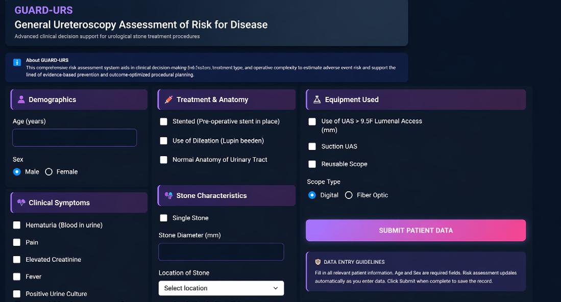
By integrating pre-operative patient metrics with planned surgical instrumentation, the platform generates a personalised risk profile to inform surgical planning.
interface integrates predictive analytics and validation transparency, supporting evidence-based perioperative planning.
External validation confirmed the robustness and clinical reliability of ensemble ML models for predicting septic risk after URS. Integration of these models into an interactive web tool enables realtime, data-driven risk assessment and supports individualised, evidence-based perioperative decision-making.
References
1. Bhojani N et al. Risk factors for urosepsis after ureteroscopy for stone disease: a systematic review with meta-analysis. J Endourol. 2021;35(7):991-1000.
2. Chugh S et al. Predictors of urinary infections and urosepsis after ureteroscopy for stone disease: a systematic review from EAU Section of Urolithiasis (EULIS). Curr Urol Rep. 2020;21(4):16.
3. Devos B et al. Risk factors of early infectious complications after ureterorenoscopy for stone disease: a prospective study. World J Urol. 2024;42(1):277.
4. Nedbal C et al. Validation and clinical implementation of a machine learning model for predicting sepsis following ureteroscopy: a web-based decision support tool from EAU urotechnology. Eur Urol. 2026;89(Suppl 1):S1-2.
5. Nedbal C et al. Predictors and associations of complications in ureteroscopy for stone disease using AI: outcomes from the FLEXOR registry. Urolithiasis. 2025;53(1):89.
6. Gauhar V et al. Indications, preferences, global practice patterns and outcomes in retrograde intrarenal surgery (RIRS) for renal stones in adults: results from a multicenter database of 6669 patients of the global FLEXible ureteroscopy Outcomes Registry (FLEXOR). World J Urol. 2023;41(2):567-74.
Authors: Kohei Yamada,¹ *Nicole Lim,¹ Zelig Ho,¹ Hira Syed,¹ Salman Anwar,¹ Sarosh Janardanan,¹ Sachin Agrawal¹
1. Ashford & St. Peter’s Hospitals NHS Foundation Trust, Chertsey, UK *Correspondence to nicolewannjie.lim@nhs.net
Disclosure: The authors have declared no conflicts of interest.
Keywords: Bladder cancer, European Organisation for Research and Treatment of Cancer (EORTC), laser, non-muscle invasive bladder cancer (NMIBC), outpatient, progression, recurrence, transurethral laser ablation (TULA).
Citation: EMJ Urol. 2026;14[1]:41-42. https://doi.org/10.33590/emjurol/7A8VN468
This study evaluates the long-term oncological outcomes of transurethral laser ablation (TULA) as primary treatment for non-muscle invasive bladder cancer (NMIBC) in a transurethral resection of bladder tumour (TURBT)-naïve population, benchmarked against the European Organisation for Research and Treatment of Cancer (EORTC) standards.1,2
This retrospective cohort study (2017–2024) used a prospectively maintained TULA clinic database at a single UK centre. A total of 151 patients with histologically confirmed NMIBC underwent TULA as initial treatment based on tumour or patient factors/comorbidities. Demographic, clinicopathological, and survival data were analysed. Primary outcomes were recurrence-free survival and progression-
free survival, stratified by EORTC risk groups.2 Kaplan–Meier analysis estimated survival outcomes, pairwise log rank tests compared risk groups, and multivariable Cox regression assessed predictors of recurrence and progression.
Mean age at initial TULA was 76.3 years (116 males/35 female), and mean Charlson Comorbidity Index (CCI) and follow-up were 6.9 and 31.4 months, respectively. Overall, 1- and 5-year recurrence-free survival rates were 72.2% and 39.1%, respectively, varying significantly across most EORTC risk groups. The overall 1- and 5-year progression-free survival was 98.5% and 89.4%, respectively, with no significant intergroup differences. The distribution of recurrences and progressions by EORTC risk groups are summarised in Table 1
During follow-up, eight patients (5.3%) required further general anaesthetic procedures, with a mean time to intervention of 7 months post-TULA. These comprised six TURBTs and two cystectomies. In multivariable analysis, age, sex, and comorbidity were not associated with recurrence or progression.
Primary TULA offers durable cancer control in carefully selected patients with NMIBC, with recurrence and progression rates closely aligning with EORTC predictions. Bladder cancer-specific mortality was rare, supporting TULA as a safe, minimally invasive alternative to TURBT in appropriately selected patients.
Table 1: Distribution of recurrences and progressions by EORTC risk groups.
Recurrence
Low-risk group (n=41)
Total recurrence in 1 year (EORTC standard)
N=5; 12.2% (15%)
Total recurrences in 5 years (EORTC standard)
N=11; 26.8% (31%)
Intermediate-risk group (n=79) N=18; 22.8% (24%) N=32; 40.5% (46%)
High-risk group (n=28) N=13; 46.4% (38%) N=14; 50.0% (62%)
Very high-risk group (n=3) N=1; 33.3% (61%) N=2; 66.7% (78%)
Progression
Low-risk group (n=65)
Total progression in 1 year (EORTC standard)
N=0; 0.0% (0.2%)
Total progressions in 5 years (EORTC standard)
N=1; 1.5% (0.8%)
Intermediate-risk group (n=52) N=1; 1.9% (1%) N=4; 7.7% (6%)
High-risk group (n=24) N=1; 4.2% (5%) N=1; 4.2% (17%)
Very high-risk group (n=10) N=0; 0.0% (17%)
EORTC: European Organisation for Research and Treatment of Cancer.
References
1. Yamada K et al. The feasibility of treating NMIBC in an outpatient-based setting with local anaesthetic TULA in a TURBT naïve population; an EORTC risk-matched 1 & 5 year recurrence & progression analysis. Abstract A0083. EAU Congress, 13-16 March, 2026.
N=1; 10.0% (45%)
2. Sylvester RJ et al. Predicting recurrence and progression in stage Ta–T1 bladder cancer using EORTC risk tables. Eur Urol. 2006;49:466-77.
Authors: Anurag Basavaraj Bellankimath,1 Sverre Branders,1 Isabell Kegel,2,3 Jawad Ali,1
Fatemeh Asadi,1 Truls E. Bjerklund Johansen,3,4 Can Imirzalioglu,2,3 Torsten Hain,2,3 Florian Wagenlehner,5,6 *Rafi Ahmad1,7
1. Department of Biotechnology, University of Inland Norway, Hamar
2. Institute of Medical Microbiology, Medical Microbiome-Metagenome Unit (M3U), Justus Liebig University Giessen, Germany
3. Institute of Clinical Medicine, University of Oslo, Norway
4. Institute of Clinical Medicine, University of Aarhus, Denmark
5. German Center for Infection Research (DZIF), Partner Site Giessen-Marburg-Langen, Germany
6. Clinic for Urology, Pediatric Urology and Andrology, Justus Liebig University Giessen, Germany
7. Institute of Clinical Medicine, Faculty of Health Sciences, UiT - The Arctic University of Norway, Tromsø, Norway
*Correspondence to rafi.ahmad@inn.no
Disclosure: Imirzalioglu has received support for the present manuscript from the German Federal Ministry of Education and Research (BMBF), with study funding within the German Center for Infection Research (DZIF) and network/project funding within the “Pandemic prevention and response within a One Health approach” framework; consulting fees from MSD and Shionogi, with payments to the author; payment or honoraria for lectures, presentations, speakers bureaus, manuscript writing, or educational events from MSD, with payments to the author; participated on a Data Safety Monitoring Board or Advisory Board for MSD, with payments to the author; and received equipment, materials, drugs, medical writing, gifts, or other services from Eumedica. Hain has received support for the present manuscript from the German Federal Ministry of Education and Research (BMBF), with study funding within the German Center for Infection Research (DZIF) and network/project funding within the “Pandemic prevention and response within a One Health approach” framework. Ahmad, Branders, and Bellankimath have received support for the present manuscript through funding from the Research Council of Norway through the projects OH-AMR-Diag (project number 336420) and UTI-Diag (project number 352514), with open access funding provided by the University of Inland Norway. Wagenlehner has received support for the present manuscript from Deutsche
Forschungsgemeinschaft (DFG) and Deutsches Zentrum für Infektionsforschung (DZIF); served as an advisor/consultant for Venatorx Pharmaceuticals, GlaxoSmithKline, Advanz Pharma, and Bionorica; received payment or honoraria for lectures, presentations, speakers bureaus, manuscript writing, or educational events from Astellas, AstraZeneca, Bionorica, Pfizer, OM-Pharma, MIP Pharma, GSK, Janssen, and Klosterfrau; support for attending meetings and/or travel from Bionorica, GSK, and MIP Pharma; participated on an Advisory Board for Achaogen, AstraZeneca, Bionorica, LeoPharma, MerLion, MSD, OM Pharma/Vifor Pharma, Pfizer, RosenPharma, Shionogi, Venatorx Pharmaceuticals, and GSK; participated on a Data Safety Board for Janssen (EXPEC 9 study); held a leadership or fiduciary role in German S3 Guideline Urinary Tract Infections and Guidelines European Association of Urology: Infections in Urology; is an employee of University Hospital Giessen and Marburg GmbH, and Justus Liebig University, Germany; participated in studies for Medice, Bionorica, Klosterfrau, and Deutsches Zentrum für Infektionsforschung (DZIF); and in a speaker research group for the German Research Foundation (DFG) Bacterial Renal Infections and Defence (FOR 5427). Johansen has received consulting fees and payment or honoraria for lectures, presentations, speakers bureaus, manuscript writing, or educational events from Advanz Pharma, GlaxoSmithKline, and Zambon; participated on a Data Safety Monitoring Board or Advisory Board for Advanz Pharma and Zambon; received equipment, materials, drugs, medical writing, gifts, or other services from Advanz Pharma; and is an Emeritus professor at the University of Aarhus and the University of Oslo. The other authors have declared no conflicts of interest.
Acknowledgements: Wagenlehner and Hain are members of the DFG (German Research Foundation), funded research group BARICADE (FOR5427/1-466687329), and members of the DZIF (German Center for Infection Research; site: Giessen–Marburg–Langen).
Keywords: Nanopore sequencing, urinary tract infection (UTI), rapid diagnosis.
Citation: EMJ Urol. 2026;14[1]:43-45. https://doi.org/10.33590/emjurol/0OE454FU
Urinary tract infections (UTI) affect an estimated 405 million people worldwide
Conventional microbiological testing
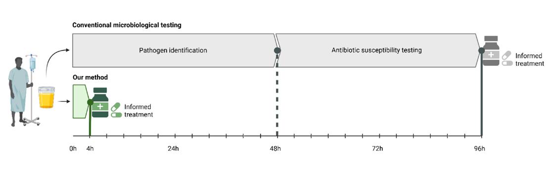
hours.
each year. Conventional diagnosis of UTIs relies on culture methods that typically require 2–4 days. Nanoporebased metagenomic next-generation sequencing (mNGS) presents a promising alternative for rapid diagnosis; however, it faces significant challenges due to the high abundance of host cells, low bacterial counts, and impurities. This study aimed to develop a rapid and cost-effective cultureindependent DNA extraction method for aiding in accurate diagnosis of complicated UTI in clinical settings.1
This study evaluated 11 cultureindependent methods for sample preparation in 78 patients with complex UTI, followed by real-time nanopore sequencing and data analysis.1 The metagenomic results are highly consistent with culture-based clinical routines, such as Matrix-Assisted Laser Desorption/Ionisation Time-of-Flight Mass Spectrometry (MALDI-TOF)/VITEK® 2 (bioMérieux S.A., Marcy-l’Étoile, France).
The optimised method demonstrated 99% accuracy (100/101) for pathogen identification and 90% (589/653) for
antimicrobial susceptibility profiling, with 95% specificity. The method’s robustness is highlighted by its ability to accurately identify pathogens with as few as 32 bacterial cells/µL and a low bacterial-to-host cell ratio limit of 0.5. Additionally, mNGS identified 13 pathogens that routine diagnostics missed, which were subsequently confirmed by Vivalytic™ (Robert Bosch GmbH, Gerlingen Schillerhöhe, Germany) or polymerase chain reaction. This method is up to 30% more economical than published studies and commercial kits. DNA yield and flow cytometry can be used for pre-screening to reduce costs, which is crucial for clinical adoption.
This research highlights a rapid and cost-effective method for processing urine samples in clinical settings, aiming to reduce turnaround time from 4 days to 4 hours (Figure 1). This could save 1.62 billion doses of broad-spectrum antibiotics annually before initiating tailored therapy, promote antimicrobial resistance stewardship, and improve UTI management.
References
1. Ahmad R et al. Bringing metagenomics to the bedside: personalized management of cUTIs for patient care. Abstract AM26-3035. EAU Congress, 13-16 March, 2026.
Authors: *Sebastiaan Remmers,1 Daan Nieboer,1
Chris Bangma,1 Monique Roobol1
1. Department of Urology, Erasmus MC Cancer Institute, Erasmus University Medical Center, Rotterdam, the Netherlands *Correspondence to s.remmers@erasmusmc.nl
Disclosure: The authors have declared no conflicts of interest.
Keywords: Mortality, prostate-specific antigen (PSA)-based screening, survivorship, treatment.
Citation: EMJ Urol. 2026;14[1]:46-47. https://doi.org/10.33590/emjurol/F8J19PN9
Data from a 15-year follow-up of the ProtecT trial, which randomised men to radical prostatectomy (RP), radiotherapy (RT), or active monitoring between 1999–2009, showed comparable prostate cancer (PCa)-specific survival (PCSS) for men treated with RP or RT (>97%).1 The ERSPC Rotterdam study was initiated in 1993 and covers a period over 20 years.2 The authors compared the restricted mean survival time (RMST) of patients with PCa treated with RP or external beam (EB)RT.3
Men in ERSPC Rotterdam were randomised between 1993–1999 to a screening arm (50%; prostate-specific antigen [PSA]based screening every 4 years) or a control arm (50%). Mortality data of men diagnosed with PCa were collected through regular linkages with Statistics Netherlands, and the cause of death was determined by a dedicated committee. Treatment was offered according to contemporary guidelines and shared decision-making. The authors identified patients with PCa treated with RP or EBRT with curative intent within 6 months of diagnosis. The authors used
inverse probability of treatment weighting to adjust for imbalance based on the age at treatment, year of treatment, the presence of any comorbidity at diagnosis (i.e., asthma, heart disease, high blood pressure, cardiovascular event, diabetes, malignancy, rheumatism, or depression), PSA at diagnosis, grade group, and cT-stage. The authors used this weight to calculate the RMST. CIs for the difference in RMST were calculated with 2,000 bootstrapped samples. Missing variables were imputed based on PSA, cT-stage, and grade group, and the authors tested for non-linear effects.
The authors identified 1,072 men treated with RP and 1,212 treated with EBRT (Table 1). Men treated with RP lived on average 1.41 years (95% CI: 0.31–2.50) longer than men treated with EBRT. The estimated 20-year PCSS was 95% (95% CI: 91–97) for RP and 91% (95% CI: 88–92) for EBRT. Using a time horizon of 15 years, the difference was 0.79 years (95% CI: 0.02–1.54) in favour of RP. The estimated 15-year PCSS was 95% (95% CI: 91–97) for RP and 92% (95% CI: 91–94) for EBRT.
In this study, the authors show that men who opted for RP have a longer RMST after propensity score weighting. However, the difference between treatments is small and the PCSS is lower than reported in the ProtecT trial. The treatments applied cover a long follow-up period in which knowledge and skills have improved, but the authors show a comparable effectiveness of both treatments on PCSS.
Table 1: Patient and tumour characteristics.
Age at treatment (years)
Year of treatment
Comorbidity
67 (63, 70) 73 (69, 76)
(1998, 2006)
(36%)
(1999, 2010)
(54%)
PSA at diagnosis (ng/mL) 6 (4, 9) 9 (5, 16)
Unknown 1 2
cT-stage T1
(55%)
(39%)
(42%)
(34%) T3
cN-stage
(100%)
ISUP GG
GG1
GG2
GG3
GG4
GG5
(65%)
(44%)
*Median (Q1, Q3); n (%).
EBRT: external beam radiotherapy; ISUP GG: International Society of Urological Pathology Grade Group; PSA: prostate-specific antigen; RP: radical prostatectomy.
References
1. Hamdy FC et al.; ProtecT Study Group. Fifteenyear outcomes after monitoring, surgery, or radiotherapy for prostate cancer. N Engl J Med. 2023;388(17):1547-58.
2. de Vos II et al.; ERSPC Rotterdam Study Group. A detailed evaluation of the effect of prostatespecific antigen-based screening on morbidity and mortality of prostate cancer: 21-year follow-up results of the Rotterdam Section of the European Randomised Study of Screening for Prostate Cancer. Eur Urol. 2023;84(4):426-34.
3. Remmers S et al. Survivorship after curative treatment: results of 20 years of follow-up from ERSPC Rotterdam. Eur Urol. 2026;89(Suppl 1):S1-2.
Authors: Stefano Bazzani,1 Marco Minelli,1 Stefania Ferretti,2 Stefano Puliatti,3 Giampaolo Bianchi,3 Cristian Secchi,1 *Federica Ferraguti1
1. Department of Sciences and Methods for Engineering, University of Modena and Reggio Emilia, Italy
2. Urology, Azienda Ospedaliero-Universitaria Modena, Italy
3. Surgical, Medical and Dental Department of Morphological Sciences related to Transplant, Oncology and Regenerative Medicine, University of Modena and Reggio Emilia, Italy *Correspondence to federica.ferraguti@unimore.it
Disclosure: The authors have declared no conflicts of interest.
Disclaimer: Figure 1A is a representative, non-identifiable CT image used for illustrative purposes only within an experimental, non-clinical workflow. No identifiable patient information is visible, and consent for publication was waived.
Keywords: Surgical robotic, treatment, urolithiasis.
Citation: EMJ Urol. 2026;14[1]:48-49. https://doi.org/10.33590/emjurol/6FRZ87E5
Percutaneous access is essential in procedures such as percutaneous nephrolithotomy, biopsy, and ablation, requiring precise needle insertion while avoiding critical organs and parts.1,2 Success depends heavily on operator skill and image guidance, typically with ultrasound (US) or fluoroscopy.3,4 However, these methods can be difficult to use and inefficient, leading to variability in accuracy and outcomes. The authors propose a robotic system that integrates US navigation to help the surgeon perform percutaneous renal access.
The main procedural steps of the proposed US-guided robotic system are shown in Figure 1. The system combines a collaborative robot with a US probe mounted in a custom holder that includes a needle guide. The surgeon first plans a safe trajectory on a pre-operative CT scan. An intraoperative US scan is then acquired, and the kidney is automatically segmented using a neural network. Because the US images are spatially tracked, a 3D point cloud of the kidney surface is reconstructed and aligned with a corresponding point cloud from CT segmentation. A registration algorithm refines the alignment, enabling precise mapping of the planned trajectory to the patient in the operating field. Once registration is complete, the system offers two workflow options: the robot automatically positions the needle guide for manual insertion along the planned path, or the surgeon continues to manually navigate the probe with real-time guidance, using 2D/3D visual feedback provided by a user interface to correctly orient the probe for accurate needle placement.
The authors conducted preliminary tests with both experienced and novice urologists, asking them to perform percutaneous renal access on a phantom, first using conventional freehand technique and then using the US-guided robotic system. Freehand: 75% of experts and 21% of novices succeeded on the first attempt; 25% of experts and 36% of novices required two attempts; 29% of novices required three attempts; and 7% of novices required four. With the robotic system, all participants succeeded on the first attempt. The authors' results indicated that robotic assistance improved performance during the renal access phase of percutaneous nephrolithotomy compared with
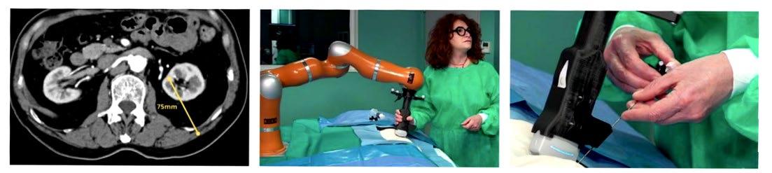
The figure illustrates the three main steps of the robotic system workflow: (A) pre-operative planning on CT scan with trajectory definition; (B) intraoperative ultrasound scan acquisition and CT-to-US registration with the robot; and (C) probe alignment and needle puncture guided by the robotic system. US: ultrasound.
the manual approach. In the manual procedure, residents required substantially longer execution times (mean: 290 sec; median: 214 sec) than experienced surgeons (mean: 73 sec; median: 70 sec) and showed high variability. With robotic assistance, execution times between the two groups became similar and variability decreased.
Accuracy also improved: during manual procedures, residents experienced several failed insertions and experts recorded one failure, whereas no failures occurred with robotic assistance. Analysis of US probe motion revealed that residents performing the manual procedure travelled longer distances and exhibited greater orientation deviations (average distance: 2.46 m; orientation deviation: 1906°) compared with experienced surgeons (0.35 m; 199°). Robotic guidance reduced both metrics for all participants. Usability assessment through the Surgery Task Load Index (SURGTLX) questionnaire indicated an overall reduction in perceived workload, particularly
for residents, narrowing the gap between inexperienced and expert surgeons.
The proposed approach reduces the performance gap between novice and expert surgeons in percutaneous access. Unlike other solutions, it does not require intraoperative CT, eliminating radiation exposure for both patients and clinical staff while providing real-time accurate guidance.
References
1. Bazzani S et al. Ultrasound navigation in robotassisted percutaneous access. Abstract AM26-3075. EAU Congress, 13-16 March, 2026.
2. Ghani KR et al. Percutaneous nephrolithotomy: update, trends, and future directions. Eur Urol. 2016;70(2):382-96.
3. Yan S et al. Percutaneous nephrolithotomy guided solely by ultrasonography: a 5-year study of >700 cases. BJU Int. 2013;112(7):965-71.
4. Kumari G et al. Radiation exposure to the patient and operating room personnel during percutaneous nephrolithotomy. Int Urol Nephrol. 2006;38(2):207-10.
Authors: Wyatt MacNevin,1 Mareen S. Kraus,2 Chrissy Gamache,2 Elena Tonkopi,2 Kathleen O’Brien,2 Karen Milford,3,4 Dawn L. MacLellan,3
*Daniel T. Keefe3
1. Department of Urology, Dalhousie University, Halifax, Canada
2. Department of Diagnostic Radiology, Izaak Walton Killam Health Centre, Dalhousie University, Halifax, Canada
3. Department of Pediatric Urology, Izaak Walton Killam Health Centre, Dalhousie University, Halifax, Canada
4. Department of Pediatric Surgery, Izaak Walton Killam Health Centre, Dalhousie University, Halifax, Canada
*Correspondence to daniel.keefe@iwk.nshealth.ca
Disclosure: The authors have received support for attending meetings and/or travel from Dalhousie University.
Keywords: CT, paediatric urology, radiation, reduced-dose, stone, urolithiasis.
Citation: EMJ Urol. 2026;14[1]:50-51. https://doi.org/10.33590/emjurol/8P9L6Q1S
Ultrasonography is the recommended first-line investigation for the diagnosis of paediatric urolithiasis.1 Despite higher sensitivity and specificity for urolithiasis detection, CT is reserved for more complex cases to limit paediatric radiation exposure.2 Although there is an increasing stone prevalence in the paediatric population, there is a lack of low-dose CT urolithiasis protocols to reduce radiation exposure in this vulnerable patient group.3 Furthermore, descriptions of low-dose protocols are sparse and often not reported in the literature. Herein, the authors report the development and implementation of a lowdose protocol to reduce radiation exposure while diagnosing and assessing paediatric stone disease.
A novel low-dose CT protocol to detect urolithiasis was designed through literature review, multidisciplinary collaboration, and experimental phantom trials. Two separate protocols were developed based on patient body mass (<45 kg and ≥45 kg). Prospective patients undergoing CT for urolithiasis assessment were evaluated using the low-dose protocol and were compared to a retrospective cohort at a single institution. Radiation reduction was characterised using descriptive statistics and comparative analysis.
Mean (±SD) age for the low-dose group was 12.6±4.2 years (n=26) compared to 12.4±3.7 years for the standard-dose group (n=15). The mean stone number detected in the <45 kg group was 4.2±3.6 (standard-dose) and 1.3±0.5 (low-dose), and in the ≥45 kg group it was 4.5±4.0 (standard-dose) and 2.6±1.5 (low-dose). There were no differences between groups with respect to stone size determined by ultrasonography and/or CT. Prior history of stone disease was higher in the standard-dose group (53.3%, n=8/15) when compared to the low-dose group (38.5%, n=10/26; p=0.03), highlighting the natural history of stone recurrence over time within this population. Treatment approach based on CT-findings did not differ between groups, with the majority of patients being managed with observation (standarddose: 60.0%, n=9/15 versus low-dose: 61.5%, n=16/26; p=0.92). Of patients who underwent ureteroscopic or percutaneous management (standard-dose: 33.3%, n=5/15; low-dose: 26.9%, n=7/26), stones were reliably encountered intra-operatively and were treated fully without complication. The low-dose protocol reduced radiation dose when compared to the standarddose group by 55.5% (≥45 kg; p=0.02)
and 27.8% (<45 kg; p=0.03). The lowdose protocol visualised stones seen on ultrasound with 100% accuracy (n=6), and in 61.5% (n=16/26) of patients. Although there was an increase in noise and lower contrast detectability, spatial resolution and detection accuracy were preserved in the low-dose patient groups.
Low-dose CT protocols are effective for assessing paediatric urolithiasis while reducing radiation exposure. Implementation of low-dose CT protocols in cases of suspected urolithiasis with
ambiguous ultrasound findings or for surgical planning is advised to limit radiation exposure in the paediatric population while maintaining diagnostic accuracy.
References
1. MacNevin W et al. Radiation dose reduction through the development of a novel pediatric lowdose computed tomography protocol for urolithiasis assessment. Abstract A0420. EAU Congress, 13-16 March, 2026.
2. Tasian GE et al. Use of and regional variation in initial CT imaging for kidney stones. Pediatrics. 2014;134(5):909-15.
3. MacNevin W et al. Radiation exposure associated with computed tomography for pediatric urolithiasis evaluation: a scoping review of the literature. J Pediatr Urol. 2024;20(3):386-94.
Authors: *Jackson J.S. Cabo,1 Andrew Amenyogbe,1 Karen L. Stern1
1. Department of Urology, Mayo Clinic Arizona, Phoenix, USA
*Correspondence to cabo.jackson@mayo.edu
Disclosure: Cabo and Stern have received an educational grant from Calyxo Inc. for this study, with payments to the institution. Amenyogbe has declared no conflicts of interest.
Acknowledgements: The authors would like to thank Victoria Edmonds, Mayo Clinic Arizona, Phoenix, USA; Christopher Ballantyne, Bon Secours, Greenville, South Carolina, USA; and the Mayo Clinic AI Core for collaboration on this work.
Keywords: Kidney stone, stone volume, suction ureteroscopy.
Citation: EMJ Urol. 2026;14[1]:52-53. https://doi.org/10.33590/emjurol/77C92T7P
With recent emphasis placed on using stone volumes in surgical outcomes research,1-6 there is a need to assess the accuracy and utility of available tools. The authors sought to assess the accuracy of AI-calculated volumes compared to semi-automated methods5,6 and evaluate whether cumulative stone diameter, semi-automated (SA) stone volume calculation, or a fully automated AI method was a better predictor of stone-free status following suctionaugmented ureteroscopy (Figure 1).
A total of 171 CT scans were included (96 pre-operation and 75 post-operation). Cumulative stone diameter was measured manually. Stone volumes were assessed using two semi-automated segmentation applications (QSAS [Mayo Clinic, Rochester, Minnesota, USA], 3D-Slicer [Chitubox, Schenzhen, China]), which require
investigator annotation of the region of interest. A Mayo Clinic-developed AI programme calculated stone volumes in a fully automated fashion. Pearson correlation assessed the association between AIestimated and semi-automated volumes. Sensitivity and specificity of the AI model were assessed for absolute stone-free rate (SFR-Grade A) on post-operative scans. Receiver operating characteristic analysis evaluated accuracy of pre-operative stone burden metrics in predicting stone-free status using SFR-Grade A (no residual fragments) and SFR-Grade C, stone-free rate with residual fragments between 2.1–4.0 mm in size (no fragments >4 mm) criteria.
AI-estimated stone volumes showed strong linear correlations with both 3D-Slicer (R=0.95; p<0.001; mean difference: -0.31 mm3; interquartile range: -13.06–25.81 mm3) and QSAS (R=0.95; p<0.001; mean difference: 0 mm3; interquartile range: -12.0–8.0 mm3) calculated volumes. Among post-operative scans, strong correlations persisted with QSAS (R=0.88; p<0.001) and 3D-Slicer (R=0.86; p<0.001).
Among 59 patients with eligible pre- and post-operative imaging, the AI model demonstrated a sensitivity of 85.7% and specificity of 88.9% for SFR-Grade A. In cases of incorrect stone-free determination, parenchymal stones were identified in both false-negative cases, and the largest false-positive residual burden (16 mm3) occurred in the right moiety of a horseshoe kidney.
Among 69 patients with a single ureteroscopy and complete follow-up imaging within 3 months, no significant difference was found between preoperative volumetric and diameter measurements in predicting SFR-Grade A. Cumulative pre-operative diameter outperformed
1: Study schema and primary aims.
Semi-automated

QSAS-calculated volume in predicting SFR-Grade C (area under the curve: 0.78 versus 0.62; DeLong Test p=0.037). Subanalysis of flexible and navigable ureteric access sheath cases revealed no difference between any measurement in predicting SFR-Grade A or -Grade C.
All pre-operative stone measurements correlated significantly with operative time; however, semi-automated volumes from 3D-Slicer (R2=0.41) and QSAS (R2=0.37) explained more variation in operative time than cumulative diameter (R2=0.25) or AI-estimated volume (R2=0.24).
A fully automated, AI-driven method for stone volume determination was highly accurate, offering an efficient option for estimating pre-operative or residual stone burden.
The AI model was 85.7% sensitive and 88.9% specific for determining SFR-Grade A without clinician annotation, with errors concentrated in small low-
Manual measurement of cumulative stone diameter
attenuation stones and anatomic variants. Pre-operative volumetric measurements did not outperform cumulative diameter in predicting stone-free status.
References
1. Cabo J et al. Comparing performance of automated and semi-automated methods for measuring kidney stone volumes: can machine replace man? Abstract A0417. EAU Congress, 13-16 March, 2026.
2. Cumpanas AD et al. Efficient and accurate computed tomography-based stone volume determination: development of an automated artificial intelligence algorithm. J Urol. 2024;211(2):256-65.
3. Geraghty R et al. Which measure of stone burden is the best predictor of interventional outcomes in urolithiasis: a systematic review and metaanalysis by the YAU Urolithiasis Working Group and EAU Urolithiasis Guidelines Panel. Eur Urol Open Sci. 2024;71:22-30.
4. Matlaga BR et al. Residual stone volume predicts health care consumption and stone events: analysis of two-year results of the ASPIRE study. J Endourol. 2026;40(3):328-34.
5. Slots C et al. Stone metrics: is stone volume the new king? Curr Opin Urol. 2026; DOI:10.1097/MOU.0000000000001376.
6. Sajdak G et al. Rock solid measurements: a comparison of three methods of kidney stone volume assessment. J Endourol. 2025;39(8):856-61.
Authors: *Etan Eigner,1,2 Nicola Fazaa,1,2 Ameer Nsair,1,2 Melissa Atallah,1,2 Kamil Malshy,3 Gilad E. Amiel,1,2 Michael Mullerad1,2
1. Rappaport Faculty of Medicine, Technion Haifa, Israel
2. The Department of Urology, Rambam Health Care Campus, Haifa, Israel
3. Department of Urology, University of Rochester Medical Center, New York, USA
*Correspondence to e_eigner@technion.ac.il
Disclosure: The authors have declared no conflicts of interest related to this work.
Keywords: CO2 laser treatment, genital warts, human papillomavirus (HPV) vaccination, predictors of repeat treatment.
Citation: EMJ Urol. 2026;14[1]:54-55. https://doi.org/10.33590/emjurol/38VL7YI4
Human papillomavirus (HPV) is the most common sexually transmitted infection worldwide,1 with most sexually active individuals acquiring infection during their lifetime.2,3 Low-risk human papillomavirus Types 6 and 11 account for the majority of anogenital warts,4 a condition that affects approximately 1% of sexually active adults at any given time.5 Although benign, genital condyloma imposes a significant psychosocial and economic burden as a result of stigma, repeated treatments, and prolonged follow-up.6 The 9-valent human papillomavirus vaccine provides highly effective primary prevention against vaccine-type infection and genital warts in both women and men.4,7 However, whether vaccination administered after treatment reduces recurrence or the need for additional procedures remains uncertain, as there was limited and inconsistent evidence in previously infected individuals, as reported in other studies.8,9 The authors therefore evaluated the clinical predictors of recurrence after CO₂ laser ablation and examined whether post-treatment human papillomavirus vaccination is associated with reduced recurrence risk in a real-world male cohort who already had the infection.
The retrospectively reviewed men (who were 17 years of age or older) were treated with CO₂ laser ablation for genital condyloma at a tertiary centre (June 2016–June 2025). The primary outcome was recurrence after documented clearance. Logistic and mixed-effects regression models tested associations between post-treatment vaccination and recurrence, adjusting for demographic and clinical factors.
Among the 560 patients included in the study (722 sessions), recurrence occurred in 28.4%. Those with recurrence were younger in age (32±10 versus 37±13 years; p<0.001) and more likely to need ≥2 sessions (18.6% versus 11.6%; p=0.02). Post-recurrence vaccination showed no significant association with reduced risk (adjusted odds ratio [aOR]: 0.49; 95% CI: 0.14–1.69; p=0.26). Independent predictors were younger age (aOR: 0.96 per year; p<0.001) and ≥2 sessions (aOR: 2.44; p=0.003).
Post-treatment human papillomavirus vaccination did not reduce recurrence of genital condyloma in patients who already had the infection. Younger age and multiple treatment sessions independently predicted recurrence, reinforcing the role of vaccinations primarily in primary prevention.
References
1. Eigner E et al. Evaluating the role of the 9-valent HPV vaccine in preventing recurrence of genital warts. Abstract A0055. EAU Congress, 13-16 March, 2026.
2. Forman D et al. Global burden of human papillomavirus and related diseases. Vaccine. 2012;30:F12-23.
3. Chesson HW et al. The estimated lifetime probability of acquiring human papillomavirus in the United States. Sex Transm Dis. 2014;41(11):660-4.
4. Joura EA et al. A 9-Valent HPV vaccine against infection and intraepithelial neoplasia in women. N Engl J Med. 2015;372(8):711-23.
5. Lewis RM et al. Estimated prevalence and incidence of disease-associated human papillomavirus types among 15- to 59-year-olds in the United States. Sex Transm Dis. 2021;48(4):273-7.
6. Raymakers AJN et al. Economic and humanistic burden of external genital warts. Pharmacoeconomics. 2012;30(1):1-16.
7. Goldstone SE al. Efficacy, immunogenicity, and safety of a quadrivalent HPV vaccine in men: results of an open-label, long-term extension of a randomised, placebo-controlled, phase 3 trial. Lancet Infect Dis. 2022;22(3):413-25.
8. Coskuner ER et al. Impact of the quadrivalent HPV vaccine on disease recurrence in men exposed to HPV infection: a randomized study. J Sex Med. 2014;11(11):2785-91.
9. Gilson R et al. Imiquimod versus podophyllotoxin, with and without human papillomavirus vaccine, for anogenital warts: the HIPvac factorial RCT. Health Technol Assess (Rockv). 2020;24(47):1-86.
EMJ is pleased to introduce two members of the European Association of Urology (EAU) 2026 Scientific Congress Committee, Cosimo De Nunzio and Veeru Kasivisvanathan, as they share cutting-edge insights in urology and the future of the EAU.
Featuring: Cosimo De Nunzio and Veeru Kasivisvanathan


Cosimo
De Nunzio
Member of Scientific Congress Committee, European Association of Urology (EAU); Associate Professor of Urology, Sapienza University of Rome, Italy
Citation: EMJ Urol. 2026;14[1]:56-58. https://doi.org/10.33590/emjurol/G253FQ8E
At the European Association of Urology (EAU) Congress each year, we see a huge range of new research and technologies. If you had to highlight one innovation or idea from this year’s meeting that could shape the future of urology, what would it be?
possible therapeutic option is also an important issue to debate.
With ageing populations across Europe, how do you expect the overall burden of urological diseases to evolve over the next decade, and what challenges will that create for healthcare systems?
The VAPOUR trial is the first trial to randomise patients between medical therapy versus minimally invasive treatments
Every year during the EAU Congress, several innovations are presented or discussed. Particularly, I would like to underline the ‘game changer session’, or ‘late breaking session’, where new, innovative research is presented and discussed. Particularly this year, Jean Nicolas Cornu from France presented the results of the VAPOUR trial. It is the first trial to randomise patients between medical therapy versus minimally invasive treatments, such as water vapour therapy. The results are innovative, as they propose minimally invasive surgical therapies (MIST) as an alternative to first-line interventional therapy (FIT) and medical treatments. FIT is a new paradigm, but costs and risk of serious complications, as well as risk of retreatment, should be evaluated in future studies. Patients’ perceptions of this new,
Several urological conditions, such as lower urinary tract symptoms (LUTS), benign prostatic hyperplasia, prostate cancer, erectile dysfunction, and incontinence, are age-related conditions and, considering the ageing effect of the current European population, their burden will continue to increase over the next years and decades. As a urologist, we also directly manage other frequent conditions, such as stone diseases, urinary tract infections, and urological cancer (including prostate cancer, one of the most frequent cancers in men). This scenario is an important challenge for a urologist, but particularly for the healthcare authorities. Urological procedures are also a technologically demanding option, and the increasing costs of several new minimally invasive treatments
will further expand this scenario. As urologists, we have to work closely with our health authorities to identify the best diagnostic and therapeutic treatment to reduce low volume care by reducing unnecessary exams or treatment and financially improve our patients’ care.
Q3 We’re seeing rapid developments in AI and digital health tools across medicine. What role do you think these technologies could play in diagnosing and managing lower urinary tract symptoms in the future?
AI is a hot topic, and I am sure that it will not replace our work if we are able to use AI in our clinical practice. In LUTS/benign prostatic obstruction (BPO) management, several applications are under investigation: to directly store and evaluate a patient’s bladder diary and questionnaires, to analyse invasive urodynamic traces, to identify possible outcomes of success, or to plan surgeries. However, its routine
use is still not established, and some ethical and legal issues are still prevalent, but I am sure, in the near future, AI will help urologists and patients to obtain better care.
Q4
There’s also growing interest in biomarkers and advanced imaging. Do you think these technologies could eventually help identify patients at risk of disease progression earlier?
It is an option, but probably more in an oncological setting, where there are several attempts to better standardise MRI reading in patients at risk of prostate cancer or to better predict kidney cancer histology reports on CT images. Integrating data from imaging, biomarkers, and clinical data will probably be used to identify patients at risk of progression. Now, on BPO, data from ultrasound or MRI, including detrusor thickness, have failed to clearly demonstrate being good predictors of progression.
Q5 Research in this field often depends on large datasets. Turning specifically to BPH, how important are international collaboration and clinical registries for advancing BPH research across Europe and globally?
It is fundamental. Now, we work in a global era, and we are able to evaluate large databases. Including data from several countries and regions can improve the possibility of generalising this data. However, I strongly believe that large databases and registries are an opportunity, but they are not a marker of quality. Identifying the right topic with the appropriate study design and appropriate team is the key to success.
Q6
In your view, what are the biggest unmet needs in BPH management today?
We are in an era of new minimally invasive treatments. They are effective and will probably be proposed as FIT in the next

In the near future, AI will help urologists and patients to obtain better care

few years. However, patients generally avoid surgeries of all types, including MISTs. We need new pharmacological treatments: innovative, effective, and with few adverse events. The latest drugs for LUTS/BOP were introduced into the market more than 10 years ago. LUTS/BPO is a benign condition, and we need to work more on patients’ perceptions and expectations.
Q7 Looking ahead, what do you think could be the biggest breakthrough in the management of BPH and LUTS over the next decade?
MISTs as FIT could be an important breakthrough, but we need more data on comparative studies, on retreatments, on the effect on BPO progression, and, finally, on appropriate cost analysis. MISTs are expensive procedures and, in some countries, they are not reimbursed. This could be a form of disparity in LUTS/BPO management.
Q8 What first inspired you to pursue a career in urology? Was there a specific mentor or experience that contributed to pursuing it?
Regarding my career, the first challenge was undoubtedly choosing a specialty. I wanted to be a surgeon, but I wasn’t sure which discipline to pursue. As is often the case in medicine, my first mentor, Giorgio Franco, currently Professor of Urology at Sapienza University, Italy, played a key role. Thanks to him, I decided to spend a fellowship in the UK, specifically in Newcastle and Sheffield, where I met another key mentor of my career, C Chapple, Consultant Urological Surgeon at Sheffield Teaching Hospitals. I was lucky as, after my residency, I was appointed as Junior Consultant at Osperdale Sant’Andrea, Sapienza University, Italy, where I worked. I still work closely with Hassan Fattahy, my father in urology, Lucio Miano, and Andrea Tubaro, from Ospedale Sant'Andrea, Sapienza University of Rome, Italy, who completely changed my career and pushed me to start an academic career. This is only to underline that mentorship is fundamental in medicine, and identifying the right mentor is the first step to improve as doctors, urologists, surgeons, and as a person.
Q9 You previously chaired the Young Academic Urologists BPH group within the EAU. From that perspective, what further advice would you give early-career researchers who want to enter this field?
My suggestion is to work hard from the beginning. Our careers are a 100-metre race, but it is also like a marathon. We should be prepared. Follow your passion, invest in your skills, and attend
courses and departments outside your country. Good interaction with mentors and colleagues is also important. Regarding LUTS/ benign prostatic hyperplasia research, it is a frequent condition, where young researchers can be involved from the basic research to the clinical and surgical management. Personally, LUTS/ BPH is the right pathway to start your urological career and to improve your clinical and surgical skills.
Q10
Looking ahead, what skills or areas of expertise do you think will become most important for the next generation of urologists?
The young generation of urologists are strongly attracted to surgery, particularly robotic surgery, and I can understand. I received training in open laparoscopic and robotic surgery and this is helpful for my current activities. My suggestion is not to focus on a single procedure or technique from the beginning, and, particularly for those interested in uro-oncology, to be prepared not only for surgery, but also to discuss with oncologists and radiotherapists the several innovative treatments which are now recommended or are in the pipeline. We urologists are not only surgeons.
Follow your passion, invest in your skills, and attend courses outside your country


Veeru Kasivisvanathan
Member of Scientific Congress Committee, European Association of Urology (EAU); Consultant Urological Surgeon, University College London Hospitals, NHS Foundation Trust; Associate Professor, University College London, UK; Professor of Urology, University of Vienna, Austria
Citation: EMJ Urol. 2026; 14[1]:59-62. https://doi.org/10.33590/emjurol/701N9G75
Q1
Your work spans prostate cancer diagnosis, focal therapy, and surgical innovation. What first drew you to urology, and particularly to prostate cancer research?
Focal therapy is a really important treatment option for patients with localised prostate cancer
When I was a medical student, I was always interested in surgery. I liked doing things where I could see my patient getting better quite quickly, and when I started working, that was reinforced. Urology was a specialty where I could get involved in carrying out procedures even as a junior doctor, so it seemed like an attractive career from a clinical point of view. In addition to that, the strength of the academic side was really clear to see. I could see the research being carried out really influencing what we did on a day-to-day basis with our patients. The combination of a range of procedures, with lots of exciting tools in a urologist’s armamentarium, and the depth of the academic side was the reason why I chose urology.
Q2 The PRECISION Trial is widely seen as transforming prostate cancer diagnosis for the first time in 25 years. When you first began working on it, did you anticipate it would have such a global impact on clinical practice?
The answer is probably no. We knew for a long time that we wanted to change the way that we diagnosed prostate cancer, because there were problems with how it was being was being diagnosed. We'd spent a long time working on MRI, and there were some places where people
felt that MRI was the right way to go forwards. The challenge was designing a trial where we could change the standard of care to something new, so we designed it initially as a feasibility study, but it became evident that the groups around the world wanted to do this as a bigger study, and the results were so clearly in favour of MRI that it led to this big change. We felt that MRI was better and hypothesised that the trial would show this, but we didn't anticipate the impact of the work, so it was nice to see. When we saw the results, we realised this would have a big impact, and it was published in one of the world's leading journals. As a result of that, guidelines changed in the next year for many international societies.
Q3
Building on studies like PRECISION and the PRIME Trial, how do you see prostate cancer diagnosis evolving over the next 5–10 years?
PRECISION introduced MRI into the pathway. PRIME allowed us to do shortened, abbreviated scans, because the problem was that we need more than four million scans per year, and, in many countries, not everyone who needs a scan can get one. Therefore, the shortened, abbreviated scan increased our global capacity from four to eight million scans overnight. The next challenge is ensuring we have a high-quality interpretation of the MRI. We have so many more scans coming through, we have an ageing population, and prostate cancer incidence is predicted to double in the next 20 years. My next study is called PARADIGM, which is looking
at AI in diagnosing cancer on MRI scans, and we're going to see whether it can reach the standards of these expert radiologists. If it can, then it shows that it could be used in the pathway.
Do you think we are moving towards a future where imaging could reduce, or even replace, traditional prostate biopsies in some patients?
One thing we know for the imaging we have so far is that the imaging signal and how strong it is relates to a patient’s prognosis. This is not just MRI, but also other imaging, like prostate-specific membrane antigen PET-CT, which has been heavily featured at the conference this year.
Biopsy itself carries some harms and risks of misclassification, so it's not inconceivable to consider that, if we treat someone on the basis of imaging, it might lead to a better outcome for the patient. Part of our work over the next 4
years will be looking at a ‘virtual biopsy’. There is no evidence that it is better, but the point is to explore that pathway and its feasibility and generate evidence to say whether or not it is better than the way we currently do things.
Focal therapy is gaining increasing attention as a treatment strategy. What key advancements in focal therapy do you expect to see over the next decade?
Focal therapy is a really important treatment option for patients with localised prostate cancer. It's an emerging treatment option, which means it's not offered everywhere, and, when it is done, it needs to be published in a registry, which means collecting data and checking outcomes as they mature. So, what changes need to happen? Firstly, I think we need to produce more data and carry out more studies supporting its role. One of the things I talked about at the conference this year was what
studies have been done so far. We have data for 7-year failurefree survival from a multinational UK study, which showed that the results of focal therapy are quite good, but it's important that we generate more data like that from other focal therapies with even longer-term follow-up to show that focal therapy can be done well. Some of the randomised trial data comparing focal therapy to radical treatment will be available in the next few years. I believe that offering focal therapy will be a marker of quality for a unit offering treatment for prostate cancer.
In
many centres around the world and many countries, robotic surgery is already the standard of care
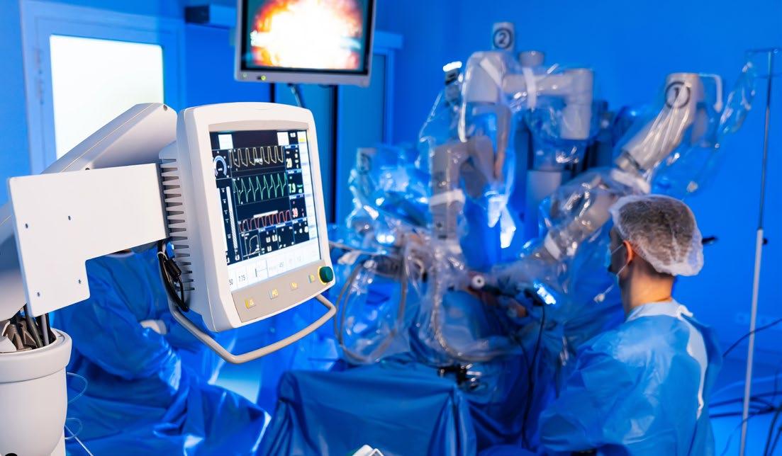
Q6
Robotic surgery continues to expand worldwide. What do you see as the biggest challenges in scaling robotic surgery globally?
Robotic surgery has caught on in a big way already. In many centres around the world and many countries, robotic surgery is already the standard of care for the way that many procedures, for example, prostatectomy, are done. Now, the challenge will be to ensure that countries that don't have as many resources can also offer robotic surgery. There are many countries in that bracket that already do offer robotic surgery, but there are many developing parts of the world that don't, because the robot itself and the maintenance of the robot are expensive. I would say that supporting these countries in being able to get access to the technology and required training is a priority. Newer robots are bringing costs down and making robotic surgery more accessible. This is really important and needs to continue to happen.
Q7
There have been lots of cases where remote robotic surgery has been done from another country thousands of kilometres away. Has this proven to be successful? Do you think this is an effective approach?
Remote robotic surgery is an exciting recent development allowing world-renowned surgeons to offer patients in distant regions a high-quality operation. However, I think the challenge in increasing uptake of robotic surgery is not the expertise required. There is an element of training that needs to happen for successful programmes. What we typically see in countries that are starting to do more robotic surgery is they

send many of their surgeons, towards the end of their training, to countries that have the robot so they can gain expertise, and then they go back to other countries to practice.
The biggest problem is getting the kit to do robotic surgery in the first place. Remote operating, with one surgeon operating from a different country, is nice, but I don't think it's the essential component required to make it successful, because you could just fly that surgeon out of the country to help with the procedure; they don't need to do it remotely. It does, however, raise the possibility of closer mentorship from experts to help centres advance their skills.
Q8 You balance clinical practice, research, and teaching at a very high level. How do you manage these different roles, and do they complement each other in your work?
I'm a clinical academic, so I spend half my time doing clinical work, and the other half doing research. For me, they really complement each other. The clinical work allows me to treat patients, which is why I became a doctor. It's what gives me satisfaction in my dayto-day life. I love seeing patients get better because of something I did. It also allows me to identify where there are problems in how we manage, diagnose, and treat patients, which then allows me to develop clinical studies to answer
that question, so they both go hand in hand. They are both enjoyable in different ways, and they have different challenges. With clinical work, I can benefit hundreds of individual patients that I personally see and treat, but with the research, when you do something that changes practice, you can then affect millions of patients, so they both have their benefits.
Q9 You received the European Association of Urology (EAU) Crystal Matula Award for the top young academic urologist in Europe. What advice would you give to medical students or early trainees who are interested in pursuing academic urology?
It's a fantastic career and I strongly recommend it. It's really important to find someone who inspires you and receive their mentorship. When you're given an opportunity, take it, be present, ask questions, and deliver on what you're asked to. If you show that you can be trusted with something to a mentor, they will give you more and more, so ally yourself with a good group, show interest, dedication, perseverance, and you'll be able to get those opportunities.
Q10As technologies such as advanced imaging, robotics, and AI reshape clinical practice, how do you think the training of the next generation of urologists will need to evolve?
I think it starts from medical school. AI has changed our world completely and understanding how to use AI in medical healthcare is probably the first thing medical school curricula should address in training. It is important that these skills are taught. For example, I lead imaging courses at the European School of Urology (ESU), and we've previously introduced MRI training courses. This year, for the first time, we’ve introduced prostate-specific membrane antigen PET-reading courses. We go out of our way to teach people how to use novel imaging technology in their daily practice. With things like robotics, I think it requires syllabus training and fellowships, so surgeons can go to centres, gain the expertise, go back to their home centre, and disseminate the information locally. I think it's a combination of changing the curriculum from very early on to changing what's being offered when they're a bit more senior and on top-level fellowships, with a dedicated syllabus and curriculums to train people.
AI has changed our world completely and understanding how to use AI in medical healthcare is probably the first thing medical school curricula should address in training
EMJ is delighted to introduce two leaders in urology: Hashim Ahmed from Imperial College London, UK; and Bertrand Tombal from Université Catholique de Louvain, Cliniques Universitaires Saint-Luc, Brussels, Belgium. Ahmed shares cutting-edge insights into prostate cancer diagnostics, whilst Tombal unpacks emerging management strategies, including prostate-specific membrane antigen-targeted radioligand therapy.
Featuring: Hashim Ahmed and Bertrand Tombal

Hashim Ahmed Chair and Professor of Urology, Imperial College London, UK
Citation: EMJ Urol. 2026;14[1]:63-66. https://doi.org/10.33590/emjurol/PZKU9925
What key experiences or clinical gaps motivated your focus on advancing prostate cancer diagnostics, particularly MRI-led pathways?
As an early trainee in urology, I used to learn about transrectal systematic biopsy. Even at that time, before I was exposed to MRI scans of the prostate, I remember thinking that the strategy made no sense: no one could actually see any potential cancers using brightness mode ultrasound, and in those days, even the prostate was a grey mass just visible from the rest of the tissue.
ultrasound (HIFU). We then realised that the bigger prize was changing the diagnostic pathway with MRI before a biopsy. We are now taking on the whole screening paradigm: can we screen with a 10-minute Prostagram MRI scan in the community instead of prostate-specific antigen (PSA)?
As an educator, which areas of prostate cancer diagnostics or focal therapy training do you plan to prioritise for the next generation of clinicians?
Urologists wanting to look after patients with prostate cancer need to be able to look at MRI scans and work out which areas are suspicious or not
Once I started my PhD at UCL with Mark Emberton, Professor of Interventional Oncology, University College London, UK, seeing MRI scans of the prostate and realising the downstream problems that a systematic (aka random) transrectal biopsy created in terms of managing the disease was quite an epiphany. At that time, our motivation was to find areas of cancer accurately so that we could focally ablate them with high-intensity focused
Urologists wanting to look after patients with prostate cancer need to be able to look at MRI scans and work out which areas are suspicious or not. This is not to say that we are doing work that is in the radiologists’ domain; however, it is complementary because we have to understand disease anatomy given its close intimacy with how we decide and then deliver treatment, whether that is focal therapy or radical prostatectomy. We are used to doing that for renal lesions with CT and for kidney and ureteric stones with CT-urograms.
There has been a tendency in the last 2 decades for young urologists to focus on one operation in the prostate cancer pathway: robotic prostatectomy. This has often led to closed thinking; learning to do robotic surgery well takes hundreds of cases, and so the race for numbers became the mark of a good or great surgeon. This has been to the detriment of our profession and patients. The next generation of prostate cancer urologists need to embrace the art of doing active surveillance and the finer skills of tissue-preserving focal therapy using a number of different ablative technologies. They also need to learn how to disentangle the management of lower urinary tract symptoms and benign prostatic hyperplasia from cancer, and reserve radical surgery for the higher-risk cases, probably as a multi-modal approach while working alongside our radiotherapy colleagues. To do all of this well, will need the creation of fellowships that cover this breadth and are not singleoperation sojourns to an institution for a year.
Q3What do you see as the most significant limitations of current prostate cancer screening approaches, particularly PSA-based strategies?
PSA, as a starting point for a screening strategy, is accepted to have many flaws. It is not specific to cancer, and therefore leads to concerns of over-testing with biopsies, as well as overdiagnosis and its concomitant over-treatment by radical therapy, and the latter’s treatment-related harms. Many of my colleagues say that treatment-related harms are low with technological improvements in radiotherapy and robotic surgery, but we know that is not quite true. Functional impact
on urinary and sexual function, as well as rectal side-effects from treatment, remain stubbornly similar, although the immediate short-term burden of and recovery from radiotherapy or surgery may have seen improvements. Focal therapy needs to be a standard option in the management of newly diagnosed men who need treatment. In the UK, this amounts to 10–12,000 every year, many of whom are not even told about it.
Whilst MRI within the secondary care pathway after a man has been referred in by their general practitioner has mitigated some of those concerns, we cannot be sure that such mitigation will hold for a population-based screening programme; further primary evidence is required.
We also don’t often talk about underdiagnosis. PSA using a threshold of 3 can miss important cancers that are potentially lifethreatening. This limited sensitivity issue might be one of the reasons why the impact of PSA-based screening on cancer-specific mortality may not be as high as we would want.
Addressing all of these issues is the reason why the Prostate Cancer UK and UK National Institute for Health and Care Research-funded TRANSFORM trial was conceived.
Q4What are the key barriers preventing the implementation of a national prostate cancer screening programme in the UK, and how do you believe these challenges should be addressed?
It is vital that we do more to stay the hand of the urologist doing biopsies. There are still too many men with marginally suspicious MRI scans scored as
equivocal being biopsied. There are still too many men with a small Grade Group 2 cancer who are being radically treated. There is still too much harm caused by treatments in intermediate and high-risk cancers that we know to only have small survival benefits over a 10–15 year period. Many colleagues seem to take a paternalistic approach to screening in advocating for it, but men entering the pathway need to be fully and transparently told of these harms.
Indeed, many are already voting with their feet. Studies in the UK show that 22–36% of White men accept an invitation for screening, whilst 10–22% of Black men accept an invitation. We need to understand why there is so much reticence about this. Reassuring men and their families that the pathway is better, but also having research such as TRANSFORM to reassure us all that screening the entire eligible population of men will not do more net harm than net good, is vital.
Q5 What role do you foresee AI playing in prostate cancer diagnostics and treatment planning, and how might these tools realistically accelerate clinical research and workflow efficiency?
There are two schools of thought with respect to AI, specifically in the prostate cancer pathway. One is that AI will be an aid for all cases, so that radiologists and pathologists, for instance, don’t miss areas of cancer. This approach will likely lead to additional time and resources, and we will need to determine whether there is a net gain for the healthcare system.
The other school of thought, which is the camp that I am in, is

that AI should be used to triage cases to identify those we are highly confident are negative. These cases would not need to be evaluated by a clinician other than a random 5–10% for quality control. For this pathway to gain favour, many have said that sensitivity and negative predictive value will need to be close to 100% (i.e., better than an expert) for acceptance. The nice thing about AI algorithms is that they can be tuned to have such high sensitivity, but then specificity will be low with lots of false positives. That might not matter if one in five, one in four, or one in three cases do not need clinician evaluation because they are negative.
Q6 Which emerging biomarkers, beyond PSA, do you believe hold the greatest potential to refine risk stratification and reduce unnecessary biopsies?
There are numerous fluid biomarkers coming to the market. Polygenic risk might also prove to be useful as a baseline risk stratification system to determine the intensity of screening. The
fluid biomarker Stockholm3 (A3P Biomedical, Stockholm, Sweden) certainly has the largest body of evidence behind it, but it needs validation in healthcare systems outside of Sweden. Most of these biomarker companies are quick to come to market on the basis of small studies, so their claims of diagnostic performance need to be looked at carefully and validated in larger studies before being used in clinical practice.
Previously, when biomarkers have been tested against MRI, they have struggled to stand up, so biomarkers need to find a complementary role in a pathway that is increasingly imaging-driven, concentrating on equivocal cases or treatment decision-making as stratification tools. For this, larger, longer-term studies will be required before the expense of using new biomarkers can be justified. It took 15 years to get MRI into the diagnostic pathway, and many in North America still don’t use it, so there can be no shortcuts for fluidic biomarkers.
Q7 Which misconceptions about focal therapy do you most frequently encounter among clinicians?
First, we treat men with low-risk cancer who don’t need treatment. In the UK, under 5% of all our cases are low risk. Furthermore, we are much more careful to select intermediate-risk cases that we think should be treated rather than monitored or men who cannot contemplate active surveillance for their Grade Group 2 cancer. In fact, a significant minority of focal therapy cases in the UK are lowvolume, high-risk cancers.
Second, the concern over multifocality still occasionally rears its head. There seems to be a cognitive dissonance that a third of the male population above the age of 50 years has multifocal low-risk cancers, but we are not suggesting that all of these men have their prostates removed. Our own UK registry data of thousands of cases have shown that new cancers emerge in about 5–10% of cases over a 5–10 year period, so the concern that the untreated prostate would be ‘unstable’ and develop lots of tumours is not correct.
Third, we do not have a safe follow-up strategy. Whilst we do not have definitions of biochemical failure, we do have MRI and biopsy that can be used robustly to guide follow-up.
Fourth, we do not have RCTs. Two attempts at RCTs have failed to accrue because patients don’t want to participate, and in those few that do accept randomisation, one in five to one in three of the men allocated to the radical arm refuse their allocation (little to no men allocated to focal refuse their allocation). The revitalised PART RCT is continuing to recruit, albeit with a significantly curtailed target figure compared to its original target. The FARP RCT in Oslo, Norway, has completed recruitment, and we await the peer-reviewed publication following its results being presented at the American Urological Association (AUA) 2 years ago.
Finally, that radical therapy cannot be carried out. A number of published case series show that both radical prostatectomy and radiotherapy can be safely and effectively carried out after focal therapy.
Q8 How aligned are UK and international guidelines with current evidence on MRI-first pathways and focal therapy?
The UK and European Association of Urology (EAU) guidelines advocate for MRI pre-biopsy, and most centres comply with this. In the USA, the guidelines are still vague enough that urologists can
do a transrectal ultrasound-guided prostate biopsy without an MRI beforehand. Rightly or wrongly, the concern in the USA seems to be about implementation and quality control.
The UK National Institute for Health and Care Excellence (NICE) Interventional Procedures Guidance permits the use of HIFU, cryotherapy, and irreversible electroporation for the focal treatment of prostate cancer, provided that all data is collated in registries. The EAU guideline in 2023 allowed for a similar approach for HIFU and cryotherapy. The USA FDA allows clinicians to use these ablative tools to ablate prostate tissue, but the companies cannot market the technologies for a cancer indication. Therefore, a number of urologists are offering focal therapy for prostate cancer as part of an informed consent, shared decision-making approach.
How can multidisciplinary teams optimise coordination in prostate cancer diagnostic and treatment pathways?
Decisions on active surveillance and focal therapy are now more nuanced than simply treating the whole prostate. Therefore, the input of our radiology and histopathology colleagues on surgery, radiotherapy, and focal therapy expertise is vital. Multidisciplinary teams should ensure they have focal therapy expertise so that men can gain advice in-house and not have to travel or seek second opinions.
Refinements in radiotherapy approaches and surgical precision also mean that planning meetings are often required to have radiological input to ensure the optimal therapeutic ratio for each individual patient.
What are your top recommendations for reducing overtreatment while maintaining patient safety and preserving quality of life?
First, try your very best to avoid a biopsy after an MRI.
Second, reassure a man with low-risk disease that active surveillance is the default option and is very safe. Spend time with them, get other colleagues, such as specialist nurses involved, so that they are happy with monitoring. Give them advice on lifestyle with aerobic exercise and diet so it is more active on their part. Do not offer focal or radical therapy to those with low-risk cancer unless there is a strong psychological issue.
Third, reassure a man with favourable intermediate-risk disease that prognosis is excellent. Reassure them that active surveillance is safe, and that treatment afterwards still means cancer-specific survival close to 100% at 10 years.
Finally, offer focal therapy to those men who cannot contemplate active surveillance for favourable intermediate-risk cancer, and to unfavourable disease that meets other criteria for focal therapy.

Bertrand Tombal Chairman, Division of Urology; Full Professor of Urology, Université Catholique de Louvain, Cliniques Universitaires SaintLuc, Brussels, Belgium
Citation: EMJ Urol. 2026;14[1]:67-70. https://doi.org/10.33590/emjurol/PZKU9925
Q1 You are recognised internationally for your work in advanced prostate cancer. What defining principles or philosophies guide your work as a clinician-scientist?
My approach is grounded in three core principles: a strong commitment to patient-centred medicine, scientific rigour and intellectual curiosity, and the promotion of guideline-driven, value-based care across health systems.
Patient-centred decision-making involves placing shared decisionmaking at the centre of care by integrating patient values, comorbidities, and life goals with high-level evidence and multidisciplinary expertise.
Scientific rigour and curiosity mean designing and contributing to clinical trials that address clinically meaningful, realworld questions, including toxicity, quality of life, and costeffectiveness, rather than focusing on efficacy alone.
development of androgen receptor pathway inhibition in prostate cancer. I recall a time when, beyond androgen deprivation therapy (ADT), treatment options were limited to firstgeneration antiandrogens such as bicalutamide and flutamide. The introduction of abiraterone and enzalutamide in the late stages of disease fundamentally transformed the management of advanced prostate cancer. This was followed by substantial efforts to move these agents, along with apalutamide and darolutamide, into earlier disease settings to improve overall survival, delaying disease progression, and preserving quality of life.
More recently, the field has entered a new and auspicious phase, shifting from largely biology-agnostic treatment strategies towards biologicallyinformed care
Finally, guideline-driven, valuebased care is about ensuring that innovations, particularly costly new drugs and technologies, are used judiciously, targeted at patients most likely to benefit, and implemented in a way that minimises unnecessary resource utilisation.
Q2 How has the management of advanced and metastatic prostate cancer evolved during your career, and which shift do you believe has been the most transformative for patient outcomes?
I have been privileged to witness and actively participate in the
More recently, the field has entered a new and auspicious phase, shifting from largely biology-agnostic treatment strategies towards biologicallyinformed care. The widespread adoption of genomic profiling, prostate-specific membrane antigen (PSMA) PET imaging, and the use of poly-ADP ribose polymerase (PARP) inhibitors in homologous recombination repairaltered disease now enables a more personalised, tailored therapeutic approach, moving decisively away from a one-sizefits-all paradigm.
Q3
Many patients remain on long-term ADT. What strategies do you advocate for to mitigate its long-term toxicity and improve patients’ quality of life?
My guiding principle for minimising the adverse effects of ADT has always been simple: “Do not use it if it is not truly needed.” I firmly believe that ADT is substantially overused, particularly in earlier
disease stages when combined with radiotherapy. Similarly, in the metastatic setting, systemic treatments could likely be interrupted more often in carefully selected exceptional responders, specifically patients who achieve a deep biochemical response, such as a prostate-specific antigen level below 0.2 ng/mL.
Prevention of cardiovascular complications is another critical priority. All patients should be systematically screened for preexisting cardiovascular disease, and when treatment options are available, therapies with a more favourable cardiovascular safety profile should be preferred.
Beyond treatment selection, supportive care interventions are essential. I consistently advocate for the early initiation of structured exercise programmes combining aerobic and resistance training, which have been shown to improve body composition, muscle strength, and physical function in men receiving ADT. In parallel, bone, metabolic, and cardiovascular health should be addressed proactively through regular dual-energy X-ray absorptiometry surveillance and the use of bone-protective agents when indicated; optimisation of blood pressure, lipid profiles, and glycaemic control; and comprehensive lifestyle counselling encompassing diet, smoking cessation, and alcohol moderation.
Q4 How do you approach discussions about sexual health, fertility preservation, and masculine identity with patients starting lifelong hormonal therapy?
Discussions start before therapy, using clear language about expected changes in libido, erectile function, body composition, and
mood, and offering early referral for sexual medicine support, devices, or penile rehabilitation where appropriate.
For men with potential future parenthood, sperm cryopreservation is presented as standard, and conversations explicitly acknowledge the impact on masculine identity, inviting partners when possible and normalising psychological support and peer groups.
PSMA-targeted radioligand therapy (RLT) has generated considerable momentum. What patient characteristics or disease features predict the best response in your experience?
I must admit that I have some concerns regarding the rapid dissemination of PSMA-based RLTs. Conceptually, these agents represent a form of targeted therapy, and I find it difficult to justify their use in situations where target expression is low or absent, or when they are coadministered with treatments known to downregulate PSMA expression. This concern directly echoes my broader commitment to guideline-driven, valuebased care, a principle that I am surprised remains insufficiently emphasised in many discussions surrounding RLT.
Evidence consistently shows that the best responses to ^177LuPSMA-617 occur in carefully selected patients: those with high and homogeneous PSMA expression on PET imaging, minimal or absent PSMA-negative/ fluorodeoxyglucose-positive disease, and adequate bone marrow and renal reserve, as demonstrated in the VISION and TheraP trials and confirmed in subsequent analyses.
Moreover, lower baseline disease burden, preserved performance status, and favourable biochemical profiles, such as lower alkaline phosphatase and lactate dehydrogenase levels, are independently associated with deeper prostate-specific antigen responses and improved survival in multivariable models.
Q6
What major research questions remain unanswered before RLT can move earlier in the treatment pathway?
The optimal timing and number of cycles of PSMA-based RLT remain poorly defined. Dedicated clinical trials are still needed to determine when RLT should be administered and how it should be integrated with ADT, androgen receptor signalling inhibitors, chemotherapy, or PARP inhibitors, both in the hormone-sensitive setting and in earlier stages of metastatic castration-resistant prostate cancer.
If we are candid, RLT has thus far demonstrated its greatest efficacy in late-stage disease, whereas studies exploring its use at earlier stages have reported more modest benefits. We may never obtain a clear answer regarding optimal timing without trials specifically designed to compare early versus late administration of RLT, incorporate treatment re-challenge, and allow systematic cross-over.
Moreover, too many RLT trials have been drug-centred rather than patient-centred, with designs optimised primarily to demonstrate benefit for the investigational agent. A notable example is PSMAfore, in which docetaxel, the most widely accepted standard comparator, was excluded. What is urgently needed is patient-oriented trials
to define the most effective sequencing and combinations for individual patients.
The optimal number of RLT cycles also remains a critical unresolved issue. The blanket approach of administering six cycles to all patients, simply because this was done in clinical trials, is inconsistent with the drug’s mechanism of action and risks unnecessary overexposure. Dose adaptation guided by serial imaging and treatment response should, in my view, become the standard of care.
Finally, several key questions remain unanswered, including the risk of long-term bone marrow toxicity, cumulative dose limits, optimal sequencing with other radiopharmaceuticals, and whether earlier use of RLT influences clonal evolution or promotes PSMA-negative escape.
How can health systems ensure equitable access to precision oncology tools, particularly in resource-limited settings?
This is a challenging issue. At a minimum, diagnostic tests should be recommended only when there is robust, evidencebased justification that their results will meaningfully influence clinical management. Too often, this is not the case, particularly with modern imaging. The use of PSMA PET has expanded rapidly and is vastly uncontrolled, despite limited evidence defining the clinical scenarios in which it truly alters disease trajectory or patient outcomes.
To address this, health systems should adopt tiered diagnostic pathways that prioritise highvalue investigations, such as targeted germline and somatic
testing or PSMA PET imaging in clearly defined indications. Healthtechnology assessment frameworks can help guide reimbursement decisions and ensure that resources are allocated where clinical benefit is demonstrable.
At a system level, the development of regional diagnostic hubs, shared image-reading platforms, and cross-border referral networks can help consolidate expertise and infrastructure. Such models allow patients who are treated in resource-limited centres to access molecular diagnostics and advanced imaging through structured referral pathways or clinical trials, promoting equity while maintaining efficiency and quality.
How can we better include patients who are older, frailer, or comorbid, and who represent much of the realworld population, in clinical trials?
This represents my primary commitment for the years ahead. The systematic exclusion of patients who are older, frailer, and comorbid is one of the most critical limitations of drugcentred randomised clinical trials. I am consistently struck by how broadly these trial results are disseminated, even though they ultimately reflect the benefit of an intervention in a highly selected population. The problem is further amplified when the same trials are repeatedly incorporated into multiple meta-analyses, leading to an expanding body of evidence that remains largely unaware of the extent of patient selection.
This issue is particularly concerning in prostate cancer, where approximately onequarter of diagnoses occur in men older than 75 years, the majority of whom have at least one significant comorbidity, and
frequently multimorbidity. Trials should therefore adopt more pragmatic inclusion criteria, relying on geriatric assessment and functional status rather than chronological age alone, and incorporate endpoints that meaningfully capture functional independence and quality of life.
In parallel, trial designs must evolve to better reflect realworld care. Decentralised visits, simplified treatment regimens, and caregiver-friendly protocols, together with dedicated studies specifically designed for frail populations, are essential to ensure that clinical trial results are applicable to the patients we actually treat, rather than to a highly selected minority.
Q9 How do you foresee workforce training in urologic oncology changing to keep pace with increasingly complex systemic therapies?
Urologic oncology, as a medicosurgical specialty, is confronted with a fundamental question: do we aspire to train ‘skilled and safe technicians’, or ‘coordinators of multiprofessional urological cancer care’? There is a real risk that, if urologists fail to embrace the full biological and systemic dimensions of the disease, they may function like factory workers, operating complex machines by pushing buttons and pulling levers without understanding the mechanisms that drive them. Such an approach undermines the ability to justify surgical interventions on sound scientific grounds or to articulate why, based on evolving insights from genomics, proteomics, and metabolomics, imposing surgical stress represents the most appropriate therapeutic choice.

I strongly oppose this narrow vision. Training in urologic oncology must instead integrate formal education in systemic therapies, molecular oncology, and theranostics, supported by structured, joint rotations across urology, medical oncology, radiation oncology, and nuclear medicine.
Looking forward, competency frameworks should increasingly emphasise proficiency in trial interpretation; the management of complex treatment-related toxicities, including those associated with PARP inhibitors and RLTs; and the effective use of digital decision-support tools and multidisciplinary tumour boards. This broader skill set is essential if urologic oncologists are to remain central, informed leaders in the care of patients with urological cancers.
Q10What do you believe will be the single biggest change in advanced prostate cancer management over the next decade?
AI is emerging as both the most exciting opportunity and the most complex challenge in prostate cancer care. In diagnostics, AI already rivals expert radiologists and pathologists in interpreting
MRI and digital pathology, creating the prospect of faster, more consistent detection and grading worldwide. By integrating imaging, pathology, genomics, and routine clinical data, AI models can generate far more granular risk profiles, helping to individualise active surveillance, choose between local and systemic options, and time treatment intensification or de escalation. In advanced disease, decision support systems built on trial and real world datasets may soon help optimise combinations and sequences of androgen receptor signalling inhibitors, chemotherapy, PARP inhibitors, and RLTs for each patient. At the same time, workflow tools for automatic contouring, PSMAPET quantification, toxicity prediction, and report generation could release precious clinician time for conversations and shared decision making rather than data administration. The central challenge is that many AI tools are trained on narrow, often homogeneous datasets, risking loss of performance and amplification of existing disparities when deployed in more diverse real world populations. Their black box nature raises legitimate concerns about explainability, accountability, and how to obtain
meaningful informed consent when an opaque algorithm influences life-changing decisions. Regulatory frameworks and reporting standards in urologic oncology are only beginning to catch up, leaving uncertainty about validation requirements, post marketing surveillance, and liability when things go wrong. There is also a human challenge: clinicians must acquire new skills to critically interrogate AI outputs, understand their limitations, and avoid automation bias while still embracing tools that can genuinely augment judgment. For health systems, particularly in Europe, a key question is how to deploy AI in a way that narrows rather than widens the gap between highly resourced centres and hospitals with limited digital infrastructure. In the coming decade, the task will be to ensure that AI becomes an ethically governed partner in prostate cancer care, enhancing precision, efficiency, and equity, without undermining the trust and human connection at the heart of the clinician-patient relationship.
Authors: *Jonas J.L. Meenderink,1 Monique J. Roobol1
1. Department of Urology, Erasmus MC Cancer Institute, University Medical Center Rotterdam, the Netherlands *Correspondence to j.meenderink@erasmusmc.nl
Disclosure: The authors have declared no conflicts of interest.
Received: 06.02.26
Accepted: 06.03.26
Keywords: Active surveillance (AS), de-escalation, prostate cancer (PCa), screening.
Citation: EMJ Urol. 2026;14[1]:71-76. https://doi.org/10.33590/emjurol/0H89FF7E
With an ever-growing world population and increasing life-expectancy, the incidence of different types of cancer is expected to rise in the coming decades. Prostate cancer (PCa) is already the most commonly diagnosed cancer among males, ranking as the first to third most common cause of cancer-related death in western countries. PCa worldwide incidence is set to grow from 1.5 million to 2.9 million by 2050, with an estimated 400,000 males already dying from PCa annually as of 2022 and an estimated 940,000 deaths by 2050.1 In Europe, as seen in Figure 1,2 this increase will also be felt with a gradual shift towards an older population, with an expected 23.6% increase in diagnoses and 42% more PCarelated deaths by 2040.3 PCa is generally, though exceptions apply, a slow-growing cancer, with 98% of patients living longer than 5 years past their initial diagnosis.4
Before the introduction of prostatespecific antigen (PSA)-based testing, up to one in five men diagnosed with PCa were diagnosed with advanced, incurable disease.5 Following the introduction of the PSA test, urologists and general practitioners gained a tool to detect PCa at an earlier stage. This led to years of
research into the early detection of PCa and, together with advancements in the treatment of advanced PCa, it led to less men being diagnosed with advanced stages of PCa and an increase in the lifeexpectancy of men with PCa.4,6 However, during this longer life-expectancy, a whole assortment of different and increasingly expensive medical interventions may take place, such as repeat visits to the urologist, novel scans, surgery, radiation therapy, and systemic therapies in metastatic disease, for which costs are several times higher than therapy for organ-confined disease.7
A challenge presents itself in the coming decades: to keep providing high-quality care to men diagnosed with PCa with a decreasing workforce. As the population ages and birthrates decline, not only are there more men at risk of PCa, but also less doctors and nurses to care for them. Currently, there is already a shortage of over one million healthcare professionals in Europe, and up to one-third is expected to retire over the coming years.8 As such, effort should not only be focused on what more can be done to improve cancer-related outcomes for patients, but also on what we can, safely, do less of: de-escalating PCa care. The following paragraphs will list a few examples of steps in the de-escalation of
Population pyramids, EU, 2022 and 2100
(% of total population)
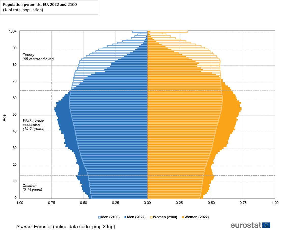
Elderly (65 years and over)
Working-age population (15–64 years)
Children (0–14 years)
Adapted from Eurostat.2
PCa care and will discuss possible future directions, focusing on early detection and active surveillance (AS).
As the proverb goes, ‘prevention is better than cure’. One of the first steps in deescalating PCa care would be to invest in the prevention of the disease. However, primary prevention is virtually impossible, as there are currently no thoroughly established modifiable risk factors for PCa.9 Through the use of organised screening programmes, it has been proven that
PCa-related morbidity and mortality can be reduced. In the latest analysis of the European Randomized Study of Screening for Prostate Cancer (ERSPC), PSA screening was found to reduce PCa-related mortality by 16% (rate ratio: 0.84; 95% CI: 0.76–0.92).10 However, PSA was first introduced as a marker for follow-up of PCa and not for screening. In the ERSPC, this came at the cost of overdiagnosis and overtreatment, as more men were diagnosed with forms of PCa that might not have caused symptoms later in life. For every averted death due to PCa, 12 men are diagnosed with PCa on top of those diagnosed in a clinical situation. On the other hand, the risk of diagnosis
for advanced, incurable PCa was lower for screened males than controls (risk ratio: 0.66; 95% CI: 0.60–0.74).10
In the ERSPC and most, if not all, of the first PCa screening trials, screening was done in a one-size-fits-all approach. If a participant’s PSA level exceeded a set threshold of 3 ng/mL, further evaluation was done through prostate biopsies.10 Because of the overdiagnosis found in these trials, organised screening programmes for PCa have been hotly contested in the past decades. To improve the characteristics of these organised screening programmes, the use of risk stratification tools is vital.
Risk stratification in screening for PCa can be done in a multitude of different ways and at different points of the screening pathway. Using risk calculators, novel biomarkers, and MRI, patients at risk of a PCa diagnosis can be identified more clearly and, just as importantly, patients at low or no risk for PCa can also be identified.11 Incorporating MRI into risk calculators has shown to be able to reduce the number of biopsies by 36%, while missing only 4% of high-grade PCa in men who did not receive a biopsy.12 To reduce overdiagnosis of insignificant PCa (i.e., forms of PCa that would not cause symptoms later in life), these modalities should be combined in an organised screening programme. As for other ways to reduce the inflow of men into these screening programmes, attention should be turned to which men should be invited for screening. Upfront selection of patients is currently only done through age restrictions on participation in a screening programme. To further de-escalate the overdiagnosis of men with insignificant PCa, attention can be turned towards who to invite for screening and what to consider as ‘clinically significant’ PCa.
No two people are the same, as much as no two cases of PCa are the same. Currently, clinical significance is defined on the pathologic characteristics of the tumour. However, an International Society of Urologic Pathology Grade Group (ISUP GG) 1 PCa diagnosis for a 74-year-old with end-stage kidney disease does not carry the same weight as the same diagnosis
for a 52-year-old man with no medical history. The same disease that could be clinicopathologically, and appropriately, defined as ‘insignificant’ or ‘not clinically significant’ for the first man, whose lifeexpectancy is to be counted in years, could very well become ‘clinically significant’ for the second man, as he may have a lifeexpectancy of several decades where the disease may become more aggressive. As symptoms of PCa often take years to manifest, in light of efficient use of resources, remaining life expectancy should be used as a tool, more so than chronological age, to determine both who to invite to screening and what to consider clinically significant PCa.
As of yet, there are no large-scale organised screening programmes. Although their use in the reduction of PCa-related mortality has been proven, much discussion still exists around overdiagnosis and overtreatment. PSA tests are, however, available outside of the organised screening setting. Through their general practitioner, informed men can get access to a PSA test. This practice, where asymptomatic men take the initiative to get a PSA test, is labelled ‘opportunistic screening’. This opportunistic screening poses several problems: first and foremost, that it may not have any effect on PCa-related mortality and has higher overdiagnosis than organised screening, with up to twice the number of men needing to be diagnosed to prevent one PCa-related death.13 Second, having opportunistic screening be dependent on public initiative could disadvantage men who are less medically informed.
A challenge in both opportunistic and organised screening is how to handle those at a higher risk of PCa, especially at a younger age due to hereditary forms of PCa. As mentioned, in opportunistic screening, initiative is dependent on a man’s own knowledge of the disease and, in this case, his increased risk. In organised screening, the pathway proposed for the general population might not be suitable for these men, as they are at greater risk of developing PCa and at younger ages than those without a genetic predisposition. In organised screening, this could be taken
into account by reaching out to these men earlier and evaluating if an adapted programme would be of greater benefit.
Organised screening is, however, not yet a widely accepted and applied practice, and opportunistic screening will probably still exist for several years, if not longer. So, de-escalating opportunistic screening is an equally important endeavour to explore. Much the same as organised screening programmes, opportunistic screening can benefit from the use of risk stratification tools. In addition, part of this risk stratification can be outsourced from the hospital, as Hogenhout et al.14 describe in their article on shifting PCa detection to primary care. By performing riskstratification at a diagnostic centre, over two-thirds of referrals could be prevented, and at a reduced cost compared to a hospital visit.
With the introduction of PSA-based screening of PCa and the subsequent inflow of men with low-risk PCa, AS was introduced as a way to defer or avoid invasive treatment by closely following these men. Critically distinct from watchful waiting, when higher-grade PCa is detected in men on AS, definitive treatment is initiated. Though initially only performed in a research setting, as time passed and results were published, AS evolved into the preferred treatment for low-grade PCa. Oncological outcomes are comparable to definitive treatment, such as radical prostatectomy or radiotherapy, for men on AS.15 The introduction of MRI in the AS pathway allows for a theoretical improvement of the initial selection of patients eligible for AS by more precisely sampling tumours during biopsy. Also, MRI may identify men with elevated PSA but no prostatic lesions, and thus reduce unnecessary biopsies in up to a quarter of patients.16 However, due to the reduction in undersampling, men who would have been diagnosed with a low-risk form of PCa could show higher grade PCa although the underlying disease was the same. Though a heterogeneous group, multiple studies
have shown that a subgroup of men with ISUP GG 2 can be safely monitored with AS.17 Even though expensive, definitive treatments can be avoided, most AS strategies include an intensive followup protocol. This consequently strains the urologist’s practice with frequent appointments for repeat PSA-tests, MRI, and biopsy results.
Currently, AS is mostly performed according to static, quite intensive, one-size-fitsall protocols. These protocols generally consist of a combination of recurring PSAtests, digital rectal examinations, MRI, and biopsies. Different centres have differing protocols, with different combinations of the above-mentioned diagnostic interventions at varying intervals.17 Given the favourable oncological outcomes observed in men undergoing AS, there is now the concept of the ‘overly surveyed patient’. To reduce the workload and keep AS a viable option for all men with low-risk PCa, AS should switch to a risk-based follow-up strategy. This approach has already shown promise with the Stratified Cancer Surveillance (STRATCANS) programme, where most men (214/297; 72%) remained treatment free at a median follow-up of 4.9 years. Apart from diagnostic interventions (digital rectal examination, MRI, and biopsies), follow-up was virtual and men in the lowest risk group (127/294; 43%) only received follow-up biopsy if indicated by PSA or MRI.18 This risk-based approach addresses the main concern with reducing follow-up: ensuring that those at highest risk of upgrading to higher-risk disease are detected at an appropriate time.
Another challenge arises around keeping men on AS. From a policy and economic point of view, providing a one-time surgery for these low-risk men is more profitable than keeping them in years-long intensive follow-up. Financial incentives to perform definitive therapy in men at low risk of clinically significant PCa are different for a given healthcare system but, for as long as these exist, there will be men at low risk of clinically significant PCa who, possibly to their detriment, undergo definitive treatment. To ensure that men are treated with a focus on what is in their best
oncological and personal interest (instead of focusing on financials), healthcare systems should ensure that renumeration for AS does not discourage inclusion over performing definitive therapies.
To have AS remain a viable treatment option and reduce definitive treatment, further research should be focused on reducing the workload of AS. Using a risk-based approach, further incorporating MRI and riskcalculators and reducing follow-up intensity for those patients who are expected to have excellent outcomes should assist in keeping PCa care accessible for all men.
Future Perspectives
More men at risk of prostate cancer
- Increasing life-expectancy
- Ageing population
Decreasing workforce
References
1. International Agency for Research on Cancer (IARC). Dataviz. Available at: https://gco.iarc.who.int/today/en/ dataviz. Last accessed: 19 February 2026.
2. Eurostat. Population projections in the EU. 2023. Available from: https:// ec.europa.eu/eurostat/statisticsexplained/index.php?title=Population_ projections_in_the_EU#Source_data_for_ tables_and_graphs. Last accessed: 28 January 2026.
3. European Commission Joint Research Centre (JRC). European Cancer Information System (ECIS). 2024. Available at: https://ecis.jrc.ec.europa.eu. Last accessed: 19 February 2026.
4. Siegel RL et al. Cancer statistics, 2026. CA Cancer J Clin. 2026;76(1):e70043.
5. Paquette EL et al. Improved prostate cancer-specific survival and other disease parameters: impact of prostate-
To continue to manage PCa care in the future, it is essential that we de-escalate parts of PCa care. As discussed, this can be done on different levels and at different points of the diagnostic pathway (Figure 2). Through timely detection, when there is opportunity for curative care, risk stratification, careful selection of who to screen, and efficient use of resources, PCa care will remain accessible and of high quality for all patients.
Current practice
- Opportunistic screening
→ High overdiagnosis
→ No proven benefit
Solutions
- Introduction of organised screening (proven prostate cancer-related mortality)
→Inclusion of multiple risk-stratification steps, also in opportunistic screening
→ Attention to life-expecancy-dependent inclusion criteria
specific antigen testing. Urology. 2002;60(5):756-9.
6. Helgesen F et al. Trends in prostate cancer survival in Sweden, 1960 through 1988: evidence of increasing diagnosis of nonlethal tumors. J Natl Cancer Inst. 1996;88(17):1216-21.
7. McLeod OD et al. Cost analysis of prostate cancer care using a biomarkerenhanced diagnostic strategy with Stockholm3. Eur Urol Open Sci. 2024;66:26-32.
8. Organisation for Economic Co-operation and Development (OECD). Health at a glance: Europe 2024: state of health in the EU cycle. 2024. Available at: https:// www.oecd.org/en/publications/healthat-a-glance-europe-2024_b3704e14-en. html. Last accessed: 19 February 2026.
9. Leitzmann MF, Rohrmann S. Risk factors for the onset of prostatic cancer: age, location, and behavioral correlates. Clin Epidemiol. 2012;4:1-11.
Active Surveillance
Current practice
- One-size-fits-all approach
- Intensive follow-up
Solutions
- Dynamic risk-based approach
→ Personalised follow-up
→ Reduction in workload
10. Roobol MJ et al. European study of prostate cancer screening23-year follow-up. N Engl J Med. 2025;393(17):1669-80.
11. Auvinen A et al. Prostate cancer screening with PSA, kallikrein panel, and MRI: the ProScreen randomized trial. JAMA. 2024;331(17):1452-9.
12. Alberts AR et al. Prediction of highgrade prostate cancer following multiparametric magnetic resonance imaging: improving the Rotterdam European Randomized Study of Screening for Prostate Cancer risk calculators. Eur Urol. 2019;75(2):310-8.
13. Arnsrud Godtman R et al. Opportunistic testing versus organized prostatespecific antigen screening: outcome after 18 years in the Göteborg randomized population-based prostate cancer screening trial. Eur Urol. 2015;68(3):354-60.
14. Hogenhout R et al. Shifting risk-stratified early prostate cancer detection to a primary healthcare setting. BJU Int. 2023;131(5):596-601.
15. Hamdy FC et al. Fifteen-year outcomes after monitoring, surgery, or radiotherapy for prostate cancer. N Engl J Med. 2023;388(17):1547-58.
16. Kasivisvanathan V et al. MRI-targeted or standard biopsy for prostatecancer diagnosis. N Engl J Med. 2018;378(19):1767-77.
17. Kinsella N et al. Active surveillance for prostate cancer: a systematic review of contemporary worldwide practices. Transl Androl Urol. 2018;7(1):83-97.
18. Gnanapragasam VJ et al. The 5-year results of the Stratified Cancer Active Surveillance programme for men with prostate cancer. BJU Int. 2025;135(5):851-9.
Editor's Pick
Neurogenic bladder is an underdiagnosed and important condition that has great, variable impacts on patients if not diagnosed and treated well. This article gives a brief summary on this subject, highlighting the complex, lifelong multidisciplinary care that is required as patients age, from psychology and physiotherapy to occupational therapy and social work. The article also explores current health system considerations and practical facilitators that support effective, patient-centred management across the lifespan.
Erdem Canda
Department of Urology, School of Medicine, Koç University, Istanbul, Türkiye


Authors: *Emily K. Clennon,1 Kelly T. Harris,1 Kyle O. Rove,1 Duncan T. Wilcox,1 Daniel Wood1
1. Children’s Hospital Colorado, Aurora, USA *Correspondence to emily.clennon@childrenscolorado.org
Disclosure: Daniel Wood is a member of the Executive Committee of the Neurogenic Bladder Research Group (NBRG). The other authors have declared no conflicts of interest.
Received: 30.12.25
Accepted: 16.02.26
Keywords: Bladder dysfunction, lower urinary tract dysfunction, neurogenic, neurogenic dysfunction, transition, transitional care.
Citation: EMJ Urol. 2026;14[1]:77-86. https://doi.org/10.33590/emjurol/1061221L
Abstract
Neurogenic dysfunction of the lower urinary tract is a complex condition with variable, often profound, and multisystem impacts that require longitudinal, multidisciplinary care that becomes more complex as patients age. This review covers key pathophysiology and outlines clinical priorities in the management of patients with neurogenic lower tract dysfunction. The current landscape of transitional urologic care is also described with attention to health system components, challenges, and patient-level tools/facilitators.
Key Points
1. Neurogenic bladder dysfunction occurs secondary to a neurologic insult and can include a wide range of urinary symptoms and their sequelae. Patients with neurogenic bladder dysfunction require lifelong multidisciplinary care.
2. Preservation of renal function is the foremost clinical priority for these patients, and management of bladder emptying, continence, sexual function/fertility, prevention of infection, and screening for sexual abuse are all essential components of their long-term comprehensive care.
3. Transition from paediatric to adult care systems remains challenging with highly variable structures and success between systems and nations. Effective transition can minimise recurrent hospitalisations and escalation of care in adulthood, and necessarily involves the expertise and active participation of the patient and their caregivers.
The term neurogenic bladder is defined as bladder dysfunction resulting from damage to the nerves that control the bladder in the central or peripheral nervous system. The term is often, mistakenly, used as a ‘diagnosis’, even though it occurs secondary to a neurological diagnosis, which could include spinal dysraphism, exstrophy, spinal cord injury, cerebrovascular accident, multiple sclerosis, iatrogenic pelvic nerve injury, etc. The impact of a neurological lesion may be impaired storage or voiding or altered bladder pressures, which may result from dis-coordinated voiding. Management of lower urinary tract neurogenic dysfunction requires attention to multiple domains of patient pathophysiology and navigation of the intersections of physiology, psychosocial environment, and health system capabilities. Longitudinal multidisciplinary care is paramount for these patients, as the character and degree of a neurologic dysfunction may evolve over time and can affect multiple systems (e.g., urological, nephrological, colorectal, orthopaedic, psychological). Involvement of psychology, physiotherapy, occupational therapy, and social work is particularly important for many of these patients. Given the complexity of pathophysiology and care models, individuals with congenital pathology require active transition between paediatric and adult care systems. This review will cover priorities in the clinical care of patients with neurogenic bladder and the current landscape of transitional care for these patients internationally.
Historically, renal failure was a source of significant morbidity and mortality among patients with neurogenic bladder; it was the leading cause of death among patients with spina bifida >16 years old and after spinal cord injury.1,2 Though patients with spina bifida do not typically suffer congenital renal dysplasia, 50% will develop renal insufficiency within 5 years without routine urologic care.3 Patients with renal
dysplasia secondary to abnormal in utero lower urinary tract mechanics are at even higher risk of renal disease. Modern series demonstrate that active management of lower tract neurologic dysfunction can substantially reduce morbidity, with severe renal impairment in as few as 1.6% of patients at one centre.4 Thus, renal preservation is the foremost clinical priority for these patients.
If neurogenic bladder is suspected, investigation often begins with uroflowmetry (if able to void volitionally) and post-void residual assessment. Adults with low-risk clinical features (suprapontine or peripheral nerve lesion, normal renal function, minimal/stable symptoms) and normal assessment can safely forego invasive testing.5,6 Importantly, the level of the neurological lesion may not correlate with clinical phenotype, so careful initial assessment is crucial. If there are abnormalities on initial testing or high clinical suspicion of neurologic pathology, quantitative evaluation of bladder mechanics via video urodynamics is recommended. Storage and detrusor leak point pressure greater than 40 cm H2O have long been used as the threshold at which bladder pressures are considered unsafe.7 However, newer analyses demonstrate increased risk of secondary vesico-ureteral reflux (VUR) and hydronephrosis with storage pressures ≥15 cm H2O,8 so many clinicians now use this finding to trigger stratification as higher risk of upper tract deterioration. Impaired compliance (<20 cm H2O), detrusor overactivity, and detrusor sphincter dyssynergia have also been associated with upper tract deterioration.6,8 In children, bladder capacity is lower and deterioration in compliance, even if absolute pressure is still within a ‘safe’ range, often prompts changes in management. In patients with established neurologic dysfunction, new or changed symptoms should prompt re-evaluation.
Renal function may be monitored with laboratory measures, ultrasonography, and functional imaging, such as nuclear renal scans. Cystatin C is the most accurate serum measure of renal function available in most clinical contexts for patients after
the first year of life.9 In patients who are non-weight-bearing, serum creatinine is not an accurate measure of renal function due to reduced muscle mass but can be useful to trend if more accurate labs, such as cystatin C and chromium-51 edetic acid, are not readily available.9 Changes in renal evaluation despite stable urinary symptoms should prompt re-evaluation of bladder dynamics and consideration of more intensive bladder management. Multisystem effects of these patients’ primary pathologies (e.g., kyphosis resulting in respiratory compromise, feeding difficulties, bone resorption), non-urologic comorbidities (e.g., obesity, cardiovascular disease), and urologic disease beyond high bladder pressures (e.g., urinary tract infections [UTI] and urolithiasis) also pose threats to renal function.9
There is historic controversy regarding reactive versus proactive management of patients with congenital neurogenic bladder. Reactive strategies involve observation without intervention until new symptoms, changes in bladder dynamics,
or renal dysfunction develop.10 This strategy theoretically confers lower risk of iatrogenic injury or infection, less risk of medical trauma and its sequelae, and less burden on caregivers and cost to the family and health system during observation. Proactive management strategies include urodynamics in the first months of life regardless of voiding symptoms, early initiation of anticholinergics and clean intermittent catheterisation (CIC), and surveillance renal function testing and ultrasound every 6–10 months (Figure 1).10,11 Reviews in recent years have demonstrated significantly lower rates of secondary VUR, UTIs, and renal deterioration with proactive strategies, and current European Association of Urology (EAU) guidelines advocate for proactive management.10-12
Incomplete bladder emptying may contribute to increased bladder pressures, secondary VUR, bladder calculi, and recurrent UTI. In children ≤6 years old, postvoid residual assessments >20 mL or >10%
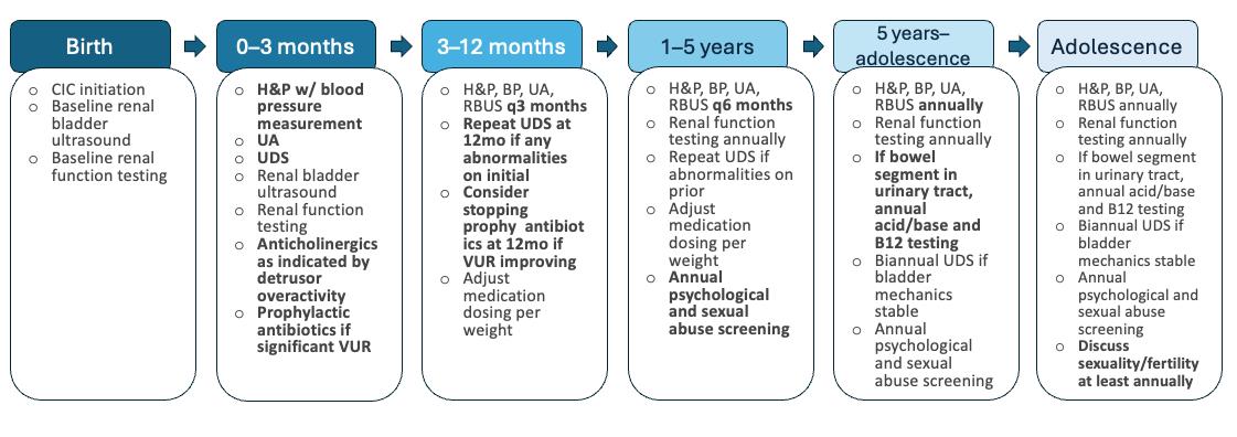
Represents a proactive management strategy with a primary goal of renal preservation. Note that UDS evaluation is performed in the first 3 months of life, ideally 2–3 months after closure of any spinal defects. For patients not undergoing spinal surgery, UDS may be performed prior to hospital discharge after birth.
Adapted from European Association of Urology (EAU)/European Society for Paediatric Urology (ESPU) guidelines on the management of neurogenic bladder in children and adolescents.11
BP: blood pressure; CIC: clean intermittent catheterisation; H&P: history and physical; mo: month; RBUS: renal and bladder ultrasound; UA: urinalysis; UDS: urodynamic studies; VUR: vesico-ureteral reflux; w/: with.
expected bladder capacity are considered abnormal.13 Residuals typically decrease in adolescence, then slowly increase in adulthood with volumes up to 100 mL being accepted as normal.14,15 Most patients with neurogenic bladder dysfunction will require some form of assistance with emptying, including vesicostomy, indwelling catheter (urethral or suprapubic), CIC via the urethra or a catheterisable channel, or creation of an incontinent urinary diversion. CIC is favoured over indwelling catheter given the lower risk of catheter-associated UTI.5,6 Impaired mobility and spasticity may challenge self-catheterisation, particularly via the urethra. Catheterisable channel creation may facilitate independent or caregiver access to the urinary tract, improve patient comfort, allow CIC despite urethral stricture or bladder neck closure, minimise caregiver interaction with the patient’s genitals, and minimise wheelchair transfers for patients with female anatomy.6
Urinary incontinence, whether from the native urethra or a catheterisable channel, affects >50% of patients with neurogenic bladder secondary to spina bifida and is a significant challenge to social life, independence, and sexuality.16-18 Not surprisingly, health-related quality of life in these patients tracks with degree of primary urinary continence.19 In patients with neurogenic bowel dysfunction or chronic constipation, optimisation of bowel management may be all that is required to improve or resolve urinary incontinence. Other interventions may include changes in catheterisation schedules, anticholinergics or β-agonists, intravesical onabotulinum toxin, bulking agents, slings or bladder neck reconstructions/closures, or continent urinary diversions with bowel pouches. If intravesical onabotulinum toxin is used, the surgeon must verify dosing by other specialties (for skeletal muscle spasticity, anal sphincter relaxation, etc.) to avoid overdose.
Care must be taken to ensure efforts toward continence do not create a bladder environment hostile to the upper tracts. For this reason, surgical interventions aimed at continence are often deferred until a patient is old enough to desire social continence,
has demonstrated the ability or willingness to catheterise as necessary post-procedure, and has undergone necessary interventions to create a safe lower urinary tract. Bladder neck closures are reserved as a terminal incontinence procedure for patients with stable symptoms and renal function, safe bladder dynamics, and an established catheterisable channel.
Diagnosis of UTI in the neurogenic bladder population is complex, as altered sensation may preclude traditional symptoms and bacteriuria secondary to urinary tract instrumentation is common. These barriers to accurate diagnosis can lead to overexposure to antibiotics and, alternatively, to diagnosis of infection only once it has spread to the upper tracts.20 Pyelonephritis and its sequelae are the most common causes of hospitalisation among patients with neurogenic bladder who experience more than 10-times the number of hospitalisations of their peers.21-23 Incomplete bladder emptying, incontinence and genital skin breakdown, VUR (in the absence of high bladder pressures), and urinary calculi are all modifiable risk factors for UTI that should be addressed as part of longitudinal care for neurogenic bladder (Figure 2).
Though the spectrum of sexual experience and reproductive potential in the setting of neurogenic bladder are outside the scope of this review, it is important to note that interventions targeting the bladder can impact these domains. Indwelling urethral catheters, urethral trauma or erosion from catheterisation, and procedures altering bladder neck anatomy can preclude antegrade ejaculation. Reconstructive surgeries may impair genital sensation and sexual function, and failure to achieve continence negatively impacts sexual experience.18,24 Women who have bowel in the urinary tract (e.g., bladder augment) have a 75% false-positive rate with urine pregnancy tests and must rely on serum human chorionic gonadotropin (hCG) testing or ultrasound to diagnose
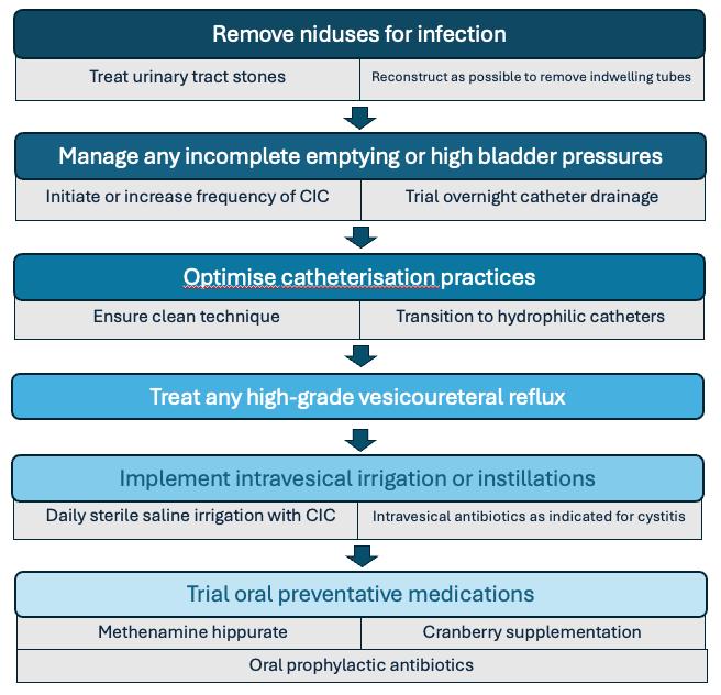
Note the preference for intravesical antibiotic administration for infections limited to the lower urinary tract and the exhaustion of other options prior to considering oral prophylactic antibiotics.
CIC: clean intermittent catheterisation.
pregnancy.25 Asymptomatic bacteriuria and UTI are more common among these women and require close attention during pregnancy to prevent obstetric complications.26 Access to the bladder via catheterisable channel may be challenged by a gravid uterus, so some women need an indwelling catheter in the third trimester. Lastly, pregnancy amongst these patients requires coordination between obstetricians and urologists to determine safe delivery
plans if pelvic anatomy is altered enough to preclude vaginal delivery or if reconstructed structures may be encountered during Caesarean section. Emergency Caesarean sections can significantly increase risks to mothers with complex reconstructions and their babies.27
Despite robust evidence that patients with neurogenic bladder are as interested in sexual activity as their peers, 9/10
paediatric urologists report their training did not include instructions on sexual and reproductive health in the context of congenitalism, and a majority of patients with spina bifida report inadequate education regarding their sexual and reproductive health from providers.28,29 Questionnaires used to screen for sexual dysfunction in the general population, such as the International Index of Erectile Function (IIEF), are not validated in and do not necessarily reflect the unique concerns of the neurogenic bladder population.30 Comprehensive and appropriately-tailored assessment and education regarding anatomy, sexuality, and fertility are indicated for these patients.29,31-33
Children with disabilities are approximately three-times more likely to be victims of sexual violence than non-disabled peers; estimates from case series of patients with spina bifida suggest rates ≥30% by early adulthood.34-36 Risk of abuse does not normalise in adulthood and is increased in the setting of intellectual and sensory disabilities.37 Reliance on caregivers for bladder management, particularly CIC via the urethra, creates vulnerability multiple times per day and increases tolerance of physical intrusion. Nearly half (44%) of assailants against this population acted as caregivers for their victims.35 Given the prevalence of sexual violence in this population and the vulnerability bladder management creates, screening for sexual abuse and education regarding (in)appropriate touch from childhood is a critical component of comprehensive care for these patients.
Patients with neurologic dysfunction of the bladder report lower quality of life in mental health domains and may have twice the risk of depression of matched peers.38,39 Caregivers of children with these diagnoses report significant burden, particularly with CIC, and high rates of burnout and distress.40-42 The availability of mental health providers for patients
and their caregivers is limited, particularly after transition to adult care settings. Medical management strategies should be adapted to minimise psychological distress, patient re-traumatisation, and burden on patients and caregivers to the extent possible without compromising upper tract preservation. Psychological care and caregiver respite should be ideally integrated into transitional programmes.
Historically, urologic care of patients with congenitalism was limited to paediatric urology given early patient mortality and limited familiarity with relevant pathophysiology and reconstruction techniques among adult providers. This paradigm is necessarily changing with substantially improved life expectancy and the subsequently increased prevalence of congenitalism. Though many management principles of congenital and acquired neurogenic bladder are shared, patients who have required lifelong management face different challenges than adults with newly developed dysfunction, and the transition between care in childhood and adulthood is a particular challenge for patients and providers alike. Though many investigations regarding the transition process, thus far, have focused on emotional/functional readiness to transition as an end point, ‘successful’ transition discussed here is defined as engagement in an adult urological care system without a significant (i.e., 2 years or more) gap from paediatric care.43
In settings where transition has not been structured or prioritised, transfer rates to adult healthcare can be below 10%.43 Across high resource nations with focus on transitional care, up to 40% of patients with congenitalism fail to move into adult specialty care without a significant interruption, and, as recently as 2005, 76% could not identify a primary care provider.9,23 Once engaged in adult urologic care, 52% of transitional patients were experiencing incontinence and 97% required some form of intervention, with 71% changing bladder
management and 38% undergoing major surgery.16,44 Analyses of hospitalisations for patients with spina bifida have estimated that 34% (equating to 182 million USD of services per year) could have been avoided with more active management.45
How transition happens depends heavily on the design and limitations of the local healthcare system. Figure 3 outlines a variety of models and some benefits and challenges of each. Differing locations, health record systems, and reimbursement models between paediatric and adult practices create significant challenges in the transition process. National or regional centres with lifelong disease-specific care likely offer the smoothest transition for patients and providers but are rare globally and may require significant travel for patients and families. Systems with divisions between both clinical providers and payors for paediatric and adult patients, such as the Children’s Health Insurance Program (CHIP) versus Medicaid/Medicare in the USA, are possibly the least well adapted to transitional care, because they involve multiple levels of barriers to collaboration.
Health coverage/reimbursement and clinic structures that adequately reflect the complex needs of these patients compared to the general urology patient population (e.g., longer appointment times, rooms that accommodate wheelchairs, etc.) are rare and can limit engagement of adult practices.47
Beyond urologic care, effective multidisciplinary cooperation needs to be maintained in transition. Multidisciplinary clinics are much less common in adult centres, and adult patients face intersectional challenges between their longstanding diagnoses and diseases of ageing. If necessary, repeat surgical interventions come with a substantial risk of multi-organ system complication; the risk of any complication exceeds 90% and the readmission rate exceeds 40%.48 Ultimately, regardless of the system, successful transition requires active and thoughtful interaction between paediatric and adult urologists, colleagues in different specialties, and patients and their families. Structured, dedicated transition programmes and clinics have demonstrated success rates exceeding 85% in varied health delivery systems.43
Lifelong care from a pediatric urologist when paediatric and adult urology departments are combined
Potentially greater nexibility in joint cases and clinics
Potentially greater flexibility in joint cases and clinics
Easy to implement in disease-specific specialty centres
Easy to implement in disease-specific specialty centres
Not financially feasible between independent children's and adult centres without established collaboration
Not financially feasible between independent children's and adult centres without established collaboration
Lifelong care from a paediatric urologist who integrates with a separate adult urology department
from a paediatric urologist who integrates with a separate adult urology department
Maximal continuity of care
Maximal continuity of care
Minimal patient anxiety
Minimal patient anxiety
Potentially more robust interaction between pediatric and adult colleagues
Potentially more robust interaction between paediatric and adult colleagues
Demand may exceed clinical capacity of pediatric urologists over time
Demand may exceed clinical capacity of pediatric urologists over time
Lifelong care in pediatric setting not socially or developmentally appropriate
Ufelong care in pediatric setting not socially or developmentally appropriate
Transition of some form still required when pediatric urologist retires
Transitio n of some form still required when paediatric urologist retires
Easiest to implement in centralised systems
Transition from a paediatric urologist to an adolesc ent/adult specialist with an interest in congenitalism
Transition from a paediatric urologist to an adolescent/adult specialist with an interest in congenitalism
Direct transfer from a paed iatric urologist to adult urology
Direct transfer from a pediatric urologist to adult urology
AI!o ,v s developmentally appropriate transittons in care enVJ'ronment
Allows developmentally appropriate transitions in care environment
Adult urologists likely better equipped to address sexual function, fertility, and ageing-related conditions
Adult urologists /1ke!y better eqwpped to address sexual function fertJ/ity, and ageing-related conditions
May ease multidisciplinary care for adult conditions
May ease multidisciplinary care or adult condl ·ons
May be naturally facilitated by patient social transillons (i e., gomg to coJJege, moving to work in another citY, losing support from parental Joss, etc.)
May be naturally facilitated by patient social transitions (i.e., going to college, moving to work in another city, losing support from parental loss, etc.)
Potentially less f/ex1bilIty m Jomt cases and clinics
Potentially less flexibility in joint cases and clinics
May require bridging of different medical record systems and reimbursement models
May reqwre bridging of different medical record systems and reimbursement models
Multidisciplinary care may be challenging ,r not mtegrated in same fashion
Multidisciplinary care may be challenging if not integrated in same fashion
Requires specialist trained in both paediatric and adult urology
Requires specialist trained in both pediatric and adult urology
Requires intentional patient-led handover process
Requires intentional patientled handover process
Highest risk of loss to follow-up
Highest risk of loss to follow-up
Most difficult model conceptually and fogisucaJJy for collaboration between paediatric and adult co/leagues
Most difficult model, conceptually and logistically - for collaboration between pediatric and adult colleagues
Difficult to identify adult urologists with mterest expertise and capac1ty to serve this population
Easiest to implement in fragmented systems , ,
Difficult to identify adult urologists with interest, expertise, and capacity to serve this population
The transition process involves not only hand-off of care between paediatric and adult providers but is, more importantly, the process by which patients learn how to assume responsibility for their healthcare. As such, there is no universally applicable transition schedule. Effective transition is a gradual process that is often initiated by patient interest, though it may require prompting by providers. Surveys of patients with spina bifida indicate up to 40% of patients have no desire to transition and require motivational interviewing and coaching through the process, often requiring additional non-clinical visits to accomplish this goal.49,50
The Spina Bifida Association (SBA) recommends starting conversations about transition as early as birth.51 Other programmes and centres may recommend initiating conversations around 12 years of age, with a goal of transition completion around 18–20 years.9,48,52-54 On average, adolescents with spina bifida develop autonomy in activities of daily living 2–5 years after their peers and tend to overestimate their ability to complete tasks independently.55-57 Validated (Transition Readiness Assessment Questionnaire [TRAQ]) and non-validated (GotTransition, Ready Steady Go, Urology Care Foundation guide) tools have been created to guide assessment of patient knowledge and ownership in care. Improvements in these domains has been associated with increased transitional readiness.43
For some patients, the goal of transition to independence may be unachievable, despite the best efforts of all concerned. By the age of 30 years, only one-third of patients with spina bifida are independent and one-third require supervision and occasional assistance, despite being able to perform some care tasks.58 Careful evaluation and acknowledgement of each patient’s cognitive and physical abilities and functional capacity/readiness are critical components of transition planning and goalsetting for long-term success.
Though urologists study and care for individuals with neurogenic bladder, they cannot truly understand life with and management of the condition as an observer. These patients and their families quickly develop expertise not available to their care teams and beyond just lived experience.59 They can detect unease, lack of familiarity, and disinterest in providers and systems, all of which can result in a loss of trust in the therapeutic relationship and preclude transition and effective care.46 Ideally, transitional programmes should be developed and designed in collaboration with patient advisors.
The effects of neurogenic lower urinary tract dysfunction and its management extend beyond the urinary system and across the lifespan. As these patients age, the primary clinical challenges remain the same, protecting renal function, ensuring adequate bladder emptying, and achieving continence, but impetuses to prevent infection, avoid hospitalisation, and manage sexuality/ fertility emerge. Screening for safety concerns and sexual abuse is necessary from early in life given the vulnerabilities created by developmental disabilities and the intimacy of bladder management. Many patients experience interruptions and avoidable escalations in care in the transition from paediatric to adult care, which is variably managed depending on health system structure and capabilities. There are tools available to clinicians, patients, and families to guide and prepare for transition, and the expertise of patients and caregivers living with neurogenic bladder should be utilised in the development of transitional programmes.
References
1. Singhal B, Mathew KH. Factors affecting mortality and morbidity in adult spina bifida. Euro J Ped Surg. 1999;9(1):31-2.
2. Hackler RH. A 25-year prospective mortality study in the spinal cord injured patient: com-parison with the long-term living paraplegic. J Urol. 1977;117(4):486-8.
3. Bauer SB, Joseph DB. Management of the obstructed urinary tract associated with neuro-genic bladder dysfunction. Urol Clin North Am. 1990;17(2):395-406.
4. Malakounides G et al. Single centre experience: long term outcomes in spina bifida. J Ped Uro. 2013;9(5):585-9.
5. European Association of Urology (EAU). EAU guidelines on neuro-urology. 2024. Available at: https://d56bochluxqnz. cloudfront.net/documents/fullguideline/EAU-Guidelines-on-NeuroUrology-2024.pdf. Last accessed: 11 September 2025.
6. Ginsberg DA et al. The AUA/SUFU guideline on adult neurogenic lower urinary tract dysfunction: diagnosis and evaluation. J Urol. 2021;206:1097.
7. McGuire EJ et al. Prognostic value of urodynamic testing in myelodysplastic patients. J Urol. 1981;126(2):205-9.
8. Swatesutipun V, Tangpaitoon T. The safety cutoff storage pressure for preventing upper urinary tract damage in neurogenic bladder from spinal cord pathology and risk factor anal-ysis. Neurourol Urodyn. 2022;41:991-1001.
9. Loftus CJ, Wood HM. Congenital causes of neurogenic bladder and the transition to adult care. Transl Androl Urol. 2016;5(1):39-50.
10. Fairchild RJ et al. Medical management of neurogenic bladder in patients with spina bifida: a scoping review. J Ped Urol. 2023;19(1):55-63.
11. Stein R et al. EAU/ESPU guidelines on the management of neurogenic bladder in children and adolescent part I diagnostics and conservative treatment. Neurourol Urodyn. 2020;39:45-57.
12. Li Y et al. Systematic review and metaanalysis to study the outcomes of proactive versus delayed management in children with a congenital neurogenic bladder. J Ped Urol. 2023;19(6):730-41.
13. Chang S et al. Age- and genderspecific nomograms for single and dual post-void residual urine in healthy children. Neurourol Urodyn. 2013;32:1014-18.
14. Lim LY et al. Age- and gender-specific normal post-void residual urine in healthy adoles-cents. J Ped Uro. 2023;19(4):367.e1-6.
15. Asimakopoulos AD et al. Measurement of post-void residual urine. Neurourol Urodyn. 2016;35(1):55-7.
16. Summers SJ et al. Urologic problems in spina bifida patients transitioning to adult care. Urol. 2014;84:440-4.
17. Verhoef M et al. High prevalence of incontinence among young adults with spina bifida: de-scription, prediction and problem perception. Spinal Cord. 2005;43:331-40.
18. Roth JD et al. Urinary and fecal incontinence during sexual activity is common and bother-some among adults with spina bifida. Urol. 2024;186:54-60.
19. Szymanski K et al. Quality of life improves with improved continence in people with spina bi-fida. S33-1. ESPU-SPU Joint Meeting, 3-6 September, 2025.
20. Kyaw TS et al. Cumulative antibiotic exposure in the pediatric spina bifida population. S40-1. ESPU-SPU Joint Meeting, 3-6 September, 2025.
21. Dicianno BE, Wilson R. Hospitalizations of adults with spina bifida and congenital spinal cord anomalies. Arch Phys Med Rehabil. 2010;91:529-35.
22. Manack A et al. Epidemiology and healthcare utilization of neurogenic bladder patients in a US claims database. Neurourol Urodyn. 2011;30:395-401.
23. Young NL et al. Youth and young adults with spina bifida: their utilization of physician and hospital services. Arch Phys Med Rehabil. 2014;95(3):466-71.
24. Tourchi A Hoebeke P. Longterm outcome of male genital reconstruction in childhood. J Ped Urol. 2013;9(6):980-9.
25. Vasquez E et al. False positive pregnancy tests in pediatric patients with augmentation en-terocystoplasty. J Ped Uro. 2022;18(5):615.e1-6.
26. Huck N et al. Pregnancy following urinary tract reconstruction using bowel segments: a re-view of the published literature. World J Urol. 2020;38:335-42.
27. Deans R et al. Reproductive outcomes in women with classic bladder exstrophy: an obser-vational crosssectional study. Am J Obstet Gynecol. 2012;206(6):496.e1-6.
28. Streur CS et al. “I don’t know what I’m doing… I hope I’m not just an idiot”: the need to train pediatric urologists to
discuss sexual and reproductive health care with young women with spina bifida. J Sex Med. 2018;15(10):1403-13.
29. Streur CS et al. “If everyone else is having this talk with their doctor, why am I not having this talk with mine?”: the experiences of sexuality and sexual health education of young women with spina bifida. J Sex Med. 2019;16(6):853-9.
30. Rague JT et al. “I just don’t have any of that”: applicability of the International Index of Erectile Function in young men with spina bifida. J Urol. 2023;210(3):538-47.
31. Dorner S. Sexual interest and activity in adolescents with spina bifida. J Child Psych Psy-chiat. 1977;18:229-37.
32. Sawyer SM, Roberts KV. Sexual and reproductive health in young people with spina bifida. Dev Med Child Neurol. 1999;41(10):671-75.
33. Verhoef M et al. Sex education, relationships, and sexuality in young adults with spina bifida. Arch Phys Med Rehabil. 2005;86(5):979-87.
34. Jones L et al. Prevalence and risk of violence against children with disabilities: a systematic review and meta-analysis of observational studies. Lancet. 2012;380(9845):899907.
35. Crawford-Jakubiak JE et al. Care of the adolescent after an acute sexual assault. Pediat. 2017;139(3):e20164243.
36. Hopson B et al. Psychometric evaluation of sexual knowledge and self-efficacy components of the SPARKS survey for adults with spina bifida. S33-2. ESPU-SPU Joint Meeting, 3-6 September, 2025.
37. Amborski M et al. Sexual violence against persons with disabilities: a meta-analysis. Traum Viol Abuse. 2021;23(4):1330-43.
38. Clark R, Welk B. Patient reported outcome measures in neurogenic bladder. Transl Androl Urol. 2016;5(1):22-30.
39. Sciscent BY et al. Mental health and substance use disorders in young adults with spina bi-fida. J Neurosurg: Ped. 2023;31(6):633-9.
40. Sadighian MJ et al. Caregiver burden among those caring for patients with spina bifida. Urol. 2021;153:339-44.
41. Flewelling KD et al. Unexpected challenges faced by caregivers of children with neurogenic bladder: a qualitative study. J Ped Urol. 2022;18(4):502.e1-9.
42. Xie B et al. Caregiver burnout in parents of children with neurogenic
bladder: prevalence and determinants. J Ped Urol. 2026;22(2):105774.
43. Akdağcık Z et al. Transitional urology in congenital and neurological conditions: a global re-view of structured care models and clinical outcomes. World J Urol. 2025;43:628.
44. Chan R et al. The fate of transitional urology patients referred to a tertiary transitional care center. Urol. 2014;84:1544.
45. Kinsman SL, Doehring MC. The cost of preventable conditions in adults with spina bifida. Eur J Pediatr Surg. 1996;6(1):17-20.
46. Wood D et al. Lifelong congenital urology: the challenges for patients and surgeons. Eurol Urol. 2019;75(6):1001-7.
47. Wood D et al. Growing up with neurogenic bladder: navigating the challenges and contro-versies in pediatric to adult transition and lifelong care: a report from the neurogenic blad-der research group (NBRG). Neurourol Urodyn. 2025;44:96-102.
48. Loftus CL et al. Postoperative complications of patients with spina bifida undergoing uro-logic laparotomy: a multi-institutional analysis. Urol. 2017;108:3-236.
49. Stephany HA et al. Transition of urologic patients from pediatric to adult care: a preliminary assessment of readiness in spina bifida patients. Urol. 2015;85:959.
50. Fremion E et al. A chronic care model for spina bifida transition. J Pediatr Rehabil Med. 2017;10(3-4):243-7.
51. Fremion E et al. Guidelines for the care of people with spina bifidatransition. 2023. Available at: https:// www.spinabifidaassociation.org/blog/ transition/. Last accessed: 11 September 2025.
52. GotTransition. 2025. Available at: https://www.gottransition.org/. Last accessed: 11 September 2025.
53. Urology Care Foundation. Pediatric urology - transitioning from pediatric to adult care. 2024. Available at: https:// www.urologyhealth.org/resources/ pediatric-urology-transitioning-frompediatric-to-adult-care. Last accessed: 11 September 2025.
54. Transition & Patient Empowerment, Innovation, Education & Research (TIER). Ready Steady Go Programme. 2025. Available at: https://www.readysteadygo. net/ready-steady-go.html. Last accessed: 11 September 2025.
55. Davis BE et al. Acquisition of autonomy skills in adolescents with myelomeningocele. Dev Med Child Neurol. 2006;48(4):253-8.
56. Psihogios AM, Holmbeck GN. Discrepancies in mother and child perceptions of spina bifida medical responsibilities during the transition to adolescence: associations with family conflict and medical adherence. J Pediatr Psychol. 2013;38(8):859-70.
57. Stromfors L et al. Experiences among children and adolescents of living with spina bifida and their visions of the future. Disabil Rehabil. 2017;39(3):261-71.
58. Oakeshott P, Hunt GM. Long-term outcome in open spina bifida. Br J Gen Pract. 2003;53(493):632-6.
59. Clifton S et al. Disability lived experience and expertise: recognising the expert contribu-tions of people with disability. Evid Policy. 2025;21(4):578-95.
Authors: Arjav Gupta,1 *Mckinley Smith,2 Blayne Welk2
1. Northern Ontario School of Medicine University, Thunder Bay, Canada 2. Department of Urology, Western University, London, Canada *Correspondence to mckinley.smith@lhsc.on.ca
Disclosure: The authors have declared no conflicts of interest. The patient provided informed consent for the publication of this case report.
Acknowledgements: Gupta was involved in writing the original draft, writing review, data analysis, and figure preparation; Smith was involved in conceptualisation, writing review, editing, data analysis, and figure preparation; and Welk was involved in the supervision and editing of the case report.
Received: 03.12.25
Accepted: 11.02.26
Keywords: CT imaging, cystolithotomy, giant bladder stone (GBS), neurogenic bladder, stone management, urinary tract infection (UTI).
Citation: EMJ Urol. 2026;14[1]:87-92. https://doi.org/10.33590/emjurol/Q69H56BC
Erratum: This article was originally published online on 10th March 2026. An erratum has since been issued and can be viewed here.
Abstract
The authors report the case of an 80-year-old Canadian male with a giant bladder stone who declined treatment. A bladder stone is defined as a giant bladder stone when it has a diameter greater than 4 cm or a weight greater than 100 g; they are rare, with less than 100 cases reported in the literature. These stones are even rarer in developed countries like Canada. This pathology usually requires open cystolithotomy for definitive management. To the authors’ knowledge, this is the only reported case of a patient declining treatment, which helps urologists understand the natural history of this condition through a novel growth analysis of the patient’s bladder stone.
Key Points
1. Giant bladder stones are a rare entity, particularly in developed countries such as Canada.
2. The authors report a case of an 80-year-old male with a giant bladder stone.
3. This case was novel as the patient declined treatment and the clinicians were able to track the stone growth kinetics.
Bladder stones are solid calculi composed most commonly of magnesium ammonium phosphate, calcium oxalate, calcium phosphate, uric acid, and cystine.1 They encompass approximately 5% of all urological stones and are primarily caused by urinary stasis in the context of benign prostatic hyperplasia (BPH) or neurogenic lower urinary tract dysfunction.2 Bladder stones may cause recurrent urinary tract infections (UTI), haematuria, lower urinary tract symptoms, pelvic pain, and urinary retention.3
A bladder stone is defined as a giant bladder stone (GBS) when it has a diameter greater than 4 cm or a weight greater than 100 g; they are rare, with less than 100 cases reported in the literature.4 These stones are even rarer in developed countries like Canada. This is primarily due to improved access to nutrition at early ages, better fluid intake, increased access
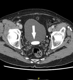
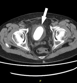
to pelvic imaging, and higher socioeconomic status.5 The primary management strategy for GBS is open cystolithotomy.6
The authors report the case of an 80-yearold male with a GBS who declined open cystolithotomy. This patient provided informed consent for his anonymised information to be published in a case report.
An 80-year-old healthy White man originally presented with lower abdominal pain and a 3.2 cm stone on abdominal/pelvic CT (Figure 1A). He was not interested in follow-up investigations and he declined treatment for his stone at the time of follow-up with another urologist. He was counselled at that time about the long-term risks of declining further investigations or treatment and did not book any follow-up care.
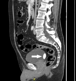
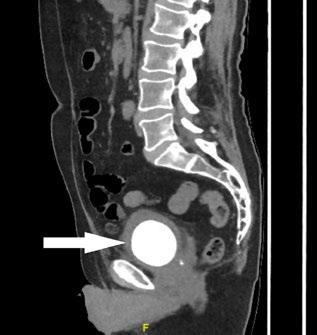
Three years later, the patient presented to the emergency department with a 24-hour history of suprapubic pain and voiding in small quantities. He complained of mild urgency and frequency, with no other infectious symptoms. He denied gross haematuria.
A new abdominal/pelvic CT scan showed a 6 cm bladder calculus, and mild left kidney pelviectasis (Figure 1B). A 3D CT reconstruction is shown in Figure 2
Creatinine was 138 µmol/L with a baseline
of 120 µmol/L. White blood cell count was 10.9x10⁹ /L and haemoglobin was 157 g/L. Urine culture had insignificant growth. Catheter placement with cystoscopy and guidewire failed as the balloon burst shortly after placement. On cystoscopy, the patient had evidence of a moderately obstructing prostate. There were no urethral abnormalities. The urothelium could not be visualised, as the stone was occupying the entire bladder space.
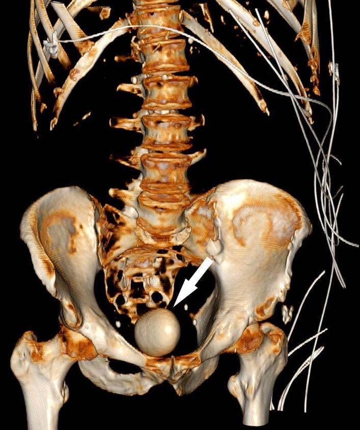
During outpatient follow-up, cystolithotomy was discussed with the patient and his family member. Risks of the procedure and estimated risks of not proceeding with treatment were reviewed, including the risk of renal dysfunction, infection or sepsis, pain, and possible bladder rupture. The patient, along with his family member, declined the treatment. He was adamant that he did not want further interventions.
Factors that may influence bladder stone formation include BPH, neurogenic bladder, anatomical defects, foreign bodies, urethral strictures, upper tract stones, and recurrent or chronic UTIs.2 Specifically, intravesical prostatic protrusion and reduced urinary flow are independent risk factors in the context of BPH.7 Chronic UTIs are a major contributor, as infected stones may grow rapidly in the presence of urease-producing bacteria.8 In the patient, the most likely aetiology is voiding dysfunction from an enlarged prostate. The patient presented with lower urinary tract symptoms both times; however, he was able to void and had no gross haematuria. He had no reported history of UTIs. While there was potentially a slight decrease in renal function, it is unlikely to be clinically significant given his age.
GBSs are exceedingly rare and mostly seen in tropical or developing countries,9 such as Türkiye, Iran, India, and China; however, even these countries have seen improved rates over the past several years.10 Gadelkareem et al.6 conducted a study on predictors of clinical and surgical characteristics in GBSs in the “stone forming belt” of Africa and Asia. Risk factors for giant stone development included male gender and older age. However, dietary factors may contribute towards GBS formation, even in developed countries. Insufficient fluid intake, high oxalate consumption, low calcium consumption, consumption of mostly carbon 3 plants (e.g., rice, wheat), and high intake of sugarsweetened beverages have been correlated to the formation of bladder stones. Dietary evaluation is an important component in
the assessment of patients with recurrent stone formation, as poor habits and a lack of resources or education can cause stone formation regardless of geography.11
In this case, no intervention was performed at the patient’s request. The literature suggests that an untreated bladder stone may lead to urinary tract damage, recurrent stone formation, bladder damage, urinary retention, infection, and sepsis.2 Further complications for GBS, specifically, have been reported in the literature. Syarif et al.12 reported a case of giant bladder diverticulum associated with a large bladder stone, requiring transurethral bladder neck incision and diverticulectomy with stone extraction. Patients have also been reported to present with renal failure and hydronephrosis in the context of GBS.3,13,14 Furthermore, additional bladder stones may be precipitated by chronic obstruction and mucosal injury associated with such large stones.14 Based on a series of CT images, the progression rate of the patient’s stone was approximately 18 cm3/year (41% per year). The progression rate of bladder stones is not well defined in the literature, and it is not clear if it would be linear. Most case reports outline the time period for which a patient had experienced symptoms, which may vary widely.15,16 Regarding actual growth rate, a recent systematic review and meta-analysis investigated stone growth in asymptomatic renal stones, and calculated a 25% stone growth over an average duration of 43.41 months.17 To the best of the authors’ knowledge, there are no similar articles investigating stone growth rates for bladder stones specifically. The authors found one other report of GBS containing serial imaging, with an initial ultrasound followed by CT imaging later. The rate of stone growth in this case was from 76.0 mm longitudinal and 73.6 mm axial, on ultrasound, to 57.26 mm by 78.25 mm coronal and 79.29 mm by 54.93 mm sagittal, on CT, over the course of 6 months.18
The main options for bladder stone management include spontaneous passage, transurethral cystolithotripsy (TUCL), percutaneous cystolithotomy, and open cystolithotomy. Asymptomatic stones of <1 cm size may be treated conservatively;
however, bladder stones are usually symptomatic and require active treatments. The most recent European Association of Urology (EAU) guidelines on bladder stone treatment state TUCL as the intervention of choice for adults and children, where feasible, with a stone-free rate (SFR) of 95–100%. A systematic review and metaanalysis showed no significant difference in SFR between TUCL and percutaneous cystolithotomy. Options for TUCL include a standard rigid cystoscope or a nephroscope, where a nephroscope allows use of large forceps to remove fragments through a 24 Fr sheath, and shorter operative times, where the mean difference is 22.74 minutes (favouring the nephroscope). The main limitation of urethral access is prolonged operative time for very large stones. Percutaneous management is particularly advantageous in cases of very large stones >5 cm or altered urethral anatomy, with an SFR of 90–100%. Tan et al.19 performed a minimally invasive percutaneous procedure using a laparoscopic entrapment bag. In a study of 25 patients with large bladder stones, all patients were stone free and clinically well after this technique. Open cystolithotomy has the highest SFR with 100%, but has a higher risk of complications, including vesicocutaneous fistula formation.20 When endoscopic approaches have higher chances of low SFR, such as in the case of GBS formation, open cystolithotomy may be
References
1. Takasaki E et al. Chemical compositions of 300 lower urinary tract calculi and associated disorders in the urinary tract. Urol Int. 1995;54(2):89-94.
2. Leslie WS et al. Bladder Stones [Internet] (2025) Treasure Island: StatPearls Publishing. Available at: https://www.ncbi.nlm.nih.gov/books/ NBK441944/. Last accessed: 16 February 2026.
3. Molla YD et al. Giant bladder stone a rare cause of renal failure, a case report. Int J Surg Case Rep. 2023;105:108085.
4. Vidhyarthy AK et al. Giant bladder calculus in an adult- a persistent problem in the developing world: a case report. Clin Pract Cases Emerg Med. 2020;4(4):544-7.
favoured.6,21 Unique to GBS, cystolithotomy may be more difficult in cases because the bladder stone can adhere to the bladder mucosa. In the study by Gadelkareem et al.,6 it was found that symptom severity, roughness of stone surface, occupation as a farmer, and stone size were correlated with stone adherence to mucosa. The management of bladder outlet obstruction, such as via transurethral resection of the prostate or of the bladder neck, is recommended in patients with multiple or large bladder calculi.22 Pharmacological treatment for bladder stones is limited. Chemical dissolution agents have no established role in the management of GBS given their size, composition, and inability to achieve sustained bladder therapeutic concentrations.23,24 These conservative treatments have had a limited role in the literature for GBS to date.
GBSs are a rare entity, particularly in developed countries. The mainstay treatment is open cystolithotomy, though percutaneous surgical approaches have been reported. In this case, 3 years of nontreatment of a GBS resulted in no serious adverse complications, and an estimated growth rate of 18 cm3/year (41% per year).
5. Mosa MM et al. Extraction of a giant bladder stone (560 g) in a young female: a case report. Int J Surg Case Rep. 2024;119:109653.
6. Gadelkareem RA et al. Predictors of clinical and surgical characteristics of giant stones of the urinary bladder: a retrospective study. BMC Urol. 2023;23(1):83.
7. Kim JW et al. Intravesical prostatic protrusion is a risk factor for bladder stone in patients with benign prostatic hyperplasia. Urology. 2014;84(5):1026-9.
8. Huang W et al. Risk factors for bladder calculi in patients with benign prostatic hyperplasia. Medicine (Baltimore). 2017;96(32):e7728.
9. Vlahopoulos L et al. Management of a giant bladder stone in a 14-years-old female with SuperPulsed thulium fiber laser. Urol Case Rep. 2025;61:103096.
10. Napitupulu T et al. Giant bladder stone: a case report and literature review. JMA J. 2022;5(3):384-8.
11. Yitgin Y et al. Role, importance and assessment of dietary habits in urolithiasis patient. World J Urol. 2023;41(5):1229-33.
12. Syarif S et al. Giant stone in a urinary bladder diverticulum in a 69-year-old male: a case report. Pan Afr Med J. 2023;45:181.
13. Lai AYH, Kuo YC. A huge pelvic calculus causing acute renal failure. Am J Emerg Med. 2008;26(2):246.e3-4.
14. Bestari MG et al. Giant bladder stone resulting in renal failure and concurrent bladder cancer: a case report. Int J Surg Case Rep. 2022;94:107170.
15. Wei X et al. Giant bladder stone: a case report. Exp Ther Med. 2022;24(2):535.
16. Ma C et al. Giant bladder stone in a male patient: a case report. Medicine (Baltimore). 2016;95(30):e4323.
17. Nooshabadi MP et al. Evaluating the natural history of incidentally recognized clinically silent nephrolithiasis in healthy adults: an updated systematic review with metaanalysis. Urolithiasis. 2025;53(1):31.
18. Miravent S et al. Pre-hospital identification of a giant bladder calculus through screening sonography: a case report. Curr Med Imaging. 2025;21:e15734056324600.
19. Tan YK et al. Minimally invasive percutaneous management of large bladder stones with a laparoscopic entrapment bag. J Endourol. 2014;28(1):61-4.
20. Donaldson JF et al. Treatment of bladder stones in adults and children: a systematic review and meta-analysis on behalf of the European Association of Urology Urolithiasis Guideline Panel. Eur Urol. 2019;76(3):352-67.
21. Franco-González CD et al. A case report and literature review of a giant bladder stone in a 43-year-old female (9 x 8 x 4 cm, 250g). Radiol Case Rep. 2024;19(11):5539-43.
22. Richter S et al. Combined cystolithotomy and transurethral resection of prostate: best management of infravesical obstruction and massive or multiple bladder stones. Urology. 2002;59(5):688-91.
23. Skolarikos A et al. European Association of Urology guidelines on the diagnosis and treatment of urolithiasis. Euro Urol. 2025;88(1):64-75.
24. Chakra MA et al. Established and recent developments in the pharmacological management of urolithiasis: an overview of the current treatment armamentarium. Expert Opin Pharmacother. 2020;21(1):85-96.
If you like it,why not share it...

Share with a colleague today