Beyond Weight Loss: The Musculoskeletal Effects of GLP-1 Therapies Congress Feature:
Interviews:
Noriko Yoshimura and Elaine Dennison discuss the future of osteoarthritis research, population ageing, and the global burden of musculoskeletal disease
05 Welcome
Congress Review
06 Review of the World Congress on Osteoporosis, Osteoarthritis and Musculoskeletal Diseases (WCO-IOF-ESCEO) 2026, 16th–19th April
Congress Feature
21 Beyond Weight Loss: The Musculoskeletal Effects of GLP-1 Therapies
Aleksandra Zurowska
26 Hyaluronic Acid Injections in Knee Osteoarthritis: What Do the Updated Recommendations Say?
Niamh Holmes
Abstract Reviews
31 Improving Fracture Prevention Through Advanced Diagnostic Tools
Kirilov N et al.
34 Development of Novel Muscle-Active Agents: A Regulatory Perspective
Ewa Bałkowiec-Iskra
36 GLP-1 Receptor Agonists and Joint Health
Ali Mobasheri
38 Pathologic Fracture Due to Osteosarcoma in an Asymptomatic Patient with Paget's Disease of Bone: A Case Report
Dhamo B et al.
42 Evidence-Based Strategies for Improving Bone Health in Older Adults Through Orthogeriatric Care and Risk Communication
Kamberi F et al.
45 Effect of Preoperative Bisphosphonate Therapy on Bone Mineral Density in Patients with Primary Hyperparathyroidism 1 Year After Parathyroidectomy
Pershina-Miliutina AP et al.
Congress Interviews
58 Elaine Dennison
60 Noriko Yoshimura
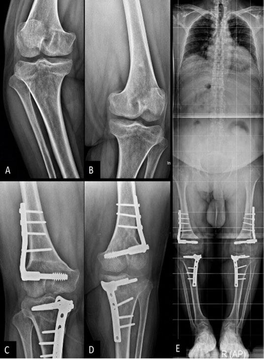
48 Simultaneous Double Femoral and Tibial Osteotomy in the Treatment of Severe Genu Varum in Young Adults
Fekih A et al.
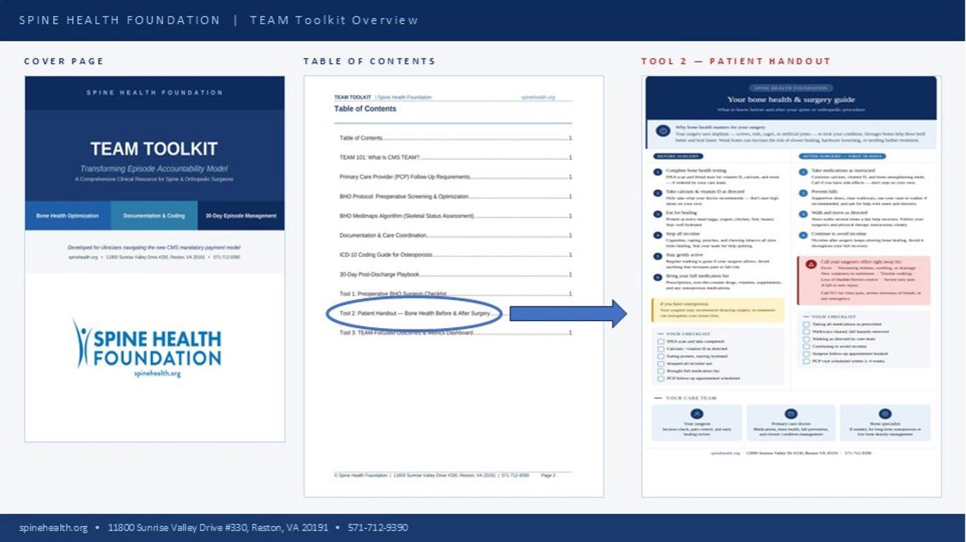
50 Transforming Episode Accountability Model (TEAM): Impact on Orthopaedic, Spine, and Bone Health Clinical Practice and Opportunities to Drive Osteoporosis Diagnosis and Treatment
Roy RT et al.
53 Meeting Global Challenges for Osteoporosis Societies: Osteoporosis Canada’s Experience Supporting Spread of Fracture Liaison Services Across Canada
Jiwa and Singh
55 Opportunistic Diagnosis of Vertebral Fractures Using Routine CT Scans: The Portuguese Experience
Barbosa and Rocha
Aims and Scope
EMJ Rheumatology is an open access, peer-reviewed eJournal committed to publishing the highest quality medical research concerning all aspects of diseases of the blood and bone marrow to help advance the development of this field.
The journal is six weeks after the World Congress on Osteoporosis, Osteoarthritis and Musculoskeletal Diseases (WCO-IOF-ESCEO), and features highlights from this congress, alongside abstract reviews and interviews with experts in the field.
EMJ Rheumatology also publishes peer-reviewed research papers, review articles, and case reports in the field. In addition, the journal welcomes the submission of features and opinion pieces intended to create a discussion around key topics in the field and broaden readers’ professional interests. The journal is managed by a dedicated editorial team that adheres to a rigorous double-blind peer-review process, maintains high standards of copy editing, and ensures timely publication.
EMJ Rheumatology endeavours to increase knowledge, stimulate discussion, and contribute to a better understanding of blood disorders. Our focus is on research that is relevant to healthcare professionals in this field. We do not publish veterinary science papers or laboratory studies not linked to patient outcomes. We have a particular interest in topical studies that advance research and inform of coming trends affecting clinical practice in haematology.
Further details on coverage can be found here: www.emjreviews.com
Editorial Expertise
EMJ is supported by various levels of expertise:
• Guidance from an Editorial Board consisting of leading authorities from a wide variety of disciplines.
• Invited contributors who are recognised authorities in their respective fields.
• Peer review, which is conducted by expert reviewers who are invited by the Editorial team and appointed based on their knowledge of a specific topic.
• An experienced team of editors and technical editors.
Peer Review
On submission, all articles are assessed by the editorial team to determine their suitability for the journal and appropriateness for peer review.
Editorial staff, following consultation with a member of the Editorial Board if necessary, identify three appropriate reviewers, who are selected based on their specialist knowledge in the relevant area.
All peer review is double blind. Following review, papers are either accepted without modification, returned to the author(s) to incorporate required changes, or rejected.
Editorial staff have final discretion over any proposed amendments.
Submissions
We welcome contributions from professionals, consultants, academics, and industry leaders on relevant and topical subjects. We seek papers with the most current, interesting, and relevant information in each therapeutic area and accept original research, review articles, case reports, and features.
We are always keen to hear from healthcare professionals wishing to discuss potential submissions, please email: editorial.assistant@emjreviews.com
To submit a paper, use our online submission site: https://emj.kriyadocs.com/welcome
Submission details can be found through our website: www.emjreviews.com/contributors/authors
Reprints
All articles included in EMJ are available as reprints (minimum order 1,000). Please contact hello@emjreviews.com if you would like to order reprints.
Distribution and Readership
EMJ is distributed through controlled circulation to healthcare professionals in the relevant fields across Europe.
Indexing and Availability
EMJ is indexed on DOAJ, the Royal Society of Medicine, and Google Scholar®
EMJ is available through the websites of our leading partners and collaborating societies. EMJ journals are all available via our website: www.emjreviews.com
Open Access
This is an open-access journal in accordance with the Creative Commons Attribution-Non Commercial 4.0 (CC BY-NC 4.0) license.
Congress Notice
Staff members attend medical congresses as reporters when required.
This Publication
Publication Date: June 2026
Online ISSN: 2056-6395
All information obtained by EMJ and each of the contributions from various sources is as current and accurate as possible. However, due to human or mechanical errors, EMJ and the contributors cannot guarantee the accuracy, adequacy, or completeness of any information, and cannot be held responsible for any errors or omissions. EMJ is completely independent of the review event (WCO-IOF-ESCEO) and the use of the organisations does not constitute endorsement or media partnership in any form whatsoever.The cover photo is of Prague, Czechia, the location of WCO-IOF-ESCEO 2026.
1. Sandoz. Our biosimilars. Available at: https://www.sandoz.com/business/biosimilars/our-biosimilars/#marketed. Accessed May 2026. 2. WCO IOF-ESCEO Sandoz breakfast symposium. Available at https://www.wco-iof-esceo.org/congress-news/sandoz_breakfast_symposium/. Accessed May 2026. (link is valid until Aug 11, 2026).
WCO IOF-ESCEO Abstract book. Availbale at https://www.wco-iof-esceo.org/download/2026/abstract-book (poster P1505, page 881-882). Accessed May 2026. #Jubbonti® is launched in the US and have begun launching across multiple European markets since December 2025, with additional rollouts planned in 2026. Jubbonti® Marketing Authorization Holder (MAH) address: Sandoz GmbH, Biochemiestr. 10, 6250 Kundl, Austria. Jubbonti® is approved for women with post-menopusal osteoporosis. For complete list of indications, kindly refer to product SmPC. Available at https://www.ema.europa.eu/en/documents/product-information/jubbonti-epar-product-information_en.pdf. Accessed May 2026.
report side effect to a Sandoz medicinal product electronically Contact Sandoz – Get in Touch with Our Team | Sandoz Explore the first real-world safety data from biosimilar denosumab usage3 Click to access Poster P1505 (page 881-882) WCO Satellite Symposium: Biosimilars in Bone Health2
Editorial Director
Andrea Charles
Editor
Sean Boyle
Managing Editor
Darcy Richards
Senior Copy Editor
Noémie Fouarge
Copy Editors
Meghan Garcka, Sarah Jahncke
Editorial Leads
Helena Bradbury, Katrina Thornber, Aleksandra Zurowska
Editorial Co-ordinators
Jess Nicholson, Bertie Pearcey, Alena Sofieva
Editorial Assistants
Niamh Holmes, Josh Lister, Roli Omamuli, Alexander Perkins
Reporter
Anna Caldwell
Head of Marketing
Stephanie Corbett
Creative Director
Tim Uden
Design Manager
Stacey White
Senior Designers
Owen Silcox, Tamara Kondolomo
Designers
Shanjok Gurung, Fabio van Paris
Junior Designers
Fraser Hoey, Helena Spicer, Caleb Wylie
Business Unit Lead
Kelly Byrne
Chief Executive Officer
Justin Levett
Chief Commercial Officer
Dan Healy
Founder and Chairman
Spencer Gore
Welcome
Dear Readers,
Welcome to this EMJ Rheumatology supplement, featuring our coverage of the World Congress on Osteoporosis, Osteoarthritis and Musculoskeletal Diseases (WCO-IOFESCEO) held in Prague, Czechia, from 16th–19th April 2026. This Congress remains the foremost international forum for advancing science and clinical practice across osteoarthritis and related musculoskeletal conditions, and this year’s programme reflects both the growing momentum in the field and the urgency of addressing a disease that affects hundreds of millions of people worldwide.
The papers gathered here represent a carefully selected body of research presented at this year’s meeting, spanning disease mechanisms, biomarkers, and both pharmacological and non-pharmacological interventions. Together, they offer a timely and authoritative snapshot of where the field stands and where it is headed. We hope this supplement proves a valuable resource for delegates and the wider clinical community alike, and we extend our sincere thanks to all authors and reviewers for their contributions.
Aleksandra Zurowska
Editorial Lead
Contact us
Editorial enquiries: editor@emjreviews.com
Sales opportunities: salesadmin@emjreviews.com
Permissions and copyright: accountsreceivable@emjreviews.com
Reprints: info@emjreviews.com
Media enquiries: marketing@emjreviews.com
WCO-IOF-ESCEO 2026 represents more than a continuation of tradition; it is a forward-looking forum addressing the modern needs of the field
Congress Review
Review of the World Congress on Osteoporosis, Osteoarthritis and Musculoskeletal Diseases (WCO-IOF-ESCEO) 2026
THIS MONTH, Prague, Czechia, welcomed the 26th edition of the World Congress on Osteoporosis, Osteoarthritis and Musculoskeletal Diseases (WCO-IOF-ESCEO). Held from 16th–19th April 2026 at the O2 Universum, this landmark event brought together a global community of clinicians, researchers, and healthcare leaders dedicated to advancing bone, joint, and muscle health. As the most influential meeting in the field, the Congress serves as the premier platform for exchanging cutting-edge scientific knowledge and emerging therapeutic strategies.
Organised under the auspices of the International Osteoporosis Foundation (IOF) and the European Society for Clinical and Economic Aspects of Osteoporosis, Osteoarthritis and Musculoskeletal Diseases (ESCEO), the 2026 Congress arrives at a pivotal moment. With high levels of international participation, the meeting showcases a comprehensive scientific programme spanning basic science, translational research, and realworld clinical practice.
The opening ceremony sets a prestigious tone, recognising scientific excellence while addressing broader global challenges. High-level sessions explore WHO’s bone health agenda and the evolving role of academic publishing, highlighting the interdisciplinary nature of the field. This integration underscores the
urgent need to align scientific discovery with global health policy and patient education.
A defining strength of WCO-IOFESCEO remains its breadth. The 2026 programme covers the full spectrum of musculoskeletal disorders, from osteoporosis and osteoarthritis to frailty and sarcopenia, with a renewed emphasis on prevention, early diagnosis, and personalised treatment. Through plenary sessions and specialised symposia, the Congress facilitates both high-level knowledge exchange and practical clinical engagement.
International collaboration remains the cornerstone of the meeting. By bridging diverse healthcare systems and research environments, WCO-IOF-ESCEO fosters
The Congress reflects the ongoing transformation of medicine through advances in genetics and therapeutic innovation
a nuanced understanding of shared challenges like ageing populations and increasing disease burdens. Beyond its scientific contributions, the Congress reflects the ongoing transformation of medicine through advances in genetics and therapeutic innovation, ultimately aiming to bridge the translational gap to provide accessible, patient-centred care.
WCO-IOF-ESCEO 2026 represents more than a continuation of tradition; it is a forward-looking forum addressing the modern needs of the field. By reaffirming its role as the key meeting point for musculoskeletal health, the Prague Congress continues to shape the future of bone and joint care worldwide.
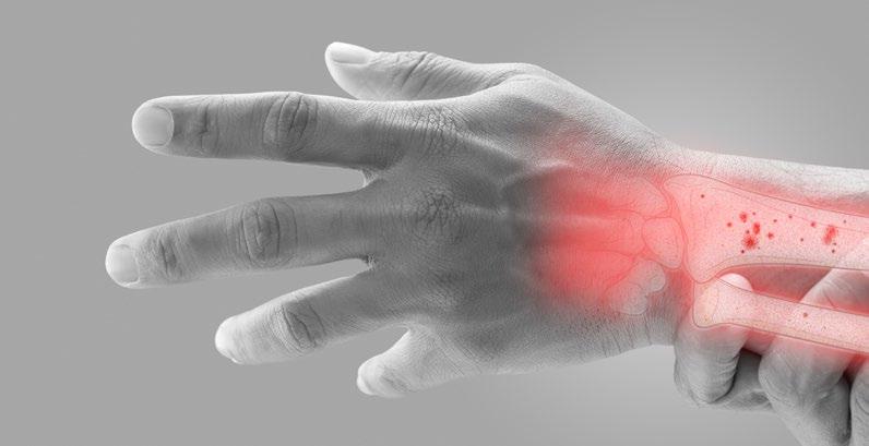
Early FLS Intervention Cuts Secondary Fracture Risk After Wrist Fracture
RESEARCH presented at WCOIOF-ESCEO 2026 has highlighted the potential of early Fracture Liaison Service (FLS) intervention following distal radius fracture (DRF) to reduce the risk of subsequent fragility fractures, including hip fractures.1
DRF is often the first clinical sign of underlying osteoporosis and is associated with a substantially increased risk of future fractures. Previous evidence suggests that the risk of hip fracture may rise by up to 3.5-fold after a DRF, particularly within the first 2 years following injury. Despite existing guidance from the National Institute for Health and Care Excellence (NICE) and the Royal Osteoporosis Society, many patients still fail to receive timely bone health assessment and preventative treatment.
In this retrospective study, researchers evaluated outcomes in adults aged 50 years and older presenting with an index DRF at Southend University Hospital, UK, between January 2018–December 2024. A total of 612 eligible patients were included, with a mean age of 72 years and women accounting for 78% of the cohort.
Patients were divided into two groups based on FLS involvement. The FLS group completed a structured pathway involving assessment within 90 days, dual-energy X-ray absorptiometry (DXA) scanning, initiation of anti-osteoporosis therapy, and falls assessment.
The non-FLS group either did not receive referral or did not complete the pathway.
Over a 24-month follow-up period, secondary fragility fractures occurred in 15.6% of patients in the non-FLS group compared with 8.9% of those receiving full FLS management. Hip fractures specifically were reported in 5.1% of non-FLS patients versus 2.4% in the FLS group.
Further statistical analysis demonstrated that FLS-led management was associated with a 34% lower hazard of secondary fracture after adjustment for factors including age, sex, comorbidities, and prior fractures (adjusted hazard ratio: 0.66; 95% CI: 0.48–0.91; p=0.01).
The authors concluded that DRF represents a critical opportunity for early secondary prevention. They emphasised that comprehensive FLS pathways may significantly reduce the burden of future fractures and support improved long-term bone health outcomes.
Hip fractures specifically were reported in 5.1% of non-FLS patients versus 2.4% in the FLS group
Late DXA Referral May Limit Osteoporosis Benefit in Men
MEN undergoing first dual-energy X-ray absorptiometry (DXA) assessment for osteoporosis had substantially poorer survival than women, despite presenting with less severe skeletal disease, according to data presented at WCO-IOF-ESCEO 2026.2 Investigators reported that only 73% of men survived 5 years after their first DXA scan, compared with 87% of women, raising concerns that men are being referred too late to benefit fully from osteoporosis treatment.
Osteoporosis in men remains underrecognised and underdiagnosed, despite increasing evidence of significant fracturerelated morbidity and mortality. To examine whether referral patterns may delay intervention opportunities in men, researchers analysed first routine-care DXA scans recorded in the Danish NOCTE cohort between 2010–2022.
The study identified 72,818 men and 172,622 women undergoing a first DXA scan and matched them with controls from the background population by age, sex, and region. Investigators assessed mortality rates, standardised mortality ratios, and 5- and 10-year survival outcomes following DXA referral.
Mean age at index scan was approximately 72 years for men and 70 years for women. Women more frequently had osteoporosisrange T-scores below –2.5 (33.4% versus 16.5%) and slightly higher rates of major osteoporotic fracture in the year before assessment.
Despite this, men experienced markedly worse outcomes. Five-year survival following DXA was 73% in men compared with 87% in women, while 10-year survival was 52% versus 71%, respectively. Mortality
rates were also substantially elevated in men relative to matched controls, with a standardised mortality ratio increase of 56%, compared with 17% in women.
Osteoporosis in men remains underrecognised and underdiagnosed, despite increasing evidence of significant fracture-related morbidity and mortality
The findings suggest that men may only be referred for osteoporosis evaluation after substantial frailty, comorbidity burden, or advanced disease progression has already developed. In contrast, women appear more likely to undergo assessment earlier in the disease course, despite presenting with lower overall mortality risk.
The authors concluded that current osteoporosis referral pathways continue to inadequately capture at-risk men and may limit the opportunity for timely intervention during the period when antiosteoporotic therapy is most likely to provide meaningful fracture prevention benefit.
Bisphosphonates Associated with Lower Mortality After Fracture
PEOPLE with multiple long-term health conditions may have better survival outcomes after fractures when treated with bisphosphonates rather than denosumab, according to a large real-world Australian study presented at WCO-IOF-ESCEO 2026.3
Multimorbidity is commonly defined as having two or more chronic health conditions. It is increasingly common in older adults; however, these patients are often underrepresented in RCTs.
Researchers analysed health data from 117,031 adults aged ≥50 years in New South Wales, Australia, who experienced fractures requiring hospital or emergency care between 2010–2018. All participants were treatment-naïve for osteoporosis therapies before their fracture.
The study compared two commonly used antiresorptive treatments: bisphosphonates (alendronate, risedronate, and zoledronic acid) and denosumab. Of the eligible participants, 6,036 initiated bisphosphonates, while 2,611 initiated denosumab within 12 months of fracture. Investigators used a target trial emulation approach designed to mimic the conditions of a randomised trial using real-world data.
The mean age of eligible participants was 75.7 years. Women receiving bisphosphonates had a 29% lower risk of all-cause mortality compared with denosumab users, whereas men showed a 21% lower mortality risk during the followup of up to 4 years.
The mortality benefit was seen consistently across different multimorbidity clusters, including cardiovascular, cardiometabolic,
geriatric, and mental health groups. Researchers found no meaningful difference in refracture risk between the two treatment approaches.
The mortality benefit was seen consistently across different multimorbidity clusters
The analysis excluded people with previous osteoporosis treatment, prior fractures, metastatic cancer, or other metabolic bone disorders in an effort to reduce confounding factors.
However, as an observational study using administrative health data, the findings cannot prove that bisphosphonates directly caused lower mortality. The researchers noted that further investigation is needed to determine whether these therapies may provide additional non-skeletal benefits beyond fracture prevention.
The study suggests that bisphosphonates may remain an important treatment option for older adults with complex health needs following fractures, while future research could help clarify the biological mechanisms underlying the observed survival differences.
Maternal Diabetes Linked to Childhood Fracture Risk
MATERNAL diabetes during pregnancy was associated with differing risks of long-bone fractures in offspring during early childhood, according to a large Australian population-based study presented at WCO-IOF-ESCEO 2026. Researchers found that children born to mothers with Type 1 diabetes had a higher fracture risk, while gestational diabetes was linked to a lower risk of fracture during the first 9 years of life.4
The study explored whether maternal diabetes exposure in utero may influence skeletal development and subsequent fracture susceptibility in children. Diabetes during pregnancy has previously been associated with altered fetal growth and bone development, but evidence regarding long-term fracture outcomes in offspring remains limited.
Researchers conducted a state-wide data linkage study using administrative health records from Victoria, Australia, including 279,067 singleton mother–offspring pairs between 2012–2015. Maternal diabetes status was identified using ICD-10-AM codes from the Victorian Perinatal Data Collection and categorised as gestational diabetes (GD), Type 1 diabetes (T1D), or Type 2 diabetes (T2D). Offspring longbone fractures presenting to emergency departments between 2013–2021 were analysed using Cox proportional hazards regression models adjusted for offspring sex, socioeconomic disadvantage, and maternal smoking during pregnancy.
Among the cohort, 22,153 mothers (8.0%) had GD, 998 (0.4%) had T1D, and 885 (0.3%) had T2D. Overall, 15,275 offspring (5.7%) experienced at least one incident long-bone fracture during follow-up. Compared with offspring of mothers without diabetes, children exposed to GD had a lower fracture hazard (hazard ratio [HR]: 0.90; 95% CI: 0.84–0.96). In contrast, offspring exposed to maternal T1D had a significantly higher fracture hazard (HR: 1.28; 95% CI: 1.00–1.64). No significant association was identified for T2D exposure (HR: 0.92; 95% CI: 0.68–1.25).
The findings highlight the potential importance of early monitoring and targeted injury-prevention strategies for children exposed to maternal T1D during pregnancy
The authors suggested that the lower fracture risk observed with gestational diabetes may reflect behavioural differences rather than direct skeletal effects. Meanwhile, the increased fracture risk associated with maternal T1D may indicate altered fetal bone development or other metabolic influences requiring further investigation.
The findings highlight the potential importance of early monitoring and targeted injury-prevention strategies for children exposed to maternal T1D during pregnancy. However, the observational design means causality cannot be confirmed, and additional research is needed to clarify the biological mechanisms underlying these associations.
European Cohort Reveals Changing Patterns in Renal Osteodystrophy
A LARGE European bone biopsy analysis, presented at WCO-IOF-ESCEO 2026, suggests renal osteodystrophy may present differently than previously believed, with normal and high bone turnover patterns more common than low turnover disease across chronic kidney disease (CKD) populations.5
Median age was 57 years, 32% were female, and 19% had diabetes
Renal osteodystrophy remains a major complication of CKD, yet understanding of its epidemiology has become limited because bone biopsy use has declined in both research and clinical practice. Earlier studies suggested low turnover bone disease had become dominant in CKD. Researchers therefore reassessed contemporary bone pathology patterns using a multinational biopsy database.
This multicentre observational analysis included bone biopsies collected between 1998–2025 from five countries. Patients represented the spectrum of kidney dysfunction, including predialysis CKD Stage 4–5, dialysis-dependent disease, and kidney transplant recipients. Investigators analysed turnover, mineralisation, and bone volume using thermostatic mixing valve classification criteria. Additional diagnostic reclassification applied Malluche and Recker cut-offs.
Of 851 biopsies identified, 155 were excluded because of duplicate records, incomplete registration, or procedures performed for clinical indications. The final cohort included 696 research-indication biopsies, comprising 135 patients predialysis, 335 patients on dialysis, and 225 transplant recipients.
Median age was 57 years, 32% were female, and 19% had diabetes.
According to original thermostatic mixing valve reports, normal bone turnover was the most common presentation across all CKD groups. However, among tetracyclinelabelled biopsies (n=341), additional definitions produced substantially different classifications. Using Malluche criteria, most patients who were pre-dialysis or on dialysis were classified as having high bone turnover. Recker criteria classified all patients as either high or normal turnover, with no low turnover category predominating. Bone mineralisation defects were also reported in a notable proportion of both patients who were pre-dialysis and transplant recipients. Anti-osteoporosis medication use remained uncommon, reported in 0% of patients pre-dialysis, 2% of patients on dialysis, and 6% of transplant recipients.
The findings challenge earlier assumptions that low turnover bone disease predominates in CKD. Researchers highlighted substantial variability depending on classification thresholds, emphasising the need for harmonised diagnostic criteria in renal osteodystrophy research and practice. Further investigation into abnormal mineralisation patterns and their clinical implications is also warranted.
Bone Strain Index Improved 10-Year Fracture Prediction
A NEW prospective study presented at WCO-IOF-ESCEO 2026 showed that the Bone Strain Index (BSI), an advanced dual-energy X-ray absorptiometry (DXA)-derived measure of bone mechanical strain, independently predicted fragility fractures in older females over 5-year and 10-year follow-up periods, beyond conventional bone mineral density (BMD) and FRAX® (WHO, Geneva, Switzerland) assessment.6
Fragility fractures remain a major cause of morbidity in ageing populations, particularly among postmenopausal females. Although areal BMD and FRAX® are widely used to estimate fracture risk, these tools do not fully capture bone strength and biomechanical integrity. The BSI was developed to address this gap by applying finite element analysis to DXA imaging, quantifying internal mechanical strain within bone tissue.
Researchers conducted a large prospective cohort study involving 3,338 communitydwelling older females aged 64–77 years. Baseline DXA scans were used to calculate BSI at the lumbar spine, total hip, and femoral neck. Incident fragility fractures and major osteoporotic
during both 5-year and 10-year followup periods. Statistical analyses included receiver operating characteristic curves, Youden index-derived cut-offs, and Cox proportional-hazards models adjusted for age, areal BMD, and FRAX® score.
During the 10-year follow-up, 803 females (24.1%) sustained at least one fragility fracture, including 525 major osteoporotic fractures. Higher BSI values were significantly associated with increased fracture risk at both 5 and 10 years, independently of age, BMD, and FRAX®. Total hip BSI demonstrated the strongest predictive performance, achieving an area under the curve of 0.63 at both timepoints, outperforming lumbar spine BSI (area under the curve: 0.58).
Each 1-SD increase in total hip BSI was associated with significantly higher fracture risk at 5 years (hazard ratio [HR]: 1.42; 95% CI: 1.32–1.53) and 10 years (HR: 1.41; 95% CI: 1.34–1.49). Importantly, these associations remained statistically significant after adjustment for FRAX® score (HR: 1.24; 95% CI: 1.14–1.35).
The findings suggest that BSI could enhance long-term fracture risk stratification in older females and provide clinically meaningful information beyond standard osteoporosis assessment tools. Researchers noted that total hip BSI appeared particularly valuable for identifying females at elevated fracture risk who may otherwise be underestimated using BMD or FRAX® alone. Further research is needed to determine how BSI-guided assessment may influence osteoporosis prevention strategies and clinical outcomes across broader patient populations.
AI-Based Bone Fragility Index Predicts Future Fracture Risk
A NEW study presented at WCO-IOF-ESCEO 2026 demonstrated that an AI-based bone fragility index (BFI) derived from routine dualenergy X-ray absorptiometry (DXA) imaging predicted incident major osteoporotic fractures (MOF) and identified patients at elevated fracture risk years before clinical events occurred.7
Researchers evaluated the performance of the TBS Reveal (Medimaps Group, Geneva, Switzerland) BFI, an AI-driven assessment tool designed to improve fracture prediction beyond conventional bone mineral density measurements. While bone mineral density forms the basis of most osteoporosis assessments, it does not fully account for bone microarchitecture, which contributes significantly to skeletal fragility and fracture susceptibility.
The retrospective study included 338 female participants aged 68.3±9.4 years who underwent baseline X-ray imaging with prospective follow-up for fracture outcomes. Investigators applied Bayesian Weibull survival models to evaluate the relationship between BFI category and future MOF incidence over time. Participants were stratified into high and very high BFI groups based on baseline imaging findings.
During a median follow-up period of 7.7 years (95% CI: 6.8–8.5), 157 participants experienced an incident MOF. The AIbased BFI showed a strong association with fracture risk, particularly during the first
5 years after baseline imaging, compared with all other participant groups. Females categorised as high BFI demonstrated a 10.3% higher 5-year fracture probability (95% CI: 1.2–19.5), increasing to 12.9% at 10 years (95% CI: 1.5–23.6). The very high BFI group showed an even more pronounced effect, with a 19.2% greater fracture probability at 5 years (95% CI: 6.3–31.8) and a 21.0% higher probability at 10 years (95% CI: 7.9–31.2).
Survival analyses revealed clearly separated fracture-free survival curves between risk categories, supporting the ability of the AI-based BFI to distinguish short- and longterm fracture risk trajectories from a single baseline X-ray assessment.
The findings suggest that AI-enhanced skeletal analysis could strengthen osteoporosis risk stratification and improve early identification of females at high fracture risk who may benefit from targeted prevention strategies or closer monitoring.
The study was observational in design, and further prospective validation is needed to determine how the AI-based BFI performs across broader populations and clinical settings. Additional research is also required to establish how integration of AI-derived fracture prediction tools may influence long-term clinical outcomes and osteoporosis management pathways.
The AI-based BFI showed a strong association with fracture risk, particularly during the first 5 years after baseline imaging, compared with all other participant groups
REMS Technology Detects Bone Changes During Pregnancy
RESEARCH presented
at
WCO-IOF-ESCEO 2026 has shown that pregnancy may be associated with measurable declines in bone density and bone microarchitectural integrity, potentially increasing the risk of future fragility fractures.8
During pregnancy, calcium requirements rise substantially to support fetal skeletal development. This increased demand can trigger enhanced maternal bone resorption, with previous studies suggesting bone mineral density reductions of up to 9% during pregnancy. However, conventional imaging methods such as dual-energy X-ray absorptiometry are not routinely recommended during pregnancy due to exposure to ionising radiation.
To address this challenge, investigators evaluated the use of radiofrequency echographic multi-spectrometry (REMS), a radiation-free ultrasound-based technology capable of assessing both bone density and bone quality.
The longitudinal study included 46 White pregnant women assessed during the first trimester and re-evaluated during the third trimester using REMS scans of the femoral neck. Participants ranged in age from 26–49 years and had BMIs between 16.59–29.76 kg/m².
Researchers assessed changes in bone quantity using REMS-derived T-scores and evaluated bone quality through the Fragility Score (FS), which estimates the probability of imminent fragility hip fractures over a
5-year period. Patients were categorised into seven escalating fracture-risk classes, ranging from R1–R7.
Analysis demonstrated significant deterioration in both bone-related parameters over the course of pregnancy. Mean T-scores declined from −1.1±0.7 during the first trimester to −1.3±0.7 in the third trimester (p<0.0001), while mean FS increased from 41.70±5.68 to 46.39±8.33 (p=0.0006). Notably, more than half of participants (55%) transitioned into a higher fracture-risk category by late pregnancy.
The authors concluded that pregnancy may negatively impact both bone density and bone microarchitecture, reinforcing the importance of monitoring maternal bone health. They highlighted REMS as a promising non-invasive tool for identifying women at increased fracture risk during pregnancy while avoiding radiation exposure.
%
More than half of participants (55%) transitioned into a higher fracture-risk category by late pregnancy
AI Spine Scans Could Expand Osteoporosis Screening
ROUTINE spine X-rays analysed using AI could help identify people at high risk of osteoporosis-related fractures, even when dual-energy X-ray absorptiometry (DXA) scans are unavailable, according to a study presented at WCO-IOF-ESCEO 2026.9
Osteoporosis is a condition in which bones become weak and more likely to fracture, particularly among older adults and postmenopausal women. Bone mineral density (BMD) is typically measured using DXA, but access to these scans can be limited in some healthcare settings.
AI-derived T-scores achieved 90% diagnostic accuracy for osteoporosis detection
Researchers retrospectively analysed adults aged ≥20 years who underwent both lumbar DXA scanning and routine spine radiographs within a 6-month period at a tertiary medical centre in Taiwan between 2014–2024. An AI tool, DeepXray™ Spina (Alpha Intelligence Manifolds, Inc., New Taipei City, Taiwan), was used to estimate lumbar BMD from standard radiographs, including lumbosacral and kidney-ureterbladder imaging.
The study included 540 participants, of whom 73.9% were women, reflecting the well-established higher prevalence of osteoporosis among females, particularly after menopause. Mean participant age was 57 years, and mean follow-up was 6.4 years. AI-derived BMD measurements showed strong agreement with DXAderived values, with a correlation coefficient of 0.943 and an intraclass correlation coefficient of 0.934. AI-derived T-scores achieved 90% diagnostic accuracy for osteoporosis detection.
The AI system also demonstrated comparable fracture prediction performance to DXA for vertebral and hip fractures over long-term follow-up. During the study period, 57 vertebral, 13 hip, and 100 all-site fractures were recorded.
AI significantly outperformed DXA for predicting all-site fractures overall.
Lower AI-derived BMD was associated with increased fracture risk, with every 0.1 g/cm2 decrease linked to a 74% higher odds of vertebral fracture, suggesting routine radiographs could double as osteoporosis screening tools.
The researchers noted that the study was retrospective and conducted at a single centre, which may limit generalisability. However, the findings suggest AI-assisted analysis of existing radiographs could support opportunistic population-level osteoporosis screening, particularly in regions where DXA access is limited.
Further prospective studies could help determine how AI-based radiograph analysis might be integrated into routine osteoporosis assessment pathways.
REMS Technology May Improve Fracture Risk Assessment
A
GROWING body of evidence, presented at WCO-IOF-ESCEO 2026, suggests that radiofrequency echographic multi-spectrometry (REMS) technology could improve fracture risk assessment in patients with osteoporosis and patients with chronic kidney disease (CKD), particularly those who are difficult to evaluate using conventional bone imaging techniques.10
Patients with CKD frequently develop CKD–mineral and bone disorder, a complication associated with impaired bone quality, skeletal fragility, and increased fracture risk. Assessing bone health in these patients remains challenging because standard diagnostic tools, including dual-energy X-ray absorptiometry (DXA), primarily measure bone mineral density and provide limited information regarding bone microarchitecture. In addition, DXA interpretation may be complicated by vascular calcifications and degenerative changes, which are common in older adults and nephrology populations.
The study reviewed emerging clinical evidence evaluating REMS technology in osteoporosis and CKD-related bone disorders. REMS is a radiation-free ultrasound-based technique that analyses raw radiofrequency ultrasound signals to assess bone status and fragility. Comparative findings against DXA focused on diagnostic accuracy, precision, and feasibility in complex patient populations.
Available evidence suggested that REMS may overcome several recognised limitations of DXA imaging. Unlike conventional bone density assessment, REMS appeared less affected by aortic calcifications and degenerative spinal disease, both of which can artificially elevate DXA measurements and reduce diagnostic reliability. REMS-derived
parameters also demonstrated strong associations with fracture risk, indicating potential clinical utility for identifying patients at increased risk of skeletal complications.
Importantly, the technology may offer practical advantages in nephrology settings where repeated imaging and long-term monitoring are often required. Because REMS does not involve ionising radiation, it may support more frequent assessment of bone fragility in high-risk patients with CKD–mineral and bone disorder.
Practical case examples included in the review illustrated how REMS could assist clinicians in tailoring monitoring strategies and therapeutic decision-making for patients with complex bone disorders. The findings also highlighted the potential value of integrating REMS into multidisciplinary care pathways involving geriatrics, endocrinology, orthopaedics, and nephrology.
While the evidence remains preliminary and largely based on comparative and observational data, the findings suggest that REMS could become a valuable adjunctive tool for fracture prevention in vulnerable CKD populations. Further prospective studies are needed to clarify how REMS-guided management may influence long-term fracture outcomes and patient care.
References
1. Elsalamwy AE et al. From wrist to hip: can early FlS intervention after a distal radius fracture prevent the next fragility fracture. Abstract OP1-P416. WCO-IOF-ESCEO, 16-19 April, 2026.
2. Hansen BB et al. Life expectancy following first DXA - are men referred too late? An analysis of the 1900-1960 birth cohort. Abstract OC2. WCO-IOFESCEO, 16-19 April, 2026.
3. Lin M et al. Bisphosphonates or denosumab after fracture? Real-world evidence from a target trial emulation in multimorbid adults. Abstract P1076. WCO-IOF-ESCEO, 16-19 April, 2026.
4. Che PN et al. Maternal diabetes in pregnancy and fracture risk in offspring: a Victoria state-wide data linkage study. Abstract P1370. WCOIOF-ESCEO, 16-19 April, 2026.
5. Ystrøm I et al. The landscape of renal osteodystrophy in Europe: a first report from the EUROD bone biopsy database. Abstract P735. WCO-IOFESCEO, 16-19 April, 2026.
6. De Filette JMK et al. Improved 10-year fracture risk prediction with the Bone Strain Index beyond bone mineral density and FRAX® in communitydwelling older women. Abstract P824. WCO-IOF-ESCEO 2026, 16-19 April, 2026.
7. Gatineau G et al. Prediction of incident fractures using the AI-based TBS Reveal bone fragility index: a Bayesian survival analysis. Abstract OC26. WCO-IOF-ESCEO 2026, 16-19 April, 2026.
8. Degennaro VA et al. Pregnancy-related changes in bone health: a longitudinal rems-based study. Abstract OC16. WCO-IOF-ESCEO, 16-19 April, 2026.
9. Hsu JY et al. Can AI-derived BMD from routine radiographs predict fracture risk? A 6-year follow-up study. Abstract OC24. WCO-IOF-ESCEO, 16-19 April, 2026.
10. Guglielmi G. Fracture prevention in high-risk patients: the clinical role of REMS technology. Abstract NSS72. WCO-IOF-ESCEO, 16-19 April, 2026.
TRANSFORMING OSTEOARTHRITIS CARE: From Early Intervention to Long-Term Outcomes
WCO Congress 2026 | Prague | ESCEO-IOF
Osteoarthritis
remains a major and growing global health burden. OA affects ~600 million people worldwide, with knee OA projected to increase by ~75% by 2050 1
OA is not a single, uniform condition, but a spectrum of phenotypes and severities. The disease disproportionately impacts older adults, women, and individuals with obesity, contributing substantially to disability and reduced quality of life.1 Awareness of OA heterogeneity is essential for meaningful, personalized care.
OA Early symptoms are often missed
Early-stage OA is characterized by subtle, episodic symptoms which are often underestimated, delaying diagnosis and treatment.2
Early diagnosis changes the paradigm
Early identification and intervention can change disease trajectories.3 Diagnostic frameworks combine patient-reported symptoms, clinical examination and risk factors.4 Predictive models and machine-learning studies show OA progression may be anticipated 2–5 years in advance.5 Understanding where a patient sits on the OA trajectory enables proactive care:3
➡ Before pain intensifies
➡ Before irreversible damage
➡ Before functional decline accelerates
Early intervention requires a holistic stepwise approach
OA management is about long-term outcome, not short-term symptom control only. An individualized, multimodal approach, including education, weight management, and exercise, is recommended. Pharmacological therapy should be used when symptomatic.6
Shifting awareness toward earlier disease stages is essential
OA should be viewed as a lifecourse disease, not only an end-stage joint condition. Disease severity evolves along a continuum with non-linear progression, periods of stability, and symptom fluctuation.7
OUR SPEAKERS
. 2021;29(12): 1632-1634.
Dr. Nicholas Fuggle
Prof. Jean-Yves Reginster
Prof. Olivier Bruyère
Beyond Weight Loss: The Musculoskeletal Effects of GLP-1 Therapies
AT THE European Society for Clinical and Economic Aspects of Osteoporosis, Osteoarthritis and Musculoskeletal Diseases (ESCEO) symposium on glucagon-like peptide-1 (GLP-1) receptor agonists and musculoskeletal health, the speakers explored the rapidly evolving evidence surrounding the effects of GLP-1 receptor agonists on bone, joints, and muscle. Originally developed for the treatment of Type 2 diabetes, these therapies are now widely used in obesity management, raising important questions about their broader impact on musculoskeletal health.
EXPANDING BEYOND METABOLIC DISEASE
Opening the session, Gülistan Bahat, Istanbul University, Capa, Türkiye, gave a short introduction on how GLP-1 receptor agonists have moved beyond diabetes care into wider metabolic medicine, driven largely by their profound and sustained effects on weight loss. However, Bahat noted that rapid weight reduction may also lead to loss of bone mineral density (BMD) and skeletal muscle mass, particularly in older adults living with frailty and multimorbidity.
Bahat also emphasised that GLP-1 receptor agonists may exert direct biological effects on musculoskeletal tissues independent of weight loss. GLP-1 receptors are expressed in osteoblasts, osteoclasts, myocytes, chondrocytes, synovium, and components of the immune system, with experimental
GLP-1 receptor agonists may exert direct biological effects on musculoskeletal tissues independent of weight loss
data suggesting anti-inflammatory, antiapoptotic, and tissue-modulating effects.
Bahat explained that this creates a complex and potentially conflicting picture. While significant weight loss may negatively affect bone and muscle, direct GLP-1 signalling could simultaneously support bone remodelling, muscle quality, and joint health. As a result, understanding the overall musculoskeletal impact of these therapies has become an increasingly important research priority.
BONE HEALTH: PROTECTIVE OR PROBLEMATIC?
Discussing skeletal outcomes, Nicholas Harvey, University of Southampton, UK, examined the intricate relationship between obesity, diabetes, weight loss, and fracture risk.
Harvey explained that obesity has traditionally been viewed as protective against fractures due to increased BMD. However, large meta-analyses suggest the relationship is more complicated, particularly at higher levels of obesity, where hip fracture risk may actually increase after adjusting for BMD.
Diabetes further complicates the issue. Harvey mentioned evidence showing that Type 2 diabetes is associated with an approximately 30–40% increased fracture risk, while Type 1 diabetes confers an even greater risk independent of BMD and inflammatory markers.1,2
Against this background, the impact of pharmacologically induced weight loss on bone becomes highly relevant. Animal studies presented during the symposium suggested potentially encouraging effects. In ovariectomised rat and mouse models mimicking postmenopausal bone loss, GLP-1 receptor agonists such as liraglutide and exenatide appeared to partially preserve bone structure and trabecular architecture.3,4
Human evidence, however, remains inconclusive. Harvey noted that most pivotal GLP-1 trials were designed primarily around weight loss and cardiovascular outcomes rather than musculoskeletal endpoints, leaving significant gaps in long-term fracture data.5
Some smaller human studies suggested that GLP-1 receptor agonists may help preserve bone mineral content during weight loss, whereas others demonstrated reductions in hip and lumbar spine BMD, particularly in patients not undertaking exercise interventions. Importantly, combination approaches involving exercise alongside pharmacological treatment appeared to mitigate some skeletal losses.6
Harvey stressed that resistance exercise may prove critical in preserving musculoskeletal health during weight reduction, particularly in older adults at risk of osteoporosis and sarcopenia.
OSTEOARTHRITIS AND THE METABOLIC JOINT
Turning to joint health, Ali Mobasheri, University of Oulu, Finland, argued that osteoarthritis should increasingly be recognised as a metabolically driven disease rather than simply a consequence of ageing and mechanical wear.
Mobasheri described how obesity contributes to osteoarthritis through both “mechanoflammation,” caused by increased mechanical loading, and “metaflammation,” a chronic low-grade inflammatory state driven by adipose tissue and metabolic dysfunction.
These inflammatory processes activate synovial cells, promote cartilage degradation, and alter the joint microenvironment. Emerging evidence also suggests that adipocytes and inflammatory mediators within subchondral bone may contribute to osteoarthritis progression.
Importantly, Mobasheri emphasised growing evidence that GLP-1 receptor agonists may exert anti-inflammatory effects beyond simple weight reduction. He referenced ongoing work investigating intra-articular liraglutide injections for osteoarthritis treatment, reflecting increasing interest in tissue-specific applications of these therapies.
A major focus of the presentation was the STEP 9 trial, published in 2024, which demonstrated clinically meaningful reductions in pain and improvements in physical function among patients with obesity-related osteoarthritis treated with semaglutide over 68 weeks. Reduced reliance on analgesics and opioids was also observed during the study period.7
Mobasheri further emphasised that obesity itself is highly heterogeneous and cannot be fully characterised using BMI alone. Different metabolic phenotypes may influence both osteoarthritis progression and treatment response, reinforcing the need for more personalised therapeutic strategies.
He predicted that obesity-associated osteoarthritis could become one of the first osteoarthritis phenotypes to receive targeted approval for GLP-1-based therapies.
MUSCLE HEALTH AND THE SARCOPENIA DEBATE
The symposium concluded with a discussion on muscle health from Gustavo Duque, McGill University, Montreal, Canada, who addressed growing concerns surrounding reports of lean mass loss during GLP-1 therapy.
Duque presented preclinical data from animal models showing largely beneficial muscular effects. GLP-1 receptor agonists appeared to reduce muscle atrophy, preserve strength, enhance exercise endurance, reduce inflammation, and improve vascularisation.8
However, translating these findings into humans remains challenging. Duque cautioned that many clinical studies rely on dual-energy X-ray absorptiometry, which measures 'lean mass' rather than true muscle tissue specifically. Since lean mass
In ovariectomised rat and mouse models mimicking postmenopausal bone loss, GLP-1 receptor agonists such as liraglutide and exenatide appeared to partially preserve bone structure
includes multiple non-fat tissues, observed reductions may not accurately reflect clinically meaningful muscle loss.
Crucially, available evidence suggests that reductions in lean mass do not necessarily translate into impaired muscle function. Several short-term studies demonstrated preservation of grip strength and physical performance despite measurable lean mass loss.9
Duque argued that many patients actually become more mobile and physically active following weight reduction, potentially improving overall muscle function despite changes in body composition.
Nevertheless, he raised concerns regarding weight regain following treatment discontinuation. Emerging
evidence suggests that regained weight may disproportionately accumulate as fat, including intramuscular fat infiltration, which could negatively affect long-term muscle quality and metabolic health.
GLP-1 receptor agonists appeared to reduce muscle atrophy, preserve strength, enhance exercise endurance, reduce inflammation, and improve vascularisation
Throughout the presentation, Duque repeatedly stressed that GLP-1 receptor agonists should not be viewed as standalone interventions. Exercise, particularly resistance training, remains essential to preserve both muscle and bone health during treatment.
A RAPIDLY EVOLVING FIELD
Across all presentations, speakers agreed that the musculoskeletal effects of GLP-1 receptor agonists are considerably more complex than initially assumed. While concerns surrounding bone loss and lean mass reduction remain valid, emerging evidence also points towards potentially beneficial anti-inflammatory and tissuemodulating effects.
As the clinical use of GLP-1 receptor agonists continues to expand, future studies will be essential to determine how these therapies can be safely integrated into long-term musculoskeletal care, particularly for ageing populations living with obesity, osteoarthritis, osteoporosis, and sarcopenia.
References
1. Harvey NC et al. Body mass index and subsequent fracture risk: a metaanalysis to update FRAX. J Bone Miner Res. 2025;40(10):1144-55.
2. Curtis EM et al. Type 1 and 2 diabetes mellitus: comprehensive fracture risk relationships in UK Biobank. J Bone Miner Res. 2025;40(11):1246-54.
3. Lu N et al. Glucagon-like peptide-1 receptor agonist liraglutide has anabolic bone effects in ovariectomized rats without diabetes. PLoS One. 2015;10(7):e0132744.
4. Pereira M et al. Chronic administration of glucagon-like peptide-1 receptor
agonists improves trabecular bone mass and architecture in ovariectomised mice. Bone. 2015;81:459-67.
5. Iepsen EW et al. GLP-1 receptor agonist treatment increases bone formation and prevents bone loss in weight-reduced obese women. J Clin Endocrinol Metab. 2015;100(8): 2909-17.
6. Jensen SBK et al. Bone health after exercise alone, GLP-1 receptor agonist treatment, or combination treatment: a secondary analysis of a randomized clinical trial. JAMA Netw Open. 2024;7(6):e2416775.
7. Bliddal H et al. STEP 9 study group. Once-weekly semaglutide in persons with obesity and knee osteoarthritis. N Engl J Med. 2024;391(17):1573-83.
8. Zheng Z et al. Glucagon-like peptide-1 receptor: mechanisms and advances in therapy. Sig Transduct Target Ther. 2024;DOI:10.1038/s41392-02401931-z.
9. Neeland IJ et al. Changes in lean body mass with glucagon-like peptide1-based therapies and mitigation strategies. Diabetes Obes Metab. 2024;26(Suppl 4):16-27.
Hyaluronic Acid Injections in Knee Osteoarthritis: What Do the Updated Recommendations Say?
The session ‘ESCEO Symposium: Update of the 2017 recommendations for the use of hyaluronic acid injections in patients with knee osteoarthritis: a patientcentered experts’ consensus’ at the World Congress on Osteoporosis, Osteoarthritis, and Musculoskeletal Diseases (WCO-IOF-ESCEO) 2026 brought together leading rheumatologists to explore the evidence for intra-articular hyaluronic acid injections in osteoarthritis (OA) care.
UMBRELLA REVIEW SUPPORTS IAHA EFFICACY
Olivier Bruyère, University of Liège, Belgium, opened the conversation on the efficacy of intra-articular hyaluronic acid (IAHA) injections, showcasing the lack of universal consensus in regard to their benefits in knee OA.
Guidelines significantly differ for this treatment. USA guidelines generally do not recommend IAHA injections, whereas guidelines from international bodies, including the Osteoarthritis Research Society International (OARSI) and the European Society for Clinical and Economic Aspects of Osteoporosis, Osteoarthritis and Musculoskeletal Diseases (ESCEO), support their use in selected patients.1,2
Bruyère outlined an umbrella analysis that he and his team published,3 in which the principal objectives were to clarify the effectiveness of IAHA in knee osteoarthritis using evidence from previously conducted systematic reviews (SR) and meta analyses, identify the characteristics of SRs and meta-analyses that may explain discrepancies in reported conclusions, and describe the outcomes that were consistent across studies.
This umbrella analysis adhered to the Cochrane guidelines for Overviews of Reviews and the Preferred Reporting Items for Overviews of Reviews (PRIOR) checklist, as well as being recorded in the Prospective Register of Systematic Reviews (PROSPERO). Inclusion criteria consisted of SRs of RCTs with or without meta-analyses, assessing pain or functional efficacy outcomes compared with placebo, including saline or any alternatives. SRs including both randomised and nonRCTs were excluded from the analysis, alongside scoping reviews and abstract, commentaries, and narrative reviews of SRs.
He also outlined A Measurement Tool to Assess Systematic Review version 2 (AMSTAR-2) checklist, which was used to assess the quality of meta-analyses and mitigate bias. The tool categorises them into critically low, low, moderate, and highquality analyses.
Out of more than 1,500 papers identified, 22 SRs or meta-analyses were included. Twenty of these 22 reported significant benefits of IAHA for improving patient symptoms. Collective significant results from these studies displayed 15 as having positive conclusions, three mixed, and four negative. The AMSTAR-2 checklist demonstrated that the five analyses
categorised as ‘high-quality’ demonstrated statistically significant benefits to the patients following IAHA treatment. However, author conclusions remained mixed, with three positive and two negative interpretations. Bruyère and his team explored this discrepancy and concluded that negative interpretations often arose when there were restrictions in the inclusion criteria, such as the number of patients in the RCT, or when the clinical relevance of the results was challenged.
Overall, the umbrella study demonstrated that high-quality SRs and meta-analyses consistently support a significant effect of IAHA on pain and function in knee osteoarthritis. However, Bruyère argued that, because responses to treatment vary, patient selection remains important.
SAFETY PROFILE OF IAHA INJECTIONS
Ali Mobasheri, University of Oulu, Finland, speaking on behalf of Emmanuel Maheu, Hôpital Saint-Antoine, Paris, France, then discussed the efficacy and safety of IAHA treatment. He highlighted the substantial placebo effect associated with IAHA, noting that injections of saline, or even a single puncture without injection, may improve OA pain.4 He argued that saline itself may act as an active treatment rather than a true placebo.
An SR by Bannuru RR et al.5 studied 74 trials and 13,032 patients aged 45–75 years. Global incidence of local adverse events was shown to be 8.5%, with the most common being transcient local reactions, pain at injection site, effusion, and arthralgia, all of which resolved spontaneously. Overall safety was therefore excellent and was comparable to the placebo. This was supported by a Cochrane collaboration, which concluded that IAHA treatment led to more local reactions, but fewer systemic adverse effects, than systemic treatments.6
Maheu et al.7 also reported an 8.5% rate of local post-injection reactions, consisting of pain, redness, and short flares. Clinical
trials and post-marketing surveys have demonstrated similar rates, while repeated cycles of injections do not increase the frequency of these reactions.
Mobasheri also highlighted flaws in studies presenting evidence against the use of IAHA injections, warning of misleading data where there is a lack of analysis of the plausibility of these serious adverse effects. He reiterated Bruyère’s statement that certain patient groups may derive greater benefit from treatment.
He then likened criticism of IAHA safety data to COVID-19 anti-vaccination movements, stating that an epidemiological, contextual perspective should be considered when looking at adverse events associated with IAHA.
He concluded by highlighting the extensive safety record of IAHA, demonstrating local adverse events comparable to placebo injections, and very few systemic effects.
NEXT-GENERATION COMBINATION THERAPIES
Finally, Nicholas Fuggle, University of Southampton, UK, discussed the newest developments in hyaluronic acid (HA) therapy for OA, specifically combination therapy. These included combinations with mannitol, sorbitol, chondroitin, polynucleotides, and tranexamic acid (TXA).
Mannitol acts as a scavenger of reactive oxygen species, reducing degradation of HA and thereby increasing its residence time within the joint, potentially prolonging the therapeutic effect. Rheological models have shown that, in chondrocytes from patients with OA, HA alone loses both its elastic and storage modulus over time, whereas with the addition of mannitol, both these properties are maintained over 30 minutes.8
Fuggle then presented a study by Maheu et al.,9 which demonstrated non-inferiority of combined HA–mannitol therapy compared with HA alone in a two-arm, single-injection study.
He then went on to outline the differential yet similar chemical structure of mannitol and sorbitol as diastereomers. Although both are six-carbon sugar alcohols, mannitol is derived from mannose, whereas sorbitol is derived from glucose. Their distinct chemical structure provides them with different medical applications, as mannitol is excreted in the urine and sorbitol is metabolised by the liver.
Similar to mannitol, sorbitol scavenges hydroxyl radical species. Mongkhon et al.10 demonstrated that sorbitol abrogated IL-1β-induced catabolism, inflammation, and reactive oxygen species production in the joint. Furthermore, baseline functional status has been shown to predict response to HA–sorbitol, with the bottom 10th percentile for baseline Western Ontario and McMaster Universities Osteoarthritis Index (WOMAC) function as the strongest predictor (odds ratio: 13.81).11
He then moved on to discuss chondroitin sulphate as a combination therapy with HA. Chondroitin sulphate is known to exert anti-inflammatory effects through reduction of nuclear factor kappa-lightchain-enhancer of activated B cell (NF-kB) activation. Given its role as a component of the cartilage matrix, it can also contribute to hydration and elasticity.12 Chondroitin combination therapy may therefore increase the viscosity of synovial fluid, improve HA tissue adherence, and enhance rheological properties of elastic and storage modulus. Combination therapy with chondroitin sulphate and HA significantly reduced IL-1β, IL-6, and TNF-α levels more than either treatment individually, while increasing levels of aggrecan and collagen-II.12
References
1. Bannuru RR et al. OARSI guidelines for the non-surgical management of knee, hip, and polyarticular osteoarthritis. Osteoarthr Cartil. 2019;27(11):1578-89.
2. Maheu E et al. Efficacy and safety of hyaluronic acid in the management of osteoarthritis: evidence from real-life setting trials and surveys. Semin Arthritis Rheum. 2026;45(Suppl 4):S28-33.
3. Bruyère O et al. Effects of intraarticular hyaluronic acid injections on pain and function in patients with knee
Fuggle next discussed polynucleotide combination therapy. Polynucleotides are highly-purified, high molecular weight polymers that act as a substrate for nucleases within the joint, and therefore potentially have a synergistic effect with HA. Stagni et al.13 reported a significantly greater difference in WOMAC pain scores versus baseline after 2, 6, and 12 months for polynucleotides and HA combination therapy.
Finally, similar to mannitol combination therapy, TXA modifies HA and protects it against degradation. It may also serve as an anti-inflammatory factor through reduction of IL-6 and C-reactive protein (CRP) via plasminogen-dependent and plasminogen-independent pathways.14 Potential cartilage benefits have also been suggested. The murine study by Brochard et al.14 found symptomatic benefits of TXAmodified HA. Structural benefits were also observed, however they were seen in both HA alone and TXA/HA combination therapy, suggesting no additional benefit of TXA combination therapy.
Fuggle concluded by summarising the range of emerging combination therapies for HA. Mannitol and sorbitol combinations have demonstrated non-inferiority, likely paving the way for superiority studies as the next steps. Chondroitin combinations, however, have been demonstrated to be effective in single-arm studies, but would require controlled studies to validate these findings. Looking ahead, polynucleotides and TXA combination therapies represent novel areas of development that may prove beneficial in osteoarthritis management.
osteoarthritis: an umbrella review of systematic reviews and meta-analyses of randomized placebo-controlled trials. Maturitas. 2025;203:108779.
4. Desmarais MH. Value of intra-articular injections in osteoarthritis. Ann Rheum Dis. 1952;11(4):277-81.
5. Bannuru RR et al. Comparative safety profile of hyaluronic acid products for knee osteoarthritis: a systematic review and network meta-analysis. Osteoarthritis Cartilage. 2016;24(12):2022-41.
6. Bellamy N et al. Viscosupplementation for the treatment of osteoarthritis of the knee. Cochrane Database Syst Rev. 2006;2006(2):CD005321.
7. Maheu E et al. Why we should definitely include intra-articular hyaluronic acid as a therapeutic option in the management of knee osteoarthritis: results of an extensive critical literature review. Semin Arthritis Rheum. 2019;48(4):563-72.
8. Conrozier et al. Mannitol preserves the viscoelastic properties of hyaluronic acid in an in vitro model
of oxidative stress. Rheumatol Ther. 2014;1(1):45-54.
9. Maheu E et al. A single intra-articular injection of 2.0% non-chemically modified sodium hyaluronate vs 0.8% hylan G-F 20 in the treatment of symptomatic knee osteoarthritis: a 6-month, multicenter, randomized, controlled non-inferiority trial. PLoS One. 2019;14(12):e0226007.
10. Mongkhon JM et al. Sorbitol-modified hyaluronic acid reduces oxidative stress, apoptosis and mediators of inflammation and catabolism in human
11. Bruyère O et al. Assessment of the response profile to hyaluronic acid plus sorbitol injection in patients with knee osteoarthritis: post-hoc analysis of a 6-month randomized controlled trial. Biomolecules. 2021;11(10):1498.
12. Ma et al. Alone or in combination, hyaluronic acid and chondroitin sulfate alleviate ECM degradation in osteoarthritis by inhibiting the NF-κB pathway. J Orthop Surg Res. 2025;20(1):11.
13. Stagni C et al. Randomised, double-blind comparison of a fixed co-formulation of intraarticular polynucleotides and hyaluronic acid versus hyaluronic acid alone in the treatment of knee osteoarthritis: twoyear follow-up. BMC Musculoskelet Disord. 2021;22(1):773.
14. Brochard S et al. A single intraarticular injection of a tranexamic acid-modified hyaluronic acid (HA/TXA) alleviates pain and reduces OA development in a murine model of monosodium iodoacetate-induced osteoarthritis. Front Pharmacol. 2024;15:1456495.
WCO-IOF-ESCEO
Abstract Reviews
Drawing on key findings presented at WCO-IOF-ESCEO 2026, the following abstract reviews highlight emerging advances in fracture prevention, musculoskeletal therapeutics, orthogeriatric care, and osteoporosis service delivery.
Improving Fracture Prevention Through Advanced Diagnostic Tools
Authors: *Nikola Kirilov,1 Mira Hristova,2 Mancho Kovachev,1 Fabian Bischoff,3 Stoyanka Vladeva,2 Elena Bischoff4
1. Department of Orthopedics and Traumatology, Medical University of Pleven, University Hospital "Dr. Georgi Stranski", Bulgaria
2. Trakia University, Stara Zagora, Bulgaria
3. Rheumatology Practice, Stara Zagora, Bulgaria
4. Ruhr University Bochum, Herne, Germany
*Correspondence to kirilov_9@abv.bg
Disclosure: The authors have declared no conflicts of interest.
Fragility fractures represent a major global health burden in patients with osteoporosis, contributing to significant morbidity, mortality, reduced quality of life, and increased healthcare costs due to hospitalisation and surgical interventions.1-3 Although dual-energy X-ray absorptiometry (DXA) remains the current gold standard for assessing bone mineral density (BMD), it has important limitations. DXA primarily evaluates bone quantity and fails to capture early microarchitectural deterioration and qualitative changes in bone tissue, which are key determinants of bone strength.4,5 Consequently, a substantial proportion of fragility fractures occur in individuals classified as having osteopenia or even normal BMD, highlighting that DXA alone fails in predicting fracture risk.4 Current clinical guidelines emphasise the need for improved diagnostic strategies that integrate both bone density and clinical risk factors.2,6 This review aims to explore how advanced diagnostic tools can enhance fracture risk prediction and contribute to early preventive strategies, potentially reducing the need for surgical interventions.
MATERIALS AND METHODS
A narrative review of 65 recent studies was conducted to evaluate advancements in osteoporosis diagnostics and fracture risk prediction. The review focused on three principal domains: first, the limitations of conventional imaging modalities, such as DXA, in accurately predicting fracture risk;4-6 second, the clinical performance and potential advantages of emerging diagnostic techniques, particularly radiofrequency echographic multi-spectrometry (REMS), in assessing both BMD and bone quality;7-9 and third, strategies for early identification of patients at high risk for fragility fractures, including the integration of clinical risk factors and validated tools such as Fracture Risk Assessment Tool (FRAX®; University of Sheffield, UK).2,5 Relevant peer-reviewed articles were selected based on their methodological quality and clinical relevance.
RESULTS
The findings demonstrate that fragility fractures frequently occur in patients with osteopenia as assessed by DXA, underscoring the need for more sensitive and comprehensive methods for evaluating bone quality.4,5 REMS has emerged as a promising, non-ionising, portable and operator-independent diagnostic technique that evaluates both BMD and bone quality through the calculation of a fragility score. This method has demonstrated excellent precision with a root mean square coefficient of variation (RMS-CV) of less than 0.5%, enabling the detection of subtle changes in bone status over relatively short follow-up intervals.
Importantly, REMS allows for early identification of individuals at high fracture risk, facilitating timely implementation of preventive strategies such as pharmacologic therapy and fall prevention programmes, which are known to significantly reduce fracture incidence. Early
intervention is crucial as it may decrease the occurrence of severe fractures that often require surgical management.
While quantitative CT also provides valuable insights into trabecular bone structure and is less affected by degenerative changes or soft tissue artefacts, its use is limited by exposure to ionising radiation, higher cost, and reduced accessibility.10,11 In contrast, REMS offers a radiation-free alternative suitable for repeated measurements
Evaluates trabecular microarchitecture
early microstructural changes
Bedside use
Main strength Standard BMD reference method Improves fracture risk prediction beyond BMD
Main limitation No bone quality assessment
and bedside use, making it particularly advantageous in routine clinical practice and in vulnerable populations. These findings are summarised in Table 1, which compares DXA (with and without trabecular bone score [TBS]), quantitative CT, and REMS in terms of their ability to assess bone mineral density, bone quality, microarchitecture, precision, radiation exposure, and clinical applicability in osteoporosis management.
Gold standard for 3D bone structure Radiation-free, high precision, combined density + quality assessment
Indirect measure; affected by artefacts High cost and radiation exposure
Still emerging validation in long-term outcomes
DXA: dual-energy X-ray absorptiometry; QCT: quantitative CT; REMS: radiofrequency echographic multispectrometry; RMS-CV: root-mean-square coefficient of variation; TBS: trabecular bone score.
CONCLUSION
Advanced diagnostic tools such as REMS have the potential to overcome the limitations of conventional imaging techniques by providing a more comprehensive assessment of bone
health, including both density and quality. By enabling earlier detection of bone deterioration and more accurate identification of high-risk individuals, these technologies support targeted preventive strategies that may significantly reduce fracture incidence and the subsequent need
Table 1: Comparison of imaging modalities for assessment of BMD and bone quality in osteoporosis.
for surgical interventions. The integration of such innovative diagnostic approaches into clinical practice aligns with current international recommendations and may enhance fracture prevention and optimise patient care.
References
1. Kirilov N et al. Improving fracture prevention through advanced diagnostic tools. Abstract NSS71. WCOIOF-ESCEO, 16-19 April, 2026.
2. Kanis JA et al. European guidance for the diagnosis and management of osteoporosis in postmenopausal women. Osteopor Int. 2019;30:3-44.
3. Curtis EM et al. The impact of fragility fracture and approaches to osteoporosis risk assessment worldwide. Bone. 2017;104:29-38.
4. Ethel SS et al. Bone mineral density thresholds for pharmacological intervention to prevent fractures. Arch Intern Med. 2004;164:1108-12.
5. Hans D et al. Bone microarchitecture assessed by TBS predicts osteoporotic fractures independent of bone density: the Manitoba study. J Bone Miner Res. 2011;26(11):2762-9.
6. McCloskey E et al. Fracture risk assessment. Clin Biochem. 2012;45(12):887-93.
7. Diez-Perez A et al. Radiofrequency echographic multi-spectrometry for the in-vivo assessment of bone strength: state of the art-outcomes of an expert consensus meeting organized by the European Society for Clinical and Economic Aspects of Osteoporosis, Osteoarthritis and Musculoskeletal Diseases (ESCEO). Aging Clin Exp Res. 2019;31(10):1375-89.
8. Pisani P et al. Fragility score: a REMS-based indicator for the prediction of incident fragility fractures at 5 years. Aging Clin Exp Res. 2023;35(4):763-73.
9. Caffarelli C et al. Clinical application of radiofrequency echographic multi-spectrometry (REMS) for diagnosis and follow-up in several rare bone disorders: a case series. BMC Med Imaging. 2025;25(1):382.
10. Fusco S et al. Comparison between quantitative computed tomography-based bone mineral density values and dual-energy x-ray absorptiometry-based parameters of bone density and microarchitecture: a lumbar spine study. Appl. Sci. 2025;15(6):3248.
11. Cosman F et al. National osteoporosis foundation. Clinician's guide to prevention and treatment of osteoporosis. Osteoporos Int. 2014;25(10):2359-81.
Development of Novel Muscle-Active Agents: A Regulatory Perspective
Author: *Ewa Bałkowiec-Iskra1
1. Medical University of Warsaw, Poland
*Correspondence to ewa.balkowiec-iskra@wum.edu.pl
Disclosure: The author has declared no conflicts of interest.
Disclaimer: The views expressed in this presentation are the personal views of the author and may not be understood or quoted as being made on behalf of or reflecting the position of the European Medicines Agency or one of its committees or working parties.
Keywords: European Medicines Agency (EMA), sarcopenia.
Sarcopenia is a severe and debilitating disease, affecting muscles and bones, as well as other organs and systems. Sarcopenia is characterised by progressive loss of skeletal muscle mass, strength, and function. It is estimated that sarcopenia affects approximately 10–40% of adults aged 60 years and older, with prevalence rates increasing in subjects over 80 years. The clinical implications of sarcopenia include, but are not limited to, increased risk of falls, fractures, functional disability, and mortality, leading to healthcare burden and reduced quality of life. Sarcopenia is a significant public health concern in the context of global population ageing.1,2
DISCUSSION
The European Working Group on Sarcopenia in Older People 2 (EWGSOP2) has established comprehensive diagnostic criteria, incorporating measures of muscle strength, mass, and physical performance. However, sarcopenia is frequently overlooked in clinical practice. Many patients with sarcopenia are not seen by geriatricians, but by general
practitioners or other specialists who may not be trained to assess muscle loss, weakness, and functional loss.3 The underlying pathophysiology of sarcopenia remains poorly understood and is complex and multifactorial, involving agerelated alterations in protein synthesis, inflammatory pathways, hormonal changes, and mitochondrial dysfunction.4
Pharmacological interventions that have been studied in sarcopenia include androgenic medications (testosterone), selective androgen receptor modulators, growth hormone, myostatin inhibitors (antimyostatin antibodies and myostatin propeptides), angiostatin-converting enzyme inhibitors, β2-adrenergic receptor agonists (formoterol), growth differentiation factor 15 monoclonal antibodies, and antidiabetic medications (glucagon-like peptide-1 receptor agonists and sodiumglucose co-transporter 2 inhibitors). Moreover, enhancement of mitochondrial function, improvement of insulin sensitivity, and regulation of muscle energy metabolism are considered possible pharmacological interventions that could be effective in patients with sarcopenia.5 Designing clinical studies in sarcopenia is very challenging due to heterogeneity of the population and a lack of any regulatory guidelines. Open questions include, but are not limited to: selection of the target population (inclusion criteria, exclusion criteria, comorbidities), outcomes (primary and secondary efficacy and safety endpoints), overall study design (dose selection, comparator, duration, biomarkers, non-pharmacological interventions, statistical analysis, minimally important clinical difference), demonstration of a maintenance of effect, and prevention of relapses.
CONCLUSION
The European Medicines Agency (EMA) offers multiple tools supporting the development of medicinal products (scientific advice and protocol assistance,
qualification advice, Priority Medicines [PRIME] Scheme).6 More constructive interactions involving different stakeholders (researchers, industry, regulators) are needed to discuss various aspects of study design.
References
1. Bakowiec-Iskra E et al. Development of the novel muscle-active agents: a regulatory perspective. Abstract MR-D12. WCO-IOF-ESCEO, 16-19 April, 2026.
2. Liu X et al. Therapeutic advances in sarcopenia management: from traditional interventions to personalized medicine. Clin Nutr. 2025;51:187-97.
3. Evans WJ et al. Sarcopenia: no consensus, no
diagnostic criteria and no approved indication—how did we get here? Geroscience. 2024;46(1):183-90.
4. Bahat G, Ozkok S. The current landscape of pharmacotherapies for sarcopenia. Drugs Aging. 2024;41(2):83-112.
5. Rolland Y et al. Current and investigational medications for the treatment of sarcopenia. Metabolism. 2023;149:155597.
6. European Medicines Agency (EMA). PRIME: priority medicines. 2026. Available at: https:// www.ema.europa.eu/en/human-regulatoryoverview/research-development/prime-prioritymedicines#:~:text=PRIME%20is%20a%20scheme%20 run%20by%20the%20European,to%20optimise%20 development%20plans%20and%20speed%20up%20 evaluation. Last accessed: 30 April 2026.
GLP-1 Receptor Agonists and Joint Health
Author: *Ali Mobasheri1-6
1. Research Unit of Health Sciences and Technology, Physics and Technology, University of Oulu, Finland
2. Department of Personalized Medicine, State Research Institute Centre for Innovative Medicine, Vilnius, Lithuania
3. Department of Joint Surgery, The First Affiliated Hospital of Sun Yat-sen University, Guangzhou, Guangdong Province, China
4. Faculté de Médecine, Université de Liège, Belgium
5. Department for Health Sciences, Medicine and Research, Center for Regenerative Medicine, Faculty of Health and Medicine, University for Continuing Education Krems, Austria
6. Department of Orthopedic Surgery, Maastricht University Medical Center, the Netherlands
*Correspondence to ali.mobasheri@oulu.fi
Disclosure: Mobasheri has received grants or contracts from the European Commission Academy of Finland, Research Council of Finland, Research Council of Lithuania, Lietuvos Mokslo Taryba, and COST Association, with payment made to the institution; holds or has held a leadership or fiduciary role for Osteoarthritis Research Society International (OARSI), and World Health Organization Collaborating Center for Public Health Aspects of Musculoskeletal Health and Aging; and is Member-at-Large on the Steering Committee Osteoarthritis Action Alliance, and Member of the Scientific Advisory Board of the European Society for Clinical and Economic Aspects of Osteoporosis, Osteoarthritis and Musculoskeletal Diseases (ESCEO).
Keywords: Disease-modifying osteoarthritis drug (DMOAD), glucagon-like peptide-1 (GLP-1) receptor agonist (RA), incretins, low back pain, musculoskeletal health, osteoarthritis (OA).
The primary objective of this research was to synthesise the current state of knowledge regarding the multi-faceted effects of glucagon-like peptide-1 (GLP-1) receptor agonists (RA) on the musculoskeletal system. By evaluating their impact on osteoarthritis (OA), osteoporosis, sarcopenia, intervertebral
disc degeneration, and low back pain, this work highlights a therapeutic potential that extends far beyond metabolic regulation. GLP-1 receptors are not merely localised to the gut or brain; they are actively expressed within the synovial joint and in spinal tissues. When these receptors are activated, they initiate a cascade that reduces inflammatory signalling, mitigates oxidative stress, and suppresses the production of catabolic enzymes in chondrocytes and potentially other cell types in the joint. In preclinical models of OA, GLP-1 RAs have demonstrated the ability to attenuate cartilage degradation and preserve subchondral bone architecture.1,2 Clinically, the weight reduction associated with these medications correlates strongly with decreased knee pain and enhanced joint function, though a notable gap persists in research that integrates advanced imaging with biochemical markers to confirm these structural benefits.
DISCUSSION
In bones, GLP-1 signalling plays a critical dual role by inhibiting the formation of bone-resorbing osteoclasts while simultaneously enhancing the activity of bone-forming osteoblasts. This biological synergy suggests that GLP-1 RAs may protect bone mineral density during periods of rapid weight reduction, which is accompanied by loss of bone. In skeletal muscle, while a reduction in lean mass often mirrors total weight loss, there is evidence that GLP-1 RAs may improve the ‘quality’ of the remaining muscle tissue, despite the loss of muscle mass. By enhancing mitochondrial function, improving insulin sensitivity, and reducing intramuscular lipid content, these agents may mitigate the typical sarcopenic decline associated with ageing and obesity.3
In the spine, adipokines are increasingly recognised for their pathogenic roles in driving degenerative spinal diseases,4
leading to profound immunometabolic alterations within disc cells.5 Crucially, emerging preclinical models of intervertebral disc degeneration demonstrate that GLP-1 RAs exert potent anti-apoptotic, anti-inflammatory, and anti-fibrotic actions, suggesting they can actively slow the structural breakdown of the discs. Clinical observations have already pointed to significant improvements in the severity of low back pain. These improvements are likely mediated by a dual mechanism: the mitigating systemic effects of reduced adiposity and lower systemic inflammation, combined with a substantial decrease in the mechanical load placed on the spinal column.
CONCLUSION
The landscape of obesity pharmacotherapy is shifting rapidly from single-pathway agents like semaglutide to multi-hormone agonists. These next-generation coagonists and tri-agonists aim to stimulate two or three metabolic pathways simultaneously, incorporating other incretin hormones such as glucose-dependent insulinotropic polypeptide along with GLP-1. For the treatment of obesity, these emerging combination therapies are designed to achieve greater weight loss and superior metabolic health, while avoiding the adverse side-effects commonly observed with monotherapy. While GLP-1 RAs demonstrate clear protective potential
for cartilage, bone, skeletal muscle, and spinal tissues, more clinical evidence is required to establish their utility for the long-term management of specific musculoskeletal diseases. To fully realise this potential, well-designed, randomised, double-blind, placebo-controlled clinical trials are urgently needed. Such trials must prioritise objective imaging and biochemical biomarker endpoints to definitively determine whether these multi-hormone agents can function as true diseasemodifying treatments capable of halting the progression of OA, low back pain, and other degenerative musculoskeletal conditions.6
References
1. Mobasheri A. GLP1 receptor antagonists and joint health. Abstract ESCEO6. WCO-IOF-ESCEO, 16-19 April, 2026.
2. Cheng J et al. Effect of glucagon-like peptide-1 receptor agonists in osteoarthritis: a systematic review of pre-clinical and human studies. Osteoarthritis and Cartilage Open. 2025;7(1):100567.
3. Old VJ et al. The effects of glucagon-like peptide-1 receptor agonists on mitochondrial function within skeletal muscle: a systematic review. J Cachexia Sarcopenia Muscle. 2025;16(1):e13677.
4. McDonnell JM et al. The role of adipokines in spinal disease: a narrative review. JOR Spine. 2025;8(2):e70083.
5. Francisco V et al. A new immunometabolic perspective of intervertebral disc degeneration. Nat Rev Rheumatol. 2022;18(1):47-60.
6. Gatto A et al. The effects of GLP-1 agonists on musculoskeletal health and orthopedic care. Curr Rev Musculoskelet Med. 2025;18(10):469-80.
Pathologic Fracture Due to Osteosarcoma in an Asymptomatic Patient with Paget's Disease of Bone: A Case Report
Disclosure: The authors have declared no conflicts of interest.
Disclaimer: The authors confirm that informed consent was obtained from the patient for the publication of this case report, including consent to publish images and other relevant clinical information. Patient anonymity was strictly maintained, and no identifying information was disclosed in this manuscript.
Keywords: Amputation, chemotherapy, osteoporosis, osteosarcoma, Paget’s disease of bone (PDB).
Paget’s disease of bone (PDB) is a focal, benign disorder of bone metabolism first described by Sir James Paget in 1876. It affects approximately 1% of the general population.1 Uncomplicated Paget’s disease is generally diagnosed in individuals aged 55 years or older and shows a slight male predominance. The associated morbidity varies depending on the site and number of lesions. The main clinical manifestations are bone pain, deformities in affected areas, and a heightened risk of fracture, although many patients remain asymptomatic.2,3 There has long been an association between Paget’s disease and an increased risk of osteosarcoma, usually occurring in individuals with long-standing, polyostotic disease and typically affecting patients in their seventh decade of life.4 While PDB lesions predominate in the axial skeleton, skull, femurs, and tibias, Pagetic osteosarcomas are more commonly found in the pelvis, femur, humerus, and skull. This
is somewhat different from osteosarcoma in adolescents, which shows a predilection for the femur. The reason for this discrepancy remains unknown. Osteosarcomas are rare cancers, predominantly occurring in adolescents and young adults during their second and third decades of life, and account for approximately 5% of childhood cancers.5 The incidence of malignancies, including sarcomas related to Paget’s disease, has been documented across numerous studies, emphasising its complex nature in certain geographic populations and suggesting varied outcomes.6
CASE PRESENTATION
A 55-year-old female presented with a sudden onset of pain in the left arm and right hip. An X-ray of the left arm (Figure 1) revealed a pathological fracture and bone tumour in the left distal humerus. A pelvic X-ray (Figure 2) showed PDB in both femoral heads. A whole-body bone scan (Figure 3) revealed images indicative of PDB in the skull, left humerus, fourth lumbar vertebra, and pelvic bones. Laboratory investigation demonstrated an elevated alkaline phosphatase (ALP) level of 671 U/L (normal range: 30–105 U/L). Biopsy samples taken from the tumour in the left humerus confirmed a high-grade osteoblastic osteosarcoma.
The patient commenced treatment with three doses of intravenous doxorubicin and cisplatin chemotherapy. Following the third dose, she received a single intravenous injection of 5 mg zoledronic acid for PDB. The chemotherapy was well tolerated without side effects, and the patient reported improvement in bone pain following the zoledronic acid infusion. The ALP level decreased to 153 U/L and normalised within 2 months. After three cycles of chemotherapy, MRI of the primary tumour and re-evaluation with the orthopaedic surgeon were conducted.
Figure 1: X-ray of the left arm showing a pathological fracture and bone tumour in the distal humerus (A-P, L-L).
A-P: anterior-posterior; L-L: latero-lateral.
Figure 2: A-P pelvis X-ray demonstrating Paget's disease of both femoral heads.
A-P: anterior-posterior.
While there was a good clinical response to chemotherapy, no significant reduction in tumour mass was observed. Due to the absence of an option for conservative surgery, amputation of the left arm was performed by the orthopaedic surgeon. After the amputation, the patient underwent
three more cycles of chemotherapy with the same regimen, which she tolerated well. She is currently under surveillance from both her oncologist and rheumatologist.
DISCUSSION
Figure 3: Radionuclide whole-body bone scan indicating PDB images in the skull, left humerus, fourth lumbar vertebra, pelvic bones, and right talocrural joint.
PDB: Paget’s disease of bone.
PDB is often diagnosed through incidental findings in routine blood chemistry panels, particularly elevated ALP levels, or through imaging performed for unrelated reasons. In this case presentation,7 PDB was diagnosed through radiological investigation prompted by a humeral fracture caused by a bone tumour. The incidence of malignant transformation in PDB can reach 30% in older patients.8 Notably, the authors’ patient is younger than the typical age when osteosarcomas usually occur, which is often in the seventh decade of life.9
The proportion of osteosarcoma cases associated with PDB in individuals over 60 years may be as high as 50%.10 This aligns with findings in Paget’s sarcoma literature, which detail outcomes from various registries and studies highlighting the significance of timely diagnosis and management.6
CONCLUSION
This case highlights the rare but serious risk of osteosarcomatous transformation in PDB, even in a patient younger than the typical age for Pagetic sarcoma. Prompt recognition through imaging and biochemical markers (markedly elevated ALP) enabled timely
biopsy and initiation of multimodal therapy. Although limb-sparing surgery was not achievable and amputation was required, systemic chemotherapy combined with a single zoledronic acid infusion resulted in clinical improvement and normalisation of ALP, and the patient remains under multidisciplinary surveillance. Clinicians should maintain a high index of suspicion for malignant change in patients with Paget’s disease who develop new localised pain, pathological fractures, or focal radiographic progression, and management should involve coordinated oncologic, orthopaedic, and metabolic bone disease expertise.
References
1. Mankin HJ, Hornicek FJ. Paget's sarcoma: a historical and outcome review. Clin Orthop Relat Res. 2005;438:97-102.
2. Seton M, Hanses MF, “Osteosarcoma in Paget’s Disease of Bone,” Reddy SV (ed.), Advances in Pathobiology and Management of Paget’s Disease of Bone (2016) 1st edition, Cambridge (USA): Academic Press, pp.89-104.
3. Paget J. Remarks on osteitis deformans. Illustr Med News. 1889;2:181-2.
4. Seton M et al. Analysis of environmental factors in familial versus sporadic Paget's disease of bone–the New England Registry for Paget's Disease of Bone. J Bone Miner Res. 2003;18:1519-24.
5. Unni KK. Dahlin's Bone Tumors: General Aspects and Data on 11,087 Cases (1996) 5th edition. Philadelphia: Lippincott‐Raven.
6. Price CH, Goldie W. Paget's sarcoma of bone. A study of eighty cases from the Bristol and the Leeds bone tumor registries. J Bone Joint Surg Br. 1969;51(2):205-24.
7. Dhamo B et al. Pathologic fracture due to osteosarcoma in an asymptomatic patient with Paget’s disease of bone: a case report. Abstract P319. WCO-IOF-ESCEO, 16-19 April, 2026.
8. Bird CE. Sarcoma complicating Paget's disease of bone: report of 9 cases, 5 with pathological verification. Arch Surg. 1927;14:1187-208.
9. Huvos AG. Osteogenic sarcoma of bones and soft tissues in older persons: a clinicopathologic analysis of 117 patients older than 60 years. Cancer. 1986;57:1442-9.
10. Choma TJ et al. Paget's disease of bone in patients younger than 40 years. Clin Orthop Relat Res. 2004;418:202-4.
Evidence-Based Strategies for Improving Bone Health in Older Adults Through Orthogeriatric Care and
The orthogeriatric concept was introduced in the 1960s to address the impact of hip fractures on health systems.¹ Over the past 15 years, orthogeriatrics has evolved into a comprehensive model of care that includes multidisciplinary management of patients with fractures, involving geriatricians, preoperative preparation, pain management, and continuity of care after discharge.² In parallel, effective risk communication with older adults has become increasingly important, as it is closely linked to decisionmaking processes and the development of health-related communication strategies throughout the ageing process.3 The objective of this study was to assess risk perception, chronic pain patterns, and bone health-related behaviours among elderly patients with chronic conditions in primary healthcare settings, and to identify key barriers and facilitators for orthogeriatric success.4 Particular emphasis was placed on the role of risk communication and a patient–family–healthcare practitionercentred approach in optimising osteoporosis prevention, fracture reduction, and engagement with health services.
MATERIALS AND METHODS
This research integrates findings from several cross-sectional and observational studies conducted in Albania between 2017–2024, including primary healthcare centres and a regional hospital. Data were collected from healthcare professionals and elderly patients with chronic conditions (mean age ≥65 years) using structured questionnaires, focus group discussions, and retrospective clinical record reviews. Variables included sociodemographic characteristics, professional backgrounds of healthcare staff, and types of chronic non-oncological pain, osteoporosis risk factors, fracture patterns, patient education strategies, and perceptions of risk communication during health emergencies. Screening tools, such as the International Osteoporosis Foundation (IOF) One-Minute Osteoporosis Risk Test, were applied in community settings. Statistical analyses were performed using SPSS (IBM, Armonk, New York, USA), with significance set at p<0.05.
RESULTS
Healthcare providers were predominantly female (86.7%), with a mean age of approximately 30 years, and over half (57.5%) had ≤5 years of professional experience. Most were based in urban centres (85%). Chronic non-oncological pain was highly prevalent, with chronic back pain (70.8%) and joint pain (61.7%) being the most frequently reported conditions. Among elderly patients, frequent falls (22%), low-impact fractures (15%), thyroid disorders (50%), diabetes (40%), low BMI, smoking, and family history were common osteoporosis-related risk factors. Fractures were more frequent in men, particularly hip fractures in those aged ≥60 years, and were predominantly caused by falls and visual impairment. Patient education relied mainly on individual counselling (68.3%) and family involvement (50.8%), while formal educational seminars were
limited (9.2%). Identified barriers included low trust in health authorities, fragmented risk communication, insufficient clinical documentation, and limited access to specialised resources (51.7%). Facilitators included physiotherapy involvement (48.3%) and cost-effective community screening tools (Figure 1).
CONCLUSION
Optimising bone health behaviours in older adults requires strengthened risk communication, interdisciplinary orthogeriatric collaboration, and active engagement of patients and families. Addressing communication gaps, improving professional training, enhancing documentation quality, and expanding access to preventive screening and rehabilitation services are essential to reducing fractures and improving outcomes in ageing populations (Figure 2).
Figure 1: Key results of the study.
Orthogeriatric care model: an integrated model based on evidence from Albania (2017–2024), AI-generated using data derived from the conclusions of this study.
References
1. Dinamarca-Montecinos JL. The orthogeriatric cycle and orthogeriatric taxonomy: definitions, classifications, and conceptual elements for a better clinical practice. Geriatr Gerontol Aging. 2023;17:e0230010.
3. Finucane ML. Emotion, affect, and risk communication with older adults: challenges and opportunities. J Risk Res. 2008;11(8):983-97.
4. Kamberi F et al. Evidence-based strategies for improving bone health in older adults through orthogeriatric care and risk communication. P603. WCO-IOF-ESCEO, 16-19 April, 2026.
Figure 2: Orthogeriatric care model.
Effect of Preoperative Bisphosphonate Therapy on Bone Mineral Density in Patients with Primary Hyperparathyroidism 1 Year After Parathyroidectomy
Pershina-Miliutina
AP et al.
Authors: Anastasiia P. Pershina-Miliutina,1
*Anna K. Eremkina,1 Ilia D. Ozhimalov,1 Anna M. Gorbacheva,1 Natalia G. Mokrysheva1
1. Endocrinology Research Centre, Moscow, Russia
*Correspondence to eremkina.anna@endocrincentr.ru
Disclosure: The authors have disclosed no conflicts of interest.
Keywords: Bisphosphonates (BPh), bone mineral density (BMD), primary hyperparathyroidism (PHPT).
Primary hyperparathyroidism (PHPT) is one of the most common endocrine disorders, characterised by excessive parathyroid hormone secretion and resulting in accelerated bone resorption, with osteoporosis affecting up to 62.5% of patients in the Russian population.1,2 Parathyroidectomy (PTE) as the only curative treatment2 is well documented to significantly improve bone mineral density (BMD) following surgery. However, bisphosphonates (BPh) remain an option for correcting hypercalcaemia and improving bone status in patients who cannot undergo surgery.3 Given the prolonged skeletal half-life of BPh (up to 10 years), a key question arises: does prior BPh exposure impair the natural bone recovery that follows successful PTE? This question remains unresolved, and no consensus exists regarding continuation or initiation of antiresorptive therapy in the peri-operative setting.2,4,5
MATERIALS AND METHODS
This single-centre, retrospective, observational study was conducted at the Department of Parathyroid Pathology and Mineral Metabolism Disorders, I.I. Dedov Endocrinology Research Centre, Moscow, Russia. Fifty patients (48 women and two men) with verified PHPT who underwent successful PTE between October 2016–February 2024 were enrolled. Participants were divided into two groups based on pre-operative treatment: Group 1 (n=25) had received BPh prior to surgery (median duration: 15 months), and Group 2 (n=25) had received no antiresorptive therapy. Groups were matched for sex, age, and baseline BMD values at three skeletal sites. Dualenergy X-ray absorptiometry (DXA) scans were assessed at the lumbar spine (LS), total hip (TH), femoral neck (FN), and distal radius. 3D-DXA analysis of the proximal femur was performed using 3D-Shaper® (3D-Shaper Medical, S.L., Barcelona, Spain) Medical software (v2.12.1), providing cortical surface BMD, cortical volumetric BMD (vBMD), cortical thickness, and trabecular vBMD. Statistical analysis included the Wilcoxon signed-rank test for within-group comparisons and the Mann–Whitney U-test for between-group comparisons, with Bonferroni correction applied for multiple testing.
RESULTS
At baseline, the two groups were comparable across all key biochemical and DXA parameters, with the exception of β-crosslaps, which was significantly higher in Group 2 (median: 1.315; interquartile range [IQR]: 1.010–1.720 ng/mL versus median: 0.531; IQR: 0.305–0.976 ng/mL; p=0.0003), reflecting greater pre-
1: 3D-shaper visualisation of cortical surface BMD over time.
A–C) Patient from Group 1; and D–F) patient from Group 2. A) and D) cortical surface BMD of the dorsal proximal femur before PTE (mg/cm²); B) and E) cortical surface BMD of the dorsal proximal femur after PTE (mg/cm²); and C) and F) change in cortical surface BMD of the dorsal proximal femur before and after PTE; the change in cortical surface BMD is expressed as percentage gain after PTE relative to pre-PTE cortical surface BMD (%).
BMD: bone mineral density; PTE: parathyroidectomy.
Figure
operative bone turnover. Pre-operative 3D-DXA parameters did not differ between groups. Between-group comparison of absolute BMD gains at 1-year postPTE revealed no statistically significant differences at any DXA site. However, 3D-DXA final absolute values showed a trend toward higher cortical thickness at the TH (median: 1.90; IQR: 1.80–1.96 mm versus median: 1.78; IQR: 1.71–1.84 mm; p=0.006) and FN (median: 1.63; IQR: 1.52–1.70 mm versus median: 1.50; IQR: 1.40–1.62 mm; p=0.013) in Group 2, approaching but not reaching the Bonferroni-corrected threshold. Within-group analysis revealed a broader and more consistent pattern of bone recovery in Group 2. Statistically significant improvements were observed at the LS, TH, and FN by DXA, and at three 3D-DXA parameters: trabecular vBMD at TH and FN, and cortical surface BMD at TH. In contrast, Group 1 demonstrated significant improvement only at the LS by DXA and in trabecular vBMD at TH by 3D-DXA, with no significant change in cortical parameters (Figure 1).
CONCLUSION
This study provides novel evidence that pre-operative BPh therapy may attenuate post-PTE bone recovery, particularly with respect to cortical bone compartments as assessed by 3D-DXA. While standard
DXA showed comparable gains between groups, 3D-DXA revealed a trend toward greater cortical restoration in patients without prior BPh exposure. BPh can suppress the osteoclast-mediated remodelling that is physiologically essential after PTE for replacing defective bone with structurally sound tissue. Thus, prior BPh use may limit the magnitude of this anabolic response. These findings highlight the clinical importance of 3D-DXA as a sensitive tool for monitoring bone microarchitecture in PHPT and underscore the need for prospective studies with larger cohorts and longer follow-up to clarify the optimal management of antiresorptive therapy in this setting.
2. Dedov II et al. Draft of clinical guidelines for the diagnosis and treatment of primary hyperparathyroidism in adult patients. J Endocr Surg. 2022;16(4):5-54.
3. Drake MT et al. Bisphosphonates: mechanism of action and role in clinical practice. Mayo Clin Proc. 2008;83(9):1032-45.
4. Orr LE et al. Skeletal effects of combined medical and surgical management of primary hyperparathyroidism. Surgery. 2020;167(1):144-8.
5. Pershina-Miliutina AP et al. Effect of preoperative bisphosphonate therapy on bone mineral density in patients with PHPT one year after parathyroidectomy. Abstract P145. WCO-IOFESCEO, 16-19 April, 2026.
Simultaneous Double Femoral and Tibial Osteotomy in the Treatment of Severe Genu Varum in Young Adults
Authors: *Aymen Fekih,1 Jacem Saadana,1
Hamad Ratib,1 Mohamed Frikha,1
Yahya Guermazi,1 Mohamed Khalil,1
Ibrahim Bouselmi,1 Hassen Ammar,1
Aziz Ben Jabria,1 Ikram Haddada2
1. Orthopedic Department, Fattouma Bourguiba University Hospital, Monastir, Tunisia
2. Physical Medicine Department, Tahar Sfar University Hospital, Mahdia, Tunisia
*Correspondence to fekih.aymen@gmail.com
Disclosure: The authors have declared no conflicts of interest.
Acknowledgements: Informed and written consent was obtained from the patient.
Severe constitutional genu varum in young adults often leads to abnormal mechanical loading across the knee, accelerating medial compartment osteoarthritis. The primary goal of surgical treatment is to restore a near-normal mechanical axis and delay joint degeneration. This report evaluates the rationale, technique, and outcomes of simultaneous double femoral and tibial osteotomy performed in a single operative session for severe varus malalignment.1
METHODS
The authors present the case of a 30-yearold male with dwarfism secondary to multiple epiphyseal dysplasia, presenting with bilateral knee pain and an intercondylar distance of 25 cm. Clinical examination showed preserved range of motion. Radiological assessment revealed preserved joint spaces but severe varus deformity, with a hip-knee-ankle (HKA) angle of 149° on the
right and 145° on the left. A simultaneous double osteotomy was performed in one operative session per limb. The procedure consisted of a lateral closing-wedge distal femoral valgus osteotomy, stabilised with a Dynamic Condylar Screw plate (DePuy Synthes, Warsaw, Indiana, USA),2 followed by a medial opening-wedge proximal tibial valgus osteotomy, stabilised with a T-plate.3 Importantly, the bone wedge resected from the femur was used as an autograft for the tibial opening wedge. Partial weight-bearing began on Day 45, with full weight-bearing permitted at 3 months after radiological confirmation of union. The contralateral limb was treated with the same technique 1 year later.
RESULTS
Postoperative recovery was uneventful, with no infections, non-unions, or neurovascular complications. At the 1-year follow-up, the patient reported high functional satisfaction with both knees, with significant improvement in gait and pain reduction. Although a slight residual varus was noted on standing long-leg radiographs, the overall mechanical axis was substantially corrected (Figure 1). The use of the femoral autograft eliminated donor-site morbidity and facilitated tibial healing.
CONCLUSION
Combined distal femoral and proximal tibial osteotomy for severe genu varum is a complex but effective procedure. Performing both osteotomies simultaneously reduces the number of anaesthetic exposures, shortens overall treatment duration, and accelerates rehabilitation compared to staged surgeries. The technique allows for precise correction of deformity at its anatomical origins (femoral and tibial) rather than attempting compensatory correction at a single level,
Figure 1: Pre-operative, postoperative, and long-term radiographic follow-up.
Significant pre-operative deformity with bilateral genu varum (A, B).
Simultaneous osteotomy performed in a single procedure: lateral femoral closing wedge osteotomy and medial tibial opening wedge osteotomy for the right knee (C), and the same procedure for the left knee 1 year later (D).
Telemetric radiograph at the latest follow-up shows good union at the osteotomy sites and satisfactory alignment of both lower limbs (E).
which can lead to joint line obliquity. Computer navigation, when available, improves precision, reproducibility, and real-time feedback during correction.
Simultaneous double femoral and tibial osteotomy is a highly effective option for young adults with severe genu varum, particularly when deformity arises from both bones. When it is well mastered and assisted by navigation, the procedure provides reliable realignment, delays osteoarthritis, and yields high patient satisfaction.
References
1. Fekih A et al. Simultaneous double femoral and tibial osteotomy in the treatment of severe genu varum in young adults. Abstract P163. WCO-IOFESCEO, 16-19 April, 2026.
2. Sautet P et al. Minimally invasive double level osteotomy in severe knee varus: pearls and pitfalls. Arthrosc Tech. 2022;11(6):e1105-9.
3. Babis GC et al. Double level osteotomy of the knee: a method to retain joint-line obliquity. Clinical results. J Bone Joint Surg Am. 2002;84(8):1380-8.
Transforming Episode Accountability Model (TEAM): Impact on Orthopaedic, Spine, and Bone Health
Clinical Practice and Opportunities to Drive
Osteoporosis Diagnosis and Treatment
Authors: Rita T. Roy,1 *Lindsay D. Orosz,1 Daniel Allen,1 Ethan Waugh,1 Paul Anderson,2 Christian Péan3
1. National Spine Health Foundation, Reston, Virginia, USA
2. University of Wisconsin School of Medicine, Madison, USA
3. Duke University Medical Center, Durham, North Carolina, USA
*Correspondence to lorosz@spinehealth.org
Disclosure: Péan has received consulting fees from Arthrex, Inc., Zimmer Biomet Holdings, Inc., and Smith & Nephew plc; serves as Chief Executive Officer, Co-Founder, and Board Member of RevelAi Health, Inc., which is his primary employer; holds founder equity (common stock and/or stock options) in RevelAi Health, Inc.; has patents planned, issued, or pending assigned to RevelAi Health, Inc., with the individual listed as inventor; and holds leadership or fiduciary roles in the Department of Orthopaedic Surgery and at the DukeMargolis Institute for Health Policy. Orosz and Roy have received research grants from Amgen, ATEC, Medimaps, and Acuity Surgical, paid to the institutions. Anderson has received consulting fees from Amgen and Angita, with payments made to the individual; honoraria for lectures or educational events from Amgen, with payment made to the individual; support for attending meetings and/or travel from Amgen, with payment made to the individual; and has participated on an advisory board for Orthofix. The other authors have declared no conflicts of interest.
Keywords: Bone health optimisation (BHO), Fracture Liaison Service (FLS), osteoporosis, preoperative optimisation, Transforming Episode Accountability Model (TEAM), value-based care.
The Centers for Medicare & Medicaid Services (CMS) Transforming Episode Accountability Model (TEAM) is a mandatory 5-year, episode-based payment model
launched in January 2026. Approximately 700 acute care hospitals across selected core-based statistical areas will be financially accountable for cost and quality across a 30-day post-discharge episode for five surgical categories, including spinal fusion and surgical hip/femur fracture treatment. Undiagnosed osteopenia or osteoporosis affects nearly half of patients undergoing spinal fusion, and poor bone quality markedly increases the risk of hardware failure, pseudarthrosis, fragility fractures, and downstream utilisation, positioning bone health optimisation (BHO) as a key driver of both clinical quality and episode-level financial performance under TEAM.
MATERIALS AND METHODS
The authors present a clinical and operational framework,1 developed by the National Spine Health Foundation’s Spine and Bone Health Task Force, to align preoperative BHO and Fracture Liaison Service (FLS) principles with TEAM accountability requirements (Figure 1). The framework comprises four integrated components: 1) a six-step preoperative screening algorithm using FRAX® (University of Sheffield, UK), dual-energy X-ray absorptiometry, Trabecular Bone Score (TBS), and CT Hounsfield Units, with risk stratification into low, moderate, and high categories; 2) a seven-step preoperative BHO checklist covering risk identification, screening documentation, International Classification of Diseases, 10th Revision (ICD-10) coding, active optimisation planning, evaluation and management coding considerations, diagnosis carry-forward, and use of modifier 22 when bone quality complicates surgery; 3) a 30-day postoperative playbook structured around the TEAM episode window with a 3–6 month bridge
The TEAM Toolkit is the clinical and operational framework developed by the Spine Health Foundation. The cover page (left), table of contents (middle), and the patient handout tool (right) are shown here.
TEAM: Transforming Episode Accountability Model.
to long-term FLS-based management; and 4) operational alignment with TEAM’s mandatory primary care provider referral requirement to enable structured handoff of osteoporosis diagnosis, dual-energy X-ray absorptiometry results, supplementation plans, and pharmacologic therapy.
DISCUSSION
Three implications follow from this framework. First, although hospitals hold formal TEAM accountability, surgeon-level workflow decisions including preoperative bone status documentation, intraoperative fixation strategy for compromised bone, discharge disposition, and postdischarge follow-up directly determine episode reconciliation outcomes. Second, systematic ICD-10 coding for osteoporosis, osteopenia, vitamin D deficiency, and
related risk factors protects targetprice calculation through accurate risk adjustment. Third, TEAM’s mandatory primary care referral creates a structural bridge previously absent under fee-forservice, supporting long-term continuation of pharmacologic osteoporosis therapy beyond the surgical episode.
CONCLUSION
TEAM creates structural and financial alignment for systematic bone health programmes in USA acute care hospitals performing spine and orthopaedic surgery. By embedding preoperative BHO, standardised coding, and structured post-acute handoff into the surgical episode, hospitals can simultaneously reduce complications, lower episode costs, and expand FLS uptake in alignment
Figure 1: The TEAM Toolkit.
with Capture the Fracture® standards (International Osteoporosis Foundation [IOF], Nyon, Switzerland). The framework presented offers a practical template for translating TEAM compliance into durable improvements in osteoporosis diagnosis and treatment for surgical patients.
References
1. Roy R et al. Transforming episode accountability model: an opportunity to drive bone health optimization and Fracture Liason Service uptake in the United States. Abstract MR-D12. WCO-IOFESCEO, 16-19 April, 2026.
Meeting Global Challenges for Osteoporosis Societies: Osteoporosis Canada’s Experience Supporting Spread of Fracture Liaison Services Across Canada
Authors: *Famida Jiwa,1 Sonia Singh2
1. Osteoporosis Canada, Toronto, Canada
2. Department of Family Practice, Faculty of Medicine, University of British Columbia, Vancouver, Canada
*Correspondence to fjiwa@osteoporosis.ca
Disclosure: Singh has received consulting fees and support for attending meetings and/or travel from Osteoporosis Canada, paid to the individual. Jiwa has declared no conflicts of interest.
Osteoporosis Canada is the only organisation in Canada serving people affected by osteoporosis. The organisation works to empower, support, and educate individuals and communities on bone and muscle health and on fracture riskreduction, with a strong focus on the spread of the Fracture Liaison Services (FLS) model of secondary fracture prevention. This presentation provides an overview of FLS development and spread across Canada, including the successes, the continuing challenges, and specific facilitators and barriers that have been identified.1
MATERIALS AND METHODS
Information to shape this presentation included: 1) data from Osteoporosis Canada’s FLS registry, audit reports, and key performance indicator development; 2) a summary report of key informant interviews with Canadian FLS leads; and 3) historical data on the development and progression of FLS in Canada since 2012.
RESULTS
Significant expansion of FLS programmes across Canada has taken place since 2012, with the number of FLS increasing from 30 in two provinces in 2012 to 55 in eight provinces in 2026. Facilitating this spread was the Brookfield Chair in Fracture Prevention (2020–2025), which provided start-up funding for new FLS, and the establishment of a national FLS medical lead to develop and sustain structured FLS processes, registries, audits, and toolkits. The context of the Canadian healthcare system poses a significant challenge to rapid and integrated FLS spread, as oversight of health is regulated by each of the 13 provinces and territories. The 2023 Osteoporosis Canada FLS National audit demonstrated high enrolment of patients with hip fracture into FLS programmes (median: 75%) and high persistence of pharmacologic treatment (median: 81%) in patients with fragility fractures who are at high risk for refracture. Common challenges encountered by FLS programmes include: 1) reluctance of patients to initiate anti-osteoporosis medication due to concerns about side effects; 2) lack of adequate administrative support for FLS operations; and 3) limited access to bone density testing and specialist support in rural and remote areas of the country.
CONCLUSION
Osteoporosis Canada has seen tremendous progress in improving access to FLS for Canadian patients with fragility fractures. Global collaboration and networking can help support Canada to overcome challenges and to achieve a vision where all patients receive appropriate secondary fracture prevention interventions so that no one is ‘left behind’.
1. Jiwa F, Singh S. Meeting global challenges for osteoporosis patient societies: Osteoporosis Canada’s experience supporting spread of Fracture Liaison Services across Canada. Abstract MTE 10. WCO-IOF-ESCEO, 16-19 April, 2026.
Opportunistic Diagnosis of Vertebral Fractures Using Routine CT Scans: The Portuguese Experience
Authors: *Ana Paula Barbosa,1,2 José Vicente Rocha1,2
1. Osteoporosis Outpatient Clinic, Endocrinology Department, ULS Santa Maria Lisboa, Portugal
2. Lisboa Faculty of Medicine, University of Lisbon, Portugal *Correspondence to anapaulabarbosa@ medicina.ulisboa.pt
Disclosure: The authors have declared no conflicts of interest.
Vertebral fractures are among the most common fragility fractures in patients with osteoporosis, accounting for nearly 50% of all osteoporotic fractures, as described in the WHO report on osteoporosis prevention and management1 and in the epidemiological analysis by Johnell and Kanis.2,3 Their clinical relevance extends far beyond the initial event, as patients who sustain a vertebral fracture are approximately five times more likely to develop subsequent vertebral fractures and have a twofold increased risk of hip fracture, according to Lindsay et al.4 These fractures are associated with substantial morbidity, mortality, reduced quality of life, and increased healthcare costs.2 Furthermore, many vertebral fractures remain clinically silent and therefore undiagnosed, contributing to the progression of osteoporosis without adequate treatment, a problem highlighted in the IMPACT study by Delmas et al.5
The mid-thoracic spine and the thoracolumbar junction are the regions that are most frequently affected, likely due to the biomechanical stresses concentrated in these transition zones. Progressive vertebral collapse often results in kyphotic spinal deformities, which may significantly
impair posture and physical function. These deformities predispose patients to chronic pain, loss of balance, increased risk of falls, muscle fatigue, and accelerated degenerative changes of the intervertebral joints. In more advanced cases, thoracic kyphosis may also contribute to restrictive pulmonary disease, decreased exercise tolerance, and progressive sedentarism or immobilisation, further worsening frailty and overall functional decline.6
Despite their importance, vertebral fractures are frequently underdiagnosed in routine clinical practice. Some authors demonstrated that radiologists often overlook or fail to report those fractures when imaging studies are performed for unrelated clinical indications, such as chest or abdominal evaluation.5 This underreporting represents a missed opportunity for early diagnosis and intervention, particularly because identification of a vertebral fracture should prompt investigation and treatment of underlying osteoporosis in order to reduce the burden of future fragility fractures, as recommended in the European guidance by Kanis et al.3
Several imaging modalities can be used to identify those fractures. Conventional radiography, including anteroposterior and lateral spine projections, remains the first-line imaging technique and is widely used for fracture detection and morphologic assessment. Genant et al.7 described the semiquantitative technique that remains one of the most widely accepted methods for vertebral fracture assessment. Conventional radiography can be used particularly if vertebral fracture assessment (on dual X-ray absorptiometry) is not available.3 CT provides excellent bony detail and can detect fractures incidentally on studies performed for other indications. MRI, particularly with short tau inversion recovery sequences, is especially valuable in differentiating acute from chronic fractures by demonstrating bone marrow
oedema. Bone scintigraphy may also aid in identifying metabolically active fractures, particularly in cases with multiple lesions or unclear symptom localisation.6
MATERIALS AND METHODS
More recently, during 1 year, in a tertiary hospital with an already implemented Fracture Liaison Service programme, two imagiologists prospectively analysed 430 thoracic, abdominal, and pelvic CT scans performed for other diagnostic purposes, in order to identify vertebral fractures.8
RESULTS
Two hundred and six patients (48%) were identified with vertebral fractures, 74 patients (36%) were eligible for osteoporosis management, and 68 patients (33%) died during the data collection; these patients were significantly older (84 versus 75 years; p<0.01) and more frequently had ≥2 vertebral fractures (47% versus 28%; p=0.02). Twelve patients (16%) had a previously known fragility fracture and had never been on anti-osteoporotic drugs. Furthermore, 28% were active smokers, 22% were receiving oral anticoagulants, 32% were taking antidepressants and/or antipsychotics, and 20% were using oral or inhaled corticosteroids.
CONCLUSION
This Portuguese work has shown that within 1 year of opportunistic CT scan evaluation, a vulnerable population could be identified for osteoporosis management and fracture risk reduction intervention measures. It also helped with the understanding that untreated and undiagnosed vertebral fractures can significantly impact a patient’s quality of life and life expectancy.
Inappropriate or ambiguous terminology in CT reports may create confusion for referring clinicians and contribute to underdiagnosis of vertebral fractures. Descriptions such as “vertebral height loss,” “wedging,” “endplate deformity,”
or “degenerative change” are often used without explicitly identifying the presence of a vertebral fracture, thereby reducing the likelihood of appropriate clinical follow-up.9 Since many of these fractures are detected incidentally on CT examinations performed for unrelated indications, failure to use clear and standardised terminology may result in missed opportunities for osteoporosis assessment and secondary fracture prevention.10
Educational initiatives directed at radiologists have been shown to improve awareness and reporting of osteoporotic vertebral fractures. Other authors emphasised the importance of adequate training in vertebral fracture assessment and standardised interpretation methods. Basic training programmes emphasising the radiological appearance of those fractures, the importance of standardised reporting, and the clinical implications of these findings could significantly enhance diagnostic accuracy and communication with referring physicians.11
The adoption of structured reporting systems and internationally recognised classification criteria may further reduce variability in interpretation and facilitate earlier diagnosis, timely referral, and initiation of anti-osteoporotic treatment, in line with the recommendations of the International Osteoporosis Foundation (IOF) through the ‘Capture the Fracture’ Best Practice Framework.12
More recently, AI-based vertebral fracture detection algorithms have been developed to operate in real-world clinical settings. Burns et al.13 demonstrated that automated CT-based systems are capable of detecting vertebral compression fractures and assessing bone density with promising accuracy. Similarly, others showed that deep learning models can automatically identify osteoporotic vertebral fractures on CT scans with high sensitivity and specificity.14 According to a systematic review, AIassisted tools may be particularly valuable in high-volume radiology workflows, where subtle fractures are more likely to be overlooked due to time constraints or competing diagnostic priorities.15
In addition to improving fracture detection rates, AI technologies have the potential to optimise the efficiency of Fracture Liaison Services by facilitating automated patient identification and referral pathways. The IOF highlighted the importance of coordinated secondary fracture prevention programmes,12 and AI-supported imaging analysis may further strengthen these initiatives. More recently, Behanova et al.16 demonstrated the clinical relevance of opportunistic AI-supported detection of vertebral fractures on routine CT scans, showing that early recognition of incidental vertebral fractures may enable prompt osteoporosis evaluation and treatment, ultimately reducing the risk of subsequent fragility fractures and their associated healthcare burden.
As these technologies continue to evolve, their integration into routine radiological practice may become an important component of comprehensive osteoporosis care and secondary fracture prevention strategies.
References
1. World Health Organization (WHO). Prevention and management of osteoporosis: report of a WHO scientific group. 2003. Available at: https://iris.who. int/server/api/core/bitstreams/d7e15236-588c4fce-ba62-0e0894c98185/content. Last accessed: 17 May 2026.
2. Johnell O, Kanis JA. An estimate of the worldwide prevalence and disability associated with osteoporotic fractures. Osteoporos Int. 2006;17(12):1726-33.
3. Kanis JA et al.; Scientific Advisory Board of the European Society for Clinical and Economic Aspects of Osteoporosis and Osteoarthritis (ESCEO) and the Committee of Scientific Advisors of the International Osteoporosis Foundation (IOF). European guidance for the diagnosis and management of osteoporosis in postmenopausal women. Osteoporos Int. 2013;24(1):23-57.
4. Lindsay R et al. Risk of new vertebral fracture in the year following a fracture. JAMA. 2001;285(3):320-3.
5. Delmas PD et al.; IMPACT Study Group. Underdiagnosis of vertebral fractures is a worldwide problem: the IMPACT study. J Bone Miner Res. 2005;20(4):557-63.
6. Old JL, Calvert M. Vertebral compression fractures in the elderly. Am Fam Physician. 2004;69(1):111-6.
7. Genant HK et al. Vertebral fracture assessment using a semiquantitative technique. J Bone Miner Res. 1993;8(9):1137-48.
8. Barbosa AP. Opportunistic diagnosis of vertebral fractures using routine CT scans: the Portuguese experience and literature review. Abstract NSS57. WCO-IOF-ESCEO, 16-19 April, 2026.
9. Bartalena T et al. Incidental vertebral compression fractures in imaging studies: lessons not learned by radiologists. World J Radiol. 2010;2(10):399-404.
10. Fink HA et al.; Fracture Intervention Trial Research Group. What proportion of incident radiographic vertebral deformities is clinically diagnosed and vice versa? J Bone Miner Res. 2005;20(7):1216-22.
11. Lewiecki EM, Laster AJ. Clinical review: clinical applications of vertebral fracture assessment by dual-energy X-ray absorptiometry. J Clin Endocrinol Metab. 2006;91(11):4215-22.
12. Åkesson K et al.; IOF Fracture Working Group. Capture the fracture: a best practice framework and global campaign to break the fragility fracture cycle. Osteoporos Int. 2013;24(8):2135-52.
13. Burns JE et al. Vertebral body compression fractures and bone density: automated detection and classification on CT images. Radiology. 2017;284(3):788-97.
14. Tomita N et al. Deep neural networks for automatic detection of osteoporotic vertebral fractures on CT scans. Comput Biol Med. 2018;98:8-15.
15. Kuo RYL et al. Artificial intelligence in fracture detection: a systematic review and meta-analysis. Radiology. 2022;304(1):50-62.
16. Behanova M et al. AI-supported opportunistic detection of vertebral fractures on routine CT scans: diagnostic performance and clinical relevance. Bone. 2026;203:117735.
Congress Interviews
EMJ spoke with Elaine Dennison and Noriko Yoshimura at the WCOIOF-ESCEO 2026 Congress to discuss the future of musculoskeletal health. Dennison explored the realities of fracture liaison services, access to anabolic therapies, and concerns surrounding GLP-1 receptor agonists and muscle loss, while Yoshimura reflected on receiving the IOF CSA Medal of Achievement and shared insights into osteoarthritis prevention, healthy ageing, and the evolving understanding of musculoskeletal disease across the life course.
Featuring: Elaine Dennison and Noriko Yoshimura
Faculty of Medicine, University of Southampton, UK
You need a holistic approach that includes fall prevention, nutrition, addressing lifestyle factors, and so on
Q1Fracture Liaison Services (FLS) have been a major focus at the World Congress on Osteoporosis, Osteoarthritis, and Musculoskeletal diseases (WCOIOF-ESCEO) for several years now, yet the treatment gap remains substantial. In your view, what separates systems that genuinely reduce secondary fractures from those that become more of a ‘referral exercise’ without longterm impact?
I think there is good evidence that FLS work, and given adequate staffing and funding, they have huge potential. Different models for an FLS exist and can work very well, but I think they all share several key features: firstly, they need to be able to find patients who have sustained a fracture reliably, easily, and efficiently. Secondly, I believe you need some dedicated coordination to support the process. Of course,
the background of that person or persons might vary in different settings, but having dedicated resources is very important. Next, you need to think about more than just prescribing medication (in a timely way). Rather, you need a holistic approach that includes fall prevention, nutrition, addressing lifestyle factors, and so on. Finally, I think you need to be able to follow up people reliably, and to be able to chart your outcomes with some metrics. The IT system and administrative support can be critical to this, along with buy-in from leadership teams. This is where you can see what is working and where the tweaks need to be. The International Osteoporosis Foundation (IOF)’s ‘Capture the Fracture’ initiative is a powerful example of places where things are working really well.
Elaine Dennison
Q2
Much of the conversation around osteoporosis still centres on treatment after fracture. Do you think the field is moving quickly enough towards a true life-course prevention model, or are we still fundamentally reacting too late?
I think treatment and life-course prevention approaches work best in partnership, i.e., lifestyle as part of the management. The area where I think we have been slow is optimisation of peak bone mass. Young people don’t perceive fragility fractures that might occur many years ahead as a problem. This is natural; I think we all believe ourselves to be infallible when young, so better education at school and college, and promotion of the combination of dairy and weight-bearing physical activity needs to be promoted. We’ve done some research looking at self-perception of fracture risk (which is well known to be underestimated in everyone) and considering whether conversations in families can help support this.
Q3
New interest is emerging around glucagonlike peptide 1 (GLP-1) receptor agonists and musculoskeletal health. Beyond bone mineral density alone, what outcomes do you think researchers should prioritise to understand their real impact on healthy ageing and fracture risk?
The big one is muscle; these drugs lead to significant muscle
loss. Given the close relationships between muscle and bone, and the consequences of loss of muscle, this is a really big concern. Understanding the impact of these drugs on body composition for an extended period is going to be very important. We’ve seen too often the negative impacts of rapid weight loss on bone health, e.g., after bariatric surgery. I think we are starting to see this recognised, and the evidence base is beginning to emerge.
Q4
Across the sessions you chaired at this year’s Congress, were there any recurring themes or tensions that reflected where the field is currently struggling, for example around implementation, access, or translating evidence into practice?
I guess the main one is sequencing and positioning of therapies. There is now a lot of evidence that the best outcomes often come from initial use of anabolic therapies followed by antiresorptives, but too often this isn’t possible because of funding. Romosozumab therapy is a slightly unusual case, but even this still requires access to specialist services for prescription, so there is a tension and a bottle neck there.
Q5 Osteoporosis care often sits between multiple specialties, meaning responsibility can become fragmented. How do we move bone health from being viewed as a niche concern to a routine component of broader healthy ageing strategies?
I am not sure I would call it niche; at conferences you often see rheumatologists and endocrinologists, but far fewer orthopaedic surgeons, and a few gerontologists. Different centres structure services differently, but often the common factor to success is a champion who pulls together like-minded individuals. Rheumatologists love working in multidisciplinary teams of course, and we rely on a lot of other healthcare professionals to work with us.
Q6
For clinicians outside of specialist bone health services attending this Congress, what is the most important takeaway you hope they leave with?
I think I would say the opportunities that are opening up: new diagnostic approaches (often using AI) and the new treatments that have emerged in recent years. And what a fantastic venue Prague, Czechia, is for a congress!
We’ve seen too often the negative impacts of rapid weight loss on bone health
Department of Preventive Medicine for Locomotive Organ Disorders, 22nd Century Medical and Research Center, University of Tokyo, Japan
Q1You were recently awarded the International Osteoporosis Foundation Committee of Scientific Advisors (IOF CSA) Medal of Achievement at the World Congress on Osteoporosis, Osteoarthritis and Musculoskeletal Diseases (WCOIOF-ESCEO) Congress 2026. What do you believe has been the most impactful contribution of your work to the field?
middle-aged and older adults. Importantly, these conditions affect not only pain and physical function, but also social participation and quality of life.
If I could change one thing, it would be shifting the focus from reactive treatment to earlier prevention across the life course
I am deeply honoured to receive this award from IOF CSA. Looking back, I believe the most impactful aspect of my work has been demonstrating, through large-scale population-based studies, that musculoskeletal diseases are not simply inevitable consequences of ageing, but major determinants of disability, loss of independence, and healthy life expectancy.
Through epidemiological studies, such as the ROAD study, we were able to clarify the prevalence, incidence, and secular trends of osteoarthritis and osteoporosis across a wide age range in the general population, including
Another important contribution of our work has been demonstrating that musculoskeletal diseases should not be viewed as isolated disorders. Through long-term longitudinal follow-up, we found that osteoarthritis, osteoporosis, sarcopenia, spinal disorders, metabolic factors, and cognitive decline are closely interconnected and together influence mobility, frailty, and overall health outcomes in ageing populations.
I also believe our work helped strengthen the concept that prevention and early intervention should be central goals in musculoskeletal health. In rapidly ageing populations such as Japan, preserving mobility is essential not only for physical function, but also for maintaining independence, cognitive health, social participation, and healthy longevity.
Q2 The award recognises your pioneering work in epidemiology and prevention of musculoskeletal disorders. Why do you think prevention has historically been under-emphasised in this field?
Historically, musculoskeletal disorders were often regarded as unavoidable age-related conditions rather than preventable diseases. Compared with illnesses associated with high mortality, such as cardiovascular disease or cancer, musculoskeletal diseases were sometimes underestimated because they primarily cause disability rather than death.
Another challenge is that prevention research requires long-term population follow-up and large cohort studies, which are resource-intensive and take many years to generate evidence. For a long time, the field also focused mainly on structural changes visible on imaging, while less attention was paid to early functional decline, lifestyle factors, or modifiable risk factors.
However, as societies age worldwide, the importance of prevention is becoming increasingly clear. We now recognise that maintaining musculoskeletal health is closely linked not only to mobility, but also to frailty prevention, cognitive function, social engagement, and overall healthy ageing. This broader perspective is becoming increasingly important for sustainable healthcare systems.
Q3 Your work on large-scale population studies, such as the ROAD study, has been widely influential. What are the most important insights these studies have provided into the burden of osteoarthritis and osteoporosis?
One of the most important contributions of the ROAD study
was clarifying the prevalence, incidence, and secular trends of osteoarthritis and osteoporosis across a broad age spectrum in the general population, including middle-aged and older adults.
Another key insight from the ROAD study was that musculoskeletal diseases should not be viewed as isolated conditions. Our findings highlighted the importance of understanding musculoskeletal health from a more integrated and life-course perspective, particularly in rapidly ageing societies such as Japan. In this context, the ROAD study may also provide important insights for other countries facing similar demographic transitions.
Q4
Osteoarthritis is often described as a ‘degenerative’ condition with limited disease-modifying options. Based on your research, do you think this perception needs to change?
Yes, I believe this perception is gradually changing.
Osteoarthritis has traditionally been viewed as a simple ‘wear-and-tear’ disease, but growing evidence suggests that it is a complex whole-joint disorder involving not only cartilage degeneration, but also inflammation, bone changes, muscle weakness, biomechanics, metabolic factors, and systemic ageing processes.
Importantly, osteoarthritis is highly heterogeneous. Different patients may have different underlying mechanisms and progression patterns. This means we may need more personalised approaches for prevention and treatment.
I also think the field is moving toward recognising earlier and potentially modifiable stages of disease. Even if we do not yet have definitive disease-modifying therapies, there is
increasing opportunity to intervene before severe disability develops.
Q5 Have any of your latest data changed how we should think about the progression or early detection of osteoarthritis?
Recent findings suggest that we should think about osteoarthritis progression more broadly than structural joint damage alone.
For example, early functional decline, muscle weakness, pain symptoms, spinal alignment, reduced physical activity, and metabolic abnormalities may all contribute to progression before advanced radiographic changes appear. This highlights the importance of identifying individuals at risk at an earlier stage.
Another important point is that osteoarthritis progression is not uniform. Some individuals remain stable for many years, while others
experience relatively rapid decline. Understanding these different trajectories may help us develop more targeted preventive strategies in the future.
Overall, I believe we are moving toward a more integrated understanding of osteoarthritis that connects imaging findings with physical function, metabolic health, cognitive ageing, and broader health trajectories across the life course.
Q6
Reflecting on this year’s WCO-IOF-ESCEO Congress, what do you see as the most important emerging themes in osteoporosis and musculoskeletal research?
One of the most important themes emerging from this year’s WCO-IOFESCEO Congress was the growing recognition that musculoskeletal health should be considered a central component of healthy ageing. Preserving mobility is increasingly understood not only as a matter of physical function, but also as a key determinant of independence and quality of life in older adults.
Another important development is the rapid advancement of technologies such as AI, imaging
One of the most important themes emerging from this year’s WCO-IOF-ESCEO Congress was the growing recognition that musculoskeletal health should be considered a central component of healthy ageing
analysis, and large-scale longitudinal cohort studies. These approaches are providing new opportunities to better understand disease risk, progression, and heterogeneity, and may contribute to earlier detection and more precise preventive strategies in the future.
At the same time, I believe an important future challenge is to better understand the interactions among musculoskeletal disorders, metabolic health, physical performance, frailty, and cognitive ageing, rather than continuing to study each condition separately.
Although research in these areas has advanced significantly, our understanding remains fragmented. Developing a more integrated perspective on healthy ageing will be essential in rapidly ageing societies.
Q7
Lastly, if you could change one thing in how musculoskeletal diseases are currently managed globally, what would it be?
if I could change one thing, it would be shifting the focus from reactive treatment to earlier prevention across the life course.
In many healthcare systems, intervention begins only after pain, fracture, or severe mobility limitation has already developed. By that stage, restoring full function can be difficult.
I believe we need a greater emphasis on maintaining musculoskeletal health earlier in life through physical activity, nutrition, risk assessment, and early identification of functional decline. Preventing disability before it becomes irreversible should be a global priority.
Ultimately, I believe musculoskeletal health should be recognised not simply as the absence of joint or bone disease, but as a fundamental foundation for maintaining independence, cognitive and social function, and healthy longevity throughout the life course.