St Barts & The Royal London Hospital, London, UK; Catholic University of Zimbabwe, Harare, Zimbabwe
Dr Richard Conway
St James Hospital, Ireland; Trinity College, Dublin
Dr Lucía Silva-Fernández
Complexo Hospitalario Universitario de A Coruña (CHUAC), A Coruña, Spain
Dr Christakis Christodoulou
University of Nicosia Medical School, Cyprus
Prof Elaine Dennison
Southampton General Hospital, UK
Prof Dr Hendrik Schulze-Koops
Ludwig Maximilian University of Munich, Germany
Dr Ajesh Maharaj
Prince Mshiyeni Memorial Hospital, Nelson R Mandela School of Medicine, Durban, South Africa
Dr Pankaj Bansal
Mayo Clinic School of Medicine and Science, USA
Dr Gyorgy Nagy
Semmelweis University, Hungary
Prof Aysen Akinci
Hacettepe University, Türkiye
Prof Özgür Kasapçopur
Istanbul University, Türkiye
Prof Prodromos Sidiropoulos
University of Crete Medical School, Greece
Aims and Scope
EMJ Rheumatology is an open access, peer-reviewed eJournal committed to publishing the highest quality medical research concerning all aspects rheumatic function and disease to help advance the development of this field.
The journal is published annually, six weeks after the European Alliance of Associations for Rheumatology (EULAR) Congress, and features highlights from this congress, alongside interviews with experts in the field, reviews of abstracts presented at the congress, as well as in-depth features on congress sessions. Additionally, this journal covers advances within the clinical and pharmaceutical arenas by publishing sponsored content from congress symposia, which is of high educational value for healthcare professionals. This undergoes rigorous quality control checks by independent experts and the in-house editorial team.
EMJ Rheumatology also publishes peer-reviewed research papers, review articles, and case reports in the field. In addition, the journal welcomes the submission of features and opinion pieces intended to create a discussion around key topics in the field and broaden readers’ professional interests.
EMJ Rheumatology is managed by a dedicated editorial team that adheres to a rigorous double-blind peer-review process, maintains high standards of copy editing, and ensures timely publication.
EMJ Rheumatology endeavours to increase knowledge, stimulate discussion, and contribute to a better understanding of rheumatic diseases. Our focus is on research that is relevant to all healthcare professionals in the field. We do not publish veterinary science papers or laboratory studies not linked to patient outcomes. We have a particular interest in topical studies that advance research and inform of coming trends affecting clinical practice in rheumatology.
Further details on coverage can be found here: www.emjreviews.com
Editorial Expertise
EMJ is supported by various levels of expertise:
• Guidance from an Editorial Board consisting of leading authorities from a wide variety of disciplines.
• Invited contributors who are recognised authorities in their respective fields.
• Peer review, which is conducted by expert reviewers who are invited by the Editorial team and appointed based on their knowledge of a specific topic.
• An experienced team of editors and technical editors.
Peer Review
On submission, all articles are assessed by the editorial team to determine their suitability for the journal and appropriateness for peer review.
Editorial staff, following consultation with either a member of the Editorial Board or the author(s) if necessary, identify three appropriate reviewers, who are selected based on their specialist knowledge in the relevant area.
All peer review is double blind.
Following review, papers are either accepted without modification, returned to the author(s) to incorporate required changes, or rejected.
Editorial staff have final discretion over any proposed amendments.
Submissions
We welcome contributions from professionals, consultants, academics, and industry leaders on relevant and topical subjects. We seek papers with the most current, interesting, and relevant information in each therapeutic area and accept original research, review articles, case reports, and features.
We are always keen to hear from healthcare professionals wishing to discuss potential submissions, please email: editorial.assistant@emjreviews.com
To submit a paper, use our online submission site: www.editorialmanager.com/e-m-j
Submission details can be found through our website: www.emjreviews.com/contributors/authors
Reprints
All articles included in EMJ are available as reprints (minimum order 1,000). Please contact hello@emjreviews.com if you would like to order reprints.
Distribution and Readership
EMJ is distributed through controlled circulation to healthcare professionals in the relevant fields across Europe.
Indexing and Availability
EMJ is indexed on DOAJ, the Royal Society of Medicine, and Google Scholar®; selected articles are indexed in PubMed Central®.
EMJ is available through the websites of our leading partners and collaborating societies. EMJ journals are all available via our website: www.emjreviews.com
Open Access
This is an open-access journal in accordance with the Creative Commons Attribution-Non Commercial 4.0 (CC BY-NC 4.0) license.
Congress Notice
Staff members attend medical congresses as reporters when required.
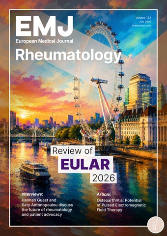
All information obtained by EMJ and each of the contributions from various sources is as current and accurate as possible. However, due to human or mechanical errors, EMJ and the contributors cannot guarantee the accuracy, adequacy, or completeness of any information, and cannot be held responsible for any errors or omissions. EMJ is completely independent of the review event (EULAR 2026) and the use of the organisations does not constitute endorsement or media partnership in any form whatsoever. The cover photo is of London, UK, the location of EULAR 2026.
Study Highlights Demographic Differences Across Myositis Subtypes
NEW insights from EULAR 2026 on idiopathic inflammatory myopathies (IIM) suggest that incidence patterns differ substantially according to disease subtype, sex, and ethnicity, offering important information about these rare autoimmune conditions.2
IIMs are a heterogeneous group of autoimmune conditions characterised by inflammation and damage to skeletal muscle, associated with substantial morbidity. Despite the introduction of EULAR and American College of Rheumatology (ACR) classification criteria in 2017, contemporary populationbased data on individual IIM subtypes remain limited.
Researchers from the UK conducted a large-scale epidemiological analysis of dermatomyositis, inclusion body myositis and other IIMs. The study identified 4,105 people with one of these conditions over a 19-year period. Among those classified as having other IIMs, 54.2% had polymyositis.
IIMs are a heterogeneous group of autoimmune conditions characterised by inflammation and damage to skeletal muscle, associated with substantial morbidity
The analysis showed that the incidence of all three subtypes increased substantially during the study period. However, while crude incidence rose over time, agestandardised rates remained broadly stable, suggesting that population ageing may be driving the increase rather than a true rise in disease occurrence.
Distinct differences emerged across demographic groups. Female patients
experienced a higher incidence of dermatomyositis and other IIMs, whereas inclusion body myositis was more common among males. Ethnicity was also associated with important variations in disease incidence. Compared with people of White ethnicity, those of South Asian, Black, and unknown ethnicity had a greater incidence of dermatomyositis. Similarly, the incidence of other IIMs was higher among South Asian, Black, and mixed ethnic groups.
The findings provide much-needed insight into myositis patterns across diverse populations, helping to address longstanding gaps in epidemiological evidence. The recorded incidence of inclusion body myositis was lower in the most deprived populations than in the least deprived. However, researchers noted that it remains unclear whether this reflects genuine differences in disease occurrence or disparities in diagnosis, detection, and access to care.
Researchers describe the study as the first population-based study to characterise the epidemiology of IIM subtypes across key sociodemographic groups. The research highlights the need for further investigation into potential barriers to diagnosis and management. Future studies could help determine whether observed differences reflect biological factors, healthcare inequalities, or a combination of both.
AI Imaging Tools Show Promise in Rheumatoid Arthritis Care
AI-DRIVEN imaging technologies demonstrated strong potential to improve the diagnosis, monitoring, and assessment of inflammatory arthritis, according to research presented at EULAR 2026.3
Accurate assessment of disease activity and structural damage remains a cornerstone of rheumatoid arthritis management. However, conventional imaging techniques often require expert interpretation and can be time-consuming, particularly when analysing large datasets. Several studies presented at the Congress explored how AI-based approaches could support clinicians by providing faster, more objective image analysis.
One study evaluated RADAR (RADAR, New York, USA), an AI algorithm developed to assess radiographic progression in rheumatoid arthritis. The model was initially trained and validated using data from the BCD cohort, comprising 7,560 hand and foot joints, before being tested in the ESPOIR cohort, a 20-year collection of X-rays. When evaluated using high-quality radiographs, the system achieved an estimated accuracy of 0.95, sensitivity of 0.93, and specificity of 0.99, demonstrating its ability to analyse large volumes of radiographic data efficiently.
Researchers also presented findings from ADMIRA, an automated deep learningbased MRI analysis system designed for patients with early inflammatory arthritis. The model was trained using visual Rheumatoid Arthritis MRI Scoring system assessments and validated in MRI scans from 180 patients. ADMIRA scores for total inflammation, synovitis, tenosynovitis, and osteitis showed good to excellent agreement with conventional visual scoring, supporting its validity as an automated assessment tool.
Ultrasound-based approaches also demonstrated encouraging results. A multivariable diagnostic model integrating musculoskeletal ultrasound findings with clinical variables achieved high diagnostic accuracy and exceptional negative predictive value across inflammatory rheumatic and musculoskeletal diseases. Investigators suggested that this targeted approach could help reduce unnecessary treatment by improving diagnostic precision in patients with suspected arthritis.
Additional research evaluated AI-assisted thermography as a non-invasive method of detecting joint inflammation. Analysis of 200 dorsal hand images from 100 patients showed that CatBoost (Yandex, Moscow, Russia) consistently outperformed other algorithms, achieving the highest levels of accuracy, sensitivity, specificity, and precision for identifying inflamed joints.
Finally, optical spectral transmission imaging was shown to correlate significantly with clinical measures of inflammatory arthritis activity. In a longitudinal analysis involving 60 patients and 1,312 wrist and finger joint assessments, optical spectral transmission scores reflected changes in disease activity over time, suggesting potential utility for long-term monitoring.
Although further validation in larger populations is required, these studies highlight the growing role of AI in rheumatology. By improving efficiency, standardisation, and diagnostic accuracy, AI-assisted imaging may help support earlier diagnosis and more effective monitoring of patients with inflammatory arthritis.
Cardiac Involvement Emerges Early in SSc
PRIMARY
cardiac involvement (pCI) affects a notable minority of patients with early systemic sclerosis (SSc) and is associated with a higher-risk clinical profile, according to findings from the SOLAR registry presented at EULAR 2026.4
Cardiac disease is a major driver of morbidity and mortality in SSc, yet early manifestations can be difficult to detect in routine clinical practice. Researchers analysed baseline data from 372 patients enrolled in the SOLAR registry, all of whom met the 2013 American College of Rheumatology (ACR)/EULAR classification criteria and had a disease duration of 3 years or less. pCI, defined as myocardial, pericardial, coronary, or conduction system abnormalities attributable to SSc, was identified in 24 patients (6.5%) at baseline. Coronary artery disease was the most commonly reported manifestation, while heart failure and pericardial effusion were rare. Most affected patients were female, and those with pCI tended to be older than patients without cardiac involvement.
The study found several clinical features associated with an increased likelihood of pCI. Patients with diffuse cutaneous SSc were significantly more likely to have pCI than those with limited cutaneous disease, while the latter was associated with a lower risk.4 Myositis, pulmonary arterial hypertension, overlap syndrome, and hypertension were all more prevalent among patients with cardiac involvement. In multivariable analyses, pulmonary
arterial hypertension emerged as the strongest associated factor, increasing the odds of pCI more than 15-fold. Myositis, overlap syndrome, diffuse skin involvement, and older age were also independently associated with a higher risk. Although interstitial lung disease and diabetes were numerically more common in the pCI group, these differences did not reach statistical significance.
Longitudinal follow-up highlighted the dynamic nature of cardiac disease in SSc. Among 275 patients who were free from pCI at baseline and had follow-up data available, six developed incident cardiac involvement during subsequent visits, representing 2.2% of the cohort. Overall, 30 patients (8.1%) had pCI documented at least once during the first four registry visits. These findings suggest that a single baseline assessment may fail to identify evolving cardiac abnormalities, particularly in patients with multisystem disease or established risk factors.
The SOLAR data indicate that pCI is uncommon but clinically significant in early SSc. The authors suggest that structured and repeated cardiac screening may improve detection and support earlier intervention in high-risk patients.
Coronary artery disease was the most commonly reported manifestation, while heart failure and pericardial effusion were rare
The theme reflected EULAR’s commitment to advancing research, improving care, and ultimately helping people maintain independence and quality of life
Towards Earlier and More Precise Systemic Sclerosis Disease Monitoring
SYSTEMIC SCLEROSIS
(SSc) continues to present a major challenge in clinical management and trial design due to its marked heterogeneity, variable organ trajectories, and evolving disease burden. New data presented at EULAR 2026 highlight the clinical relevance of early disease activity assessment and the need for more standardised outcome measures in SSc. Findings from two presentations explored how improved stratification and reporting frameworks could strengthen evaluation of disease progression and therapeutic outcomes.5,6
An oral abstract presentation from Santiago et al.5 reported findings from a longitudinal observational analysis of the EUSTAR database, a large multicentre registry prospectively following more than 25,000 patients across more than 200 international centres. The study assessed whether baseline disease activity, measured using the modified disease activity index (mDAI), could help predict subsequent organ involvement and disease progression.
Of the 3,214 patients included, 59.1% had inactive disease at baseline (<2.5 mDAI), with patients with active disease (>2.5 mDAI) demonstrating a higher 1-year cumulative incidence of organ-specific and multi-system involvement. This included skin progression, digital ulcers/gangrene, interstitial lung disease, and cardiac involvement. Active disease was also associated with shorter time to clinical events and greater use of immunosuppressive therapies, biologics, and antifibrotic agents. Together, this study underscores the importance of early disease activity monitoring.
A presentation by Campochiaro et al.6 examined reporting standards for digital ulcer studies, an area where inconsistent definitions and outcome assessments
remain a challenge. Developed by the World Scleroderma Foundation (WSF) digital ulcer committee, the recommendations identified seven core domains for clinical trials, including ulcer classification, eligibility criteria, healing outcomes, patient-reported outcomes, background therapies, and long-term recurrence monitoring.
The framework emphasises the importance of standardising local wound care protocols, clearly defining assessment timepoints, and incorporating environmental and seasonal variables. Collectively, these considerations aim to reduce inter-study variability and improve the robustness, reproducibility, and interpretability of trial outcomes in digital ulcer research.
Taken together, the two presentations reinforce a broader shift in SSc research towards earlier and more precise disease characterisation, alongside improved methodological consistency in outcome reporting. Future research may further clarify how early disease activity indices, such as the mDAI, can be integrated into trial design and routine clinical assessment, potentially supporting more targeted treatment strategies and improved longitudinal disease monitoring in SSc.
Of the 3,214 patients included, 59.1% had inactive disease at baseline (<2.5 mDAI), with patients with active disease (>2.5 mDAI) demonstrating a higher 1-year cumulative incidence of organspecific and multisystem involvement
Studies Highlight Challenges of Long-Term Glucocorticoid Use in Vasculitis
NEW RESEARCH presented at EULAR 2026 has raised questions about the role of ongoing low-dose glucocorticoid therapy in vasculitis, with findings suggesting limited benefit in antineutrophil cytoplasmic antibody-associated vasculitis (AAV) and identifying a subgroup of patients with difficult-to-treat giant cell arteritis (GCA) who remain dependent on glucocorticoids despite treatment escalation.7,8
In an oral presentation, Chavatza et al.7 evaluated the impact of low-dose glucocorticoids during maintenance treatment in AAV. The study included 171 patients receiving maintenance therapy at three referral centres in Greece, with a median follow-up of just over 7 years.7
During follow-up, 65 major flares occurred in 48 patients, corresponding to an incidence rate of 8.1 per 100 patientyears. Rituximab use was associated with a lower risk of major flares, while higher disease activity at diagnosis was linked to an increased risk. Importantly, concomitant glucocorticoid use at doses below 7.5 mg/day did not reduce the risk of major flares. However, low-dose glucocorticoid exposure was associated with increased risks of chronic damage accumulation and hospitalisation.
The investigators concluded that lowdose glucocorticoids did not reduce the risk of major flares during maintenance therapy but were associated with increased risks of chronic damage accumulation and hospitalisation, supporting earlier glucocorticoid withdrawal following remission.
A separate poster presentation by Dhrif et al.8 proposed the first definition of difficultto-treat GCA using data from 546 patients in the French Large Vessel Vasculitis Study Group cohort. Patients included in the analysis had at least 24 months of followup and comprehensive documentation of glucocorticoid dosing and relapse history.
The proposed definition incorporates three criteria: ongoing disease activity
preventing withdrawal of at least one immunosuppressive therapy after 24 months, inability to taper glucocorticoid dosage below 5 mg/day because of persistent disease activity, and the occurrence of at least two relapses during follow-up.
Using this definition, difficult-to-treat disease was identified in 9.7% of patients. This subgroup was characterised by predominant large-vessel involvement, a higher proportion of female patients, and greater difficulty reducing glucocorticoid treatment beyond 6 months. At Month 24, patients with difficult-to-treat disease required a mean maintenance glucocorticoid dose of 8.8 mg/day compared with 2.68 mg/day in those without difficult-to-treat GCA. Glucocorticoid dosage at Month 6 was also found to predict progression to difficult-to-treat disease among women with large-vessel vasculitis.
The authors noted that the proposed definition captures key unmet needs in GCA, including recurrent relapses and insufficient glucocorticoid sparing, and may help identify patients who could benefit from earlier treatment optimisation. They emphasised that the predictive model requires validation in independent cohorts.
At Month 24, patients with difficult-to-treat disease required a mean maintenance glucocorticoid dose of 8.8 mg/day compared with 2.68 mg/day in those without difficult-to-treat GCA
ULT Continuation Lowers Gout
Flares and Cardiometabolic Risk
At EULAR 2026, new data challenged and refined contemporary thinking in gout management while also providing mechanistic insight into how body composition may influence risk across gout and rheumatoid arthritis (RA), with implications for cardiometabolic disease.9,10
Researchers carried out a pragmatic, open-label, randomised superiority trial from nine rheumatology centres in the Netherlands evaluating treat-to-target (T2T) urate-lowering therapy (ULT) continuation versus a structured attempt at discontinuation in patients with gout in remission.9 Current guidelines recommend lifelong ULT with a T2T approach, but real-world adherence is variable, and discontinuation is common, reflecting uncertainty about the necessity of indefinite therapy in sustained remission.
Over 24 months, continued T2T ULT demonstrated more durable disease control. During the final 6-month followup period, remission criteria were met in 79.2% of patients continuing ULT compared with 62.9% in those attempting discontinuation. Across the full study period, cumulative flare incidence was substantially lower with continued therapy (12.3% versus 31.8%). In the discontinuation arm, nearly a quarter of patients required re-initiation of ULT after a median of 392 days, and more antiinflammatory medication was needed overall. A modest renal benefit was also observed in the continuation group. Notably, while a substantial proportion of patients maintained remission off therapy, the findings support continued T2T ULT at a population level and provide a foundation for shared decision-making, particularly in balancing long-term flare prevention against potential treatment minimisation strategies.
In parallel, a large imaging-based analysis from the UK Biobank examined the relationship between adiposity distribution, muscle composition, and risk of gout and RA.10 Using MRI data from individuals with gout (n=281), RA (n=308), and matched controls, the study demonstrated distinct patterns of body composition. Gout was characterised by increased visceral, hepatic, and intramuscular fat, while RA showed greater fat infiltration within muscle tissue. These metabolically adverse patterns were associated with increased risk of Type 2 diabetes and coronary heart disease.
Prospective analyses further showed that higher visceral and ectopic fat, alongside lower subcutaneous fat, increased the risk of incident gout, while increased intramuscular fat with reduced muscle volume was associated with future RA development. The findings support a model in which abnormal fat distribution and impaired muscle quality contribute to systemic inflammation and cardiometabolic risk in rheumatic disease.
Together, these studies reinforce the importance of sustained urate control in gout and highlight metabolic health as a shared therapeutic target across inflammatory arthritis.
79.2%
During the final 6-month follow-up period, remission criteria were met in 79.2% of patients continuing ULT compared with 62.9% in those attempting discontinuation
Real-World Evidence Highlights Gaps and Opportunities in Rheumatology Care
REAL-WORLD evidence is providing important insights into treatment implementation, weight management, lung disease detection, and smoking patterns among patients with rheumatic and musculoskeletal diseases, according to research presented at EULAR 2026.12-15
Observational data are increasingly complementing findings from RCTs and helping clinicians understand how recommendations are applied in routine practice. One study from Italy12 evaluated adherence to a treat-to-target strategy across 1,494 outpatient visits involving patients with rheumatoid arthritis, psoriatic arthritis, and spondyloarthritis.
The analysis found that adherence was suboptimal overall and was markedly lower among patients with spondyloarthritis, where adherence reached only 40%, compared with more than 70% in rheumatoid arthritis and psoriatic arthritis. The principal barrier to implementation was the absence of documented disease activity assessments using validated indices. This accounted for approximately 90% of nonadherence cases. A smaller proportion resulted from failure to adjust treatment according to disease activity findings.
The analysis found that adherence was suboptimal overall and was markedly lower among patients with spondyloarthritis
Researchers also identified factors associated with better adherence. Treatment with targeted synthetic or biologic disease-modifying antirheumatic drugs was linked to improved implementation across all disease groups, while younger age was associated with adherence in rheumatoid arthritis and spondyloarthritis.
The second study13 examined glucagonlike peptide 1 receptor agonist use in more than 60,000 patients with rheumatic and musculoskeletal diseases. Data were analysed from individuals receiving semaglutide or tirzepatide, with a mean baseline BMI of 36.3 kg/m2, and approximately two-thirds had diabetes.
Across all disease cohorts, weight-loss use was most common among patients with psoriatic arthritis, followed by ankylosing spondylitis and rheumatoid arthritis. At 12 months, non-diabetic users of tirzepatide lost 8% of baseline body weight compared with 6% among semaglutide users. A similar pattern was observed in individuals with diabetes, although overall weight loss was lower. Weight reduction plateaued after 12 months.
Further real-world evidence emerged from the international ANCHOR-RA study,14 which enrolled 1,169 patients with rheumatoid arthritis who had at least two risk factors for interstitial lung disease but no known diagnosis. Investigators found that 9.1% had previously undiagnosed interstitial lung disease.
Factors associated with interstitial lung disease included older age, male sex, greater tobacco exposure, higher rheumatoid arthritis disease activity, lower oxygen saturation, abnormal lung diffusion findings, crackles on auscultation, and the MUC5B promoter variant.
Separately, an analysis15 of 5,523 registry visits from Switzerland demonstrated a decline in current smoking prevalence among patients with rheumatoid arthritis between 2012–2022. However, lifetime smoking exposure increased among men with rheumatoid arthritis over the same period, contrasting with declining rates in the wider population. The findings suggest that smoking prevention efforts may have been less effective among men who later developed rheumatoid arthritis.
Together, these studies demonstrate the growing value of real-world evidence in informing clinical decision-making and identifying opportunities to improve outcomes for patients with rheumatic and musculoskeletal diseases.
References
1. Wilhelmi T et al. Turning guidelines to answers: patient evaluation of AI-based guideline chatbots in rheumatology. Abstract OP0256-PARE. EULAR Congress, 3-6 June, 2026.
2. Gordon P et al. Idiopathic inflammatory myopathies subtypes have clear epidemiological differences across sex and ethnicity subgroups: a populationbased cohort in England, 2002-2021. Abstract POS0262. EULAR Congress, 3-6 June, 2026.
3. Pensec H et al. Artificial intelligencebased analysis of 20-year radiographic progression in the ESPOIR rheumatoid arthritis inception cohort. Presentation OP0344. EULAR Congress, 3-6 June, 2026.
4. Amikishiyev S et al. Primary cardiac involvement in early systemic sclerosis: baseline profile of the patients in the SOLAR registry. Oral presentation OP0215. EULAR Congress, 3-6 June, 2026.
5. Santiago T et al. Clinical value of modified EUSTAR disease activity index to stratify for clinical outcome: a longitudinal observational analysis of the EUSTAR database. Presentation OP0213. EULAR Congress, 3-6 June, 2026.
6. Campochiaro C et al. Points to consider for reporting outcome measures in systemic sclerosis interventional studies on digital ulcers: an initiative from the World Scleroderma Foundation digital ulcer ad hoc committee. Presentation POS0321. EULAR Congress, 3-6 June, 2026.
7. Chavatza K et al. In patients with ANCA-associated vasculitides, low-dose glucocorticoids added to maintenance treatment, does not reduce the risk of major flares and is associated with an increased risk for chronic damage and hospitalization. Presentation OP0229. EULAR Congress, 3-6 June, 2026.
8. Dhrif O et al. Identifying difficult-totreat giant cell arteritis: results from a French Large Vessel Vasculitis Study Group cohort. Presentation POS0023. EULAR Congress, 3-6 June, 2026.
9. Peeters IR et al. Treat−to−target continuation of urate−lowering therapy versus a urate−lowering therapy discontinuation attempt strategy in gout patients in remission (GO TEST Finale): a pragmatic open label randomised superiority trial. Presentation OP0001. EULAR Congress, 3-6 June, 2026.
10. Ferguson LD et al. Gout and rheumatoid arthritis are associated with adverse body fat distribution and muscle
11. Bardan I et al. Long-term outcomes of juvenile idiopathic arthritis in adulthood: a population-based matched comparator study from Norway. Abstract OP0190. EULAR Congress, 3-6 June, 2026.
12. Arese M et al. Treat-to-target in inflammatory arthritis: a recommended strategy, an incomplete practice. Abstract OP0351. EULAR Congress, 3-6 June, 2026.
13. McCormick N et al. GLP-1 receptor agonists to facilitate weight loss and improve disease activity, pain and function in patients with rheumatic and musculoskeletal disease: real-world evidence from the Rheumatology Informatics System for Effectiveness (RISE) Registry. Abstract OP0112. EULAR Congress, 3-6 June, 2026.
14. Sparks JA, et al. Risk factors for interstitial lung disease in patients with rheumatoid arthritis: results from the international ANCHOR-RA study. Abstract OP084. EULAR Congress, 3-6 June, 2026.
15. Berthouzoz EC et al. Increasing lifetime smoking exposure among male rheumatoid arthritis patients despite declining current use in Switzerland. Abstract OP0109. EULAR Congress, 3-6 June, 2026.
Congress Beyond the Joint: Weight, Ultrasound, and the Therapeutic Frontier in Psoriatic Arthritis
Author: *André Lucas Ribeiro1
1. Hospital de Clínicas de Porto Alegre, Brazil
*Correspondence to Lucasribeiro.andre@gmail.com
Disclosure: The author has disclosed no conflicts of interest.
PSORIATIC arthritis (PsA) at the European Alliance of Associations for Rheumatology (EULAR) 2026 Congress was defined less by a single breakthrough than by a shift in framing. Across the ‘Optimising care in Psoriatic Arthritis’ and late-breaking sessions, three questions dominated: what we treat, when we should intervene, and how aggressively we should combine therapies. The unifying message was that drug choice, long the centre of the PsA conversation, is now only one variable among several that determine whether a patient reaches remission.
WEIGHT IS A TREATMENT TARGET, NOT A BACKGROUND VARIABLE
The most provocative reframing came from a machine-learning reanalysis of the SPEED trial, an open-label study of early PsA with poor prognostic factors.¹ Using recursive partitioning across baseline variables in 192 patients, the investigators found that BMI (median: 28.96 kg/m²) was the single most influential predictor of 1-year PASDAS, while treatment arm ranked only sixth. The derived decision rules were clinically legible: patients with a BMI below 25 kg/m² and lower baseline disease activity (PASDAS <5.4) achieved the best outcomes irrespective of treatment, whereas the combination of obesity (BMI: ≥27 kg/m²), longer disease duration, and polyarticular disease predicted PASDAS scores as high as 6.0 at Week 48. This is a sub-analysis of a single trial and should be read as hypothesis-generating; recursive partitioning is sensitive to cohort composition and cannot disentangle
adiposity from its metabolic and behavioural correlates. Even so, the signal aligns with prior observational data linking obesity to lower remission rates, reframing weight as a modifiable treatment component.
If SPEED implicated weight, the Phase IIIb TOGETHER-PsA trial tested whether acting on it changes outcomes.² Adults with active PsA and obesity or overweight with a weight-related comorbidity (mean BMI: 37.2–38.4 kg/m²) were randomised to ixekizumab with concomitant tirzepatide (IXE+TZP) or ixekizumab alone. The composite primary endpoint at Week 36, ACR50 (American College of Rheumatology 50% improvement response) plus at least 10% weight reduction, was achieved by 31.7% of the combination group versus 0.8% with ixekizumab alone (p<0.001). The contrast is partly mechanical, since the weight-loss component is unattainable with ixekizumab monotherapy, but the articular signal was independent and early: ACR50 separated by Week 4 (11.1% versus
3.9%; p=0.029) and minimal disease activity (MDA) favoured the combination at Week 36 (26.3% versus 15.3%; p=0.026). Even though TOGETHER-PsA does not establish whether benefits were driven by weight loss, the metabolic effects of glucagon-like peptide-1/glucose-dependent insulinotropic polypeptide agonism, or improved drug pharmacodynamics, but it provides the first randomised evidence that addressing adiposity alongside cytokine blockade is feasible and clinically active.
The DIPSA trial helped adjudicate which of those mechanisms is doing the work.³ In 92 patients with moderately active PsA and a BMI of 25–40 kg/m², a Mediterranean diet, a low-calorie dietary approach to stop hypertension diet, and standard dietary advice were compared. All three arms produced modest weight loss and significant improvement in DAPSA by Week 24, with no differences between them, and the early MDA advantage for the dietary arms at Week 12 had dissipated by Week 24. The interpretable finding was a dose-response, with the magnitude of weight loss, not the dietary strategy, predicting improvement in DAPSA independent of arm. Read alongside SPEED and TOGETHER-PsA, DIPSA implies that
weight reduction itself, however achieved, is the active ingredient and the dietary pattern secondary.
ULTRASOUND SHARPENS THE QUESTIONS OF WHO AND WHEN
If weight reframed what we treat, imaging reframed when we should act and ultrasound was the connecting thread. Zabotti et al.4 compared clinical and ultrasound features across the psoriatic disease continuum in 183 patients spanning psoriasis, stable and progressing subclinical PsA, and early clinical disease.⁴ The central finding was that clinical assessment failed where imaging succeeded: tender joint count, Bath Ankylosing Spondylitis Disease Activity Index (BASDAI), pain, and Health Assessment Questionnaire (HAQ) increased progressively across disease states but were unable to distinguish patients with arthralgia who progressed to PsA from those who remained stable. What discriminated were ultrasound-detected active synovitis (odds ratio: 3.40; p=0.019) and active enthesitis (odds ratio: 3.45; p=0.049), alongside older age. Progression to clinically manifest PsA occurred a mean of 13.7 months after enrolment.
If weight reframed what we treat, imaging reframed when we should act and ultrasound was the connecting thread
The diagnostic counterpart was the DUET study, a 17-centre effort to build and validate a PsA-specific sonographic enthesitis score.⁵ Across 213 patients with early PsA, 100 with psoriasis, and 106 controls, the optimal score combined weighted inflammatory and structural lesions at the Achilles, patellar, and triceps insertions. Performance was modest in the whole cohort (area under the curve: 0.65; age-specific specificity: 73–74%; sensitivity: 47–49%) but improved in the validation cohort (specificity up to 100%) and in patients with tender entheses (sensitivity up to 63%). Two messages matter for practice. First, DUET is a specificity-oriented adjunct best deployed in symptomatic entheses rather than a standalone screen. Second, the finding that large-joint enthesitis is not a universal feature of early PsA tempers the assumption that the enthesis is uniformly the earliest target and refines where the probe should be placed.
THE THERAPEUTIC CEILING: COMPARISON AND COMBINATION
On treatment itself, EULAR 2026 moved past placebo comparisons. The latebreaking BE BOLD trial was the first headto-head study of an IL-17A/F inhibitor against an IL-23 inhibitor in PsA.⁶ Among 553 patients, bimekizumab was superior to risankizumab for the primary endpoint of ACR50 at Week 16 (49.1% versus 38.4%; p=0.0078), with early separation by Week 4 (19.9% versus 7.2%). The hierarchy stopped at the first ranked secondary endpoint, however, as MDA did not differ significantly (43.0% versus 39.9%; p=0.4408). The result narrows, but does not close, the mechanistic debate, since a joint-focused advantage at 16 weeks must be weighed against tolerability and longer-term outcomes.
The combination strategy advanced on two fronts. Exploratory MRI from the Phase IIa AFFINITY study, in TNFinhibitor-inadequate responders, showed numerically greater reductions in PsAMRIS inflammation in hand and foot phalangeal joints with guselkumab plus golimumab than with guselkumab alone, with stable structural damage and no
separation at the heel enthesis.⁷ The paired biomarker analysis found that reductions in TNF activity, C-reactive protein, and IL-6 tracked with combination therapy and that responders carried a higher baseline inflammatory burden, suggesting the dual approach may raise the efficacy ceiling selectively in the most inflamed patients.⁸ Building on this logic, the COMBo trial design was presented: a Phase IV, placebocontrolled study of deucravacitinib added to ongoing TNF-inhibitor therapy in difficult-to-control PsA, powered to detect a doubling of low-disease-activity
References
1. Garaïman A et al. Lighten the load: artificial intelligence reveals BMI outranks treatment in one-year PsA outcomes—findings from the SPEED trial. Abstract OP0187. EULAR Congress, 3-6 June, 2026.
2. Coates LC et al. Ixekizumab and concomitant tirzepatide achieved early disease control in adults with PsA and obesity/overweight: phase 3b TOGETHER-PsA trial. Abstract OP069. EULAR Congress, 3-6 June, 2026.
3. Eder L et al. Dietary Interventions in Psoriatic Arthritis (DIPSA): a randomised controlled clinical trial. Abstract OP070. EULAR Congress, 3-6 June, 2026.
attainment.⁹ With recruitment still to come, it will provide the first controlled test of dual TNF and TYK2 inhibition.
Taken together, EULAR 2026 sketched a more dimensional PsA: a disease in which the patient’s metabolic state, imaging phenotype, and the combinatorial logic of immunomodulation are converging on the same goal of deeper, earlier, and more durable control. The trials that will test whether this integration improves long-term outcomes are, fittingly, still to come.
4. Zabotti A et al. Clinical and imaging features of subclinical and early PsA reveal synovio-entheseal inflammation as a marker of psoriatic arthralgia progression. Abstract OP0183. EULAR Congress, 3-6 June, 2026.
5. Eder L et al. The development and validation of a diagnostic ultrasound enthesitis tool (duet) for psoriatic arthritis. Abstract OP0180. EULAR Congress, 3-6 June, 2026.
6. McInnes IB et al. Bimekizumab efficacy and safety versus risankizumab in active PsA: 16-week results from the head-to-head BE BOLD study. Abstract LB0001. EULAR Congress, 3-6 June, 2026.
7. Wellen J et al. Effect of guselkumab and golimumab combination therapy
on MRI-detected inflammation and damage in PsA: findings from the phase 2a AFFINITY study. Abstract OP0186. EULAR Congress, 3-6 June, 2026.
8. McInnes IB et al. Biomarker-driven insights from the phase 2a AFFINITY study evaluating guselkumab + golimumab combination versus guselkumab monotherapy in PsA. Abstract POS0479. EULAR Congress, 3-6 June, 2026.
9. Merola JF et al. The COMBo study: a randomised trial of deucravacitinib–TNF inhibitor combination versus TNF inhibitor monotherapy in difficult-tocontrol psoriatic disease. Abstract POS0499. EULAR Congress, 3-6 June, 2026.
Highlights from the European Alliance of Associations for Rheumatology (EULAR) 2026 Congress
Author: *Fareeha Tariq¹
1. King's College Hospital, London, UK
*Correspondence to fareeha.tariq@kcl.ac.uk
Disclosure: Tariq has declared no conflicts of interest.
THE EUROPEAN Alliance of Associations for Rheumatology (EULAR) 2026 Congress was held in London, UK, from 3rd–6th June. The sessions spanned a broad array of clinical and basic science topics across rheumatoid arthritis (RA), systemic lupus erythematosus (SLE), psoriatic arthritis, spondylarthritis, vasculitis, Sjögren's disease, myositis, juvenile idiopathic arthritis, systemic sclerosis, and osteoarthritis. This feature highlights some of the emerging concepts across rheumatology in 2026.
DECIPHERING IMMUNE TRENDS IN PRECLINICAL RHEUMATIC DISEASES
Multi-omic Approaches Reveal How
Immune Cells Go Awry
Loss of B cell tolerance emerged as an early driver of systemic autoimmunity in both SLE and RA.1 Bylinska et al.1 profiled the pre-SLE continuum: autoantibody-negative controls, autoantibody-positive individuals, incomplete lupus erythematosus, and established SLE using 137-plex cellular Indexing of Transcriptomes and Epitopes by Sequencing. They reported a loss of inhibitory checkpoints in naïve B cells (leukocyte-associated Ig-like receptor 1 [LAIR1] down, cluster of differentiation [CD]83-high) early during the autoantibodypositive stage, but a slower transition to effector phenotype, with metabolic reprogramming and B cell receptor activation emerging in incomplete lupus erythematosus and SLE. In contrast, antigen-experienced memory, CD11chi,
and CD24hi subsets acquire mitochondrial and Type I interferon signature early in the antibody-positive stage and maintain these signatures throughout the preSLE to SLE continuum. This B cell shift is coupled to the myeloid compartment, which shows the largest transcriptional change of all: IL1B+ CD14+ monocytes show the largest transcriptional changes. As disease progresses, they lose TIMP1–CD63 prosurvival signalling to B cells and instead gain S100A9-mediated inflammatory signalling through the B cell receptor, alongside increased TNF and IL-6 signalling. T cell involvement is staged in parallel, with CD4 central memory cells marking the earliest transition, and CD4 and CD8 effector memory cells progressively engaging the activated B cell subsets.
In at-risk RA, Takada et al.2 ran mass cytometry on 42 anti-cyclic citrullinated peptide (CCP)-positive individuals, 15 of whom converted to clinical disease within 2 years. Eight lymphocyte subsets were expanded relative to controls, but only
two predicted conversions. High baseline frequencies of CXCR5+-activated naïve B cells correlated with 47% RA-free survival at 1 year against 91% in the low group, and Ki-67+ proliferating peripheral helper T cells, at 53% versus 84%, with both curves separating inside the first year. The predictive unit, an activated naïve B cell population paired with a proliferating T-helper population, mirrors the naïve-B activation and effector-memory T cell engagement seen in pre-SLE.
Proteomics anchors the convergence at the soluble level. Pu et al.3 screened 2,920 plasma proteins in 46,687 UK Biobank participants with 606 incident RA cases over a median of 8.3 years; 1,520 proteins were associated with future onset. Mendelian randomisation nominated 11 proteins with genetic risk for causality, including TNF, IL1RN, CD40, human leukocyte antigen (HLA)-DRA, CD72, TNFRSF14, AIF1, RNASET2, ICAM3, CCL21, and ACRBP, four of them already drug targets. Two molecules recur across both diseases with hard, molecule-level support: TNF, monocyte-induced in pre-SLE and genetically causal in pre-RA, and CD72, lost as a B cell checkpoint in pre-SLE and flagged as a causal protein in pre-RA. The shared antigen-presentation pathway (HLADRA, CD40) reinforces the overlap, but rests on weaker, pathway-level evidence.
The Gut–Joint Axis
In the session ‘Novel Insights into PreRA Mechanisms: What Happens Where and When?’, Zaiss4 made the case that RA may begin in the gut, with antiacetylated protein antibodies (AAPA) as the connecting thread. Drawing on the TIRx cohort, consisting of individuals who are AAPA-positive and have musculoskeletal symptoms but no clinical synovitis, he showed that AAPA significantly separated those who progressed to RA from those who did not, pointing to a diagnostic and possibly predictive role, while established disease was marked by high levels of tissue protein acetylation. To explain how this might arise, he turned to the collageninduced arthritis model, where artificially acetylating gut Escherichia coli heightened its antigenicity, while intestinal acetylation, in turn, drove damage to the epithelial barrier. At the sites where acetylation occurred, myeloid cells gathered in the mucosal villi and CD11c+ cells presented to B cells, while the acetylated proteins themselves appeared to push those myeloid cells away from antigen presentation and towards an inflammatory effector role. Taken together, the data positioned gut protein acetylation as an upstream trigger of the autoantibody response that precedes joint disease.
Advances in multi-omic profiling are uncovering the key immune cells and molecular pathways driving autoimmune rheumatic diseases long before clinical diagnosis
Relevance to Practice
These studies demonstrate how advances in multi-omic profiling are uncovering the key immune cells and molecular pathways driving autoimmune rheumatic diseases long before clinical diagnosis. By defining the earliest stages of immune dysregulation, these approaches provide a valuable window of opportunity for disease interception before autoimmunity becomes tissue-centric and difficult to cure. This concept is already being translated into clinical practice through prevention trials, such as the ALTO trial, which showed that treatment with abatacept for 1 year delays arthritis progression for up to 4 years in individuals who are at very high risk of developing RA. Ongoing trials, including the ExIST trial, evaluating baricitinib in individuals at risk of inflammatory arthritis will determine whether targeting the JAK pathway can similarly delay arthritis onset in anti-CCP-positive individuals at moderateto-high risk of disease.5,6
BEYOND CD19: SEQUENTIAL B-LINEAGE TARGETING IN TREATMENT-REFRACTORY RHEUMATIC DISEASE
Relapse After CD19 CAR-T and Sequential BCMA-Directed Rescue
CD19 CAR-T cell therapy showed promise in inducing deep immune reset in patients with refractory SLE, but follow-up data presented at EULAR 2026 established that maintenance of long-term remission may not be a reality for all patients. Data presented by Wirsching reported a relapse at a median of 13 months (range: 9–24) in 1/28 patients with disease, or arthritis,
respectively, B cell maturation antigen (BCMA) CAR-T (idecabtagene vicleucel) with sustained remission, obinutuzumab plus nintedanib, and tofacitinib followed by BCMA CAR-T in the CARAMBA study. The patient with SLE (double-stranded DNApositive) relapsed with transverse myelitis, arthritis, and skin disease. BCMA CAR-T cell therapy successfully cleared the neurological deficit, however, leaving behind residual cutaneous disease (despite double-stranded DNA seroconversion), which was cleared by anifrolumab. Both patients with SSc (Scl70positive) relapsed with multi-organ disease and received the BCMA T cell engager teclistamab, with early improvement. Relapse after CD19 CAR-T is infrequent but real and late; IIM is the least durable setting, and BCMA-directed combinatorial therapy is the dominant rescue.
The rescue pattern points to the compartment CD19 leaves behind. CD19 CAR-T spares CD19-BCMA+ long-lived plasma cells, and the most systematic test of targeting them came from Boeltz et al.,8 who treated 16 patients with multidrug-resistant disease, eight with SSc, two with IIM, two with IgG4-related disease, two with RA, one with Sjögren’s disease, and one with Graves’ disease, with the CD3×BCMA T cell engager teclistamab in a named-patient programme. Clinical responses occurred in 15/16 patients, with seroconversion of autoantibodies that characteristically survive CD19 monotherapy, including anti-Sjögren’s-syndrome-related antigen A, Scl-75/100, anti-CCP, rheumatoid factor, and Mi-2, alongside normalised IgG4. Nine patients reached drug-free remission (median: 11 months), though four progressed by a median of 5 months, and all 16 developed hypogammaglobulinaemia requiring Ig replacement.
Off-the-Shelf CAR-T Cell Therapy
Looking beyond autologous, single-target constructs, Shiff et al.9 presented FT839 (Fate Therapeutics, San Diego, California, USA), an off-the-shelf, induced pluripotent stem cell-derived dual-CAR-T cell designed to address three limitations of current CAR-T at once: single-antigen targeting, the need for conditioning chemotherapy, and perpatient manufacturing.
It expresses two CARs against the B cell lineage marker CD19 and the activation marker CD38, the latter spanning plasmablasts, plasma cells, and activated T and natural killer cells. FT839 is engineered with synthetic alloimmune defence receptor plus CD58 deletion to eliminate alloreactive immune cells and resist host rejection. In preclinical assays, FT839 eliminated CD19+ B cells, plasmablasts, plasma cells, and CD38+ activated CD4/CD8 T cells (>93–99%) while sparing resting CD38⁻ T cells. In HLA-mismatched cultures, FT839 demonstrated greater persistence than unmodified controls, consistent with the effects of its alloevasion engineering (2.4×10⁶ versus 1.3×10⁴ cells; p<0.0001).
The data are preclinical, but FT839 is one example of the off-the-shelf, multi-target, conditioning-free CAR-T cell therapy.
CD19 CAR-T cell therapy showed promise in inducing deep immune reset in patients with refractory SLE
Relevance to Clinical Practice
While relapse following CD19 CAR-T therapy is uncommon, these findings show that sequential BCMA-directed therapies can successfully restore disease control in selected patients. Emerging off-theshelf CAR-T platforms may also improve accessibility by reducing manufacturing delays and broadening immune-cell targeting. Although these approaches remain investigational, they represent an important step towards more personalised,
mechanism-based treatment for refractory rheumatic diseases.
AI IN RHEUMATOLOGY
Machine Learning Identifies Subgroups in JPsA From Multicentre Inception
Cohorts
Shoop-Worrall et al.10 used machine learning to test whether juvenile psoriatic arthritis (JPsA) separates into distinct subgroups, pooling five multinational inception cohorts: CAPS, ICON, NPRD, ReACCh-Out, and CAPRI, including 3,282 children diagnosed with JPsA, enthesitisrelated juvenile idiopathic arthritis, or undifferentiated juvenile idiopathic arthritis, between 2001–2023. Applying latent class analysis to active joint count, psoriasis, dactylitis, and nail involvement within the first 24 months of symptom onset, they consistently recovered three clusters: a low-burden low joint–psoriasis group, a high-burden psoriasis–dactylitis group, and an intermediate all features group. The psoriasis–dactylitis cluster was the most severely affected, with the highest enthesitis (70–83%), the greatest proportion reporting a family history of psoriasis (57–85%), and the worst patient-reported global, pain, and disability scores; these children were younger (median: 10–11 years) and more often female (65% versus 44%). The key methodological strength was testing the clusters across three case definitions: ILAR JPsA criteria, physician diagnosis, and ClASsification criteria for
Psoriatic ARthritis (CASPAR). The same broad phenotypes recurred under all three, supporting their robustness, but their proportions shifted markedly:
the low-burden group made up 26% under International League of Associations for Rheumatology (ILAR) criteria, yet 59% under physician diagnosis, and the CASPAR-defined population yielded an additional small high joint–dactylitis group not seen in the others. JPsA is therefore clinically heterogeneous, but how the subgroups are partitioned and how large each appears depends partly on the diagnostic definition applied.
Relevance to Clinical Practice
AI can identify clinically meaningful disease subgroups beyond conventional classification criteria. As machine learning becomes increasingly integrated into rheumatology, such approaches may improve patient stratification, prognostication, and treatment selection, supporting more personalised care and more efficient clinical trial design in JPsA.
CONCLUSION
Research presented at EULAR 2026 highlighted the rapid progress being made towards precision medicine in rheumatology. Advances in multiomic technologies are improving our understanding of the earliest stages of autoimmune disease and identifying new opportunities for disease interception
References
1. Bylinska A et al. Early disruption of B cell tolerance pathways and immune regulatory circuits in preclinical systemic autoimmune disease. Abstract OP0155. EULAR Congress, 3-6 June, 2026.
2. Takada H et al. Elevation of circulating proliferative Tph cells and activated naïve B cells precedes development of clinical rheumatoid arthritis in CCPpositive at-risk individuals. Abstract POS0171. EULAR Congress, 3-6 June, 2026.
before irreversible tissue damage occurs. At the same time, innovations in cellular therapies are expanding treatment options for patients with severe, treatmentrefractory autoimmune diseases, while AI is helping to define clinically meaningful patient subgroups that may support more personalised care. Although many of these approaches remain investigational, together they demonstrate how technological advances are transforming both our understanding and management of rheumatic diseases. Continued clinical studies will be essential to determine how these discoveries can be translated into routine practice and ultimately improve outcomes for patients.
3. Pu Y et al. Decoding the proteomic landscape of rheumatoid arthritis onset: from causal drivers to clinical prediction. Abstract POS0259. EULAR Congress, 3-6 June, 2026.
4. Zaiss MM. The gut–joint axis (invited lecture). Session: Novel insights into pre-RA mechanisms: what happens where and when? EULAR Congress, 3-6 June, 2026.
5. Cope AP et al.; ALTO Study Investigators. Long-term outcomes of abatacept in individuals at risk of developing rheumatoid arthritis
(ALTO): a randomised, double-blind, placebo-controlled trial. Lancet Rheumatol. 2026;8(3):e171-80.
6. NHS Health Research Authority. ExIST. Available at: https://www.hra.nhs.uk/ planning-and-improving-research/ application-summaries/researchsummaries/exist/. Last accessed: 2 July 2026.
7. Wirsching A et al. Management of relapses after autologous CD19 CAR T-cell therapy in SLE, SSc and IIM – a case series. Abstract OP076. EULAR Congress, 3-6 June, 2026.
8. Boeltz S et al. BCMA-targeted T-cell engager teclistamab as rescue therapy for severe refractory autoimmune diseases. Abstract OP078. EULAR Congress, 3-6 June, 2026.
9. Shiff NJ et al. Off-the-shelf dualCAR T-cell therapy: targeting B and T cells in autoimmune disease without preconditioning. Abstract OP0156. EULAR Congress, 3-6 June, 2026.
10. Shoop-Worrall S et al. Distinct subgroups of early juvenile psoriatic arthritis identified using machine learning: results from a multinational collaboration of inception cohorts. Abstract OP0195. EULAR Congress, 3–6 June, 2026.
Updates from EULAR 2026: Gastrointestinal Involvement in Systemic Sclerosis
GASTROINTESTINAL (GI) involvement affects up to 90% of patients with systemic sclerosis (SSc) and ranks among the most disabling internal organ manifestations, contributing substantially to morbidity, malnutrition, and impaired quality of life. Unlike skin and lung, the GI tract has long been the underserved frontier of SSc research: clinically ubiquitous, mechanistically obscure, and short of validated biomarkers and effective therapies. The European Alliance of Associations for Rheumatology (EULAR) Congress 2026 in London, UK, suggested this is changing, with a body of work that, taken together, drew a new track: predicting GI disease early, dissecting its biology and biomarkers, measuring it objectively rather than by symptoms alone, and, for the first time, showing that a disease-modifying immunotherapy can improve gut dysfunctions.
PREDICTING GI DISEASE AND ITS PROGNOSTIC WEIGHT
Two large cohorts converged on early prediction. In the GENISOS cohort, Ayla et al.1 followed 450 patients with early SSc (≤5 years from the first non-Raynaud’s symptom) over a mean of 5.3 years. Baseline gastro-oesophageal reflux disease (GORD; hazard ratio [HR]: 4.15) and diarrhoea (HR: 2.25) independently predicted progression to significant GI involvement, and each additional baseline GI manifestation raised the hazard by 48%. The same symptoms carried prognostic weight for survival: peptic ulcer (HR: 1.89) and diarrhoea (HR: 1.66) independently predicted death. Approaching the question from the vascular and cutaneous side, Bonomi et al.2 and European Scleroderma Trials and Research
Group (EUSTAR) colleagues analysed 7,789 patients: among those GI-negative at baseline, 11.7% developed new-onset GI involvement over a median of 4.8 years, with highest incidence in the first 5 years. Puffy fingers and digital ulcers independently predicted GI onset, and, critically, incident GI involvement was independently associated with increased all-cause mortality (HR: 1.99). Together, these cohorts frame GI disease as an early, predictable, and prognostically weighty event.
These cohorts frame GI disease as an early, predictable, and prognostically weighty event
BIOLOGY AND BIOMARKERS
The most mechanistically ambitious contribution came from McMahan et al.,3 who identified two enteric nervous systemrelevant autoantibodies defining clinically distinct GI phenotypes. Antibodies to Argonaute (AGO) proteins (anti-AGO1/2), present in 9–14% of patients and absent in controls, marked severe lower-GI dysmotility and constipation, with dual positivity producing a particularly severe phenotype and titres tracking symptom severity. Antibodies to dihydrolipoamide branchedchain transacylase (DBT) instead marked an upper-GI signature of impaired oesophageal transit. Mechanistically, AGO proteins localised to myenteric ganglia and were enriched in neurone-derived extracellular vesicles, implicating disrupted vesiclemediated RNA signalling, while DBT was concentrated in mesoderm-derived enteric neurones. Complementing this antigenbased work, Makol et al.4 applied targeted serum proteomics to 84 patients, defining a signature of GI symptom severity in which inflammatory and tissue-remodelling proteins (hepatocyte Growth Factor [HGF], extracellular newly identified receptor for advanced glycation end-products-binding protein [EN-RAGE], oncostatin M [OSM], TGF-α) were elevated in moderate-tosevere disease. A third strand came from the gut itself: Spinella et al.5 used full-length 16S metagenomic sequencing in 25 patients and 25 controls to show reduced microbial
richness and a dysbiotic profile that tracked with symptom severity. It was a preliminary single-centre study, but a reminder that the microbiome remains an under-explored axis. Together, these antigen-, protein-, and microbe-level readouts point toward circulating biomarkers and candidate therapeutic targets.
MEASURING IT OBJECTIVES
The year’s richest seam was objective measurement, which repeatedly exposed the limits of symptom scores. Bonomi et al.6 applied high-resolution manometry across 109 patients including very early (Very Early Diagnosis of Systemic Sclerosis [VEDOSS]) cases: fatigue worsened stepwise with oesophageal motor severity (manometric dysmotility independently associated with lower Functional Assessment of Chronic Illness Therapy – Fatigue [FACIT-F]; β=−3.87), and University of California Los Angeles (UCLA) Gastrointestinal Tract 2.0 (GIT 2.0) burden did not differ across manometric patterns, emphasising that patient-reported symptoms can miss underlying motor dysfunction. Moving to the lower gut, Alcala-González et al.7 used high-resolution jejunal manometry in 46 patients, finding overt small-bowel dysmotility in 67%. Severe hypocontractility defined a high-risk subgroup characterised by diffuse disease, active microangiopathy, exclusive anti-Ro positivity, greater
symptom burden, and progression to intestinal pseudo-obstruction. The same drive toward physiology ran through studies of objectively-defined GORD phenotypes, which dissociated acid from non-acid reflux and argued for assessment beyond empiric acid suppression (FelixTellez et al.),8 magnetic resonanceenterography quantifying small-bowel motility heterogeneity (Minerba et al.),9 and ultrasound of hyoid excursion as an early, and potentially modifiable, marker of dysphagia detectable already in VEDOSS (Peretti et al.).10
THE FIRST THERAPEUTIC SIGNAL IN THE GUT
Perhaps the most striking translational result linked the year’s headline therapy to GI disease. Auth et al.,11 from Erlangen, Germany, studied eight patients with progressive diffuse cutaneous SSc treated with cluster of differentiation 19 (CD19)targeting CAR-T cells within the CASTLE study, all with baseline GI involvement. UCLA GIT 2.0 scores improved significantly by 3 months, exceeding the minimally important difference, with the clearest gains in distention/bloating and, by 6 months, in constipation and social functioning with reflux being the most refractory domain. Critically, oesophageal gallium-68-labeled fibroblast activation protein Inhibitor (68GaFAPI)-PET uptake, a marker of fibroblast activation, declined progressively (median
standardised uptake value mean reductions of −0.747 at 6 months and −1.222 at 12 months). This is among the first evidence that deep B cell depletion can modify not only skin and lung, but also the fibrotic and functional GI compartment, although the eight-patient sample warrants cautious interpretation.
Deep B cell depletion can modify not only skin and lung, but also the fibrotic and functional GI compartment
FROM BENCH TO BEDSIDE: DIFFICULT-TO-TREAT MANIFESTATIONS
The clinical counterpoint came in the dedicated ‘Challenges in Clinical Practice’ session, 'Difficult-to-treat GI manifestations in SSc',12 chaired by Alain Lescoat, Institut National de la Santé et de la Recherche Médicale, Paris, France; and Muriel Elhai, Department of Rheumatology, University Hospital Zurich, Switzerland. Structured around two of the most demanding scenarios: gastric antral vascular ectasia (presented by Artur Ejsmont, and discussed by Otylia Kowal-Bielecka, Department of Rheumatology and Internal Medicine, Medical University of Białystok, Poland) and small intestinal bacterial
overgrowth (presented by Marco Minerba, Rheumatology and Clinical Immunology, Department of Medicine, University of Rome Campus Bio-Medico, School of Medicine, Rome, Italy, and discussed by Francesco Del Galdo, Leeds Raynaud’s and Scleroderma Program, NIHR Biomedical Research Centre Leeds, UK). It underscored a practical reality: even as measurement and prediction advance, management of refractory gastric antral vascular ectasia and recurrent small intestinal bacterial overgrowth remains largely empirical. For gastric antral vascular ectasia, the takehome message was simple: any unexplained anaemia or iron deficiency in a patient with SSc should prompt gastroscopy, as the lesion is often subclinical. Argon plasma coagulation, together with iron and transfusion support, should be firstline therapy. In the index case, it achieved complete endoscopic resolution over 18 months, alongside immunosuppression. For small intestinal bacterial overgrowth (pooled prevalence ~35%, roughly ten-fold the general population), cyclical rifaximin remained the mainstay, outperforming rotating antibiotics for eradication (77.8% versus 44.8%) and working best with an added probiotic. The discussion reframed it not as an isolated infection but as one facet of a heterogeneous dysmotility syndrome,
References
1. Ayla A et al. Early gastrointestinal manifestations predict outcomes in patients with systemic sclerosis. Abstract POS0533. EULAR Congress, 3-6 June, 2026.
2. Bonomi F et al. Incidence, timing, and predictors of new-onset gastrointestinal involvement in systemic sclerosis: a longitudinal cohort study from the EUSTAR database. Abstract POS0562. EULAR Congress, 3-6 June, 2026.
3. McMahan Z et al. Autoimmune targeting of enteric neurons and extracellular vesicles defines distinct gastrointestinal phenotypes in systemic sclerosis. Abstract POS0542. EULAR Congress, 3-6 June, 2026.
4. Makol A et al. Distinct serum proteomic signatures associated with gastrointestinal symptom severity in systemic sclerosis. Abstract POS0577. EULAR Congress, 3-6 June, 2026.
with magnetic resonance-enterography and bowel ultrasound phenotyping, notably segmental small-bowel volume loss, offering a way to explain antimicrobial failure and, in future, to target motility-directed therapy. The unifying caution was that diseasemodifying treatments can themselves trigger or worsen GI symptoms, reinforcing the case for multidisciplinary management and controlled trials with GI-specific endpoints.
CONCLUSION
EULAR 2026 marked a quiet inflection point for GI involvement in SSc. The conversation is shifting from documenting a near-universal problem to predicting it early from clinical and symptom profiles, dissecting its biology through antigenspecific autoantibodies and serum proteomics, measuring it objectively where symptoms fall short, and, for the first time, showing that a disease-modifying immunotherapy may reach the gut. The outstanding gap remains therapeutic: these maturing biomarkers and endpoints must now be harnessed in interventional trials so that this long-neglected domain finally receives treatments matched to its clinical weight. On the evidence from London, the foundations for that next step are being laid.
5. Spinella A et al. Evaluation of gastrointestinal microbiome in patients with systemic sclerosis: our experience and emerging research. Abstract POS1090. EULAR Congress, 3-6 June, 2026.
6. Bonomi F et al. Fatigue severity relates to esophageal manometric patterns beyond gastrointestinal symptoms in systemic sclerosis. Abstract POS0539. EULAR Congress, 3-6 June, 2026.
7. Alcala-González LG et al. Objective assessment of small bowel involvement identifies high-risk phenotypes in systemic sclerosis. Abstract POS0560. EULAR Congress, 3-6 June, 2026.
8. Felix-Tellez FA et al. Objective physiological assessment across the GERD spectrum in systemic sclerosis: a multicenter international study. Abstract POS0576. EULAR Congress, 3-6 June, 2026.
9. Minerba M et al. Marked heterogeneity of small bowel motility in systemic sclerosis with gastrointestinal involvement: a pilot MR enterography study. Abstract POS1083. EULAR Congress, 3-6 June, 2026.
10. Peretti S et al. Hyoid dysfunction as an overlooked mechanism of dysphagia in systemic sclerosis from the early disease stages. Abstract POS1091. EULAR Congress, 3-6 June, 2026.
11. Auth J et al. Analysis of esophageal 68Ga-FAPI-uptake and gastrointestinal symptoms in patients with diffuse cutaneous systemic sclerosis upon CD19-targeting CAR T cell therapy. Abstract POS0505. EULAR Congress, 3-6 June, 2026.
This collection of abstract reviews showcases some of the most impactful research presented at this year's European Alliance of Associations for Rheumatology (EULAR) Congress, offering concise insights into emerging evidence, innovative therapies, and the latest developments shaping the future of rheumatology.
Engagement of Patient Research Partners in a 7-Year Clinical Research Project: Points to Consider and Lessons Learned
Authors: *Coralie Bouillot,1 Alena Piatrova2
1. Sjögren Europe, Gland, Switzerland
2. AP-HP, Le Kremlin Bicêtre, France
*Correspondence to coralie@sjogreneurope.org
Disclosure: The authors have declared no conflicts of interest.
NEw Clinical Endpoints in primary Sjögren’s Syndrome: an Interventional Trial based on stratifiYing patients (NECESSITY)1 is a public–private research consortium involving 25 partners: 20 academics, four pharma companies, and one patient association. Seven patient research partners (PRP) have collaborated since the beginning of the project, which ended in December 2025.
The aim was to draw up an unfiltered assessment of patient partnership, identify the strengths and weaknesses of such collaboration, and raise points to consider to improve future collaborative projects.
MATERIALS AND METHODS
The questionnaire assessing patient partner involvement was designed using four engagement frameworks: PPEET (main structure, domains, response scales), GRIPP2 (quality and impact of engagement), PCORI Engagement Rubric (training and logistical support), and CIHR Patient Engagement Framework (mutual respect
and inclusiveness). Items were adapted and merged for the NECESSITY project context, producing a brief survey with structured and open-ended questions. The six sections covered background, role clarity, collaboration, impact, practical support, and final reflections. All the PRPs received the anonymous survey link.
RESULTS
The seven PRPs responded to the survey, six totally and one partially, due to her recent arrival on the project (Table 1).
Most respondents reported clear role understanding (100%), alignment between tasks and expertise (100%), and perceived added value of their involvement (100%). Collaboration indicators were high (83.3% felt treated as equal partners), and all felt comfortable expressing disagreement (100%). However, fewer respondents reported receiving adequate training (85.8%), effective use of their time (71.5%), and feedback on how their input was used (33.3%).
Regarding perceived impact, most respondents reported being able to identify concrete outcomes resulting from patient partners’ input (83.3%) and reported increased understanding of research (83.3%). Overall satisfaction with involvement was very high (100%), and all respondents would recommend similar collaborations to other patients (100%). However, only 66.7% felt genuinely involved in decision-making processes, with no respondents strongly agreeing, suggesting room for improvement in shared decision-making.
Table 1: Patient partners’ perceptions of engagement, collaboration, and research impact.
CONCLUSION
PRPs generally reported positive experiences in terms of understanding their roles, collaborating with research teams, and recognising the impact of their involvement. Despite these strengths, there remain important areas for improvement to ensure meaningful patient partnership in research
for effective collaboration. Specifically, the findings indicate that limited involvement in decision-making, inadequate feedback mechanisms, and underutilisation of PRP contributions are actionable targets that should be addressed.
The results of this survey also underscore the importance of designating a PRP coordinator
within such projects. The coordinator would play a crucial role in liaising on behalf of all PRPs involved with different research teams and thus ensuring that all PRPs are fully satisfied with their participation, Underlining the importance of consistent communication between PRPs and the research team, ensuring to deliver necessary training, provide updates on key project milestones, offer timely feedback, and promote overall cohesion within the group.2
References
1. NECESSITY. NEw Clinical Endpoints in primary Sjögren’s Syndrome: an Interventional Trial based on stratifiYing patients. Available at: https://www.necessity-h2020.eu/. Last accessed: 8 March 2026.
2. Bouillot C, Piatrova A. Engagement of patient research partners (PRPs) in a 7 years clinical research project: points to consider and lessons learned! Abstract OP0255-PARE. EULAR Congress, 3-6 June, 2026.
Development and Evaluation of a Self-Management Website to Support Everyday Wellbeing of People Living with RMDs
Authors: *Sanni Huttunen,1 Heta Rissanen1
1. Omahoitopolku Project, Finnish Rheumatism Association, Helsinki, Finland
*Correspondence to sanni.huttunen@reumaliitto.fi
Disclosure: The authors have declared no conflicts of interest. The Omahoitopolku Project is funded by the Funding Centre for Social Welfare and Health Organisations.
Keywords: Non-pharmacological interventions, patient organisations, rheumatic and other musculoskeletal diseases (RMD), self-management.
Self-management refers to the ability of people living with chronic conditions to manage symptoms, treatment, and the physical, psychological, and lifestyle impacts of their disease.1,2 It aims to promote patient independence and is ideally supported by others, such as healthcare professionals, patient organisations, and family members.3 For people living with rheumatic and musculoskeletal diseases (RMD), lifestyle changes alongside medical treatment can meaningfully improve wellbeing and may help reduce the risk of comorbidities.4 Accessible and motivating resources are needed to strengthen everyday self-management.
The aim of this study was to develop a functional and useful self-management website in collaboration with the target groups: people living with RMDs, carers of people living with RMDs (friends and family members), and healthcare professionals. The objective of the website was to increase awareness of self-management methods and help people living with RMDs find ways to promote their own wellbeing. The website was developed as part of the Omahoitopolku project of the Finnish Rheumatism Association in 2023–2025.
MATERIALS AND METHODS
The website was developed using service design methods in close collaboration with the target groups. The needs of people living with RMDs and carers were explored through an online survey (N=74) and a design group (N=9), which participated in four workshops. Perspectives of rheumatology nurses were collected through surveys and discussions. The website’s contents were created in collaboration with a wide range of experts, including rheumatologists, nurses, nutritionists, psychologists, physiotherapists, and specialists in exercise psychology and preventive substance abuse work. The website was published in Finnish language on the Finnish Rheumatism Association’s website in November 2024. Feedback has been collected via an online survey.
RESULTS
Based on the website design process, the key information needs of people living with RMDs and carers included pain management, nutrition, mental wellbeing, sleep and recovery, and physical activity. There was a preference for video and audio formats, along with a need for both evidence and experience-based content, and for selfmanagement monitoring tools. Furthermore, 79% of online survey respondents wished for peer support to help them improve their wellbeing. Healthcare professionals emphasised motivation for self-care and medication adherence. The final website included lecture videos, practical exercises, and a seven-episode podcast combining peer-support and professional guidance. The contents were developed especially for people with inflammatory rheumatic diseases, but can be beneficial to anyone despite diagnosis. Between 11th November 2024–11th January 2026, the website received 9,241 visitors. The website feedback survey received 154 responses. Respondents
included 124 people living with RMDs, 19 carers, five healthcare professionals, and 19 others, with some respondents belonging to more than one category. Most respondents found the website beneficial: 83% reported gaining knowledge on self-management, 77% identified ways to support their wellbeing, and 84% considered the website useful. The content areas rated as most useful were ‘pain self-management’, ‘sleep and recovery’, and ‘physical activity’ (Table 1). Qualitative feedback highlighted usability and the value of targeted, evidence-based content.
CONCLUSION
A functional and coherent website was developed to support the overall wellbeing of people living with RMDs. Early usage data and feedback suggest that the materials are feasible, well received, and useful for the target groups. Dissemination of the materials will continue to ensure that more people living with RMDs receive reliable information and support for self-management.
Respondents could select multiple options (129 respondents; 516 total responses).
References
1. Huttunen S, Rissanen H. Development and evaluation of a self-management website to support everyday well-being of people living with RMDs. Abstract OP0254-PARE. EULAR Congress, 3-6 June, 2026.
2. Barlow J et al. Self-management approaches for people with chronic conditions: a review. Patient Educ Couns. 2002;48(2):177-87.
3. Nikiphorou E et al. 2021 EULAR recommendations for the implementation of self-management strategies in patients with inflammatory arthritis. Ann Rheum Dis. 2021;80(10):1278-85.
4. Gwinnutt JM et al. 2021 EULAR recommendations regarding lifestyle behaviours and work participation to prevent progression of rheumatic and musculoskeletal diseases. Ann Rheum Dis. 2023;82(1):48-56.
Table 1: Website sections rated as most useful in the feedback survey.
Primary Cardiac Involvement in Early Systemic Sclerosis: Baseline Profile of the Patients in the SOLAR Registry
Authors: *Shirkhan Amikishiyev,1 Betül Dikkanoğlu Demirok,2 Ayse Cefle,2 Mehmet Engin Tezcan,3 Esra Genç,3 Duygu Ersözlü,4 Burak Okyar,4 Gezmis Kimyon,5 Cemal Bes,6 Aysenur Yilmaz,6 Erdem Bektas,6 Tahsin Murat Turgay,7 Müçteba Enes Yayla,7 Mehmet Akif Baltaci,8 Busra Firlatan Yazgan,9 Reyhan Bilici,10 Fatma Alibaz-Oner,11 Aysegul Avcu,11 Abdurrahman Tufan,12 Abdulsamet Erden,12 Ibrahim Karaduman,12 Atalay Dogru,13 Zubeyde Ugurlu,13 Abdurrahman Soner Senel,14 Tuğba Kahraman Denizhan,14 Ozlem Dogan Agbuga,15 Ahmet Karatas,16 Gulsah Yamancan,16 Orhan Zengin,17 Fatih Albayrak,17 Berna Yurttas,18 Ali Sahin,19 Arif Babayigit,19 Didem Arslan,20 Ovgu Bickici,20 Sukran Erten,21 Orhan Kucuksahin,21 Serdar Can Güven,21 Gulen Hatemi,22 Bercemhan Sulu,22 Bengisu Aslan,23 Timuçin Kaşifoğlu,24 Dilara ISIYEL,24 Tahir Saygin Ögüt,25 Mustafa Ozmen,26 Sinem Burcu Kocaer,26 Aslihan Avanoglu Guler,27 Veli Yazısız,28 Handan Yarkan Tugsal,29 Merih Birlik,29 Süleyman Serdar Koca,16 Alper Sari,8 Mustafa Erdogan,11 Duygu Temiz Karadag,2 Ali Akdogan,9 Yasemin Yalçınkaya,1 Murat Inanc;1 on behalf of SOLAR (Systemic sclerOsis Longitudinal Assessment Registry) database
1. Division of Rheumatology, Department of Internal Medicine, Istanbul Faculty of Medicine, Istanbul University, Türkiye
2. Division of Rheumatology, Department of Internal Medicine, Faculty of Medicine, Kocaeli University, Türkiye
3. Division of Rheumatology, Department of Internal Medicine, Kartal Dr. Lutfi Kirdar City Hospital, Istanbul, Türkiye
4. Division of Rheumatology, Department of Internal Medicine, Adana City Hospital, Türkiye
5. Division of Rheumatology, Department of Internal Medicine, Faculty of Medicine, Hatay Mustafa Kemal University, Türkiye
6. Division of Rheumatology, Department of Internal Medicine, Basaksehir Cam and Sakura City Hospital, Istanbul, Türkiye
7. Division of Rheumatology, Department of Internal Medicine, Faculty of Medicine, Ankara University, Türkiye
8. Division of Rheumatology, Department of Internal Medicine, Ankara Etlik City Hospital, Türkiye
9. Division of Rheumatology, Department of Internal Medicine, Faculty of Medicine, Hacettepe University, Ankara, Türkiye
10. Division of Rheumatology, Department of Internal Medicine, Konya City Hospital, Türkiye
11. Division of Rheumatology, Department of Internal Medicine, Faculty of Medicine,
Marmara University, Istanbul, Türkiye
12. Division of Rheumatology, Department of Internal Medicine, Faculty of Medicine, Gazi University, Ankara, Türkiye
13. Division of Rheumatology, Department of Internal Medicine, Faculty of Medicine, Suleyman Demirel University, Isparta, Türkiye
14. Division of Rheumatology, Department of Internal Medicine, Faculty of Medicine, Erciyes University, Kayseri, Türkiye
15. Department of Rheumatology, Osmaniye Training and Research Hospital, Türkiye
16. Division of Rheumatology, Department of Internal Medicine, Faculty of Medicine, Firat University, Elazig, Türkiye
17. Division of Rheumatology, Department of Internal Medicine, Faculty of Medicine, Gaziantep University, Türkiye
18. Division of Rheumatology, Department of Internal Medicine, Tekirdag Ismail Fehmi Cumalioglu City Hospital, Türkiye
19. Division of Rheumatology, Department of Internal Medicine, Faculty of Medicine, Sivas Cumhuriyet University, Türkiye
20. Division of Rheumatology, Department of Internal Medicine, Faculty of Medicine, Cukurova University, Adana, Türkiye
21. Division of Rheumatology, Department of Internal Medicine, Ankara Bilkent City Hospital, Türkiye
22. Division of Rheumatology, Department of Internal Medicine, Cerrahpasa Faculty of Medicine, Istanbul University-Cerrahpasa, Türkiye
23. Division of Rheumatology, Department of Internal Medicine, Antalya Training and Research Hospital, Türkiye
24. Division of Rheumatology, Department of Internal Medicine, Faculty of Medicine, Eskisehir Osmangazi University, Türkiye
25. Division of Rheumatology, Department of Internal Medicine, Antalya City Hospital, Türkiye
26. Division of Rheumatology, Department of Internal Medicine, Faculty of Medicine, Izmir Katip Celebi University, Türkiye
27. Division of Rheumatology, Department of Internal Medicine, Ufuk University Faculty of Medicine, Ankara, Türkiye
28. Division of Rheumatology, Department of Internal Medicine, Faculty of Medicine, Akdeniz University, Antalya, Türkiye
29. Division of Rheumatology, Department of Internal Medicine, Faculty of Medicine, Dokuz Eylul University, Izmir, Türkiye
*Correspondence to shirkhan.amikishiyev@karabakh.edu.az
Disclosure: The authors have declared no conflicts of interest.
Primary cardiac involvement (pCI) is a major contributor to morbidity and mortality in systemic sclerosis (SSc). In routine practice, early cardiac manifestations may be underrecognised, particularly when systematic screening is not performed.
This study aimed to determine the baseline prevalence and clinical correlates of pCI in the SOLAR (Systemic Sclerosis Longitudinal Assessment Registry) and to summarise incident pCI during follow-up.1
MATERIALS AND METHODS
The authors conducted a cross-sectional baseline analysis with a longitudinal followup description using data from the SOLAR. The baseline cohort comprised 372 patients who fulfilled the 2013 American College of Rheumatology/European Alliance of Associations for Rheumatology (ACR/ EULAR) classification criteria for SSc,2 representing an early SSc population with a disease duration of up to 3 years. pCI was defined as myocardial, pericardial, coronary, or conduction system abnormalities attributable to SSc. Baseline demographic, clinical, and laboratory variables were compared between patients with and without pCI using Fisher’s exact test or Welch’s t-test, as appropriate. Follow-up was assessed using available follow-up visits conducted at 6-month intervals to identify incident pCI among patients who were pCI-negative.
RESULTS
At baseline, pCI was present in 24/372 patients (6.5%), and most patients who were pCI-positive were female (79.2%). In available component fields, coronary artery disease was the most frequently recorded manifestation (16/24), whereas heart failure (1/24) and pericardial effusion (1/24) were uncommon. Patients with pCI were older than those without (56.7±15.4 versus [vs] 50.4±12.8 years; p=0.063). Skin subtype distribution differed (p=0.018), with diffuse cutaneous and sine scleroderma proportionally more frequent among patients who were pCI-positive. Myositis (12.5% vs 1.7%; p=0.016), pulmonary arterial hypertension (33.3% vs 3.2%; p<0.001), overlap syndrome (33.3% vs 9.8%; p=0.003), and hypertension (41.7% vs 16.1%; p=0.004) were also more common in the pCI group, while interstitial lung disease showed a non-significant numerical increase (52.2% vs 34.1%; p=0.112). Capillaroscopy patterns were available in 276 patients and were similar between groups (p=0.195), although late patterns were numerically more frequent with pCI (21.1% vs 8.6%; Table 1).
Diabetes was numerically higher in the pCI group (16.7% vs 8.3%) but did not reach statistical significance (p=0.252).
Associated factors for pCI included older age (odds ratio [OR]: 1.04 per year; 95% CI: 1.00–1.07; p=0.024), diffuse cutaneous subtype (OR: 2.47; 95% CI: 1.04–5.88; p=0.041), myositis (OR: 8.07; 95% CI: 1.89–34.56; p=0.005), pulmonary arterial hypertension (OR: 15.27; 95% CI: 5.40–43.20; p<0.001), overlap syndrome (OR: 4.62; 95% CI: 1.84–11.58; p=0.001), and hypertension (OR: 3.72; 95% CI: 1.58–8.81; p=0.003). Limited cutaneous SSc was associated with a lower risk of pCI (OR: 0.34; 95% CI: 0.14–0.78; p=0.011). Among patients without pCI at baseline and with followup pCI data (n=275), six (2.2%) developed incident pCI, first detected at visit two in five patients and at visit three in one; overall, 30/372 patients (8.1%) had pCI recorded at least once across visits one to four.
Table 1: Baseline characteristics of patients with and without pCI.
In this early SSc registry cohort, pCI was documented in a minority of patients at baseline, which may partly reflect real-world reporting practices and the likelihood that subclinical disease is under-captured in a registry setting. Nevertheless, pCI clustered with a higher-risk clinical profile, particularly diffuse skin involvement, pulmonary arterial hypertension, myositis/overlap features, and hypertension. The emergence of new pCI cases during follow-up suggests that a single baseline assessment may miss evolving cardiac involvement, supporting structured and repeated cardiac evaluation in SSc, especially in patients with multisystem disease.
References
1. Amikishiyev S et al. Primary cardiac involvement in early systemic sclerosis: baseline profile of the patients in the SOLAR registry. Abstract OP0215. EULAR Congress, 3-6 June, 2026.
2. van den Hoogen F et al. 2013 classification criteria for systemic sclerosis: an American College of Rheumatology/European League Against Rheumatism collaborative initiative. Ann Rheum Dis. 2013;72(11):1747-55.
Table 1: Baseline characteristics of patients with and without pCI. (continued)
syndesmophytes per patient was scored across the entire spine on ldCT, along with 6.2 non-marginal syndesmophytes and 10.3 osteophytes. At Year 1, mean ldCT scores per patient increased to 22.7 marginal syndesmophytes, 6.5 non-marginal syndesmophytes, and 11 osteophytes, corresponding to mean increases of 1.0, 0.3, and 0.7, respectively.
On sCT, baseline whole-spine scores per patient were 29.6 for marginal syndesmophytes, 4.8 for non-marginal syndesmophytes, and 5.7 for osteophytes. At Year 1, these increased to 32.0, 5.5, and 6.5, corresponding to mean per-patient increases of 2.4 marginal syndesmophytes,
0.7 non-marginal syndesmophytes, and 0.8 osteophytes (Figure 1). Progression of marginal syndesmophytes was observed in seven patients on ldCT and 21 on sCT; regression in two and 13, respectively. Similar ldCT/sCT differences were seen for the other NBF-types, with most changes in the thoracic spine.
Using ldCT as the reference standard, the sensitivity of sCT for detecting marginal syndesmophytes was 0.81/0.81 (baseline/Year 1); for non-marginal syndesmophytes, 0.54/0.54; and for osteophytes, 0.41/0.42. Specificity and accuracy were ≥0.91.
Circled regions demonstrate progression of new bone formation on both imaging modalities, with a non-bridging lesion at baseline (A,C) evolving into a bridging lesion at follow-up (B, D)
Figure 1: Low-dose CT and MRI-based synthetic CT images of the lumbar spine from the same patient at baseline (A, C) and at 1-year follow-up (B, D).
Intraclass correlation coefficients between ldCT and sCT were 0.90/0.87 (baseline/ Year 1) for marginal syndesmophytes, 0.79/0.81 for non-marginal syndesmophytes, and 0.50/0.58 for osteophytes.
CONCLUSION
One-year progression of spinal NBF in patients with axSpA was observed on both sCT and ldCT. More patients showed progression on sCT than on ldCT; however, the overall magnitude of change for all lesions was comparable between modalities. Given the radiation-free nature of sCT and the excellent specificity and accuracy, this method, together with ldCT, holds promise for longitudinal assessment of spinal structural damage in axSpA.
References
1. Willesen ST et al. One-year changes of spinal new bone formation in axial spondyloarthritis assessed by low-dose CT and MRI-based synthetic CT – a descriptive evaluation. Abstract OP0104. EULAR Congress, 3-6 June, 2026.
2. de Koning A et al. Low-dose CT detects more progression of bone formation in comparison to conventional radiography in patients with ankylosing spondylitis: results from the SIAS cohort. Ann Rheum Dis. 2018;77(2):293-9.
3. Willesen ST et al. MRI-based synthetic CT: a new method for structural damage assessment in the spine in patients with axial spondyloarthritis - a comparison with low-dose CT and radiography. Ann Rheum Dis. 2024;83(6):807-15.
The following highlights showcase some of the most impactful research presented at the European Alliance of Associations for Rheumatology (EULAR) 2026 Congress. Covering topics from precision medicine and novel therapeutic strategies to disease mechanisms, comorbidities, and patient-centred care, these studies reflect the latest advances shaping the future of rheumatology.
Autoimmune Rheumatic Disease Linked to Worse Post-COVID Outcomes
PATIENTS with pre-existing rheumatologic and dermatologic autoimmune diseases experienced significantly poorer outcomes following COVID-19 infection than those without autoimmune disease, according to research presented at EULAR 2026.1
Although the acute effects of COVID-19 are well characterised, less is known about long-term outcomes in patients with autoimmune disease. To address this, researchers analysed data from the TriNetX (TriNetX, LLC, Cambridge, Massachusetts, USA) Research Network to compare post-acute mortality, hospitalisation, and respiratory symptoms in adults with and without pre-existing autoimmune disease.
The retrospective study included more than 4.2 million adults diagnosed with COVID-19 between March 2020–January 2026. Of these, 177,311 had a rheumatologic or dermatologic autoimmune disease diagnosed at least 6 months before COVID-19 infection.
Between 30–180 days after infection, patients with autoimmune disease experienced almost double the risk of death compared with those without autoimmune disease (1.64% versus 0.84%; risk ratio: 1.96). Survival analysis also demonstrated a significantly higher risk of mortality over the 180-day follow-up period.
Hospital utilisation was similarly increased. More than 6% of patients with autoimmune disease required hospital admission following COVID-19, compared with 2.9% of those without autoimmune disease, representing more than a two-fold increase in risk.
More than 6% of patients with autoimmune disease required hospital admission following COVID-19, compared with 2.9% of those without autoimmune disease
Patients with autoimmune disease who were admitted also experienced a greater number of hospital encounters.
Persistent respiratory symptoms were also more common in the autoimmune cohort. Dyspnoea affected 10.97% of patients with autoimmune disease compared with 4.58% of those without, corresponding to a 2.4-fold increased risk.
The authors concluded that pre-existing rheumatologic and dermatologic autoimmune disease is associated with significantly increased mortality, hospital utilisation, and persistent respiratory symptoms following COVID-19. These findings highlight the need for enhanced post-COVID monitoring and targeted strategies to reduce long-term complications in patients living with autoimmune rheumatic disease.
Patients with autoimmune disease experienced almost double the risk of death compared with those without autoimmune disease
Lifetime Smoking Exposure Rising in Men with Rheumatoid Arthritis
WHILE current smoking rates among people with rheumatoid arthritis (RA) in Switzerland have declined over the past decade, lifetime smoking exposure has increased among men with the disease, according to research presented at EULAR 2026.2
Smoking is a well-established environmental risk factor for RA and contributes to cardiovascular and respiratory comorbidities. Although smoking prevalence has fallen in the Swiss general population, it has remained unclear whether patients with RA have experienced similar trends.
Researchers analysed data from the Swiss Clinical Quality Management registry, including 5,523 clinical visits from 3,983 patients with documented smoking status between 2012–2022. Smoking patterns were compared with national population data from the Swiss Federal Statistical Office, with additional analyses accounting for age, sex, and education.
Current smoking prevalence among patients with RA fell from 22.9% in 2012 to 18.1% in 2022, mirroring the decline observed in the general population. Throughout the study period, current smoking remained consistently less common among patients with RA than in the wider Swiss population.
However, important differences emerged when lifetime smoking exposure was examined. Among men with RA, the proportion who had ever smoked increased from 64.9% in 2012 to 68.8% in 2022, while lifetime smoking declined among
Swiss men overall. After adjusting for age and education, the gap between men with RA and the general population widened significantly over the 10-year period.
Educational inequalities were also more pronounced in the RA cohort. Men with lower educational attainment had particularly high rates of lifetime smoking, while a similar, although less marked, gradient was observed among women with RA.
The authors suggest that although public health efforts have successfully reduced current smoking, men who go on to develop RA continue to have disproportionately high lifetime exposure to tobacco. These findings reinforce the role of smoking as a major risk factor for RA, and highlight the need for more targeted smoking prevention and cessation strategies, particularly among men and individuals with lower educational attainment.
Men with lower educational attainment had particularly high rates of lifetime smoking
Serum Proteomics Identifies Candidate Biomarkers for Anti-TNF Response in Rheumatic Diseases
BASELINE serum protein profiling may help predict which patients with inflammatory rheumatic diseases will respond to anti-TNF therapy, according to research presented at EULAR 2026.4
Although anti-TNF therapies have transformed the management of conditions such as rheumatoid arthritis (RA) and axial spondyloarthritis (axSpA), treatment response varies considerably between individuals. Currently, there are no validated biomarkers capable of predicting which patients are most likely to benefit from these therapies.
To address this unmet need, researchers performed serum proteomic profiling in 86 individuals, including 25 patients with RA, 45 with axSpA, and 16 healthy controls. Baseline blood samples were collected before anti-TNF treatment, and clinical response was assessed after 6 months using established disease-specific response criteria.
Using a high-throughput assay measuring more than 1,000 inflammation-related proteins, investigators identified several proteins associated with treatment response. While principal component analysis found no clear overall differences between responders and non-responders, more detailed analyses revealed distinct protein signatures linked to therapeutic outcomes.
In the combined cohort, 16 proteins were significantly associated with anti-TNF response. Disease-specific analyses identified 22 candidate proteins in RA and a further 22 in axSpA. Receiver operating characteristic analysis highlighted one protein that consistently predicted treatment response across both diseases, with higher baseline levels observed in responders.
Several additional biomarkers demonstrated strong predictive performance in RA, with the top three proteins achieving area under the curve values of 0.85–0.86. In axSpA, the leading candidates showed moderate
predictive accuracy, with area under the curve values ranging from 0.73–0.78. Statistical modelling confirmed independent associations between these candidate proteins and treatment response.
The authors concluded that baseline serum proteomic profiling has the potential to identify biomarkers capable of predicting response to anti-TNF therapy across inflammatory rheumatic diseases. Although the findings require validation in larger independent cohorts, they represent an important step towards precision medicine, with the potential to improve patient stratification before treatment begins.
In the combined cohort, 16 proteins were significantly associated with anti-TNF response
Metabolomic Signatures Link Liver Disease and Cardiovascular Risk in Inflammatory Arthritis
HEPATIC steatosis is common in patients who are not obese who have inflammatory arthritis, and is associated with distinct metabolic changes that may contribute to cardiovascular risk, according to research presented at EULAR 2026.5
Patients with rheumatoid arthritis (RA) and psoriatic arthritis (PsA) have an increased risk of cardiovascular disease, but the contribution of subclinical liver disease remains poorly understood. Researchers investigated the prevalence of hepatic steatosis and fibrosis using transient elastography and examined whether these abnormalities were associated with specific serum metabolomic profiles.
The cross-sectional study included 88 patients who were not obese and had inflammatory arthritis, comprising 49 with RA and 39 with PsA. All participants underwent liver assessment using FibroScan® (Echosens, Paris, France) and controlled attenuation parameter (CAP; Echosens), alongside comprehensive serum metabolomic profiling.
Despite generally low disease activity, hepatic steatosis was detected in 61.2% of patients with RA and 48.7% of those with PsA. Liver fibrosis was less common, affecting 4.1% of patients with RA and 12.8% of those with PsA.
Metabolomic analysis identified 74 metabolites that differed significantly between patients with and without hepatic steatosis. Higher CAP values were associated with elevated triglycerides,
triglyceride-rich lipoproteins, branchedchain amino acids, alanine, and lactate, while patients with steatosis had lower levels of high-density lipoprotein cholesterol; fewer large, high-density lipoprotein particles; and reduced concentrations of polyunsaturated fatty acids.
Distinct metabolic signatures were also identified for liver fibrosis. Patients with fibrosis demonstrated alterations in very-low-density lipoprotein subfractions alongside changes in amino acids involved in energy metabolism. Multivariate analysis confirmed that these metabolomic profiles remained independently associated with hepatic steatosis and fibrosis after adjustment for age, sex, metabolic comorbidities, and treatment exposure.
The authors concluded that subclinical liver disease is common in inflammatory arthritis, even among patients who are not obese and have well-controlled disease. The associated metabolomic changes suggest an atherogenic and insulin-resistant phenotype, supporting the hypothesis that hepatic involvement may contribute to the increased cardiovascular risk observed in patients with RA and PsA.
Despite generally low disease activity, hepatic steatosis was detected in 61.2% of patients with RA and 48.7% of those with PsA
Teclistamab Demonstrates Durable Responses in Refractory Autoimmune Disease
THE B CELL maturation antigen (BCMA)-targeted T cell engager teclistamab produced clinical responses in patients with severe multidrug-resistant autoimmune diseases in new research presented at EULAR 2026.6
Multidrug-resistant autoimmune diseases remain a major challenge in rheumatology with many patients often failing multiple immunosuppressive and biologic therapies. Plasma cells are increasingly recognised as drivers of refractory autoimmunity because they continue producing pathogenic autoantibodies, despite conventional B cell depletion. Teclistamab as a bispecific T cell engager is already approved for multiple myeloma and is now being investigated in autoimmune diseases due to its potential to eliminate autoreactive B cells and plasma cells.
In this study, 16 patients with active multi-drug-resistant autoimmune diseases, including systemic sclerosis, idiopathic inflammatory myopathies, IgG4-related disease, rheumatoid arthritis, Sjögren's disease, and Graves' disease, received teclistamab through a named-patient use programme using an inpatient step-up dosing schedule followed by maintenance treatment. During therapy, glucocorticoids were tapered to low doses (<5 mg/day), and all other immunosuppressive medications were discontinued. Patients were followed for 9–100 weeks to assess safety and clinical outcomes.
Clinical responses were observed in 15 of the 16 patients, spanning multiple autoimmune diseases. Treatment led to rapid peripheral B cell depletion, alongside declines in disease-specific autoantibodies, with several patients achieving autoantibody seroconversion and normalisation of IgG4
levels. Nine patients achieved drug-free remission after a single course of treatment, with responses lasting a median of 11 months, while four patients experienced disease progression after a median of 5 months. Two patients who relapsed were retreated with teclistamab and both regained clinical responses.
The safety profile was consistent with the drug's mechanism of action. Cytokine release syndrome occurred in 13 of 16 patients, although all cases were Grade 1 or 2, developed within 48 hours of treatment initiation, and resolved after a single dose of tocilizumab. No Grade 3 or 4 cytokine release syndrome or neurotoxicity was reported. As expected with plasma cell depletion, all patients developed hypogammaglobulinaemia, which was managed with Ig replacement therapy. Two infections requiring antibiotic treatment, a urinary tract infection and pneumonia, were reported during follow-up.
Although the findings are limited by the small, heterogeneous patient cohort and the absence of a control group, they provide early evidence that BCMA-targeted T cell engagement may offer a potential treatment strategy for patients with severe, multidrugresistant autoimmune diseases. Larger prospective studies with longer follow-up will be needed to confirm the durability of responses, further characterise the safety profile, and determine which patient populations are most likely to benefit.
Clinical responses were observed in 15 of the 16 patients, spanning multiple autoimmune diseases
Dietary Fibre Supplement May Boost Methotrexate Response in Rheumatoid Arthritis
THIRTY days of dietary fibre supplementation improved clinical response and immune regulation in patients with rheumatoid arthritis (RA) receiving methotrexate (MTX), according to findings from the randomised, placebo-controlled Superfibres trial presented at EULAR 2026.7 The results suggest that a simple nutritional intervention may enhance treatment efficacy through modulation of the gut microbiome and restoration of immune balance.
In MTX users, mean change in DAS28 was –1.00 with fibre compared with –0.34 with placebo
Increasing evidence indicates that gut dysbiosis contributes to RA pathogenesis by disrupting the balance between pro-inflammatory T helper 17 (Th17) cells and regulatory T (Treg) cells. Previous research has also suggested that the composition of the gut microbiota may influence response to MTX, the cornerstone treatment for RA. As dietary fibre promotes production of anti-inflammatory short-chain fatty acids, researchers investigated whether supplementation could improve disease activity and immune regulation.
The Superfibres trial enrolled 49 adults with active RA despite stable conventional synthetic disease-modifying antirheumatic drug (csDMARD) therapy. Participants were randomised to receive either 12 g/ day of inulin fibre or placebo for 30 days. Investigators assessed EULAR treatment response, changes in circulating Th17 and Treg cells, and alterations in the gut microbiota using 16S ribosomal RNA sequencing.
Patients receiving dietary fibre were significantly more likely to achieve a EULAR response than those receiving placebo (odds ratio: 4.65; 95% CI: 1.15–18.90; p=0.03). Fibre supplementation also reduced circulating Th17 cells and improved the Th17/Treg ratio compared with placebo (p<0.02). Importantly, reductions in Th17 cells were observed only among patients who responded clinically to fibre supplementation.
The greatest benefit was seen among patients receiving background MTX therapy. A significant interaction between fibre supplementation and MTX treatment was identified (p=0.011). In MTX users, mean change in Disease Activity Score-28 (DAS28) was –1.00 with fibre compared with –0.34 with placebo, whereas no treatment effect was observed in the small subgroup not receiving MTX.
Exploratory microbiome analyses identified distinct bacterial signatures associated with both fibre supplementation and clinical response. Taxa linked to fibre supplementation included Lachnospiraceae NK4A136, Paraprevotella, Romboutsia ilealis, and Clostridium celatum. Clinical responders also demonstrated enrichment of several potentially beneficial bacteria, including Bacteroides vulgatus, Bifidobacterium adolescentis, Bifidobacterium longum, Faecalibacterium prausnitzii, Collinsella aerofaciens, Lactococcus lactis, and Veillonella parvula. Ongoing analyses of short-chain fatty acids and metagenomic data aim to further clarify the underlying mechanisms.
The intervention was well tolerated, with more than 90% compliance. The investigators concluded that dietary fibre may represent a safe, inexpensive adjunct to MTX therapy capable of improving both clinical outcomes and immune regulation in RA. If confirmed in larger studies, these findings could influence future clinical practice by incorporating dietary strategies alongside pharmacological treatment.
Congress Interview
Gain expert insights from Antoni Chan, Royal Berkshire NHS Foundation Trust, Reading, UK, as he reflects on the key themes and scientific advances emerging from the European Alliance of Associations for Rheumatology (EULAR) 2026 Congress. From the latest research findings and evolving treatment strategies to the challenges and opportunities facing the field, he shares his perspective on how innovation and collaboration are shaping the future of rheumatology and improving patient care.
Featuring:
Antoni Chan
University of Reading; Royal Berkshire NHS Foundation Trust, Reading, UK
Q1 What inspired the Rheumatology Academy and Collaborative Network (RheumACaN) and what unmet need were you hoping to address?
Q2 What were the most important findings, and how could they influence routine practice?
RheumACaN was designed to close that gap systematically, combining education with pathway redesign rather than treating them as separate problems
The inspiration came directly from patients, with many arriving in my clinic having already spent months with undiagnosed inflammatory arthritis, including axial spondyloarthritis (SpA). I had worked over 16 years to improve the time to diagnosis. National audit data confirmed this was systemic, with only half of patients being referred within 3 working days, and fewer than 60% starting disease-modifying antirheumatic drugs (DMARD) within 6 weeks of referral. Primary care clinicians were seeing early inflammatory arthritis and not recognising it because it presents heterogeneously and features rarely in postgraduate education. RheumACaN was designed to close that gap systematically, combining education with pathway redesign rather than treating them as separate problems.
The mean clinician confidence in recognising and referring early inflammatory arthritis nearly doubled, and over 95% of participants reported being likely to change their practice. Referrals within 3 working days improved from 52% to 70% and have been sustained at 68% consistently, above the national benchmark of 53%. DMARD initiation within 6 weeks reached 66% locally against 60% nationally. The lesson for practice is that pathway redesign without education produces temporary change. Embedding new knowledge within restructured referral systems produces sustained improvement, and that combination is replicable in any rheumatology service willing to invest in it.
Antoni Chan
Q3
What are the greatest barriers preventing earlier diagnosis today?
Three barriers dominate. The first is recognition, as inflammatory arthritis presents heterogeneously and a general practitioner (GP) who sees it twice a year will miss the pattern that a rheumatologist sees daily. The second is confidence, as even when inflammatory arthritis is suspected, the clinical commitment required to make an urgent referral into an overstretched specialty is significant, and uncertainty leads to watchful waiting rather than action. The third is system design, as most GP practices lack a standardised inflammatory arthritis referral pathway, a validated triage tool, or direct access to rheumatology advice without a formal referral. Changing all three simultaneously is what RheumACaN is attempting to do, and the sustained improvement in referral rates suggests that the combination works.
Q4
Which areas of rheumatology care are most in need of transformation?
Three stand out. First, treat-totarget implementation presented at the European Alliance of Associations for Rheumatology (EULAR) 2026 Congress showed that only 40% of SpA outpatient visits were conducted with a validated disease activity index documented. You cannot hit a target if you have not measured it. Second, the integration of mental health and pain psychology into routine care showed that one in six patients with SpA has nociplastic pain and nearly 70% have depression by 6 months, yet pain phenotyping is not standard in any inflammatory arthritis assessment. Third, equity of
While
we celebrate science, we must simultaneously invest in validation infrastructure
access, as the diagnostic and therapeutic advances are not reaching patients in deprived communities, minority ethnic groups, or geographically underserved areas at the same rate. Measuring and publishing equity performance data should be a focus of future research.
Q5
Which AI and digital health developments are most promising, and where should expectations remain realistic?
The developments I find most promising are those addressing real clinical bottlenecks. The ADMIRA deep learning MRI reader achieving near-expert accuracy for early arthritis synovitis scoring addresses a genuine capacity problem, including radiology backlogs, inter-reader variability, and lack of specialist expertise outside academic centres. Our own machine learning triage pipeline for early inflammatory arthritis, supported by UK Research and Innovation (UKRI) funding, sits in the same category: augmenting clinical judgement at the GP referral
decision point, embedded in existing systems, and requiring no additional steps from the clinician. Where expectations should remain realistic is in large-scale predictive analytics. The distance between a published algorithm and a deployed clinical tool is orders of magnitude greater than the scientific literature suggests, and that gap has eroded credibility in the field before. While we celebrate science, we must simultaneously invest in validation infrastructure.
Q6
Which presentations or themes stood out as having the greatest potential to influence near-term clinical practice?
Three stand out beyond the headline BE BOLD and FASTLANE results. The psoriatic arthritis (PsA) interception data showing that IL-23 inhibitors reduce PsA development risk by 84% versus TNF inhibitors over 17 years is relevant to joint dermatologyrheumatology decision-making for patients with psoriasis with musculoskeletal symptoms. The JAK-SPARE baricitinib data in polymyalgia rheumatica, with 65% glucocorticoid-free remission versus 17% on standard steroid taper, could transform long-term outcomes for one of the most common inflammatory conditions in older adults, reducing the cumulative steroid burden that
drives so much preventable iatrogenic harm. The RheumACaN and CARE tool early referral data also stood out, not because of scientific novelty, but because they demonstrate that simple, implementable interventions can measurably improve care quality at the earliest and most critical point in the patient journey.
Q7 What research priorities should shape the future of inflammatory arthritis care over the next decade?
Four priorities feel most urgent. First is precision medicine at the tissue level, moving synovial pathotype-guided treatment from proof of concept to validated clinical practice in refractory inflammatory arthritis. Second is structural prevention in early disease, using formal randomised trials with imaging endpoints to test whether treating axial SpA and PsA within the first year of diagnosis permanently reduces irreversible structural damage. Third is the biology and treatment of persistent symptoms despite remission: nociplastic pain, fatigue, and sleep disturbance require dedicated mechanistic and intervention research programmes, not just the assumption that better inflammation control will resolve them. Lastly, AI implementation science moving validated algorithms through regulatory approval and real-world
deployment should have equity built into the design from the outset, not addressed retrospectively.
Q8
If you could see one major change in rheumatology practice emerge from EULAR 2026, what would it be?
A 2-week referral standard for suspected inflammatory arthritis, equivalent to the 2-week wait that transformed cancer diagnosis in the UK. Not within 3 months on a generic musculoskeletal list; within 2 weeks, using a validated triage tool at the point of referral, with a structured early inflammatory arthritis pathway receiving the patient at the other end and a DMARD prescription within 6 weeks if the diagnosis is confirmed.
This requires no new drugs and no new technology. It requires investment in education, system redesign, and political will. Everything we celebrated at EULAR 2026, the head-to-head biologic trials, the molecular imaging, the AI tools, and precision medicine, all of it depends on the patient arriving in time for it to matter. The drugs we have are extraordinary. The pathways delivering patients to those drugs are not. That is the single change I want to see come out of EULAR 2026.
Interviews
Discover insights from Hannah Guest and Katy Antonopoulou as they discuss the power of patient advocacy, the importance of lived experience, and the evolving role of patients in shaping the future of rheumatology. They reflect on the progress made in patient engagement, the challenges that remain, and how collaboration can drive more person-centred care.
Featuring: Katy Antonopoulou and Hannah Guest
Antonopoulou President, Sjögren Europe
Sjögren Europe has grown into a strong network of national patient organisations working together across the continent
What inspired you to take on the role of president of Sjögren Europe, and what keeps you engaged in this work?
My journey in patient advocacy is both personal and long-standing. I live with osteoarthritis myself, and for more than 45 years, I have also been a caregiver to my mother, who lives with rheumatoid arthritis. Growing up in a family affected by a chronic rheumatic disease gave me an early understanding of how deeply these conditions affect everyday life for patients and their families.
Alongside my role as President of Sjögren Europe, I also serve as President of the Hellenic League Against Rheumatism (ELEANA) and as a member of the European Alliance of Associations for Rheumatology (EULAR) PARE Committee and the EULAR Advocacy Committee. Through these roles, I work to strengthen the voice of people living with rheumatic diseases in research, healthcare policy, and clinical discussions across Europe.
Sjögren disease, historically referred to as Sjögren’s syndrome, is a systemic autoimmune condition that remains under-recognised and often underestimated across healthcare systems. What motivates me most is helping ensure that the experiences and needs of people living with the disease are recognised when decisions about care and research are made.
Q2
Although you don’t live with Sjögren disease yourself, how have patients’ stories shaped your approach to leadership and advocacy?
Listening to patients has always been central to my work and leadership. Through the member organisations of Sjögren Europe, we hear many stories about delayed diagnosis, complex symptoms, and the everyday challenges of living with a disease that is still not widely recognised.
Advocacy must always start from lived experience. These experiences shape our priorities
Katy
and guide our actions, ensuring that what we do reflects real needs.
Q3
How has Sjögren Europe evolved since its founding in 2019, and what are the organisation’s biggest accomplishments so far?
Since 2019, Sjögren Europe has grown into a strong network of national patient organisations working together across the continent. One of our most important achievements has been building a European platform where organisations can collaborate, share knowledge, and advocate collectively.
This has helped bring greater visibility to Sjögren disease and ensured that the patient voice is increasingly recognised in European discussions on research priorities and healthcare policy.
Q4
What are the main unmet needs of people living with Sjögren disease across Europe today, and how is Sjögren Europe addressing them?
One of the most pressing challenges is delayed diagnosis. Many people living with Sjögren disease spend years searching for answers before receiving the correct diagnosis, often seeing multiple specialists along the way. No patient should have to wait years to understand what is happening to their health.
One of the most pressing challenges is delayed diagnosis
These delays often lead to fragmented care and missed opportunities for early intervention, significantly affecting both quality of life and disease management. Improving awareness among healthcare professionals and strengthening referral pathways are, therefore, essential.
Sjögren Europe works to address these gaps by promoting awareness, supporting patient education, and collaborating with clinicians, researchers, and European stakeholders to encourage earlier recognition and more coordinated care.
Q5 What concrete policy changes would you most like to see European healthcare systems adopt to improve diagnosis and care pathways for patients with Sjögren’s disease?
Improving awareness of Sjögren disease among healthcare professionals is a crucial first step, so that patients can be referred earlier to appropriate specialists. Earlier recognition can significantly improve the patient journey and help prevent years of uncertainty.
Stronger multidisciplinary care models are also essential, as Sjögren disease is a systemic condition that can affect multiple organs and requires coordinated management across specialties. It is important that this complexity is recognised within national healthcare planning.
At the same time, greater investment in research and clinical trials is needed. We still require a deeper understanding of the disease and the development of more effective treatments.
Patients must be research partners, not only participants in studies. Including patient organisations in research design and policy discussions helps ensure that scientific progress truly reflects real-world needs.
Q6 What’s one misconception about living with Sjögren disease that you wish more people understood?
Many people still believe that Sjögren's disease only causes dry eyes and dry mouth. In reality, it is a complex systemic autoimmune disease that can affect multiple organs and significantly impact quality of life. Recognising this broader impact is essential if we want to improve diagnosis, care, and research.
Q7
How does Sjögren Europe ensure that diverse patient voices from different countries are heard and valued in its decision-making?
Sjögren Europe is a federation of national patient organisations, each representing the experiences and priorities of patients in their own country.
Through collaboration and shared governance, these perspectives shape our strategic direction, ensuring that the diversity of patient experiences across Europe is meaningfully reflected in our work.
Many people still believe that Sjögren's disease only causes dry eyes and dry mouth. In reality, it is a complex systemic autoimmune disease
Q8 Lastly, if you could share one message with every clinician in Europe about Sjögren disease, what would it be?
My message would be simple: listen carefully to your patients and consider Sjögren disease when symptoms suggest it, even if the clinical picture is not immediately clear. Listening is often the first step towards an earlier diagnosis.
Greater awareness and stronger collaboration between clinicians and patients can make a real difference in improving outcomes and quality of life.
Looking ahead, there is growing momentum in research across Europe, with increasing interest in understanding disease mechanisms and developing new therapeutic approaches. Strengthening collaboration between researchers, clinicians, and patient organisations will be essential to accelerate progress.
Hannah Guest
Lupus Clinical Nurse Specialist, Queen Elizabeth Hospital Birmingham (QEHB), UK; Chair, Lupus UK Nurse Network
Patients often face a number of challenges, including accepting that they have a chronic, incurable condition and feeling lonely, isolated, and afraid
Could you tell us about your career journey and what led you to specialise in lupus care?
I qualified as a nurse with a FirstClass Honours degree from the University of Birmingham in 2013. My final placement as a student nurse was on the renal medical ward at Queen Elizabeth Hospital Birmingham (QEHB), UK. At that time, I didn’t know what I wanted to specialise in, but found myself really enjoying the placement and was subsequently offered a job as a Band 5 Staff Nurse upon qualification.
I was keen to experience all aspects of Nephrology nursing, so between 2013–2017, I rotated through renal medicine, renal surgery, dialysis, and transplantation. Advancement into a Clinical Nurse Specialist role appealed to me, and I began looking out for opportunities. In 2017, I saw an advert for a Lupus Clinical Nurse Specialist. This was a brand new, 5-year fixed-term post, funded by Lupus UK. The task, in summary, was to develop a cross-speciality (nephrology and rheumatology) nursing service for patients with systemic lupus erythematosus and to lead the lupus research portfolio. I was drawn to both the opportunities and challenges presented by this role, which included but were not limited to: advancing my knowledge and skills in the context of a highly specialised service; the variation of clinical and research work that aligned with my ambition to develop a clinical-academic career; and the future leadership opportunities which I hoped to use to support the development of services for
patients with lupus not only at University Hospitals Birmingham (UHB) but regionally/nationally.
Q2
You became University Hospital Birmingham’s first Lupus Clinical Nurse Specialist in 2017. What were some of the biggest opportunities and challenges involved in establishing this role?
A significant opportunity in developing the Lupus Clinical Nurse Specialist role was securing charity funding. A grant from Lupus UK funded the role for the first 5 years, after which I was able to demonstrate its value and secure permanent Trust funding through the development of strategic business cases. I also receive funding from Lupus UK to support the delivery of educational meetings for members of the Lupus UK Nurse Network.
Another key opportunity has been working within an exceptionally experienced, friendly, and supportive multidisciplinary team. They have supported me in gaining the knowledge, skillset, and confidence necessary to carry out the role to a high standard and to strive continually to improve myself and the service. This includes both formal education (e.g., completing the non-medical prescribing course) and informal on-the-job training.
Developing strong relationships with industry partners has also provided valuable opportunities. These collaborations have led to invitations to attend and speak at regional and national meetings and conferences, facilitating my continuing professional development, expanding my
Some of the ways in which the Lupus UK Nurse Network and Spec-NET aim to address the issues raised above include: advocating for the value of Lupus specialist nurses, showcasing successful career pathways, developing disease-specific competency frameworks, and providing educational opportunities and peer support.
Q8
Looking ahead, what areas of Lupus care do you believe require the greatest attention from clinicians, researchers, policymakers, and patient organisations?
Priorities for clinicians should include: cohorting patients, ensuring that they have access to a regional MDT, advocating for the value of specialist nurses, and supporting the development of nurses within their centres.
Priorities for researchers should include developing new treatment options that support rapid proteinuria reduction and steroid minimisation whilst also being convenient for patients (e.g., reducing pill/appointment burden).
Priorities for policymakers should include strategic planning to urgently address the shortage of lupus specialist nurses in the UK.
Priorities for patient organisations should include developing patient support services that both complement specialist nurse services (where these exist) and help reduce the inequalities faced by patients who do not have access to these services.
Q9
Finally, what message would you like healthcare professionals to take away about the value of specialist nursing in the management of lupus?
Specialist nursing in the management of lupus is a challenging, highly specialised but hugely rewarding role that makes a real difference to patients’ experience and clinical outcomes.
The following quotes are from some of the patients whom I personally have cared for, with one patient stating, “Thank you for helping me receive the most suitable treatment and monitoring my situation, making me no longer
feel afraid and anxious.” Others have commented, “You are always there and are supportive, caring, empathetic and reassuring,” “Thank you for your time today. I really felt you listened to me,” and “Thank you for all that you do, you truly go above and beyond.” Another patient shared, “I have had fantastic service, support and clinical care since being diagnosed. I will always be eternally grateful.”
Priorities for researchers should include developing new treatment options that support rapid proteinuria reduction and steroid minimisation
References
1. Royal College of Nursing (RCN). A Competency Framework for Rheumatology Nurses: Second edition. 2024. Available at: https://www.rcn. org.uk/Professional-Development/ publications/rcn-competencyframework-for-rheumatology-nursesuk-pub-011-572. Last accessed: 21 June 2026.
2. Sammaritino LR et al. 2025 American College of Rheumatology (ACR) guideline for the treatment of systemic lupus erythematosus. Arthritis Care Res (Hoboken). 2025;DOI:10.1002/ acr.25690.
3. Fanouriakis A et al. EULAR recommendations for the management of systemic lupus erythematosus with kidney involvement: 2025 update. Ann Rheum Dis. 2026;85(1):75-90.
4. Kidney Disease: Improving Global Outcomes (KDIGO) Lupus NephritisWork Group. KDIGO 2024 Clinical Practice Guideline for the management of lupus nephritis. Kidney Int. 2024;105(1S):S1-S69.
5. Chen YE et al. Value of a complete or partial remission in severe lupus nephritis. Clin J Am Soc Nephrol. 2008;3(1):46-53.
6. Bovin BH et al. Efficacy and safety of voclosporin versus placebo for lupus nephritis (AURORA 1): a double-blind, randomised, multicentre, placebocontrolled, phase 3 trial. Lancet. 2021;397(10289):2070-80.
7. Furie RA et al. Efficacy and safety of obinutuzumab in active lupus nephritis. N Engl J Med. 2025;392(15):1471-83.
8. Saxena A et al. Safety and efficacy of long-term voclosporin treatment for lupus nephritis in the phase 3 AURORA 2 clinical trial. Arthritis Rheumatol. 2024;76(1):59-67.
9. Department of Health and Social Care. England Rare Diseases Action Plan 2025: main report. 2025. Available at: https://www.gov.uk/government/ publications/england-rare-diseasesaction-plan-2025/england-rarediseases-action-plan-2025-mainreport. Last accessed: 23 June 2026.
10. Rees F et al. The incidence and prevalence of systemic lupus erythematosus in the UK, 1999-2012. Ann Rheum Dis. 2016;75(1):136-41.
11. Finney D et al. The missing link in connective tissue disease care: addressing the shortage of specialist nurses in the UK. Rheumatol Adv Pract. 2026;10(2):rkag031.
Author:
Osteoarthritis: Potential of Pulsed Electromagnetic Field Therapy
*Hung Van Le1
1. Biologics & Drug Targets, ProSci LLC, Rockaway, USA *Correspondence to hung.v.le@prosciconsulting.com
Disclosure: The author has declared no conflicts of interest.
Pulsed electromagnetic field (PEMF) therapy has emerged as a promising non-invasive modality for the management of osteoarthritis (OA), particularly as an adjunct to standard care. This narrative review examines current clinical evidence, mechanistic insights, and future directions for PEMF in OA based on a theoretical framework. Clinical studies consistently show encouraging results that PEMF reduces pain, stiffness, and functional limitations, supporting its role in symptomatic management. However, evidence for disease-modifying effects, such as cartilage preservation or regeneration, remains limited and inconclusive, with most trials underpowered relative to Phase III standards and characterised by substantial variability in treatment protocols.
Mechanistic studies indicate that PEMF modulates key biological pathways relevant to OA, including calcium–calmodulin-dependent nitric oxide signalling, inflammatory cytokine regulation, and metabolic reprogramming of chondrocytes. Emerging evidence further implicates targets such as hexokinase 2 and 15-hydroxyprostaglandin dehydrogenase in catabolic and regenerative processes. These findings suggest that PEMF has the potential to influence both symptom expression and underlying disease biology.
Building on these insights, the authors propose a framework for the rational design of targeted PEMF protocols based on molecular mechanisms and disease stage. Theoretical modelling of enzyme–substrate interactions provides preliminary estimates of field strengths required to modulate key enzymatic drivers of cartilage degradation. Such an approach, although computationally based and with inherent limitations, may facilitate the transition from empirically applied PEMF therapies to mechanism-based interventions. Future research should prioritise standardised protocols, mechanistic validation, and adequately powered clinical trials to fully evaluate the potential of PEMF as a diseasemodifying therapy in osteoarthritis.
Key Points
1. Osteoarthritis is a leading cause of pain and disability, and current treatments primarily provide symptomatic relief without modifying disease progression.
2. This narrative review summarises current clinical and mechanistic evidence on pulsed electromagnetic field (PEMF) therapy for osteoarthritis and proposes a mechanism-based framework for designing targeted PEMF protocols.
3. Current evidence supports PEMF as a safe adjunct for reducing pain and improving function, but diseasemodifying effects remain unproven. A mechanism-informed approach to protocol design may improve future clinical outcomes and should be evaluated in adequately powered trials.
INTRODUCTION
In clinical practice, first-line pain management relies predominantly on pharmacological agents, including nonsteroidal anti-inflammatory drugs (NSAID), corticosteroids, and opioids. These medications are effective for short-term relief but carry well-documented risks. Long-term NSAID use is associated with gastrointestinal injury, renal dysfunction, and increased cardiovascular risk.1 Corticosteroids can suppress immune function and promote osteoporosis, and opioids carry a substantial risk of dependency and addiction.2,3 Despite these limitations, pharmacological management remains the dominant approach for both acute and chronic pain.
These concerns have prompted growing interest in non-pharmacological alternatives. Physical therapy, acupuncture, mindfulness-based interventions, and dietary modification are increasingly recognised for their capacity to reduce pain and improve quality of life without systemic adverse effects.4 Among these alternatives, electromagnetic stimulation therapies have gained particular attention. Therapies such as transcutaneous electrical nerve stimulation and pulsed electromagnetic field (PEMF) therapy are becoming more widely used as drug-free pain relief solutions.5,6 The National Centre for Complementary and Integrative Health (NCCIH), part of the NIH, provides a public fact sheet discussing the use of magnetic fields for pain management, reflecting growing public and scientific interest in this field.7
This narrative review examines the emerging application of PEMF in osteoarthritis (OA), with particular attention to knee osteoarthritis. The authors summarise the available clinical evidence, mechanistic insights from preclinical studies, and current limitations of the field, before proposing a theoretical framework for future advances.
PEMF IN PAIN MANAGEMENT
What Is PEMF?
To understand PEMF therapy, it is helpful to distinguish it from static magnetic therapy (SMT). Although both approaches involve magnetic fields interacting with biological tissues, their mechanisms and clinical evidence differ substantially.
SMT uses permanent magnets that generate a constant, unchanging magnetic field. These magnets are typically embedded in bracelets, insoles, mattress pads, or wearable devices and placed on or near the body. The steady magnetic field may influence the alignment of charged particles in tissues and potentially affect ion transport across cell membranes. In SMT, the ‘dose’ is primarily defined by magnetic flux density, measured in Gauss (G) or Tesla (T). Most commercially available therapeutic magnets range from 300–5,000 Gauss. The depth of magnetic field penetration depends on both field strength and distance from the target tissue. However, despite widespread commercial availability, high-quality clinical evidence supporting SMT for pain relief
remains limited. Published studies often report modest or inconclusive results, and SMT is considered less biologically active than time-varying electromagnetic therapies.
In contrast, PEMF therapy uses time-varying magnetic fields generated by electrical currents passing through coils. Because the magnetic field changes over time, it can induce small electrical currents within tissues (via electromagnetic induction). These induced currents are believed to stimulate cellular signalling pathways involved in inflammation control, tissue repair, and pain modulation. Compared to static magnets, PEMF devices can penetrate deeper into tissues, deliver dynamic electromagnetic stimulation, and allow precise control of multiple treatment parameters.
Key PEMF Control Parameters
Unlike SMT, PEMF dosing is defined by several independently adjustable variables: frequency, waveform, field intensity, and treatment duration.8 There are typically two frequency components in PEMF systems: the high-frequency electromagnetic oscillation within each pulse, often in the kHz or even MHz range, and the pulse repetition frequency (PRF), which is the rate at which pulses are delivered, typically ranging from a few Hz to several hundred Hz, depending on the application (e.g., bone healing versus pain management). Different frequencies may influence biological tissues in distinct ways.
The waveform or shape of the electromagnetic pulse, such as sinusoidal, square, rectangular, or sawtooth, may also alter the biological response. Waveform characteristics can affect how electrical currents are induced in tissues. The intensity or field strength is measured in Gauss or Tesla. Therapeutic PEMF devices can operate at low intensities (microtesla range) or significantly higher strengths, depending on the clinical indication. Finally, the duration of the treatment sessions typically lasts from minutes to over an hour, depending on the device and therapeutic goal. The total treatment course length may span days to weeks.
Unlike static magnetic therapy, PEMF therapy is not defined by magnetic strength alone. Instead, its biological effects depend on the combination of frequency, waveform, intensity, and treatment duration. This multi-parameter control allows for more targeted therapeutic strategies, but it also makes comparing devices and clinical studies more challenging.
PEMF Dose-Response
As noted above, PEMF acts through magnetically induced tissue currents that alter ion movement and trigger downstream signalling. Decades of experimental research suggest that low-frequency pulsed fields are more bioactive than static magnetic fields, and that pulsed signals may produce stronger biological responses than continuous waveforms. Higher pulse repetition frequencies can enhance cellular responsiveness, while greater magnetic flux densities (within therapeutic ranges) tend to produce stronger biological effects. In addition, repeated exposure over multiple days, particularly beyond 10 days, amplifies cumulative responses. When waveform data are available, triangular waveforms have sometimes demonstrated stronger cellular effects compared to sinusoidal or square waves.
A meta-analysis of 92 studies demonstrated that PEMF responsiveness varies considerably by cell type and tissue origin.8 Interpretation of this literature is complicated by inconsistent reporting of key parameters, including waveform, pulse duration, frequency, and field strength, which limits cross-study comparisons and slows progress towards standardised protocols.6
CLINICAL TRIALS SUPPORTING EFFECTIVENESS IN OSTEOARTHRITIS, INCLUDING KNEE OSTEOARTHRITIS
Evidence supporting the use of PEMF for pain reduction and functional improvement in musculoskeletal conditions, including OA, is summarised in a health fact sheet produced by the National Centre for Complementary and Integrative Health
(NCCIH).7 The cautious recommendation by the NCCIH that ‘Electromagnetic therapy may be a beneficial complementary therapy for treating osteoarthritis’ was based primarily on the meta-analysis of 12 clinical studies by Wu et al.9 and the systematic review of 15 additional studies by Paolucci et al.10 encompassing a total of 1,370 patients with osteoarthritis affecting primarily the knees, ankles, hands, neck, and lower back. Overall, these studies showed that electromagnetic therapy (mostly PEMF) reduced pain, improved physical function and reduced stiffness while being well tolerated. It could be a useful addition to the standard of care currently available to patients, although much remains to be done in optimising protocols and validating them with larger trials.
Since these reviews, several additional randomised controlled trials have been published, reflecting sustained interest in PEMF as a therapeutic modality. Table 1 summarises the key characteristics of these trials. Hashemi et al.11 conducted a small study with 70 female patients with primary knee OA. It investigated the effect of low-frequency PEMF in addition to a regular schedule of physical therapy. Exposure was 30 minutes every weekday for 3 weeks. Evaluations were conducted at baseline, after 3 weeks of treatment, and at 7 weeks of follow-up. The results showed that the PEMF group experienced less pain as measured by the Visual Analog Scale (VAS), lower functional limitation, and reduced stiffness at 7 weeks compared to the sham group. Sham refers to a fake therapeutic intervention that looks, feels, and sounds identical to the real treatment being tested, but deliberately omits the active component to serve as a baseline comparison. Physician Global Assessment (PGA) scores were also superior in the PEMF group versus sham. Similar to the previous study, Wang et al.12 investigated the effect of PEMF in a group of 60 patients with confirmed end-stage osteoarthritis (Kellgren–Lawrence [KL] Grade ≥3) in one or both knees. PEMF treatment was administered in addition to home-based stretching and strengthening exercises designed by physiotherapists. PEMF
treatment consisted of two 10-minute sessions per week for 8 weeks, delivered by a Quantum Tx machine (QuantumTX, Singapore) generating a uniform 1 mT field intensity at 50 Hz pulse frequency. Both knees were treated in alternate sessions, so each knee was exposed to a total of 8 sessions. Evaluations were conducted at baseline and at 4 and 8 weeks of treatment. The results showed improved knee muscle strength and reduced pain, as well as a promising tendency to improve performance-based physical function (as measured by 6-metre walk plus sit-to-stand time) in the PEMF group versus sham.
Collectively, these studies support supplementing standard physical therapy with PEMF in knee OA. Effective protocols used field intensities not exceeding 10 mT and frequencies below 100 Hz, with sessions of 10–30 minutes delivered two to five times weekly for up to 8 weeks. The durability of the benefit beyond this period and the need for maintenance sessions remain to be established.17
Maghroori et al.13 further investigated the utility of PEMF as adjunct therapy in a more complex setting where the patient population was already subjected to two forms of therapy: an exercise regimen plus a nonsteroidal anti-inflammatory drug, meloxicam 15 mg daily. PEMF treatment consisted of a 30-minute session with a pulse intensity of 50 Gauss (5 mT) and a frequency of 75 Hz. Each patient in a group of 60 diagnosed with Grade 2 or 3 knee OA was treated with eight sessions of PEMF or sham PEMF over 3 weeks. Evaluations at baseline, end of treatment, and follow-up at 6 weeks and 3 months after treatment showed that the addition of PEMF therapy enhanced pain relief and physical function with no reported side effects. The findings support the use of PEMF as adjunct therapy for patients with knee OA who are concomitantly treated with exercise and an NSAID agent.
In a similar complex setting comprising a population of 120 patients suffering from knee OA, KL Grade ≥2, Wang et al.14 investigated the effect of adding PEMF to two established therapies in China: exercise
Study Authors Design
Hashemi et al.11
a. Double-blinded RCT
b. Cohort of 70 female patients with KOA
c. PEMF+PT vs Sham PEMF+PT
d. Sham: Identical PEMF device but inactive
a. ASV Magnetotherapy Device (ASA Laser, Arcugnano, Italy)
40% intensity, 10–100 Hz pulse frequency
b. 30 minutes duration, 15 sessions over 3 weeks
2–3
Wang et al.12
Maghroori et al.13
a. Double-blinded RCT
b. Cohort of 60 endstage patients with KOA
c. PEMF+exercise vs Sham PEMF+exercise
d. Sham: Identical PEMF device but inactive RFID card
a. Single-blinded RCT
b. Cohort of 60 KOA patients
c. PEMF+ exercise+meloxicam vs Sham PEMF+ exercise+meloxicam
d. Sham: Identical PEMF device but inactive
a. Quantum TX PEMF Device (QuantumTX, Singapore)
b. 1 mT field strength
c. 50 Hz pulse frequency
d. 10 minutes exposure for each leg per session
e. Two sessions per week over 8 weeks, alternating leg in consecutive sessions
a. Magno 915G PEMF Device (Novin Medical Engineering Company, Tehran, Iran)
b. 5 mT field strength
c. 75 Hz pulse frequency, protracted waveform, 44% duty cycle
d. 30 minutes per session
e. 8 sessions over 3 weeks
>3 on one or both knees
Primary: pain intensity based on VAS score
Secondary: Function based on WOMAC and PGA
Primary: Knee muscle strength (flexion/ extension assessed with dynamometer)
Secondary: Pain based on VAS score
2–3 a. Pain intensity based on VAS score
b. Pain, stiffness, functions based on WOMAC
Primary vs secondary not specified
Evaluation and followup Limitations
Baseline, end of Week 3 and 7
a. Small cohort b. Limited follow-up period
c. All female cohort
d. No reporting of structural outcome or imaging biomarkers
e. Exact field strength and pulse frequency not provided
Baseline, end of Week 4 and 8
Baseline, end of Week 3, 6, and 12
a. Small cohort
b. Limited follow-up period
c. No reporting of structural outcome or imaging biomarkers
Wang et al.14
a. Single-blinded RCT, multicentre
b. Cohort of 120 elderly (60–80 yrs old) patients with KOA
c. EMF+ECHT+exercise vs ECHT+exercise vs PEMF+exercise vs exercise+education (control)
d. Sham: not specified
a. Haobro Co. PEMF Device (uzhou Haobro Medical Device Co., Ltd., Suzhou, China)
b. 0.8 mT field strength
c. 50 Hz pulse frequency
d. 30 minutes per session
e. 5 sessions per week for 4 weeks
2–4
Primary: Pain and functions based on VAS and WOMAC scores, respectively
Secondary: knee muscle strength and ROM
Baseline, end of Week 4 and 8
a. Small cohort
b. Age imbalance
c. Singleblinded No reporting of structural outcome or imaging biomarkers
a. Singleblinded RCT
b. Age is biased towards the elderly
c. Limited follow-up
d. No sham specified
e. No reporting of structural outcome or imaging biomarkers
Hackel et al.15
Durtschi, et al.16
a. Unblinded RCT, multicentre (NTC05244187)
b. Cohort of 120 patients with joints and soft tissue pain
c. PEMF vs SOC
d. SOC crossover allowed after 14 days
e. Sham: none
a. Double-blinded RCT (NCT05315297)
b. Cohort of 54 CMC patients with OA PEMF vs Sham
a. Orthocor Active System (OrthoCor Medical, Inc., Blaine, Minnesota, USA; FDA approved)
b. Radiofrequency (27.12 MHz) PEMF
c. 2 ms pulse, 2 Hz pulse frequency
d. Two 30-minute sessions per day for 14 days
e. Patient selfadministered
a. ActiPatch (BioElectronics Corporation, Frederick, Maryland, USA; FDA approved)
b. Radiofrequency (27.12 MHz) PEMF
c. 1,000 Hz pulse frequency
d. 4.4 µW/cm2 average power density
e. 4 weeks of nightly exposure
f. Patient selfadministered
a. Undefined b. Mankoski Pain Scale is used instead for patient selection
Primary: Pain, patient self-report using Mankoski scale
Secondary: Adverse events, patient selfreport
a. Undefined b. NPRS pain scale 3–8 is used instead for patient selection
Primary: Change in daily average NPRS from baseline to 4 weeks
Secondary: PRWHE, SANE, and oral use of analgesics at 4 and 6 weeks
Baseline, then daily for 14 days
a. Unblinded RCT
b. Not restricted to OA
c. No follow-up d. No sham control
e. No reporting of structural outcome or imaging biomarkers
Baseline, end of Week 4 and 6
a. Small cohort b. Limited follow-up c. No reporting of structural outcome or imaging biomarkers
CMC: carpometacarpal; ECHT: external Chinese herbal therapy; KOA: knee osteoarthritis; NPRS: numeric pain rating scale; OA: osteoarthritis; PEMF: pulsed electromagnetic field therapy; PRWHE: Patient-Rated Wrist/Hand Evaluation; PT: physical therapy; RFID: radiofrequency identification; ROM: range of movement; SANE: Single Assessment Numeric Evaluation; SOC: standard of care; VAS: visual analog scale; vs: versus; WOMAC: The Western Ontario and McMaster Universities Osteoarthritis Index.
and external Chinese herbal therapy (Sanqi Shengyu External Application Cream).
The PEMF protocol was distinct in using a much higher magnetic pulse intensity of 800 mT at a pulse frequency of 50 Hz for 30 minutes. Patients in the PEMF group were treated for a total of 20 sessions over 4 weeks. Evaluations were conducted at baseline, end of treatment, and at 4-week follow-up. The results of 4 treatment arms (exercise only as control, exercise + PEMF, exercise + external Chinese herbal therapy, and combination of exercise + PEMF + external Chinese herbal therapy) showed that the combined therapy group demonstrated superior outcomes, especially at the 4-week follow-up. The beneficial effect of external Chinese herbal therapy alone was durable, while that of PEMF
diminished over time after intervention. The exercise-only control group also showed significant improvements, but less pronounced than the active intervention groups.
Direct comparison of these two trials is not possible given differences in PEMF protocols and control design; nonetheless, both support the use of PEMF as an adjunct to multimodal regimens combining exercise and anti-inflammatory therapy. Systematic optimisation of dosing parameters, including carrier frequency, pulse intensity, and treatment duration, across specific clinical endpoints, will be essential for advancing PEMF as a treatment option. To that effect, Yang et al.18 reported that at fixed magnetic field
intensity (3.8 mT), higher PEMF pulse frequency (75>50>8 Hz) resulted in better recovery in a rat model of knee osteoarthritis.
Two further trials warrant separate consideration because of the distinct characteristics of the devices employed. Unlike the protocols described above, which use field intensities in the millitesla range and pulse frequencies below 100 Hz, these trials employed devices operating at a 27.12 MHz radiofrequency carrier with field intensities in the microtesla range and pulse frequencies between 2–1,000 Hz. Such devices are more accurately classified as pulsed radiofrequency or shortwave therapy systems; they retain the capacity to penetrate soft tissue and disrupt chronic pain signalling, but whether they operate through the same cellular mechanisms as conventional PEMF remains to be established. Whether the two distinct sets of protocols operate similarly at the cellular or molecular level remains to be determined for particular biological applications.
Hackel et al.15 reported a prospective study with 120 patients suffering from diverse soft tissue or joint pain (ankle, back, knee, wrist, elbow, shoulder, foot, hip, or neck), although knee pain predominated (43%). Patients were randomised, but the study was not blinded and included two arms: PEMF and standard of care (SOC). Patients in the PEMF arm self-administered the treatment with a commercial device (Orthocor Active System, Orthocor Medical, Blaine, Minnesota, USA) operating at 27.12 MHz carrier radiofrequency, pulse frequency of 2 Hz, pulse duration of 2 ms, and classified by the FDA as a short-wave diathermy device. The treatment consisted of daily 2-hour sessions for 14 days as per the manufacturer’s instructions. After 14 days, patients in the SOC arm were allowed to cross over. The two key endpoints for the study were efficacy, measured by changes in pain score from baseline, and safety, measured by the number of adverse events. The overall results showed that use of the Orthocor device was safe and led to significant reductions in pain and medication use compared to the standard of care for joint and soft tissue pain.
Durtschi et al.16 reported on a single-centre, double-blind RCT with 61 patients suffering from thumb carpometacarpal (CMC) osteoarthritis to test the effectiveness of another commercial PEMF device, the ActiPatch® made by BioElectronics Corporation, Frederick, Maryland, USA, classified by the FDA as a Non-Thermal Shortwave Device. The device operates at a 27.12-MHz carrier radiofrequency, a pulse frequency of 1,000 Hz, and a pulse duration of 0.1 ms. The study consisted of two arms: patients in a treatment group wore the device overnight for 4 weeks, whereas patients in the control group similarly wore a sham device that did not emit the radio waves. Two endpoints were considered: pain reduction and function improvement. Evaluations were conducted at baseline, at 4 weeks, at the end of treatment, and at 6 weeks of follow-up. The results showed that both PEMF and sham groups achieved pain reduction and function improvement at the end of treatment (4 weeks), but no statistical difference could be discerned between the two groups. At 6-week follow-up, however, the pain reduction was sustained in the PEMF group only. The placebo effect was substantial but did not extend beyond the treatment period.
Collectively, these studies suggest that PEMF therapy may provide moderate symptomatic benefits in osteoarthritis, particularly for pain reduction, improvement in physical function, enhancement of muscle performance in advanced disease, and reduction in analgesic utilisation. However, the evidence base remains constrained by methodological heterogeneity, relatively small cohorts, and short-term followup. Current clinical evidence is stronger for symptomatic management than for demonstrating true disease-modifying effects on osteoarthritis progression. Future trials would benefit from larger multicentre cohorts, standardised PEMF dosing protocols, robust sham controls, longterm imaging endpoints, biomarker-based assessments of cartilage preservation and inflammation, and stratification by OA phenotype and severity.
Mechanistic Aspects
Early mechanistic studies identified three principal effects of PEMF on inflamed joint tissues.19-21 First, PEMF modulates the calmodulin (CaM)-dependent nitric oxide (NO) signalling cascades in cells of inflamed joints, leading to an increase in the production of anti-inflammatory cytokines (e.g., IL-10, IL-13, sIL-1R) and a decrease in pro-inflammatory cytokines (e.g., IL-1 α/β, IL-6, TNF-α) by inhibiting the NF-κB pathway. The overall effect is a decrease in physiological pain mediators (bradykinin, prostaglandins, histamine) and enzymes responsible for degrading the cartilage matrix (matrix metalloproteinases [MMP] and ADAMTS). In addition, PEMF up-regulates the adenosine receptors A2A and A3 to stimulate chondrocyte proliferation, differentiation, and extracellular matrix synthesis through the release of anabolic morphogens, such as bone morphogenetic proteins and antiinflammatory cytokines. Finally, acting through calmodulin (CaM)-dependent nitric oxide (NO) signalling cascades, PEMF also increases microvascular perfusion and oxygenation of tissues.
More recent work has further characterised the molecular responses of osteoarthritic tissue to PEMF. Using a cellular model of human chondrocytes (C28/I2) stimulated with IL-1β and a mouse model of osteoarthritis, Zhou et al.22 demonstrated that upregulation of SIRT1 (a transcription factor deacetylase) by PEMF blocks the activation of the pivotal pro-inflammatory NF-κB signalling pathway, which is often overactive in osteoarthritic joints. This finding is significant: it identifies a discrete molecular mechanism by which PEMF suppresses joint inflammation and potentially slows disease progression, and it reinforces NF-κB inhibition as a therapeutic target in OA.
Working with primary human chondrocytes, Bao et al.23 showed that the glycolysis rate increased in chondrocytes from arthritic cartilage, accompanied by upregulation of hexokinase II (HK2). Overexpression of HK2 promotes an inflammatory response and catabolism while inhibiting anabolic activities. Concomitant with
the increased expression of HK2 was a decrease in expression of HMGA2, a DNAbinding protein capable of transcriptional regulation. The authors also showed that PEMF inhibits the expression of HK2 and increases HMGA2, leading to a reversal of the inflammatory and catabolic state in the chondrocytes. Lonidamine, an inhibitor of HK2, performed similarly. In a mouse model of osteoarthritis, lonidamine in combination with PEMF more effectively reversed cartilage degeneration. Finally, HK2 was proposed as a potential target for developing therapeutic agents for the treatment of osteoarthritis.
The beneficial effects of PEMF extend beyond bone and cartilage to tendons and periarticular muscle. Because tendinopathy and muscle atrophy increase mechanical stress on joints and contribute to OA progression, mechanistic findings in these tissues are directly relevant to the joint disease context.
Using proteomic analysis of rat muscles subjected to experimentally induced tendinopathy, Torretta et al.24 reported that glycolysis in these tissues is enhanced. When exposed to PEMF, changes in the pattern of cellular proteins support a switch towards oxidative phosphorylation, as evidenced by the increase in lactate dehydrogenase B (LDHB), which converts lactate to pyruvate, boosting NAD+ signalling, ATP production, and beta-oxidation of fatty acids. PEMF also increases the level of antioxidant proteins, which control the damage caused by reactive oxygen species. Two key transcriptional co-activators, PGC-1α and YAP, were upregulated by PEMF. The former is clearly linked to the increase in oxidative metabolism, anti-inflammatory state, and antioxidant effects. The latter supports tissue repair and regeneration, and cell proliferation.25
DISCUSSION
Clinical Trials and Mechanistic Insights
The expanding body of clinical evidence supports the use of PEMF therapy as an
adjunctive intervention in osteoarthritis, particularly in combination with standard care, including physical therapy and pharmacological management. Across studies, PEMF has consistently demonstrated promising efficacy in reducing pain and inflammation, outcomes that are clinically meaningful and directly relevant to patient quality of life. However, evidence supporting structural modification of joint tissues, such as cartilage preservation or regeneration, remains limited and confined to preclinical models.
At present, clinical data supporting disease-modifying effects of PEMF remain insufficient and inconsistent. Most trials involve small cohorts, typically no more than several hundred patients, rendering them more comparable to Phase II studies than to adequately powered Phase III trials required for regulatory approval. Accordingly, no PEMF devices or treatment protocols have received full U.S. FDA approval as disease-modifying therapies for osteoarthritis. Consistent with this evidence base, PEMF is currently positioned from a regulatory standpoint as a non-invasive adjunct for symptomatic management. Several systems (e.g., ActiPatch, OrthoCor, and related wearable or wrap-based devices) have been FDA-cleared for the management of musculoskeletal pain, including knee OA; however, their indications remain restricted to symptom relief rather than modification of the underlying disease process.
A major limitation of the current clinical literature is the substantial heterogeneity in PEMF protocols, including differences in carrier and pulse frequencies, field intensities, waveforms, and treatment duration. This variability complicates cross-study comparisons and limits the interpretability of meta-analyses, thereby impeding the development of standardised treatment guidelines. Progress will require a more systematic classification of PEMF protocols grounded in biological mechanisms and aligned with defined clinical endpoints, distinguishing, for example, parameters optimised for symptom relief from those intended to modify disease progression.
Mechanistic studies provide a critical foundation for such an approach and have identified several biological pathways responsive to PEMF exposure. Broadly, these investigations cluster into two amplitude regimes. The first involves millisecond radiofrequency pulses (e.g., 27.12 MHz) with magnetic field strengths in the microtesla range. These protocols have been shown to promote calcium–calmodulin (Ca²⁺/CaM) interactions and activate nitric oxide (NO) signalling cascades, resulting in downstream anti-inflammatory and anticatabolic effects in diseased joint tissues.
A second body of work has examined PEMF protocols characterised by magnetic field strengths in the millitesla range and pulse frequencies below 100 Hz. Applied across a range of biological systems, including chondrocytes, stem cells, articular cartilage, tendons, and ligaments, these protocols consistently demonstrate anti-inflammatory and anti-catabolic effects as well as regenerative responses. These include enhanced extracellular matrix synthesis and an increase in cartilage and subchondral bone thickness.10,19 Taken together, these findings suggest that PEMF has the capacity in preclinical disease models to engage both symptom-modifying and tissue-restorative processes, a combination that is highly relevant to osteoarthritis pathophysiology.
Potential modes of action include modulation of intracellular signalling cascades, transcriptional regulation, and direct perturbation of molecular interactions; however, these mechanisms have not yet been integrated into a unified framework. Continued reliance on heterogeneous and empirically derived protocols risks generating fragmented insights, thereby limiting mechanistic clarity and slowing clinical translation.
Addressing these limitations will require a strategic shift in both clinical and preclinical research. Future clinical trials should be scaled for larger patient populations, comparable to Phase III studies, and incorporate more rigorous stratification by disease stage. Critically, these patient subgroups should be prospectively matched with PEMF protocols designed
to target specific biological processes. In parallel, mechanistic studies should transition toward hypothesis-driven designs in which stimulation parameters are selected based on established features of osteoarthritis pathology.
Such an integrated, mechanism-informed framework would enable a more precise definition of therapeutic parameters, improve reproducibility across studies, and enhance clinical efficacy. This approach could reposition PEMF from an empirically applied adjunct therapy to a rationally designed, mechanism-based intervention with the potential to achieve disease modification in osteoarthritis.
Targeted PEMF Therapy
For Osteoarthritis
OA is driven by a combination of biomechanical stress and cellular senescence, leading to metabolic reprogramming within joint tissues. This altered cellular state promotes a phenotype characterised by chronic inflammation and progressive degradation of articular cartilage, resulting in pain, functional impairment, and reduced mobility.
Inflammatory signalling is mediated in part by pro-inflammatory cytokines, primarily IL-1β, TNF-α, and IL-6, which stimulate the production of pain mediators (e.g., bradykinin, prostaglandins, histamine) as well as matrix-degrading enzymes, including ADAMTS family members and MMPs. Pharmacological strategies targeting downstream mediators, particularly prostaglandins, have achieved clinical success and form the basis of current symptomatic treatments. In contrast, efforts to directly target upstream cytokines have failed, reflecting both the multifactorial nature of OA and redundancy within inflammatory signalling networks. In addition, inadequate patient stratification in clinical trials may have obscured potential benefits, as patients with a strong inflammatory endotype were diluted within more heterogeneous study populations.26,27
These limitations have prompted a shift toward targeting structural drivers of disease progression, particularly cartilage-
degrading enzymes. OA progression is increasingly understood as a sequential process involving initial inflammation and cytokine activation, followed by aggrecan degradation, mechanical weakening of cartilage, and collagen breakdown. This framework suggests a stage-specific therapeutic strategy, in which aggrecanase inhibitors (notably ADAMTS-5 and, to a lesser extent, ADAMTS-4) may be most effective in early disease, whereas collagenase inhibitors (e.g., MMP-13, with contributions from MMP-3 and MMP-1) may be required to limit structural deterioration in more advanced stages.28,29-31 Additional enzymes, such as ADAMTS-7, may contribute to matrix degradation through effects on cartilage oligomeric matrix protein (COMP), although their therapeutic relevance remains less clearly defined.32
Beyond matrix degradation, metabolic dysregulation has emerged as a key feature of OA pathophysiology. Notably, upregulation of 15-hydroxyprostaglandin dehydrogenase (15-PGDH), the principal enzyme responsible for prostaglandin E₂ (PGE₂) inactivation, may limit endogenous repair processes despite the known regenerative roles of PGE₂ in stem cell function, tissue repair, and cell survival. Inhibition of 15-PGDH by SW033291 resulted in cartilage regeneration in experimental models of OA.33 Additional translational studies could lead to the development of drugs addressing cartilage repair and regeneration in patients with OA. Concurrently, increased glycolytic activity in osteoarthritic chondrocytes, driven in part by HK2, has been linked to a pro-inflammatory, catabolic phenotype.23 Importantly, recent experimental evidence suggests that inhibition of HK2, particularly in combination with PEMF, can attenuate cartilage degeneration in preclinical models. Together, 15-PGDH and HK2 represent promising targets for restoring metabolic balance and promoting tissue regeneration in OA.23,33
Against this backdrop, the development of PEMF protocols designed to modulate specific enzymes represents a mechanistically grounded therapeutic strategy. A rational development pathway
Table 2: Estimated static magnetic field strengths required for disrupting interactions between enzymes and substrates responsible for cartilage degeneration.
Aggrecanases
ADAMTS-5 N/A
N/A
Collagenases
N/A
(THpeptide)
PGE2 inactivation
Q9UNA0_267854:6qig.1.C
O75173_217805:6qig.1.C
P08254_32477:1su3.1.A
Driver of glycolysis HK2 (glucose) 5HG1 N/A
aCalculation of B (mT) was performed as previously reported.34
bThe dipole moment (μ) of the proteins was calculated from coordinates provided in the corresponding PDB or Swiss-Model files shown. Charge distribution at pH 7.4 of the appropriate protein chain(s) and bound ligands was first calculated using the PDB2PQR module of the APBS server.35 The resulting .pqr file was subsequently read by a Python script (dipole_calculator, available upon request), which computes the dipole vector following equation:
cThe K eq used for calculating ΔG0 is 1/Km for enzyme-substrate interaction. The catalytic Km values used as a first approximation of substrate affinities were obtained from the following sources: ADAMTS-5,36,37 ADAMTS-4,36,38-40 MMP-13,38, 41-46 MMP-3,43 MMP-1,39,42-44, 46 15-PGDH,47-52 and HK2.53,54 A median was selected when multiple Km values were reported for variant synthetic substrates.
dProof of inhibition by PEMF to be determined empirically. eIndirect proof of inhibition.33
APBS: Adaptive Poisson–Boltzmann Solver; ATP: adenosine triphosphate; B: static magnetic field strength (magnetic flux density); CoM: center of mass; ΔG°: Standard Gibbs free energy change; HK2: hexokinase 2; K_eq: equilibrium constant; K_m: Michaelis constant; mT: millitesla; MMP: matrix metalloproteinase; M⁻¹: inverse molar (per mole per liter); μ: electric dipole moment; N/A: not applicable; NAD⁺: nicotinamide adenine dinucleotide (oxidized form); PDB: Protein Data Bank; PDB2PQR: Protein Data Bank to Protein Quantity Relationship; PEMF: pulsed electromagnetic field; pH: potential of hydrogen; PGE₂: prostaglandin E₂; PQR: protein structure file format containing atomic coordinates, charges, and radii; TBD: to be determined; 15-PGDH: 15-hydroxyprostaglandin dehydrogenase.
progression towards disease-modifying claims. Alongside this, advances in the mechanistic understanding of PEMF, particularly its effects on inflammatory signalling, matrix degradation, and metabolic dysregulation, provide a rational basis for the development of more precisely targeted protocols.
The framework proposed in this review is best understood as hypothesis-generating. By integrating biophysical modelling with mechanistic insight, it identifies specific molecular targets, including aggrecanases, collagenases, and metabolic regulators such as HK2 and 15-PGDH, that may, in principle, be amenable to PEMF-based modulation. These proposals have not been validated experimentally, and the field strength estimates derived from enzyme–substrate modelling carry inherent limitations. The value of this framework
lies not in its predictive precision, but in its capacity to generate testable hypotheses and to reorient PEMF research from empirical application towards mechanisminformed design.
Ultimately, realising the full potential of PEMF in osteoarthritis will require coordinated progress in protocol standardisation, patient stratification, and adequately powered clinical trials. For now, PEMF should be regarded as a promising and well-tolerated adjunct for symptom management, and the mechanistic framework advanced here as a starting point for hypothesis-driven investigation. Whether PEMF can achieve disease modification in osteoarthritis remains an open question, one that the field is not yet equipped to answer, but which a more rigorous, mechanism-informed research agenda may, in time, resolve.
1. Sostres C et al. Nonsteroidal antiinflammatory drugs and upper and
lower gastrointestinal mucosal damage. Arthritis Res Ther. 2013;15(Suppl 3):S3.
2. Schimmer BP, Parker KL. “Adrenocorticotropic hormone; adrenocortical steroids and their
Figure 1: Rational framework for PEMF treatment at various OA stages.
synthetic analogs; inhibitors of the synthesis and actions of adrenocortical hormones”, Goodman & Gilmans the pharmacological basis of therapeutics, 11th ed. (2006), New York: McGraw Hill, pp. 1587-612.
3. Volkow ND et al. Prevention and treatment of opioid misuse and addiction: a review. JAMA psychiatry. 2019;76(2):208-16.
4. Heather T et al. Evidence-based nonpharmacologic strategies for comprehensive pain care: the Consortium Pain Task Force White Paper. Explore. 2018;14(3):177-211.
5. Johnson MI et al. Efficacy and safety of transcutaneous electrical nerve stimulation (TENS) for acute and chronic pain in adults: a systematic review and meta-analysis of 381 studies (the meta-TENS study). BMJ open. 2022;12(2):e051073.
6. Markov MS. Pulsed electromagnetic field therapy history, state of the art and future. Environmentalist. 2007;27:465-75.
7. National Center for Complementary and Integrative Health. Magnets For Pain: What You Need to Know. Available at: https://www.nccih.nih. gov/health/magnets-for-pain-whatyou-need-to-know. Last accessed: 28 March 2026.
8. Flatscher J et al. Pulsed electromagnetic fields (PEMF)— physiological response and its potential in trauma treatment. Int J Mol Sci. 2023;24(14):11239.
9. Wu Z et al. Efficacy and safety of the pulsed electromagnetic field in osteoarthritis: a meta-analysis. BMJ open. 2018;8(12):e022879.
10. Paolucci T et al. Electromagnetic field therapy: a rehabilitative perspective in the management of musculoskeletal pain–a systematic review. J Pain Res. 2020;13:1385-400.
11. Hashemi SE et al. Efficacy of pulsed electromagnetic field therapy in the treatment of knee osteoarthritis: a double-blind, randomized-controlled trial. Turk J Phys Med Rehabil. 2024;71(1):66-73.
12. Wang QW et al. The effects of pulsed electromagnetic field therapy on muscle strength and pain in patients with end-stage knee osteoarthritis: a randomized controlled trial. Front Med (Lausanne). 2024;11:1435277.
13. Maghroori R et al. Efficacy of pulsed electromagnetic field therapy as an adjunct to meloxicam and exercise in grade ii and iii knee osteoarthritis: a randomized, single-blind clinical trial. Middle East J Rehabil Health Stud. 2026;13(1):e164013.
14. Wang R et al. Effects of sanqi shengyu external application cream and pulsed electromagnetic field on knee osteoarthritis in older adults: a randomized controlled trial. Physiother Res Int. 2026;31(1):e70121.
15. Hackel JG et al. Evaluating noninvasive pulsed electromagnetic field therapy for joint and soft tissue pain management: a prospective, multicenter, randomized clinical trial. Pain Ther. 2025;14(2):723-35.
16. Durtschi MS et al. Clinical efficacy of pulsed electromagnetic field therapy on thumb carpometacarpal joint pain: a double-blind, randomized, controlled trial. HAND. 2025;0(0): 15589447251371088.
17. Bhutada G et al. Impact of pulsed electromagnetic field therapy combined with traditional exercises on knee osteoarthritis pain, range of motion, and functional activities-a review article. Res J Sci Technol. 2025;17(3):220-4.
18. Yang et al. Effects of whole‐body exposure to pulsed electromagnetic field at different frequencies on knee osteoarthritis. Bioelectromagnetics. 2025;46(6):e70016.
19. Pilla A et al. Electromagnetic fields as first messenger in biological signaling: application to calmodulindependent signaling in tissue repair. Biochim Biophys Acta Gen Subj. 2011;1810(12):1236-45.
20. Iwasa K, Reddi A. Pulsed electromagnetic fields and tissue engineering of the joints. Tissue Eng Part B Rev. 2018;24(2):144-54.
21. Bragin DE et al. Increases in microvascular perfusion and tissue oxygenation via pulsed electromagnetic fields in the healthy rat brain. J Neurosurg. 2015;122(5):1239-47.
22. Zhou S et al. Pulsed electromagnetic field ameliorates the progression of osteoarthritis via the Sirt1/NFκB pathway. Arthritis Res Ther. 2025;27(1):33.
23. Bao C et al. Hexokinase 2 Suppression Alleviates the Catabolic Properties in Osteoarthritis via HMGA2 and Contributes to Pulsed Electromagnetic Field-mediated Cartilage Protection. Int J Biol Sci. 2025;21(4):1459-77.
24. Torretta E et al. Effects of pulsed electromagnetic field treatment on skeletal muscle tissue recovery in a rat model of collagenaseinduced tendinopathy: results from a proteome analysis. Int J Mol Sci. 2024;25(16):8852.
25. Maiullari S et al. Pulsed electromagnetic fields induce skeletal muscle cell repair by sustaining the expression of proteins involved in
the response to cellular damage and oxidative stress. Int J Mol Sci. 2023;24(23):16631.
26. Brandt MD et al. Advances and Challenges in the Pursuit of DiseaseModifying Osteoarthritis Drugs: A Review of 2010–2024 Clinical Trials. Biomedicines. 2025;13:355.
27. Jenei-Lanzl Z et al. Emerging concepts and challenges in the development of disease-modifying osteoarthritis drugs – a more refined perspective. Arch. Pharm. Res. 2025;48:467-94.
28. Cuffaro D et al. Targeting aggrecanases for osteoarthritis therapy: from zinc chelation to exosite inhibition. J Med. Chem. 2022;65(20):13505-32.
29. Hu Q, Ecker M. Overview of MMP13 as a Promising Target for the Treatment of Osteoarthritis. Int. J. Mol. Sci. 2021;22:1742.
30. Álvaro I et al. Profile of matrixremodeling proteinases in osteoarthritis: impact of fibronectin. Cells. 2020;9(1):40.
31. Rose BJ, Kooyman DL. A tale of two joints: the role of matrix metalloproteases in cartilage biology. Dis Markers. 2016;2016(1):4895050.
32. Zhang Y et al. The function and roles of ADAMTS-7 in inflammatory diseases. Mediators Inflamm. 2015;2015(1):801546.
33. Singla, M et al. Inhibition of 15-hydroxy prostaglandin dehydrogenase promotes cartilage regeneration. Science. 2026;391:1053-62.
34. Le HV. Pulsed electromagnetic fields in cancer therapy: a review of experimental and clinical evidence. Cancer Res J. 2026;14(1):6-16.
35. Jurrus E et al. Improvements to the APBS biomolecular solvation software suite. Protein Sci. 2018;27(1):112-28.
36. Fowkes MM, Lim NH. “Purification and Activity Determination of ADAMTS-4 and ADAMTS-5 and Their Domain Deleted Mutants”, Apte S (ed), ADAMTS Proteases, Methods Mol Biol, vol. 2043 (2020), New York, NY: Humana, pp 75-91.
37. Fowkes MM et al. Development of selective ADAMTS-5 peptide substrates to monitor proteinase activity. J Med Chem. 2023;66(5):3522-39.
38. Wayne GJ et al. TIMP-3 inhibition of ADAMTS-4 (Aggrecanase-1) is modulated by interactions between aggrecan and the C-terminal domain of ADAMTS-4. J Biol Chem. 2007;282(29):20991-8.
39. Lauer-Fields JL et al. Substrate conformation modulates aggrecanase (ADAMTS-4) affinity and sequence
specificity: Suggestion of a common topological specificity for functionally diverse proteases. J Biol Chem. 2007;282(1):142-50.
40. Santamaria S et al. Development of a fluorogenic ADAMTS-7 substrate. J Enzyme Inhib Med Chem. 2021;36(1):2160-9.
41. Knäuper V et al. Biochemical characterization of human collagenase-3 (*). J Biol Chem. 1996; 271(3):1544-50.
42. Lauer-Fields JL et al. Hydrolysis of triple-helical collagen peptide models by matrix metalloproteinases. J Biol Chem. 2000;275(18):13282-90.
43. Lauer-Fields JL et al. Kinetic analysis of matrix metalloproteinase activity using fluorogenic triplehelical substrates. Biochemistry. 2001;40(19):5795-803.
45. Jha AK et al. Matrix metalloproteinase-13 mediated degradation of hyaluronic acid-
based matrices orchestrates stem cell engraftment through vascular integration. Biomaterials. 2016; 89:136-47.
46. Tokmina-Roszyk M, Fields GB. Dissecting MMP P10′ and P11′ subsite sequence preferences, utilizing a positional scanning, combinatorial triple-helical peptide library. J Biol Chem. 2018; 293(43):16661-76.
47. Schlegel W, Greep RO. Kinetic studies on 15-hydroxyprostaglandin dehydrogenase from human placenta. Adv Prostaglandin Thromboxane Res. 1976;1:159-62.
48. Schlegel W et al. Partial purification of human placental 15-hydroxyprostaglandin dehydrogenase: kinetic properties. Prostaglandins. 1974;5(5):417-33.
49. Niesen FH et al. High-affinity inhibitors of human NAD+dependent 15-hydroxyprostaglandin dehydrogenase: Mechanisms of inhibition and structure-activity relationships. PLoS One. 2010; 5(11):e13719.
50. Ensor CM, Tai, HH. Bacterial expression and site-directed
mutagenesis of two critical residues (tyrosine-151 and lysine-155) of human placental NAD+dependent 15-hydroxyprostaglandin dehydrogenase. Biochim Biophys Acta Protein Struct Mol Enzymol. 1994;1208(1):151-6.
51. Zhou H et al. C‐terminal region of human NAD+‐dependent 15‐hydroxyprostaglandin dehydrogenase is involved in the interaction with prostaglandin substrates. Eur J Biochem. 2001;268(12):3368-74.
52. Cho H et al. Key NAD+binding residues in human 15-hydroxyprostaglandin dehydrogenase. Arch Biochem Biophys. 2005;433(2):447-53.
53. Nawaz MH et al. The catalytic inactivation of the N-half of human hexokinase 2 and structural and biochemical characterization of its mitochondrial conformation. Biosci Rep. 2018;38(1):BSR20171666.
54. Ahn KJ et al. Enzymatic properties of the N-and C-terminal halves of human hexokinase II. BMB Rep. 2009;42(6):350-5.
Sjögren’s Disease in Men and Juvenile Patients: A Review of Clinical and Laboratory Features and Their Complications
Authors: Igor Mesquita Lameira,1 *Carolina Almeida Paradela,1 Esther Castro de Menezes,2 Carina Barros do Amaral,2 Tatiana Ferreira Foscaldo,1,3 Adriano Maia Correa,2 Anderson Mauricio Paiva Costa,2 Hélder Antônio Rebelo Pontes,1,2 Flávia Sirotheau Correa Pontes1
1. Department of Oral Diagnosis (Pathology and Semiology Areas), Faculdade de Odontologia de Piracicaba, Universidade de Campinas, Brazil
2. Department of Oral Surgery and Pathology, Hospital Universitário João de Barros, Universidade Federal do Pará, Belém, Brazil
3. Department of Oral Surgery and Pathology, School of Dentistry, Federal University of Minas Gerais (UFMG), Belo Horizonte, Minas Gerais, Brazil
*Correspondence to carina.amaral@ics.ufpa.br.
Disclosure: The authors have disclosed no conflicts of interest.
Sjögren’s Disease (SjD) is a chronic autoimmune disorder that predominantly affects women but may also occur in men and children, often with distinct clinical presentations. This review aimed to describe the main clinical findings, complications, and therapeutic approaches of SjD in these two underrepresented populations and to compare them with the classical phenotype observed in adult women. A total of 117 studies were analysed, comprising 74 adult male cases and 65 paediatric patients. In men, glandular manifestations were frequent but often incomplete, with isolated oral and/or ocular symptoms associated with significant systemic involvement, particularly neurological (38.2%), pulmonary (31.1%), and nephro–urological (21.6%) complications. In children, enlargement of the major salivary glands was the most common manifestation (75.4%), followed by musculoskeletal (44.6%), haematological (44.6%), cutaneous (29.2%), renal (20.0%), and neurological (13.8%) involvement. Complete sicca syndrome was less frequent in both groups; however, isolated glandular symptoms and early systemic manifestations were common and clinically relevant. SjD in men and paediatric patients demonstrates greater clinical heterogeneity and a higher burden of systemic manifestations compared with the classical adult female phenotype, underscoring the need for heightened clinical awareness and tailored diagnostic strategies in these populations.
Key Points
1. Although Sjögren's disease predominantly affects middle-aged women, adult men and juvenile patients represent underrecognised populations with distinct clinical characteristics.
2. This review analysed 117 published studies to characterise the clinical presentation, systemic involvement, and diagnostic challenges of Sjögren's disease in adult men and juvenile patients.
3. Adult men and juvenile patients frequently present with incomplete sicca syndrome and early systemic manifestations, highlighting the need for tailored diagnostic strategies and earlier recognition.
INTRODUCTION
Sjögren’s disease (SjD) derives its name from the Swedish ophthalmologist Henrik Samuel Conrad Sjögren, who, in 1933, was the first to describe the association between keratoconjunctivitis sicca, xerostomia, and polyarthritis.1 SjD is a chronic, systemic autoimmune disorder of unknown aetiology characterised by dysfunction of the salivary and lacrimal glands, resulting in oral dryness (xerostomia) and ocular dryness (xerophthalmia). Dryness may also affect other mucosal surfaces, including the respiratory tract, gastrointestinal tract, and vagina, resulting in a broader spectrum of sicca manifestations beyond the salivary and lacrimal glands.2
In addition to glandular involvement, SjD presents with systemic manifestations affecting multiple organs, such as arthritis, interstitial pneumonitis, interstitial nephritis, isosthenuria or renal tubular acidosis, thyroiditis, involvement of the central, peripheral, and autonomic nervous systems, vasculitis, and an increased risk of lymphoma.3 Throughout this review, the term ‘extraglandular manifestations’ refers specifically to systemic organ involvement outside the exocrine glands.
SjD may occur as an isolated condition or in association with other autoimmune diseases, such as rheumatoid arthritis or systemic lupus erythematosus.4,5 Although SjD may occur at any age, approximately two-thirds of cases are diagnosed between 30–60 years of age, predominantly affecting middleaged women, with an estimated prevalence
ranging from 0.01–0.72%. In children, SjD is less well characterised in terms of clinical presentation and long-term outcomes.5,6
The disease is rare in childhood and may be underrecognised. When onset occurs before the age of 18 years, it is referred to as early-onset, childhood, or juvenile SjD.7 Clinical manifestations in children may differ from those observed in adults, such as recurrent parotid enlargement. However, pathological and laboratory findings are similar to those in adults, including characteristic lymphocytic infiltration of exocrine glands, hypergammaglobulinemia, elevated erythrocyte sedimentation rate, positivity for anti-Ro and anti-La antibodies, antinuclear antibodies (ANA), and rheumatoid factor.8,9
With regard to male patients, few cases have been reported in the literature describing their clinical characteristics, complications, and therapeutic approaches. The female-to-male ratio may reach up to 20:1 in Asian populations, although this proportion varies across different regions.10,11
Therefore, this study aimed to conduct a review of the English language literature addressing the clinical characteristics and associated complications of SjD in male patients and children, in order to identify key differences and unique features compared with the typical manifestations observed in adult women.
METHODOLOGY
The present study consists of a narrative literature review with a qualitative and
descriptive approach, conducted with the aim of gathering, analysing, and synthesising the available scientific evidence regarding SjD in male patients and in the paediatric population. This design was chosen to allow for a broad, critical, and interpretative analysis of published findings, enabling clinical, diagnostic, and therapeutic contextualisation of the cases described in the literature.
The bibliographic search was conducted through electronic databases like PubMed, ScienceDirect, Web of Science, Embase, and Scopus, without restriction on the initial publication date, including studies published up to January 2026. Additionally, a complementary search was performed using Google Scholar, considering the first 100 results ranked by relevance. Controlled descriptors and free-text terms were combined using Boolean operators according to the following search strategy: (“Sjögren’s syndrome” OR “Sjögren Syndrome” OR “Sicca Syndrome”) AND ([male OR man] OR [child OR children]) AND (“Complications” OR “associated disease” OR “Coexistent Disease” OR “Associated conditions” OR “Concomitant disease”). The strategy was adapted according to the indexing characteristics of each database to enhance search sensitivity and comprehensiveness.
Case reports, case series, and observational studies published in English that described male and/or paediatric patients with SjD and that provided sufficient clinical, laboratory, and/or histopathological information to support the clinical diagnosis were included. Patients were considered eligible when they fulfilled the 2016 American College of Rheumatology/European League Against Rheumatism (ACR/EULAR) classification criteria for SjD, which were used for research classification purposes rather than as diagnostic criteria. As clinical diagnosis of SjD relies on expert clinical assessment and should be distinguished from research classification, studies were included if patients met these criteria and the diagnosis was adequately described by the authors. For studies originating from Asian countries, diagnoses based on the 1999 Japanese criteria, which are considered diagnostic criteria, were also
accepted, provided they were clearly described. Studies with inadequate or inconclusive diagnostic criteria; publications lacking sufficient information to support the clinical diagnosis; exclusively laboratory-based, immunohistochemical, histomorphometric, gene expression, or in vitro experimental studies without detailed clinical descriptions; review articles; and publications in languages other than English were excluded.
Study selection was initially performed through screening of titles and abstracts identified in the databases, followed by full-text assessment of potentially eligible articles. In cases of uncertainty regarding diagnostic adequacy or study eligibility, a careful analysis of the methodological and clinical information provided was conducted, considering the consistency of the reported data and adherence to recognised diagnostic criteria.
Data from the included studies were extracted in a standardised manner and organised in an electronic spreadsheet developed using Microsoft Excel (Microsoft Corporation, Redmond, Washington, USA). The variables collected included author and year of publication, country of origin, total number of patients, sex, age at diagnosis, disease duration, initial clinical manifestations, glandular and extraglandular signs and symptoms, associated complications, diagnostic criteria used, laboratory and serological findings, results of minor salivary gland biopsy, including the presence of lymphocytic foci when available, therapeutic approaches employed, follow-up duration, and clinical outcome.
The collected data were analysed descriptively, with emphasis on clinical characterisation and associated complications. The synthesis was conducted in a narrative and interpretative manner, aiming to identify recurrent clinical patterns, diagnostic particularities, and potential differences in disease presentation between male and paediatric patients, thereby contributing to a more comprehensive understanding of these less frequently described manifestations of SjD.
RESULTS
The results are summarised in Table 1.
The literature search identified 2,943 records from electronic databases and indexed sources, and 100 from gray literature. After the removal of 91 duplicates, 2,952 records underwent title and abstract screening. Of these, 2,416 were excluded for irrelevance and 59 (including seven from Google Scholar) were excluded due to unavailable full texts, resulting in 477 articles assessed for eligibility. Following full-text evaluation, 408 were excluded due to inappropriate study design (n=78), publication type (n=58), non-English language (n=37), inadequate population (n=203), or insufficient diagnostic/clinical information (n=32). Consequently, 69 studies were included in the adult male analysis (50 from databases and 19 from gray literature).
For the paediatric population, 2,916 records were identified. After removing 94 duplicates, 2,822 records were screened, yielding 367 potentially relevant articles. Full-text review led to exclusion of 319 studies due to inappropriate design (n=46), publication type (n=77), foreign language (n=32), inadequate population (n=82), insufficient information (n=45), or failure to meet inclusion criteria (n=37). A total of 48 studies were included in the paediatric subgroup analysis.
Study Characteristics
Adult male population
Sixty-nine studies (64 case reports and five case series) comprising 74 patients were analysed. Isolated SjD was identified in 63 cases, whereas SjD associated with another autoimmune disease was identified in 11 cases. The mean age was 57.15 years. Japan contributed 30 studies and the USA 12, with an additional 14 countries reporting isolated cases.
Juvenile population
Forty-eight studies (41 case reports and seven case series) included 65 patients. Females represented 66.2% and males
33.8% (approximately a 2:1 ratio). Isolated SjD was present in 49 cases, whereas SjD associated with another autoimmune disease was identified in 16 cases. The USA contributed 27 publications and Japan 14, with the remaining cases distributed across Europe, Asia, and Latin America. The mean age was 11.78 years.
Clinical Features and Complications
Adult male population
Oral manifestations occurred in 53 patients, predominantly xerostomia, frequently associated with mucosal alterations and salivary gland enlargement. Ocular involvement was observed in 55 patients, mainly xerophthalmia. Complete sicca syndrome was reported in 42 patients, whereas 11 presented isolated oral symptoms and 13 isolated ocular symptoms.
General symptoms were reported in 47 patients, most commonly fever (approximately 18 cases) and fatigue/ asthenia (approximately 16 cases). Respiratory symptoms (cough and dyspnoea) occurred in approximately 15 cases. Lymphadenopathy was identified in 9.5%.
Cutaneous manifestations were present in 22 patients, mainly xerosis and erythematous eruptions; purpura, vasculitis, oedema, and dermatomyositis were less frequent. Musculoskeletal involvement occurred in 26 cases, predominantly arthralgia and joint pain, followed by muscle weakness and myalgia; severe cases included myopathy and atrophy.
Cardiovascular complications were observed in 13 patients, mainly hypertension and orthostatic hypotension; less frequent findings included Raynaud’s phenomenon, vasculitis, inflammatory periaortitis, constrictive pericarditis, and one case of acute eosinophilic myocarditis with cardiogenic shock.
Nephro-urological involvement occurred in 16 patients (21.6%), mainly glomerular disease (proteinuria ± hematuria), pauciimmune glomerulonephritis, and severe proteinuria. Tubulointerstitial disease included chronic tubulointerstitial nephritis,
Table 1: Demographic, clinical, and laboratory characteristics of juvenile and adult male SjD. ANA: antinuclear antibodies;
SjD: Sjögren's disease.
renal tubular dysfunction, distal renal tubular acidosis, nephrocalcinosis, hypokalaemia, and hypokalaemic paralysis. One patient required renal transplantation. Lower urinary tract symptoms were less frequent.
Neurological involvement was common: peripheral nervous system involvement occurred in 23% (mainly sensory neuropathy), and central nervous system involvement in approximately 19% (ataxia, encephalopathy, autoimmune encephalitis, limbic encephalitis, cerebral vasculitis, demyelinating disease, hemiplegia, language disturbances, and cerebrovascular events).
Pancreatic complications were reported in four patients. Haematological abnormalities occurred in 25 of 74 patients (33.8%), including anaemia, leukopenia, thrombocytopenia, pancytopenia, hypergammaglobulinemia, elevated IgG, cryoglobulinemia, hypocomplementemia, and elevated inflammatory markers. T cell lymphoma and MALT lymphoma were rarely reported. Otorhinolaryngological manifestations were observed in 12 patients (16.2%), mainly parotid enlargement.
Juvenile population
Oral symptoms were present in 43 patients (xerostomia in 37), and ocular symptoms in 43 (xerophthalmia in 32). General symptoms were reported in 52.3% of cases, and lymphadenopathy in approximately 8%.
Otorhinolaryngological involvement was the most frequent extraglandular manifestation (75.4%), particularly parotid enlargement (approximately 46 patients), often bilateral and recurrent. Submandibular enlargement occurred in about seven cases, and recurrent/chronic parotitis in approximately eight; structural abnormalities were less frequent. Thyroid involvement was rare.
Musculoskeletal manifestations occurred in 29 patients, mainly arthralgia and polyarticular inflammatory arthritis; muscle weakness and osteopenia were less common. Haematological abnormalities were also reported in 29 patients, primarily elevated erythrocyte sedimentation rate/CRP (approximately 18 cases) and hypergammaglobulinemia (approximately
16 cases). Anaemia, cytopenias, hypocomplementemia, and haematologic malignancies were rare.
Cutaneous involvement occurred in 19 patients. Renal involvement was reported in 13 cases (20%), mainly proteinuria and membranous glomerulonephritis. Neurological involvement occurred in nine cases, pulmonary in four, and cardiovascular and pancreatic complications in one case each.
Laboratory Findings
Among adult men, anti-Ro antibodies were positive in 50 patients (64.1%), anti-La antibodies in 33 (42.3%), and combined anti-Ro/anti-La positivity was observed in 44.6% of cases. ANA positivity was identified in 45.5% of patients, and Schirmer’s test was positive in a similar proportion. Rheumatoid factor was positive in 22% of cases when reported. Minor salivary gland biopsy showed positive findings in 55.4% of patients.
In juvenile patients, anti-Ro positivity reached 84.6% (55/65), anti-La antibodies were identified in 38 cases, and combined anti-Ro/anti-La positivity occurred in 37 cases. ANA was positive in 75.4% (49 patients), and minor salivary gland biopsy was positive in 49 patients.
Risk of Bias
Among adult male studies, 45 of 64 case reports were low risk, 15 moderate, and four high; of five case series, two were low risk and three moderate. In the juvenile group, 39 of 41 case reports were low risk and two moderate; among seven case series, five were low risk, one moderate, and one high.
DISCUSSION
SjD is a chronic, systemic autoimmune connective tissue disorder mediated by B and T lymphocytes, primarily affecting the salivary and lacrimal glands. Clinically, it is characterised by sicca manifestations, with xerostomia and xerophthalmia as the central features.1 The disease demonstrates
a marked female predominance, with a reported female-to-male ratio of approximately 12:1 in different populationbased studies.12,13 This epidemiological profile has contributed to the fact that most of the available knowledge regarding SjD derives from cohorts composed predominantly of adult women, in whom glandular manifestations are present in more than 90% of cases and complete sicca syndrome is identified in approximately 89% of patients. This may limit the understanding of the disease in other populations.14
In this context, the present review aimed to synthesise and compare the demographic, clinical, and laboratory characteristics of SjD in two historically underrepresented populations: juvenile patients and adult men. Although previous reviews have addressed these groups separately, the comparative approach proposed in this study allows the identification of distinctive clinical patterns and diagnostic challenges that are not fully characterised when relying exclusively on the classical model based on adult female cohorts, particularly regarding the frequency of incomplete sicca presentations and the predominance of systemic manifestations.
This review adopted the two most widely used sets of criteria for SjD: the 2016 ACR/EULAR classification criteria and the Japanese diagnostic criteria (1999). The main differences between them lie in the inclusion of anti-La antibodies and lacrimal gland biopsy in the Japanese diagnostic criteria, whereas these items are not included in the 2016 ACR/EULAR classification criteria. In the latter, positivity for anti-Ro antibodies or a minor salivary gland biopsy with a focus score >1 is required for classification.15,16 It should be emphasised that the 2016 ACR/EULAR criteria were developed for research classification purposes and are not intended to establish a clinical diagnosis of SjD.
Based on the classification criteria applied in this study, 74 adult male patients who fulfilled the inclusion criteria were analysed, predominantly derived from case reports and small case series, with a mean age of 57.15 years and a clear predominance of isolated SjD. A broad geographic
distribution was observed, with the highest concentration of cases reported in Japan and the USA, reflecting both disease prevalence and potential publication bias due to underreporting. Clinically, glandular manifestations were frequent, with ocular symptoms in 55 patients and oral manifestations in 53, and complete sicca syndrome identified in 42 individuals. Nevertheless, a proportion of patients presented isolated glandular symptoms, with 11 reporting only xerostomia and 13 only xerophthalmia, indicating that incomplete presentations also occur in men and may contribute to diagnostic delay in selected cases.
The occurrence of SjD in children and adolescents is considered rare, accounting for approximately 1% of patients.7 In this population, 65 patients were analysed, with a mean age of 11.78 years and a female predominance of approximately 2:1. As observed in adult men, most patients had isolated SjD rather than SjD associated with another autoimmune disease. The USA and Japan accounted for the majority of publications, with additional cases reported in other countries across Europe, Asia, and Latin America.
In young patients, initial signs and symptoms are often nonspecific and may mimic other autoimmune or systemic inflammatory disorders, rendering diagnosis challenging and frequently delayed.17 The authors' findings reflect this complexity, as despite the high frequency of glandular manifestations, complete sicca syndrome occurred in a lower proportion of cases, while isolated xerostomia or xerophthalmia was observed in approximately 23.3% of patients. Moreover, enlargement of major salivary glands, particularly parotid and submandibular glands, was identified in up to 91.6% of juvenile patients, representing the most frequent clinical sign in this group. A substantial burden of extraglandular manifestations was also observed, including musculoskeletal, neurological, renal, and cutaneous involvement, suggesting that in the juvenile population SjD may present as a systemic disease from early stages, frequently preceding or masking classical sicca symptoms.
When analysed comparatively, the data from this review suggest that SjD exhibits greater clinical heterogeneity in juvenile patients and adult men than that traditionally described in adult female cohorts. While in the latter, glandular manifestations and complete sicca syndrome are considered predominant clinical features, in juvenile and male populations, a greater variability in presentation was observed, with a relevant proportion of cases characterised by incomplete sicca symptoms and early systemic involvement.2,12,14 This pattern may contribute to additional diagnostic challenges in these groups, particularly when initial clinical suspicion of SjD is low.
In the juvenile population, extraglandular manifestations, particularly musculoskeletal and neurological, were frequent. Musculoskeletal complaints included arthralgia in approximately 15% of patients and arthritis in 6.6%, while cutaneous manifestations were observed in about 30% of paediatric cases. This profile reinforces the need to consider SjD in the differential diagnosis of children and adolescents presenting with recurrent systemic manifestations associated with autoimmune findings, even in the absence of overt sicca complaints.
Conversely, adult men demonstrated a higher frequency of pulmonary, renal, and peripheral neurological involvement. Pulmonary involvement was identified in approximately 29.4% of cases, while neurological manifestations occurred in 38.2%, predominantly in the form of painful sensory neuropathy and cranial neuropathies. Renal involvement was also significant, present in approximately 20.5% of patients, with proteinuria and tubular dysfunction as the predominant manifestations. These findings may reflect delayed diagnosis in this group, in whom SjD is often recognised only after systemic manifestations become apparent. Additionally, an increased prevalence of Klinefelter syndrome has been reported among men with SjD, supporting the hypothesis that sex chromosome abnormalities may contribute to disease susceptibility. Some authors have even suggested that men with SjD or systemic
lupus erythematosus who have not fathered children should be considered for screening for Klinefelter syndrome. Furthermore, men with SjD remain underrepresented in clinical studies, although evidence suggests a higher prevalence of neuropathy, fatigue, interstitial lung disease, and rheumatologic comorbidities, with greater functional impact compared to women.18,19
From a laboratory perspective, although anti-Ro positivity was high in both groups, its diagnostic relevance appears particularly significant in the juvenile population, in whom classical clinical criteria are often not fully met. It should be noted that, although current nomenclature recommends distinguishing between anti-Ro52 and anti-Ro60 antibodies, the data available in the included studies only allowed the identification of anti-Ro positivity. In juvenile SjD, anti-Ro antibodies were detected in approximately 81.6% of cases and ANA in about 80%, even in the absence of complete sicca manifestations. In contrast, in adult men, there was greater heterogeneity in the performance and reporting of complementary tests, with anti-Ro positivity in approximately 64.7%, ANA positivity in 45.5%, and rheumatoid factor in 22% of cases, suggesting less diagnostic standardisation compared to female cohorts.
Among the main strengths of this review are the direct comparative approach between two underrepresented populations, the systematic integration of demographic, clinical, and laboratory data, and the updating of the current body of evidence. However, inherent limitations must be acknowledged. The analysed data in both juvenile patients and adult men were derived exclusively from case reports and small case series, limiting generalisability and increasing susceptibility to publication bias, with possible overrepresentation of severe or atypical presentations. Additionally, incomplete and heterogeneous reporting of clinical and laboratory data restricted the possibility of more robust quantitative analyses. Therefore, the findings should be interpreted with caution, reinforcing the need for prospective, multicentre studies applying standardised diagnostic criteria to better characterise SjD in these populations.
In summary, although SjD is classically associated with adult women, it presents relevant clinical and laboratory manifestations in children, adolescents, and men, often with distinct profiles, early systemic involvement, and greater diagnostic complexity. Recognition of these differences is essential to reduce diagnostic delays and optimise clinical management. Future studies, preferably multicentre and employing standardised diagnostic criteria, are necessary to further elucidate the natural history of SjD in these groups and to guide more precise therapeutic strategies.
CONCLUSION
The findings of this review contribute to the characterisation of SjD in less-studied populations, such as adult men and juvenile patients, by highlighting specific clinical and laboratory profiles that influence diagnostic recognition and disease management in these groups.
In adult men, glandular manifestations were frequent, although complete sicca syndrome was not consistently present. A high frequency of systemic manifestations was observed, including pulmonary,
References
1. Sjögren H. Zur Kenntnis der Keratoconjunctivitis Sicca (Keratitis filiformis bei Hypofunktion der Tränendrüsen). Acta Ophthalmol. 1933;11(Suppl 2):1-151.
2. Brito-Zerón P et al. Sjögren syndrome. Nat Rev Dis Primers. 2016;2:16047.
4. Cimaz R et al. Primary Sjögren syndrome in the paediatric age: a multicentre survey. Eur J Pediatr. 2003;162(10):661-5.
5. Voulgarelis M, Moutsopoulos HM. Mucosa-associated lymphoid tissue lymphoma in Sjögren's syndrome: risks, management, and prognosis. Rheum Dis Clin North Am. 2008;34(4):921-33.
6. Brito-Zerón P et al. Influence of geolocation and ethnicity on the phenotypic expression of primary Sjögren’s syndrome at diagnosis
cutaneous, musculoskeletal, neurological, and nephro-urological involvement, as well as haematological complications. From a laboratory perspective, high positivity rates for anti-Ro and anti-La antibodies were identified, reinforcing their diagnostic relevance in this population.
In the juvenile population, SjD exhibited a distinct profile, with frequent glandular manifestations but less consistent presentation of complete sicca syndrome. Otorhinolaryngological alterations, particularly those associated with salivary gland enlargement, were especially prevalent. Extraglandular manifestations, including musculoskeletal, cutaneous, neurological, and renal involvement, were also common, as were haematological abnormalities. Serological evaluation demonstrated high positivity rates for ANA and anti-Ro antibodies.
Taken together, these results indicate that SjD in adult men and juvenile patients presents greater clinical heterogeneity and a higher burden of systemic manifestations compared with the classical phenotype described in adult women, underscoring the need for tailored diagnostic strategies in these populations.
in 8310 patients: a cross-sectional study from the Big Data Sjögren Project Consortium. Ann Rheum Dis. 2017;76:1042-50.
7. Virdee S et al. A systematic review of primary Sjögren's syndrome in male and paediatric populations. Clin Rheumatol. 2017;36(10):2225-36.
8. Nikitakis NG et al. Primary Sjögren syndrome in childhood: report of a case and review of the literature. Oral Surg Oral Med Oral Pathol Oral Radiol Endod. 2003;96:42-7.
9. Houghton KM et al. Primary Sjögren syndrome in dizygotic adolescent twins: one case with lymphocytic interstitial pneumonia. J Rheumatol. 2005;32:1603-6.
10. Ramos-Casals M et al. Childhood-onset of primary Sjögren's syndrome: phenotypic characterization at diagnosis of 158 children. Rheumatology (Oxford). 2021;60(10):4558-67.
11. Brandt JE et al. Sex differences in Sjögren's syndrome: a comprehensive
review of immune mechanisms. Biol Sex Differ. 2015;6:19.
12. Ramírez Sepúlveda JI et al. Difference in clinical presentation between women and men in incident primary Sjögren's syndrome. Biol Sex Differ. 2017;8:16.
13. Fujibayashi T et al. Revised Japanese criteria for Sjögren's syndrome (1999): availability and validity. Mod Rheumatol. 2004;14:425-34.
14. Negrini S et al. Sjögren’s syndrome: a systemic autoimmune disease. Clin Exp Med. 2022;22:9-25.
15. García-Carrasco M et al. Primary Sjögren syndrome: clinical and immunologic disease patterns in a cohort of 400 patients. Medicine (Baltimore). 2002;81(4):270-80.
16. Pertovaara M et al. A longitudinal cohort study of Finnish patients with primary Sjögren's syndrome: clinical, immunological, and epidemiological aspects. Ann Rheum Dis. 2001;60(5):467-72.
17. Bartůnková J et al. Primary Sjögren's syndrome in children and adolescents: proposal for diagnostic criteria. Clin Exp Rheumatol. 1999;17(3):381-6.
18. Sjögren’s Foundation. Men with Sjögren’s disease. Available at: https:// sjogrens.org/living-with-sjogrens/menwith-sjogrens-disease. Last accessed: 25 January 2026.
19. Harris VM et al. Klinefelter's syndrome (47,XXY) is in excess among men with Sjögren's syndrome. Clin Immunol. 2016;168:25-29.