AI-Enhanced Approaches to Interstitial Lung Disease
Kian Fan Chung, Peter Barnes, and Lucilla Piccari discuss the future of respiratory medicine Interviews:
10 Review of the European Respiratory Society (ERS) Congress 2025, 27th September-1st October 2025
Congress Features
24 Counting Coughs: ERS 2025 Highlights on Objective Cough Monitoring
Galvosas and Small
30 ERS 2025: From Growth to Decline – How Small Lungs Shape the Future of Respiratory Health
Ouriana S Kotsiou
33 Vaping Versus Smoking: A Debate Session and ERS Guidance
Katrina Thornber
Symposium Reviews
38 Challenges in Advanced Lung Disease: Perspectives on Alpha 1 Antitrypsin Deficiency and Bronchiectasis
49 IL-33 Is Not Just IL-33: There Is More Than One Side to the COPD Story
Poster Review
59 The Burden of Exacerbations in Non-cystic Fibrosis Bronchiectasis: Real-World Evidence from the UK, France, and Japan
Abstract Reviews
68 AI-Driven Identification of High-Risk Patients with COPD for Biologic Therapy: Pathway Development Opportunities
Taylor A et al.
69 Effect of 8-Week Exercise-Based Rehabilitation on Immune Cell Counts in Post-COVID Syndrome Following Hospitalisation: An RCT
Bishop NC et al.
71 Perceptions, Expectations, and Adherence in Interstitial Lung Disease: Insights from a Multicentre Mixed-Methods Study
Volpato E et al.
73 Role of the Fibroblast Activation Protein as a Biomarker of Fibrotic Lung Diseases: Interim Analysis of a Prospective Exploratory Multi-Cohort Study
Deleu AL et al.
76 Abstract Highlights
Congress Interview
85 Mary Morrell Interviews 88 Kian Fan Chung
Lucilla Piccari 98 Sir Peter Barnes
100 Personalised Treatment Approaches in Asthma
103 Sex Differences in Obstructive Lung Disease
Patricia Silveyra
Articles
109
Editor's Pick: AI-Enhanced Approaches to Interstitial Lung Disease: A Review of Machine Learning Advances
Maule G et al.
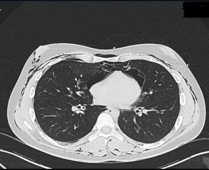
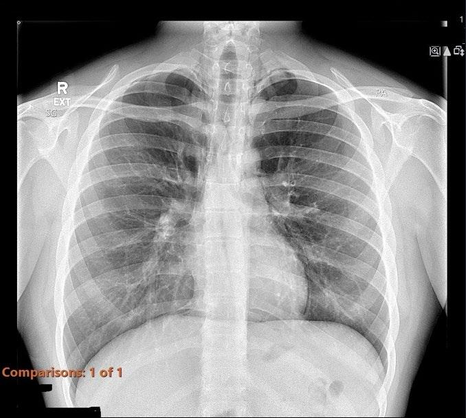
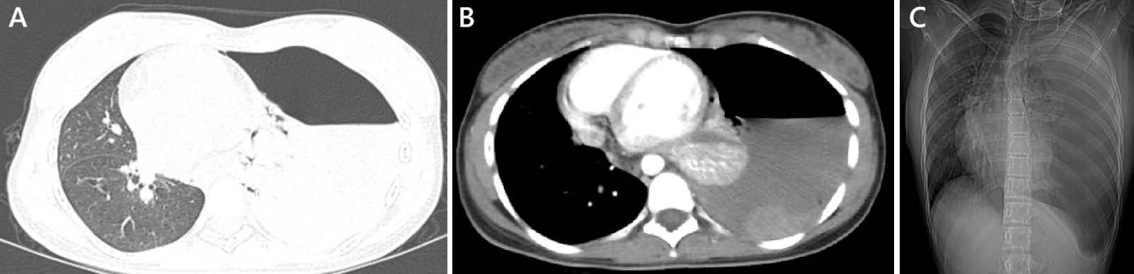
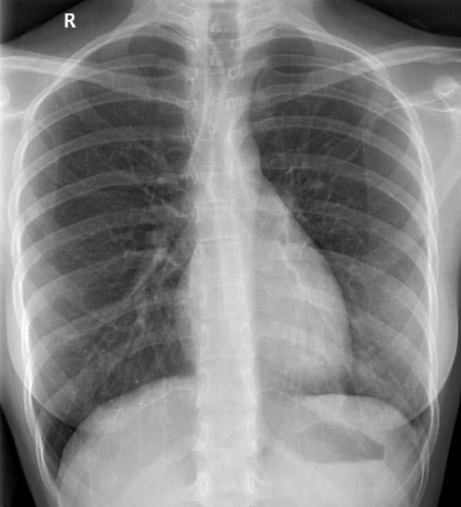
123 Chest Pain Beyond Asthma: A Case of Spontaneous Pneumomediastinum in a Young Adult with Asthma
Chidambaram A
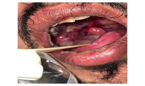
129 Mycoplasma Pneumoniae-Induced Rash and Mucositis in an Adult Male from Saudi Arabia: A Case Report and Literature Review
AlKhawajah S et al.
135 Pneumocystis Jirovecii Pneumonia in Patients Without HIV: A UK 5-Year Retrospective Study
Ebere and Sewell
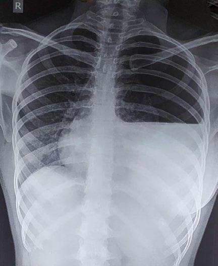
144 Spontaneous Haemopneumothorax in a Young Female: A Case Report of Birt–Hogg–Dubé Syndrome
Baby UR et al.
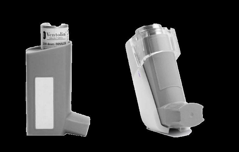
152 Opportunities and Challenges in the Implementation of Smart Inhalers to Improve Asthma Outcomes
Crawford AL et al.
Editorial Board
Editor-in-Chief
Antonio Rossi
Senior Medical Director, Oncology Center of Excellence, Therapeutic Science & Strategy Unit, IQVIA, Milan, Italy
Catharina Belge
University Hospitals Leuven, Belgium
Jacques Bouchard Université Laval, Canada
Andrew Bush
Imperial College London, UK
Nicholas Hill Tufts University School of Medicine, USA
Martin Balzan University of Malta, Malta
Giorgio Walter Canonica Humanitas Research Hospital, Italy
Enrico Clini University of Modena, Italy
Oliver Eickelberg University of Pittsburgh, USA
Atul Gupta King's College London, UK
Dario Olivieri University of Parma, Italy
Neil Holden University of Lincoln, UK
Mohammad Azizur Rahman Dhaka University, Bangladesh
Islam Ibrahim University of California, USA
Paraschiva Postolache
Peter Barnes
Imperial College London, UK
Grigore T. Popa University of Medicine and Pharmacy, Romania
Aims and Scope
EMJ Respiratory is an open access, peer-reviewed ejournal committed to helping elevate the quality of practices in interventional cardiology globally by informing healthcare professionals on the latest research in the field.
The journal is published annually, six weeks after the European Respiratory Society (ERS) Congress, and features highlights from this event, alongside interviews with experts in the field, reviews of abstracts presented at ERS, as well as in-depth features on sessions from this event. The journal also covers advances within the clinical and pharmaceutical arenas by publishing sponsored content from congress symposia, which is of high educational value for healthcare professionals. This undergoes rigorous quality control checks by independent experts and the in-house editorial team.
EMJ Respiratory also publishes peer-reviewed research papers, review articles, and case reports in the field. In addition, the journal welcomes the submission of features and opinion pieces intended to create a discussion around key topics in the field and broaden readers’ professional interests. The journal is managed by a dedicated editorial team that adheres to a rigorous double-blind peer-review process, maintains high standards of copy editing, and ensures timely publication.
EMJ Respiratory endeavours to increase knowledge, stimulate discussion, and contribute to a better understanding of practices in the field. Our focus is on research that is relevant to all healthcare professionals in this area. We do not publish veterinary science papers or laboratory studies not linked to patient outcomes. We have a particular interest in topical studies that advance knowledge and inform of coming trends affecting clinical practice in the respiratory field.
Further details on coverage can be found here: www.emjreviews.com
Editorial Expertise
EMJ is supported by various levels of expertise:
• Guidance from an Editorial Board consisting of leading authorities from a wide variety of disciplines.
• Invited contributors who are recognised authorities in their respective fields.
• Peer review, which is conducted by expert reviewers who are invited by the Editorial team and appointed based on their knowledge of a specific topic.
• An experienced team of editors and technical editors.
Peer Review
On submission, all articles are assessed by the editorial team to determine their suitability for the journal and appropriateness for peer review.
Editorial staff, following consultation with either a member of the Editorial Board or the author(s) if necessary, identify three appropriate reviewers, who are selected based on their specialist knowledge in the relevant area.
All peer review is double blind. Following review, papers are either accepted without modification, returned to the author(s) to incorporate required changes, or rejected.
Editorial staff have final discretion over any proposed amendments.
Submissions
We welcome contributions from professionals, consultants, academics, and industry leaders on relevant and topical subjects. We seek papers with the most current, interesting, and relevant information in each therapeutic area and accept original research, review articles, case reports, and features.
We are always keen to hear from healthcare professionals wishing to discuss potential submissions, please email: editorial.assistant@emjreviews.com
To submit a paper, use our online submission site: www.editorialmanager.com/e-m-j
Submission details can be found through our website: www.emjreviews.com/contributors/authors
Reprints
All articles included in EMJ are available as reprints (minimum order 1,000). Please contact hello@emjreviews.com if you would like to order reprints.
Distribution and Readership
EMJ is distributed through controlled circulation to healthcare professionals in the relevant fields across Europe.
Indexing and Availability
EMJ is indexed on DOAJ, the Royal Society of Medicine, and Google Scholar®; selected articles are indexed in PubMed Central®
EMJ is available through the websites of our leading partners and collaborating societies. EMJ journals are all available via our website: www.emjreviews.com
Open Access
This is an open-access journal in accordance with the Creative Commons Attribution-Non Commercial 4.0 (CC BY-NC 4.0) license.
Congress Notice
Staff members attend medical congresses as reporters when required.
This Publication Launch Date: 2013 Frequency: Yearly Online ISSN: 2054-3166
All information obtained by EMJ and each of the contributions from various sources is as current and accurate as possible. However, due to human or mechanical errors, EMJ and the contributors cannot guarantee the accuracy, adequacy, or completeness of any information, and cannot be held responsible for any errors or omissions. EMJ is completely independent of the review event (ERS 2025) and the use of the organisations does not constitute endorsement or media partnership in any form whatsoever. The cover photo is of Amsterdam, the Netherlands, the location of ERS 2025.
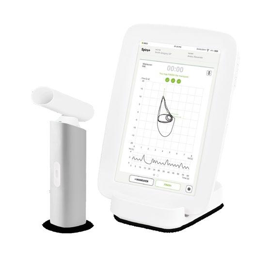
Wireless mode of operation for practice or home visit use – easily expandable with ECG, BP, SpO₂, ABI, and more
Pneumotachograph technology with integrated self-calibration
Automatic best breath selection with a clear and intuitive measurement review
The MESI mTABLET SPIRO delivers quick and reliable results while simplifying my diagnostic workflow.
Dr. Jan-Ove Sanders Germany
BOOK A DEMO
Managing Editor
Darcy Richards
Copy Editors
Noémie Fouarge, Sarah Jahncke
Editorial Leads
Helena Bradbury, Ada Enesco
Editorial Co-ordinators
Bertie Pearcey, Alena Sofieva
Editorial Assistants
Niamh Holmes, Katrina Thornber, Katie Wright, Aleksandra Zurowska
Creative Director
Tim Uden
Design Manager
Stacey White
Senior Designers
Tamara Kondolomo, Owen Silcox
Creative Artworker
Dillon Benn Grove
Designers
Shanjok Gurung, Fabio van Paris
Junior Designer
Helena Spicer
Head of Marketing
Stephanie Corbett
Business Unit Lead
James Murphy
Chief Executive Officer
Justin Levett
Chief Commercial Officer
Dan Healy
Founder and Chairman
Spencer Gore
Welcome
Dear Readers,
We are delighted to welcome you to the 2025 issue of EMJ Respiratory, where we bring you the latest advances from the European Respiratory Society (ERS) Congress 2025, held in Amsterdam, the Netherlands. This year, the Congress put the spotlight on global respiratory health.
Alongside our review of the Congress, you can find several cutting-edge research abstracts, insightful features looking at how the field can reshape the way it thinks about small airway disease and chronic cough, and an exclusive interview with the ERS 2025 Presidential Award winner, Mary Morrell, who discusses sleep-related health and its unmet needs.
Among our peer-reviewed content is a timely review of machine learning advances and how these could transform the way in which interstitial lung disease is managed in the future. You can also discover a thought-provoking feature highlighting recent advancements in the understanding of sex-based differences in respiratory disease and the potential strategies that could aid future research, prevention, and treatment.
Be sure not to miss our infographic covering personalised treatment approaches in asthma, and expert interviews that dive deeper into the topics of pulmonary hypertension associated with lung disease, cellular senescence in COPD, and biomarker-driven precision medicine.
We would like to take this opportunity to thank our Editorial Board, the authors, peer reviewers, and interviewees for their contributions to this comprehensive issue. We hope you find valuable insights to take forward into your own clinical practice.
Contact us
Editorial enquiries: editor@emjreviews.com
Sales opportunities: salesadmin@emjreviews.com
Permissions and copyright: accountsreceivable@emjreviews.com
Reprints: info@emjreviews.com Media enquiries: marketing@emjreviews.com
Foreword
Dear Colleagues,
It is my pleasure to introduce the latest issue of EMJ Respiratory. In this edition, you will find peer-reviewed articles alongside a review of the European Respiratory Society (ERS) Congress 2025, which took place in Amsterdam, the Netherlands, between 27th September–1st October. The hybrid event saw experts from around the globe come together to present research findings and key updates across a variety of disciplines within respiratory medicine.
The theme for this year’s ERS Congress was ‘Respiratory Health Around the Globe’, with the programme including several sessions on this topic, such as the global impact of lung disease from infancy to old age, global health challenges like smoking and air pollution, and how to improve respiratory health through strategies like vaccination and risk identification.
In line with the Congress theme, this issue covers a variety of topics, starting with a feature article which reviews sex differences in obstructive lung disease. The journal also includes an article that explores an AI-enhanced approach to interstitial lung disease and reviews the transformative potential of machine learning advances in the management of this disease.
Additionally, you can find insightful interviews with key opinion leaders in the field of respiratory medicine, covering topics such as a new era in asthma and COPD, precision medicine for airway diseases, and redefining pulmonary hypertension.
The hybrid event saw experts from around the globe come together to present research findings and key updates across a variety of disciplines within respiratory medicine
I would like to take this opportunity to thank all those who have contributed to this issue, including all of the authors, peer reviewers, interviewees, and the Editorial Board. I hope you enjoy reading this journal.
Antonio Rossi Oncology Center of Excellence, Therapeutic Science & Strategy Unit, IQVIA, Milan,
Italy
ERS 2025
Under the theme ‘Respiratory Health Around the Globe’, ERS 2025 highlighted the Society’s commitment to addressing respiratory disease on a truly international scale
Review of the European Respiratory Society (ERS) Congress 2025 Congress Review
FROM 27th September–1st October 2025, Amsterdam, the Netherlands, welcomed over 22,000 respiratory professionals to the European Respiratory Society (ERS) Congress. Set against the city’s historic canals and vibrant streets, the Congress became a dynamic forum for sharing knowledge, fostering collaborations, and exploring the latest advances in respiratory medicine. Under the theme ‘Respiratory Health Around the Globe’, ERS 2025 highlighted the Society’s commitment to addressing respiratory disease on a truly international scale.
The Congress showcased the latest developments across all areas of respiratory medicine, from lung cancer, COPD, and asthma to interstitial lung disease, sleep-disordered breathing, and airway diseases. Delegates explored innovations in early detection, screening programmes, and personalised treatments, alongside emerging therapies aimed at improving outcomes for patients worldwide. Prevention, holistic self-management, and smoking cessation remained central themes, while sessions addressing global disparities, environmental risk factors, and air pollution reinforced the need for collaboration and equitable care across regions.
Sleep and airway medicine was a major focus of the programme, with discussions on disease variability, sex differences, and the impact of environmental and lifestyle factors. Delegates also learned about the latest research on microbiome-targeted therapies in COPD, trajectories of lung function, and potential asthma remission.
Highlighted sessions provided insights into the challenges and opportunities in sleepdisordered breathing, supporting more personalised and precise approaches to patient care.
Sustainability was front and centre throughout the Congress. Attendees were encouraged to explore ways to reduce their environmental footprint, from encouraging greener travel to adopting eco-friendly hospital practices. A special session on sustainable healthcare initiatives in the Netherlands illustrated how collaboration and innovation can benefit both patients and the planet.
Finally, the global dimension of the Congress was evident in every session. Faculty from multiple continents shared their expertise in sessions tagged as ‘global’, highlighting diverse approaches to respiratory medicine and emphasising the importance of international collaboration. The Global Voices initiative provided a platform for people living with respiratory
disease to share their experiences, bringing patient perspectives to the forefront and reinforcing the Congress’s patientfocused mission. Poster sessions, studio discussions, and interactive workshops offered delegates countless opportunities to exchange ideas, network, and translate research into clinical practice.
A special session on sustainable healthcare initiatives in the Netherlands illustrated how collaboration and innovation can benefit both patients and the planet
ERS 2025 also spotlighted the Society’s commitment to supporting early-career professionals. From interactive workshops to mentoring opportunities, young researchers and clinicians had the chance to showcase their work, gain feedback, and connect with global leaders in the field.
Read on for key insights into ERS 2025, and make sure to join us next year as ERS heads to Barcelona, Spain, from the 5th–9th September, 2026.
Exercise-Based Rehabilitation May Restore Immune Balance in Post-COVID Syndrome
AN RCT presented at the ERS Congress 2025 suggests that exercise-based rehabilitation may help correct immune dysregulation in people recovering from severe COVID-19.1
The study, led by Neil Bishop, Loughborough University, UK, in collaboration with researchers from the NIHR Leicester Biomedical Research Centre, investigated how an 8-week structured exercise programme affected immune cell recovery in patients with post-COVID syndrome following hospitalisation.
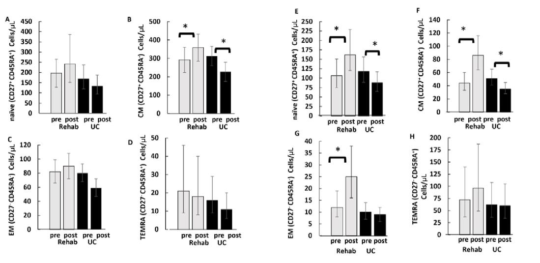
In this assessor-blinded RCT, 31 participants (13 male) were assigned either to an 8-week supervised exercise and education programme (n=13) or to usual care (n=18). The intervention included personalised aerobic and resistance training sessions delivered under clinical supervision. Blood samples were collected at baseline and after 8 weeks to assess CD4+ and CD8+ T cell subsets using flow cytometry, and results were analysed with linear mixed models.
After 8 weeks, only participants in the exercise group showed significant increases in central memory CD4+ T cells and naïve, central, and effector memory CD8+ T cells, suggesting a shift toward a healthier and more balanced immune profile. No comparable changes were seen in the usual care group.
These findings indicate that targeted exercise-based rehabilitation may support the recovery of immune function in individuals with post-COVID syndrome, complementing its well-established benefits for physical performance and quality of life. This approach could represent a promising therapeutic avenue for addressing the lingering immune effects of severe COVID-19 infection.
After 8 weeks, only participants in the exercise group showed significant increases in central memory CD4+ T cells and naïve, central, and effector memory CD8+ T cells
Air Pollution Increases Severity of Obstructive Sleep Apnoea
NEW RESEARCH presented at the ERS Congress 2025 has found that long-term exposure to air pollution may worsen the severity of obstructive sleep apnoea (OSA), a condition characterised by repeated pauses in breathing during sleep.2
The study, which analysed data from across Europe, highlights the potential impact of particulate matter (PM10) on sleeprelated breathing disorders and raises concerns about environmental influences on respiratory health.
Using records from the European Sleep Apnoea Database (ESADA), researchers examined data from 19,325 patients with OSA and linked it with PM10 concentration levels extracted from the Copernicus Atmosphere Monitoring Service. The study assessed how 1-year average exposure to PM10 was associated with OSA severity, measured through key indicators such as the apnoea–hypopnoea index, oxygen saturation, and the time spent with oxygen levels below 90%. Statistical models were adjusted for factors including age, sex, BMI, smoking status, humidity, temperature, and season.
The analysis revealed that for every oneunit increase in long-term PM10 exposure, there was a corresponding 0.41 increase in the apnoea-hypopnoea index (95% CI:
0.21–0.67), indicating a clear link between particulate air pollution and more severe sleep apnoea. However, no significant associations were observed for oxygen saturation or T90, suggesting that air pollution primarily affects the frequency rather than the depth of apnoeic events.
Interestingly, the relationship between PM10 exposure and OSA severity varied significantly between regions, with some European centres showing stronger associations than others. These differences may reflect variations in air quality, urbanisation, or environmental policies across countries.
Overall, the findings support the growing body of evidence connecting air pollution to worsening respiratory and sleep health. They also underline the importance of addressing environmental risk factors in the management of OSA and other chronic conditions influenced by air quality.
For every one-unit increase in longterm PM10 exposure, there was a corresponding 0.41 increase in the apnoea–hypopnoea index
Despite its ‘green’ image, domestic wood combustion has become the primary contributor to PM2.5
Domestic Wood Burning Now Major UK Air Pollution Source
NEW DATA presented at the ERS Congress 2025, combining national property and health datasets, has found that domestic wood burning has become the UK’s leading source of harmful particulate pollution, with solid fuel use concentrated among affluent, older populations, raising challenges for air quality policy and public health messaging.3
Wood is increasingly marketed as a renewable and cheaper energy source; however, new evidence highlights its growing environmental and health burden. Researchers report that emissions from domestic wood burning more than doubled between 2009–2022, from 6,800 tonnes to 14,000 tonnes of fine particulate matter (PM2.5), now exceeding those from road vehicles. The findings underscore an emerging paradox: despite its ‘green’ image, domestic wood combustion has become the primary contributor to PM2.5, a pollutant linked to respiratory diseases such as lung cancer.
Using data from the English Longitudinal Study of Ageing (ELSA; n=11,000), 25 million Energy Performance Certificates (EPC), and UK Census records, the study identified clear sociodemographic and geographic patterns in solid fuel use. Wood-burning appliances were most common in affluent, predominantly White urban areas, including Smoke Control Areas originally established after the 1952 Great London Smog. The ELSA data further
revealed that use of solid fuels doubles with age, even among people with preexisting respiratory disease, suggesting gaps in awareness or risk perception.
Although preliminary analyses showed unexpectedly higher lung function among solid fuel users, likely reflecting socioeconomic differences or selection bias, the authors warn that long-term harms may be underestimated. They propose using EPC data to guide targeted public health interventions aimed at reducing domestic emissions and promoting cleaner heating alternatives. The researchers also note that quasi-experimental designs will be essential to isolate the health effects of wood smoke in high-income countries where exposures are lower but widespread.
The study highlights a pressing need for nuanced air quality strategies that recognise domestic wood burning as an environmental issue. Tailored public health campaigns could help reduce carbon emissions, reduce solid fuel emissions exposure, and address environmental injustice.
Paternal Smoking in Puberty Linked to Accelerated Ageing in Offspring
NEW findings presented at the ERS Congress 2025 suggest that fathers who begin smoking during puberty may pass on signs of accelerated biological ageing to their children, adding a new dimension to the understanding of intergenerational health risks.4
The research, part of the multigenerational RHINESSA study, involved 892 offspring (mean age: 28 years) from across Europe
The research, part of the multi-generational RHINESSA study, involved 892 offspring (mean age: 28 years) from across Europe. It was led by an international collaboration including researchers from the University of Bergen, Norway; University of Southampton, UK; Aarhus University, Denmark; Uppsala University, Sweden; and the University of Melbourne, Australia, among others.
Investigators analysed blood DNA methylation to estimate biological ageing using validated epigenetic clocks (GrimAge, PhenoAge, and DunedinPACE). Paternal smoking histories were obtained directly from fathers, while offspring provided personal smoking data. Linear regression models adjusted for sex and grandparental education were used to assess associations between fathers’ age at smoking initiation, categorised as never, ≤15 years, or >15 years, and biological age acceleration in offspring.
Results showed that offspring whose fathers started smoking before age 15 years had significantly higher biological age estimates compared to those whose fathers
never smoked. GrimAge (β=0.7; 95% CI: 0.03–1.30) and PhenoAge (β=1.0; 95% CI: -0.40–2.30) indicated accelerated ageing in this group, with more pronounced effects when offspring who smoked themselves were excluded. No consistent associations were observed in the maternal line.
These findings suggest that paternal smoking during puberty, a critical developmental window, may influence biological ageing markers in the next generation. The mechanisms may involve epigenetic alterations in germ cells that persist into offspring DNA methylation patterns.
The study adds to growing evidence linking paternal environmental exposures during adolescence with offspring health outcomes and highlights the potential long-term biological consequences of early smoking initiation. Further research is needed to determine whether this accelerated biological ageing mediates previously observed associations with reduced lung function and asthma risk in offspring.
Global Childhood Disease Burden from SecondHand Smoke
AN ANALYSIS presented at the ERS Congress 2025 examined global and regional trends in disease burden among children attributable to second-hand smoke (SHS) exposure from 1990–2021, using updated data from the Global Burden of Disease (GBD) study.5
SHS remains a major cause of preventable childhood illness and death, with WHO estimates indicating over 1.2 million premature deaths annually, including around 65,000 among children under 15 years.
Researchers measured disability-adjusted life years (DALY) linked to SHS exposure across major disease categories from 1990–2021 in children aged 0–14 years. Temporal trends were additionally assessed via estimated annual percentage change, with its 95% uncertainty intervals. Associations with the Socio-demographic Index (SDI), which reflects income, education, and fertility rates, were examined.
In 2021, SHS exposure was responsible for approximately 3.79 million DALYs from lower respiratory infections, 0.80 million from otitis media, and 3.86 million from
respiratory infections and tuberculosis worldwide among children. Findings revealed marked inequalities: low-SDI regions had significantly higher agestandardised DALY rates, 302.43 and 305.40 per 100,000 for lower respiratory infections and respiratory infections/ tuberculosis, respectively, compared to high-SDI regions, which reported rates of just 7.64 and 10.25 per 100,000.
The authors concluded that SHS continues to inflict a severe and unequally distributed toll on children’s respiratory health, particularly in low-SDI regions. They call for strengthened tobacco control measures and targeted health equity initiatives to protect children and reduce preventable harm globally.
Growing Up with Dogs May Protect Against Childhood Asthma
BABIES who grow up in homes with dogs may have a significantly lower risk of developing asthma by the age of 5 years, according to research presented at the ERS Congress 2025.6 The study, led by Makiko Nanishi and presented by Jacob McCoy from The Hospital for Sick Children (SickKids), Toronto, Canada, found no similar protective effect for babies exposed to cats.
The research team analysed data from 1,050 children enrolled in the Canadian CHILD Cohort Study. Dust samples were collected from each child’s home between 3 and 4 months of age, and the researchers measured levels of dog allergen (Can f1), cat allergen (Fel d1), and endotoxin, a bacterial surface molecule. When the children reached the age of 5 years, they underwent clinical assessments for asthma, lung function tests, and genetic analysis for asthma-related risk variants.
The findings revealed that infants exposed to higher levels of Can f1, the major dog allergen, had around a 48% lower risk of developing asthma by the age of 5 years compared with those exposed to lower levels. These children also demonstrated better lung function, particularly those carrying genetic markers associated with poorer lung development. No such association was observed for cat allergen exposure or endotoxin levels, suggesting that early exposure to dog
allergens may play a role in preventing allergic sensitisation or modifying immune development, potentially through changes to the nasal microbiome.
The findings revealed that infants exposed to higher levels of Can f1, the major dog allergen, had around a 48% lower risk of developing asthma by the age of 5 years compared with those exposed to lower levels
The research adds to growing evidence that early-life environmental exposures, including interactions with household pets, can shape immune development and respiratory health, offering new insights into potential strategies for asthma prevention in children.
Online Singing Therapy Enhances Quality of Life in COPD
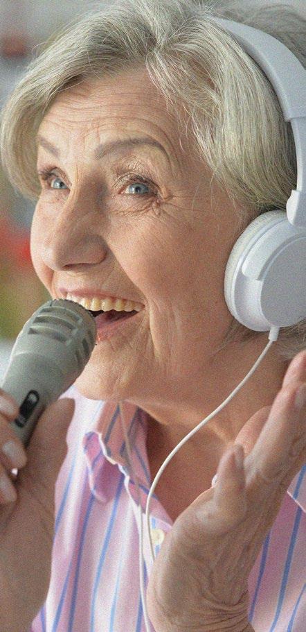
A WORLD-FIRST randomised controlled trial, presented at the ERS Congress 2025, has demonstrated that online group singing can significantly improve quality of life in people living with COPD and interstitial lung disease (ILD).7 Both conditions are characterised by persistent breathlessness and reduced physical and emotional wellbeing, highlighting the need for accessible interventions that can complement medical care.
74 %
Participants were predominantly women (74%), with an average age of 69.6 years
Conducted in Australia, the Phase III trial investigated the impact of a 12-week guided group singing programme delivered entirely online. A total of 101 participants were randomised, with 50 allocated to the singing intervention and 51 to usual care. Participants were predominantly women (74%), with an average age of 69.6 years, and included individuals with both COPD (64%) and ILD (37%). The primary outcome was quality of life, assessed using the SF-36 questionnaire, while secondary outcomes included anxiety, depression, dyspnoea, and loneliness.
Results revealed a meaningful improvement in the singing group, whose SF-36 scores were 7.4 points higher than those of the control group (95% CI: 0.6–14.1; p=0.031).
In participants who attended at least eight sessions, representing 56% of the intervention arm, the difference increased to 11.0 points (95% CI: 3.8–18.3; p=0.003), surpassing the threshold for clinical significance. Subgroup analyses showed even greater benefits among women, those with existing anxiety or depression, and participants with no prior pulmonary rehabilitation experience.
While the intervention did not produce significant changes in secondary outcomes, no adverse events were reported, indicating that online singing is a safe and welltolerated approach.
This study provides compelling evidence that guided, online group singing can enhance quality of life in individuals with chronic lung disease, particularly when participation is consistent. It supports the
integration of creative, non-pharmacological therapies into respiratory care, offering patients an engaging and socially connected way to manage breathlessness and improve wellbeing.
Large UK Study Shows Fruit Intake May Mitigate the Effects of Air Pollution
A LARGE cross-sectional study presented at the ERS Congress 2025 has revealed that higher fruit intake may help mitigate the harmful effects of fine particulate air pollution on lung function, with the greatest benefit seen among women who consume four or more fruit portions per day.8
Chronic exposure to air pollution is strongly linked to reduced lung function and elevated respiratory disease risk. Independent of pollution, a healthy diet has been shown to enhance lung function. Therefore, this study sought to determine whether a healthy diet can offset the adverse impacts of air pollution, focusing on fruit, vegetable, and whole grain consumption.
The researchers analysed data from the UK Biobank, consisting of approximately 150,000 adults, with complete information on diet, lung function, and air pollution exposure. Lung function was assessed via Forced Expiratory Volume in 1 second (FEV1) and Forced Vital Capacity (FVC), while annual average ambient particulate matter (PM2.5) at each participant’s residence was modelled based on data from the 2010 EXPANSE project. A healthy diet score was constructed based on reported intake, and linear regression models adjusted for sex, age, height, BMI, income, education attainment, alcohol intake, smoking, passive smoking, physical activity, and ethnicity.
The results demonstrated that participants in the highest healthy diet score category had higher FEV1 values than those in the lowest category (+41.7 mL; 95% CI: 35.4–48.1), showing that a healthy diet was associated with better lung function. Conversely, increased air pollution corresponded with lower lung function. Whilst interactions between healthy diet and lung function were not found, an interaction between total fruit intake and air pollution was observed in females (p=0.004) who had higher average intakes than men. Additionally, women with the highest fruit intake showed a reduced impact of PM2.5 exposure, with lung function declining by just 57.5 mL per 5 μg/m³ increase in PM2.5, compared to 78.1 mL in the low fruit intake group. Men did not show a statistically significant protective effect.
Women with the highest fruit intake showed a reduced impact of PM2.5 exposure
The study demonstrates that while a healthy diet is beneficial for lung function irrespective of pollution exposure, high fruit intake in particular appears to mitigate the adverse effects of air pollution among women. Further research should explore long-term trajectories, underlying mechanisms, and how dietary promotion can be integrated into respiratory disease prevention strategies.
A MULTICENTRE cohort study presented at the ERS Congress 2025 investigated the long-term impact of remote monitoring (RM) on healthcare consumption and asthma control in paediatric patients across four Dutch hospitals from 2017–2023.9 While RM is increasingly used in respiratory care, its real-world, large-scale effects remain underexplored.
A total of 1,278 children aged 6–18 years, each with at least 2 years of follow-up, were included. Of these, 687 (53.8%) used RM during the study period. Data on outpatient visits, emergency visits, hospitalisations, and asthma control were compared between RM and regular care groups. Analyses included incidence rate ratios (IRR), number needed to treat (NNT), and interrupted time series analysis (ITSA) to evaluate trends over time following RM adoption.
Patients in the RM group had significantly fewer outpatient visits, with a median reduction of 0.62 visits per year (p<0.001). Emergency visits were reduced by 45% (IRR: 0.55; 95% CI: 0.44–0.68; NNT: 24), and hospitalisations by 58% (IRR: 0.42; 95% CI: 0.30–0.59; NNT: 35), favouring RM. ITSA indicated a quarterly reduction of 0.90
outpatient visits per 100 patients after RM implementation. Asthma control markedly improved, with the proportion of wellcontrolled cases rising from 68.6% to 90.1% within 43 months of RM introduction.
The findings demonstrate that RM in routine paediatric asthma care can reduce healthcare utilisation while achieving high rates of asthma control over the long term. These results support RM as an effective tool to optimise respiratory care and resource use in children, aligning with the growing focus on digital health strategies and AI in clinical practice.
Asthma control markedly improved, with the proportion of well-controlled cases rising from 68.6% to 90.1% within 43 months of RM introduction
Robotic Bronchoscopy with Cone-Beam CT Boosts Lung Lesion Diagnosis
A RANDOMISED controlled trial presented at the ERS Congress 2025 demonstrates that robotic-assisted bronchoscopy (RAB) combined with cone-beam CT (CBCT) dramatically improves diagnostic yield compared with conventional bronchoscopy for detecting peripheral pulmonary lesions (PPL).10
The trial, conducted at University Hospital Zurich, Switzerland, compared RAB+CBCT using the ion endoluminal system with conventional bronchoscopy employing ultrathin bronchoscopes and 2D fluoroscopy. A total of 78 patients with 127 PPLs were enrolled from June–November 2024 and randomised 1:1 to either procedure group. The primary endpoint was diagnostic yield, defined per the American Thoracic Society (ATS) 2024 consensus criteria.
Results revealed a substantial advantage for the RAB+CBCT approach. Diagnostic yield reached 84.6% (33/39) compared to only 23.1% (9/39) for conventional bronchoscopy, an absolute difference of 61.5% (95% CI: 44.1–78.9%; P<0.001).
Among non-diagnostic conventional cases, 92.9% were successfully diagnosed upon subsequent RAB+CBCT, either within the same anaesthesia event or during a separate session. Median lesion diameter was 11 mm, with 27.6% classified as pure ground-glass opacities. Importantly, adverse event rates were similar across both groups (p=0.09).
These findings position robotic-assisted bronchoscopy integrated with CBCT as a transformative diagnostic tool for peripheral lung lesions
Overall, 53.5% of all lesions were diagnosed as lung cancer, with nearly 40% identified at Stage IA, highlighting the potential of RAB+CBCT to facilitate earlier diagnosis and improve clinical outcomes. These findings position robotic-assisted bronchoscopy integrated with CBCT as a transformative diagnostic tool for peripheral lung lesions. Its enhanced precision, especially for small or ground-glass lesions, could lead to significant stage shifts in lung cancer detection and management.
The investigators note that continued evaluation of procedural efficiency, costeffectiveness, and training implications will be essential as RAB+CBCT moves toward wider clinical adoption.
References
1. Bishop NC et al. Effect of 8-week exercise-based rehabilitation on immune cell counts in Post-COVID syndrome following hospitalisation: a randomised controlled trial. Abstract OA6534. ERS Congress, 27 September-1 October, 2025.
2. Pengo M et al. Association between air pollution and sleep disordered breathing severity across Europe. Abstract PA5662. ERS Congress, 27 September-1 October, 2025.
3. Horsfall L et al. The rising threat of domestic wood burning to Europe’s respiratory health: a case study using longitudinal data from England and Wales. PA5850. ERS Congress, 27 September–1 October, 2025.
4. López-Cervantes JP et al. Father’s smoking initiation in puberty as associated with accelerated ageing in offspring. Abstract PA3770. ERS
Congress, 27 September-1 October, 2025.
5. Jin R et al. Global trends and regional differences in the burden of disease attributable to secondhand smoke exposure in children during 1990-2021: an analysis of the Global Burden of Disease study. Abstract OA5371. ERS Congress, 27 September-1 October, 2025.
6. Nanishi M et al. Association of earlylife dust allergens and endotoxin with childhood asthma and lung function: an analysis of the CHILD study. Abstract OA1265. ERS Congress, 27 September-1 October, 2025.
7. Smallwood N et al. The SINFONIA study: a phase III randomised controlled trial of an online group singing intervention for people with COPD or ILD. Abstract RCT2291. ERS Congress, 27 September-1 October, 2025.
8. Kaewsri P et al. Does diet modify the effects of air pollution on lung function? A large cross-sectional study. Abstract PA495. ERS Congress, 27 September-1 October 2025.
9. Oppelaar M et al. Assessment of healthcare consumption and asthma control after implementation of remote monitoring in long-term multicentre paediatric asthma care. Abstract OA2346. ERS Congress, 27 September-1 October, 2025.
10. Steinack C et al. Robotic-assisted bronchoscopy with integrated cone-beam CT vs. conventional bronchoscopy for diagnosing peripheral pulmonary lesions: an open-label randomized controlled trial. Abstract OA 1178. ERS Congress, 27 September-1 October 2025.
Counting Coughs: ERS 2025 Highlights on Objective Cough Monitoring
Authors: *Mindaugas Galvosas,1 Peter M. Small1,2
1. Research & Development Department, Hyfe, Inc., Wilmington, Delaware, USA
2. Department of Global Health, University of Washington, Seattle, USA
*Correspondence to mindaugas@hyfe.com
Disclosure:
Galvosas and Small are employees at and stock option holders of Hyfe, Inc.
Keywords: Cough, cough monitoring, digital health.
THE EUROPEAN Respiratory Society (ERS) Congress 2025 showcased continuous cough monitoring across sensor modalities, a pragmatic 7-day monitoring standard, and early signals for efficacy and tolerability in real studies, positioning cough as a scalable biomarker and clinical endpoint for respiratory care.
WHY COUGH, WHY NOW
In precision health, clinically meaningful parameters are quantified to individualise therapies and track response. Cough, one of the most common reasons for seeking care and a key respiratory sign, has historically been hard to measure reliably: 24-hour 'snapshots' are noisy, day-to-day variability is high, and manual annotation is slow, inconsistent, and privacy-intrusive. The ERS Congress 2025 highlighted how continuous, largely automated, and privacypreserving technologies now enable objective, at-scale cough measurement suitable for clinical research and care.
AUTOMATED APPROACHES SIGNAL RAPID PROGRESS IN COUGH DETECTION AND QUANTIFICATION
The Automated Cough Counting Algorithm (ACCA) demonstrated strong agreement with ground truth, showing high sensitivity (97%) and a positive predictive value exceeding 75% across heterogeneous
clinical conditions (refractory chronic cough, COPD, idiopathic pulmonary fibrosis, interstitial lung disease, and asthma), along with a low median absolute error compared to human-annotated counts.1 Presenters noted that such models may surpass human annotators in consistency by eliminating inter-rater variability and enabling faster data analysis.1 Such validated automated algorithms for cough counting can greatly accelerate clinical development and further expand the use of cough monitoring in trials.
Another signal processing algorithm for identifying coughs in respiratory audio recordings was presented during the Congress. The RESP biosensor-based algorithm (Strados Labs, Philadelphia, Pennsylvania, USA), trained on audio recordings from multiple chronic cough conditions (cough variant asthma, atopic cough, eosinophilic bronchitis, and refluxrelated cough), demonstrated high precision (94.9%) and sensitivity (95%), and strong correlation with human annotation, with a mean absolute error of 3.26 coughs/hour.2
A new cough monitoring wearable device (C-mo, Caparica, Portugal) validation reported the performance of an automated cough monitoring algorithm against expertannotated reference, adding another modality to the growing objective cough monitoring ecosystem. The wearable chest-patch device uses electromyography to trigger audio recording during cough events and was evaluated for feasibility in patients with acute cough wearing the device for up to 4 hours, showing 93.7% sensitivity for detecting cough events in real-world environments.3 The same poster featured another group of study subjects wearing the device for 24 hours, showing 95% sensitivity in cough detection performance.3
A smartwatch-based, automated, and fully privacy-preserving cough-counting monitor (Hyfe Inc., Delaware, USA) was also featured across multiple posters. As shown earlier this year by Chaccour et al.,4 the correlation between manually annotated (human) and cough-counting monitormeasured hourly cough counts was very high, with a Pearson correlation coefficient of 0.99. The monitor’s on-device algorithm achieved an overall sensitivity of 90.4% with 1.03 false positives per hour as users went about their usual activities.4
It was evident at ERS 2025 that presented cough monitoring approaches differ in sensor placement, acoustic and
accelerometric features used to enhance adherence, and in whether they rely on post-processed audio or fully privacypreserving on-device processing with no audio recordings, with several technologies now showing strong agreement with human annotations and readiness for scaled use.
FROM RAW COUNTS TO ENDPOINTS: ANALYTICS THAT DETECT CHANGE
This year’s event also emphasised the importance of how cough counts (e.g., cough timestamps) are analysed after detection. Patients with cough often report that periods of intense coughing (termed bouts or epochs) are the most distressing aspect, as “everyone notices that you’ve got a cough,” and derived measures such as cough bouts and cough-free time remain exploratory endpoints in many clinical trials.
The RESP biosensor-based algorithm demonstrated high precision (94.9%) and sensitivity (95%), and strong correlation with human annotation, with a mean absolute error of 3.26 coughs/hour.
In a TRPM8-agonist (AX-8) study5 using an audio recording system with humanannotated cough events (Vitalograph, Ennis, Ireland), investigators categorised cough bout metrics into count- and duration-based hierarchies to assess treatment-induced changes in cough patterns. Placebo reduced mean coughs per bout while maintaining bout frequency, whereas treatment reduced bout frequency while maintaining mean coughs per bout. Similarly, placebo shortened the mean bout duration without affecting frequency, while treatment reduced frequency without altering duration, suggesting that countbased metrics might provide detailed granularity, whereas duration-based metrics may yield complementary insights in specific clinical contexts.5
This year’s event also emphasised the importance of how cough counts are analysed after detection
Another group presented a head-to-head comparison between a log-transformed linear mixed model and a negative binomial generalised linear mixed-effects model (GLMM) for handling skewed and overdispersed cough frequency data, and
assessing their impact on treatment interpretation. Using 24-hour cough frequency data collected with an audio recording system, the analysis showed that log-transformation reduced skewness and produced more precise estimates than assuming a negative binomial distribution, without altering the overall interpretation of treatment effects.6
Multi-day cough monitoring featured prominently during the Congress. Evidence supporting a 7-day standard showed that most participants with problematic cough reached accurate frequency estimates within 7 days.7 This duration captures inter- and intraindividual cough variability, enhancing the accuracy of cough-related endpoints in research, as well as variation that may result from daily habits. 'Metronomic' phenotypes stabilised sooner; highly variable phenotypes needed closer to 8 days, supporting 7 days as a pragmatic default balancing statistical stability and operational feasibility.6 While future work may explore tailored monitoring durations for specific populations, a 7-day period is recommended as the default for most studies involving cough frequency assessment.7
Future research should establish standardised definitions for cough bouts and other timestamp-derived clinical outcome assessments, as current presentations use varying definitions, which remains a significant limitation.
OPERATIONAL READINESS: ADHERENCE, PRACTICALITY, AND PRIVACY
Continuous endpoints are only as good as the time truly monitored. A poster presented at ERS 2025 highlighted that the use of an on-device photoplethysmography-based wear detector enabled the aforementioned smartwatch-based cough-counting monitor (Hyfe Inc., Delaware, USA) to achieve 98.93% overall accuracy for wear versus non-wear classification (sensitivity: 98.94%; specificity: 98.92%; negative predictive value: 99.45%; positive predictive value: 97.94%) across 28,185 5-minute observations.8 The study also found that accurate wear detection allows objective, device-verified adherence (including onwrist via photoplethysmography sensor and bedside charging), supports per-protocol and missing-data strategies, and reduces reliance on self-report.8
Given recent advancements in continuously evaluating adherence to cough monitoring, future studies could explore how adherence
metrics correlate with participants’ adherence to prescribed interventions. Additionally, researchers could examine whether adherence insights gathered during screening could be leveraged to train participants for improved compliance in subsequent study phases.
Several platforms presented privacypreserving designs (e.g., on-device processing and no storage of raw conversational audio). This directly addresses concerns from patients and Institutional Review Boards about audio data handling.
Future studies should compare adherence across various device form factors, such as smartwatches, pendants, and adhesive sensors placed on the abdomen or chest, and assess how adherence is influenced by user age and study duration.
APPLICATIONS OF CONTINUOUS COUGH MONITORING: SIGNALS OF EFFICACY AND TOLERABILITY
Objective monitoring is already finding treatment signals and flagging tolerability issues. An open-label exploratory trial with azithromycin was presented at the event. In this trial, 30 patients with chronic respiratory disease who underwent continuous smartwatch monitoring (Hyfe
Inc., Delaware, USA), showed a significant reduction in median coughs/hour by Week 4 (p<0.01), alongside improvements in patient-reported outcomes and 'relief-ofcough' time.9 Notably, the trajectory of change was visible from Week 1, supporting continuous cough endpoints for early signal detection.9
Continuous cough monitoring to assess tolerance to inhaled antibiotics in chronic bronchial infection was also presented by a group from Spain. Among eight participants monitored with a coughmonitor smartwatch (Hyfe Inc., Delaware, USA), individual cough trajectories across 'before–during–after' periods showed progressive increases in cough in those who did not tolerate treatment, followed by reductions after discontinuation, illustrating how continuous cough monitoring can serve as an objective tolerability marker complementing patient-reported symptoms.10 In the future, continuous cough monitoring insights could help personalise treatment approaches by identifying those that are best tolerated and most effective for individual patients.
Lastly, a poster on patient-reported outcomes versus objective cough frequency was presented. This longitudinal analysis examined day-to-day variability in VAS/NRS and objective cough frequency captured via a cough monitoring pendant (SIVA Health, Zurich, Switzerland).11 Correlations were modest (0.42 for VAS and objective cough frequency; and 0.41 for NRS and objective cough frequency), and observations were patient-specific, underlining that PROs and objective counts are complementary.11
To date, most work has focused on chronic cough, but there is growing interest in extending objective cough
References
1. Ferreira J et al. Automated cough counting algorithm for chronic cough: accelerating clinical development. Poster PA453. ERS Congress, 27 September-1 October, 2025.
monitoring to additional respiratory and systemic conditions.
CONCLUSIONS
ERS 2025 data show that objective cough monitoring has crossed a practical threshold: algorithms now demonstrate high agreement with human annotation, devices are accessible and privacyaware, adherence can be verified at scale, and week-scale windows yield stable, responsive endpoints. These capabilities are already implemented across observational studies and interventional drug trials in chronic cough and related conditions, and broader adoption of objective cough endpoints across therapeutic areas now appears both scientifically justified and operationally foreseeable.
In the future, clinical studies to compare and contrast the advantages and limitations of various devices, such as watches, pendants, and adhesives, in terms of accuracy and acceptability are needed. In addition, we can expect that continuous monitors may not only count coughs but also classify them; for example, distinguishing between dry and wet coughs. ‘Coughomics’ could further enable the endotyping of patients with chronic cough: some may display a neurogenic profile (high cough frequency, normal lung function, heightened cough reflex sensitivity), while others show an inflammatory profile (moderate cough frequency, abundant sputum, and airway inflammation). Once considered a background symptom, cough is now emerging as a valuable data stream, and continuous monitoring is what makes this transformation possible.
2. deLaubenfels T et al. Automated cough identification in chronic cough from real-world recordings by a custom machine learning algorithm. Poster PA461. ERS Congress, 27 September-1 October, 2025.
3. Neuparth N et al. Validation of a new automated cough monitoring device: results of a pilot study. Poster PA455. ERS Congress, 27 September-1 October, 2025.
4. Chaccour C et al. Validation and accuracy of the Hyfe cough monitoring system: a multicenter clinical study. Sci Rep. 2025;15(1):880.
5. Taylor T et al. Novel cough bout metrics to support assessment of treatment response to AX-8. Poster PA3594. ERS Congress, 27 September-1 October, 2025.
6. Kum E et al. Comparison of statistical methods for analyzing cough frequency in a trial of refractory chronic cough. Poster PA466. ERS Congress, 27 September-1 October, 2025.
7. Rudd M et al. Optimizing cough frequency assessment: a 7-day monitoring standard. Poster PA4969. ERS Congress, 27 September-1 October, 2025.
8. Kosharnyi M et al. Objective assessment of adherence in
continuous cough monitoring: wear detection on the Hyfe CoughMonitor smartwatch. Poster PA2876. ERS Congress, 27 September-1 October, 2025.
9. Sykes D et al. Azithromycin and continuous cough monitoring: an open-label trial. Poster PA469. ERS Congress, 27 September-1 October, 2025.
10. Santisteve S et al. Continuous cough monitoring to evaluate tolerance to inhaled antibiotic treatment in patients
with chronic bronchial infection. Poster PA3875. ERS Congress, 27 September-1 October, 2025.
11. Gomez Camblor J et al. Correlations between patient-reported outcomes and objective cough frequency: insights from longitudinal monitoring using a novel digital device. Poster PA468. ERS Congress, 27 September-1 October, 2025.
ERS 2025: From Growth to Decline –How Small Lungs Shape the Future of Respiratory Health
Author: *Ourania S. Kotsiou¹
1. Laboratory of Human Pathophysiology, Department of Nursing, University of Thessaly, Larissa, Greece
*Correspondence to okotsiou@uth.gr
Disclosure: The author has declared no conflicts of interest.
Keywords:
Asthma, COPD, European Respiratory Society (ERS) 2025, interstitial lung disease, low lung volumes, lung function, small airways disease.
Citation: EMJ Respir. 2025;13[1]:30-32.
https://doi.org/10.33590/emjrespir/UCOR4266
THIS YEAR'S European Respiratory Society (ERS) Congress 2025, held in Amsterdam, the Netherlands, placed a strong spotlight on the ‘forgotten zone’ of the respiratory system: the small airways. The session entitled ‘Small lungs, big consequences: the origins, consequences and clinical implications of low lung volumes’ captivated clinicians and researchers alike, urging a paradigm shift from traditional spirometry-centred perspectives to a holistic appreciation of distal airway physiology.
SMALL LUNG SYNDROME: THE NEED TO RECLASSIFY CHRONIC LUNG DISEASE
Opening the session, Peter Burney, Imperial College London, UK, called for a fundamental reclassification of chronic lung diseases around lung size and developmental potential, rather than solely spirometric ratios. He argued that “the term ‘obstructive’ has served us well, but it obscures the fact that many patients begin life with small lungs, not damaged ones.”
Drawing on population studies, Burney proposed that ‘small lung syndrome’ represents a common pathway linking asthma, COPD, and restrictive patterns through impaired growth and airway remodelling. Early-life nutrition, pollution, and maternal smoking were identified as root causes, demanding a shift from late-stage diagnosis to lifelong lung health monitoring.1,2
TRACKING LUNG FUNCTION AND RESPIRATORY DEVELOPMENT FROM
BIRTH TO HEALTHY OLD AGE
Shyamali Dharmage, University of Melbourne, Australia, presented compelling longitudinal data from the TAHS and CHILD cohorts,3,4 tracking lung function trajectories from birth into late adulthood. Her findings revealed that low maximal lung function in early adulthood predicts premature respiratory decline, regardless of smoking or occupational exposure.
Many patients begin life with small lungs, not damaged ones
She underscored that early interventions targeting respiratory infections, allergens, and environmental pollutants during critical
developmental windows could alter lifetime lung trajectories. The message resonated strongly: the foundation for chronic lung disease is laid not in midlife, but in the first 1,000 days of life.
SMALL LUNGS: IMPLICATIONS FOR LOW- AND MIDDLE-INCOME COUNTRIES
Obianuju Ozoh, University of Lagos, Nigeria, shifted the focus to global inequities, revealing the high prevalence of small lungs in low- and middle-income countries. Her multi-country analyses showed that reduced lung volumes, often interpreted as restrictive or mixed patterns, may reflect stunted lung growth due to malnutrition, biomass exposure, and early-life infections rather than classic interstitial disease.5
Ozoh warned that misclassification of small lungs as ‘normal variants’ perpetuates underdiagnosis and undertreatment in vulnerable populations.6 She urged the ERS community to adopt regionally appropriate reference values and advocate for lung health equity as a global priority.
ARE CLINICIANS MISSING TREATABLE CARDIOVASCULAR DISEASE IN PATIENTS WITH LOW FORCED VITAL CAPACITY?
Closing the session, Christer Janson, Uppsala University, Sweden, highlighted a powerful clinical overlap: low forced vital capacity not only predicts respiratory morbidity, but also signals unrecognised cardiovascular disease.
Citing epidemiological data, he demonstrated that individuals with low forced vital capacity, even in the absence of airflow limitation, show higher rates of hypertension, heart failure, and metabolic syndrome.7 He called for integrated screening strategies combining spirometry with cardiovascular risk assessment, emphasising that “low lungs may be the first warning sign of a failing heart.”
CONVERGING THEMES: A PARADIGM SHIFT
Across these talks, one theme was unmistakable: lung volume matters. Whether
The foundation for chronic lung disease is laid not in midlife, but in the first 1,000 days of life
References
1. Agustí A, Faner R. Lung function trajectories in health and disease. Lancet Respir Med. 2019;7(4):358-64.
2. Schultz ES et al. Early life determinants of lung function change from childhood to adolescence. Respir Med. 2018;139:48-54.
3. Bui DS et al. Childhood predictors of lung function trajectories and future COPD risk: a prospective cohort study
reduced by early developmental insults, environmental exposures, or systemic comorbidities, small lungs translate into big clinical consequences.
Speakers agreed that traditional dichotomies (obstructive versus restrictive, paediatric versus adult) are blurring. The future may demand a new taxonomy of lung disease rooted in developmental biology, environmental context, and systemic interaction.
Individuals with low forced vital capacity, even in the absence of airflow limitation, show higher rates of hypertension, heart failure, and metabolic syndrome
CONCLUDING REMARKS
As the lights dimmed in the Amsterdam Congress Centre, the audience left with a renewed appreciation that the smallest lungs may tell the biggest stories. The session’s unifying message was clear: “Rethinking small airways disease means rethinking what we call normal.”
For clinicians and researchers alike, ERS 2025 marked a turning point, from treating damaged lungs to understanding how they grow, adapt, and interact across the lifespan. The challenge now lies in transforming these insights into early detection, equitable diagnostics, and therapies that truly breathe life into global respiratory health.
from the first to the sixth decade of life. Lancet Respir Med. 2018;6(7): 535-44.
4. Dai R et al. Wheeze trajectories: determinants and outcomes in the CHILD Cohort Study. J Allergy Clin Immunol. 2022;149(6):2153-65.
5. Ozoh OB et al. Pulmonary dysfunction among adolescents and adults with sickle cell disease in Nigeria: implications for monitoring. Ann Thorac Med. 2019;14(4):269-77.
6. Meghji J et al. Improving lung health in low-income and middle-income countries: from challenges to solutions. Lancet. 2021;397(10277):928-40.
7. Janson C et al; BOLD Collaborative Research Group. Lung function and onset of cardiometabolic diseases in the longitudinal Burden of Obstructive Lung Disease study. BMJ Open Respir Res. 2025;12(1):e002442.
Vaping Versus Smoking: A Debate Session and ERS Guidance
Author: Katrina Thornber, EMJ, London, UK
Citation: EMJ Respir. 2025;13[1]:33-37.
https://doi.org/10.33590/emjrespir/DECB3629
THIS YEAR at the European Respiratory Society (ERS) Congress 2025, the Pro-Con debate session titled ‘Are e-cigarettes effective smoking cessation tools or public health hazards?’ sparked a lively discussion on a controversial topic. Experts presented the latest research on the use of e-cigarettes, or vapes, as they are commonly referred to, through the lenses of respiratory health, harm reduction, and population-level risk.
INTRODUCTION
The session, chaired by Stamatoula Tsikrika, Medical School of the National and Kapodistrian University of Athens, Greece; and Didier Cataldo, University of Liège, Belgium, brought together opposing perspectives on a rapidly evolving issue: e-cigarettes as a ‘healthier’ alternative to smoking. To tackle a topic which encompasses respiratory health, public health, and socio-political issues, experts were asked to present opposing perspectives on the role of e-cigarettes in smoking cessation and public health. Tsikrika opened by acknowledging the polarising narrative surrounding e-cigarettes: initially promoted as a safer alternative to smoking, they are now under scrutiny for their addictive nature, unknown long-term risks, and rising use among younger populations.
Speaking in favour of e-cigarettes, Hayden McRobbie, Professor of Population Health, Queen Mary University, London, UK, argued that we cannot ignore that e-cigarettes are one of the most effective smoking cessation tools. McRobbie prefaced his talk by acknowledging that e-cigarettes are by no means a magic cure, but they are less harmful than smoking.1 His position is that e-cigarettes reduce smoking prevalence, but
he admitted that there needs to be tighter regulation, with strict restrictions on access for non-smokers, particularly young people. On the other hand, Aslı Görek Dilektaşlı, Department of Pulmonary Medicine, Uludag University Faculty of Medicine, Bursa, Türkiye, presented evidence challenging the notion that vaping serves as an effective smoking cessation strategy. She emphasised the significant health risks associated with e-cigarette use and outlined why it does not constitute a beneficial alternative to conventional smoking.
TOBACCO VERSUS VAPING: WHICH IS MORE HARMFUL?
McRobbie argued that smoked tobacco is “public health enemy number one.” In particular, he emphasised the cumulative decline in lung function, particularly forced expiratory volume in 1 second, attributable to smoking.2 He stressed that cessation, even later in life, slows this decline. Acknowledging the risks associated with e-cigarettes, McRobbie emphasised that e-cigarettes are most beneficial for current smokers, but for non-smokers, they are associated with increased harm. However, according to the Royal College of Physicians (RCP), using medicinal nicotine is classed as low harm risk, whereas combustible tobacco
is high risk.1 Additionally, McRobbie argued that any studies reporting an association between e-cigarette use and respiratory illness lack causality, as “practically all vapers were smokers or ex-smokers.”
He also highlighted that in late 2019, over 2,800 cases of severe lung injury in the USA were initially linked to vaping, termed e-cigarette or vaping product use-associated lung injury, but were later found to be linked to tetrahydrocannabinol-containing devices rather than vaping nicotine products.3
Conversely, Dilektaşlı addressed the emerging health risks associated with e-cigarettes. Despite perceptions of reduced risk, e-cigarettes deliver nicotine alongside hundreds of toxic and carcinogenic compounds. Nicotine itself is linked to metabolic dysregulation, cardiovascular strain, and neurological impairment, and in vitro studies suggest chronic vaping may induce renal, cardiac, and hepatic fibrosis, and trigger pro-inflammatory responses in the airway epithelium.4,5
In addition to the toxic effects of nicotine, the flavouring agents used in e-cigarettes may drive cytotoxicity, as shown by a study in embryonic adult models.6
Clinically, Dilektaşlı expressed that patients frequently report cough and throat irritation, while epidemiological data associate e-cigarette use with increased risks of bronchitis, asthma, and e-cigarette or vaping product use-associated lung injury.7,8 Additionally, a large Korean cohort (over four million individuals) reported a 2.7fold increase in lung cancer among former smokers using e-cigarettes.9 Furthermore, in the absence of long-term safety data, vaping cannot be endorsed as a cessation aid, she urged.
Despite perceptions of reduced risk, e-cigarettes deliver nicotine alongside hundreds of toxic and carcinogenic compounds
ARE E-CIGARETTES AN EFFECTIVE SMOKING CESSATION TOOL?
McRobbie explained that modern e-cigarette products can deliver very similar blood nicotine levels to smoking, meaning they can act as a good replacement and a harm reduction approach.10 He reviewed current scientific evidence supporting the use of e-cigarettes for cessation, including data from a systematic review consisting of multiple RCTs comparing nicotine-containing e-cigarettes with conventional nicotine replacement therapy (NRT). In this systematic review, the authors concluded that e-cigarettes were more effective than NRT in promoting smoking cessation.11 However, McRobbie emphasised that evidence remains limited regarding differences in the incidence of serious adverse events between the two approaches, urging the need for ongoing safety monitoring. He also highlighted research that accounted for socioeconomic factors, referencing a study by Courtney et al.,12 explaining that individuals from
lower socioeconomic groups face greater challenges in quitting smoking. In this context, the use of e-cigarettes was shown to be more effective than single-form NRT (gum or lozenge) in achieving biochemically validated 6-month abstinence rates.
Whilst McRobbie implored that smoking prevalence is still high, Dilektaşlı argued that global tobacco use has been steadily declining for decades, and this decline was achieved before the introduction of e-cigarettes in the UK in 2007. Beyond the direct health risks of vaping, there are broader public health concerns. Specifically, there are three well-established public health harms linked to e-cigarettes: the gateway effect, dual use, and relapse risk among former smokers.
The gateway effect refers to the increased likelihood of initiating smoking following e-cigarette use. In a large cohort study, 30-day e-cigarette use was associated with a four-to-six-fold increase in the odds of starting smoking, providing clear evidence of this effect.13 Furthermore, the study demonstrated an eight-fold increase in the odds of continued smoking, indicating a pattern of dual use among new smokers. Finally, exposure to e-cigarettes was linked to a three-to-five-fold increase in the risk of relapse among former smokers, demonstrating that e-cigarettes are not an effective way to quit smoking.
Another significant public health concern regarding e-cigarettes is their role in sustaining nicotine addiction at the population level, Dilektaşlı argued. A study utilising data from three UK birth cohorts (1950s, 1970s, and Millennium) demonstrated this clearly.14 Across these cohorts, overall smoking prevalence declined from 33% to 13%. When the Millennium cohort was stratified by e-cigarette use, predicted smoking
30-day e-cigarette use was associated with a four-to-six-fold increase in the odds of starting smoking
probabilities revealed that youth who never used e-cigarettes exhibited very low smoking risk, whereas current e-cigarette users had a 33% likelihood of initiating smoking. These findings depict that e-cigarettes cause sustained nicotine addiction at the population level.
Dilektaşlı next drew attention to the limitations of studies supporting e-cigarettes as cessation tools. One of the most cited trials, led by McRobbie, compared e-cigarettes to user-selected NRT combined with 4 weeks of behavioural counselling.15 At 1 year, abstinence was 18% for those in the e-cigarette group versus 10% for those receiving NRT. However, a closer examination revealed that four out of five participants in the e-cigarette group were still vaping at the end of the first year. This raises the question: ‘Can we really call it smoking cessation, or is it just product switching?’.
Additionally, several design features biased this trial in favour of e-cigarettes, Dilektaşlı argued. Participants could select device type and flavour, perhaps encouraging continued engagement, whereas NRT was provided in fixed quantities. This may explain the difference in adherence rates between groups: 53% in the e-cigarette group versus 10% in the NRT group. Dilektaşlı also identified limitations in several systematic reviews that have been published in favour of e-cigarettes. For example, in one review, only 10 of 90 included trials were at low risk of bias, follow-up periods were short, and real-world cohort studies were excluded. She implored that real world data show
a contrasting picture: smokers who use e-cigarettes are 28% less likely to quit compared with smokers who did not vape.16
From a public health perspective, Dilektaşlı argued that endorsing e-cigarettes aligns with the financial interests of the tobacco industry, re-normalises smoking behaviour, and enables the industry to position itself as a ‘solution’ to a crisis it created. Dilektaşlı stated: “History reminds us how doctors once stood in cigarette advertisements claiming safety. So do we really want to be recalled by our future colleagues in the same way?” She concluded that e-cigarettes pose substantial public health risks, and do not meet the principle of “first, do no harm.” Instead, policy should prioritise nicotine-free abstinence and evidence-based cessation strategies.
EUROPEAN RESPIRATORY SOCIETY REAFFIRMS OPPOSITION TO E-CIGARETTES
Tsikrika concluded the provocative debate with a reminder of the stance of the ERS. She stated that the ERS does not endorse novel tobacco products or nicotine products, including e-cigarettes, as a safe and effective tool for tobacco cessation. Instead, the safest option for a smoker to quit smoking is to use counselling and evidencebased medicine. The ERS emphasises the fact that these products pose health risks, contribute to nicotine dependence, and that they should be under strict regulation to protect public health.
References
1. Royal College of Physicians (RCP). E-cigarettes and harm reduction: an evidence review. 2024. Available at: https://www.rcp.ac.uk/media/n5skyz1t/ e-cigarettes-and-harm-reduction_fullreport_updated_0.pdf. Last accessed: 15 October 2025.
2. McRobbie H, Kwan B. Tobacco use disorder and the lungs. Addiction. 2021;116(9):2559-71.
3. Centers for Disease Control and Prevention (CDC). Outbreak of lung injury associated with the use of e-cigarette, or vaping, products. Available at: https://archive.cdc. gov/#/details?url=https://www.cdc. gov/tobacco/basic_information/ecigarettes/severe-lung-disease.html. Last accessed: 15 October 2025.
4. Bhatnagar A et al. Electronic cigarettes: a policy statement from the American Heart Association. Circulation. 2014;130(16):1418-36.
5. Alexander LEC et al. Chronic inhalation of e-cigarette vapor containing nicotine disrupts airway barrier function and induces systemic inflammation
and multiorgan fibrosis in mice. Am J Physiol Regul Comp Physiol. 2018;314(6):R834-47.
6. Bahl V et al. Comparison of electronic cigarette refill fluid cytotoxicity using embryonic and adult models. Reprod Toxicol. 2012;34(4):529-37.
7. Cho JH, Paik SY. Association between electronic cigarette use and asthma among high school students in South Korea. PLoS One. 2016;11(3):e0151022.
8. Joshi D et al. Impact of electronic cigarette ever use on lung function in adults aged 45-85: a crosssectional analysis from the Canadian Longitudinal Study on Aging. BMJ Open. 2021;11:e051519.
9. Kim YW et al. Association of electronic cigarette use after conventional smoking cessation with lung cancer risk: a nationwide cohort study. Am J Resp Crit Care Med. 2024;209:A3051.
10. Hajek P et al. Nicotine delivery to users from cigarettes and from different types of e-cigarettes. Psychopharmacology (Berl). 2017;234(5):773-9.
11. Lindson N et al. Electronic cigarettes for smoking cessation. Cochrane Database Syst Rev. 2024;1(1):CD010216.
12. Courtney RJ et al. Vaporized nicotine products for smoking cessation among people experiencing social disadvantage: a randomized clinical trial. Ann Intern Med. 2025;178(8):1085-94.
13. McMillen R. E-cigarette use and future cigarette initiation among never smokers and relapse among former smokers in the PATH study. Public Health Rep. 2019;134(5):528-36.
14. Mongilo J et al. Risk of adolescent cigarette use in three UK birth cohorts before and after e-cigarettes. Tob Control. 2025;DOI:10.1136/TC-2024059212.
15. Hajek P et al. A randomized trial of e-cigarettes versus nicotinereplacement therapy. N Engl J Med. 2019;380(7):629-37.
16. Kalkhoran S, Glantz SA. E-cigarettes and smoking cessation in real-world and clinical settings: a systematic review and meta-analysis. Lancet Respir Med. 2016;4(2):116-28.
Challenges in Advanced Lung Disease: Perspectives on Alpha 1 Antitrypsin Deficiency
and Bronchiectasis
This industry symposium took place during the European Respiratory Society (ERS) International Congress held in Amsterdam, the Netherlands, from 27th September–1st October 2025
Support: The publication of this article was funded by CSL Behring. The views and opinions expressed are exclusively those of the speakers.
Chairs: Noel Gerard McElvaney,1 Michal Shteinberg2
Speakers: Noel Gerard McElvaney,1 Michal Shteinberg,2 Sanjay H. Chotirmall,3 Emily F.A. van ‘t Wout,4 Thomas5
1. Royal College of Surgeons in Ireland (RCSI), University of Medicine and Health Services, Dublin, Ireland
2. Pulmonology Institute and CF Center, Carmel Medical Center and Technion- Israel Institute of Technology, The B. Rappaport Faculty of Medicine, Haifa, Israel
3. Lee Kong Chian School of Medicine, Nanyang Technological University (NTU), Singapore
4. Department of Pulmonology, Leiden University Medical Centre, the Netherlands
5. German AATD Patient Group, Flensburg, Germany
Disclosure: McElvaney has received grants and/or research support from Chiesi, CSL Behring, Grifols, Kamada, pH Pharma, and Vertex; and honoraria and/or consultation fees from Dicerna, Inhibrx, Intellia, and Vertex. Shteinberg has received grants and/or research support from GSK, Novartis, Trudell Medical International, Tel Aviv League for Lung Diseases, and the G. Baum Foundation; honoraria and/or consultation fees from AstraZeneca, Boehringer Ingelheim, CSL Behring, Dexcel Pharma, GSK, Insmed, Kamada, Novartis, Sanofi, Synchrony Medical, Teva, and Zambon; and is on the management board of the European Multicenter Bronchiectasis Audit and Research Collaboration (EMBARC) and the Israeli Society for Tuberculosis and Nontuberculosis Mycobacterium. Chotirmall has received grants and/ or research support from the Singapore Ministry of Education, the Singapore Ministry of Health’s National Medical Research Council, and the Singapore’s National Research Foundation; and honoraria and/or consultation fees from Astra Zeneca, Boehringer Ingelheim, Chiesi Farmaceutici, CSL Behring, GSK, Inovio Pharmaceutics Inc., Imam Abdulrahman Bin Faisal University, Pneumagen Ltd, Sanofi, and Zaccha Pte Ltd. van ‘t Wout has received grants and/or research support from Alpha-1 Foundation, the Brus Foundation, ZonMw, and Stichting Alpha1 International Registry; and honoraria and/or consultation fees from Airna, Chiesi, CSL Behring, GSK, and Tessera Therapeutics. Thomas has received expense allowances from CSL on several occasions as a patient expert, in accordance with German law; and is a board member of the patient organisation Alpha1 Deutschland e.V., which has received funds from public sources and from the pharmaceutical industry. The surname of Thomas has not been disclosed to maintain anonymity.
Acknowledgements: Writing assistance was provided by Nicola Humphry, Nottingham, UK.
Taking place during the 2025 European Respiratory Society (ERS) International Congress held in Amsterdam, the Netherlands, this symposium presented up-to-date expert perspectives on the challenges of advanced lung disease, focusing on alpha 1 antitrypsin deficiency (AATD) and bronchiectasis.
Experts discussed the latest findings regarding the real-world management of AATD, including how to assess lung and liver function in patients, when and how to initiate augmentation therapy, and the importance of exercise and pulmonary rehabilitation. The current understanding of bronchiectasis was discussed, and experts shared their thoughts on differentiating disease severity from disease activity, and the value of precision medicine in managing the disease.
Key messages from the symposium included the importance of recognising that AATD can manifest with extra-pulmonary disease, the growing evidence supporting a survival benefit of augmentation therapy in AATD, and the rapid advances in the field of bronchiectasis that are revealing new treatment targets and opportunities for precision medicine.
Perspectives on Alpha 1 Antitrypsin Deficiency
One of the most common genetic diseases, AATD, involves the retention of misfolded alpha 1 antitrypsin (AAT) in hepatocytes, which can cause liver cirrhosis. In case of an inflammatory insult, the consequent deficiency of AAT in the circulation results in unopposed protease activity, perpetuating inflammation and leading to progressive tissue damage, including emphysema in the lungs and panniculitis in the skin (Figure 1).1,2
The latest findings regarding the real-world management of AATD were discussed by Gerry McElvaney, Professor of Medicine, Royal College of Surgeons in Ireland (RCSI), University of Medicine and Health Sciences, Dublin, Ireland; and Emily F.A. van ‘t Wout,
Assistant Professor and treating physician, Department of Pulmonology, Leiden University Medical Centre, the Netherlands.
Real-World Alpha 1 Antitrypsin
Deficiency: What to Monitor, Measure, and Manage in the Clinic
Assessing lung function
McElvaney presented data from a 2024 Delphi consensus of 103 members of the European Alpha 1 Research Collaboration (EARCO) group, which focused on the optimal follow up of patients with AATD and advanced lung disease.3
In terms of assessing lung function in worsening disease (defined as one or more of moderate-severe dyspnoea,
Created with BioRender.com.
Adapted from McElvaney et al.2
1–2 exacerbations per year requiring hospitalisation, or forced expiratory volume in one second [FEV1] decline of ≥50 mL/year), the panel agreed that both pre- and post-bronchodilator spirometry measurements should be performed at baseline, whereas subsequent measurements could be limited to postbronchodilator application.3 Spirometry parameters should include FEV1, forced vital capacity (FVC), the FEV1/FVC ratio, inspiratory vital capacity (VC), maximal VC, and inspiratory capacity. To assess lung decrement prior to changes in FEV1 and FVC, forced expiratory flow rates (FEF25, FEF50, and FEF75) as a percentage of FVC, and maximal expiratory flow rates (MEF75, MEF50 and MEF25) as a percentage of remaining VC, should also be measured. Body plethysmography parameters should include total lung capacity (TLC), residual volume, residual volume/TLC, airway resistance, specific airway resistance, functional residual capacity, and functional residual capacity/ TLC. Chest X-ray should be performed only when clinically indicated, and arterial blood
gas parameters (pH, partial pressure of O2, partial pressure of CO2, hydrogen carbonate ion) should be assessed via arterial puncture.
While the panel consensus was that FEV1 should be used to follow up patients with AATD, McElvaney stressed that changes in this parameter may be minimal among patients with lung disease at diagnosis (lung-index cases), since real-world evidence suggests that these patients experience a plateau in FEV1 decline from their mid-40s (Figure 2).4
When and how should augmentation therapy be initiated?
The EARCO panel agreed that patients should have AAT levels <11 µM and CTconfirmed emphysema prior to initiation of AAT augmentation therapy, and that patients should abstain from smoking for >6 months.3 McElvaney also noted that AAT levels alone are not sufficient to make a diagnosis of AATD and should be accompanied by genotyping, phenotyping, and/or gene sequencing as indicated. The
Figure 1: Sites of disease in alpha 1 antitrypsin deficiency.
Healthy liver
Healthy lungs
Healthy skin
Liver cirrhosis
Emphysema
Panniculitis
Forced expiratory volume in one second decline is non-linear, with an inflection point in the late 40s and different trajectories for lung-index and non-lung index patients.
Adapted from Fraughen et al.4
FEV1: forced expiratory volume in one second.
age of the patient and the deterioration of FEV1 should also be considered, and it was generally agreed that therapy should not be initiated following lung transplantation. During augmentation therapy, clinicians should consider an extended dosing interval (off-label; follow local guidelines), and AAT levels should be monitored at trough levels whenever there are changes in dose or treatment interval, or significant changes in the patient’s BMI.3
In a large observational study, augmentation therapy has been shown to confer a survival advantage in AATD,4 with a substantial, unadjusted life-years gain of 6.9 years versus standard care over a 50-year horizon.5 However, longitudinal lung function measurements identified distinct AATD phenotypes.4 In non-lung-index patients, augmentation therapy was not associated with a reduction in FEV1 decline, while in lung-index patients, the only group for whom therapy was associated with a significant reduction of decline was those with relatively well-preserved lung function.4 McElvaney emphasised that these data further highlight the potential difficulties of using FEV1 as a primary follow-up measure.
What about alpha 1 antitrypsin deficiency liver disease?
Liver disease associated with AATD can range from mild, self-limiting cholestasis in infancy to chronic hepatitis, cirrhosis, hepatocellular carcinoma (HCC), or even fulminant hepatic failure.6 Approximately 4% of children with AATD experience lifethreatening liver disease,6,7 and McElvaney stressed that infants with severe liver disease may enter a ‘honeymoon period’ where they experience normal growth and few signs or symptoms of the disease, followed by renewed progressive injury and decompensation in their teenage years.6 McElvaney emphasised the need for early involvement of hepatologists in the care of people with AATD.
Risk factors for liver fibrosis in adults with protease inhibitor ZZ (PiZZ)-genotype
AATD include age >50 years, male sex, obesity, diabetes, high BMI, and metabolic syndrome.8 A retrospective review found that 7.9% of explant livers from adults undergoing liver transplantation had periodic acid-Schiff globules suggestive of AATD, yet less than two-thirds of these patients were tested for AATD, indicating
Figure 2:
that diagnosis of AATD is often overlooked in this patient population.9
Vibration-controlled transient elastography is the preferred non-invasive test for liver fibrosis.10 While magnetic resonance elastography was considered highly sensitive and specific for detecting advanced fibrosis, this technique is limited by cost and lack of general availability.10 Traditional ultrasound can be useful in screening for HCC or liver cirrhosis; the detection of steatosis; the identification of nodular, structural, or biliary liver disease; and the monitoring of fibrosis progression in patients with BMI >35 kg/m2, but it is considered to have limited sensitivity for differentiating progression in early fibrosis.10 Recommendations regarding the frequency of liver monitoring depend on the degree of fibrosis. Mild fibrosis should be evaluated every 2–3 years, whereas moderate-tosevere fibrosis should be evaluated every 6–12 months.10
The transition from F1 (portal fibrosis without septa) to F2 (portal fibrosis with few septa) on the Meta-analysis of Histological Data in Viral Hepatitis (METAVIR) scale is considered to be the most important aspect of risk stratification for liver disease in AATD.10 Clinical signs of advanced liver disease (including portal hypertension, hepatic encephalopathy, and sarcopenia) or HCC may indicate that a patient with AATD should be considered for liver transplant.10
Does alpha 1 antitrypsin deficiency always present with lung or liver disease?
To emphasise that not all incidents of AATD present with one of these two common manifestations, McElvaney described the case of a 23-year-old male who initially presented to a tertiary referral centre with an acutely swollen right knee and joint effusion (mainly neutrophilic). The patient was discharged on non-steroidal antiinflammatory medication; however, 8 weeks later, he presented to a different centre with chest pain and severe dyspnoea, and was found to have bilateral pleural effusions (low pH exudate, mainly neutrophilic). C-reactive protein levels remained very high after attempted drainage of the
effusions, and he was admitted to the ICU with respiratory failure. At this stage, the patient recalled that he had had jaundice as an infant and had been told he had a rare disease. The medical team tested for AATD, which led to a diagnosis of acute serositis/ panniculitis secondary to PiZZ AATD. Due to inflammation, the patient’s AAT levels were unusually high. Treatment with high-dose intravenous AAT was initiated (off-label), and the patient’s symptoms resolved within 12–24 hours. When the patient presented again, 6 weeks later, with a diffusely swollen, tender right upper limb, AAT augmentation therapy was reinstituted, and the patient remains on maintenance high-dose therapy.
The Nuts and Bolts of Caring for Patients with Alpha 1 Antitrypsin Deficiency
van ‘t Wout explained that AAT mutations can vary considerably, and in the most frequent phenotype, PiZZ, this results in both retention of AAT in the liver (leading to liver disease) and a deficiency in circulating AAT (predisposing individuals to early onset emphysema).1
However, van ‘t Wout emphasised that, while medical textbooks state that emphysema in AATD is typically panacinar and basal, in real life, ‘classical’ centrilobular emphysema of the upper lobes can also be found, especially with increasing age.1 Patients may also present with other pulmonary manifestations, such as bronchiectasis, asthma, or vasculitis associated with anti-neutrophil cytoplasmic antibody.1,11 Of note, in a retrospective cohort study in the USA, only 5.6% of patients with newly diagnosed COPD and liver disease were tested for AATD between 2012–2021,12 and the testing rates for patients with asthma and bronchiectasis are even lower.
Inhalation therapy
Inhalation therapy is commonly used to treat pulmonary symptoms of AATD, although no AATD-specific therapies are available. van ‘t Wout explained that clinicians generally follow COPD guidelines regarding the use of long-
acting β-agonist or muscarinic antagonists to treat their patients with AATD.13-15 In cases with comorbid asthma, inhaled corticosteroids may also be used.14 This can increase FEV1 and exercise capacity, and decrease dyspnoea.16 Treatment may also depend on the phenotype of the AATDassociated lung disease, since frequent exacerbations are associated with a greater decline in lung function compared with infrequent exacerbations,17 and bacterial colonisation of the lower respiratory tract is associated with a substantially increased risk of exacerbations compared with no colonisation.18 van ‘t Wout cautioned clinicians to consider the impact of inhaler choice on climate change, and stressed that pressurised measured dose inhalers contain hydrofluorocarbons, which impact global warming.19
Alpha 1 antitrypsin augmentation therapy
AAT augmentation therapy in PiZZ AATD has been associated with a significant reduction in lung density loss, but with less impact on exacerbation frequency and quality of life (QoL).20,21 Findings vary in terms of the impact of augmentation therapy on FEV1 decline,22,23 with differences potentially depending on the AAT variant (van ‘t Wout, unpublished data). Real-world, long-term data show that augmentation therapy is associated with reduced mortality,4 but not with improvements in QoL.24
Exercise and pulmonary rehabilitation
High levels of sedentary behaviour are associated with poorer cardiometabolic health, whereas high levels of physical activity are associated with improved cardiometabolic health.25 Given that patients with AATD-associated severe COPD are generally younger than those with usual COPD,26 it is crucial to support this population to engage in physical activity.25
Pulmonary rehabilitation can help to improve QoL in patients with COPD.27 However, differences in muscle adaptation to pulmonary rehabilitation exercise regimens have been observed in patients with AATDassociated COPD versus those with usual COPD,28 and further research is needed to
understand the impact of this intervention in patients with AATD.29
While high-intensity and moderate-intensity exercise training appear to be equally effective on exercise capacity, QoL, and dyspnoea in patients with AATD-associated COPD,28 high-intensity exercise training may be associated with greater benefits in terms of psychological comorbidities.30 This is an important consideration in this population, as anxiety and depression may be common in AATD.11,30,31
van ‘t Wout concluded that AATD is a heterogenous disease, and that treatment is partly dependent on phenotype and genotype. Clinicians should encourage physical activity in their patients with AATD-associated lung disease, as this can improve exercise capacity, QoL, and dyspnoea. van ‘t Wout also emphasised that treating physicians should not overlook the psychological impact of AATD.
Perspectives on Bronchiectasis
Bronchiectasis is defined as a chronic, abnormal dilation of the bronchi accompanied by classical symptoms.32,33 Typically caused by chronic airway inflammation and/or infection, the disease can be associated with multiple comorbidities, including airway diseases (asthma, COPD, chronic rhinosinusitis), cardiovascular disorders, and gastrooesophageal reflux disease. Anxiety and depression are common comorbidities in bronchiectasis.31,32,34,35
The silent drivers of progression in bronchiectasis and the value of precision medicine in managing the disease were discussed by Michal Shteinberg, Clinical Associate Professor of Medicine, Technion, Israel Institute of Technology, and the Pulmonology Institute and CF Centre, Carmel Medical Centre, Haifa, Israel; and Sanjay H. Chotirmall, Associate Professor of Molecular Medicine, Lee Kong Chian School of Medicine, Nanyang Technological University, Singapore.
Silent
Drivers of Progression in Bronchiectasis: What are We Still Missing?
Shteinberg discussed how to classify the severity of bronchiectasis, how to decide on treatment goals, and how to determine the risk for complications.
What is severe bronchiectasis?
Disease severity is a reflection of the lung damage that bronchiectasis has already caused, measured mainly through a combination of FEV1, extent of radiological dilatation, and the presence of emphysema.36,37 Several different disease severity scores are used to assess bronchiectasis, including the Bronchiectasis Severity Index (BSI);38 FEV1, Age, Colonisation, Extension, Dyspnoea (FACED);39 and extended FACED (E-FACED).40 Disease severity scores classify bronchiectasis as mild, moderate, or severe, and can be used to predict mortality and hospitalisations.38-40 However, each system uses different parameters and different weighting.38-40 In addition,
Shteinberg emphasised that severity scores are heavily influenced by age, and young people are therefore scored as having milder disease than older individuals with similar parameters.
Disease activity, on the other hand, is a reflection of current airway inflammation and can be used to predict disease progression.36,37 Common measures of disease activity include sputum purulence, frequency of exacerbations, severity of daily symptoms, neutrophil elastase concentrations, and blood or sputum eosinophil counts.36
The green colour in the sputum of patients with chronic inflammatory lung disease is thought to reflect the accumulation of myeloperoxidase, an enzyme released during neutrophilic inflammation.37 Sputum purulence can be used for the noninvasive assessment of disease activity in bronchiectasis, and to predict the future risk of exacerbations and mortality.41
Figure 3: Identifying specific endotypes in bronchiectasis.
The severity of daily symptoms can also be used to predict future pulmonary exacerbations.42 For example, real-world data from the EMBARC registry (N=9,466) showed that for each previous exacerbation, and for each 10-point reduction in QoL score, the rate ratios for future exacerbations were 1.11 (p<0.0001) and 1.10 (p<0.0001), respectively.42
What are our most impactful treatment goals?
Shteinberg explained that clinical trials in bronchiectasis most often demonstrate efficacy through achieving microbiology goals and reducing pulmonary exacerbations, but that improvements in lung function and QoL are less common, despite QoL being important to patients in real life.43,44 In addition, patients who do respond with FEV1 improvement often do not have a corresponding QoL improvement, and vice versa.45 Clinical trial data also demonstrate a significant placebo effect on QoL, with 26–56% of patients who receive placebo showing clinically important improvements in QoL.45
Who are the patients at high risk for complications?
In patients with bronchiectasis, comorbidities such as COPD, connective tissue disease, inflammatory bowel disease, and asthma are associated with a particularly high mortality risk.34 The Bronchiectasis Aetiology Comorbidity Index (BACI), in which comorbidities are weighted according to their impact on mortality, can be used to predict 5-year mortality rate, hospitalisation, exacerbations, and healthrelated QoL, and can inform decisions about which patients should be closely followed up.34 Comorbidity with rheumatoid arthritis occurs in 1–23% of patients with bronchiectasis, and is associated with more frequent exacerbations, higher disease severity, and elevated mortality compared to those without rheumatoid arthritis.46
Symptom duration and congenital aetiologies have been shown to correlate with disease severity in bronchiectasis, indicating that the longer a patient has been living with symptoms, the more severe the disease.47 Patients with paediatric-
onset bronchiectasis in the EMBARC registry (n=249) were shown to have a longer disease duration, worse lung function, greater radiological extent, higher bacterial infection rate, and an increased exacerbation frequency compared to those with adult-onset bronchiectasis (n=1,173).
Symptom duration was also shown to be independently associated with exacerbation frequency, hospitalisations, Pseudomonas infection, and lung function. Congenital aetiologies such as primary ciliary dyskinesia and primary immune deficiency also impacted disease severity.
Shteinberg emphasised the importance of looking for the silent drivers of severity alongside severity scores. This includes markers of disease activity such as sputum purulence, symptom score, and exacerbation frequency, and predictors of complications such as comorbidities, longer symptomatic disease duration, and congenital aetiologies.
Bronchiectasis: Is Precision Medicine the Answer?
There has been a significant increase in the number of global registries and clinical trials for bronchiectasis over the past 20 years,48 accompanied by an exponential growth in the quantity and quality of bronchiectasis research.49
Chotirmall explained that the key lesson learned is that bronchiectasis has a high degree of clinical heterogeneity, yet there remain several unanswered questions. For example, more research is needed to understand the fundamental origins, mechanisms, and natural history of bronchiectasis, potentially different types of the disease, whether any are reversible, and if some have origins in early life.
The understanding of the pathogenesis of bronchiectasis has evolved from a ‘vicious cycle’ of disease to a ‘vicious vortex’ model, in which infection, inflammation, and airway dysfunction intersect concurrently, rather than sequentially, with structural disease.35,50 The ‘vicious vortex’ model provides a framework to develop personalised pharmaceuticals
for bronchiectasis, such as muco-active agents to treat airway dysfunction, antiinflammatory agents to treat inflammation, and antimicrobial agents to treat infection.35 Chotirmall emphasised that in precision medicine, it is necessary to use the right intervention at the right time for each individual patient to get the best treatment response.
Multi-omics technologies, covering genomics, epigenomics, transcriptomics, metabolomics, and microbiomics, support the individualisation of therapeutic options through the analysis of integrated datasets, permitting the identification of the type and severity of disease, and predicting individual treatment response.51 Multi-omics endotype profiling (Figure 3) can offer the potential to identify novel therapeutic targets, as well as treatable and targetable traits in each individual patient with bronchiectasis.
Chotirmall explained that once clinical disease traits have been identified at the individual patient level, and based on the ‘vicious vortex’ framework, disease targets can be prioritised. For example, a patient might have a clinical trait of frequent exacerbations, an inflammatory trait of neutrophilic inflammation, or a microbial trait of chronic Pseudomonas infection.35 Each of these characteristics can be treated with macrolides, dipeptidyl peptidase 1 inhibition, or eradication therapies, respectively; however, it is important to remember that once treatment is initiated, the same patient’s disease may evolve to develop different traits, and precision treatment will need to be reassessed.48
It is advisable to consider a broad range of clinical disease traits as having potential for treatment in clinical practice, since a complex intersection of disease traits exists in bronchiectasis, and targeting one aspect of the disease may have an unexpected impact on others. As an example, Chotirmall described a therapeutic agent currently in development, nebulised polyclonal human IgG, that demonstrated an exploratory endpoint of a reduction in bacterial sputum load in patients with noncystic fibrosis bronchiectasis in a Phase
I study (NCT04643587).53 A Phase II trial to further explore a potential therapeutic benefit with a primary endpoint of reduced exacerbations is ongoing (NCT07048262).54
Chotirmall summarised four steps in the personalised medicine approach to bronchiectasis:
• Understand that the disease presents with different forms and complexity in individual patients.
• Use unique algorithms to support a precision approach to different disease targets based on the ‘vicious vortex’ framework.
• Consider precision medicine an ongoing dynamic process that requires periodic reassessment.
• Continuously monitor the disease domains, severity (multi-dimensional scores), activity (sputum purulence, exacerbations, body weight), and impact (cough, dyspnoea, comorbidities), as part of a holistic precision medicine approach to management.
The Patient Voice: Living with Advanced Lung Disease
Thomas, a patient representative from Flensburg, Germany, discussed living with advanced lung disease from his own perspective. He was diagnosed with AATD at the age of 36 years, with an FEV1 of 70% at the time. Ten years later, his FEV1 had declined by 20%, prompting the initiation of ongoing augmentation therapy. Today, at the age of 65 years, Thomas’ FEV1 is 35%, constituting a 15% decline over nearly 20 years, which he feels shows that augmentation therapy has been beneficial.
Thomas explained that training is one of the most important ways to get the most out of his damaged lungs. He pursues three main training goals: strengthening his auxiliary muscles to maximise his lung capacity, increasing/maintaining his endurance to stabilise his overall physical condition, and general strength training to maintain muscle mass.
He also considers rehabilitation to be important. He, however, finds inpatient programmes to be more effective than outpatient programmes because they allow patients to focus intensively without having to cope with everyday life at the same time. Thomas has had four sessions of 3–4-week inpatient rehabilitation over the past 20 years, and uses the essential exercises and behaviours he has learned to help him through daily life. He also performs pulmonary rehabilitation techniques at his local gym and has regular connective tissue massages.
Thomas explained that he has a great deal of autonomy in managing his disease, including self-administration of his AAT therapy, and he is supported in this by his family doctor. He considers the optimal approach to be a combination of substantial patient autonomy and self-management, complemented by close collaboration with the family doctor in more challenging situations.
References
1. Strnad P et al. Alpha1-antitrypsin deficiency. N Engl J Med. 2020;382(15):1443-55.
2. McElvaney OF et al. Alpha-1 antitrypsin deficiency: current therapy and emerging targets. Expert Rev Respir Med. 2023;17(3):191-202.
3. Miravitlles M et al. Assessment and monitoring of lung disease in patients with severe alpha 1 antitrypsin deficiency: a European delphi consensus of the EARCO group. Respir Res. 2024;25(1):318.
4. Fraughen DD et al. Augmentation therapy for severe alpha-1 antitrypsin deficiency improves survival and is decoupled from spirometric decline-a multinational registry analysis. Am J Respir Crit Care Med. 2023;208(9):964-74.
5. McElvaney NG et al. Real-world evidence for severe alpha-1 antitrypsin deficiency: life-years gained for augmentation therapy versus standard care. Late-Breaking Abstract 29725. ERS Congress, 27 September-1 October, 2025.
Key Takeaways
AATD is a heterogenous disease, and treatment partly depends on phenotype and genotype. While AATD is commonly associated with pulmonary manifestations such as emphysema,1,11 it is important to recognise that it can also present as liver disease6 or panniculitis.2 There is increasing evidence to support a substantial survival benefit of AAT augmentation therapy in patients with AATD,4,5 yet the association with FEV1 decline is unclear.4,22,23 To improve QoL in patients with AATD-associated lung disease, clinicians should encourage physical activity.25,30
The past 25 years have seen substantial growth in the understanding of bronchiectasis.48,49 We are entering an exciting period in the field, with the emergence of mature international registries, advances in multi-omics technologies and endotype-based classification, and new pipeline therapeutics (Chotirmall, personal communication). The coming years will likely see the emergence of a new era of precision medicine in bronchiectasis.
6. Suri A et al. Alpha-1 antitrypsin deficiency liver disease. Clin Liver Dis. 2022;26(3):391-402.
7. Sveger T et al. The liver in adolescents with alpha 1-antitrypsin deficiency. Hepatology. 1995;22(2):514-7.
8. Clark VC et al. Clinical and histologic features of adults with alpha-1 antitrypsin deficiency in a non-cirrhotic cohort. J Hepatol. 2018;69(6):1357-64.
9. Shah RS et al. Alpha-1 antitrypsin deficiency is under-recognized in individuals with cirrhosis undergoing liver transplantation. Eur J Gastroenterol Hepatol. 2021;33(1S Suppl 1):e233-8.
10. Clark VC et al. Diagnosis and monitoring pathways using noninvasive tests in patients with alpha-1 antitrypsin deficiency-associated liver disease: results from an expert delphi panel. United European Gastroenterol J. 2025;DOI: 10.1002/ueg2.70009.
11. Miravitlles M et al. Disease burden associated with alpha-1 antitrypsin deficiency: systematic and structured literature reviews. Eur Respir Rev. 2022;31(163):210262.
12. Riley L et al. Testing patterns and disparities for alpha-1 antitrypsin deficiency. Am J Med. 2023;136(10):1011-7.
13. Glaister P et al. Alpha 1 antitrypsin augmentation for alpha 1 antitrypsin deficiency associated lung disease. Cochrane Database Syst Rev. 2024;10(10):CD015930.
14. Izquierdo M et al. Alpha-1 asthma overlap syndrome: a clinical overview. Curr Allergy Asthma Rep 2022;22(9):101-11.
15. Barjaktarevic I, Campos M. Management of lung disease in alpha-1 antitrypsin deficiency: what we do and what we do not know. Ther Adv Chronic Dis. 2021;12_ suppl:20406223211010172.
16. Corda L et al. Inhaled corticosteroids as additional treatment in alpha-1antitrypsin-deficiency-related COPD. Respiration. 2008;76(1):61-8.
17. Hiller AM et al. Decline in FEV1 and hospitalized exacerbations in individuals with severe alpha-1 antitrypsin deficiency. Int J Chron Obstruct Pulmon Dis. 2019;14:1075-83.
18. Spittle DA et al. Bacterial colonisation doubles the risk of exacerbation in alpha-1 antitrypsin deficiency. Respir Med. 2025;240:108025.
19. Wilkinson A et al. The environmental impact of inhalers for asthma: a green challenge and a golden opportunity. Br J Clin Pharmacol. 2022;88(7):3016-22.
20. Chapman KR et al. Intravenous augmentation treatment and lung density in severe α1 antitrypsin deficiency (RAPID): a randomised, double-blind, placebo-controlled trial. Lancet. 2015;386(9991):360-8.
21. McElvaney NG et al. Long-term efficacy and safety of α1 proteinase inhibitor treatment for emphysema caused by severe α1 antitrypsin deficiency: an open-label extension trial (RAPID-OLE). Lancet Respir Med. 2017;5(1):51-60.
22. Schouten IGM et al. Long-term effect of α1-antitrypsin augmentation therapy on the decline of FEV1 in deficient patients: an analysis of the AIR database. ERJ Open Res. 2021;7(3):00194-2021.
23. Seersholm N et al. Survival in relation to lung function and smoking cessation in patients with severe hereditary alpha 1-antitrypsin deficiency. Am J Respir Crit Care Med. 1995;151(2 Pt 1):369-73.
24. Ellis PR et al. Quality of life and mortality outcomes for augmentation naïve and augmented patients with severe alpha-1 antitrypsin deficiency. Chronic Obstr Pulm Dis. 2023;10(2):139-147.
25. O’Shea O et al. Physical activity, exercise capacity and sedentary behavior in people with alpha-1 antitrypsin deficiency: a scoping review. Int J Chron Obstruct Pulmon Dis. 2023;18:1231-50.
26. Green CE et al. PiSZ alpha-1 antitrypsin deficiency (AATD): pulmonary phenotype and prognosis relative to PiZZ AATD and PiMM COPD. Thorax. 2015;70(10):939-45.
27. Sandhaus RA et al. Improving the lives of patients with alpha-1 antitrypsin deficiency. Int J Chron Obstruct Pulmon Dis. 2020;15:3313-22.
28. Jarosch I et al. Different traininginduced skeletal muscle adaptations in COPD patients with and without alpha-1 antitrypsin deficiency. Respiration. 2016;92(5):339-47.
29. Alwadani FA et al. Pulmonary rehabilitation for chronic obstructive pulmonary disease patients with underlying alpha-1 antitrypsin deficiency: a systematic review and practical recommendations. Chronic Obstr Pulm Dis. 2024;11(1):121-32.
30. Jarosch I et al. A high-intensity versus moderate-intensity exercise training programme in alpha-1 antitrypsin deficiency-related COPD (IMAC): a randomized, controlled trial. Respiration. 2025;104(3):200-5.
31. Beiko T et al. Anxiety and depression in patients with alpha-1 antitrypsin deficiency: current insights and impact on quality of life. Ther Clin Risk Manag. 2019;15:959-64.
32. Martínez-García MÁ et al. Bronchiectasis in COPD patients: more than a comorbidity? Int J Chron Obstruct Pulmon Dis. 2017;12:1401-11.
33. Bankier AA et al. Fleischner Society: glossary of terms for thoracic imaging. Radiology. 2024;310(2):e232558.
34. McDonnell MJ et al. Comorbidities and the risk of mortality in patients with bronchiectasis: an international multicentre cohort study. Lancet Respir Med. 2016;4(12):969-79.
35. Flume PA et al. Advances in bronchiectasis: endotyping, genetics, microbiome, and disease heterogeneity. Lancet. 2018;392(10150):880-90.
36. Long MB et al. Rethinking bronchiectasis as an inflammatory disease. Lancet Respir Med. 2024;12(11):901-14.
37. Im Y et al. Disease severity and activity in bronchiectasis: a paradigm shift in bronchiectasis management. Tuberc Respir Dis (Seoul). 2025;88(1):109-19.
38. Chalmers JD et al. The bronchiectasis severity index. An international derivation and validation study. Am J Respir Crit Care Med. 2014;189(5):57685.
39. Martínez-García MÁ et al. Multidimensional approach to non-cystic fibrosis bronchiectasis: the FACED score. Eur Respir J. 2014;43(5):1357-67.
40. Martínez-García MÁ et al. Predicting high risk of exacerbations in bronchiectasis: the E-FACED score. Int J Chron Obstruct Pulmon Dis. 2017;12:275-84.
41. Aliberti S et al. Objective sputum colour assessment and clinical outcomes in bronchiectasis: data from the European Bronchiectasis Registry (EMBARC). Eur Respir J. 2024;63(4):2301554.
42. Sibila O et al. Symptoms, risk of future exacerbations, and response to long-term macrolide treatment in bronchiectasis: an observational study. Lancet Respir Med. 2025;13(10):911-20.
43. Crichton ML et al. A systematic review of pharmacotherapeutic clinical trial
end-points for bronchiectasis in adults. Eur Respir Rev. 2019;28(151):180108.
44. Dudgeon EK et al. “The missing ingredient”: the patient perspective of health related quality of life in bronchiectasis: a qualitative study. BMC Pulm Med. 2018;18(1):81.
45. Sibila O et al. Heterogeneity of treatment response in bronchiectasis clinical trials. Eur Respir J. 2022;59(5):2100777.
46. De Soyza A et al. Bronchiectasis rheumatoid overlap syndrome is an independent risk factor for mortality in patients with bronchiectasis: a multicenter cohort study. Chest. 2017;151(6):1247-54.
47. Khalaili L et al. Greater disease severity in adults with paediatriconset versus adult-onset bronchiectasis: a multicenter EMBARC registry study. Eur Respir J. 2025;DOI:10.1183/13993003.006652025.
48. Chotirmall SH et al. The precision medicine era of bronchiectasis. Am J Respir Crit Care Med. 2024;210(1):2434.
49. Narayana JK et al. Characterizing research trends in bronchiectasis through AIpowered analytics. Eur Respir J. 2025;DOI:10.1183/13993003.008942025.
50. Chandrasekaran R et al. Geographic variation in the aetiology, epidemiology and microbiology of bronchiectasis. BMC Pulm Med. 2018;18(1):83.
51. Chotirmall SH. Future directions: the next 10 years in research. European Respiratory Society. 2018;DOI: 10.1183/2312508X.10017217.
52. Mersha TB et al. Resolving clinical phenotypes into endotypes in allergy: molecular and omics approaches. Clin Rev Allergy Immunol. 2021;60(2):20019.
53. Downey DG et al. CSL787, a novel nebulized IgG therapy, in patients with bronchiectasis: a phase 1 trial. Late-Breaking Abstract OA2309. ERS Congress, 27 September-1 October, 2025.
54. CSL Behring. Dose range finding, efficacy, and safety study of nebulized CSL787 in adults with non-cystic fibrosis bronchiectasis (NCFB). NCT07048262. https://clinicaltrials. gov/study/NCT07048262.
IL-33 Is Not Just IL-33: There Is More Than One Side to the COPD Story
This non-promotional AstraZeneca-sponsored symposium intended for healthcare professionals took place on 28th September 2025 as part of the European Respiratory Society (ERS) Congress held in Amsterdam, the Netherlands
Support: The ERS symposium was organised and funded by AstraZeneca. This article was commissioned and funded by AstraZeneca.
Chairperson: Claus Vogelmeier 1
Speakers: Andrew Menzies-Gow,2 Rebecca D’Cruz,3 Dave Singh,4,5 Stephanie Christenson6
1. University Hospital Marburg, Germany
2. AstraZeneca, Cambridge, UK
3. Guy’s and St Thomas’ NHS Foundation Trust, London, UK
4. University of Manchester, UK
5. Medicines Evaluation Unit an IQVIA business, Manchester, UK
6. University of California, San Francisco, USA
Disclosure: Vogelmeier has given presentations and/or served on scientific advisory boards sponsored by AstraZeneca, Boehringer Ingelheim, Chiesi, GSK, Grifols, Insmed, Menarini, Novartis, Nuvaira, Aerogen, Sanofi, and Roche; and has unrestricted grants from AstraZeneca, Boehringer Ingelheim, GSK, Grifols, and Novartis. Menzies-Gow is an employee of AstraZeneca. D’Cruz has received grants/research support from Fisher & Paykel Healthcare and Philips Respironics; and honoraria from AstraZeneca, Aerogen, Fisher & Paykel Healthcare, and ResMed. Singh has received personal fees from Adovate, Aerogen, Almirall, Apogee, Arrowhead, AstraZeneca, Bial, Boehringer Ingelheim, Chiesi, Cipla, Connect Biopharma, Covis, CSL Behring, DevPro Biopharma, Elpen, Empirico, EpiEndo, Genentech, Generate:Biomedicines, GSK, Glenmark, Kamada, Kinaset Therapeutics, Kymera, Menarini, MicroA, OM Pharma, Orion, Pieris Pharmaceuticals (now merged with Palvella Therapeutics), Pulmatrix, Revolo, Roivant Sciences, Sanofi, Synairgen, Tetherex, Teva, Theravance Biopharma, Upstream, and Verona Pharma. Christenson has received grant support from the NIH, American Lung Association, COPD Foundation, and Department of Defense; consulting and advisory board fees from AstraZeneca, Sanofi, Regeneron, GSK, Verona Pharma, Apogee Therapeutics, Amgen, DevPro Biopharma, Kymera Therapeutics, Genentech, and Uniquity Bio; non-branded speaking fees from AstraZeneca, GSK, Sanofi, Regeneron, Medscape, and Horizon CME; and writing fees from UpToDate.
Acknowledgements: Writing assistance was provided by Stevan Rakovic, Witney, UK.
Disclaimer: The opinions expressed in this article belong solely to the speakers. This article refers to investigational products that are not currently approved for the treatment of COPD in any country.
At a non-promotional AstraZeneca-sponsored symposium at the European Respiratory Society (ERS) 2025 Congress, five experts were invited to discuss the current unmet need for biologic therapy in COPD, explore the roles of IL-33 in COPD pathogenesis, and review newly developed biologics for COPD that target IL-33 pathways. Andrew Menzies-Gow, Vice President, Respiratory & Immunology, Global Biopharmaceuticals Medical, AstraZeneca, Cambridge, UK, highlighted barriers to effective COPD management and emphasised the need for proactive, integrated, patient-centred care. Rebecca D’Cruz, Pulmonologist, Guy’s and St Thomas’ NHS Foundation Trust, London, UK, explained why novel COPD therapies are required and addressed the role of mucus dysfunction in COPD progression. Dave Singh, Pulmonologist and Clinical Pharmacologist, University of Manchester and Medicines Evaluation Unit, UK, described how the reduced (IL-33RED) and oxidised (IL-33OX) forms of IL-33 act through distinct pathways to promote inflammation, mucus dysfunction, and impaired epithelial repair in COPD. Stephanie Christenson, Pulmonologist, University of California, San Francisco, USA, summarised aspects of clinical trials evaluating IL-33-targeted biologics for COPD. Finally, Claus Vogelmeier, Pulmonologist, University Hospital Marburg, Germany, chaired a panel discussion of how targeting distinct IL-33 pathways might change the future landscape of COPD.
Introduction
Adequate management of COPD is important because hospitalisation for an acute exacerbation of COPD (AECOPD) is associated with high rates of pooled 365-day hospital readmission (38.2%), in-hospital mortality (6.2%), and pooled 365-day post-discharge mortality (12.2%).1 However, despite optimisation of maintenance treatment with combination inhalers, many patients continue to experience exacerbations,2 highlighting an unmet need for additional treatments. Biologics targeting immune system components, including IL-33 signalling, have been developed as potential new COPD therapies.3
The main objectives of the symposium described in this article were to raise awareness of the current unmet need for
biologic therapy in COPD despite optimised standard-of-care treatment, explore the roles of IL-33RED and IL-33OX in COPD pathogenesis, and review new biologics for COPD that target IL-33 pathways.
Transforming Care in COPD
Andrew Menzies-Gow
Globally, around 468 million people lived with chronic respiratory diseases in 2021.4 Lung disease is a major driver of health inequalities.5,6 The number of deaths, disability-adjusted life years, and hospitalisations due to chronic respiratory diseases has increased during the past 3 decades.4,7,8 Healthcare systems have similar barriers to care for chronic respiratory diseases, including
delayed diagnosis and fragmented care pathways.9-11 As a result, many individuals with chronic lung diseases are undiagnosed or undertreated.12,13 The main stakeholders in healthcare delivery, including healthcare providers, pharmaceutical companies, professional societies, and patient advocacy groups, need to work together to provide proactive, integrated, patient-centred care for chronic respiratory diseases, as early diagnosis and initiation of guideline-directed medical therapy decreases the rates of AECOPD and hospital admissions, and may reduce the rate of premature deaths.
The Unmet Need for Novel Therapies in COPD
Rebecca D’Cruz
COPD affects millions of people worldwide, but many patients receive suboptimal therapy and continue to experience moderate-to-severe exacerbations.14 Exacerbations have profound effects on patients’ disease progression,15 their physical and mental wellbeing,16,17 and their caregivers’ lives.18
A retrospective USA cohort study (SIRIUS I) included 4,920 patients with COPD on triple therapy with a history of ≥2 moderate or ≥1 severe exacerbations per year.14 During the first year of followup, 69% of participants experienced ≥1 moderate and/or severe exacerbation, while 25% of patients experienced ≥1 severe exacerbation requiring hospitalisation for ≥2 days.19 These arresting statistics highlight COPD as a devastating disease with serious implications.
Approximately 90% of patients in the SIRIUS I study received an oral corticosteroid (OCS) during baseline for a mean cumulative duration of 73 days.14 OCS exposure is associated with significantly elevated risks of various adverse outcomes, including pneumonia (adjusted hazard ratio [aHR]: 2.90; 95% CI: 2.77–3.03 versus no exposure), osteoporosis (HR: 1.80; 95% CI: 1.70–1.92), Type 2 diabetes (HR: 1.44; 95% CI: 1.37–1.51), and cardiovascular/
cerebrovascular disease (HR: 1.26; 95% CI: 1.21–1.30).20 Notably, relative all-cause mortality rates were 74% higher for patients exposed to cumulative OCS doses of 0.5–<1.0 g (aHR: 1.74; 95% CI: 1.65–1.83) and 145% higher for patients exposed to cumulative OCS doses of 1.0–<2.5 g (aHR: 2.45; 95% CI: 2.33–2.58), in comparison to those exposed to <0.5 g of OCS.20
D’Cruz emphasised that patients with COPD frequently die of cardiovascular disease, and the management of cardiopulmonary risk in patients with COPD remains suboptimal. An AECOPD increases the risks of subsequent exacerbations and cardiovascular events, both of which are associated with premature death.21,22 A retrospective UK cohort study of 213,466 patients with COPD concluded that there was an approximately two-fold increase in the risk of acute coronary syndrome (aHR: 2.07; 95% CI: 1.39–3.09) and nearly threefold elevations in the risks of arrhythmia (aHR: 2.86; 95% CI: 2.36–3.47) and heart failure (aHR: 2.87; 95% CI: 2.36–3.50) during the first 14 days after a moderate/severe exacerbation.23 Even a single moderate exacerbation may increase the risk of future exacerbations and is associated with a higher risk of premature mortality. In an observational analysis of 340,515 patients with COPD in the UK, one moderate exacerbation was associated with a 17% increase in the adjusted incidence rate ratio (aIRR) for COPD-related death (1.17; 95% CI: 1.04–1.33 versus no exacerbation) and a 23% higher aIRR for cardiovascular-related death (1.23; 95% CI: 1.07–1.42).24 Notably, the risks were even greater after one severe exacerbation, with aIRR increases of 138% for COPD-related death (2.38; 95% CI: 2.08–2.73 versus no exacerbation) and 65% for cardiovascular-related death (1.65; 95% CI: 1.34–2.02).24
Patients with COPD continuing to experience exacerbations on triple therapy have a severe and underrecognised disease burden. In an analysis of data (drawn from an international cross-sectional study) for 399 patients on triple therapy with productive cough and ≥2 moderate/≥1 severe exacerbations in the prior year,
54% exhibited severe-to-very-severe airway obstruction, 78% had breathlessness with a Modified Medical Research Council (mMRC) Dyspnoea Scale score ≥2, and 35% required O2 therapy.25,26 Unfortunately, the devastating implications of COPD are underrecognised by clinicians: a recent survey revealed that 73% of physicians considered their patients’ COPD to be somewhat/ well/completely controlled, despite these patients exhibiting exacerbations on triple therapy.26 This highlights an incongruence between patients’ experiences and clinicians’ perceptions.
The Role of Mucus Dysfunction in COPD Progression
Rebecca D’Cruz
Mucus dysfunction is central to COPD pathology and includes mucus hypersecretion and mucus plugging.27-31 Mucus hypersecretion is associated with dysregulation of basal cell differentiation, mucin-5AC (MUC5AC) overproduction by bronchial epithelial goblet cells, impaired ciliary clearance, airway infection, and symptoms such as productive cough and dyspnoea.27,28 Viscous mucus plugs obstruct airways to increase airway resistance and drive hyperinflation, which raises the work of breathing and manifests as breathlessness.30,32,33 Mucus plugs in CT scans are associated with accelerated lung function decline,34 an elevated risk of exacerbations,34 and increased all-cause mortality.29
The COPDGene study is an observational prospective cohort study that included analysis of 4,363 patients with COPD at 21 centres in the USA.35 This analysis revealed that 74.5% of participants had signs of mucus dysfunction (cough, phlegm, and/or CT-detected mucus plugs).35 Interestingly, only approximately 35% of patients with mucus dysfunction had cough and/or phlegm as well as mucus plugs. Approximately 45% of participants had cough and/or phlegm only (indicative of mucus hypersecretion), while approximately 20% of participants had silent mucus plugs without cough or phlegm
(Figure 1).35 This suggests that although mucus dysfunction is common in patients with COPD, mucus hypersecretion and mucus plugs may be distinct, and there is a poor correlation between the two.
Productive cough and excess sputum are associated with numerous negative outcomes, including airflow limitation, exacerbations, dyspnoea, fatigue, physical activity limitation, depression, anxiety, and social isolation.36-39 Mucus plugs occlude the lumen of the airways.31,32,40 Furthermore, there is evidence that mucus plugs are more common in severe COPD: the prevalence of ≥1 mucus plug in CT images of 18 lung segments increased progressively from 22.1% in patients with mild COPD (Global Initiative for Chronic Pulmonary Obstructive Disease [GOLD] Stage I) to 63.1% in patients with very severe disease (GOLD Stage IV).29,41
Although some patients with COPD exhibit resolution of mucus plugging, others have persistent plugs or develop new mucus plugs. In a subgroup of 2,118 patients with COPD in the COPDGene study, who were followed up for 5 years including CT and spirometry assessments, the annual mean decline in forced expiratory volume in 1 second was faster in participants with persistent (60.4 mL/year) or newly formed (54.9 mL/year) mucus plugs than in those with resolved mucus plugs (39.3 mL/year) or absent mucus plugs (reference group; 37.2 mL/year).42 There is evidence that mucus plugs are associated with elevated risks of AECOPD and death. For example, a retrospective observational study of 374 propensity-score-matched patients with COPD demonstrated that CT-detected mucus plugs significantly increased the risk of moderate-to-severe exacerbations by 50% (aHR: 1.50; 95% CI: 1.12–2.02) and the risk of severe exacerbations by 111% (aHR: 2.11; 95% CI: 1.43–3.10 ) versus no mucus plugs.34 Furthermore, an observational retrospective analysis of COPDGene data found that the presence of mucus plugs was associated with significantly higher hazards of all-cause mortality: aHR 1.15 (95% CI: 1.02–1.29) for plugs in 1–2 lung segments and aHR 1.24 (95% CI: 1.10–1.41) for plugs in three or more segments versus none.29
Figure 1: Mucus dysfunction is common in COPD, with a poor correlation between mucus hypersecretion and mucus plugs.
COPDGene study,* approximately 75% of patients had signs of mucus dysfunction. † Of these patients:
Both cough/phlegm, and mucus plugs (n=1151)
Cough/phlegm only (n=1,473)
Silent mucus plugs only
Not all have both
*Based on data from 4,363 patients with COPD (current or former smokers) across the full spectrum of COPD severity. Patients were recruited from the COPDGene study, an observational prospective cohort study conducted across 21 centres in the USA, and included 45–80-year-old non-Hispanic White or non-Hispanic Black patients with COPD and with a ≥10 pack-year smoking history.29,35
†Signs of mucus dysfunction included cough, phlegm, and/or mucus plugs.
Adapted from Mettler et al., 35 licensed under CC BY 4.0 (https://creativecommons.org/licenses/by/4.0/).
Many patients with COPD continue to experience exacerbations on triple therapy, indicating an unmet need for novel therapies that target the broader mechanisms underlying COPD pathogenesis.43-46
The Distinct Pathways of IL-33 Driving COPD Pathogenesis
Dave Singh
IL-33-mediated signalling is a key pathway driving COPD pathogenesis. IL-33 is highly expressed in lung tissue homogenate from patients with severe COPD: IL-33 levels were significantly higher in patients with GOLD Stage III/IV COPD (forced expiratory volume in 1 second: <50% predicted) than in healthy controls (p<0.001).41,47 IL-33 levels correlate with an increased risk of future COPD exacerbations and prevalence of productive cough.48,49 In a prospective study of 62 patients with COPD in Korea followed for 1 year, participants in the highest
quartile of plasma IL-33 concentration had a significantly higher exacerbation rate than patients with lower IL-33 levels (1.00±1.16 versus 0.40±0.62 exacerbations/year; p=0.01).48 Furthermore, a multicentre study of 307 people with COPD observed that productive cough (phlegm for ≥3 months/ year) was significantly more prevalent in those with a plasma IL-33 concentration above the median than in those with lower levels (46.8% versus 32.7%; p=0.016).49
IL-33 is found in two forms in the body: IL-33RED and IL-33OX. IL-33RED is stored in the nuclei of structural cells such as epithelial and endothelial cells, and is rapidly released upon tissue injury and cell damage induced by trauma, infections, pollutants, and allergens, for example.46,50,51 IL-33RED undergoes a conformational switch to IL-33OX upon exposure to the extracellular environment.51
IL-33RED binds to the serum-stimulated-2 (ST2) receptor on immune and endothelial cells, which recruits IL-1 receptor accessory 1
(n=627)
protein (IL-1RAP) to form a heterodimer that activates various inflammatory pathways, including Type 1 inflammation via cell types such as Th1 cells, Type 2 inflammation via cell types such as eosinophils, and Type 3 inflammation via cell types such as neutrophils and macrophages.45,50,52,53
IL-33 can also stimulate endothelial cells to release cytokines involved in Type 1 and Type 3 inflammation.54,55
IL-33RED is a potent inflammatory cytokine, and multiple homeostatic mechanisms regulate its activity. Firstly, IL-33RED is retained in the nuclei of airway epithelial cells and is inactivated by caspase 3/7 during apoptosis to prevent initiation of an immune response.56 Secondly, ST2 is found not only as a membrane-bound form but also as a soluble form (sST2) that acts as a ‘decoy’ receptor to dampen the immune response to IL-33RED.56 sST2 acts as an endogenous regulator of inflammation, and it is thought that reduced sST2 levels may cause an imbalance between IL-33RED and sST2 that promotes uncontrolled inflammation.57-59 Thirdly, the conformational switch that occurs on oxidation of IL-33RED to IL-33OX prevents it from binding to membrane-bound ST2 receptors.46,51
In vitro experiments have shown that IL-33OX signals via the receptor for advanced glycation end-products (RAGE)/ epidermal growth factor receptor (EGFR) and is involved in mucus hypersecretion and airway remodelling.46 Experiments using human bronchial epithelial cells cultured in an air-liquid interface revealed that goblet cell MUC5AC/B expression was upregulated by IL-33OX, inducing a human epithelial mucin hypersecretion phenotype similar to that observed in COPD.46 MUC5AC secretion was also increased by IL-33OX but not by an oxidation-resistant form of IL-33RED (p≤0.01).46 The effects of IL-33OX and IL-33RED were also evaluated in a model of airway epithelial wound healing, which measured the extent of wound closure 24 hours after a scratch injury to cultured primary human bronchial epithelial cells. IL-33OX, but not IL-33RED, inhibited wound closure, indicating that IL-33OX impairs epithelial repair mechanisms.46
IL-33 dysregulation is a key driver of COPD pathogenesis, with IL-33RED-mediated pathways causing inflammation, and IL-33OX-mediated signalling leading to mucus hypersecretion and impaired epithelial repair (Figure 2).44,46,50
Figure 2: IL-33 dysregulation is a key driver of inflammation and mucus dysfunction in COPD.44,46,50,60
Smoking status has complex effects on IL-33 signalling. A recent study reported significantly higher sputum IL-33 levels for 80 people with COPD than for 20 healthy controls (median [interquartile range]: 38.7 [16–80] versus 14.1 [8–37] pg/mL; p<0.05).61 Interestingly, among patients with COPD, active smokers had significantly lower sputum IL-33 concentrations than former smokers (median [interquartile range]: 23 [11–53] versus 64 [32–108] pg/mL; p=0.002).61 Nonetheless, patients with severe COPD (GOLD Stage III/IV) had higher airway IL-33 levels compared to healthy controls, irrespective of their smoking status.61
An analysis of the association between smoking status and IL-33 gene expression across eight different studies also provided evidence of lower IL-33 gene expression in current smokers with COPD than in former smokers with COPD.62 However, there also appeared to be a trend toward lower ST2 gene expression in active smokers than in former smokers.62 The impact of this interplay between IL-33 and ST2 gene expression levels needs further elucidation, particularly given that ST2 is expressed not only as a membrane-bound receptor that mediates IL-33 signalling but also as a soluble protein that can lower the activity of this pathway. Notably, further analyses of gene set variation in bronchial epithelial air-liquid interface cultures have indicated that IL-33OX signalling is higher in patients with COPD than in healthy controls, and higher in current smokers than in former smokers, irrespective of COPD status.46 These data raise the possibility that active smoking may be associated with enhanced activation of IL-33OX signalling relative to former smokers.
Given its role as a key orchestrator of the inflammatory cascade (Figure 3), IL-33 is a strategic therapeutic target for COPD.45,46,50,51,63,64 Several novel biologics have been developed that inhibit IL-33 activity with differing mechanisms of action. Phase II and Phase III studies of these biologics are either completed or ongoing, and the results of these studies will provide important insights into the potential of these agents as novel COPD therapies.
Exploring Clinical Development of IL-33-Targeted Biologics
Stephanie Christenson
Phase II and Phase III clinical trials have been undertaken to evaluate the different types of biologics targeting IL-33 pathways in the management of COPD.67-83 Christenson began by summarising the clinical development programmes for the three types of biologics described above by Singh. They went on to present data from some of the Phase II studies that have been published.67,68,72,77,82,83 Although the primary endpoint was not met in the Phase II clinical trials for these biologics, important signals of clinical efficacy were observed, supporting the initiation of large Phase IIb/ III programmes. The results of these Phase IIb/III studies have yet to be fully published, so Christenson focused on describing the important features of the design of each of these clinical trials.69,70,73,74,78-80 The results from the Phase IIb/III studies will help to better understand these molecules and elucidate the impact of their different mechanisms of action.
Panel Discussion
D’Cruz described the importance of treating both inflammation and mucus dysfunction in COPD. Although therapies are available to break down mucus and promote its expectoration, pharmacological interventions inhibiting mucus overproduction are lacking. Given the adverse effects of long-term OCS use,20 D’Cruz was of the opinion that new treatments are needed to reduce the risk of further exacerbations and steroid exposure in patients with COPD who exhibit exacerbations on triple therapy.
Singh discussed the COPDGene study35 and the fact that mucus plugs are associated with higher exacerbation and mortality rates.29,34 Establishing whether interventions that reduce mucus plugging improve outcomes was highlighted as important. Additionally, Singh addressed
Figure 3: IL-33 is a key orchestrator of the inflammatory cascade in COPD and a strategic therapeutic target.45,46,50,51,63-66
*Damage induced by smoke, pollutants, and viral or bacterial exposure.
†Eosinophils are elevated in 10–40% of patients with COPD.45
potential biomarkers for responders to antiIL-33 therapy, highlighting exacerbation frequency as a disease severity measure that might help identify patients requiring more aggressive therapy.
Christenson explained that IL-33 is a good therapeutic target for COPD because it is involved in driving heterogeneous types of inflammation as well as mucus dysfunction, all of which are relevant to COPD. Discovering whether differences in mechanisms between biologics are reflected by differences in clinical trial outcomes will be enlightening. Christenson also stressed the importance of acquiring data for both current and former smokers in clinical trials.
References
1. Waeijen-Smit K et al. Global mortality and readmission rates following COPD exacerbation-related hospitalisation: a meta-analysis of 65 945 individual patients. ERJ Open Res. 2024;10(1):00838-2023.
Conclusion
Vogelmeier emphasised the unmet need for novel treatments targeting the mechanisms underlying COPD, including inflammation and mucus dysfunction. IL-33 is a potential therapeutic target for COPD because it promotes all these pathogenetic mechanisms through IL-33RED and IL-33OX. Novel biologics inhibiting the IL-33 pathway have differing mechanisms of action, and it will be interesting to establish whether these differences are reflected in the outcomes of Phase III trials.
2. Sansbury LB et al. Disease burden and healthcare utilization among patients with chronic obstructive pulmonary disease (COPD) in England. Int J Chron Obstruct Pulmon Dis. 2022;17:415-26.
3. Kersul AL, Cosio BG. Biologics in COPD. Open Respir Arch. 2024;6(2):100306.
4. Cao Z et al. Burden of chronic respiratory diseases and their attributable risk factors in 204 countries and territories, 1990-2021: results from the global burden of disease study 2021. Chin Med J Pulm Crit Care Med. 2025;3(2):100-10.
5. Public Health England. Respiratory disease: applying all our health. 2022. Available at: https://www.gov.uk/ government/publications/respiratorydisease-applying-all-our-health/ respiratory-disease-applying-all-ourhealth. Last accessed: 24 October 2025.
6. World Health Organization (WHO). Chronic respiratory diseases in the WHO region. 2025. Available at: https://www. who.int/europe/publications/i/item/ WHO-EURO-2025-12340-52112-79990. Last accessed: 24 October 2025.
7. Al Rajeh AM. Trend of admissions due to chronic lower respiratory diseases: an ecological study. Healthcare (Basel). 2023;11(1):65.
8. Dairi MS. Hospitalization pattern for chronic lower respiratory diseases in Australia: a retrospective ecological study. Cureus. 2022;14(12):e33162.
9. Kiley J, Gibbons G. Developing a research agenda for primary prevention of chronic lung diseases-an NHLBI perspective. Am J Respir Crit Care Med. 2014;189(7):762-3.
10. Kostikas K et al. Clinical impact and healthcare resource utilization associated with early versus late COPD diagnosis in patients from UK CPRD database. Int J Chron Obstruct Pulmon Dis. 2020;15:1729-38.
11. Young KC et al. Care fragmentation is associated with increased chronic obstructive pulmonary disease exacerbations in a U.S. urban care setting. Am J Respir Crit Care Med. 2022;206(8):1044-7.
12. Diab N et al. Underdiagnosis and overdiagnosis of chronic obstructive pulmonary disease. Am J Respir Crit Care Med. 2018;198(9):1130-9.
13. Foda HD et al. Inverse relationship between nonadherence to original GOLD treatment guidelines and exacerbations of COPD. Int J Chron Obstruct Pulmon Dis. 2017;12:209-14.
14. Nordon C et al. Characteristics and outcomes of people with COPD who experience exacerbations while on inhaled triple therapy: results of the SIRIUS I cohort study in the US (20152019). Int J Chron Obstruct Pulmon Dis. 2025;20:1851-64.
15. Suissa S et al. Long-term natural history of chronic obstructive pulmonary disease: severe exacerbations and mortality. Thorax. 2012;67(11):957-63.
16. Machado A et al. Giving voice to people – experiences during mild to moderate acute exacerbations of COPD. Chronic Obstr Pulm Dis. 2022;9(3):336-48.
17. Hurst JR et al. Understanding the impact of chronic obstructive pulmonary disease exacerbations on patient health and quality of life. Eur J Intern Med. 2020;73:1-6.
18. Suresh M et al. Caregiver experiences and roles in care seeking during COPD exacerbations: a qualitative study. Ann Behav Med. 2022;56(3):257-69.
19. Nordon C et al. Exacerbation and mortality in COPD patients on triple inhaler and at high exacerbation risk. Eur Resp J. 2024;64(Suppl 68):PA1287.
20. Tse G et al. A long-term study of adverse outcomes associated with oral corticosteroid use in COPD. Int J Chron Obstruct Pulmon Dis. 2023;18:2565-80.
21. Daniels K et al. Risk of death and cardiovascular events following an exacerbation of COPD: the EXACOS-CV US study. Int J Chron Obstruct Pulmon Dis. 2024;19:225-41.
22. Singh D et al. Implications of cardiopulmonary risk for the management of COPD: a narrative review. Adv Ther. 2024;41(6):2151-67.
23. Graul EL et al. Temporal risk of nonfatal cardiovascular events after chronic obstructive pulmonary disease exacerbation: a population-based study. Am J Respir Crit Care Med. 2024;209(8):960-72.
24. Whittaker H et al. Frequency and severity of exacerbations of COPD associated with future risk of exacerbations and mortality: a UK routine health care data study. Int J Chron Obstruct Pulmon Dis. 2022;17:427-37.
25. Janssens W et al. Characteristics of exacerbating COPD patients with productive cough and on inhaled triple therapy, by smoking status: a realworld multi-country study. Eur Respir J. 2024;64(Suppl 68):PA1288.
26. De Soyza A et al. Perception of COPD control by physicians managing symptomatic exacerbating patients receiving inhaled triple therapy: a realworld multi-country 2022 survey. Eur Resp J. 2024;64(Suppl 68):PA1289.
27. Raby KL et al. Mechanisms of airway epithelial injury and abnormal repair in asthma and COPD. Front Immunol. 2023;14:1201658.
28. Kotlyarov S. Involvement of the innate immune system in the pathogenesis of chronic obstructive pulmonary disease. Int J Mol Sci. 2022;23(2):985.
29. Diaz AA et al. Airway-occluding mucus plugs and mortality in patients with chronic obstructive pulmonary disease. JAMA. 2023;329(21):1832-9.
30. Fahy JV, Dickey BF. Airway mucus function and dysfunction. N Engl J Med. 2010;363(23):2233-47.
31. van der Veer T et al. Association between automatic AI-based quantification of airway-occlusive mucus plugs and all-cause mortality in patients with COPD. Thorax. 2025;80(2):105-8.
32. Okajima Y et al. Luminal plugging on chest CT scan: association with lung
function, quality of life, and COPD clinical phenotypes. Chest. 2020;158(1):121-30.
33. Gagnon P et al. Pathogenesis of hyperinflation in chronic obstructive pulmonary disease. Int J Chron Obstruct Pulmon Dis. 2014;9:187-201.
34. Jin KN et al. Mucus plugs as precursors to exacerbation and lung function decline in COPD patients. Arch Bronconeumol. 2025;61(3):138-46.
35. Mettler SK et al. Silent airway mucus plugs in COPD and clinical implications. Chest. 2024;166(5):1010–9.
36. Stott-Miller M et al. Defining chronic mucus hypersecretion using the CAT in the SPIROMICS cohort. Int J Chron Obstruct Pulmon Dis. 2020;15:2467-76.
37. Hughes R et al.; NOVELTY study investigators. Frequent productive cough: symptom burden and future exacerbation risk among patients with asthma and/or COPD in the NOVELTY study. Respir Med. 2022;200:106921.
38. Choate R et al. The burden of cough and phlegm in people with COPD: a COPD patient-powered research network study. Chronic Obstr Pulm Dis. 2020;7(1):49-59.
39. Cook N et al. Impact of cough and mucus on COPD patients: primary insights from an exploratory study with an online patient community. Int J Chron Obstruct Pulmon Dis. 2019;14:1365-76.
40. Dunican EM et al. Mucus plugs and emphysema in the pathophysiology of airflow obstruction and hypoxemia in smokers. Am J Respir Crit Care Med. 2021;203(8):957-68.
41. Global Initiative for Chronic Obstructive Lung Disease (GOLD). Global strategy for prevention, diagnosis and management of COPD: 2025 report. 2025. Available at: https://goldcopd. org/2025-gold-report/. Last accessed: 24 October 2025.
42. Mettler SK et al.; COPDGene Investigators; COPDGene site investigators. Longitudinal changes in airway mucus plugs and FEV1 in COPD. N Engl J Med. 2025;392(19):1973-5.
43. Wang Y et al. Role of inflammatory cells in airway remodeling in COPD. Int J Chron Obstruct Pulmon Dis. 2018;13:3341-8.
44. Tian PW, Wen FQ. Clinical significance of airway mucus hypersecretion in chronic obstructive pulmonary disease. J Transl Int Med. 2015;3(3):89-92.
45. Brightling C, Greening N. Airway inflammation in COPD: progress to precision medicine. Eur Respir J. 2019;54(2):1900651.
46. Strickson S et al. Oxidised IL-33 drives COPD epithelial pathogenesis via ST2-independent RAGE/EGFR signalling complex. Eur Respir J. 2023;62(3):2202210.
47. Kearley J et al. Cigarette smoke silences innate lymphoid cell function and facilitates an exacerbated type I interleukin-33-dependent response to infection. Immunity. 2015;42(3): 566-79.
48. Joo H et al. Association between plasma interleukin-33 level and acute exacerbation of chronic obstructive pulmonary disease. BMC Pulm Med. 2021;21(1):86.
49. Kim SW et al. Factors associated with plasma IL-33 levels in patients with chronic obstructive pulmonary disease. Int J Chron Obstruct Pulmon Dis. 2017;12:395-402.
50. Calderon AA et al. Targeting interleukin-33 and thymic stromal lymphopoietin pathways for novel pulmonary therapeutics in asthma and COPD. Eur Respir Rev. 2023;32(167):220144.
51. Cohen ES et al. Oxidation of the alarmin IL-33 regulates ST2dependent inflammation. Nat Commun. 2015;6:8327.
52. Rabe KF et al. Targeting type 2 inflammation and epithelial alarmins in chronic obstructive pulmonary disease: a biologics outlook. Am J Respir Crit Care Med. 2023;208(4):395-405.
53. Keddache S et al. Inflammatory and immunological profile in COPD secondary to organic dust exposure. Clin Immunol. 2021;229:108798.
54. Aoki S et al. ST2 gene expression is proliferation-dependent and its ligand, IL-33, induces inflammatory reaction in endothelial cells. Mol Cell Biochem. 2010;335(1-2):75-81.
55. Gajewski A et al. IL-33 augments the effect of rhinovirus HRV16 on inflammatory activity of human lung vascular endotheliumpossible implications for rhinoviral asthma exacerbations. Allergy. 2021;76(7):2282-5.
56. Liew FY et al. Interleukin-33 in health and disease. Nat Rev Immunol. 2016;16(11):676-89.
57. Alhallak K et al. Mast cells control lung type 2 inflammation via prostaglandin E2-driven soluble ST2. Immunity. 2024;57(6):1274-88.e6.
58. Zhao J, Zhao Y. Interleukin-33 and its receptor in pulmonary inflammatory diseases. Crit Rev Immunol. 2015;35(6):451-61.
59. Hayakawa H et al. Soluble ST2 blocks interleukin-33 signaling in allergic airway inflammation. J Biol Chem. 2007;282(36):26369-80.
60. MacNee W. Pathology, pathogenesis, and pathophysiology. BMJ. 2006;332(7551):1202-4.
61. Abdo M et al. Association of airway inflammation and smoking status with IL-33 level in sputum of patients with asthma or COPD. Eur Respir J. 2024;64(3):2400347.
62. Faiz A et al. IL-33 expression is lower in current smokers at both transcriptomic and protein levels. Am J Respir Crit Care Med. 2023;208(10):1075-87.
63. Wechsler ME, Wells JM. What every clinician should know about inflammation in COPD. ERJ Open Res. 2024;10(5):00177-2024.
64. Zhou Y et al. Role of IL-33-ST2 pathway in regulating inflammation: current evidence and future perspectives. J Transl Med. 2023;21(1):902.
65. Marcuccio G et al. Endothelial dysfunction in chronic obstructive pulmonary disease: an update on mechanisms, assessment tools and treatment strategies. Front Med (Lausanne). 2025;12:1550716.
66. Gabryelska A et al. IL-33 mediated inflammation in chronic respiratory diseases-understanding the role of the member of IL-1 superfamily. Front Immunol. 2019;10:692.
67. Yousuf AJ et al. Astegolimab, an antiST2, in chronic obstructive pulmonary disease (COPD-ST2OP): a phase 2a, placebo-controlled trial. Lancet Respir Med. 2022;10(5):469-77.
68. University of Leicester. Anti-ST2 (MSTT1041A) in COPD (COPD-ST2OP). NCT03615040. https://clinicaltrials.gov/ study/NCT03615040.
69. Genentech, Inc. A study to evaluate the efficacy and safety of astegolimab in participants with chronic obstructive pulmonary disease. NCT05037929. https://clinicaltrials.gov/study/ NCT05037929.
70. Hoffmann-La Roche. A study to evaluate astegolimab in participants with chronic obstructive pulmonary disease (ARNASA). NCT05595642. https://clinicaltrials.gov/study/ NCT05595642.
71. Hoffmann-La Roche. A study to evaluate the long-term safety of astegolimab in participants with chronic obstructive pulmonary disease (COPD). NCT05878769. https://clinicaltrials.gov/ study/NCT05878769.
72. Sanofi. Proof-of-concept study to assess the efficacy, safety and tolerability of SAR440340 (anti-IL-33 mAb) in patients with moderate-tosevere chronic obstructive pulmonary disease (COPD). NCT03546907. https:// clinicaltrials.gov/study/NCT03546907.
73. Sanofi. Study to assess the efficacy, safety, and tolerability of SAR440340/ REGN3500/itepekimab in chronic obstructive pulmonary disease (COPD) (AERIFY-1). NCT04701983. https:// clinicaltrials.gov/study/NCT04701983.
74. Sanofi. Study to assess the efficacy, safety, and tolerability of SAR440340/ REGN3500/itepekimab in chronic obstructive pulmonary disease (COPD) (AERIFY-2). NCT04751487. https:// clinicaltrials.gov/study/NCT04751487.
75. Sanofi. Mechanistic study of the effect of itepekimab on airway inflammation in patients with COPD (AERIFY-3). NCT05326412. https://clinicaltrials.gov/ study/NCT05326412.
76. Sanofi. A study to investigate long-term safety and tolerability of itepekimab in participants with COPD (AERIFY-4). NCT06208306. https://clinicaltrials.gov/ study/NCT06208306.
77. AstraZeneca. A phase II, randomized, double-blind, placebo-controlled study to assess MEDI3506 in participants with COPD and chronic bronchitis (FRONTIER-4). NCT04631016. https:// clinicaltrials.gov/study/NCT04631016.
78. AstraZeneca. Efficacy and safety of tozorakimab in symptomatic chronic obstructive pulmonary disease with a history of exacerbations (OBERON). NCT05166889. https://clinicaltrials.gov/ study/NCT05166889.
79. AstraZeneca. Efficacy and safety of tozorakimab in symptomatic chronic obstructive pulmonary disease with a history of exacerbations. (TITANIA). NCT05158387. https://clinicaltrials.gov/ study/NCT05158387.
80. AstraZeneca. Efficacy and safety of tozorakimab in symptomatic chronic obstructive pulmonary disease with a history of exacerbations (MIRANDA). NCT06040086. https://clinicaltrials.gov/ study/NCT06040086.
81. AstraZeneca. Long-term efficacy and safety of tozorakimab in participants with chronic obstructive pulmonary disease with a history of exacerbations (PROSPERO). (PROSPERO). NCT05742802. https://clinicaltrials.gov/ study/NCT05742802.
82. Rabe KF et al. Safety and efficacy of itepekimab in patients with moderateto-severe COPD: a genetic association study and randomised, double-blind, phase 2a trial. Lancet Respir Med. 2021;9(11):1288-98.
83. Singh D et al. A phase 2a trial of the IL-33 monoclonal antibody tozorakimab in patients with COPD: FRONTIER-4. Eur Respir J. 2025;66(1):2402231.
Z4-77844 / November 2025
The Burden of Exacerbations in Noncystic Fibrosis Bronchiectasis: Real-World Evidence from the UK, France, and Japan
These posters were presented during the European Respiratory Society (ERS) Congress held in Amsterdam, the Netherlands, from 27th September–1st October 2025
Support: The publication of this article was sponsored by Insmed Incorporated.
Presenters:
Michael R. Loebinger,1,2 Pierre-Régis Burgel,3 Takanori Asakura4
1. Imperial College London, UK
2. Royal Brompton Hospital, London, UK
3. Hôpital Cochin AP-HP and Université Paris Cité, France
4. Division of Pulmonary Medicine, Department of Medicine, Keio University School of Medicine, Tokyo, Japan
Disclosure: Loebinger has received consulting fees from 30 Technology, AN2 Therapeutics, Armata, AstraZeneca, and Boehringer Ingelheim. Burgel has received consulting fees from AstraZeneca, Chiesi, GSK, Insmed Incorporated, Sanofi, Vertex, and Viatris. Asakura has declared no conflicts of interest.
Acknowledgements: Writing assistance was provided by Rachel Danks, RSD Medical Communications Ltd, Lydbrook, UK.
Disclaimer: Insmed Incorporated participated in the study design, research, analysis, data collection, and interpretation of data, as well as review and approval of this article.
Bronchiectasis is a progressive respiratory condition that contributes to significant morbidity, reduced quality of life, and increased healthcare utilisation. At the European Respiratory Society (ERS) Congress held in Amsterdam, the Netherlands, from 27th September–1st October 2025, three posters were presented offering real-world, routine-care insights into the clinical characteristics, treatment patterns, and exacerbations of patients with bronchiectasis across the UK, France, and Japan. Michael R. Loebinger, Professor of Practice (Respiratory Medicine) at Imperial College London, and Consultant Respiratory Physician at Royal Brompton Hospital, London, UK; Pierre-Régis Burgel, Professor of Respiratory Medicine at the Hôpital Cochin AP-HP and Université Paris Cité, France; and Takanori Asakura, Assistant Professor in the Division of Pulmonary Medicine at Keio University School of Medicine, Tokyo, Japan, presented recent real-world data from the UK, France, and Japan, respectively.
PHARMA
PARTNERSHIP
Overall Summary
The findings of three retrospective studies provide valuable insights into the burden of bronchiectasis across different healthcare settings geographically.1-3 In the UK and France, patients who experienced ≥2 exacerbations during baseline were more likely than those with <2 exacerbations to present with comorbidities, including asthma in the UK and COPD in both countries, and to carry a greater treatment burden.1,2 Moreover, the presence of ≥2 exacerbations, COPD, or asthma in the UK and having ≥2 exacerbations or COPD in France during baseline was associated with an increased risk of further exacerbations during followup.1,2 Significant burden of exacerbations was also presented in Japan, regardless of age or preexisting respiratory diseases.3 Taken together, these studies highlight the unmet need for effective management strategies aimed at reducing and preventing exacerbations as a means of alleviating the overall burden of bronchiectasis.1-3
Background
Bronchiectasis is a chronic, progressive, and inflammatory respiratory condition characterised by permanent dilatation of the bronchi, and is typically associated with persistent cough, sputum production, and exacerbations.4 Pulmonary exacerbations experienced by patients with bronchiectasis necessitate antibiotic therapy or hospitalisation, thereby contributing to the substantial disease burden and high utilisation of healthcare resources.4,5 Updated ERS guidelines define patients at high risk of exacerbations as those having a history of ≥2 exacerbations in the prior year or one severe exacerbation or one exacerbation plus severe daily symptoms.6
Existing evidence in the USA and Europe has demonstrated that exacerbations are associated with considerable morbidity, including heightened hospitalisation rates and impaired quality of life.7,8 Despite these insights, there remains a paucity of research examining morbidity and treatment patterns explicitly related to bronchiectasis exacerbations in the UK, France, and Japan.
Morbidity
in Patients with Non-cystic Fibrosis Bronchiectasis and Exacerbations in The Health Improvement Network® (Cegedim, Paris, France)
UK and France Databases1,2
Two studies explored the association of exacerbations with morbidity and treatment patterns among patients with bronchiectasis in the UK and France, respectively, using primary care electronic health record (EHR) data.
Methodology
Two retrospective studies using data from 2018 to the end of 2022 were conducted via The Health Improvement Network (THIN) UK and France, respectively; comprehensive longitudinal databases comprising EHR extracts recorded by primary care physicians. Eligible participants were individuals aged 12 years or older who had a diagnosis of bronchiectasis in 2018 without cystic fibrosis.
Study assessments included patient demographics, comorbidities, and treatment patterns. Exacerbations were defined as primary care physician visits with codes for exacerbations, haemoptysis, lower respiratory tract infection, or antibiotic prescription with either bronchiectasis or ≥1 lower respiratory tract infection symptom. Exacerbation incidence during follow-up was estimated using the Kaplan–Meier method, while the association of baseline characteristics with exacerbations at followup was estimated using hazard ratios by means of a multivariable Cox model.
Clinical Characteristics
A total of 12,106 patients were included in the UK and 6,194 in France, of which 1,881 (15.5%) and 774 (12.5%), respectively, experienced ≥2 exacerbations during baseline (Table 1). Patients with ≥2 exacerbations during the baseline period were significantly more likely to have comorbidities such as asthma, COPD, gastro-oesophageal reflux disease, and heart failure compared with those with <2 exacerbations (Table 1).
Table 1: Demographics and comorbidities during the 12-month baseline period (2018).
Comorbidities, n (%)
Symptoms/complications, n (%)
THIN®: The Health Improvement Network® (Cegedim, Paris, France).
1: Risk of exacerbations during follow-up by exacerbation frequency during the baseline period (2018). p<0.0001
<2 exacerbations in 2018
<2 exacerbations in 2018
exacerbations in 2018
Exacerbation Risk Factors
Approximately 70% of patients in the UK and 75% of patients in France with ≥2 exacerbations during the baseline period experienced a subsequent exacerbation within the first year of follow-up. This compares with only around 30% and 25% of patients in the UK and France, respectively, who had <2 exacerbations during baseline (Figure 1).
After adjusting for covariates, the presence of ≥2 exacerbations during baseline was associated with a 160% increased risk of further exacerbations during follow-up in the UK and a 309% increased risk in France (Table 2). In the UK, the presence of COPD was also associated with a 48% increased risk of further exacerbations, while asthma increased the risk by 21%. In France, COPD and heart failure at baseline increased the risk of further exacerbations by 27% and 21%, respectively.
Treatment Patterns
In both the UK and France, patients who experienced ≥2 exacerbations during baseline were more likely to have received treatments such as antibiotics (oral and inhaled), inhaled corticosteroids, oral steroids, mucolytics, and bronchodilators, either at baseline or during followup, compared with those with <2 exacerbations. The use of long-term antibiotics remained consistent between 2018–2022 among patients with ≥2 exacerbations during baseline in the UK, with approximately one in four patients receiving these therapies (23.8% and 24.5%, respectively). In France, although the overall antibiotic prescriptions declined over time in patients with ≥2 exacerbations, from 91.9% of patients in 2018 to 80.4% in 2022, the use of long-term antibiotics remained relatively stable across this period (15.5% versus 13.5%, respectively).
No: number; THIN®: The Health Improvement Network®.
Figure
CHD: congestive heart disease; ref: reference; THIN®: The Health Improvement Network®.
Limitations
Several limitations should be considered when interpreting these findings. First, a database-specific definition of exacerbations was employed to minimise the likelihood of false-positive classifications. As a result, the actual exacerbation frequency in clinical practice may be higher, particularly as self-managed episodes were unlikely to be captured in EHRs. Second, data were derived exclusively from participating primary care sites with no linkage to secondary care hospital data; thus, exacerbations or treatments managed in hospital settings may not have been represented. Finally, treatment patterns observed during follow-up may have been influenced by the COVID-19 pandemic, which disrupted healthcare utilisation globally, as well as the loss of some patients at follow-up.
Conclusion
Data from the THIN UK and France databases reveal that patients with bronchiectasis who had ≥2 exacerbations during the baseline period were at the highest risk of experiencing exacerbations during follow-up. In addition, patients with ≥2 exacerbations during baseline had more comorbidities, including asthma in the UK and COPD in both countries, and a higher overall treatment burden than those with <2 exacerbations. Overall, these data illustrate the importance of effective strategies aimed at reducing and managing exacerbations as a means of alleviating the overall burden of bronchiectasis.
2:
Exacerbations Among Incident Cases of Bronchiectasis in Japan3
This study was conducted to evaluate bronchiectasis-associated exacerbations over incident cases and in subgroups of patients with preexisting respiratory diseases in Japan.
Methodology
This retrospective analysis utilised two administrative claims databases provided by JMDC, Inc., Tokyo, Japan, which included insured individuals aged <75 years from February 2015–April 2023 and aged ≥75 years from April 2019–March 2023.
Patients with bronchiectasis were identified according to claims-based diagnostic criteria. Incident cases with bronchiectasis, defined as meeting bronchiectasis diagnosis criteria (index date) and without any other bronchiectasis-related claim in the year prior to the index date, were included.
Study outcomes of interest were pulmonary exacerbations, defined as either a bronchiectasis-related hospitalisation or bronchiectasis-related ambulatory visit followed by administration of oral or intravenous (IV) antibiotics. Exacerbations were further categorised into those requiring oral antibiotic use and those requiring hospitalisation or IV antibiotic therapy. The burden of bronchiectasis during follow-up was quantified by examining the proportion of patients who experienced exacerbations, the annualised rate of exacerbations, and the time from index +1 day to first exacerbation.
Results
A total of 6,288 patients aged <75 years and 1,127 patients aged ≥75 years identified as incident cases of bronchiectasis were included in the analysis. Compared with those aged <75 years, a higher proportion of patients aged ≥75 years had all-cause hospitalisations, respiratory-related hospitalisations, and long-term macrolide use during the 1-year period prior to the diagnosis of non-cystic fibrosis bronchiectasis.
Exacerbation outcomes during the follow-up period are summarised in Table 3. Overall, 63.6% of patients aged <75 years (mean follow-up duration of 2.8 years) and 67.2% of patients aged ≥75 years (mean followup of 1.4 years) experienced at least one exacerbation. The proportion of patients who required hospitalisation or IV antibiotics for an exacerbation was approximately three times higher in the ≥75-year group compared with the <75-year group (67.6% versus 23.0%, respectively). Similarly, the annualised rate of exacerbations requiring hospitalisation or IV antibiotics was around six times higher among patients aged ≥75 years than among those aged <75 years (0.43 versus 0.07 per person-year, respectively).
Compared with the full population, the patients with pre-existing respiratory diseases (COPD, asthma, nontuberculous mycobacterial pulmonary disease, or chronic rhinosinusitis) presented higher proportions of patients with exacerbations, higher annualised rates, and a shorter time to first exacerbation, leading to a greater burden of exacerbations. Trends in exacerbations were generally similar across both age cohorts in the four subpopulations.
Limitations
There are several limitations of this study that should be considered. First, underreporting of pulmonary exacerbations may result from the absence of claims with bronchiectasis International Classification of Diseases (ICD) codes. Second, the incident cases were required to have only a 1-year washout period without any claims with a bronchiectasis diagnosis, which may have resulted in the inclusion of prevalent cases with earlier diagnoses. Conversely, patients with a relatively shorter disease history may have experienced fewer exacerbations, leading to potential underestimation of the overall exacerbation burden. Finally, the study period encompassed the COVID-19 pandemic, during which declines in nonCOVID-19 respiratory-related hospitalisations were reported. This phenomenon may have contributed to the underreporting of bronchiectasis-related hospitalisations.
Patients with exacerbation requiring oral antibiotic use, n (% of patients with any exacerbation)
Patients with exacerbation requiring hospitalisation or IV antibiotic use, n (% of patients with any exacerbation)
Annualised rate of exacerbations, PPPY, mean (95% CI)
Annualised rate of exacerbation requiring oral antibiotic use, PPPY, mean (95% CI)
Annualised rate of exacerbation requiring hospitalisation or IV antibiotic use, PPPY, mean (95% CI)
Time from index +1 day to first exacerbation, median (95% CI), months
Patients with exacerbation on index date, n (%)
(7.2)
(24.1)
*Baseline risk factors associated with ≥2 exacerbations during follow-up were assessed using multivariate regression analysis. The risk factors were bronchitis, GORD, COPD, asthma treated with ICS, pneumonia, chronic rhinosinusitis, long-term macrolide use, NTM-PD, and idiopathic lung disease in patients aged <75 years; and pneumonia, long-term macrolide use, and idiopathic interstitial lung disease in patients aged ≥75 years (with an OR with a p value <0.05).
GORD: gastro-oesophageal reflux disease; ICS: inhaled corticosteroids; IV: intravenous; NTM-PD: nontuberculous mycobacterial pulmonary disease; OR: odds ratio; PPPY: per person per year.
Conclusion
This first large-scale, real-world, populationbased study analysed exacerbations among incident patients with bronchiectasis across all age populations in Japan. The findings demonstrate a substantial burden of
exacerbations, evident irrespective of patient age or the presence of preexisting respiratory diseases. These results underscore the unmet need for effective management strategies aimed at reducing or preventing exacerbations in order to lessen the overall burden of bronchiectasis in Japan.
Table 3: Pulmonary exacerbations among incident cases of bronchiectasis in Japan.*
References
1. Loebinger MR et al. Morbidity in patients with non-cystic fibrosis bronchiectasis (NCFB) and exacerbations in the THIN® UK Database. PA6002. ERS Congress, 27 September-1 October, 2025.
2. Burgel PR et al. Morbidity in patients with non-cystic fibrosis bronchiectasis (NCFB) and exacerbations in the THIN® France Database. PA6001. ERS Congress, 27 September-1 October, 2025.
3. Asakura T et al. Exacerbations among incident cases of bronchiectasis in
Japan. PA3953. ERS Congress, 27 September-1 October, 2025.
5. Tkacz J et al. Real-world treatment patterns, health care resource utilization, and costs in a US Medicare population with bronchiectasis. J Manag Care Spec Pharm. 2024;30(9):967-77.
6. Chalmers JD et al. European Respiratory Society clinical practice
guideline for the management of adult bronchiectasis. Eur Respir J. 2025;DOI:10.1183/13993003. 01126-2025.
7. Chalmers JD et al; EMBARC Registry Investigators. Bronchiectasis in Europe: data on disease characteristics from the European Bronchiectasis registry (EMBARC). Lancet Respir Med. 2023;11(7):637-49.
8. Flume PA et al. Pulmonary exacerbations in insured patients with bronchiectasis over 2 years. ERJ Open Res. 2023;9(4):00021-2023.
MED-ALL-BE-00048 October 2025
ERS 2025
Abstract Reviews
This year’s ERS Congress showcased cutting-edge research shaping the future of respiratory medicine. From AI-driven patient stratification to immunological insights in post-COVID care and novel biomarkers in lung fibrosis, the following abstracts highlight innovation across clinical and translational domains.
AI-Driven Identification of High-Risk Patients with COPD for Biologic Therapy: Pathway
Development Opportunities
Authors: Anna Taylor,1 Andrew Cushing,1 Eve Walker,1 David J. Lowe,1,2 *Chris Carlin1
1. NHS Greater Glasgow and Clyde, UK
2. University of Glasgow, UK *Correspondence to Chris.Carlin3@nhs.scot
Disclosure: Taylor has received presentation fees from AstraZeneca; travel, accommodation, and registration support for the COPD Leadership Forum from AstraZeneca; and has joint working agreements with AstraZeneca and GlaxoSmithKline. Carlin has served on advisory boards for AstraZeneca, GlaxoSmithKline, Chiesi, and Sanofi Regeneron; received speaker fees for AstraZeneca, GlaxoSmithKline, Chiesi, and Sanofi Regeneron; travel and registration support from AstraZeneca to attend the European Respiratory Society (ERS) Congress and the British Thoracic Society (BTS) Conference; and has joint working agreements with AstraZeneca and GlaxoSmithKline. Cushing, Walker, and Lowe have joint working agreements with AstraZeneca and GlaxoSmithKline.
Biologic therapies targeting eosinophilic inflammation hold promise for COPD management. Realising their benefits will require effective patient identification and pathway development to improve access. AI-based risk prediction models offer a novel approach to stratify patients and optimise treatment delivery.
METHODS
Using de-identified routine clinical data from Glasgow Safe Haven, UK, the authors established a cohort of approximately 38,000 patients with a coded diagnosis of COPD.1 AI-based models were applied to the 2021 dataset to identify 3,639 patients at the highest risk of hospital admission within 6 months or mortality within 12
months. Among these, 382 patients had an eosinophil count >300 cells/μL in the preceding 12 months, despite using triple inhaler therapy, suggesting eligibility for biologic treatment.
RESULTS
The high-risk group’s adverse deprivation demographics mirrored the COPD burden in the wider population. Most biologic-eligible high-risk patients were aged >60 years and resided >5 km from central hospital sites where biologic therapies are typically initiated. However, a high proportion live <5 km from community vaccination hubs, presenting an opportunity to adapt treatment initiation locations. Based on RCT data, a projected reduction of 520 hospital admissions per year could be achieved in the authors’ organisation if biologic therapy were provided to this highest-risk cohort.
CONCLUSION
AI-driven risk prediction enables the targeted identification of patients with COPD who may benefit from biologic therapy. Model-derived insights can support pathway reconfiguration to improve access and equality, particularly via decentralised treatment initiation, facilitating timely intervention and better outcomes.
Reference 1. Taylor A et al. AI-driven identification of highrisk COPD patients for biologic therapy: pathway development opportunities. Abstract OA1190. ERS Congress, 27 September-1 October, 2025.
Effect of 8-Week Exercise-Based Rehabilitation on Immune Cell Counts in Post-COVID Syndrome Following Hospitalisation: An RCT
Authors: Nicolette C. Bishop,1 Malik Hamrouni,1
*Enya Daynes,2 Molly M. Baldwin,2 George Mills,2 Rachael Evans,3 Chris E. Brightling,3 Sally J. Singh,3 Matthew J. Roberts1
1. National Centre for Sport and Exercise Medicine, School of Sport, Exercise and Health Sciences, Loughborough University, UK
2. National Institute for Health and Care Research (NIHR) Leicester Biomedical Research Centre- Respiratory, University Hospitals of Leicester NHS Trust, UK
3. NIHR Leicester Biomedical Research CentreRespiratory, University of Leicester, UK
*Correspondence to e.daynes@nhs.net
Disclosure: Bishop has received support for the present manuscript from the National Institute for Health Research (NIHR)-Leicester Biomedical Research Centre. Hamrouni has received support for the present manuscript from the NIHR –Leicester Biomedical Research Centre. Brightling has received support for the present manuscript from the NIHR-Leicester Biomedical Research Centre and PHOSP-COVID NIHR UKRI; and grants and consultancy fees from 4D Pharma, Areteia, AstraZeneca, Chiesi, Genentech, GlaxoSmithKline, Mologic, Novartis, Regeneron Pharmaceuticals, Roche, and Sanofi, with payment to the institution. Daynes has received consulting fees from the Neurosciences and Mental Health Institute (NMHI) Grant Committee for Long COVID; payment or honoraria for lectures from ClinicalPhysio; and held a leadership or fiduciary role in the British Thoracic Society (BTS) Pulmonary Rehabilitation Specialist Advisory Group and as a Quality Lead in the Royal College of Physicians (RCP) Pulmonary Rehabilitation Services Accreditation Scheme. Evans has received support for the present manuscript from UK Research and Innovation (UKRI), the Medical Research Council (MRC), and the NIHR; grants from the Wolfson Foundation and Genentec/Roche, outside of the current work, with payment to the author; consulting fees from AstraZeneca/Evidera for Long COVID; speaker fees from Boehringer and Moderna; support for attending meetings and/or travel from Chiesi; and held unpaid leadership roles as Chair of the ERS Group 01.02 (Pulmonary Rehabilitation and Chronic Care) and the ATS Pulmonary Rehabilitation Assembly. Roberts has received support for the present manuscript from the NIHR-Leicester Biomedical Research Centre. The other authors have declared no conflicts of interest.
Acknowledgements: The authors would like to thank the contributions of the Pulmonary
Rehabilitation teams at the University Hospitals of Leicester for their support in delivering COVID-19 rehabilitation.
Survivors of severe COVID-19 may exhibit immune dysregulation, characterised by reduced naïve and increased senescent and exhausted T cell populations.1 Regular exercise is associated with increased naïve and reduced exhausted and senescent T cell populations, and may therefore help resolve post-COVID-19 immune dysregulation.
The aim of this study was to explore the effect of an 8-week exercise-based rehabilitation intervention on cluster of differentiation (CD)4 and CD8 T cells and subsets in participants with postCOVID syndrome following hospitalisation, compared with usual care.2
MATERIALS AND METHODS
This was a sub-study in a single-blind RCT comparing an 8-week supervised rehabilitation programme to usual care alone. The rehabilitation programme consisted of twice weekly, individually prescribed and progressed aerobic and strength training, and a programme of education/self-management techniques, supplemented by a home exercise programme. Venous blood samples were collected pre- and post- intervention, and T cell immunophenotyping via flow cytometry was performed and analysed using linear mixed models.
Figure 1: Effect of exercise versus usual care on cluster of differentiation 4+ (A–D) and cluster of differentiation 8+ (E–H) subset changes from pre- to post-trial.
*Significant difference from pre and within trial.
Data are mean (95% CI) and are adjusted for sex and baseline value of the dependent variable.
CD: cluster of differentiation; CM: central memory; EM: effector memory; TEMRA: terminally differentiated effector memory; UC: usual care.
RESULTS
Thirty-one participants (n=13 male; 42%) completed blood samples pre- and postintervention: 13 in the rehabilitation group and 18 in usual care alone. At 8 weeks, there were statistically significant differences in the number of central memory and naive CD8+ T cells, with an increase observed in rehabilitation and a decrease observed in usual care (all p<0.05; Figure 1). The number of CD8+ effector memory T cells increased in rehabilitation only (p<0.05), with no change in usual care alone (Figure 1). There were statistically significant differences in natural killer cells in the rehabilitation group compared to usual care alone (mean [CI]; rehabilitation pre: 271 [198–345] cells/µL; rehabilitation post: 378 [298–459] cells/ µL; usual care pre: 302 [236–367] cells/ µL; usual care post: 265 [200–331] cells/ µL; p<0.05). There were no statistically
significant differences in inflammatory and cardiometabolic biomarkers, including C-reactive protein, IL-6, cholesterol, triglycerides, and glucose.
CONCLUSION
Exercise-based rehabilitation could be a potential therapy for restoring postCOVID-19 immune dysregulation.
References
1. Lord JM et al.; PHOSP-COVID Study collaborative group; ISARIC4C investigators. Accelerated immune ageing is associated with COVID-19 disease severity. Immun Ageing. 2024;21(1):6.
2. Bishop NC et al. Effect of 8-week exercise-based rehabilitation on immune cell counts in post-COVID syndrome following hospitalisation: a randomised controlled trial. Presentation 6534. ERS Congress, 27 September-1 October, 2025.
Perceptions, Expectations, and Adherence in Interstitial Lung Disease: Insights from a Multicentre Mixed-Methods Study
Authors: *Eleonora Volpato,1-3 Agata A.M.D. Buscemi,4 Claudia De Petro,5,6 Margherita S. Silani,5,6 Marco Mantero,5,6 Francesco Blasi,5,6 Paolo I. Banfi2
1. Department of Psychology, Università Cattolica del Sacro Cuore, Milan, Italy
2. Heart-Respiratory Rehabilitation Unit, IRCCS Fondazione Don Carlo Gnocchi, Milan, Italy
3. Research Group Health Psychology, KU Leuven, Belgium
5. Respiratory Unit and Cystic Fibrosis Adult Center, Fondazione IRCCS Ca' Granda Ospedale Maggiore Policlinico, Milan, Italy
6. Department of Pathophysiology and Transplantation, Università degli Studi di Milano, Italy
*Correspondence to eleonora.volpato@unicatt.it
Disclosure: Volpato has received an ERS Long Term Research Fellowship 2024 grant (ID Number: LTRF202404-01155), with payment to the author, unrelated to the current study; payment or honoraria for a workshop at the ERS Congress 2025, unrelated to the current study; and payment for expert testimony from ISTUD, unrelated to the current study. Blasi has received grants from AstraZeneca, Chiesi, and Insmed unrelated to the present study; personal fees from Menarini; and personal fees for lectures and advisory boards from AstraZeneca Chiesi, Boehringer Ingelheim, GSK, Guidotti, Grifols, Insmed, Menarini, Novartis, OM Pharma, Pfizer, Sanofi, Vertex, and Zambon. The other authors have declared no conflicts of interest.
Acknowledgements: The authors wish to sincerely thank all participants for their generous involvement and commitment, which made this study possible.
Interstitial lung diseases (ILD) are a heterogeneous group of chronic disorders leading to progressive lung damage and disability. While their physiological impact is well documented, the effects on quality of life, fatigue, and daily functioning remain less clear. Understanding how patients perceive their illness and treatments may offer new insights into adherence and self-management.1
This study examined whether illness perceptions and expectations influence adherence to antifibrotic therapy, oxygen therapy, and non-invasive ventilation. It also explored emotional responses, perceived barriers, and perceived benefits associated with illness and treatment in individualswith ILD.2
MATERIALS AND METHODS
This multicentre, prospective, mixed-methods study was conducted by IRCCS Fondazione Don Carlo Gnocchi, Milan, in collaboration with the Ospedale Maggiore Policlinico of Milan, Italy. Respiratory function, comorbidities, and treatment adherence were assessed at baseline (T0), 6 months (T1), and 12 months (T2). Psychological measures included the Brief Illness Perception Questionnaire (B-IPQ), Beliefs about Medication Questionnaire (BMQ), Medication Adherence Rating Scale5I (MARS-5I), and Illness Cognition Questionnaire (ICQ). In parallel, semistructured interviews were analysed using Interpretative Phenomenological Analysis to capture patients’ lived experiences and meanings attached to illness and treatment.
RESULTS
A total of 125 adults with ILD were enrolled (median age: 73 years; 72% male). The present report focuses on preliminary data from 48 participants who completed at least one follow-up assessment. Full adherence was observed in 61.5% of patients for antifibrotic therapy and 68.0% for oxygen therapy. A significant negative correlation emerged between diffusing capacity for carbon monoxide and antifibrotic adherence (r=−0.571; p<0.001). No significant linear associations were detected between illness perceptions, medication beliefs, and adherence scores. However, exploratory analyses suggested a non-linear trend: patients with moderate illness perception scores showed the highest adherence across time, whereas those with very low or very high perceived illness threat tended to adhere less. From a cognitive perspective, ICQ scores remained relatively stable, but higher ‘Acceptance’ and ‘Perceived Benefits’ scores were observed in participants with sustained adherence.
The qualitative strand identified four major themes describing patients’ lived experiences: 1) illness perception and uncertainty; 2) illness and treatment knowledge; 3) relationship with therapy and healthcare professionals; and 4) expectations of the future. Together, these findings suggest that adherence is not merely a behavioural outcome, but a dynamic process shaped by how patients understand and emotionally integrate their illness.3,4
CONCLUSION
Adherence in ILD appears to be influenced by both medical and psychological factors, with illness perception profiles and perceived benefits being particularly relevant. Extreme perceptions, either minimising or catastrophising illness, may undermine long-term engagement with therapy. Strengthening education, emotional support, and individualised communication could help realign patients’ beliefs and expectations, thereby promoting adherence and improving quality of life.1 Future studies with the full sample will clarify whether specific cognitive–emotional patterns predict adherence trajectories, and may inform tailored psychosocial interventions in ILD care.
References
1. Swigris JJ et al. Patients’ perceptions and patient-reported outcomes in progressivefibrosing interstitial lung diseases. Eur Respir Rev. 2018;27(150):180075.
2. Volpato E et al. Impact of illness perceptions and expectations on adherence in interstitial lung diseases: a mixed-methods multicenter study. Poster 3410. ERS Congress, 27 September-1 October, 2025.
3. Tikellis G et al. Barriers to and facilitators of the use of oxygen therapy in people living with an interstitial lung disease: a systematic review of qualitative evidence. Eur Respir Rev. 2023;32(169):230066.
4. Viswanathan VK et al. Patient profile-based management with nintedanib in patients with idiopathic pulmonary fibrosis. Pulm Ther. 2024;10(4):377-409.
Role of the Fibroblast Activation Protein as a Biomarker of Fibrotic Lung Diseases: Interim Analysis of a
Prospective Exploratory Multi-Cohort Study
Authors: Anne-Leen Deleu,1 Zéna Wimana,1
Clémentine Marin,2 Bruno Vanderlinden,2
Philomène Lavis,3 Sigrid Vercauteren,1 Bastien
Lesire,4 Samuel De Bontridder,5 Ani Garabet,6 Patrick Flamen,1 *Benjamin Bondue5
1. Department of Nuclear Medicine, Institut Jules Bordet, Hôpital Universitaire de Bruxelles (H.U.B.), Université Libre de Bruxelles (ULB), Belgium
2. Medical Physics Department, Institut Jules Bordet, Hôpital Universitaire de Bruxelles (H.U.B.), Université Libre de Bruxelles (ULB), Belgium
3. Department of Pathology, Hôpital Universitaire de Bruxelles (H.U.B.), Université Libre de Bruxelles (ULB), Belgium
4. Department of Pneumology, CHU Tivoli, La Louvière, Belgium
5. Department of Pneumology, Hôpital Universitaire de Bruxelles (H.U.B.), Université Libre de Bruxelles (ULB), Belgium
6. Inflammation and Cell Death Signalling Group, Signal Transduction and Metabolism Laboratory, Université libre de Bruxelles (ULB), Belgium
*Correspondence to benjamin.bondue@hubruxelles.be
Disclosure: The authors have received funding for this project from Fonds Erasme, with payments to the institution; and a contract with GE HealthCare to obtain the FAPI tracer. De Bontridder has received honoraria for lectures from AstraZeneca; support for attending congress from GSK; and participated in advisory boards for Sanofi. Deleu has received congress and travel support for participation to the ERS Congress 2025 from Boehringer Ingelheim; congress and travel support for participation in the EANM Congress 2025 from Novartis; and is a member of the Belgian Association of Nuclear Medicine Board. Bondue has received funding to support participation in the ERS Congress from Boehringer Ingelheim
Fibrotic interstitial lung disease (fibrotic ILD) entails a spectrum of different clinical entities characterised by an irreversible destruction of the alveolar wall, eventually leading to respiratory failure.1 Despite the development of antifibrotic treatments, overall survival rates remain low and good predictive and prognostic biomarkers are lacking.2 The aim of this prospective, multicohort study is to evaluate the value of the fibroblast activation protein (FAP) as a biomarker of fibrotic lung diseases with FAP inhibitor (FAPI) PET/CT as a minimally invasive imaging tool.3
MATERIALS AND METHODS
This is a prospective, exploratory, multicohort study including patients with fibrotic ILD and assigning them into separated cohorts depending on the underlying pathology and clinical evolution. An [18F] FAPI-74 or [68Ga]Ga-FAPI-46 PET/CT was performed at different time points depending on the patient cohort (Figure 1A and 1B). Quantitative PET parameters were compared before and during treatment. Finally, uptake values were compared to the clinical evolution of the patient in terms of pulmonary function tests. At followup, patients were classified as having progressive or non-progressive fibrotic ILD according to European Respiratory Society (ERS)/American Thoracic Society (ATS) criteria, and differences in baseline FAPI uptake parameters between the two groups were analysed using a two-sample independent t-test.4
RESULTS
At the time of the interim analysis, 31 patients underwent a baseline FAPI PET/ CT, which was repeated after treatment initiation in 21 of these patients. A significant negative correlation was found between the mean standardised uptake value (SUVmean) and the forced vital capacity and diffusing capacity of the lungs at baseline (r=−0.55; p=0.001 and r=−0.64; p<0.0001, respectively). SUVmean values were significantly higher in patients suffering from an exacerbation of the disease compared to patients with stable disease (p<0.0001; Figure 1C). Moreover, a significant strong negative correlation was found between the change in SUVmean values and the change in diffusing capacity
after 3 months of antifibrotic treatment (n=8; r=−0.87; p=0.005). Finally, a significant higher SUVmean at baseline was seen in patients with a progressive fibrotic ILD phenotype compared to the nonprogressive phenotype (p=0.07; Figure 1D).
CONCLUSION
Preliminary results point out that FAPI PET/ CT can be used as a minimally invasive imaging tool to monitor disease activity in different clinical entities of the fibrotic ILD spectrum. Further inclusion and followup of patients is ongoing to confirm the prognostic value of FAPI PET/CT in a larger patient cohort.
Figure 1: [68Ga]Ga-FAPI-46 PET/CT in patient with fibrotic interstitial lung disease with an acute exacerbation (A, B), and higher [68Ga]Ga-FAPI-46 uptake in patients with acute exacerbation (C) and those with progressive fibrotic disease phenotype (D).
MIP image (A) and corresponding axial section of the fused PET/CT images (B) of the [68Ga]Ga-FAPI-46 PET/CT in a fibrotic patient with ILD with an acute exacerbation of the disease. Two-sample independent t-tests showing a significant higher SUVmean in patients suffering from an exacerbation compared to patients in basal conditions (p<0.0001) (C) as well as a significant higher baseline SUVmean in patients with a progressive phenotype compared to patients with a non-progressive phenotype of the disease (D).
FAPI: fibroblast activation protein inhibitor; ILD: interstitial lung disease; IPF: idiopathic pulmonary fibrosis; MIP: maximum intensity projection; SUVmean: mean standardised uptake value.
References
1. Wijsenbeek M, Cottin V. Spectrum of fibrotic lung diseases. N Engl J Med. 2020;383(10):958-68.
2. Cottin V et al. Presentation, diagnosis and clinical course of the spectrum of progressivefibrosing interstitial lung diseases. Eur Respir Rev. 2018;27(150):180076.
3. Deleu AL et al. Role of the fibroblast activation protein as biomarker of fibrotic lung diseases: interim analysis of a prospective exploratory multi-cohort study. Oral Presentation OA1279. ERS Congress, 27 September-1 October, 2025.
4. Raghu et al. Idiopathic pulmonary fibrosis (an update) and progressive pulmonary fibrosis in adults: an official ATS/ERS/JRS/ALAT Clinical Practice Guideline. Am J Respir Crit Care Med. 2022;205(9):e18-47.
Each year, the European Lung Foundation (ELF) recognises outstanding research that advances patient-centred care and promotes lung health, celebrating studies with the potential to make a real impact on respiratory wellbeing. The following highlights feature the winner of ELF’s Best Abstract Grant for Healthy Lungs for Life at the European Respiratory Society (ERS) Congress 2025, along with the two recipients of the ELF Travel Grant for Best Abstract in Patient-Centred Research.
Extreme Heat and Air
Pollution Worsen
Outcomes in Chronic Airway Diseases
THE STUDY honoured with the ELF’s Best Abstract Grant for Healthy Lungs for Life at the ERS Congress 2025 revealed that extreme heat waves and poor air quality significantly worsen symptoms, inflammation, and lung function in individuals with chronic airway diseases, including asthma and COPD.1
The research, conducted by Tuğçe Karamustafalıoğlu, Eylem Sercan Özgür, and Sibel Nayci from Mersin University, Türkiye, provides valuable insights into how environmental stressors exacerbate respiratory disease outcomes in real-world conditions.
In this prospective cross-sectional study, 52 patients with asthma and 44 with COPD were evaluated between February–December 2024. Meteorological data and air quality indices were obtained from official national sources. Pulmonary function tests and fractional exhaled nitric oxide (FeNO) measurements were performed during periods of average weather and repeated during extreme heat wave conditions. Disease control and quality of life were assessed using validated tools: the Asthma Control Test (ACT) and Asthma Quality of Life Questionnaire (AQLQ) for asthma, and the modified Medical Research Council (mMRC) scale and COPD Assessment Test (CAT) for COPD.
During extreme heat events, both patients with asthma and patients with COPD exhibited marked increases in airway inflammation and symptom burden. In
asthma, FeNO rose significantly (β=23.50; 95% CI: 17.17–29.82), alongside declines in ACT (p=0.005) and AQLQ scores (β=–13.60; 95% CI: –21.95–-5.42). Patients with COPD showed comparable deterioration, with FeNO (β=16.60; 95% CI: 9.93–23.38), mMRC (β=0.25; 95% CI: 0.08–0.41), and CAT (β=2.02; 95% CI: 1.04–3.00) all worsening during heat waves.
High air pollution levels further compounded these effects, causing measurable declines in pulmonary function. Among patients with asthma, forced expiratory volume in 1 second (FEV1; β=–0.18; 95% CI: –0.03–-0.32) and forced vital capacity (β=–0.87; 95% CI: –0.31–-2.06) decreased, while patients with COPD experienced reductions in FEV1/forced vital capacity ratio (β=–1.70; 95% CI: –0.62–4.10) and FEV1 (β=–0.08; 95% CI: 0.00–-0.17).
The findings emphasise that extreme heat and air pollution jointly impair respiratory health, exacerbating inflammation, worsening symptom control, and diminishing quality of life in people with chronic airway diseases. The study reinforces the urgent need for integrated climate and air quality policies to protect vulnerable respiratory patients.
Chat-GPT Leads in Accuracy and Reliability for Bronchiectasis Patient Education
THE AIR-BE study received the 2025 ELF Travel Grant for Best Abstract in Patient-Centred Research at the ERS Congress 2025, recognising its assessment of AI tools in responding to disease management questions from people with bronchiectasis.2
AI chatbots, particularly Chat-GPT, can deliver accurate, clear, and patient-friendly information on bronchiectasis management
Bronchiectasis is a chronic condition characterised by airway damage, persistent cough, sputum production, and recurrent infections. Patients often turn to the internet for accessible information, including AI chatbots, to meet their educational needs. While these tools have the potential to complement clinician-led care, their quality, accuracy, and ability to provide understandable responses tailored to patients’ needs remain uncertain. The AIRBE study was designed to evaluate how effectively three widely available AI models could address patient-focused queries relating to bronchiectasis management.
Fifteen questions of varying complexity, developed by ELF patient representatives, were submitted to Chat-GPT (OpenAI, San Francisco, California, USA), Google Bard (now Gemini; Google, Mountain View, California, USA), and Microsoft Copilot (Microsoft, Redmond, Washington, USA). Reliability was assessed by investigators through repeated submissions of the same question. Responses were independently evaluated for accuracy and comprehensiveness by 28 respiratory experts from ERS Assemblies and the ERS CONNECT Clinical Research Collaboration, and for understandability by 33 patients.
The results revealed that Chat-GPT achieved 14 reliable answers, Google Bard achieved 10, and Microsoft Copilot achieved 13. On a scale of 0–10, the median accuracy scores ranged from 7–9 for Chat-GPT, 6–8 for Google Bard, and 6–8 for Microsoft Copilot, with Chat-GPT scoring higher than the others (p<0.0001). Comprehensiveness scores ranged from 7.5–9.0 for ChatGPT, 6.5–9.0 for Google Bard, and 6.0–8.0 for Microsoft Copilot, with Chat-GPT leading again (p<0.0001). For median understandability scores, both Chat-GPT (range: 8–9) and Google Bard (range: 8–10) surpassed Microsoft Copilot (range: 7–9; p=0.0002).
These findings show that AI chatbots, particularly Chat-GPT, can deliver accurate, clear, and patient-friendly information on bronchiectasis management. In clinical practice, such tools could complement consultations, reinforce patient education, and potentially improve adherence when integrated with professional guidance. Future research should explore the stability of these outputs over time and their use in real-world care environments to ensure safety and reliability.
Comorbidities Caused by Occupational Exposure to Chromium
THE SECOND study to receive the ELF Travel Grant for Best Abstract in Patient-Centred Research at the ERS Congress 2025 investigated the relationship between skin and respiratory conditions, referred to as the skin–lung axis, in workers exposed to hexavalent chromium.3
Hexavalent chromium, used in electroplating, welding, painting, metalworking, and construction, is classified as both a skin and respiratory sensitiser. Occupational exposure has been linked to chronic immune-mediated diseases such as contact dermatitis, occupational asthma, and rhinitis.
The research assessed whether airway symptoms and dermal disorders coexisted in individuals evaluated for chromium-related illnesses under a national compensation programme. Exposure was quantified using a job exposure matrix. Allergic sensitisation was determined through skin prick tests (SPT), where a positive result indicated allergic occupational asthma or rhinitis due to chromium exposure. Delayedtype hypersensitivity, a marker of contact dermatitis, was evaluated through skin
patch testing, considered the diagnostic gold standard.
Thirty male participants underwent 37 allergy tests, including 12 SPTs and 25 patch tests. Five workers had only SPTs, 18 had only patch tests, and seven received both. Of all the tests, four (10.8%) were positive, equally divided between SPT and patch results. Symptom prevalence was notable, with 23 participants (76.7%) reporting skin problems, 17 (56.7%) experiencing asthma-like symptoms, and 22 (73.3%) reporting rhinitis.
The findings suggest that workers exhibiting Type IV hypersensitivity to chromium may also display concurrent respiratory symptoms. This supports the concept of a skin–lung axis, where immune responses in the skin can influence airway conditions and vice versa.
Thirty male participants underwent 37 allergy tests, including 12 SPTs and 25 patch tests
The following abstract highlights spotlight innovative findings from this year’s Abstracts Leading to Evolution in Respiratory Medicine Trials (ALERT) sessions, which present pivotal and late-breaking clinical trial data across all areas of respiratory disease. These dynamic sessions encourage discussion among experts and attendees, showcasing research that is shaping future clinical practice.
Biweekly Astegolimab Shows Promise in COPD Management
THE PHASE Ib ALIENTO trial, presented at the ERS Congress 2025, investigated the efficacy and safety of astegolimab, an anti-suppression of tumourigenicity 2 (ST2) monoclonal antibody, in patients with COPD.4
The randomised, double-blind, placebocontrolled study enrolled 1,301 participants aged 40–90 years, all current or former smokers with a history of frequent exacerbations. Participants were assigned 1:1:1 to receive subcutaneous astegolimab 476 mg every 2 weeks (Q2W), every 4 weeks, or placebo for 52 weeks. At baseline, 79% of participants were receiving inhaled corticosteroids, long-acting β-agonists, and long-acting muscarinic antagonist therapy.
The primary endpoint (annualised rate of moderate-to-severe COPD exacerbations) was significantly reduced by 15% with astegolimab Q2W compared with placebo (p=0.0494), whereas no significant reduction was observed with the
participants receiving astegolimab every 4 weeks. With Q2W dosing, exploratory analyses indicated numerical reductions in moderate/severe COPD exacerbation rates in those with baseline eosinophil counts below 300 cells/µL, in severe exacerbation rates, and in the change from baseline to Week 52 in St George’s Respiratory Questionnaire for COPD (SGRQ-C) scores, although these differences did not reach statistical significance. Other secondary endpoints at Week 52 showed no meaningful differences between treatment arms.
The primary endpoint (annualised rate of moderate-tosevere COPD exacerbations) was significantly reduced by 15% with astegolimab Q2W.
The findings indicate that biweekly administration of astegolimab can reduce COPD exacerbations in a broad population of patients
Astegolimab was generally well tolerated, with adverse events balanced across treatment groups, suggesting a favourable safety profile. The findings indicate that bi-weekly administration of astegolimab can reduce COPD exacerbations in a broad population of patients, irrespective of smoking status or eosinophil level, while also showing trends towards improvement in other clinical outcomes. These results support further evaluation of astegolimab
as a potential therapeutic option for patients with COPD, particularly those with frequent exacerbations despite standard inhaled therapy.
Overall, the ALIENTO trial demonstrates that targeting the ST2 pathway with astegolimab may offer a modest but clinically meaningful reduction in exacerbation risk, highlighting the potential for novel biologic approaches in the management of COPD.
Online Cognitive Behavioural Therapy Reduces Anxiety and Improves Asthma Control
A LATE-BREAKING clinical trial presented at the ERS Congress 2025 revealed that internet-delivered cognitive behavioural therapy (ICBT) significantly reduces anxiety related to asthma and improves disease control and quality of life.5
The study, led by Marianne Bonnert, Karolinska Institute, Stockholm, Sweden, represents the first RCT to target asthmarelated anxiety through a fully digital psychological intervention.
Anxiety associated with asthma is common and often manifests as catastrophising thoughts and avoidance of symptomtriggering situations, both of which can worsen disease management. However, such psychological factors are rarely addressed in standard asthma care.
In this 8-week randomised trial, 90 adults with asthma-related anxiety were assigned to either therapist-guided ICBT or treatment as usual combined with online medical education. The ICBT programme included psychoeducation, exposurebased exercises, and affective labelling, all delivered through an online platform with
control (asthma control test [ACT] scores), avoidance behaviour, asthma-related quality of life, and lung function (forced expiratory volume in 1 second) measured via digital spirometry.
Participants receiving ICBT also showed notable improvements in asthma control, reduced avoidance behaviours, and enhanced quality of life
Results demonstrated that ICBT led to a significantly greater reduction in catastrophising compared to treatment as usual combined with online medical education (mean difference: –18.53; 95% CI: –25.54–-11.53; p<0.001). Participants receiving ICBT also showed notable improvements in asthma control, reduced avoidance behaviours, and enhanced quality of life. Lung function remained stable across both groups, indicating that the psychological improvements were achieved without physiological deterioration. Importantly, all treatment gains were maintained at 6-month follow-up.
The authors concluded that ICBT is a scalable and effective adjunct to standard asthma care, capable of addressing the psychological dimensions of asthma that often go untreated. By integrating mental health support into respiratory management, this digital approach could help bridge a major gap in comprehensive asthma care and empower patients to manage their symptoms with greater confidence.
FIBRONEER-IPF: Promising Results for Nerandomilast in Reducing Forced Vital Capacity Decline
IDIOPATHIC pulmonary fibrosis (IPF) is a progressive and ultimately fatal lung disease characterised by irreversible scarring of lung tissue, leading to declining lung function and respiratory failure. Despite current therapies, many patients continue to experience disease progression.6
The FIBRONEER-IPF trial was designed to evaluate the efficacy and safety of nerandomilast, a selective phosphodiesterase 4B inhibitor, as a potential treatment for IPF. New findings presented at the ERS Congress 2025 confirmed that the trial met its primary endpoint, demonstrating that nerandomilast significantly reduced the decline in forced vital capacity (FVC) over 52 weeks compared with placebo.
To further explore the potential clinical benefits of nerandomilast, time-to-event analyses were conducted. These included time to first acute exacerbation, respiratoryrelated hospitalisation, or death, as well as other time-to-event endpoints. A total of 1,177 patients were included in the analysis. Patients continued their assigned treatment beyond 52 weeks until the end of the trial. Mean exposure to study drug was 14.8 months, and mean follow-up was 16.4 months.
Although nerandomilast did not demonstrate a statistically significant effect on the key secondary endpoint, a numerical reduction in the risk of death was
observed in the 18 mg group compared to placebo (hazard ratio: 0.66; 95% CI: 0.41–1.08). Notably, nerandomilast 18 mg was associated with a statistically significant reduction in the composite outcome of a >10% decline in FVC % predicted, or death, with a hazard ratio of 0.75 (95% CI: 0.59–0.95). FVC trajectories continued to diverge between treatment and placebo groups beyond Week 52, supporting sustained benefit in lung function over time.
These findings suggest that nerandomilast 18 mg may offer long-term benefits in slowing disease progression in IPF, with potential implications for future treatment strategies. However, the absence of statistically significant findings in most time-to-event endpoints, including mortality, emphasises the need for further investigation. The study’s limitations include a relatively short follow-up for long-term outcomes, and the exploratory nature of some endpoints. In clinical practice, nerandomilast may provide an additional therapeutic option for patients with IPF, particularly in preserving lung function over time.
References
1. Karamustafalıoğlu T et al. The impact of extreme heat waves and air pollution on the prognosis of chronic airway diseases. Poster 1344. ERS Congress, 27 September-1 October, 2025.
2. Nigro M et al. Performance of artificial intelligence (AI) answering questions on disease management from people with bronchiectasis: results from the AIR-BE study. Poster 4927. ERS Congress, 27 September1 October, 2025.
3. Dehghani A et al. Comorbidities caused by occupational exposure to chromium: the skin-lung axis. Poster 3739. ERS Congress, 27 September-1 October, 2025.
4. Greening N et al. Randomised, placebo-controlled trial of astegolimab for COPD with frequent exacerbations: ALIENTO. Presentation 1116. ERS Congress, 27 September1 October, 2025.
5. Bonnert M et al. Online cognitive behaviour therapy for anxiety in asthma: a randomised controlled trial.
Presentation 2290. ERS Congress, 27 September-1 October, 2025.
6. Oldham J et al. Effect of nerandomilast on clinical outcomes in patients with idiopathic pulmonary fibrosis (IPF): data from final database lock of the FIBRONEER-IPF trial. Presentation 5336. ERS Congress, 27 September-1 October, 2025.
Congress Interview
EMJ had the privilege of speaking with Mary Morrell, recipient of the European Respiratory Society (ERS) 2025 Presidential Award in recognition of her outstanding contributions to sleep-related health. In this conversation, she reflects on how her research has shaped care guidelines for sleep apnoea, her dedication to medical education, and the vital role the ERS plays in driving progress within the field of respiratory medicine.
Mary Morrell
Professor of Sleep and Respiratory Physiology, National Heart and Lung Institute, Imperial College London; Head of the Pears Cumbria School of Medicine, UK
You were recently honoured with the European Respiratory Society (ERS) 2025 Presidential Award for your contributions to sleeprelated health and respiratory medicine. What does this recognition mean to you, and how do you hope it will inspire the wider respiratory community?
When I started my career, I did not ever imagine being one of those people up on stage. It was definitely emotional, more so than I expected; I felt really humbled by it. Having watched the ceremony for many years, I reflected afterwards on why it meant so much to me.
I also hope that, since the award recognised both research and education, it will inspire others to contribute in either of those areas.
Q2 Your group’s work on sleep-disordered breathing has influenced national care guidelines and improved treatment options for patients with sleep apnoea. Which findings do you consider most impactful, and what gaps remain in patient care?
Treatment of mild sleep apnoea can be beneficial for patients who have symptoms
Part of the reason was that I’ve always admired the way the ERS supports not just research but also education. My award was partly for education and mentorship, and I think that the ERS’s efforts to make sure that people, particularly those with less access to education, can still benefit from its courses is really admirable. It’s a community that works together, and that made the award even more meaningful.
I think that the most impactful work has been showing that treatment of mild sleep apnoea can be beneficial for patients who have symptoms. Even if someone has relatively few respiratory events per night, they can still be quite symptomatic, and if they are, treatment can make a big difference. It took rigorous studies to demonstrate that. The fact that our data were included in the National Institute for Health and Care Excellence (NICE) guidelines1 meant there is now a national standard in the UK, and that had a global ripple effect. Colleagues from around the world contacted me about extending similar guidance in their own countries.
My group has also done innovative studies on the role of intermittent hypoxia in cognitive decline and on alternative treatments, such as positional therapy for certain types of sleep apnoea. But yet, the work on mild sleep apnoea has probably made the biggest impact overall.
Q3
One of your goals has been to increase awareness of sleep-related health. What do you think are the biggest misconceptions about sleep disorders among clinicians and the public, and how can these be addressed?
One of the most important recent insights is that sleep apnoea doesn’t look the same in everyone. For example, in women, symptoms can present differently. They may not always report excessive sleepiness. Some may even describe difficulty sleeping rather than feeling sleepy. So, it’s really about broadening people’s understanding and helping clinicians recognise that sleep apnoea can look different across populations. In particular, it’s important not to dismiss the possibility of sleep apnoea in women.
More generally, we’ve made progress in breaking down stereotypes about who gets which diseases, and that’s a good thing. Greater media attention has also helped raise awareness.
Q4
You set up a UK respiratory sleep research network to connect teams and translate findings into practice. How has this network strengthened research and clinical care, and do you see potential for similar initiatives?
It sounds quite grand, but our network is an informal group than has worked together to collaborate on some of the larger funded trials.
The idea came from recognising that, in a relatively small country, we could achieve much more by working together. I took a lot of inspiration from the Spanish Sleep Network, who were doing fantastic, networked research that allowed them to collect data from larger populations. So, we started linking up with colleagues across the UK to work on shared studies. Our first collaboration looked at sleep apnoea in older adults: there is sometimes an assumption that older people just get sleepy naturally, but we showed that treatment of sleep apnoea can improve symptoms for this group.
From there, we moved on to studies of mild sleep apnoea, and others explored mandibular advancement devices and positional devices. It was a really good mode for working together. We learned a lot from our colleagues in Spain and France, who already had strong collaborative structures. Ultimately, you achieve more, and enjoy it more, when you work together.
Q5
As Former Director of Phase One at Imperial College London; and Head of the Pears Cumbria School of Medicine, Carlisle, UK, you’ve led significant curriculum reforms. How do you ensure that medical students are prepared to meet the evolving challenges of respiratory medicine and patient care?
I think it is important to highlight to students the importance of prevention. We often focus heavily on treatment, of lung cancer, chronic airway diseases, asthma, etc., but prevention is crucial. This includes smoking cessation, clean air initiatives, cardiovascular and respiratory fitness, and even
We’ve made progress in breaking down stereotypes about who gets which diseases
I
think it is important to highlight to students the importance of prevention
understanding how breathing and being outdoors can support mental health. That might sound a little offbeat, but it’s an important point.
I suppose my focus on prevention also reflects the wider context; at the ERS Congress, there was a strong emphasis on global health, prevention, climate change, and community care, all of which link closely to respiratory health.
Q6Finally, looking ahead, what are the most exciting opportunities and pressing challenges in sleep and respiratory medicine? How do you see research, education, and professional societies like the ERS shaping the future of the field?
It’s tempting to point to a particular treatment area, but what’s really important is innovation, especially from people coming into the field. When you’re new to a discipline, you see things differently, and that’s where breakthroughs come from. I often tell my students: “You’ll be the one to make the next big discovery. If I were going to do it, I’d have done it by now!”
Fresh perspectives and the courage to question assumptions are essential. I really admire how the ERS supports people
in the early stages of their careers, giving them a platform to share ideas, connect, and learn from others in an open and supportive environment. That’s vital in a world where people are increasingly siloed. The society brings everyone together and fosters genuine communication, and that’s something to celebrate. And I have to say, Silke Ryan, the ERS president, has been truly inspirational in the way that she’s brought people together in what can currently feel like quite a difficult world. That kind of leadership really matters.
Reference
1. National Institute for Health and Care Excellence (NICE). Obstructive sleep apnoea/hypopnoea syndrome and obesity hypoventilation syndrome in over 16s. Available at: https://www. nice.org.uk/guidance/ng202. Last accessed: 28 October 2025.
Interviews
EMJ is delighted to share exclusive interviews with three leading voices in the field of respiratory medicine. Kian Fan Chung discusses biomarker-driven precision medicine, corticosteroid resistance, and digital tools in asthma care. Lucilla Piccari highlights advances in understanding and managing pulmonary hypertension associated with lung disease. Sir Peter Barnes reflects on breakthroughs in asthma and COPD, from biomarkers to senotherapies.
Professor of Respiratory Medicine and Head of Experimental Studies Medicine, National Heart & Lung Institute, Imperial College London; Respiratory Physician, Royal Brompton & Harefield Hospital, Guy’s and St Thomas’ NHS Foundation Trust, London, UK
As co-leader of the European Unbiased BIOmarkers in PREDiction of respiratory disease outcomes (U-BIOPRED) Consortium, you have been instrumental in redefining how we classify severe asthma. What are the most important biomarker discoveries to come out of this project, and how are they shaping the treatment of severe asthma?
the heterogeneity of the immune and inflammatory pathways that drive asthma at an individual level, emphasising that this condition is not a black-and-white situation when considering Type 2 (eosinophilic) versus non-Type 2 (non-eosinophilic) inflammation or immune responses.
Extensive omics data can provide a more granular definition of molecular phenotypes or endotypes of severe asthma
The U-BIOPRED Consortium cohort database has now been in existence for the past 10 years.1 Various analyses of the study cohort continue to be published from this dataset, which remains an important repository of omics data on severe asthma and has been instrumental in redefining the classification of the condition.
Together with work coming from places such as the USA Severe Asthma Research Program (SARP), this has allowed us, for the first time, to define the various phenotypes. Extensive omics data can provide a more granular definition of molecular phenotypes or endotypes of severe asthma. These approaches have confirmed
U-BIOPRED has been instrumental in initiating the application of precision medicine in asthma, though this has not yet reached the clinic. As an example of the approach that we have taken in our work, starting with sputum omics data, we first defined groups of eosinophilic or neutrophilic inflammatory phenotypes based on transcriptomics or proteomics data. We subsequently integrated various omics platforms, including transcriptomics, proteomics, and metagenomics, to obtain further granularity of these endotypes. This integrative omics approach to dissecting severe asthma is, in fact, one of the great achievements of U-BIOPRED, made possible by a collaborative academic-industrial publicprivate partnership,2 as well as
Kian Fan Chung
the decision to have a central repository for all data and biobanking.
Another concept that has come out of U-BIOPRED is that, despite the description of various endotypes using this integrative approach, it is unwise to view asthma in terms of definite boxes. For instance, there is a prevalent idea in the clinic that asthma is either a Type 2-high or a Type 2-low condition. Using techniques that allow us to determine the extent of involvement of particular pathways or cells, it is clear that each of these pathways overlaps between any defined endotypes. In addition, we now have evidence that many interactions occur between these different immune and inflammatory cells or pathways that could determine particular clinical aspects of the disease.
Are these new findings shaping
Real hard precision medicine has yet to reach the clinic. We need to validate and confirm all these findings, and though this has been done to some extent, we need to start applying it to severe asthma. With the increasing granularity of endotypes, we need more specific biomarkers with greater precision to define the driving pathways. This means using biomarkers that will transcend measurements dependent on just a number of given circulating cells. There is no reason why we cannot introduce these findings into the clinic now.
Q2
The issue of corticosteroid resistance in asthma and COPD remains a major clinical challenge. What mechanisms do you believe are most critical in this resistance, and how close are we to overcoming it with new therapies?
The issue of corticosteroid resistance in asthma has been a major challenge, and there continues to be interest in
understanding the underlying causes in order to find ways of reversing it. However, the introduction of Type 2 biologic therapies has likely reversed corticosteroid resistance in eosinophilic asthma.
A wide variety of mechanisms have been implicated, starting with the reduced expression of the transducing glucocorticoid receptor (GR), GRα, and the increase in non-transducing GR, GRβ, which acts as an antagonist of GRα. We and others have also provided evidence that cytokines of the Type 2 pathway, such as IL-4 and IL-13, were able to interfere with GR binding affinity, and that for IL-4, this occurs through the phosphorylation of GR, particularly by p38 mitogenactivated protein kinase (MAPK).
Corticosteroid resistance in neutrophilic asthma has also been described, implicating the role of T helper 17 cells associated with PI3K and histone deacetylase 2
(HDAC2) mechanisms. Chlamydia or Haemophilus influenzae infections have also been implicated in this.
Finally, oxidative stress remains a very important mechanism, particularly in relation to the corticosteroid resistance we see in COPD.
The introduction of biologic therapies, such as anti-IL-5, anti-IL-5 receptor α (IL-5Rα), and anti-IL-4Rα antibodies, not only reduced or abolished asthma exacerbations in those with high blood eosinophil counts and/or high fractional exhaled nitric oxide (FeNO), but has also allowed a reduction in maintenance oral corticosteroid therapy while maintaining improvement in asthma. The interpretation of this effect is that the patients’ response to corticosteroids has been restored through the inhibition of these Type 2 cytokines, particularly IL-4 and IL-13, which were implicated in the induction of corticosteroid resistance many years ago.
It would be interesting to see whether these biologic treatments would be effective in reversing corticosteroid resistance in neutrophilic asthma or COPD, or whether targeting certain nonType 2 targets would be successful.
Q3Your research touches on the impact of environmental pollutants and nanoparticles on respiratory health. How should clinicians be thinking about environmental exposure when diagnosing and managing chronic airway diseases?
There is little doubt that environmental pollutants represent one of the biggest threats to human health, particularly respiratory health. The lungs are the site of entry for these toxic particles and gases, and we all breathe up to 30,000 times every day. This threat will be increased by climate change, which is already impacting the whole ecosystem.
My work in this field has been about trying to understand the
impact of these toxic particles and gases, both at the cellular and the whole-person level. At the cellular level of the airway epithelium, it has been fascinating to understand how these nanoparticles of pollution interact with the cell membrane to induce the production of oxygen radicals, which have damaging properties from which the whole cascade of inflammation and tissue damage is initiated. At the whole person level, my work has focused on the extent to which real-life exposures affect the cardiovascular–respiratory system in order to seek any early warning signs that environmental pollution is starting to cause a problem. I am hoping that this work will be helpful for the community and for patients, providing guidance on how to mitigate the effects of environmental pollutants on their health.
It is clear in my mind that clinicians play a key frontline role in thinking about environmental pollution in the clinic when diagnosing and managing patients, particularly those with respiratory and/or cardiovascular diseases, because
it has an impact on the lungs and the heart; these patients are more susceptible to the detrimental effects of pollution than others.
However, there are some issues here, as many clinicians feel that they are not knowledgeable enough about the effects of pollution. They are unsure what advice to give patients regarding avoidance and how to determine the contribution of pollution to their medical conditions. These hurdles can be overcome with the inclusion of pollution and climate change as topics of continuing medical education. This should be essential for all clinicians and should include theoretical and practical aspects. I believe that the European Respiratory Society (ERS) could also play a role in this regard.
There has been a lot of progress in our understanding of chronic cough, which is defined as a cough that has lasted for >8 weeks
person and patient to breathe clean air, because they can see how their patients are directly affected by pollution. There will be a session at the ERS Congress in Amsterdam, the Netherlands, on this topic: ‘Patients and pollution: the science and best practices for care providers’ advice’.
Q4
You've also been at the forefront of developing novel cough suppressants and exploring the neural pathways behind cough hypersensitivity. How is this changing the way that we understand and treat chronic cough?
There has been a lot of progress in our understanding of chronic cough, which is defined as a cough that has lasted for > 8 weeks. It is a worldwide problem, with up to 5–10% of the adult population in many countries affected by chronic cough. The realisation that many patients with chronic cough do not have any conditions that we associate with causing cough led to the concept of unexplained or refractory chronic cough (UCC/RCC). This was contrary to the belief held by some that a chronic cough is highly treatable if the cause is diagnosed and treated.
from chillies that causes a burning sensation in the throat upon ingestion or inhalation.
This realisation around UCC/ RCC has received increasing scientific support, with a greater understanding of the underlying mechanisms. These involve peripheral neuroinflammatory mechanisms in the upper airways affecting cough channels and receptors, and central neural pathways in the brain stem, midbrain, and cerebrum, which are assessed by brain MRI techniques.
From this understanding, the development of specific targeted therapies has ensued; for example, cough sensory neuron-targeting P2X3 receptor antagonists, the first of which, gefapixant, has shown efficacy in improving cough in UCC/RCC.
Advising patients on how to reduce their exposure is an active area of research at the moment, with ongoing investigations into the need for personal monitoring, the value of protective measures, and the contribution of diet, lifestyle, and various medicines. This means that all clinicians need to keep abreast of any developments happening.
Finally, I would say that clinicians should also play a role in advocating for the right of every
So far, a large proportion of people who have UCC/RCC attend cough clinics. The recognition of UCC/ RCC has led to the realisation that this condition is underpinned by cough hypersensitivity, which has been defined as a clinical syndrome characterised by troublesome coughing that is often triggered by low levels of thermal, mechanical, or chemical exposure.3 This concept has been around for a while, since it was known that these patients were more sensitive to chemical tussive agents such as citric acid or capsaicin, which is the extract
Another important aspect of chronic cough and cough hypersensitivity that is currently gaining attention is the objective measurements of cough. The current advent of automatic analysis of cough events using digital tools and machine learning, allowing for continuous unobtrusive recordings over long periods of time, will provide some more information on the basis and mechanisms underlying cough hypersensitivity at an individual level.
These important advances have pushed forward the consideration that chronic cough is a distinct condition with specific underlying mechanisms and new targeted therapies that justify the label of a disease. This reclassification of people with chronic cough will have substantial positive implications for the patient, improving our clinical approach, shaping healthcare policy, and advancing research outcomes
to improve the care of cough hypersensitivity. There will be a symposium on ‘Cough phenotyping: from the clinic to treatment’ on the 29th of September 2025, at the ERS Congress in Amsterdam, the Netherlands.
Q5Can you tell us more about the myAirCoach project? How do you see mobile-health (mHealth) tools transforming personalised care in asthma management?
The myAirCoach4 study was a project funded by EU Horizon 2020 and run by 12 European partners to develop an mHealth system on an app-based platform. The aim was for the system to facilitate data collection and asthma self-management through the use of home monitoring and the prediction of asthma exacerbation occurrences. It was one of the earlier pioneering studies that started to look at mHealth systems with the aim of improving the self-management of asthma through the collection of data, specifically regarding asthma control, inhalation technique, biomarker measures,
and environmental exposure. This data would give the patient a wider insight into their condition and how it’s affected by their environment and behaviour.
MyAirCoach first reported that compliance with measuring domiciliary spirometry and FeNO varied widely among patients, even in a research study involving a group of people with moderateto-severe asthma. Despite this, we reported that changes in FeNO and forced expiratory volume were associated with asthma exacerbations and asthma control, making these measurements clinically valuable in determining asthma control and exacerbation onset. In a subsequent study,4 we went on to show that mHealthsupported self-management aided by the myAirCoach system was effective in clinically improving asthma control, exacerbation rates, and quality of life. In addition, end-users of this mHealth platform reported generally positive attitudes towards the system.
Since then, there have been improvements in mobile technologies, from healthcare
home-monitoring systems to wearable devices. Several larger studies, similar to the type conducted by myAirCoach, have shown the benefit of this mHealth approach in both children and adults with asthma, resulting in improvements in adherence and a reduction in exacerbations, emergency department visits, and the use of rescue medication.
In addition, machine learning has more recently been used to process large amounts of data that have been collected to identify patterns so that tools or techniques can learn how to do a given task. For example, these tools could be used to predict when an exacerbation of asthma might happen based on previous data. Furthermore, the development of wearable devices that measure a wide range of specific parameters relating to asthma (e.g., detection of wheeze, dyspnoea, and cough) will facilitate the collection of all the necessary information.
I believe that the future of asthma management will be determined by the judicious use of homebased self-monitoring, combining
mHealth with machine learning. However, there will need to be validation of these new digital technologies and approaches in clinical trials and studies. This will emerge as a valuable means of delivering medical care to patients suffering from asthma.
Q6Looking ahead, what do you believe will be the key breakthroughs in precision medicine for asthma and COPD over the next 5–10 years?
I think that bioinformatic analysis, coupled with machine learning, will improve the clustering of molecular phenotypes or endotypes of asthma with more accurate biomarkers, which are essential tools needed for the practice of precision medicine in the clinic. There is already great interest in building up techniques in multi-omics integration, which may provide more granularity of
the phenotype. In addition, we now have patients undergoing biologic therapies. This will allow us to understand the perturbations of inflammatory and immunological pathways, induced by blocking a single Type 2 pathway, that link up to biological and therapeutic responses. There is also the possibility of predicting which drugs could be used in specific endotypes. The ultimate aim of precision medicine is to give the right treatments to the right patient at the right time.
Finally, the major breakthrough will be the application of all these approaches to precision medicine in the clinic. Currently, the use of blood eosinophil count as the main biomarker in clinics is not accurate enough to predict who will respond to existing biologic treatments. Therefore, we need to use more refined biomarkers for a wider variety of targets. I hope
that this occurs soon, as we can then include the use of biologic therapies against so-called nonType 2 targets. This will also apply to COPD.
References
1. Shaw DE et al.; U-BIOPRED Study Group. Clinical and inflammatory characteristics of the European U-BIOPRED adult severe asthma cohort. Eur Respir J. 2015;46(5):130821. Erratum in: Eur Respir J. 2017;49(6):1550779.
2. Riley JH et al.; Industry Representatives of the U-BIOPRED Study Group. U-BIOPRED: evaluation of the value of a public-private partnership to industry. Drug Discov Today. 2018;23(9): 1622-34.
3. Chung KF et al. Cough hypersensitivity and chronic cough. Nat Rev Dis Primers. 2022;8(1):45.
4. Khusial RJ et al.; myAirCoach study group. Effectiveness of myAirCoach: a mHealth self-management system in asthma. J Allergy Clin Immunol Pract. 2020;8(6):1972-9.e8. Erratum in: J Allergy Clin Immunol Pract. 2021;9(4):1767-8.
Lucilla Piccari
Research Physician, Pulmonary Hypertension Unit, Hospital del Mar, Barcelona, Spain; Coordinator of the Spanish Registry of Pulmonary Hypertension
Q1 As coordinator of the REHAR registry, you've had a unique perspective on pulmonary hypertension (PH) associated with respiratory diseases in Spain. What have been the most revealing insights from this national data, and how are they influencing clinical strategies?
Some of the most interesting data are precisely those illustrating the reality of clinical practice of physicians attending patients diagnosed with Group 3 PH in Spain
Some of the most interesting data are precisely those illustrating the reality of clinical practice of physicians attending patients diagnosed with Group 3 PH in Spain. A registry is obviously not a strict depiction of practice, as it relies on the voluntary reporting of data, which investigators do in their own spare time, but it offers an idea of the characteristics and challenges of this diagnosis. For instance, there is an obvious bias towards diagnosing severe PH, which is usually overrepresented in registries, but interestingly only about half of the patients with lung disease and severe PH seem to receive pulmonary vasodilator treatment, which is striking considering that this is precisely the category of patients in which the current European Respiratory Society (ERS)/European Society of Cardiology (ESC) guidelines for PH recommend considering treatment. In the guidelines, the diagnosis of Group 3 PH with right heart catheterisation is advised to be considered if severe PH is suspected, and for phenotyping or management strategy purposes, to be performed in an expert PH centre, with pharmacologic treatment to be considered on a case-to-case basis.
Looking at the other end of the spectrum, we find that even taking into account underreporting,
many patients diagnosed with mild-to-moderate PH associated with lung disease are not followed up in the PH centres where they were diagnosed, possibly because they are usually also not treated with vasodilator therapy. We are witnessing a growing interest in these forms of PH, though, so hopefully there will be greater attention to these patients in future practice.
Q2
Your recent Spanish Society of Pulmonology and Thoracic Surgery (SEPAR) position paper offers systematic guidance on diagnosing pulmonary hypertension associated with lung disease. What are the key takeaways for clinicians from this review?
This was a very interesting experience, led by Diego A. Rodríguez, Head of the Pulmonology Department at Hospital del Mar, Barcelona, Spain, and prominently involving the pulmonary circulation assembly of SEPAR, in collaboration with the assemblies for chronic obstructive pulmonary disease (COPD), interstitial lung disease (ILD), sleep disorders, mechanical ventilation, and lung transplants. The intended audience for this document was the general pulmonologist and/ or pulmonologist specialised in other diseases, who is in doubt about when to suspect PH in their patient with respiratory disease; unfortunately, symptoms and signs are very non-specific and it is hard to discern whether a patient is experiencing a progression of their underlying lung disease, or whether they are developing PH at the same time. We developed GRADE-tiered
recommendations for each of the three questions, focusing on COPD, ILD, and hypoventilationsyndrome associated PH: what clinical signs lead to the suspicion of PH; which tests to perform when the suspicion arises; and when to refer a patient for right heart catheterisation. For each associated condition, we also developed a flow chart to help clinicians navigate the diagnostic process. One of the main takeaways, though, is that there is not a lot of highquality data on the diagnosis of Group 3 PH, as the only Class Ia recommendations in the whole document regard the opportunity to perform right heart catheterisation when listing for lung transplant or lung volume reduction surgery. As the saying goes, more studies are needed.
Q3
You also coordinate the SUPPORT project, which aims to unify international efforts in Group 3 PH. What are the biggest challenges and opportunities in fostering global collaboration on such
a complex and often underrecognised condition?
The first and foremost challenge in both clinical practice and research on Group 3 PH is that, up until not long ago, it was considered that PH was the “natural” evolution of respiratory diseases, almost like an inevitability. We now know that not only is it not inevitable (in fact, many patients with advanced lung disease never develop PH, so there must be different phenotypes), but that pulmonary vascular disease starts at a very early stage in both COPD and ILD, and there is an interdependence in their pathogenesis. If we can understand which patients will develop PH, how PH will progress, and whether it will respond to pulmonary vasodilators, we will be able to help patients with Group 3 PH much more effectively. The possibility of a lung transplant aside, therapeutic options for these patients are still very few and far between, especially in Europe. In this context, the partnership with John Wort, Clinical Lead for
Pulmonary Hypertension at the Royal Brompton Hospital, London, UK, and Patrizio Vitulo, Head of the Pulmonology Department at ISMETT, Palermo, Italy, was created to pool data from Spain, Italy, and the UK in order to identify phenotypes in what are still underdiagnosed disease groups.
We published the first manuscript arising from this collaboration in 2022,1 where we were able to identify a fundamentally different prognosis profile in patients with COPD and ILD based on their haemodynamic strata: patients with COPD had significantly worse survival only in the presence of severe PH, compared to not having PH or suffering only mildto-moderate PH; in contrast, patients with ILD presented worse survival for any haemodynamic level of PH, from the mildest forms to the most severe. We concluded that it is fundamental to analyse patients with COPD and ILD with PH separately because important differences between the two diseases might get lost in the
noise. Now we’re moving onto the next step of the project, involving deep imaging phenotyping.
Q4
Your research also touches on the intersection of pulmonary vascular disease, environmental pollution, and climate change. How do you think respiratory clinicians should be responding to these emerging environmental threats in both research and patient care?
I think that the crucial intersection between the health of the people and the health of the planet cannot be emphasised enough. We have known for decades that pollution is a causative factor of many prevalent disorders, including cardiovascular, neurologic, and metabolic diseases, but there is much work that needs to be done to illustrate the severity of the problem. When we started looking into the effect of environmental pollution and climate change on the pulmonary vasculature, my colleague Mona Licthblau, Vice Head of Pulmonary Hypertension at University Hospital Zurich, Switzerland, and I never thought we would find so many preclinical and clinical evidence; however, when it came to clinical studies involving patients with PH, observational data were much scarcer. We, therefore, need to increase research efforts in this field. In Spain, we will start a new project looking at “external” factors affecting PH, among which are environmental pollution and climate change.
On the other hand, I think that most people don’t appreciate the extent to which extreme weather events (which are becoming more common) and environmental pollution are linked to the same root causes, primarily burning
We have the solutions, we just need the political and social will to implement them
fossil fuels. This is why, with Naftali Kaminsky, Chief of Yale Medicine Pulmonary, Critical Care and Sleep Medicine, New Haven, USA, and renowned climatologist Michael E. Mann, University of Pennsylvania, USA, we wrote an opinion piece 2 years ago,2 suggesting that respiratory societies, such as the American Thoracic Society (ATS) and the European Respiratory Society (ERS), should enforce a ban on fossil fuel-sponsored research and researchers just as they did with tobacco-sponsored endeavours 20 years ago. Ultimately, doctors have a duty to advocate for their patients’ health, and it is patently clear how this involves demanding to stop burning fossil fuels and polluting our environment as much as possible. We have the solutions, we just need the political and social will to implement them.
Q5The voice of the patient is a growing focus in your recent publications. What have you learned from listening to patients with PH and chronic lung disease, and how should this shape research and therapeutic development?
One side of the coin is that, if clinicians are puzzled as to where parenchymal lung disease stops and PH starts, patients are even more confused. This has many practical implications. For one, it is hard to even contact patients who suffer from Group 3 PH and
are willing to help researchers better understand their point of view. However, on the clinical practice side, patients who have spoken to us have illustrated how ILD and PH are often treated in parallel ‘silos’, where the specialists of each condition treat their own side of the lung and don’t adequately communicate with each other. Some patients were given sparse information, and some only realised they had PH during subsequent visits, long after the diagnosis. This has led me and many colleagues to start promoting the idea of multidisciplinary committees, where physicians with different expertise can discuss clinical cases and bring their own knowledge as well as learn from their colleagues. From a research point of view, we also need to involve patients in observational and pharmacologic studies to better understand what matters to them in terms of diagnostic process and treatment outcomes.
The other side of the coin in this journey has been realising how profound and pervasive the mark of PH is on patients with lung disease. We are used to considering symptoms and quality of life as things that can be measured through a quick questionnaire, but really, the implications of deep breathlessness, reduced ability to perform even simple tasks, oxygen therapy, and the repercussions these have on their social, financial, and emotional lives are hard to overstate. Listening to patients’ voices is the best way to renew our resolve to treat, and hopefully, one day, even cure PH associated with lung disease.
Q6Looking ahead, what developments in PH treatment, particularly for Group 3 PH, are you most hopeful about, and how close are we to seeing meaningful breakthroughs in clinical practice?
In the last few years there have been several breakthroughs in PH management that make me hopeful for the future. First and foremost, the success of the INCREASE trial with inhaled treprostinil, which provided a potential treatment to patients with ILD-PH who suffer the worst survival within Group 3 PH, itself the group with the worst prognosis among all patients with PH. This success has been a formidable boost in bringing about renewed interest in randomised clinical trials in the field, with at least three other compounds or formulations already in the research pipeline for patients with ILD-PH. Unfortunately, an analogous randomised clinical trial to test inhaled treprostinil in
COPD-PH had negative results, and despite these patients likely representing one of the biggest groups of patients with PH, research is still lagging behind in this field. However, the momentum is there to build on previous evidence and possibly mark a before and after in the treatment of Group 3 PH, just as the beginning of the 2000’s was for pulmonary arterial hypertension, with the rise of the first oral compounds.
Furthermore, in my opinion, some newer agents and formulations that have recently appeared in the PAH space may be considered for pharmacological research in some patients with Group 3 PH, provided a careful selection of the phenotypes most likely to respond to these drugs is carried out. This is why research on Group 3 PH phenotypes, as well as a recognition of which clinical trial endpoints are more appropriate in this particular PH group, is so crucial to designing future RCTs.
In this respect, we absolutely must involve patient representatives in observational and pharmacologic research to make sure that our efforts translate into outcomes that are meaningful to the people who are receiving the intervention. The participation of patients in research is well established in other fields of medicine (oncology comes to mind), and it is time we applied this to pulmonary vascular disease research as well.
References
1. Piccari L et al; REHAR Registry Investigators. The effect of borderline pulmonary hypertension on survival in chronic lung disease. Respiration. 2022;101(8):717-27.
2. Piccari L et al. Fossil industry and respiratory societies: time for a clear stand. Am J Respir Crit Care Med. 2024;209(3):245-7.
Sir Peter Barnes
Senior
Research Investigator;
Professor of Thoracic Medicine, National Heart & Lung Institute, Faculty of Medicine, Imperial College London, UK
Q1 As a leading figure in asthma and COPD research, how has the clinical landscape for these diseases changed most significantly under your watch, and what shifts still need to occur?
For more than 40 years, I have been involved in asthma and COPD research. The clinical management of these diseases has changed considerably, particularly in asthma, where inhaled corticosteroids (ICS) are now the mainstay of asthma control, combined with a rapidacting β2-agonist as a reliever. Recently, the introduction of biologics has transformed the management of severe asthma. In COPD, the early use of longacting inhaled bronchodilator combinations has greatly improved symptoms, but we still lack safe and effective antiinflammatory treatments, so there are no treatments that significantly reduce disease progression and mortality.
In COPD, the early use of long-acting inhaled bronchodilator combinations has greatly improved symptoms
Q2 You are currently investigating accelerated lung ageing and cellular senescence in COPD. Could you elaborate on the clinical implications of these mechanisms, and how they might inform future therapeutic strategies?
There is increasing evidence that accelerated lung ageing and
cellular senescence are key driving mechanisms in COPD. Every cell type in the peripheral lungs of patients with COPD shows senescence, and these cells accumulate over time. Senescent cells secrete a distinct pattern of inflammatory mediators known as the senescence-associated secretory phenotype, which mirrors the pattern of inflammation we see in COPD lungs. That is why we believe that senescence, induced by the oxidative stress of cigarette smoke and air pollution, drives the low-grade neutrophilic inflammation seen in COPD.
We now understand the molecular pathways involved in cellular senescence in COPD, and this has identified new therapeutic targets that may lead to new treatments (known as senotherapies) that may reduce disease progression and mortality in COPD. We have also shown that senescence is spread from cell to cell by extracellular vesicles, which could contribute to disease progression and comorbidities that are also accelerated ageing diseases involving the same molecular pathways as in COPD.
Q3 As a member of the Scientific Committee of Global Guidelines on Asthma (GINA) and COPD (GOLD), how do you see the translation of complex molecular and cellular research into actionable clinical recommendations evolving? Are there areas where the gap between science and practice is most concerning?
Both GINA and GOLD have been very important in improving the diagnosis and management of
asthma and COPD throughout the world. Their recommendations are based on the best available evidence, and their reports are updated every year.
GINA and GOLD have greatly increased awareness of these diseases and provide a rational scientific basis for management recommendations and treatment choices. Of course, there is still a lot that needs to be understood about the underlying mechanisms of asthma and COPD. We do not yet have a cure for asthma, but with effective therapy, some patients may go into long-term remission. In a complex disease like COPD, we still have a lot to learn about different endotypes and phenotypes, so that personalised therapies can be developed in the future.
Q4
Biomarker discovery is a key focus in your work. What do you see as the most promising biomarkers currently in development for asthma or COPD, and how close are we to integrating them into routine clinical practice?
In asthma, our research identified fractional exhaled nitric oxide (FeNO) as a very useful biomarker for eosinophilic inflammation in the airways in patients with asthma. Since the test is easy for patients to perform, it is increasingly used in the diagnosis and management of asthma, and is now recommended in various guidelines, such as the UK asthma guidelines, as an essential biomarker for the management of asthma.
In COPD, blood eosinophil counts have proven useful in indicating which patients are more likely to respond to ICS. This simple test is now widely used in clinical practice, and hopefully, this will reduce the widespread overuse
of ICS in the management of COPD.
Q5
You have contributed significantly to the development of novel inhaled therapies, including through RespiVert, an Imperial College spin-out company. What are the current challenges in respiratory drug development, and how are emerging technologies helping overcome them?
For asthma and COPD, the inhaled route is preferred in order to reduce systemic side effects. Developing inhaled therapies is more challenging than oral therapies because the pharmacokinetics are more challenging, and toxicology studies are expensive. It is also difficult to find molecules that are topically active and retained in the airways. In patients with COPD and severe asthma, the inhaled drug needs to reach the peripheral airways. So far, no new inhaled therapies for airway diseases have been approved, although several inhaled anti-inflammatory molecules are being assessed in clinical studies.
Q6
Given the global burden of COPD and asthma, especially in low-resource settings, what do you believe are the most urgent research or policy priorities to improve outcomes at the population level?
There is an urgent need for more clinical studies in low-resource settings, where the cost of drugs may be a major barrier to treatment. The importance of inhaled delivery and correct use of inhalers needs to be promoted, and the importance of adherence to drug treatment should be emphasised. The use of nurse practitioners to manage asthma in the community has been very
successful in the UK, and this approach should be extended to low-resource settings in the future.
Q7
With >1,500 peerreviewed publications and >150,000 citations, what recent developments in respiratory medicine do you believe are most likely to reshape the field in the next 5–10 years?
AI will be increasingly used to aid the diagnosis of respiratory diseases and develop new drug therapies that may be personalised to the individual patient. There is increasing evidence that accelerated ageing and cellular senescence are important driving mechanisms for chronic lung diseases, leading to the future use of senotherapies. Of particular interest are senolytic drugs, which lead to the elimination of senescent cells from tissues. These drugs have the potential to stop the progression of chronic lung diseases and may be effective when given intermittently, making it easier for drug development.
Q8
Finally, as a mentor to many and a leader in the field, what advice would you offer to young scientists striving to build meaningful careers in a rapidly evolving research landscape?
Research in respiratory disease is at a very exciting stage, with many new and powerful research techniques available to probe underlying disease mechanisms and to recognise different phenotypes/endotypes of lung disease. There are many unsolved questions in chronic respiratory disease and so now is a great time to undertake research in this area.
• Asthma is a common, chronic, non-communicable disease affecting >260 million people globally, and responsible for >450,000 deaths each year1
• Asthma is heterogeneous in nature, with differences in endotypes and phenotypes
1. T2-high asthma is driven by Type 2 inflammation (IL-4, IL-5, IL-13), leading to eosinophilic inflammation. It accounts for ~60% of cases of SA and includes allergic asthma and non-allergic eosinophilic asthma2-4
• Late-onset eosinophilic: non-atopic, often with CRSwNP, often steroid-resistant
• AERD: adult-onset, nasal polyps, aspirin/NSAID sensitivity, typically severe
Early-onset allergic
Childhood
What’s Next?
The treatment goal for asthma is shifting from symptom control to clinical remission, defined as “very mild or no asthma symptoms, no exacerbations, and no use of systemic corticosteroids for at least 12 months.”11
Abbreviations:
• In up to 10% of cases, patients are affected by severe asthma, defined as asthma that remains uncontrolled despite optimised high-dose ICS and LABA treatment
2. T2-low asthma is characterised by either neutrophilic inflammation or a lack of significant granular inflammation (paucigranulocytic asthma)2-4
• Obesity-related: non-atopic, middle-aged females, often severe symptoms
• Smoking-related: older adults, frequent exacerbations, reduced lung function
• Very late onset: >50–65 years, linked with immunosenescence
Obesity-related
Emerging priorities:
Early mAb use in mild T2-high asthma to prevent airway remodelling and disease progression8
AEC: airway epithelial cell; AERD: aspirin-exacerbated respiratory disease; CRSwNP: chronic rhinosinusitis with nasal polyps; FeNO: fractional exhaled nitric oxide; ICS: inhaled corticosteroids; ILC2: innate lymphoid cell type 2; IFN-γ: interferon gamma; LABA: long-acting beta agon mAb: monoclonal antibody; NSAID: non-steroidal antiinflammatory drug; ppb: parts per billion; Rα: receptor alpha; SA: severe asthma; SEA: severe eosinophilic asthma; T2: Type 2; Th: T helper; TSLP: thymic stromal lymphopoietin.
References
Elucidating pathogenesis of T2-low asthma for biomarker and biologic discovery6
Chung 7. Ramakrishnan
Nolasco
Reddel
in Asthma
Personalised Care: Where Are We Now?
While diagnostic biomarkers for T2-high asthma are now well established, specific biomarkers for T2-low asthma have yet to be identified. Similarly, biologics (mAbs) are revolutionising treatment for severe T2-high asthma, but identification of targeted therapies for T2-low asthma is an unmet need.
T2-high asthma
• Sputum eosinophils ≥2–3%
• FeNO ≥20 ppb
• Blood eosinophils ≥150–300/ µL
• Elevated serum total IgE, usually >100 IU/mL (allergic asthma)
• Anti-IgE (omalizumab) for severe allergic asthma
• Anti-IL-5/Rα (mepolizumab, reslizumab, benralizumab) for SEA ; benralizumab can also be used as a treatment of acute eosinophilic exacerbations7
• Anti-IL-4Rα (dupilumab) for severe T2-high asthma
• Anti-TLP (tezepelumab) targets upstream inflammation, making it a potential treatment option for patients with severe T2-high and T2-low asthma
1. Department of Environmental and Occupational Health, Indiana University School of Public Health, Bloomington, USA *Correspondence to psilveyr@iu.edu
Disclosure: The author has declared no conflicts of interest.
Sex differences in lung diseases represent a critical yet historically overlooked dimension in pulmonary research and clinical care. From infancy to older adulthood, males and females experience distinct patterns of disease incidence, severity, progression, and therapeutic response. These disparities are shaped by both biological sex (driven by genetic, hormonal, and developmental factors) and gender-related influences, such as health behaviours, occupational exposures, and access to healthcare. Despite mounting evidence that sex significantly impacts nearly every facet of respiratory health,1,2 most diagnostic criteria and treatment guidelines continue to follow a ‘one-size-fits-all’ approach. This perspective highlights recent advances in our understanding of sex-based mechanisms in respiratory conditions, identifies persistent knowledge gaps, and proposes strategies for incorporating sex and hormonal variables into research, treatment, and prevention efforts.
FROM DEVELOPMENTAL DIVERGENCE TO LIFELONG DISPARITY
The roots of sex differences in lung diseases can be traced back to the fetal stage of life.3 Anatomical and functional disparities between male and female lungs are evident as early as the initial stages of organogenesis. Female fetuses generally produce pulmonary surfactant earlier than their male counterparts, a process promoted by the positive regulatory effects of oestrogens on alveolar Type II cells. Conversely, androgens inhibit surfactant synthesis, contributing to the delayed pulmonary maturation observed in male fetuses. Clinically, this translates into a markedly higher risk of respiratory distress syndrome among preterm male infants, who are nearly twice as likely to develop the condition compared to females of the same gestational age. Additionally, in utero exposures such as maternal asthma, smoking, air pollution, obesity, and hyperglycaemia can impair fetal lung development and increase the risk of respiratory disease in offspring.4-6
After birth, male infants remain at elevated risk for bronchopulmonary dysplasia, a chronic lung disease commonly linked to premature birth and mechanical ventilation. This susceptibility is partly due to sexbased differences in lung injury repair mechanisms and immune regulation. In contrast, girls tend to exhibit more efficient lung development during infancy and early childhood, with superior baseline airway function and greater resistance to environmental injury. However, this early female advantage fades at puberty, when rising levels of gonadal sex hormones lead to a shift in disease patterns. Notably, asthma becomes more prevalent and severe in adolescent and adult females than in males, with greater airway hyperresponsiveness and symptom burden.7
An additional structural factor likely contributing to these differences is dysanapsis, defined as the disproportionate relationship between airway calibre and lung parenchyma volume.8 In general, boys tend to have larger lung volumes relative to airway size even in childhood, whereas girls generally exhibit proportionally larger airways for a given lung volume.1 During adolescence, rapid lung growth in boys further accentuates this mismatch, while girls maintain a more balanced airway-lung relationship. This developmental divergence contributes to sex-specific patterns of airflow limitation and helps contextualise shifts in respiratory disease prevalence across the lifespan.9
With ageing, lung function declines in both sexes, but sex-specific trajectories become increasingly pronounced, particularly during midlife transitions. Menopause is a critical inflection point for many women, marked by accelerated declines in pulmonary function and increased risk of new-onset asthma and COPD progression.10 These changes are modulated by hormonal shifts, notably declining oestrogen levels, as well as by exogenous influences such as air pollution and the use of hormone replacement therapy (HRT). The net impact of these factors varies depending on exposure history, comorbidities, and timing of hormonal interventions.
Overall, while sex hormones play an important role in shaping respiratory health throughout life, other modifying factors such as smoking, obesity, and environmental exposures also substantially influence susceptibility, severity, and treatment responses in lung disease.11,12
HORMONAL INFLUENCES ON LUNG DISEASE
As outlined above, sex hormones do not exert uniform or static effects on respiratory health. Rather, their roles shift across the life course, influenced by developmental stage, reproductive status, and environmental exposures. Oestrogen, progesterone, and androgens have been shown to modulate lung physiology through multiple mechanisms, including airway smooth muscle tone, mucociliary clearance, immune cell activity, and gene expression.
In reproductive-aged women, asthma symptoms frequently fluctuate with the menstrual cycle.13 Up to 40% of women with asthma report symptom worsening in the days preceding menstruation, a condition termed premenstrual asthma. This exacerbation typically occurs during the luteal phase, when oestrogen and progesterone levels drop, potentially leading to heightened airway inflammation and reduced bronchodilation. Similar patterns have been observed in female athletes, where hormonal fluctuations are postulated to influence the severity of exerciseinduced bronchoconstriction.14
The use of oral contraceptives (OC) can ameliorate or worsen these symptoms, as research studies have reported conflicting results.15,16 For example, one study found that among women without asthma, OC use was associated with an increased odds of current wheeze, whereas in women with asthma history, OC use was inversely associated with wheeze.15 More recent work reported a dual potential for oestrogen and progesterone to either exacerbate or ameliorate airway inflammation, depending on context and dose.16 Among women with asthma, the use of OC has also been linked to the risk of severe exacerbations,
although findings were not uniform across populations and can be modified by exogenous factors such as obesity.17,18
Pregnancy also introduces profound hormonal changes, with substantial elevations in oestrogen and progesterone that can affect immune function and lung disease presentation.19 Interestingly, about one-third of patients who are pregnant report symptom improvement, one-third report worsening, and the remainder experience no change.20 Oestrogen is known to enhance vascular permeability and promote airway relaxation, while progesterone increases respiratory drive. Despite these physiological benefits, uncontrolled asthma during pregnancy is associated with serious complications, including preeclampsia, low birth weight, and preterm birth, necessitating close monitoring and multidisciplinary care.
In the postmenopausal period, the decline in endogenous oestrogen is associated with a range of adverse respiratory outcomes. These include accelerated lung function decline, late-onset asthma, and greater susceptibility to COPD progression. However, some women with asthma experience a decline in symptoms and even remission after the menopausal transition.21 While HRT has been explored as a potential modifier of these outcomes, studies report inconsistent findings, depending on dose, composition, and delivery method.21 Given this uncertainty, the author refrains from making definitive clinical recommendations regarding the use of HRT solely for lung protection. Further research is needed to clarify the risks and benefits of HRT in the management of respiratory diseases.
Together, these hormonal dynamics across menarche, pregnancy, menopause, and ageing highlight the need for individualised treatment strategies that consider reproductive history, hormone levels, and timing of exposures.
GENOMICS AND SEX-SPECIFIC IMMUNE PATHWAYS
The immune system is deeply influenced by biological sex, contributing to welldocumented differences in susceptibility, severity, and treatment response across respiratory diseases.22 Females generally mount more robust humoral and cellmediated immune responses than males, which can result in enhanced pathogen clearance but also a heightened risk of autoimmune and allergic conditions, including asthma. Oestrogen contributes to this immune amplification by modulating T cell differentiation, cytokine expression, and B cell survival. In contrast, testosterone tends to exert immunosuppressive effects, dampening inflammatory responses and providing a degree of protection against hyperactive immune-mediated disease.
Advances in transcriptomics have further elucidated the molecular basis of these sexbased immune differences.23 Large-scale studies have identified thousands of genes with sex-biased expression across human tissues, including the lung. In asthma, sex-specific transcriptomic signatures reveal that males and females activate distinct inflammatory pathways in response to allergen exposure.23 For instance, IL17 signalling, a pathway associated with neutrophilic inflammation and particularly relevant in obesity-related asthma, is more prominent in females than males.24 In contrast, hypoxia-inducible factor-1 and interferon pathways tend to be more active in males, consistent with studies highlighting sex-dependent regulation of these axes.7,25 Beyond T helper 17 biology, females also mount more robust Th2 responses and harbour higher numbers of group 2 innate lymphoid cells, which are implicated in eosinophilic, non-allergic, late-onset asthma phenotypes.25 Together, these observations underscore that asthma is not a uniform disease but rather a spectrum of phenotypes influenced by sexspecific immune pathways. Translationally, recognition of these pathways holds
promise for precision medicine, as targeted biologic therapies may yield different efficacy depending on the patient’s sexspecific immune profile.
Genome-wide association studies (GWAS) also support the role of sex in shaping genetic risk for asthma and other lung conditions.23 Several asthma-associated loci demonstrate sex-specific effects, some of which vary by ancestry, further complicating the genetic landscape. These findings emphasise the importance of incorporating sex-stratified analyses into all-omics studies, not only to ensure biological relevance but also to uncover precision medicine insights that might otherwise be masked in aggregated data.
THE ENVIRONMENTAL INTERFACE: WHERE GENDER AND SEX INTERSECT
While biological sex shapes the intrinsic immune and physiological landscape of lung disease, gender plays an equally critical role by influencing exposures, behaviours, and healthcare access. Gender refers to the sociocultural norms, roles, and expectations associated with being male, female, or nonbinary, and these constructs significantly impact health outcomes. The intersection of sex and gender is particularly salient in respiratory health, where environmental exposures and social determinants compound biological vulnerabilities.
In low- and middle-income countries, for example, women are disproportionately exposed to biomass smoke due to traditional cooking practices, resulting in elevated rates of COPD.26 In more industrialised settings, occupational roles shaped by gender can lead to differential exposure to industrial dust, cleaning chemicals, or second-hand smoke.27 Gendered patterns of tobacco use, stress exposure, and physical activity also contribute to disparities in lung health and disease progression.
Moreover, gender affects health-seeking behaviours and access to care. While men may be more likely to underreport
symptoms or delay seeking medical attention, women may encounter diagnostic bias, particularly in diseases like COPD, where they are historically underdiagnosed or misdiagnosed. Additionally, women have higher rates of anxiety and depression, conditions that are known to worsen asthma control and COPD outcomes.28 These psychosocial factors often intersect with biological susceptibility, creating a layered vulnerability that is uniquely gendered.
THERAPEUTIC IMPLICATIONS: THE NEXT FRONTIER IN PERSONALISED RESPIRATORY CARE
Sex differences in lung disease mechanisms strongly suggest that one-size-fits-all treatment paradigms may be suboptimal. In asthma, for example, adult women are more likely to present with corticosteroidresistant, non-eosinophilic phenotypes.29 Emerging data from clinical trials indicate that androgen supplementation may improve asthma control in women with low levels of dehydroepiandrosterone sulfate, pointing to a potential role for sex hormone modulation in refractory disease.30 These examples underscore the need for sex-informed phenotyping when selecting or tailoring asthma therapies. Similarly, in COPD, sex-specific cytokine profiles and patterns of airway inflammation may underlie differential responses to bronchodilators, inhaled corticosteroids, and anti-inflammatory agents.31,32 Yet, few studies stratify treatment responses by sex, limiting the generalisability of findings and potentially overlooking critical therapeutic insights. This highlights opportunities for sex-stratified treatment algorithms that optimise efficacy for both women and men.
Lung cancer also illustrates the clinical relevance of sex differences.33 Women are more likely than men to harbour EGFR mutations, which respond well to targeted tyrosine kinase inhibitors. Additionally, sex-based differences in carcinogen metabolism, tumour microenvironment, and oestrogen receptor expression in lung tumours suggest a potential role for antioestrogen therapies in selected cases. Taken together, these findings show
how sex-informed therapy selection can enhance precision oncology approaches. However, most clinical trials fail to analyse or report outcomes by sex, representing a missed opportunity for precision medicine. Future treatment strategies must account for sex-specific pharmacokinetics, receptor expression profiles, and the influence of endogenous and exogenous hormones to optimise efficacy and safety.
CHALLENGES AND OPPORTUNITIES
Despite growing recognition of the importance of sex and gender in respiratory research, several challenges continue to hinder meaningful progress. Many studies are underpowered to detect sex differences due to limited sample sizes and the absence of sex-stratified analyses. Even when both sexes are included, researchers often fail to analyse or report sex-disaggregated results, reducing the interpretability and clinical applicability of findings. Moreover, hormonal data, such as menstrual phase, menopausal status, or the use of hormonal therapies, are frequently excluded from study designs, despite their relevance to disease mechanisms and treatment responses.
There is also a notable lack of mechanistic research using models that reflect hormonal variability across the lifespan, particularly in ageing populations. The continued reliance on male-dominated datasets, especially in preclinical research, perpetuates a biased view of disease processes and limits the discovery of sex-specific targets. These gaps are compounded by limited funding dedicated to sex- and gender-informed research and a lack of formal training in how to incorporate these variables into study design, analysis, and interpretation.
Nonetheless, the field is advancing. Federal funding policies, along with updated journal editorial standards, are promoting more equitable research practices.
Moving forward, sustained progress will require dedicated funding mechanisms to support sex- and gender-aware studies, expanded education and mentorship in sex-based research methodologies, and the routine inclusion of sex and hormonal variables across all stages of the research process. Interdisciplinary collaboration, bringing together experts in pulmonology, immunology, endocrinology, genomics, and social sciences, will be essential to build a more comprehensive and inclusive understanding of lung health.
CONCLUSION
Sex and gender influence nearly every aspect of lung health, from prenatal development to late adulthood. Yet, these factors remain underrepresented in both research and clinical practice.
Decades of male-dominated studies and sex-agnostic trial designs have led to treatment guidelines and disease models that fail to reflect the full spectrum of respiratory pathophysiology. Integrating sex and hormonal biology into the study of pulmonary conditions will not only improve diagnostic accuracy but also enhance the precision and equity of respiratory care.
As we enter an era of personalised medicine, it is essential to advance beyond sex-neutral paradigms. This requires the systematic inclusion of sex- and gender-based variables in study design, hormonal profiling in clinical cohorts, and intentional recruitment of underrepresented populations, including transgender individuals. By embracing sex-informed research and inclusive methodologies, we can build a more equitable framework for understanding, preventing, and treating lung disease.
References
1. Silveyra P et al. Sex, hormones, and lung health. Physiol Rev. 2025;106(1):53-86.
2. Reddy KD, Oliver BGG. Sexual dimorphism in chronic respiratory diseases. Cell Biosci. 2023;13(1):47.
3. Gortner L et al. Sexual dimorphism of neonatal lung development. Klin Padiatr. 2013;225(2):64-9.
4. Robinson JL et al. The impact of maternal asthma on the fetal lung: outcomes, mechanisms and interventions. Paediatr Respir Rev. 2024;51:38-45.
5. Wang B et al. Why do intrauterine exposure to air pollution and cigarette smoke increase the risk of asthma? Front Cell Dev Biol. 2020;8:38.
6. Denizli M et al. Maternal obesity and the impact of associated early-life inflammation on long-term health of offspring. Front Cell Infect Microbiol. 2022;12:940937.
7. Chowdhury NU et al. Sex and gender in asthma. Eur Respir Rev. 2021;30(162):210067.
8. Sheel AW et al. Revisiting dysanapsis: sex-based differences in airways and the mechanics of breathing during exercise. Exp Physiol. 2016;101(2):213-8.
9. Debban CL et al. Dysanapsis genetic risk predicts lung function across the lifespan. Am J Respir Crit Care Med. 2024;210(12):1421-31.
10. Real FG et al. Lung function, respiratory symptoms, and the menopausal transition. J Allergy Clin Immunol. 2008 Jan;121(1):72-80.e3.
11. Sideleva O, Dixon AE. The many faces of asthma in obesity. J Cell Biochem. 2014;115(3):421-6.
12. Eckhardt CM, Wu H. Environmental exposures and lung aging: molecular mechanisms and implications for improving respiratory health. Curr Environ Health Rep. 2021;8(4):281-93.
13. Farha S et al. Effects of the menstrual cycle on lung function variables in women with asth-ma. Am J Respir Crit Care Med. 2009;180(4):304-10.
14. Rodriguez Bauza DE, Silveyra P. Asthma, atopy, and exercise: sex differences in exercise-induced bronchoconstriction. Exp Biol Med (Maywood). 2021;246(12):1400-9.
15. Salam MT et al. Endogenous and exogenous sex steroid hormones and asthma and wheeze in young women. J Allergy Clin Immunol. 2006;117(5):1001-7.
16. Jung WJ et al. Population-based study of the association between asthma and exogenous female sex hormone use. BMJ Open. 2021;11(12):e046400.
17. Nwaru BI et al. Hormonal contraceptives and onset of asthma in reproductive-age women: Populationbased cohort study. J Allergy Clin Immunol. 2020;146(2):438-46.
18. Matheson MC et al. Hormonal contraception increases risk of asthma among obese but de-creases it among nonobese subjects: a prospective, population-based cohort study. ERJ Open Res. 2015;1(2):00026-2015.
19. Robinson DP, Klein SL. Pregnancy and pregnancy-associated hormones alter immune re-sponses and disease pathogenesis. Horm Behav. 2012;62(3):263-71.
20. Tamási L et al. Asthma in pregnancy - from immunology to clinical management. Multidiscip Respir Med. 2010;5(4):259-63.
21. Eliyahu E et al. Effects of hormone replacement therapy on women's lung health and disease. Pulm Ther. 2023;9(4):461-77.
22. Klein SL, Flanagan KL. Sex differences in immune responses. Nat Rev Immunol. 2016;16(10):626-38.
23. Zein JG et al. A between-sex comparison of the genomic architecture of asthma. Am J Respir Cell Mol Biol. 2023;68(4):456-8.
24. Hynes GM, Hinks TSC. The role of interleukin-17 in asthma: a protective response? ERJ Open Res. 2020;6(2):00364-2019.
26. Jenkins C. Differences between men and women with chronic obstructive pulmonary dis-ease. Clin Chest Med. 2021;42(3):443-56.
27. Biswas A et al. Sex and gender differences in occupational hazard exposures: a scoping re-view of the recent literature. Curr Environ Health Rep. 2021;8(4):267-80.
28. Pumar MI et al. Anxiety and depression- Important psychological comorbidities of COPD. J Thorac Dis. 2014;6(11):1615-31.
29. Gonzalez-Uribe V et al. Asthma phenotypes in the era of personalized medicine. J Clin Med. 2023;12(19):6207.
30. Marozkina N et al. Dehydroepiandrosterone supplementation may benefit women with asthma who have low androgen levels: A Pilot Study. Pulm Ther. 2019;5(2):213-20.
31. Wei C et al. Analysis of immune characteristics and inflammatory mechanisms in COPD pa-tients: a multi-layered study combining bulk and single-cell transcriptome analysis and ma-chine learning. Front Med (Lausanne). 2025;12:1592802.
32. Athanazio R. Airway disease: similarities and differences between asthma, COPD and bron-chiectasis. Clinics (Sao Paulo). 2012;67(11): 1335-43.
33. Gee K, Yendamuri S. Lung cancer in females-sex-based differences from males in epidemi-ology, biology, and outcomes: a narrative review. Transl Lung Cancer Res. 2024;13(1):163-78.
AI-Enhanced Approaches to Interstitial Lung Disease: A Review of Machine Learning Advances
Editor's Pick
I have selected this review as my Editor’s Pick for this issue of EMJ Respiratory because there is a growing interest in the integration of AI into healthcare, with machine learning playing a pivotal role in reshaping the landscape of disease management. Interstitial lung diseases represent a heterogeneous group for which an accurate diagnosis requires a multidisciplinary approach, combining clinical, radiological, and pathological assessments. This article aims to explore and discuss the transformative potential of machine learning in the management of interstitial lung disease.
Antonio Rossi IQVIA,
Milan, Italy
Authors: *Geran Maule,1,2 Joseph Ayim Zamora,3 Aseed Mestarihi,1,2 Akil Augustus,3 Kyle Da Silva,3 John Rickards,4 Mohammad Khraisat,1,2 Jon Beacher2
1. University of Central Florida College of Medicine, Orlando, USA
2. HCA Florida North Florida Hospital, Graduate Medical Education Internal Medicine Residency Program, Gainesville, USA
3. Trinity Medical Sciences University School of Medicine, Ribishi, Saint Vincent and the Grenadines
4. Department of Internal Medicine, Mercer University School of Medicine, Macon, Georgia, USA
*Correspondence to geran.maule@hcahealthcare.com
Disclosure:
Disclaimer:
The authors have declared no conflicts of interest. This research did not receive any specific grant from funding agencies in the public, commercial, or not-for-profit sectors.
The work was supported by HCA Healthcare and/or an HCA Healthcare-affiliated entity. The views expressed in this publication represent those of the author(s) and do not necessarily represent the official views of HCA Healthcare or any of its affiliated entities.
The integration of AI into healthcare has marked a transformative era, with machine learning (ML) playing a pivotal role in reshaping the landscape of interstitial lung disease (ILD) management. ML models excel in analysing complex datasets, such as medical imaging and electronic health records, offering unprecedented advancements in the diagnosis, prognosis, and treatment of ILDs. These models have demonstrated superior accuracy compared to traditional methods, particularly in diagnosing idiopathic pulmonary fibrosis, where delays
in diagnosis significantly impact patient outcomes. Early and precise diagnosis through ML-driven tools allows for the timely initiation of therapy, which is crucial for improving prognosis and extending patients' quality of life. Despite the challenges in data quality and model interpretability, the future of ML in pulmonary healthcare is promising, with continued advancements poised to enhance patient management and outcomes. This article aims to examine the transformative potential of ML in the management of ILD.
Key Points
1. Interstitial lung diseases (ILD) cause significant morbidity, yet diagnosis is often delayed. AI offers potential to improve early detection, prognostication, and treatment selection, addressing major unmet needs in ILD care.
2. This narrative review synthesised 26 primary peer-reviewed studies applying machine learning to ILD, covering diagnostic imaging, biomarker discovery, and prognostic modelling, with comparisons to human readers and evaluation of emerging AI tools.
3. Machine learning can match or surpass expert performance in ILD diagnosis, predict progression, and identify novel biomarkers, but widespread clinical adoption requires prospective validation, interpretability, and integration into real-world workflows.
INTRODUCTION
In recent years, the convergence of AI and healthcare has ushered in a transformative era marked by unprecedented advancements.1 At the heart of this revolution lies machine learning (ML), a subset of AI that empowers systems to learn from data, identify patterns, and make decisions with minimal human intervention.1 ML, broadly defined, involves algorithms that enable computers to learn from and analyse large volumes of data.1 These algorithms improve their performance through iterative processes, making predictions or decisions based on historical data.1 In healthcare, ML models offer the potential to enhance diagnostic accuracy, prognostic insights, and treatment planning.
Traditional diagnostic methods often rely on subjective interpretation of symptoms and diagnostic tests, which can be influenced by human error and variability. ML algorithms, however, can analyse complex datasets, including medical imaging, genetic information, and electronic health records (EHR), to identify subtle patterns that may elude human practitioners. This capability can lead to earlier and more accurate diagnoses, ultimately improving patient outcomes.
Additionally, ML enables personalised prognostic modelling by integrating diverse data such as patient demographics, lifestyle factors, and clinical history. This allows for individualised risk assessments and predictions about disease progression and treatment response, helping to optimise clinical decision-making and potentially prevent adverse outcomes.2
Interstitial lung diseases (ILD) represent a heterogeneous group of approximately 200 pulmonary disorders, characterised by varying degrees of inflammation and fibrosis affecting the lung interstitium.3 Accurate diagnosis often requires a multidisciplinary approach, combining clinical, radiological, and pathological assessments, given overlapping imaging patterns and heterogeneous presentations.4 The traditional diagnostic process for ILD is often lengthy and invasive. Patients may undergo multiple evaluations, and in complex cases, a surgical lung biopsy is sometimes required to establish a definitive diagnosis. Even with multidisciplinary discussions, misdiagnoses or significant diagnostic delays are common. Notably, emerging AI systems may facilitate the detection of pulmonary fibrosis even before overt clinical or radiological manifestations appear. For instance, a new
AI-driven screening tool was able to predict pulmonary fibrosis up to 4 years before a conventional diagnosis (area under the receiver operating characteristic curve [AUROC]: ~0.84 at 4 years),5 underscoring the potential of ML in identifying ILD at a pre-fibrotic stage, when early intervention could be most beneficial.
Idiopathic pulmonary fibrosis (IPF) is often diagnosed late, with a median delay of 2.1 years, largely due to misdiagnosis and delays at multiple healthcare levels, including general practitioners and community hospitals.6 This delay is particularly concerning, given that early initiation of therapy in IPF is associated with better outcomes and slower disease progression.7
In summary, ML holds transformative potential in the realm of ILDs, offering advancements in diagnosis, prognosis, and treatment.8 As the technology continues to evolve, it is poised to enhance patient management and outcomes, marking a significant leap forward in the quest for more effective and personalised healthcare. As highlighted by Barnes et al.,9 ML represents a ‘new frontier’ in radiology for ILD, offering a shift from subjective pattern recognition toward reproducible, highthroughput analysis.9
METHODOLOGY
Literature Search Strategy
To explore the impact of ML techniques in ILD, a comprehensive search of relevant literature was conducted. The authors searched the following databases from 1999–20 June 2025:
• PubMed;
• IEEE Xplore;
• Google Scholar; and
• Cochrane Library.
Boolean operators were used to combine relevant terms, such as:
• ‘interstitial lung disease’ or ‘idiopathic pulmonary fibrosis’ or ILD or IPF; and
• ‘artificial intelligence’ or ‘machine learning’ or ‘deep learning’ or ‘neural networks’ or ‘radiomics’ or ‘computer-assisted diagnosis’.
Filters were applied to restrict results to English-language, peer-reviewed, humansubject studies. Titles and abstracts were screened for relevance, followed by fulltext review. Reference lists of key studies were also manually screened for additional sources. A Preferred Reporting Items for Systematic Reviews and Meta-Analyses (PRISMA)-style flowchart (Figure 1) illustrates the selection process.
Studies included in this review met the following criteria:
• Involved human subjects diagnosed with any subtype of ILD;
• Applied AI or ML techniques for diagnostic, prognostic, or biomarkerdiscovery purposes; and
• Reported specific outcomes, such as model performance metrics (e.g., AUROC, sensitivity, specificity), diagnostic accuracy, or clinical utility.
A total of 26 primary studies were included in the authors’ results, each applying ML techniques to human subjects with ILD and reporting relevant diagnostic, prognostic, or biomarker outcomes. An additional set of systematic reviews and expert perspectives was referenced in the discussion to contextualise findings, but was not included in the formal study count.
Exclusion criteria included studies not directly related to ML applications in ILDs, studies focused solely on non-human subjects, studies lacking sufficient detail on methodology or results, and non-peerreviewed material, including editorials, commentaries, conference abstracts, and technical white papers.
Data Synthesis and Analysis
A narrative synthesis approach was used due to the substantial heterogeneity among included studies in terms of ML models, data types, ILD subtypes, and outcome measures. Given the early-stage
Records identified from: Databases (N=136)
PubMed=70
Google Scholar=18
IEEE Xplore=16
Cochrane Library=32
Records screened (n=112)
Reports assessed for eligibility (n=48)
Studies included in qualitative synthesis (n=26) (Table 1)
ML:
and exploratory nature of AI applications in ILD, with limited standardisation across methodologies, a narrative framework allowed for meaningful comparison, critical appraisal, and integration of diverse findings that would not be suitable for quantitative meta-analysis.
A total of 26 studies were included and analysed for this review, focusing on the application of ML in ILD. A summary of the results is shown below in Table 1.
DISCUSSION
The application of ML in ILD represents a significant advancement in healthcare, offering promising improvements in diagnosis, prognosis, and treatment. As ML technologies continue to evolve, they provide increasingly sophisticated tools for analysing complex medical data, potentially addressing some of the longstanding challenges in ILD management. A pivotal aspect of these advancements is the comparison of ML models with
Records removed before screening: Duplicate records removed (n=24)
Records excluded (n=64): Irrelevant disease (e.g., COPD, asthma)
Did not use ML/AI
Reports excluded (n=48): Not primary research (review, editorial
Animal/in vitro study
Duplicate of included paper
traditional diagnostic methods, including human readers, and the impact of these comparisons on clinical practice. Mekov et al.34 offered an early overview, outlining how AI tools could bridge radiologic and clinical domains by supporting differential diagnosis and care planning in respiratory medicine, while Chan and Auffermann35 emphasised the potential of AI to unify multimodal imaging in diffuse lung diseases.
Machine Learning in the Diagnosis of Interstitial Lung Disease
Radiological evaluation of ILD, particularly the identification and characterisation of pulmonary fibrosis, presents persistent challenges, even for experienced thoracic radiologists.36 A key difficulty lies in detecting honeycombing, a defining feature of usual interstitial pneumonia (UIP), which is central to diagnosis but subject to high interobserver variability and diagnostic uncertainty.36 This diagnostic ambiguity is especially pronounced in patients who do not meet criteria for a definitive UIP pattern.37
Figure 1: PRISMA flow diagram of literature selection for a review of machine learning applications in interstitial lung disease.
machine learning.
Table 1: Summary of key studies on machine learning applications in interstitial lung disease. (Continued)
Chang et al.38 addressed this clinical gap by training an ML classifier on CT scans labelled with pathology- and clinicalsupported diagnoses, intentionally excluding cases with clear UIP to focus on patients who are classified as ‘grey zone’, where radiologic interpretation alone may be insufficient.38 Such models are especially valuable in real-world practice, where diagnostic confidence varies and multidisciplinary discussion is often required.
Building on this, Castillo-Saldana et al.39 applied quantitative CT metrics to distinguish fibrotic ILD from emphysema, a common diagnostic dilemma. By leveraging densitometric and histogrambased features, their model captured subtle structural differences not readily appreciated by visual inspection, suggesting a role for quantitative imaging in phenotyping patients with overlapping clinical or radiographic features.39
Complementing this, Ukita et al.31 developed a deep learning (DL)–based computer-aided detection system to identify fibrosing ILD on plain chest radiographs. Though the study did not directly compare ILD to emphysema, its use in a broad screening context highlights the potential of computer-aided detection tools to navigate diagnostic ambiguity and improve early identification.31
Convolutional neural networks (CNN) are particularly effective in medical imaging
due to their structure and functionality.40 They start by representing an input image as a grid of numbers, with each number indicating the brightness of a pixel. CNNs use small squares called filters that slide across the image, performing a mathematical operation known as convolution to highlight specific features like edges or colours.40 Following this, pooling reduces the image’s size by keeping only the most important parts, allowing the network to focus on key features while enhancing processing speed.40 CNNs consist of multiple layers, with each layer recognising increasingly complex patterns, from basic edges in early layers to complete objects in later ones. Ultimately, the network classifies the image, identifying it as, for example, a cat or a dog, based on the features it has learned. This process enables CNNs to analyse and interpret a wide variety of images effectively. One of the key advantages of CNNs is their translation invariance, meaning they can recognise objects regardless of their position in the image. Beyond medical imaging, CNNs are widely used in realworld applications, including facial recognition and autonomous driving.40
In a pivotal study by Mei et al.,4 CNN and vision transformer models were evaluated for both ILD subtype classification and survival prediction.4 Using CT scans and clinical data, the joint CNN–multilayer
perceptron model achieved an AUROC of 0.94, significantly outperforming a panel of seven human readers, whose combined AUROC was 0.88. The panel included radiologists and pulmonologists with varying experience levels, all of whom were provided identical CT scans and clinical metadata. The model also demonstrated higher sensitivity (90%) and specificity (87%) for diagnosing UIP compared to readers (sensitivity: 80%; specificity: 83%).4 These findings show the potential of ML to enhance diagnostic precision in ILD, particularly in complex or borderline cases. Still, the single-centre nature of this study warrants cautious interpretation until external validation is achieved.
In an early study from India, Agarwala et al.29 developed a DL algorithm to detect ILD patterns on high-resolution CT, achieving an AUROC of 0.91.29 This work is particularly notable for demonstrating the scalability and adaptability of AI models across diverse healthcare systems, including resourcelimited environments.
Ahmad et al.12 developed Fibresolve, an ML tool designed to identify IPF from other ILDs using thin-slice CT imaging.12 Notably, the algorithm outperformed clinical panels in cases with atypical UIP patterns that often require surgical biopsy for definitive diagnosis. Among patients who did not meet imaging criteria for IPF but had ≤3 mm CT slices, Fibresolve achieved a diagnostic yield of 53.1% and a specificity of 85.9%. These figures are particularly meaningful, considering that traditional diagnostic pathways for such cases are often prolonged and invasive. By reducing the median time to diagnosis (213 days), Fibresolve could meaningfully expedite care and reduce the need for invasive procedures. The system has since received FDA approval, further supporting its potential utility in clinical practice.
Walsh et al.33 developed a DLalgorithm using 1,157 anonymised, high-resolution CT scans to classify fibrotic lung disease.33 The algorithm achieved an accuracy of 76.4%, surpassing 66% of 91 thoracic radiologists, whose median accuracy was 70.7%. Additionally, the algorithm showed
good interobserver agreement (weighted kappa [κw ]=0.69), exceeding 62% of the radiologists (κw=0.67), and offered nearinstantaneous diagnoses, taking only 2.31 seconds to evaluate 150 four-slice montages. This rapid and reproducible performance highlights the efficiency and reliability of ML algorithms compared to human readers,33 but we must bear in mind that real-world performance may vary, and such systems would require rigorous external validation. As Yu et al.32 point out, real-world performance may diverge from training data benchmarks. Their study retrospectively evaluated DL models for IPF diagnosis and found variability in performance when applied to different institutions and CT acquisition protocols, emphasising the importance of cross-site robustness testing before clinical deployment.32
A notable development in ML for ILD diagnosis was achieved by researchers at Sapporo Medical University Hospital, Japan, who created a DL model for detecting chronic fibrosing ILDs using chest radiographs.15 This model, which is the first to employ chest radiographs instead of CT scans, achieved an impressive area under the curve (AUC) of 0.979, with a sensitivity of 0.896 and specificity of 1.000. This performance is comparable to that of experienced radiologists and pulmonologists, demonstrating the model’s potential as a valuable diagnostic tool.15 In the realm of histopathology, Fukuoka et al.41 conducted a large international study demonstrating that AI could help standardise histopathologic diagnoses of UIP by reducing interobserver variability among expert pathologists, establishing a potential reference framework for future diagnostic tools.41 Complementing this, Chung et al.42 evaluated a genomic classifier capable of identifying UIP even in patients lacking classic high-resolution CT patterns, reinforcing the value of AIdriven molecular diagnostics in complex or ambiguous cases.42
It is also worth mentioning that, although most current AI applications in ILD are geared toward recognition of fibrotic disease patterns, there is increasing recognition that identifying pre-fibrotic
interstitial abnormalities, such as interstitial lung abnormalities or early non-specific interstitial pneumonia, can improve clinical outcomes by enabling earlier intervention.13 Incorporating AI into radiologic and histopathologic pipelines may enhance pattern recognition of subtle pre-fibrotic changes and support multidisciplinary team decision-making before irreversible damage occurs. AI’s application in identifying pre-fibrotic conditions remains an underexplored but crucial frontier. Early detection, even before irreversible fibrosis sets in, could substantially improve longterm outcomes and reduce the need for invasive diagnostics.
Finally, content-based image retrieval systems are emerging as novel AI tools with both diagnostic and educational value. Choe et al.27 developed a DL-based contentbased image retrieval system that retrieves visually similar annotated CT scans to assist with ILD subtype recognition, achieving an AUROC of 0.922 for distinguishing UIP from nonspecific interstitial pneumonia.27 These tools may enhance radiologists’ confidence, reduce ambiguity in borderline cases, and promote standardisation across institutions.
Machine Learning in Biomarker Discovery for Idiopathic Pulmonary Fibrosis
While imaging remains the cornerstone of ILD diagnosis, biomarker discovery through ML is an increasingly active and promising frontier. These approaches aim to augment diagnostic accuracy, stratify risk, and ultimately tailor treatment by extracting patterns from high-dimensional molecular data, spanning transcriptomics, proteomics, and gene expression profiling.
A notable example is the work by Kim et al.,19 who applied ML to high-dimensional transcriptional data to classify UIP versus non-UIP patterns in ILD.19 Their model demonstrated high diagnostic accuracy, supporting the potential of molecular classifiers as adjuncts to radiologic and histopathologic assessment. This early, yet pivotal, study laid the groundwork for multi-omic ML models, bridging the gap between molecular pathology and clinical
phenotyping in ILD. Building on this, Huang et al.18 extended the scope to plasma proteomics, applying ML to quantify over 1,300 proteins from patients with ILD and controls.18 Their model achieved nearperfect discrimination (AUROC: 0.99 for ILD versus control; AUROC: 0.90 for IPF versus non-IPF), suggesting that proteomic signatures may soon complement imaging in classifying ILD subtypes. Importantly, this study highlights how proteomics could enable earlier and less invasive diagnosis if validated in external cohorts.
Fanidis et al.10 employed the eXtreme gradient boosting ML algorithm on gene expression data to explore potential molecular signatures associated with pulmonary fibrosis.10 The model achieved an encouraging accuracy (range: 0.85–0.95) and identified several candidate genes, including IL13Rα2 and PAPSS2, with possible roles in fibrotic pathways. IL13Rα2 is a key receptor that IL-13 uses to induce fibrosis, and its signalling is crucial for the production of TGF-β,43 a major contributor to fibrotic processes in chronic inflammatory diseases.44,45 To interpret the model’s predictions, Shapley additive explanation analysis was utilised, quantifying the contribution of each feature (gene) to the overall prediction. This methodology offers insight into model decision-making and helped to identify 76 candidate genes potentially associated with fibrosis. While these findings highlight promising avenues for further investigation, it is important to note that these biomarkers remain exploratory and have yet to undergo validation in large, prospective cohorts.
Wu et al.11 conducted a parallel study focusing on differentially expressed genes and identified four critical biomarkers: FHL2, HPCAL1, RNF182, and SLAIN1 11 These genes have demonstrated validated predictive value, particularly highlighting SLAIN1 for its potential role in informing future therapeutic strategies. Notably, FHL2 has been associated with tissue remodelling and fibrosis, further emphasising its significance within the context of IPF. Although these genes demonstrate predictive potential within retrospective datasets, their utility as diagnostic or
therapeutic biomarkers also requires further clinical validation, including reproducibility across diverse populations.
Lastly, Qin et al.28 focused on rheumatoid arthritis-associated ILD, developing ML classifiers using support vector machines and random forests to detect transcriptomic signatures specific to this subset.28 Their model yielded strong diagnostic performance (AUROC: 0.89), reinforcing the idea that ML can help surface diseasespecific molecular fingerprints in clinically overlapping ILD phenotypes.
Machine Learning in Interstitial Lung Disease Prognosis
In their expert review, Bendstrup et al.45 emphasised the importance of structured ILD monitoring using symptoms, spirometry, and imaging. These routine clinical touchpoints offer a natural opportunity for ML to augment care, whether by automating change detection on chest CTs or flagging subtle declines in pulmonary function tests before they cross conventional thresholds.45 Within this prognostic domain, Chutia et al.14 developed a model to predict lung function decline in IPF by analysing 1,554 forced vital capacity (FVC) records from 176 patients, along with demographic data, smoking status, and CT scans.14 Using quantile regression combined with CNNs, the model achieved a striking 92% accuracy in forecasting lung function decline, supporting ML’s potential to inform timely intervention and improve disease monitoring.
Imaging-based models have also shown significant promise. Chen et al.25 trained a DL algorithm topredict mortality in IPF using chest CT features, achieving high predictive accuracy and reinforcing the role of imaging biomarkers in prognosis.25 Similarly, Aoki et al.16 demonstrated that a DL-based quantification tool correlated strongly with FVC and diffusing capacity of the lungs for carbon monoxide, and achieved an AUROC of 0.78 for predicting ILD progression.16 These findings highlight the utility of quantitative CT metrics as surrogates for physiologic decline when automated via deep learning pipelines.
Expanding this work, Teramachi et al.46 developed a longitudinal DL model that incorporated clinical data and environmental exposures to predict acute exacerbations and mortality in patients with ILD.46 Walsh et al.,13 who had earlier applied DL to classify fibrotic lung disease, extended their model to predict mortality in progressive fibrotic ILD, demonstrating the broader applicability of AI-derived radiologic scores in outcome prediction.13 Likewise, Moran-Mendoza et al.30 found that their ML–derived CT classifier score correlated significantly with mortality in a real-world ILD cohort, highlighting the prognostic potential of AI beyond simple subtype classification.30
Other models have extended ML-based prognostic prediction to rare ILD phenotypes. For instance, Qiang et al.20 trained a random forest model on CT and serum biomarkers to predict rapid progression in idiopathic inflammatory myopathy-associated ILD, achieving an AUC of 0.883.20 Oh et al.21 similarly demonstrated that DL-derived fibrosis extent on CT predicted transplantfree survival independently of radiologistassigned pattern.21 A related application of radiomics-based ML was explored by Karampitsakos et al.,17 who developed a random forest classifier trained on quantitative CT features to predict fibrotic ILD progression in survivors of COVID-19.17 Their model achieved robust predictive performance at 3 and 6 months (AUC: 0.827 and 0.851, respectively), demonstrating the adaptability of ML-based prognostic tools beyond idiopathic disease and into viralinduced ILD phenotypes. Other radiomics applications further support the utility of quantitative imaging. Chassagnon et al.37 developed an automated DL system to assess ILD severity in systemic sclerosis using CT imaging.37 Maciukiewicz et al.22 demonstrated that radiomic features from high-resolution CTs could predict FVC decline in systemic sclerosis-associated ILD using a random forest classifier.22 Sun et al.24 applied ensemble learning in connective tissue disease–associated ILD, integrating demographics, radiographic data, and pulmonary function tests to predict longterm mortality.24
Recent studies have highlighted the prognostic potential of imaging-based ML models. In a post hoc analysis of a Phase II trial, Devaraj et al.23 used the e-Lung platform to derive the weighted reticulovascular score (WRVS), which outperformed traditional metrics such as diffusing capacity of the lungs for carbon monoxide in predicting disease progression in patients with IPF over 52 weeks.23
In the study conducted by Mei et al.,4 they also developed a model for ILD prognosis.4 The study aimed to predict 3-year mortality in patients with ILD using advanced ML models: a Long Short-Term Memory model and a transformer model. Both models incorporated 165 features, 32 high-level CT features extracted from chest CT scans using a pretrained CNN model, and 18
clinical variables, such as medication history and treatment details. These features were longitudinally assessed to create dynamic models for survival prediction.4 The transformer model consistently outperformed the Long Short-Term Memory model, showing a 15.8% better performance.
By the third year, the transformer model’s AUROC of 0.868 indicated strong predictive performance for 3-year mortality, signifying that the model could distinguish between patients who would survive and those who would not with high accuracy.4 The model’s negative predictive values (ranging from 89.66–94.55%) suggest that it was particularly reliable at identifying patients who would survive, minimising
Figure 2: Challenges and future directions of machine learning in interstitial lung disease.
false negatives. The increase in sensitivity from 54.55% after 1 year to 72.73% by the third year further demonstrates that the model became more accurate in identifying patients at risk of death as more followup data was added. This improvement highlights the importance of continuous clinical monitoring, with the model gaining more predictive power as patient history and response to treatment accumulate over time. This suggests that longerfollow-up periods allow for more accurate prognosis and could help clinicians make more informed decisions about patient management.4
Radiomics has also shown promise in detecting subclinical progression. Poynton et al.26 applied radiomic analysis to serial chest CTs in high-risk individuals and successfully differentiated progressive interstitial lung abnormalities from stable cases, often before overt clinical or functional decline was apparent.26 This highlights the role of radiomics in early surveillance strategies. Molecular markers may further augment prognostic models. Libra et al.47 recently proposed candidate plasma biomarkers for IPF progression using ML-based analysis, offering a glimpse into how future prognostic tools might integrate multi-omic data to personalise risk stratification.47
Finally, the Duke EMPOWER app (Duke University Health System, Durham,North Carolina, USA) exemplifies the integration of digital tools with AI to enhance patient engagement and research participation.48 By offering ILD-specific education, enabling self-screening for research studies, and collecting longitudinal data on patient outcomes and biometric measures, the app illustrates a practical use of technology in managing rare diseases.48 Its success in increasing study enrolment and promoting healthy behaviours underscores the potential for AI-driven tools to improve clinical research and patient management. This direct-to-patient approach presents a promising model for other conditions, bridging the gap between technology and personalised healthcare.
Challenges and Future Directions
Despite the promising advancements in ML for diagnosing and managing ILD, several challenges must be addressed to fully realise its potential (Figure 2). One significant challenge is the integration of ML systems with existing electronic health record systems. Seamless integration is crucial for ensuring that ML tools are easily accessible and effectively utilised in clinical practice. However, this integration often involves complex technical and regulatory hurdles, including interoperability issues and the need for robust data exchange protocols. Addressing these challenges will be essential for the widespread adoption of ML tools in healthcare.
The generalisability of current models remains a significant limitation.49 Of the seven studies included in this review, four focus exclusively on IPF, which, though clinically significant, represents only a small subset of the more than 200 ILD subtypes. As a result, current ML models are often optimised for the recognition of fibrotic patterns associated with IPF, potentially limiting their diagnostic performance when applied to less common or non-fibrosing ILDs such as sarcoidosis, hypersensitivity pneumonitis, or connective tissue diseaseassociated ILD. Also, expanding model training and validation across the full ILD spectrum is essential to ensure broader clinical applicability and to prevent inequities in diagnosis and treatment. In parallel with these diagnostic innovations, Soffer et al.50 conducted a comprehensive systematic review of AI applications in ILD, highlighting the growing body of work focused on chest CT analysis.50 Their review emphasises the heterogeneity in model architectures, training datasets, and outcome definitions, which collectively pose barriers to reproducibility and clinical adoption. Importantly, they call for greater standardisation and transparency in AI development, a theme echoed throughout the authors’ review. These findings reinforce the need for interpretable and externally validated models before widespread implementation in clinical workflows.
Additionally, a challenge lies in the availability and quality of training data.
ILDs are relatively rare and heterogeneous diseases, and the development of robust ML models requires large, well-annotated, and structured datasets, resources typically available only at large academic or tertiary care centres. This concentration of data introduces potential biases, as models may underperform in community settings or underserved populations where disease presentations, imaging protocols, and EHRs may differ. Furthermore, smaller centres often lack the infrastructure to collect highresolution imaging data or comprehensive clinical annotations necessary for model development.49 Addressing this limitation will require greater collaboration across institutions, federated learning approaches, and efforts to democratise access to highquality ILD datasets.
Another obstacle is acceptance of AI among clinicians, hospitals, and patients. Many healthcare professionals may be hesitant to rely on AI due to concerns about its reliability and the potential for reduced human oversight.51,52 Building trust in AI systems will require demonstrating their efficacy through rigorous validation studies and providing adequate training for users.51,52 Additionally, patient acceptance of AI-driven diagnostics will depend on transparency about how these tools work and how patient data is handled.
Liability concerns also pose a significant challenge.53 Determining accountability when AI systems make mistakes is complex. If an AI system provides an incorrect diagnosis or treatment recommendation, it raises questions about who is responsible: the developers, the healthcare providers, or the institutions using the technology. Clear guidelines and legal frameworks will be necessary to address these issues and ensure that patients receive safe and effective care.53
Interpretability remains a key challenge to AI adoption in ILD, as many highperforming models, such as CNNs and transformers, operate as ‘black boxes’ with limited transparency. This lack of explainability can hinder clinician trust and complicate integration. Clinicians are understandably hesitant to act on an AI’s
prediction without understanding the basis, especially in high-stakes diagnoses like ILD subtyping or prognostication.
Regulatory Considerations
To advance clinical impact, future research should prioritise prospective validation and implementation studies that assess ML tools in real-world settings, measuring outcomes like diagnostic accuracy, workflow integration, and patient benefit. These studies are essential to move beyond retrospective analysis and ensure meaningful clinical adoption. Privacy concerns are another critical issue. The use of AI in healthcare involves processing vast amounts of sensitive patient data and ensuring the protection of this data; addressing potential privacy breaches is paramount.54 Strategies such as federated learning, which allows models to be trained on decentralised data without compromising privacy, offer promising solutions but require further development and validation.55
From a regulatory and ethical perspective, compliance with evolving frameworks is critical. The FDA has released guidance on Software as a Medical Device (SaMD) and adaptive AI systems, emphasising transparency, clinical validation, and postmarket monitoring.53,56 ML tools for ILD must align with these standards, particularly when offering diagnostic suggestions that could influence patient care. Likewise, adherence to data privacy regulations, such as the Health Insurance Portability and Accountability Act (HIPAA) in the USA, and the General Data Protection Regulation (GDPR) in the European Union, is paramount. These frameworks mandate strict governance over patient data, informed consent, and secure data storage and transfer, especially when training models on multi-institutional or international datasets.
Finally, model deployment should emphasise ethical use, explainability, and integration into clinical workflows. Collaboration with regulatory bodies, clinicians, and patients will be key to ensuring that AI tools are safe, effective, and trusted in practice.57 AI should be viewed not as a replacement for expert
clinical judgment but as an assistive tool aimed at improving diagnostic consistency and efficiency in diverse care settings.58
CONCLUSION
ML is revolutionising the diagnosis and management of ILD. From CNNs and vision transformers that have outperformed experienced human experts, to integrative models that combine imaging and clinical data, ML has demonstrated substantial potential for improving diagnostic accuracy and patient outcomes. Applications like the Fibresolve system and biomarker discovery pipelines also highlight the versatility and promise of ML in ILD care.
Although these advancements exist, real-world clinical adoption remains limited. To bridge this gap, future research must prioritise prospective validation studies across diverse clinical settings. Such studies should assess not only diagnostic and prognostic accuracy, but also the impact of ML tools on clinical
References
1. Davenport T, Kalakota R. The potential for artificial intelligence in healthcare. Future Healthc J. 2019;6(2):94-8.
2. Alowais SA et al. Revolutionizing healthcare: the role of artificial intelligence in clinical practice. BMC Med Educ. 2023;23(1):689.
3. Althobiani MA et al. Interstitial lung disease: a review of classification, etiology, epidemiology, clinical diagnosis, pharmacological and nonpharmacological treatment. Front Med (Lausanne). 2024;11:1296890.
4. Mei X et al. Interstitial lung disease diagnosis and prognosis using an AI system integrating longitudinal data. Nat Commun. 2023;14(1):2272.
5. Onishchenko D et al. Screening for idiopathic pulmonary fibrosis using comorbidity signatures in electronic health records. Nat Med. 2022;28(10):2107-16.
6. Hoyer N et al. Risk factors for diagnostic delay in idiopathic pulmonary fibrosis. Respir Res. 2019;20(1):103.
7. Alsomali H et al. Early diagnosis and treatment of idiopathic pulmonary
decision-making, patient outcomes, and workflow efficiency. Furthermore, model deployment studies are needed to evaluate integration with EHRs, clinician usability, and patient acceptability.
Future work should also prioritise the development of interpretable models to enhance clinician trust and transparency, external validation across multicentre and multinational cohorts to strengthen generalisability, the adoption of federated learning and secure data-sharing frameworks to address privacy concerns and expand access to high-quality ILD datasets, and early regulatory engagement to ensure alignment with standards set by the FDA, HIPAA, and GDPR.
Realising the full clinical potential of ML in ILD will require a coordinated approach that emphasises prospective evaluation, ethical implementation, and multidisciplinary collaboration. Progress in this space will depend on integration into real-world practice.
fibrosis: a narrative review. Pulm Ther. 2023;9(2):177-93.
8. Mlodzinski E et al. Machine learning for pulmonary and critical care medicine: a narrative review. Pulm Ther. 2020;6(1):67-77.
9. Barnes H et al. Machine learning in radiology: the new frontier in interstitial lung diseases. Lancet Digit Health. 2023;5(1):e41-50.
10. Fanidis D et al. An explainable machine learning-driven proposal of pulmonary fibrosis biomarkers. Comput Struct Biotechnol J. 2023;21:2305-15.
11. Wu Z et al. Identifying potential biomarkers of idiopathic pulmonary fibrosis through machine learning analysis. Sci Rep. 2023;13(1):16559.
12. Ahmad Y et al. A machine learning system to indicate diagnosis of idiopathic pulmonary fibrosis noninvasively in challenging cases. Diagnostics (Basel). 2024;14(8):830.
13. Walsh SLF et al. Deep learning-based outcome prediction in progressive fibrotic lung disease using highresolution computed tomography. Am J Respir Crit Care Med. 2022;206(7):883-91.
14. Chutia U et al. Analysis of idiopathic pulmonary fibrosis through machine learning techniques. 2021;DOI:10.1109/ ICSCC51209.2021.9528243.
15. Nishikiori H et al. Deep-learning algorithm to detect fibrosing interstitial lung disease on chest radiographs. Eur Respir J. 2023;61(2):2102269.
16. Aoki R et al. Effects of automatic deep-learning-based lung analysis on quantification of interstitial lung disease: correlation with pulmonary function test results and prognosis. Diagnostics (Basel). 2022;12(12):3038.
17. Karampitsakos T et al. Post-COVID-19 interstitial lung disease: insights from a machine learning radiographic model. Frontiers in Medicine. 2023;9:1083264.
18. Huang Y et al. Machine learning of plasma proteomics classifies diagnosis of interstitial lung disease. Am J Respir Crit Care Med. 2024;210(4):444-54.
19. Kim SY et al. Classification of usual interstitial pneumonia in patients with interstitial lung disease: assessment of a machine learning approach using high-dimensional transcriptional data. Lancet Respir Med. 2015;3(6):473-82.
20. Qiang Y et al. Development of a machine learning model in prediction of the rapid progression of interstitial lung disease in patients with idiopathic inflammatory myopathy. Quant Imaging Med Surg. 2024;14(12):9258-75.
21. Oh AS et al. Deep learning–based fibrosis extent on computed tomography predicts outcome of fibrosing interstitial lung disease independent of visually assessed computed tomography pattern. Ann Am Thorac Soc. 2024;21(2):218-27.
22. Maciukiewicz M et al. OP0150: Machine learning approaches for risk modelling in interstitial lung disease associated with systemic sclerosis using high dimensional image analysis. Ann Rheum Dis. 2021;80(Suppl 1):90
23. Devaraj A et al. e-Lung computed tomography biomarker stratifies patients at risk of idiopathic pulmonary fibrosis progression in a 52-week clinical trial. Am J Respir Crit Care Med. 2024;209(9):1168-9.
24. Sun D et al. Prediction of long-term mortality by using machine learning models in Chinese patients with connective tissue disease-associated interstitial lung disease. Respir Res. 2022;23(1):4.
25. Chen Z et al. Applications of CT with artificial intelligence in the prognostic model of idiopathic pulmonary fibrosis. Ther Adv Respir Dis. 2024;18:17534666241282538.
26. Poynton C et al. Radiomics detects interstitial lung abnormalities progression in high-risk patients. Am J Respir Crit Care Med. 2021;203(9):A3521.
27. Choe J et al. Content-based image retrieval by using deep learning for interstitial lung disease diagnosis with chest CT. Radiology. 2022;302(1):187-97.
28. Qin Y et al. Identification of biomarkers by machine learning classifiers to assist diagnose rheumatoid arthritisassociated interstitial lung disease. Arthritis Res Ther. 2022;24(1):115.
29. Agarwala S et al. Deep learning for screening of interstitial lung disease patterns in high-resolution CT images. Clin Radiol. 2020;75(6):481.e1-481.e8.
30. Moran-Mendoza O et al. Computed tomography machine learning classifier correlates with mortality in interstitial lung disease. Respir Investig. 2024;62(4):670-6.
31. Ukita J et al. Detection of fibrosing interstitial lung disease-suspected chest radiographs using a deep learning-based computer-aided detection system: a retrospective, observational study. BMJ Open. 2024;14(1):e078841.
32. Yu W et al. Evaluating the robustness of deep learning models trained to diagnose idiopathic pulmonary fibrosis using a retrospective study. Medical Physics. 2025;52(6):4239-49.
33. Walsh SLF et al. Deep learning for classifying fibrotic lung disease on high-resolution computed tomography: a case-cohort study. Lancet Respir Med. 2018;6(11):837-45.
34. Mekov E et al. Artificial intelligence and machine learning in respiratory medicine. Expert Rev Respir Med. 2020;14(6):559-64.
35. Chan J, Auffermann WF. Artificial intelligence in the imaging of diffuse lung disease. Radiol Clin North Am. 2022;60(6):1033-40.
36. Levin DL. Deep learning and the evaluation of pulmonary fibrosis. Lancet Respir Med. 2018;6(11):803-5.
37. Chassagnon G et al. Deep learningbased approach for automated assessment of interstitial lung disease in systemic sclerosis on CT images. Radiol Artif Intell. 2020;2(4):e190006.
38. Chang M et al. Analysis of validation performance of a machine learning classifier in interstitial lung disease cases without definite or probable usual interstitial pneumonia pattern on CT using clinical and pathologysupported diagnostic labels. J Imaging Inform Med. 2024;37(1):297-307.
39. Castillo-Saldana D et al. Using quantitative computed tomographic imaging to understand COPD and fibrotic interstitial lung disease. J Thorac Imaging. 2020;35(4):246-54.
40. Sharma A. A comprehensive guide to convolutional neural networks – the ELI5 way. Towards Data Science. 2018. Available at: https://medium.com/ data-science/a-comprehensive-guideto-convolutional-neural-networksthe-eli5-way-3bd2b1164a53. Last accessed: 8 August 2025.
41. Fukuoka J et al. Finding “ground truth” for the diagnosis of UIP with artificial intelligence: standardization of diagnoses by 16 expert pathologists from 9 countries. Mod Pathol. 2020;DOI:10.1038/s41379-020-0485-4.
42. Chung J et al. Evaluating clinical utility of a UIP genomic classifier in subjects with and without a HRCT pattern of UIP. Chest. 2019;156(4):A175-8.
43. Fichtner-Feigl S et al. IL-13 signaling through the IL-13alpha2 receptor is involved in induction of TGF-beta1 production and fibrosis. Nat Med. 2006;12(1):99-106.
44. Wynn TA. Fibrotic disease and the T(H)1/T(H)2 paradigm. Nat Rev Immunol. 2004;4(8):583-94.
45. Bendstrup E et al. Current best clinical practices for monitoring of interstitial
46. Teramachi R et al. Deep learning for predicting acute exacerbation and mortality of interstitial lung disease. Ann Am Thorac Soc. 2025;22(5):689-97.
47. Libra A et al. Highlights on future treatments of IPF: clues and pitfalls. Int J Mol Sci. 2024;25(15):8392.
48. Swaminathan AC et al. Development of Duke Empower: a platform to engage, inform, probe, promote wellness, and foster community among patients with rare diseases. Am J Respir Crit Care Med. 2023;207:A4962.
49. Trusculescu AA et al. Deep learning in interstitial lung disease-how long until daily practice. Eur Radiol. 2020;30(11):6285-92.
50. Soffer S et al. Artificial intelligence for interstitial lung disease analysis on chest computed tomography: a systematic review. Acad Radiol. 2022;29 Suppl 2:S226-35.
51. Chen M et al. Acceptance of clinical artificial intelligence among physicians and medical students: a systematic review with cross-sectional survey. Front Med (Lausanne). 2022;9:990604.
52. Lambert SI et al. Author correction: an integrative review on the acceptance of artificial intelligence among healthcare professionals in hospitals. NPJ Digit Med. 2023;6(1):125.
53. Cestonaro C et al. Defining medical liability when artificial intelligence is applied on diagnostic algorithms: a systematic review. Front Med (Lausanne). 2023;10:1305756.
54. Yadav N et al. Data privacy in healthcare: in the era of artificial intelligence. Indian Dermatol Online J. 2023;14(6):788-92.
55. Rahman A et al. Federated learningbased AI approaches in smart healthcare: concepts, taxonomies, challenges and open issues. Cluster Comput. 2022;DOI:10.1007/s10586022-03658-4.
56. Administration USFaD. Proposed regulatory framework for modifications to artificial intelligence/machine learning (AI/ML)-based software as a medical device (SaMD). 2019. Available at: https://www.fda.gov/ media/122535/download?attachment. Last accessed date. 8 August 2025.
57. Calandriello L, Walsh SLF. Artificial intelligence for thoracic radiology: from research tool to clinical practice. Eur Respir J. 2021;57(5):2100625.
58. Handa T. The potential role of artificial intelligence in the clinical practice of interstitial lung disease. Respir Investig. 2023;61(6):702-10.
Chest Pain Beyond Asthma: A Case of Spontaneous Pneumomediastinum in a Young Adult with Asthma
Author: *Anoopkishore Chidambaram1
1. Emergency Department, Queen Alexandra Hospital, Portsmouth Hospitals University NHS Trust, UK *Correspondence to anoop.chidam@gmail.com
Disclosure: The author has declared no conflicts of interest. Written informed consent was obtained from the patient for the publication of this case report and accompanying images.
Background: Spontaneous pneumomediastinum is an uncommon cause of chest pain and breathlessness, often seen in young adults with asthma. Its symptoms may mimic asthma exacerbations or chest infections, leading to a diagnostic delay.
Case Presentation: The author reports the case of a 30-year-old man with asthma who initially presented to an out-of-hours service with cough, sputum, and shortness of breath. He was treated for a chest infection, but subsequently developed pleuritic chest pain and worsening breathlessness. On arrival at the emergency department, he was tachypnoeic with palpable neck crepitus. Chest X-ray and CT thorax confirmed pneumomediastinum, subcutaneous emphysema, and a small apical pneumothorax.
Management and Outcomes: The patient was admitted under the cardiothoracic team and managed conservatively with O2, bronchodilators, and corticosteroids. A chest tube was not required as the pneumothorax was small and stable. He was discharged after 3 days, with complete radiological resolution at 2 weeks and no recurrence.
Conclusion: Spontaneous pneumomediastinum should be considered in young adults with asthma presenting with acute chest pain and breathlessness. Clinical examination and imaging are essential for diagnosis, and most cases resolve with supportive treatment. Increased awareness can prevent unnecessary antibiotics or invasive interventions.
Key Points
1. Spontaneous pneumomediastinum is a rare cause of chest pain and breathlessness in patients with asthma. Increased awareness of this condition can improve diagnostic accuracy and prevent inappropriate treatments, such as unnecessary antibiotics or invasive procedures.
2. Clinical examination remains essential. In this case, neck crepitus on palpation prompted early imaging. CT thorax confirmed the diagnosis and ruled out life-threatening complications, such as Boerhaave syndrome.
3. Most cases of spontaneous pneumomediastinum resolve with supportive management alone. This case highlights that careful inpatient monitoring, patient education, and planned outpatient follow-up are important components of optimal care to ensure complete recovery.
INTRODUCTION
Spontaneous pneumomediastinum (SPM) refers to the presence of free air in the mediastinum without preceding trauma or iatrogenic cause. It is uncommon, with an estimated incidence ranging from one in 14,000 to one in 25,000 emergency presentations, and is particularly associated with young adult males and patients with underlying lung disease, such as asthma.1,2 The mechanism is explained by the Macklin effect, whereby alveolar rupture secondary to sudden rises in intrathoracic pressure results in air dissecting along bronchovascular sheaths into the mediastinum.3 Clinical manifestations are often non-specific, overlapping with asthma exacerbations, chest infection, or cardiac conditions, making diagnosis challenging.
Here, the author presents a case of SPM in a young man with asthma, initially misdiagnosed as a chest infection, to highlight the importance of clinical suspicion, careful examination, and appropriate imaging.
CASE PRESENTATION
A 30-year-old man with a history of asthma contacted an out-of-hours general practitioner with cough, green sputum, and shortness of breath. He was diagnosed with a chest infection and prescribed antibiotics. Over the following 48 hours, his symptoms worsened, with pleuritic chest pain and progressive breathlessness despite the use of inhaled bronchodilators.
EXAMINATION
On arrival, he was alert but tachypnoeic and complained of sharp central chest pain. His heart rate was 120 beats per minute, blood pressure was 132/87 mmHg, respiratory rate was 22 per minute, and O2 saturation was 96% on room air. Examination revealed a mild wheeze on auscultation, with palpable neck crepitus. The trachea was central, and there was no stridor.
INVESTIGATIONS
Blood tests were unremarkable (normal white cell count, C-reactive protein, and renal and liver profiles). ECG showed sinus tachycardia. Viral swabs, including SARSCoV-2, were negative.
Chest X-ray demonstrated extensive pneumomediastinum with subcutaneous emphysema extending into the neck (Figure 1). CT thorax with contrast confirmed widespread mediastinal air, subcutaneous emphysema, and a small apical pneumothorax (Figure 2A and 2B). No oesophageal or tracheobronchial injury was evident.
MANAGEMENT AND OUTCOMES
The patient was admitted under the cardiothoracic team and managed conservatively with high-flow O2, nebulised bronchodilators, and systemic corticosteroids. A chest tube was not inserted, as the pneumothorax was small, stable, and did not impair oxygenation.
Figure 1: Chest X-ray at presentation showing extensive pneumomediastinum and subcutaneous emphysema extending into the neck (arrows).
Figure 2: CT thorax with contrast demonstrating spontaneous pneumomediastinum and subcutaneous emphysema.
A) Axial view at the thoracic inlet, showing mediastinal air with extensive subcutaneous emphysema in the neck and chest wall. B) Axial view at mid-chest level, showing mediastinal air with subcutaneous emphysema. A small apical pneumothorax was present on more superior slices, not shown.
A B
Figure 3: Follow-up chest X-ray at 2 weeks demonstrating near-complete resolution of pneumomediastinum and subcutaneous emphysema.
He remained clinically stable and was discharged after 3 days. At the 2-week outpatient review, a repeat chest X-ray showed near-complete resolution of pneumomediastinum and subcutaneous emphysema (Figure 3). His symptoms had fully resolved, with no recurrence.
DISCUSSION
SPM is an uncommon but recognised complication of asthma exacerbations. It occurs more frequently in young men and is associated with underlying lung disease, smoking, drug inhalation, or vigorous Valsalva manoeuvres.1,2,4,5 Recent reviews report asthma in about 20–25% of cases.2,6
The underlying mechanism of SPM is best explained by the Macklin effect, first described in 1944. A sudden rise in intraalveolar pressure, typically triggered by coughing, forced expiration, vomiting, or an asthma flare, can cause alveolar rupture. The released air dissects along the perivascular
interstitium towards the mediastinum and may extend into the neck and subcutaneous tissues. On CT, this appears as air tracking along the bronchovascular bundles, a classic but often under-recognised sign.3,7
Various other risk factors have been described in the literature, including recreational drug inhalation, blunt chest trauma, or infections such as H1N1 influenza. Cases linked to severe asthma exacerbation, as in the author’s patient, are well documented. A series by Panigrahi et al.8 highlighted the role of underlying lung disease in complicating the course of SPM. Published case reports (Elmoqaddem et al.9 and Lee et al.10) also demonstrate that early diagnosis, supportive management, and avoidance of unnecessary interventions result in favourable outcomes.
Clinical features most commonly include acute chest pain, dyspnoea, and subcutaneous emphysema. Careful examination is essential; in the author’s case, palpable neck crepitus was the
key clue. Hamman’s sign, a crunching sound synchronous with the heartbeat, is described but uncommon.1
Chest radiography detects many cases, but CT is preferred because it detects small pneumothoraces or pneumopericardium and helps exclude secondary causes such as oesophageal rupture.7,11
Conservative treatment is usually sufficient. O2 can accelerate reabsorption of mediastinal air, and bronchodilators and corticosteroids are used to treat the underlying asthma.1,5,11 Antibiotics are not required unless there is clinical concern about infection or risk of mediastinitis.11
SPM is generally self-limiting with excellent outcomes. Most patients recover within days to weeks. Recurrence is uncommon but more likely in people with asthma or other chronic lung disease.6,12
This case reinforces the need to consider SPM in patients with asthma who present with atypical chest pain, particularly when standard therapy does not help, as early recognition avoids unnecessary investigations, antibiotics, or invasive procedures.
LEARNING POINTS
This case report highlights a few practical lessons for everyday clinical practice. Careful examination is still invaluable. In this patient, the finding of neck crepitus was the key clue that something more than asthma was going on. In young people with asthma who present with unusual chest
References
1. Susai CJ et al. Spontaneous pneumomediastinum: current perspectives. J Thorac Dis. 2023.
2. Rachapudi S et al. Spontaneous pneumomediastinum: systematic review of 1134 cases. Clin Respir J. 2024.
3. Macklin MT, Macklin CC. Malignant interstitial emphysema of the lungs and mediastinum as an important occult complication in many respiratory diseases and other
pain, spontaneous pneumomediastinum should be kept in mind. Imaging is essential: a chest X-ray may suggest the diagnosis, but CT is more sensitive and will also show small pneumothoraces or exclude more serious problems. Most cases can be managed without invasive treatment, and even a small, stable pneumothorax can be safely observed. Recognising the condition early avoids unnecessary antibiotics or procedures and usually leads to a quick and complete recovery.
PATIENT PERSPECTIVE
“When I first became unwell, I thought it was just another asthma flare or a chest infection. The pain in my chest came on suddenly and was very worrying. I had never heard of this condition before and was frightened when the doctors told me that air had leaked into my chest and neck. They explained it clearly, which helped me to understand and feel reassured. I was relieved that I didn’t need any procedures, and with oxygen and my usual asthma treatment, I quickly felt better. I have made a full recovery, and I am thankful for the care I received.”
CONCLUSION
SPM is a rare but important differential in young adults with asthma presenting with acute chest pain. Prompt recognition, clinical examination, and imaging are essential for accurate diagnosis. Conservative management is usually sufficient, with an excellent prognosis.
conditions: an interpretation of the clinical literature in the light of laboratory experiment. Medicine. 1944;23(4):281-358.
4. Potz BA et al. Clinical significance of spontaneous pneumomediastinum. Ann Thorac Surg. 2017;104(2):431-5.
5. Yamairi K et al. Clinical analysis of 71 spontaneous pneumomediastinum cases: an observational study from a tertiary care hospital in Japan. Respir Investig. 2021;59(4):530-4.
6. Yu J et al. Clinical course and recurrence risk factors of spontaneous pneumomediastinum: a 237-case study. Respiration. 2023.
7. Murayama S, Gibo S. Spontaneous pneumomediastinum and Macklin effect: overview and appearance on computed tomography. World J Radiol. 2014;6(11):850-4.
8. Panigrahi MK et al. Clinical profile and outcomes of spontaneous pneumomediastinum: a case series and review of literature. Lung India. 2016;33(1):26-31.
9. Elmoqaddem A et al. Pneumomediastin spontané chez un asthmatique. Pan Afr Med J. 2016;25:94.
10. Lee YJ et al. A case of spontaneous pneumomediastinum and pneumopericardium in a young adult. Korean J Intern Med. 2001;16(3):205-9.
12. Kumeda Y et al. Recurrence risk of SPM: clinical analysis of 30 cases. J Thorac Dis. 2023. Article
11. Takada K et al. Spontaneous pneumomediastinum: an algorithm for diagnosis and management. Ther Adv Respir Dis. 2009;3(6):301-7.
Mycoplasma Pneumoniae-Induced Rash and Mucositis in an Adult Male from Saudi Arabia: A Case Report and Literature Review
1. Internal Medicine Unit, National Guard Hospital, Al-Mubarraz, Saudi Arabia *Correspondence to fareethab@mngha.med.sa
Disclosure: The authors have declared no conflicts of interest. Written informed consent was obtained from the patient for the publication of this case report and any accompanying images.
Mycoplasma pneumoniae (MP) is a well-known respiratory pathogen that can lead to various extrapulmonary complications, including a rare form of MP-induced rash and mucositis. This case report discusses a 24-year-old male who presented with symptoms of oral mucosal swelling, sore throat, cough, and dysphagia following a recent tooth extraction. He received initial empirical therapy with amoxicillin-clavulanate, which failed to alleviate symptoms, necessitating further workup. This case initially raised concerns about other potential causes of oral ulcers (e.g., viral infections, Stevens–Johnson syndrome). However, the diagnosis was established by means of PCR testing for MP on a throat swab specimen, while blood cultures for Group A Streptococcus were negative. The patient subsequently received azithromycin with metronidazole and was advised to use nystatin mouthwash. Remarkably, he experienced complete resolution of mucositis symptoms within 2 weeks, with only mild residual pigmentation noted on follow-up. This case underscores the importance of recognising MP-induced mucositis, which can be easily misdiagnosed as Stevens–Johnson syndrome. It highlights the necessity for precise diagnostic methods and timely intervention to ensure optimal patient outcomes.
Key Points
1. Mycoplasma pneumoniae-induced rash and mucositis (MIRM) is a rare but important differential for severe mucocutaneous eruptions. Accurate differentiation from Stevens–Johnson syndrome is crucial, as MIRM has a distinct infectious trigger and a more favourable prognosis.
2. MIRM is characterised by prominent mucosal involvement with limited skin detachment. This case, diagnosed using established clinical criteria and PCR testing, demonstrated rapid improvement with macrolide therapy. PCR testing for M. pneumoniae is essential for diagnosis in patients presenting with severe mucositis, even without radiographic pneumonia.
3. While most patients with MIRM recover fully, mucosal sequelae can occur. The authors’ patient achieved complete mucosal healing within 2 weeks, with only transient hyperpigmentation. MIRM typically follows a benign course with macrolide treatment, but follow-up is recommended to monitor potential ocular or genital complications.
CASE PRESENTATION
A 24-year-old male with no history of immune deficiency, diabetes, or drug allergies presented to the emergency department. He experienced oral swelling and a dry cough. He denied any recent travel. His symptoms began 4 days earlier with a sore throat, dry cough, tooth pain, and whitish mucus in his mouth. The oral swelling, which was mild at first, gradually worsened and caused dysphagia. Notably, he had a tooth extraction a week prior without prophylactic antibiotics. In the emergency department, he was empirically covered with broad-spectrum amoxicillinclavulanate, along with acetaminophen for symptom relief, pending further diagnostic
evaluation. On examination, the patient was alert, oriented, and febrile (38.3 °C), with a heart rate of 92 bpm and a blood pressure of 120/70 mmHg. No significant signs of systemic involvement, such as hypotension or tachycardia, were observed. Oral examination revealed congested mucosa, a whitish membrane over the tonsils, and an oral ulcer with black discolouration, as shown in Figure 1
INVESTIGATIONS
The white blood cell (WBC) count was mildly elevated (13.7×10⁹ /L), and the C-reactive protein (CRP) level was 79.1 mg/L (reference: <5 mg/L), indicating a significant inflammatory response consistent with
1: Oral mucosal ulcerations on the buccal mucosa and tongue, consistent with Mycoplasma pneumoniaeinduced mucositis.
The oral mucosa appeared congested, with erythematous patches, ulcerative lesions, and areas covered by a whitish pseudomembrane (Day 1). The patient presented with erythematous patches and ulcerative lesions that gradually improved following treatment with azithromycin.
Figure
infection. Liver and renal function tests were within normal limits. Blood and throat cultures were negative for Group A Streptococcus; however, PCR testing of a throat swab confirmed the presence of Mycoplasma pneumoniae (MP). The chest X-ray was unremarkable, with no evidence of consolidation.
TREATMENT
The patient was admitted to the general medical ward and was started on treatment. Blood and throat cultures were ordered, and amoxicillin-clavulanate was discontinued as the authors decided to wait for the culture results before starting antibiotics. Acetaminophen was administered as needed, and an urgent dermatologist consultation was arranged. Once the PCR test results for MP on a throat swab specimen returned positive, the patient was commenced on intravenous (IV) azithromycin 500 mg daily for 5 days. Additionally, metronidazole 500 mg three times daily was prescribed for 5 days due to the possibility of secondary anaerobic infection following the patient’s recent dental procedure, although no confirmed bacterial superinfection was identified. Nystatin mouthwash was also initiated empirically to cover potential fungal infection.
FOLLOW-UP AND OUTCOMES
The patient demonstrated rapid clinical improvement, with complete resolution of mucositis symptoms within 2 weeks, evidenced by the disappearance of oral ulcers and pseudomembranous ulcers. Mild post-inflammatory hyperpigmentation persisted as the only residual finding. Laboratory markers showed significant improvement: CRP levels normalised from 79.1 mg/L to 8.2 mg/L, while the WBC count decreased from 13.7×10⁹ /L to 11.5×10⁹ /L. During the 4-week follow-up, both inflammatory markers had completely normalised (CRP: <5 mg/L; WBC: 5.2×10⁹ /L), with no evidence of active mucosal lesions.
Follow-up diagnostics revealed normal findings, including the resolution of the previously noted oral mucosal ulcers and a return to baseline appearance of the throat and buccal mucosa. No signs of active infection or new lesions were observed (Figure 2)
During the 4-week follow-up, mild pigmentation was noted on the buccal mucosa. This was attributed to the healing process and was not associated with scarring. The pigmentation was considered a normal variation following mucosal ulceration and did not require further monitoring (Figure 3)
DISCUSSION
MP is a well-known respiratory pathogen that can lead to various intrapulmonary and extrapulmonary complications, including a rare mucocutaneous entity termed MP-induced rash and mucositis (MIRM).1,2 Extrapulmonary complications occur in approximately 25% of MP infections, and can affect multiple systems, including dermatological, gastrointestinal, cardiovascular, neurological, haematological, and musculoskeletal systems.3 Among these, dermatological complications are relatively rare, accounting for only 1–5% of cases.4 Common cutaneous manifestations include exanthematous eruptions, erythema nodosum, urticaria, and erythema multiforme major or Stevens–Johnson syndrome (SJS).5,6 Less frequently, bullous erythema multiforme, pityriasis rosea, toxic epidermal necrolysis (TEN), and mucositis are observed.7 Both cutaneous and mucosal involvement have been reported in MIRM, though mucosal manifestations, such as oral ulcers and erythematous patches, are commonly observed. Cutaneous findings like rash and lesions may accompany mucosal involvement but are less frequently observed in the absence of systemic involvement.
The authors’ patient developed oral mucosa swelling concurrently with cough and sore throat. The swelling was progressive and associated with dysphagia. An oral ulcer
At the 2-week follow-up, mild pigmentation was observed on the buccal mucosa. This residual pigmentation was a normal finding in the healing process of oral mucositis and did not suggest scarring or further complications.
with a black, reddish spot developed. There was no involvement of the ocular, genital, or skin mucosa. The patient’s presentation raised initial concern for SJS, prompting microbiological testing. The testing, including a respiratory panel multiplex PCR, was positive for MP, confirming the diagnosis of MIRM. The patient started as an inpatient on IV azithromycin 500 mg, nystatin, IV fluid, and acetaminophen.
He was discharged with 500 mg of azithromycin for an additional 2 days to complete a 5-day course of antibiotics. He was educated about good oral hygiene, and 2 weeks later was followed as an outpatient. He showed complete recovery of the swelling and ulcers, with only mild pigmentation seen following these lesions. This case exemplifies several classic features of MIRM: predominant mucosal
Figure 2: Mild oral pigmentation during mucositis healing.
Figure 3: Complete resolution of mucositis buccal mucosa.
involvement without significant cutaneous lesions, a clear infectious trigger (MP), rapid response to macrolide therapy, and complete resolution.
MIRM was established as a distinct clinical entity from erythema multiforme and SJS/ TEN in 2015 by Canavan et al.2 It primarily involves mucosal sites, especially oral (94%), ocular (82%), and genital (63%) mucosa.2,8 Unlike SJS/TEN, MIRM typically presents in younger males and lacks significant skin involvement. Cutaneous findings, if present, are pleomorphic and commonly consist of vesicles or bullous lesions (77%), targetoid lesions (48%), and, less frequently, papular, macular, or morbilliform lesions.9 Accurate differentiation between MIRM and other mucocutaneous disorders, such as SJS/TEN, is vital due to differences in prognosis and management.10 SJS/ TEN typically presents in adult females, with large purpuric necrotic lesions triggered by drugs such as allopurinol, lamotrigine, anticonvulsants, sulfonamides, and non-steroidal anti-inflammatory drugs.11 In contrast, MIRM is associated with direct infectious triggers and an immunopathogenesis involving immune complex deposition, complement activation, or molecular mimicry.12
Canavan et al.2 proposed diagnostic criteria for MIRM, which include: 1) <10% body surface area skin detachment; 2)
involvement of two or more mucosal sites; 3) limited vesiculobullous or targetoid skin lesions; and 4) confirmed MP infection via IgM serology or PCR.2 Recent recognition of MIRM has led to updates in diagnostic and therapeutic recommendations. A 2023 systematic review highlighted the broad spectrum of MP-related extrapulmonary complications, including MIRM. MIRM represents one of the most clinically significant extrapulmonary presentations, often mimicking other mucocutaneous disorders (e.g., SJS). This review emphasised the diagnostic complexity of atypical presentations (e.g., isolated mucositis without rash or pneumoniae).13 In parallel, the 2022 international consensus guidelines defined refined diagnostic parameters for MIRM, emphasising the importance of laboratory confirmation (e.g., PCR) and the exclusion of druginduced aetiologies.4
Treatment remains primarily supportive. While systemic corticosteroids, IV Ig, plasmapheresis, and TNF-α inhibitors have been used, data supporting their routine use are limited.7,14 Most patients respond well to macrolides such as azithromycin or clarithromycin, although there are reports of relapses or inadequate responses.14,15
Generally, MIRM has a favourable prognosis, with complete resolution reported in 81% of patients. However, approximately 11% may experience mucosal
Table 1: Treatment timeline and laboratory trends.
CRP: C-reactive protein; IV: intravenous; WBC: white blood cell.
sequelae such as pigmentary changes (hyperpigmentation), synechiae, and recurrent pseudomembranous conjunctivitis or ulceration. A systematic review analysed 119 cases of MIRM and found that approximately 81% of patients achieved full recovery without long-term complications.16 This strengthens the evidence supporting the treatment response observed in the authors’ case.
The remaining cases exhibited varying degrees of residual effects, such as skin scarring or ocular sequelae (recurrent pseudomembranous or conjunctival ulceration).13 One case report described severe complications, including herpes simplex virus dissemination and bacteraemia (e.g., Staphylococcus epidermidis), highlighting the need for vigilant follow-up.12
References
1. Poddighe D. Extra-pulmonary diseases related to Mycoplasma pneumoniae in children: recent insights into the pathogenesis. Curr Opin Rheumatol. 2018;30(4):380-7.
2. Canavan TN et al. Mycoplasma pneumoniae-induced rash and mucositis as a syndrome distinct from Stevens-Johnson syndrome and erythema multiforme: a systematic review. J Am Acad Dermatol. 2015;72(2):239-45.
3. Bajantri B et al. Mycoplasma pneumoniae: a potentially severe infection. J Clin Med Res. 2018;10(7):535-44.
4. Ramien ML. Reactive infectious mucocutaneous eruption: Mycoplasma pneumoniae-induced rash and mucositis, and other parainfectious eruptions. Clin Exp Dermatol. 2021;46(3):420-9.
5. Vanfleteren I et al. Stevens-Johnson syndrome: a diagnostic challenge in the absence of skin lesions. Pediatr Dermatol. 2003;20(1):52-6.
6. Lerch M et al. Current perspectives on Stevens-Johnson syndrome and toxic
CONCLUSION
This case has a few limitations. The authors conclude that a mucosal biopsy could have given a histopathological correlation, but it was postponed because of rapid clinical improvement. Furthermore, the use of metronidazole and nystatin, while clinically justified, might have complicated the evaluation of azithromycin’s effectiveness. This article reports on the potential impact on patient care, scientific integrity, and the inclusion of pertinent information intended to evoke critical inquiry and further investigation into MIRM, which serves to enrich discourse and deepen understanding within the field.
7. Santos RP et al. Mycoplasma pneumoniae-induced rash and mucositis: a recently described entity. BMJ Case Rep. 2017;DOI:10.1136/bcr2017-220768.
8. Amode R et al. Clinical and histologic features of Mycoplasma pneumoniaerelated erythema multiforme: a single-centre series of 33 cases compared with 100 cases induced by other causes. J Am Acad Dermatol. 2018;79(1):110-7.
9. Ramien ML, Bruckner AL. Mucocutaneous eruptions in acutely ill pediatric patients-think of Mycoplasma pneumoniae (and other infections) first. JAMA Dermatol. 2020;156(2):124-5.
10. Trapp LW et al. A 13-year-old boy with pharyngitis, oral ulcers, and dehydration. Mycoplasma pneumoniae-associated mucositis. Pediatr Ann. 2013;42(4):148-50.
11. Shah PR et al. Ophthalmic manifestations of mycoplasmainduced rash and mucositis. Cornea. 2019;38(10):1305-8.
12. Agnihotri G et al. Mycoplasma pneumoniae-associated mucositis complicated by herpes simplex virus dissemination and Staphylococcus Epidermidis bacteremia. Pediatr Dermatol. 2020;37(4):769-70.
13. Haseeb A et al. Ocular involvement in mycoplasma induced rash and mucositis: a systematic review of the literature. Ocul Surf. 2023;28:1-10.
14. Bukhari EE et al. Mycoplasma pneumoniae- associated mucositis syndrome: a rare and clinically challenging disease in a Saudi child. J Taibah Univ Med Sci. 2017;12(4):3569.
15. Gonçalves R et al. Mycoplasma pneumoniae-associated mucositis. BMJ Case Rep. 2021;14(4):e239086.
16. Meyer Sauteur PM, Beeton ML; European Society of Clinical Microbiology and Infectious Diseases (ESCMID) Study Group for Mycoplasma and Chlamydia Infections (ESGMAC), ESGMAC Mycoplasma pneumoniae Surveillance (MAPS) study group. Mycoplasma pneumoniae: delayed re-emergence after COVID-19 pandemic restrictions. Lancet Microbe. 2024;5(2):e100-1.
Authors:
Pneumocystis Jirovecii Pneumonia in Patients Without HIV: A UK 5-Year
Retrospective Study
*Ikenna Ebere,1 Edward Sewell2
1. Great Western Hospital, Swindon, UK
2. Royal Hampshire Hospital, Winchester, UK *Correspondence to ikenna.ebere7@nhs.net
Disclosure: The authors have declared no conflicts of interest.
Pneumocystis jirovecii pneumonia (PCP) is an opportunistic fungal infection that affects patients who are immunocompromised, which commonly includes people with HIV, haematological malignancies, a history of organ transplantation, and patients on long-term immunosuppressive therapies. England, as well as other countries, has seen a steady increase in the incidence of PCP. The authors’ study aimed to understand which medical conditions were predominant in their patient population diagnosed with PCP. The authors conducted a retrospective and observational study in adult patients with a diagnosis of PCP from January 2019–December 2023, a 5-year period, in a District General Hospital in the UK. The results showed that the majority (55%) of patients diagnosed with PCP did not have any malignancy, and that these patients were mainly individuals with rheumatological or autoimmune disease. Among patients with a prior diagnosis of neoplasia, 71% and 29% had solid organ tumours and haematological malignancies, respectively. The in-hospital fatality rate was 27% among all patients with PCP. There is a rising incidence of PCP among patients on longterm immunomodulating drugs, such as people with autoimmune disorders. A high index of suspicion is required for early disease identification and treatment to reduce mortality.
Key Points
1. There is a rising trend of Pneumocystis jirovecii pneumonia (PCP) infection among patients without HIV with or without cancer.
2. Patients with rheumatological and autoimmune diseases are increasingly at risk of PCP.
3. PCP prophylaxis should be considered in high-risk patients on long-term immunomodulating therapies.
INTRODUCTION
Pneumocystis jirovecii pneumonia (PCP) is an opportunistic fungal infection commonly affecting immunocompromised patients, most notably those with HIV, haematological malignancies, a history of organ transplantation, and chronic immunosuppression.1 PCP is caused by the fungus P. jirovecii, previously Pneumocystis carinii, a commensal organism of the respiratory tract.1
In England, the annual incidence of PCP has risen from 2.2 admission episodes per 100,000 population in 2012/2013 to 3.9 admission episodes per 100,000 population in 2021/2022.1 A rising incidence of PCP has also been noted in a German study by Kolbrink B et al.,2 who reported an increase in PCP incidence from 2.3 per 100,000 in 2014 to 2.6 per 100,000 in 2019.
Historically, PCP was predominantly associated with patients with HIV/AIDS. However, with the widespread use of PCP prophylaxis and highly active antiretroviral therapy, the incidence and mortality of PCP in patients with HIV have plummeted.3 Therefore, this rising trend in PCP incidence is related to the increase in the size of the at-risk non-HIV population, a composite of an increasing ageing population, rising prevalence of autoimmune diseases and malignancies, and increased use of immunosuppressive therapies such as B cell-depleting drugs.1 For context, in 2022, Scott et al.4 reported a 42.5% increase in the estimated prevalence of rheumatoid arthritis in England between 2004–2020. This rising trend led to increased exposure to biologics and disease-modifying therapies that, in turn, led to long-term immunosuppression, increasing risks of opportunistic infections such as PCP.5
In a meta-analysis of risk factors for mortality in patients with PCP who are HIV negative, it was noted that haematological malignancies accounted for the majority (29.1%) of non-HIV PCP cases, followed by autoimmune diseases (20.1%; notably systemic lupus erythematosus and rheumatoid arthritis), organ or bone marrow transplantation (14.0%), and
solid tumours (6.0%).6 Additionally, most of the study population had a history of receiving immunosuppressive therapies (e.g., steroids or steroids in combination with chemotherapy).6 PCP has high fatality, with a reported mortality rate of 30.6–84.2% in patients without HIV.6-8 Given the rising trend of PCP among patients with autoimmune and rheumatological diseases, the European Alliance of Associations for Rheumatology (EULAR) recommends prophylaxis against PCP in patients treated with daily doses of 15–30 mg of prednisolone or equivalent for 2–4 weeks.9
In this study, the authors examined the characteristics of patients diagnosed with PCP over a 5-year period in their hospital. This study aimed to reaffirm the known disease risk factors for PCP.
METHODS
The authors conducted a retrospective, observational, and descriptive study in adult patients with a diagnosis of PCP between January 2019–December 2023, a 5-year period, in a District General Hospital in the UK. Electronic clinical notes for patients with a diagnosis of PCP were retrieved using International Classification of Diseases, 10th Revision (ICD-10) code B59.
The authors defined a PCP case as a patient diagnosed and treated for PCP based on either clinical manifestations, imaging features or microbiological confirmation, or a clinico-pathological combination.
A descriptive analysis of the variables under study was performed. Two groups were established for comparison: patients with cancer and patients without cancer. The patients with cancer were further grouped into haematological malignancy or non-haematological malignancy. Analysis was performed using Stata 19 statistical software (StataCorp, College Station, Texas, USA).
The study was approved by the Clinical Audit and Research Department of the hospital, and it was exempt from the need for informed consent as it was a retrospective
study that did not involve explicit reporting of data for individual patients.
RESULTS
A total of 33 patients were diagnosed with PCP over the 5-year period. Most patients with PCP were men (55%), and the mean age was 62.85 years (SD: 13.80). There was a statistically significant difference between the mean age of patients who died in hospital compared to survivors (two-sample student t-test, one-tailed p=0.03; Table 1). Shortness of breath was the most prevalent symptom (67%), followed by fever (33%).
The majority of the patients diagnosed with PCP did not have any malignancy (55%; Table 1). Among patients with a prior diagnosis of neoplasia, 71% and 29% had solid organ tumour and haematological malignancy, respectively. Patients with solid organ tumours made up 33% of all patients in this study, compared to patients with haematological malignancies, who made up 15% of the study group.
Autoimmune/rheumatological conditions were the most common diseases among patients with PCP without known cancer diagnosis (83%), followed by patients with COPD (61%), diabetes (33%), solid organ transplant (11%), and hepatitis C (6%; Figure 1). The majority of patients who had no prior diagnosis of malignancy were on diseasemodifying anti-rheumatic drugs and steroids.
Chest X-ray was the most performed radiology procedure (91%), with 70% of the chest X-rays showing new abnormalities. Only 48% of the patients had a non-contrast chest CT scan or high-resolution chest CT, and all were reported as abnormal. Findings on the CT scans were heterogeneous, ranging from bilateral reticular interstitial opacification, to extensive diffuse patchy ground glass opacities, bilateral pleural effusions, mosaic interstitial changes, bilateral upper lobe interstitial thickening, atelectasis, ground-glass nodularity, etc.
Seventy-six percent (19/25) of the patients had an initial procalcitonin (PCT) level <0.5 ng/mL (Table 1). β-D-glucan (BDG) was positive in 45% (9/20) of the patients (assay cut-off for positive report was ≥80 pg/mL). There was no difference in in-hospital fatality between patients with positive BDG and those with negative results. Similarly, there was no difference in fatality rates between patients with PCT <0.5 ng/mL compared to those with ≥0.5 ng/mL. Conversely, patients who died in hospital had a statistically significant higher total white blood cell and neutrophil count. However, statistical difference was not observed following a multivariate logistic regression that adjusted for age and underlying medical conditions.
Sputum induction was performed in 18% of the cohort and a positive identification of the P. jirovecii DNA was noted in 50% of the patients. Conversely, bronchoscopy with bronchoalveolar lavage (BAL) was only undertaken in 39% of the patients. Detection of pathogen occurred in 85% of those patients. Forty-two percent of the patients had neither bronchoscopy nor sputum induction.
Microbiological diagnosis of PCP made based on positive identification of the causative organism for PCP, P. jirovecii, in sputum or BAL sample was made in 42% of the patients. Therefore, most patients (58% of the study population) were diagnosed with PCP based on either clinical and/ or radiological grounds. All patients were treated with high dose cotrimoxazole for 21 days, except for the patients who died prior to completing the antibiotics. All the patients were additionally treated adjunctively with prednisolone.
In-hospital fatality rate was 27% among all patients with PCP. Death was higher in patients without a cancer diagnosis (39%) compared to patients with known cancer diagnoses (13%). This difference in death rate was not statistically significant (Fisher’s exact test, one-tailed p=0.105; Table 1).
Table 1: Baseline characteristics and in-hospital mortality.
Table 1: Baseline characteristics and in-hospital mortality. (Continued)
*p value at 5% significance level was derived from a one-tailed Fisher exact test, except for age, total WBC, neutrophils, and C-reactive protein, where a two-sample t-test was undertaken.
†Mean and standard error.
‡No statistical significance after adjustment for comorbidities and other variables.
PCP, a disease known historically to affect patients with AIDS,10 now has an increasing prevalence among people with diagnoses of malignancy, autoimmune diseases, and other conditions requiring long-term use of immunosuppressive medications, such as in COPD.11
In the authors’ study, the diagnosis of PCP was more common among patients without cancer (55%), with the majority of them having autoimmune and rheumatological conditions, followed by COPD and diabetes (Figure 1). Their findings support the rising incidence of PCP among patients without HIV. In a study by Matsumura et al.,12 the team retrospectively analysed the clinical phenotypes of patients without HIV who had a PCP diagnosis. Of 82 patients, 61% were reported to have an underlying inflammatory disease such as rheumatoid arthritis, systemic lupus erythematosus, vasculitis, inflammatory myopathy, etc. In the same study, 21%, 15%, and 22% had solid organ malignancy, haematological malignancy, and pulmonary disease (other than lung cancer), respectively.12 Conversely, in a retrospective multicentre
study by Lécuyer et al.,13 18.1% and 21% of the patients were recorded to have immune-mediated inflammatory disease and COPD, respectively.13 Furthermore, a 2-year prospective cohort study looking at PCP in an ICU reported that the majority of patients (66/158; 42%) had a diagnosis of solid tumour or haematological malignancy.14 This result is similar to the authors’ finding that 45% of their study population had a diagnosis of a malignancy; however, PCP was more prevalent in patients without malignancy in the authors’ study.
The diagnostic gold standard for PCP is the demonstration of the organism in a deep respiratory sample from either induced sputum or bronchoscopy with BAL.15 The authors’ results showed that this was performed infrequently. Although the reason for this was not explored in their cohort, it has been reported that it is often difficult to obtain BAL samples in patients without HIV who have severe and rapidly progressive disease.16,17 In the authors’ study, among the patients who underwent a sputum induction or bronchoscopy with BAL, the majority had positive tests for PCP.
Given the challenges and potential delays in obtaining a bronchoscopy, the use of adjunctive biochemical markers for the early identification of patients with PCP is warranted. BDG is a frequently used test to predict the likelihood of PCP in a patient.15 In the authors’ cohort, BDG was positive (≥80 pg/mL) in 45% of the patients with a test result. Conversely, in a study by Iikuni et al.,18 BDG was reported to be elevated in 78% of 18 patients without HIV with a PCR-based diagnosis. The role of BDG in predicting mortality in patients with PCP has been explored in a few studies with mixed results.12,18-20 In the authors’ study, there was no difference in in-hospital mortality between patients with raised BDG compared to those without. BDG is a cell wall component of P. jirovecii; therefore, the level of BDG may reflect the organism burden directly, not the severity of PCP.21 More studies are required to further clarify the role of BDG in the diagnosis and prognosis of PCP in patients without HIV.
Similarly, serum PCT could represent a useful adjunctive test in the diagnosis of PCP. In the authors’ study, the majority of patients with a diagnosis of PCP had a low
PCT level (<0.5 ng/mL; 19/25 patients). There was no statistically significant difference in inpatient mortality between patients whose PCT level was below or above the threshold. The role of PCT in supporting the diagnosis of PCP in patients without HIV is not well reported.
In contrast, several studies have explored the diagnostic utility of PCT in patients with HIV and PCP. For example, Nyamande et al.22 reported significantly lower PCT levels in patients with HIV and PCP compared to those with bacterial pneumonia. In another study, involving lung transplant patients, Zeglen et al.23 demonstrated a correlation between bronchial tree colonisation by PCP and increased serum PCT. Conversely, in a study by Salerno et al.,24 there was no difference in PCT levels between patients who were PCP positive and those who were PCP negative in a HIV population. The group reported median serum PCT levels of 0.16 ng/mL (interquartile range: 0.06–0.44) and 0.13 ng/mL (interquartile range: 0.05–0.52) for patients who were PCP positive and negative, respectively, which did not reach a statistically significant difference. The group concluded that the plausible cause
1: Prevalent conditions in patients with Pneumocystis jirovecii pneumonia without a cancer diagnosis.
Figure
Disease conditions in patients without cancer with PCP Proportion
PCP: Pneumocystis jirovecii pneumonia.
of lack of statistical significance might be due to unmeasured bacteria presence in the alveolar space. Studies exploring the diagnostic role of serum PCT in patients who are HIV negative are needed.
All-cause in-hospital fatality of patients with PCP remains high. In patients who are HIV positive, highly active antiretroviral therapy may contribute to more rapid control of the infectious process through faster recovery of the immune system. In contrast, HIVnegative patterns of immunosuppression and long-term steroid therapy could be associated with an inappropriate inflammatory response in cases of high fungal load.25 The authors’ study reported an in-hospital fatality rate of 27%, which is slightly lower compared to other studies reporting a fatality rate of 30.6–84.2%.6-8 Furthermore, they observed that patients without cancer had a higher in-hospital mortality than patients with known cancer, but this difference in fatality did not reach a statistical significance (Fisher’s exact test, one-tailed p=0.105; Table 1). The lack of statistically significant difference in mortality in the authors’ study could be due to a lack of adequate statistical power.
A higher hospital death rate was also reported in patients without cancer in a French multicentre prospective cohort study. In this study, Kamel et al.14 reported an in-hospital fatality rate of 37.9% for patients with haematological or solid organ malignancy, 10.3% in patients with HIV infection, 57.1% in solid organ transplant patients, and 40.8% in ‘others’, with the between group rate differences reaching statistical significance (p=0.005). The ‘others’ were made up of patients with diverse inflammatory/autoimmune diseases, including patients on corticosteroid therapy. This study therefore demonstrates higher mortality risk among patients with PCP and no prior malignancy, especially those with rheumatological disease and organ transplant, compared to patients with a cancer diagnosis.
A univariate analysis in the authors’ study revealed that the risk factors for inpatient death in patients with PCP without HIV were advancing age, raised total white
blood cell count, and neutrophils (Table 1). However, following a multivariate logistic regression (adjusting for underlying diseases and other confounding variables), the authors’ study did not identify factors that predict in-hospital fatality. In published systematic reviews and meta-analyses of risk factors for mortality in patients with PCP without HIV, age, underlying pulmonary diseases at diagnosis of PCP, solid tumours, cytomegalovirus co-infection, lactate dehydrogenase, lymphocyte count, invasive ventilation during hospitalisation, and pneumothorax were reported to predict mortality.26
The main risk factors for developing PCP are deficiencies in cellular immunity and the use of immunosuppressive agents, especially corticosteroids.27-29 The evolving epidemiology of non-HIV PCP revealed in the authors’ study has important implications for prophylaxis strategies. Among patients without HIV, the routine use of PCP prophylaxis in haematological malignancy and transplant patients explains the higher prevalence of rheumatological conditions, as noted in the authors’ study. Similarly, Matsumura Y et al.12 noted that nearly two-thirds of patients with PCP had inflammatory diseases and fewer patients had haematological malignancies or transplantation.
The EULAR currently recommends prophylaxis against PCP in patients with autoimmune inflammatory rheumatic diseases in whom high doses of glucocorticoids are used, especially in combination with immunosuppressants.9 The EULAR cautioned that there was currently a lack of robust evidence of the benefits of prophylaxis, as most studies do not focus on a specific autoimmune inflammatory rheumatic disease. Therefore, it was not possible to make recommendations for PCP prophylaxis in individual diseases. Thus, there is a strong clinical need for practical and nuanced risk assessment tools that incorporate age, comorbidity burden, and disease-specific factors to support robust and timely identification of high-risk patients who fall outside traditional prophylaxis criteria. This approach would enable more targeted prophylaxis strategies that optimise
the balance between preventing PCP and mitigating unnecessary antimicrobial exposure. The most commonly used prophylaxis for PCP is a combination of trimethoprim and sulfamethoxazole (480 mg/ day or 960 mg three times a week).
LIMITATIONS
This study presents several limitations due to its retrospective nature. The first limitation is the small sample size, which potentially affected the strength of the conclusions that could be drawn from the study and reduced the statistical power to determine a clinical relationship between underlying diseases and in-hospital mortality. However, the descriptive data remain of interest.
Furthermore, due to reliance on ICD coding, there could be a potential underreporting of PCP cases, especially if appropriate diagnostic codes were not assigned.
The absence of detailed information on immunosuppressive regimens and duration of therapies may have influenced derived outcomes. Unfortunately, the collection of extensive data on immunosuppressants is limited by the accessible information on the clinical care system. Similarly, potential confounding factors that influence susceptibility to PCP were not fully accounted
References
1. Pates K et al. Rising incidence of Pneumocystis pneumonia: a population-level descriptive ecological study in England. J Infect. 2023;86(4):385-90.
2. Kolbrink B et al. Evolving epidemiology of pneumocystis pneumonia: findings from a longitudinal population-based study and a retrospective multi-center study in Germany. Lancet Reg Health Eur. 2022;18:100400.
3. Morris A et al. Improved survival with highly active antiretroviral therapy in HIV-infected patients with severe Pneumocystis carinii pneumonia. AIDS. 2003;17(1):73-80.
4. Scott IC et al. Rheumatoid arthritis, psoriatic arthritis, and axial spondyloarthritis epidemiology in England from 2004 to 2020: an observational study using primary care
for, such as nutritional state, smoking history, and severity of underlying diseases.
The disparity in the diagnostic criteria applied to the cases is an important limitation. More than half of the patients in the authors’ study did not have a positive identification of the causative organism in a respiratory sample. Unfortunately, this reflects current challenges faced in the diagnostic work up of patients suspected of having PCP.
CONCLUSION
The authors’ study highlights a rising incidence of PCP in patients without HIV. This trend is linked to the increased use of immunosuppressive therapies to treat autoimmune and rheumatological conditions. Furthermore, the increase in cancer diagnosis and treatment also contributes to the growing cases of PCP. In patients with PCP, inhospital fatality remains high. Therefore, it is important that clinicians have a low threshold of clinical suspicion of PCP in at-risk clinical groups, such as individuals with known rheumatological disease, cancer, or those who are on long-term immunosuppressive therapies. Future studies are warranted to develop more comprehensive risk assessment tools that reflect current changes in the population at risk of PCP.
electronic health record data. Lancet Reg Health Eur. 2022;23:100519.
5. Mori S, Sugimoto M. Pneumocystis jirovecii infection: an emerging threat to patients with rheumatoid arthritis. Rheumatology (Oxford). 2012;51(12):2120-30.
6. Liu Y et al. Risk factors for mortality from pneumocystis carinii pneumonia (PCP) in non-HIV patients: a meta-analysis. Oncotarget. 2017;8(35):59729-39.
7. Morris A, Norris KA. Colonization by Pneumocystis jirovecii and its role in disease. Clin Microbiol Rev. 2012;25(2):297-317.
8. Wang Y et al. Risk factors of mortality from pneumocystis pneumonia in nonHIV patients: a meta-analysis. Front Public Health. 2021;9:680108.
9. Fragoulis GE et al. 2022 EULAR recommendations for screening and prophylaxis of chronic and opportunistic infections in adults with autoimmune inflammatory rheumatic diseases. Ann Rheum Dis. 2023;82(6):742-53.
10. Roux A et al. Pneumocystis jirovecii pneumonia in patients with or without AIDS, France. Emerg Infect Dis. 2014;20(9):1490-7.
11. Festic E et al. Acute respiratory failure due to pneumocystis pneumonia in patients without human immunodeficiency virus infection: outcome and associated features. Chest. 2005;128(2):573-9.
12. Matsumura Y et al. Clinical characteristics of Pneumocystis pneumonia in non-HIV patients and prognostic factors including microbiological genotypes. BMC Infect Dis. 2011;11(1):76.
13. Lécuyer R et al.; PRONOCYSTIS Study Group. Characteristics and prognosis factors of Pneumocystis jirovecii pneumonia according to underlying disease: a retrospective multicenter study. Chest. 2024;165(6):1319-29.
14. Kamel T et al.; PCP-MULTI Study group. Pneumocystis pneumonia in intensive care: clinical spectrum, prophylaxis patterns, antibiotic treatment delay impact, and role of corticosteroids. A French multicentre prospective cohort study. Intensive Care Med. 2024;50(8):1228-39.
15. BMJ Best Practice. Pneumocystis jirovecii pneumonia. 2024. Available at: https://bestpractice.bmj.com/topics/ en-gb/19. Last accessed: 8 August 2024.
16. Limper AH et al. Pneumocystis carinii pneumonia. Differences in lung parasite number and inflammation in patients with and without AIDS. Am Rev Respir Dis. 1989;140(5):1204-9.
17. Kovacs JA et al. Pneumocystis carinii pneumonia: a comparison between patients with the acquired immunodeficiency syndrome and patients with other immunodeficiencies. Ann Intern Med.1984;100(5):663-71.
18. Iikuni N et al. Evaluation of Pneumocystis pneumonia infection risk factors in patients with connective
tissue disease. Mod Rheumatol. 2006;16(5):282-8.
19. Saito K et al. Detection of Pneumocystis carinii by DNA amplification in patients with connective tissue diseases: re-evaluation of clinical features of P. carinii pneumonia in rheumatic diseases. Rheumatology (Oxford). 2004;43(4):479-85.
20. de Boer MGJ et al. β-D-glucan and S-adenosylmethionine serum levels for the diagnosis of Pneumocystis pneumonia in HIV-negative patients: a prospective study. J Infect. 2011;62(1):93-100.
21. Kelley CF et al. Trends in hospitalizations for AIDS-associated Pneumocystis jirovecii pneumonia in the United States (1986 to 2005). Chest. 2009;136(1):190-7.
22. Nyamande K, Lalloo UG. Serum procalcitonin distinguishes CAP due to bacteria, Mycobacterium tuberculosis and PJP. Int J Tuberc Lung Dis. 2006;10(5):510-5.
23. Zeglen S et al. Procalcitonin serum concentration during Pneumocystis jiroveci colonization or Pseudomonas aeruginosa infection/colonization in lung transplant recipients. Transplant Proc. 2009;41(8):3225-7.
24. Salerno D et al. Serum and BAL beta-D-glucan for the diagnosis of Pneumocystis pneumonia in HIV-positive patients. Respir Med. 2014;108(11):1688-95.
25. Romani L. Immunity to fungal infections. Nat Rev Immunol. 2004;4(1):1-23.
26. Kang JS. Changing trends in the incidence and clinical features of Pneumocystis jirovecii pneumonia in non-HIV patients before and during the COVID-19 era and risk factors for mortality between 2016 and 2022. Life (Basel). 2023;13(6):1335.
27. Yale SH, Limper AH. Pneumocystis carinii pneumonia in patients without acquired immunodeficiency syndrome: associated illnesses and prior corticosteroid therapy. Mayo Clin Proc. 1996;71(1):5-13.
28. Roblot F et al. Analysis of underlying diseases and prognosis factors associated with Pneumocystis carinii pneumonia in immunocompromised HIV-negative patients. Eur J Clin Microbiol Infect Dis. 2002;21(7):523-31.
29. Komano Y et al. Pneumocystis jiroveci pneumonia in patients with rheumatoid arthritis treated with infliximab: a retrospective review and case–control study of 21 patients. Arthritis Rheum. 2009;61(3):305-12.
Spontaneous Haemopneumothorax in a Young Female: A Case Report of Birt–Hogg–Dubé Syndrome
Authors: *Unni R. Baby,1 Anjaly Anoopkumar,1 Supriya Adiody,1 Vishnu Narayanan1
1. Department of Respiratory Medicine, Jubilee Mission Medical College & Research Institute, Thrissur, India *Correspondence to unnirbaby@gmail.com
Disclosure: The authors have declared no conflicts of interest.
Birt–Hogg–Dubé syndrome (BHDS), also known as Hornstein–Knickenburg syndrome, is a rare autosomal dominant disorder caused by mutations in the FLCN gene, characterised by cutaneous lesions, pulmonary cysts, spontaneous pneumothoraces, and renal tumours. The authors report a case of a 23-year-old, previously healthy female who presented with acute left-sided chest pain and was diagnosed with spontaneous haemopneumothorax. Imaging revealed a hydropneumothorax with complete left lung collapse and a breast mass, later confirmed as a fibroadenoma. She also exhibited skeletal features and mitral valve prolapse, raising suspicion of Marfan syndrome, which was excluded through genetic testing. Instead, a pathogenic FLCN mutation (c.1285del, p.His429ThrfsTer39) confirmed BHDS. No renal abnormalities were identified on MRI. This case highlights the diagnostic challenges of BHDS, particularly in the absence of family history or overt cutaneous signs. Prompt recognition and genetic confirmation are crucial for appropriate management and surveillance of associated complications, including renal malignancies and recurrent pneumothoraces.
Key Points
1. Birt–Hogg–Dubé syndrome (BHDS) is a rare autosomal dominant condition caused by FLCN gene mutations, with a triad of skin lesions, pulmonary cysts, and renal tumours.
2. This case describes a young female presenting with spontaneous haemopneumothorax and no prior respiratory history, which led to a diagnosis of BHDS. Such presentations are uncommon and highlight the need to consider BHDS in differential diagnoses of spontaneous pneumothorax, especially when associated with other systemic features.
3. Genetic confirmation of an FLCN mutation was pivotal in diagnosing BHDS. This shows the value of molecular diagnostics and the necessity for multidisciplinary surveillance, including renal imaging and pulmonary follow-up, to manage potential complications and screen at-risk individuals effectively.
INTRODUCTION
Birt–Hogg–Dubé syndrome (BHDS) is a rare autosomal dominant genetic disorder caused by mutations in the FLCN gene, which encodes folliculin, a protein involved in regulating several cellular processes, including mTOR signalling and energy homeostasis. First recognised for its distinctive triad of cutaneous lesions, renal tumours, and pulmonary cysts, BHDS has since been shown to present with wide clinical variability, posing significant diagnostic challenges, particularly in patients without a suggestive family history or overt symptoms.1-3
The genetic basis of BHDS was first clarified in the early 2000s. In 2001, Khoo et al.4 mapped the disease locus to chromosome 17p12–q11.2 through a genome-wide linkage study in a Swedish family, while Schmidt et al.5 independently identified a similar region in unrelated families. In 2002, Nickerson et al.6 narrowed this to a 700 kb segment on 17p11.2 and identified FLCN as the causative gene. Schmidt et al.7 later found germline FLCN mutations in 84% of affected families, mostly truncating variants, supporting its role as a tumour suppressor gene. Toro et al.8 confirmed these findings in 88% of cases across 50 families. Expanding to a Japanese cohort, Kunogi et al.9 found FLCN mutations in 69.4% of patients with pulmonary cysts, noting fewer skin and renal features, suggesting ethnic and geographic variability in expression. To date, over 140 unique FLCN mutations have been identified in BHDS.10,11
Here, the authors present a case report on an uncommon initial manifestation of BHDS, spontaneous haemopneumothorax, in a young woman without a suggestive family history or skin findings. It underscores the diagnostic complexity of BHDS in atypical presentations and highlights the critical role of genetic testing in identifying rare hereditary disorders.
CASE DESCRIPTION
A 23-year-old woman with no known comorbidities presented to the pulmonology outpatient department with complaints of
left-sided chest pain for 2 days, which had progressively worsened over time. The patient had no complaints of fever, cough, expectoration, or shortness of breath. She denied any history of trauma, smoking, illicit drug use, or other addictions, and had no prior respiratory illness. Her past medical history was notable for prognathism, which had been surgically corrected.
On physical examination, she appeared tall and thin, with long, tapering extremities and a high-arched palate. Oxygen saturation on room air was 96%, and other vital parameters were stable. There were no signs of respiratory distress; however, auscultation revealed decreased breath sounds over the left hemithorax.
Cardiovascular examination revealed a mid-systolic murmur, best heard over the left parasternal area. A chest radiograph demonstrated a hyperlucent left hemithorax with an air–fluid level, consistent with a left-sided hydropneumothorax (Figure 1).
The patient was admitted for further evaluation and was managed with surgical intervention in the form of intercostal drain (ICD) insertion for left-sided hydropneumothorax; approximately 100 mL of haemorrhagic fluid was drained. Pleural fluid investigations showed low adenosine deaminase and high lactate dehydrogenase, with numerous/full-field red blood cells (Table 1). Pleural fluid haematocrit was 14.5%. Initial laboratory investigations revealed anaemia (haemoglobin: 7.3 g/ dL; haematocrit: 25%). The white blood cell count was elevated at 13,460/μL, suggestive of a possible inflammatory or infectious process. Platelet count was within normal limits (252,000/μL). Renal function test, liver function tests, and serum electrolytes were normal.
A high-resolution CT of the thorax was done for further evaluation, which revealed a gross left-sided hydropneumothorax with complete collapse of the left lung and a 27×9 mm well-defined soft tissue density in the left breast (Figure 2). The breast lesion, confirmed as a fibroadenoma on histopathology, was considered an incidental finding unrelated to the current presentation.
Culture (bacterial/mycobacterial/fungal) No growth after 48–72 hrs
ADA: adenosine deaminase; AFB: acid-fast bacilli; LDH: lactate dehydrogenase; RBC: red blood cells; WBC: white blood cells.
Figure 2: High-resolution CT of the thorax showing a gross left-sided hydropneumothorax with complete collapse of the left lung.
A B C
A 2D echocardiogram echocardiogram revealed mitral valve prolapse with a moderate eccentric jet of mitral regurgitation. Pleural fluid analysis was inconclusive (transudative, low adenosine deaminase). A repeat high-resolution CT of the chest showed mild residual left-sided hydropneumothorax and emphysematous bullae in the left upper lobe.
Given the constellation of clinical features, tall stature, skeletal anomalies, mitral valve prolapse, and spontaneous pneumothorax, Marfan syndrome was initially suspected. Genetic testing was done for further evaluation of the patient. Molecular analysis revealed a pathogenic FLCN gene deletion (c.1285del, p.His429ThrfsTer39), consistent with BHDS (Table 2). A heterozygous single base pair deletion in exon 11 of the FLCN gene (chr17:g.17216402del; Depth:104x) that results in a frameshift and premature truncation of the protein 39 amino acids downstream to codon 429 (p.His429ThrfsTer39; ENST00000285071.9) was detected. Subsequently, the patient underwent further testing to assess for other manifestations of BHDS. MRI of the abdomen showed no renal abnormalities. Family history was enquired; however, there was no history of any genetic disorders or similar illnesses among family members. No significant psychosocial stressors were reported. The patient is a student and lives with her family.
Clinically, the patient showed complete resolution of symptoms, including chest
pain, with full lung re-expansion confirmed by imaging. Based on clinical and radiological evidence of lung re-expansion and symptomatic improvement, the ICD was removed after 13 days (Figure 3). She was discharged in stable condition. Table 3 shows the case timeline. The patient reported marked symptomatic improvement, no discomfort post-ICD removal, and no new complaints on follow-up. No adverse or unanticipated events occurred during hospitalisation or in the post-discharge period. The patient remained stable throughout the clinical course and follow-up. The patient adhered well to all interventions. Tolerability was assessed through daily clinical evaluations and patient-reported feedback during hospitalisation and outpatient visits.
DISCUSSION
BHDS is an uncommon autosomal dominant disorder first described by Birt, Hogg, and Dubé in 1977, characterised by fibrofolliculomas, pulmonary cysts, spontaneous pneumothoraces, and renal neoplasms.12 The disease results from germline mutations in the FLCN gene located on chromosome 17p11.2. FLCN encodes folliculin, a tumour suppressor protein that regulates mTOR and adenosine monophosphate-activated protein kinase pathways involved in cellular metabolism, proliferation, and adhesion.6,13
Pulmonary manifestations are among the earliest and most frequent clinical features of BHDS, with thin-walled cysts seen in up to 85% of affected individuals.14 These cysts tend to be bilateral, subpleural, and predominantly located in the lower lobes and mediastinal regions, distinct from the apical pattern typically observed in primary spontaneous pneumothorax.15 Pneumothorax occurs in approximately 25–35% of patients with BHDS, often recurrent and bilateral.7 However, spontaneous haemopneumothorax, as in the authors’ case, is exceedingly rare, with only isolated reports in the literature.16,17 The haemorrhagic nature likely results from the rupture of vascularised bullae or torn pleural adhesions associated with underlying cysts.
In the present case, the patient was a 23-year-old female with no family history
or cutaneous lesions, which contributed to diagnostic uncertainty. The initial suspicion of Marfan syndrome was based on her tall habitus, skeletal features, and mitral valve prolapse. Such phenotypic overlap between connective tissue disorders and BHDS has been described previously, underscoring the need for molecular testing to establish a definitive diagnosis.18
Genetic confirmation revealed a pathogenic FLCN deletion, c.1285del (p.His429ThrfsTer39), a well-documented hotspot mutation associated with BHDS across diverse populations.9,19 Frameshift variants like this lead to truncated folliculin protein and loss of tumoursuppressor function, contributing to the pleuropulmonary and renal manifestations of the syndrome. Interestingly, the authors’ patient lacked both cutaneous lesions
Table 3: Case timeline.
and renal involvement, aligning with studies showing that Asian populations may exhibit a pulmonary-predominant phenotype with fewer skin and renal findings.20,21 The variable expression of BHDS reflects incomplete penetrance of FLCN mutations. Although FLCN encodes a tumour-suppressor protein involved in mTOR and adenosine monophosphateactivated protein kinase signalling, the extent of pulmonary, cutaneous, and renal involvement differs widely among individuals.7,12 Pulmonary cysts occur in up to 85–90% of patients, while spontaneous pneumothorax develops in about 25–35%, often as the first manifestation.15 Cutaneous fibrofolliculomas are absent in nearly one-third of cases, particularly in Asian cohorts, leading to under-recognition of the disorder.20 The global prevalence is estimated at roughly one in 200,000, though this is likely underestimated. In the authors’ patient, the absence of skin or renal findings despite a pathogenic FLCN mutation illustrates incomplete penetrance and the pulmonary-predominant phenotype described in Asian populations, reinforcing the need for genetic evaluation even in atypical presentations.20
The pleural fluid in this case was haemorrhagic yet biochemically transudative, likely due to the rupture of subpleural bullae with limited inflammation. Similar findings have been reported in non-infective secondary spontaneous pneumothoraces related to cystic lung diseases.22 Early drainage with intercostal tube insertion and supportive management led to full lung re-expansion and clinical recovery.
References
1. Menko FH et al. Birt-HoggDubé syndrome: diagnosis and management. Lancet Oncol. 2009;10(12):1199-206.
2. Kluger N et al. Birt-Hogg-Dubé syndrome: clinical and genetic studies of 10 French families. Dermatology. 2010;221(4):298-310.
3. Baba M et al. Folliculin targets AMPK and regulates mTOR signaling in cellular metabolic pathways. Nat Genet. 2006;38(6):715-17.
Early recognition of BHDS is vital due to its association with renal tumours, including chromophobe renal cell carcinoma and hybrid oncocytic tumours, which may occur even in the absence of cutaneous signs.23 Genetic counselling and family screening are essential, as asymptomatic carriers may present later with renal malignancy or recurrent pneumothorax.1 Current guidelines recommend baseline and periodic renal imaging (preferably MRI every 1–2 years) and surveillance for pulmonary complications.24
This case highlights several key learning points: 1) BHDS should be suspected in young, non-smoking patients with spontaneous or recurrent pneumothorax, especially when accompanied by skeletal or cardiac anomalies; 2) phenotypic variability necessitates a high index of suspicion even in the absence of skin or family history; and 3) genetic confirmation enables risk stratification and longitudinal monitoring for life-threatening renal tumours.
CONCLUSION
This case underscores the diagnostic challenges of BHDS, particularly in patients lacking family history or cutaneous signs. Spontaneous haemopneumothorax, though rare, can be a presenting feature. Early recognition through genetic testing is essential for appropriate management, surveillance of renal malignancies, and prevention of recurrent pneumothoraces.
4. Khoo SK et al. Birt-Hogg- Dubé syndrome: mapping of a novel hereditary neoplasia gene to chromosome 17p12- q11.2. Oncogene. 2001;20(37):5239-42.
5. Schmidt LS et al. Birt- Hogg-Dubé syndrome, a genodermatosis associated with spontaneous pneumothorax and kidney neoplasia, maps to chromosome 17p11.2. Am J Hum Genet. 2001;69(4):876-82.
6. Nickerson ML et al. Mutations in a novel gene lead to kidney tumors, lung wall defects, and benign tumors of the
hair follicle in patients with the BirtHogg-Dubé syndrome. Cancer Cell. 2002;2(2):157-64.
7. Schmidt LS et al. Germline BHDmutation spectrum and phenotype analysis of a large cohort of families with Birt-Hogg-Dubé syndrome. Am J Hum Genet. 2005;76(6):1023-33.
8. Toro JR et al. BHD mutations, clinical and molecular genetic investigations of Birt-Hogg-Dubé syndrome: a new series of 50 families and a review of published reports. J Med Genet. 2008;45(6):321-31.
9. Kunogi M et al. Clinical and genetic spectrum of Birt-HoggDube syndrome patients in whom pneumothorax and/or multiple lung cysts are the presenting feature. J Med Genet Apr. 2010;47:281-7.
10. Lim DH et al. A new locus- specific database (LSDB) for mutations in the folliculin (FLCN) gene. Hum Mutat. 2010;1:E1043-51.
11. Schmidt LS, Linehan WM. Molecular genetics and clinical features of BirtHogg-Dubé syndrome. Nat Rev Urol. 2005;2(12):797-804.
12. Birt AR et al. Hereditary multiple fibrofolliculomas with trichodiscomas and acrochordons. Arch Dermatol. 1977;113(12):1674–7.
13. Baba M et al. Folliculin encoded by the BHD gene interacts with a binding protein, FNIP1, and AMPK, and is involved in AMPK and mTOR signaling. Proc Natl Acad Sci U S A. 2006;103(42):15552–7.
14. Toro JR et al. Lung cysts, spontaneous pneumothorax, and genetic
associations in Birt–Hogg–Dubé syndrome. Am J Respir Crit Care Med. 2007;175(10):1044-53.
15. Gupta N et al. Pulmonary manifestations of Birt–Hogg–Dubé syndrome. Fam Cancer. 2013;12(3):387-96.
16. Okamoto S et al. Spontaneous hemopneumothorax as an initial manifestation of Birt–Hogg–Dubé syndrome. Respir Med Case Rep. 2020;31:101186.
17. Kobayashi T et al. Hemopneumothorax associated with Birt– Hogg–Dubé syndrome: a case report. Intern Med. 2016;55(18):2667-70.
18. Zbar B et al. Risk of renal and colonic neoplasms and spontaneous pneumothorax in the Birt–Hogg–Dubé syndrome. Cancer Epidemiol Biomarkers Prev. 2002;11(4):393-400.
19. Sattler EC, Steinlein OK. Clinical and molecular genetics of Birt–Hogg–Dubé syndrome. Dermatol Clin. 2010;28(1):145-54.
20. Furuya M et al. Genetic, epidemiologic and clinicopathologic studies of Japanese Birt–Hogg–Dubé syndrome patients with pneumothorax and/ or multiple lung cysts. Respirology. 2016;21(2):312-18.
21. Johannesma PC et al. Clinical characterization and natural course of Birt–Hogg–Dubé syndrome: a national study of 50 families in the Netherlands. Chest. 2015;147(2): 350-6.
22. Hallifax RJ et al. State-of-theart: management of spontaneous pneumothorax. Eur Respir J. 2021;58(3):2004088.
23. Pavlovich CP et al. Evaluation and management of renal tumors in the Birt–Hogg–Dubé syndrome. J Urol. 2005;173(5):1482-6.
24. National Comprehensive Cancer Network (NCCN). Kidney Cancer. 2025. Available at: https://www.nccn. org/patients/guidelines/content/PDF/ kidney-patient.pdf. Last accessed:.
Opportunities and Challenges in the Implementation of Smart Inhalers to Improve Asthma Outcomes
Authors: *Alice L. Crawford,1,2 Zoe Castillo,1 John D. Blakey1,2
1. Department of Respiratory Medicine, Sir Charles Gairdner Hospital, Perth, Australia
2. Curtin Medical School, Curtin University, Perth, Australia *Correspondence to alice.crawford@curtin.edu.au
Disclosure: Crawford has received honoraria for general practitioner and respiratory specialist education from Chiesi and AstraZeneca. Blakey has received project support from Chiesi outside of the submitted work; honoraria for educational activities and advisory from AstraZeneca, Boehringer Ingelheim, Chiesi, GSK, and Sanofi; support for travel and medical writing from GSK; and fees for medical advisory to Asthma Australia, with payment to the institution. Castillo has declared no conflicts of interest.
Asthma remains a leading cause of preventable morbidity and healthcare use worldwide, despite the availability of effective preventative inhaled therapies. A major contributor to this persistent gap between expected and observed outcomes is suboptimal adherence to inhaler treatment and failure to act appropriately when symptom control is lost. Inhalers with integrated or attached electronic components that record medication use and related information are promising technologies to address these challenges. Evidence from adult and paediatric studies indicates that smart inhalers can improve adherence. They may also improve self-management; for example, through integration with a digital personalised asthma action plan. Advances in digital connectivity and data integration also open opportunities for predicting exacerbations, as well as optimising and tailoring treatment decisions. Despite their promise and increasing evidence, there has not been a widespread adoption of this technology. Barriers to adoption include patient acceptability, data privacy, interoperability of digital platforms, clinician workload, and environmental sustainability related to electronic waste. While economic modelling and early implementation studies suggest potential costeffectiveness, the key question of ‘who pays’ remains unresolved. Overall, smart inhalers offer a promising avenue to reduce preventable harm in asthma, but their integration into guidelines and practice requires ongoing coordinated progress in technological design, healthcare system readiness, and sustainability planning.
Key Points
1. Asthma is highly prevalent, and effective treatments are outlined in established guidelines. However, poor symptom control and exacerbations remain common. Key drivers of adverse outcomes include low concordance with preventer therapy and lack of timely action when control deteriorates.
2. Smart inhalers are those that include electronics that can provide reminders, record usage, and assess technique. This provides physicians and users objective evidence around concordance. Linking information to other data streams has the potential to predict serious events and allow earlier intervention.
3. While the use of smart inhalers has shown potential for several years, implementation has been limited. This review highlights uncertainties that persist regarding justification of their additional cost, data and management issues, and their environmental impact.
INTRODUCTION
Asthma is a highly prevalent condition, affecting over 250 million people worldwide.1 Despite widespread recognition and the availability of effective therapies, asthma continues to cause significant global harm. Each year, hundreds of thousands of deaths are attributed to asthma, alongside millions of instances of urgent healthcare use and substantial individual morbidity from the disease and oral corticosteroid treatment.1 This harm occurs in the context of decades of availability of effective preventative therapy, including in countries where such medications are readily accessible and are broadly affordable.2 The use of these therapies is supported by international guidelines describing appropriate deployment and monitoring, and cataloguing the extensive available supporting study data. The persistent gap between the expected positive treatment outcomes and observed reality is multifactorial, but not using preventative treatment as prescribed is a key driver. It has also been repeatedly shown that most people with asthma and their general practitioners accept regular symptoms,3 and often do not recognise poor control as a prompt to act to avoid more serious deterioration.4
This review will consider the two centrally important issues of inhaler adherence and early appropriate intervention during exacerbations, before giving examples of the research evidence describing the potential for smart inhalers to begin
to address these problems. Given the encouraging data supporting their use, it is notable that the widespread adoption of smart inhalers has not occurred. The barriers to incorporating such devices into the standard ecosystem of asthma care will thus be outlined.
WHAT ARE SMART INHALERS?
Inhalers may be described as ‘smart’ if they include embedded or attached electronic devices that can record drug dosing, timing, and, in some cases, the appropriateness of the inhalation pattern. Since their development over 30 years ago,5 there have been advances both in these devices and the technological infrastructure to support them.6 Today, smart inhalers are capable of unobtrusively recording relevant information and wirelessly connecting to a smartphone to allow data to be analysed and presented back to the user through an app, before being rapidly transferred to their clinician. Commercially available smart inhalers have varying capabilities, such as recording reliever and/or preventer inhaler usage, location, inhaler technique, and audiovisual reminders. Smart devices are available for common device types, including pressurised metered-dose inhalers, soft mist inhalers, various dry powder device types, or as attachable sensors to existing inhalers. This opens up the possibility for meaningful patient interaction, such as medication use reminders and inhaler technique feedback. It also gives clinicians a better insight into how the patient is using preventative
inhaled therapy before considering a stepup to (usually) more costly and complex add-on therapies.7 Figure 1 gives examples of initial smart inhalers that were largely a casing for a pressurised metered-dose inhaler cannister, and newer devices that clip or stick on to standard inhalers.
HOW IS ADHERENCE AND CONCORDANCE MEASURED?
Adherence is broadly defined by the WHO as “the extent to which a person’s behaviour corresponds with agreed recommendations from a health care provider.”8 Suboptimal adherence to therapy has long been a barrier to optimised care, and is strongly correlated with high morbidity, mortality, healthcare resource utilisation, and reduced quality of life.9 Within the EU, medication non-adherence has generally been associated with almost 200,000 deaths annually.10 It is important to emphasise that adherence is not a static characteristic; shifts in patient perspectives, behaviours, experiences, and settings can influence
how medications are taken. For example, there may be greater medication use during high-risk seasons, in response to worsening symptoms, or following new information (accurate or otherwise). In this context, although there are some associations identified, there is no reliable panel of predictive factors for determining suboptimal adherence.11 Patterns of medication use can be affected by age, gender, comorbidities, personal behaviours, symptom perception, and the individual’s knowledge of their underlying disease. External factors such as socioeconomic status, access to healthcare, disease burden, and the complexity of prescribed therapy also play significant roles.
The term ‘concordance’ is also used in discussions of treatment behaviour and refers to a more patient-centred approach to care.12 Unlike adherence, which focuses on whether medications are taken exactly as directed by the clinician, concordance emphasises a collaborative process where healthcare providers and patients agree on treatment decisions. Using the guiding
Figure 1: Examples of smart inhalers illustrating older devices that encompass pressurised metered-dose inhaler cannisters (Smartinhaler Tracker) and newer devices that attach to standard devices (Hailie® Smartinhaler®).
principle that the best inhaler is the one the patient takes (and takes correctly), concordance can be improved by unveiling potential barriers to inhaler use and using shared decision-making during the prescribing process.
Despite its clinical relevance, concordance is inherently difficult to quantify and is rarely monitored in routine practice or research settings. Instead, adherence is measured; this has its recognised limitations, as it uses methods such as self-reporting, dose counting, and medication possession ratios. To improve consistency across clinical research, the Ascertaining Barriers to Compliance taxonomy initiative was created, providing standardised terminology around adherence patterns and ‘real-world’ medication use.13 Some common adherence patterns are demonstrated in Figure 2 (a theoretical one-puff, twice-daily dosing regimen). Adherence patterns include optimal adherence, with each dose being taken using the correct technique every day, and primary non-adherence, where a patient fails to initiate treatment, such as by not filling their prescription. Estimates of primary non-adherence vary between study designs and settings, but appear to be in the range of 10–30%.14-16 Nonpersistence, where treatment is initiated correctly but ceased by patients without medical consultation, and unintentional non-adherence, where patients struggle with inhaler technique or lack the resources to maintain regular use, are also observed adherence patterns.17,18 Poor inhaler technique is common, and even apparently minor errors are associated with worse outcomes. Some people also show nonconformance, which is characterised by a self-adjustment of their regimen, e.g., taking more or fewer doses than recommended.
Identifying and understanding adherence patterns in busy clinical settings can be challenging. Validated questionnaires, such as the Test of Adherence to Inhalers (TAI), can be completed prior to an appointment, and allow patients to self-report their inhaler use behaviours.19 Although helpful, they do not replace objective measures,
and in the authors’ experience, overreporting of medicine use is still common.
DO SMART INHALERS IMPROVE CONCORDANCE?
Digital technologies can support improved adherence to all aspects of respiratory care, including medication use.20 Smart inhalers equipped with or initiating audiovisual reminder feedback (AVRF) have demonstrated an increase in medicine adherence and provide healthcare professionals with valuable insights into the use of the smart inhaler, be that preventer, reliever, or both. For example, in 2007,21 a randomised, open-label, controlled trial using the Smartinhaler device investigated the impact of AVRF from a smart inhaler on treatment adherence. There was a notable improvement in adherence in the intervention group, but the difficulty of studying this real-world issue in a trial was also highlighted: adherence was high, at over 70% in both arms. Further randomised studies in adults (using the SmartTrack system by Nexus6 [now Adherium, Melbourne, Australia]; and Propeller Health clip [Propeller Health, acquired by ResMed, San Diego, California, USA] on devices) have supported these findings.22,23
Positive outcomes with smart inhalers were also seen in a paediatric asthma cohort following their emergency department presentations.24 Improved adherence to preventer therapy was seen in children who received smart inhalers with enabled AVRF (84%) compared to those who received a smart inhaler without AVRF (30%). Further studies in paediatric respiratory clinics demonstrated that children who also received feedback from their clinicians had higher adherence rates than those without feedback.25,26
Following these insights, a key question remained: is smart inhaler technology better than intensive patient education alone? This was explored by Sulaiman et al.,27 who investigated the benefits of INCA™ smart inhalers, Vitalograph, Ennis, Ireland, on adherence and inhaler technique in
Figure 2: Patterns of inhaler use demonstrating the types of suboptimal adherence.
Optimal inhaler adherence: Graph of optimal inhaler adherence for a twice daily inhaler over a 2-week period.
Dose administered incorrectly
Dose administered correctly
Primary non-adherence: Script issued to patient but medications never commenced.
Dose administered incorrectly
Dose administered correctly
Non-persistence:
Medicine commenced but ceased by patient without medical advice to do so.
Dose administered incorrectly
Dose administered correctly
Unintentional non-adherence:
Patient intends to adhere but has poor inhaler technique or insufficient resources.
Dose administered incorrectly
Dose administered correctly
Non-conforming:
Patient does not take the medicine as prescribed; missing doses or taking more than prescribed.
Dose administered incorrectly
Dose administered correctly
These are representative graphics to illustrate concepts, not based on individual patients’ data. The first graph represents optimal adherence, the second graph illustrates primary non-adherence, the third graph depicts nonpersistence, the fourth graph reflects unintentional non-adherence, and the fifth graph shows non-conformance.
comparison to intensive education. Adults with severe uncontrolled asthma were given smart inhaler preventers with repeated inhaler training, adherence education, and disease management, with monthly reeducation. The intervention group received the same education but enhanced with (bio) feedback-guided training based on their smart inhaler data, with inhaler sensors able to detect critical inhaler errors in addition to missed doses. The addition of (bio)feedback using smart inhaler data significantly improved adherence (73% versus 63%; p≤0.01) and provided valuable information to the clinician on whether adherence was a significant factor regarding poorly controlled asthma for individual patients. Although smart inhalers, their various sensors and capabilities, accessible smartphone apps, digital platform linkage to clinicians, dosing reminders, (bio)feedback mechanisms, and opportunities for education are potentially valuable to patients who are engaged in their healthcare, they are unlikely to make significant behaviour changes in people who are disengaged.
SMART INHALERS AS A TOOL FOR MONITORING AND EDUCATION
In recent years, there has been an increase in the uptake of anti-inflammatory reliever therapy and maintenance and reliever therapy regimens, which are the preferred step one and step two treatments for asthma.28 Smart inhalers can be integrated into inhaled corticosteroid (ICS)-formoterol regimens; rather than the clinician needing to decide between monitoring reliever use or preventer use, both could potentially be assessed with one device, provided only one inhaler is in use, which is unlikely in the clinical setting.29
Monitoring preventer use with a smart inhaler can potentially provide insights into the barriers to optimal adherence and facilitate more positive health behaviours. Simple reminders and documentation of usage are helpful to both the clinician and the person with asthma, but smart devices can assist in other ways. Monitoring flow rates can assess inhaler technique, allowing
documentation of whether the inhaler was used, and used properly.
Information on reliever use can also be combined with symptom tracking to more reliably allow individuals to follow their personalised asthma action plans (PAAP), and to alert their prescribing clinician when control is being lost. Patients with daily symptoms from uncontrolled asthma are at higher risk of exacerbations; however, poor insight and disease knowledge remain common barriers to actioning PAAPs early. Smart inhalers provide an opportunity to enhance the effectiveness of PAAPs, especially if linked to educational materials and providing a record of the time spent in each PAAP zone.
The wider potential benefits of smart inhalers can be appreciated if considered through the lens of the National Institute for Health and Care Research (NIHR)’s Practical systematic Review of Self-Management Support (PRISMS) components relevant to successful self-management.30 Objective inhaler use data linked to other electronic support materials and medical records can support effective self-management, peer support, and appropriate seeking of healthcare professional input (Figure 3).
CAN SMART INHALERS PREDICT ASTHMA EXACERBATIONS?
Lugogo et al.31 demonstrated that smart inhalers attached to relievers (ProAir Digihaler, Teva Pharmaceuticals, Tel Aviv, Israel [discontinued]) can help predict the risk of asthma exacerbation up to 5 days prior to an attack using a machine learning model. The strongest predictor of impending exacerbation was unsurprisingly the mean number of daily reliever inhalations; however, peak inspiratory flow, duration and volume of inspiration, and time to peak inspiratory flow were all important variables. The information gained from smart inhalers may allow for a migration from reactive management of asthma exacerbations towards a proactive prevention approach.
Data from smart inhalers can guide treatment decisions made by healthcare practitioners. Differentiating difficult-to-treat asthma that readily responds to ICS from asthma that is refractory to ICS remains a clinical challenge, and is an important treatment decision in proceeding to longterm biologic therapy. Fractional exhaled nitric oxide (FeNO) is an ICS-responsive biomarker and a useful phenotyping test. FeNO suppression testing can be used to discern if asthma is responsive or refractory to standard ICS/long-acting β-agonist therapy, and hence guide therapeutic decisions.7 Aligning smart inhaler data with FeNO, spirometry, and symptoms can effectively identify the population with difficult-to-treat asthma who can respond well to ICS-based regimens and those who need to progress to biologics.
OPTING IN: SMART INHALERS FOR PERSONALISED MEDICINE
It is exciting to consider the possibilities surrounding smart inhalers in the contribution to personalised management
of airway disease. This is especially the case when considering additional linked technologies such as Global Positioning System location information, which could help to identify particular triggers or warn if there is a general increase in reliever use in a local area due to a viral outbreak. However, the first step of personalisation is to decide whether a smart inhaler is right for the individual at all. Constant monitoring of inhaler use, timing, technique, and location may raise concerns around privacy and data protection. Receiving alarms and notifications can feel intrusive, as many people already feel that their step-counting smart watches are. For example, a study assessing the effect of smart inhaler use with feedback in children and adolescents showed that the intervention group had higher rates of broken devices (50% versus 19%), or devices broken beyond repair (37% versus 5%), compared to a group with the same device but no feedback.26
Integrating smart inhalers into other input streams, PAAPs and tailored education may be overwhelming or burdensome for some patients. For example, the iPREDICT
Appropriate presentation of stored data
Acquisition and local analysis
Data sharing
Figure 3: Connected inhaler devices can enhance self-management and personalised care across all categories suggested by the National Institute for Health and Care Research review.30
trial was a monitoring study involving home spirometry, monitors, smart inhalers (Adherium Smart Touch, Adherium, Melbourne, Australia; and/or the ProAir Digihaler), and phone apps.32 Of the 82% of participants who completed the training required, 39% were withdrawn within the first 4 weeks due to lack of recorded data or response.
Strong physician recommendations to engage in smart inhaler monitoring can also feel coercive, potentially straining patientclinician rapport. Research published in 201733 investigated the attitudes towards smart inhaler monitoring in adolescents with asthma, a population recognised to be at risk for poor adherence. Although overall positive attitudes towards smart inhaler monitoring were noted, the primary motivator for using the device was to dispel disbelief, through proof of adherence, independence, and responsibility, in their guardians and medical team.
The potential for smart inhaler use is clear, but a blanket deployment appears unwise. Targeted or tailored use of smart systems may prove a more productive and efficient use of the resources required.
CURRENT BARRIERS TO IMPLEMENTING SMART INHALERS
Although smart inhalers have been available since the 1980s, they are not commonly used in clinical practice.5 Indeed, the first smart inhaler approved for use by the FDA (the ProAir Digihaler) was discontinued in 2024, only 5 years later.34 In the preceding section, the acceptability of such monitoring to individuals was highlighted. However, to deliver solutions for the majority who would accept some form of smart inhaler, several major barriers to a widespread roll out have not been fully overcome. Key considerations are highlighted in Figure 4
As with many medical technologies, some companies have adopted a ‘solution first’ approach and have not actively engaged with patient or clinician groups to produce a product with maximum usability and benefit.35 More recently, projects such as
MyAirCoach in the EU and the DIGITAL TEAM RCT in Australia have brought a welcome focus on the development of these technologies to users. However, co-design does not simply include those who hold a physical device.
A key limitation of many technologies and platforms is their lack of interoperability that manifests in three important ways. Firstly, it is unworkable for people with respiratory diseases to download and use different apps for different devices. This is not to imply that they are incapable of using multiple apps simultaneously, but rather that the added complexity reduces the likelihood of meaningful engagement in a real-life setting. The increased complexity introduced by managing workflows including multiple pieces of software also contributes to clinician burnout.36 Secondly, prescribers should not feel constrained in preferred treatment choice by the consideration of which devices’ software is compatible with which medications. Thirdly, many healthcare institutions (particularly public hospitals) have outdated IT infrastructure, run older versions of software and operating systems, and have restrictive security measures; these currently limit the use of a wide variety of diagnostic and monitoring devices across all specialities.37
The increasing use of Health Level Seven (HL7) Fast Healthcare Interoperability Resources standards goes some way towards addressing these issues,38 and towards making data more Findable, Accessible, Interoperable, and Reusable,39 but much work remains.
It is crucial to consider the economics of any healthcare intervention, with devices estimated to cost no less than 100 GBP per year per person, and costs in the intervention arms of studies therefore being higher.40,41 The cost to train staff in newer technologies is a also important; it is not always incorporated into models, but is highlighted as a need by physicians.42 There is a paucity of evidence on the cost-effectiveness of smart inhalers, but the available research appears to be supportive.31,43 This may be expected given the high costs of preventable
asthma exacerbations, and of the add-on therapies used for uncontrolled asthma. More practically, in many countries it is not clear who should bear the cost of these connected devices. Manufacturers are understandably unwilling to absorb the costs, most state healthcare systems and insurers are not yet convinced of the benefits of the additional outlay, and devices are usually not directly available to the public. Continuous positive airway pressure devices for sleep apnoea are, in some way, analogous personal connected devices in healthcare: the evidence for their use is more robust, and they are readily available to consumers. However, their cost is not directly covered by Medicare in Australia unless deemed essential to life.44 Should the devices become more widely available to the public, those at higher risk of serious adverse outcomes, such as the socially disadvantaged and elderly people, may not be able to cover these further, out-of-pocket expenses.45 It is perhaps only when the key question of ‘who pays?’ is answered, that we can move forward with appropriately comprehensive economic modelling.
A further important financial consideration is the long delay between the production and approval of medical interventions.46 Developers would have to invest huge sums of money to keep new technologies up to date over the usual approval timescales for medicines,47 and it would seem likely that systems linked to advice, such as an escalation on a PAAP, would require approvals that take years.
In addition to the reservations of users outlined above, clinicians may not wish to be exposed to this additional data. Excess data load is an increasing concern for physicians dealing with electronic health records.48 In areas of medicine where remote monitoring has been introduced, such as with home non-invasive ventilation devices, there also remains a degree of uncertainty as to the extent of responsibility of the prescribing healthcare professional, and to the effectiveness of intensive monitoring.49 More research is required on how to effectively manage and respond to the huge amounts of data arising from connected smart inhaler systems across millions of people with asthma.
Economics
• Who pays?
• When is the intervention cost effective?
Infrastructure
• Connection with existing IT
• Implementation in current practice
Person factors
• Can and will the software be used?
• How does it impact medicine choice?
• Can and will the device be used?
Disease factors
• For which disease types is it effective?
• Can the devices effectively do more than just remind? (e.g., prediction)
Figure 4: Graphic to illustrate some key barriers that impede the adoption of connected smart inhalers.
SUSTAINABILITY CONSIDERATIONS FOR IMPLEMENTATION
Climate change is having an increasing impact on respiratory health.50 However, the inhaled medicines used to treat the most common conditions are also a source of potent greenhouse gases and single use plastics.51,52 These issues are worsened by inhalers commonly being discarded after partial use, and rarely being either incinerated or recycled to minimise their long-term impact.53 Smart inhalers evidently increase electronic waste, adding a further challenge if incorporated into the billions of devices sold annually. The current transition to new propellants in metered dose inhalers is expensive and complex, and only partially addresses the issues that currently exist.54 To avoid a similar retroactive effort, a great deal of planning and a new infrastructure of recycling is required before widespread adoption of smart devices can reasonably be considered as part of an overall strategy to improve the environmental impact of asthma care.55
References
1. Wang Z et al. Global, regional, and national burden of asthma and its attributable risk factors from 1990 to 2019: a systematic analysis for the Global Burden of Disease Study 2019. Respir Res. 2023;24(1):169.
2. Hew M et al. Cumulative dispensing of high oral corticosteroid doses for treating asthma in Australia. Med J Aust. 2020;213(7):316-20.
3. Prieto L et al. Asthma control: do patients' and physicians' opinions fit in with patients' asthma control status? J Asthma. 2007;44(6):461-7.
4. Blakey JD et al. Observational cohort study to investigate the unmet need and time waiting for referral for specialist opinion in adult asthma in England (UNTWIST asthma). BMJ Open. 2019;9(11):e031740.
5. Coutts JA et al. Measuring compliance with inhaled medication in asthma. Arch Dis Child. 1992;67(3):332-3.
6. Kikidis D et al. The digital asthma patient: the history and future of inhaler based health monitoring devices. J Aerosol Med Pulm Drug Deliv. 2016;29(3):219-32.
7. Heaney LG et al; Medical Research Council UK Refractory Asthma Stratification Programme (RASP-UK).
SUMMARY
Preventable harm from asthma is common. Key drivers for these adverse outcomes are low adherence to preventative inhaled therapies and failure to respond in an appropriate or timely manner to deterioration in control. Smart inhalers offer an opportunity to remind people to take regular doses, to educate and monitor correct technique, and to tailor support to reduce the frequency of and harm from exacerbations.
Despite high quality supportive research, there has not been a widespread adoption of smart inhalers due to considerations around the acceptability and practical issues with the deployment of technologies. Although progress is being made on these fronts, uncertainties around bearing their cost and in relation to their environmental impact persist. Further work involving multiple stakeholders is required before smart inhalers find their place in guidelines that the supporting evidence suggests they warrant.
Remotely monitored therapy and nitric oxide suppression identifies nonadherence in severe asthma. Am J Respir Crit Care Med. 2019;199(4):454-64.
8. Burkhart PV, Sabaté E. Adherence to long-term therapies: evidence for action. J Nurs Scholarsh. 2003;35(3):207.
9. Braido F et al. Determinants and impact of suboptimal asthma control in Europe: the International Cross-Sectional and Longitudinal Assessment on Asthma Control (LIAISON) study. Respir Res. 2016;17(1):51.
10. Commission/Medi-Voice E. MEDIVOICE report summary: a low cost, environmentally friendly, smart packaging technology to differentiate European SME suppliers to service the needs of the blind, illiterate and Europe’s aging population. 2011. Available at: https://cordis.europa.eu/ project/id/17893/reporting. Last accessed: 17 April 2025.
11. George M. Adherence in asthma and COPD: new strategies for an old problem. Respir Care. 2018;63(6):818-31.
12. Bell JS et al. Concordance is not synonymous with compliance or adherence. Br J Clin Pharmacol. 2007;64(5):710-1; author reply 711-3.
13. Vrijens B et al. What we mean when we talk about adherence in respiratory medicine. J Allergy Clin Immunol Pract. 2016;4(5):802-12.
14. Kardas G et al. Primary non-adherence to inhaled medications measured with e-prescription data from Poland. Clin Transl Allergy. 2020;10:39.
15. Aberhe W et al. Non-adherence to inhaled medications among adult asthmatic patients in Ethiopia: a systematic review and meta-analysis. Asthma Res Pract. 2020;6:12.
16. Williams LK et al. Patients with asthma who do not fill their inhaled corticosteroids: a study of primary nonadherence. J Allergy Clin Immunol. 2007;120(5):1153-9.
17. Usmani OS et al. Critical inhaler errors in asthma and COPD: a systematic review of impact on health outcomes. Respir Res. 2018;19(1):10.
18. Hatley RH et al. Variability in delivered dose from pressurized metered-dose inhaler formulations due to a delay between shake and fire. J Aerosol Med Pulm Drug Deliv. 2017;30(1):71-9.
19. Plaza V et al. Validation of the 'Test of the Adherence to Inhalers' (TAI) for asthma and COPD patients. J Aerosol Med Pulm Drug Deliv. 2016;29(2):142-52.
20. Blakey JD et al. Digital technologies and adherence in respiratory diseases: the road ahead. Eur Respir J. 2018;52(5):1801147.
21. Charles T et al. An audiovisual reminder function improves adherence with inhaled corticosteroid therapy in asthma. J Allergy Clin Immunol. 2007;119(4):811-6.
22. Foster JM et al. Inhaler reminders improve adherence with controller treatment in primary care patients with asthma. J Allergy Clin Immunol. 2014;134(6):1260-8.e3.
23. Moore A et al. A randomised controlled trial of the effect of a connected inhaler system on medication adherence in uncontrolled asthmatic patients. Eur Respir J. 2021;57(6):2003103.
24. Chan AH et al. The effect of an electronic monitoring device with audiovisual reminder function on adherence to inhaled corticosteroids and school attendance in children with asthma: a randomised controlled trial. Lancet Respir Med. 2015;3(3):210-9.
25. Burgess SW et al. Providing feedback on adherence increases use of preventive medication by asthmatic children. J Asthma. 2010;47(2):198-201.
26. Morton RW et al. STAAR: a randomised controlled trial of electronic adherence monitoring with reminder alarms and feedback to improve clinical outcomes for children with asthma. Thorax. 2017;72(4):347-54.
27. Sulaiman I et al. A randomised clinical trial of feedback on inhaler adherence and technique in patients with severe uncontrolled asthma. Eur Respir J. 2018;51(1):1701126.
28. Global Initiative for Asthma. 2024 GINA Main Report - Global Strategy for Asthma Management and Prevention. 2024. Available at: https://ginasthma. org/2024-report/. Last accessed: 17 April 2025.
29. Patel M et al; SMART Study Group. Efficacy and safety of maintenance and reliever combination budesonideformoterol inhaler in patients with asthma at risk of severe exacerbations: a randomised controlled trial. Lancet Respir Med. 2013;1(1):32-42.
30. Taylor SJC et al. A rapid synthesis of the evidence on interventions supporting self-management for people with long-term conditions: PRISMS – Practical systematic Review of SelfManagement Support for long-term conditions. Health Soc Care Deliv Res. 2014;DOI:10.3310/hsdr02530.
31. Lugogo NL et al. A predictive machine learning tool for asthma exacerbations: results from a 12-week, open-label study using an electronic multi-dose dry powder inhaler withintegrated sensors. J Asthma Allergy. 2022;15:1623-37.
32. Castro M et al. iPREDICT: patient experience and compliance with digital technologies measuring uncontrolled asthma events and potential triggers in a 6-month study. Chest. 2019;156(Suppl 4):A208-10.
33. Howard S et al. See I told you I was taking it! – Attitudes of adolescents with asthma towards a device monitoring their inhaler use: implications for future design. Appl Ergon. 2017;58:224-37.
34. Asthma and Allergy Foundation of America. Teva's digihaler products to be discontinued. 2024. Available at: https://community.aafa.org/blog/tevadigihaler-discontinued. Last accessed: 20 October 2025.
35. Asthma UK. Smart asthma: real-world implementation of connected devices in the UK to reduce asthma attacks. 2017. Available at: https://www. asthmaandlung.org.uk/sites/default/ files/2023-03/auk_smartasthma_ feb2017.pdf. Last accessed: 30 July 2025.
36. Thomas Craig KJ et al. The burden of the digital environment: a systematic review on organization-directed workplace interventions to mitigate physician burnout. J Am Med Inform Assoc. 2021;28(5):985-97.
37. Borges do Nascimento IJ et al. Barriers and facilitators to utilizing digital health technologies by healthcare professionals. NPJ Digit Med. 2023;6(1):161.
38. Griffin AC et al. Clinical, technical, and implementation characteristics of realworld health applications using FHIR. JAMIA Open. 2022;5(4):ooac077.
39. Wilkinson M et al. The FAIR Guiding Principles for scientific data management and stewardship. Sci Data. 2016;3:160018.
40. National Institute for Health and Care Excellence (NICE). Asthma: diagnosis, monitoring and chronic asthma management (update). 2024. Available at: https://www.nice.org.uk/guidance/ ng245/evidence/evidence-review-rsmart-preventermaintenance-inhalersfor-the-management-of-asthmapdf-488093282397. Last accessed: 20 October 2025.
41. Vasbinder EC et al. e-Monitoring of Asthma Therapy to Improve Compliance in children (e-MATIC): a randomised controlled trial. Eur Respir J. 2016;48(3): 758-67.
42. Ruiz Morilla MD et al. Implementing technology in healthcare: insights from physicians. BMC Med Inform Decis Mak. 2017;17(1):92.
43. van de Hei SJ et al. Long-term cost-effectiveness of digital inhaler adherence technologies in difficult-totreat asthma. J Allergy Clin Immunol Pract. 2023;11(10):3064-73.e15.
44. Services Australia. Essential Medical Equipment Payment (EMEP): who can get it and how much you can get. 2025. Available at: https://www. servicesaustralia.gov.au/who-canget-essential-medical-equipmentpayment-and-how-much-you-canget?context=21831. Last accessed: 20 October 2025.
45. Callander EJ. Out-of-pocket fees for health care in Australia: implications for equity. Med J Aust. 2023;218(7):294-7.
46. Fargen KM et al. The FDA approval process for medical devices: an inherently flawed system or a valuable pathway for innovation? J Neurointerv Surg. 2013;5:269-75.
47. Brown DG et al. Clinical development times for innovative drugs. Nat Rev Drug Discov. 2022;21(11):793-4.
48. Asgari E et al. Impact of electronic health record use on cognitive load and burnout among clinicians: narrative review. JMIR Med Inform. 2024;12:e55499.
49. van der Kleij S et al. Effectiveness of remote monitoring in improving CPAP compliance and the impact of preexisting organisation of standard care: a randomised controlled trial. Sleep Breath. 2024;28(4):1715-21.
50. Vicedo-Cabrera AM et al. Climate change and respiratory health: a European Respiratory Society position statement. Eur Respir J. 2023;62(2):2201960.
51. Woodcock A et al. The environmental impact of inhaled therapy: making informed treatment choices. Eur Respir J. 2022;60(1):2102106.
52. Wurzel DF et al. Environmental impact of inhaled medicines: a Thoracic Society of Australia and New Zealand position statement. Respirology. 2025;30(2):101-12.
53. Murphy A et al. Understanding the feasibility and environmental effectiveness of a pilot postal inhaler recovery and recycling scheme. NPJ Prim Care Respir Med. 2023;33(1):5.
54. Roche N et al. Pharmaceutical, clinical, and regulatory challenges of reformulating pressurized metereddose inhalers to reduce their environmental impact. J Aerosol Med Pulm Drug Deliv. 2025;38(1):26-36.
55. Asthma Australia. The national sustainable asthma care roadmap –roundtable report. 2024. Available at: https://iht.deakin.edu.au/wp-content/ uploads/sites/153/2024/09/AsthmaRoundtable-Report_DIGITAL_FINAL.pdf. Last accessed: 20 October 2025.