Andrea R. Genazzani, Tomasso Simoncini, Basil C. Tarlatzis, Peter A. Chedraui, and Roberta Diaz Brinton share insights from ISGE 2026
Congress Feature:
Early Menopause and Brain Health: From Clinical Risk to Neuroscience
Size
Personalised Approach for MHT
Outcomes of Menopause Hormone Therapy Initiated in Women Aged ≥60 Years, or ≥10 Years Postmenopause: A Systematic Review of the Literature
52 Frequency of Genitourinary Syndrome of Menopause Symptoms in ChineseSpeaking Sex Workers Attending an Urban Sexual Health Clinic in Sydney, Australia
Jones E et al.
54 Fertility Preservation in Young Oncology Patients: Reproductive Outcomes Across Cancer Types
Kondrashkina and Axenova
56 The Cytokine Profile of the Endometrial Fluid in Women with Primary Infertility
Burac M et al.
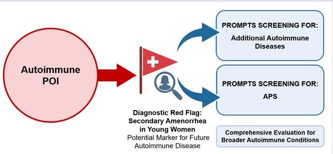
58 Primary Ovarian Insufficiency as an Early Sentinel in Autoimmune Polyglandular Syndrome Type 2
Vudu S et al.
60 Transcriptomic Stratification of Advanced Primary Endometrial Tumours According to Mismatch Repair Status
Gjorgoska and Rižner
62 Endocrine Disruptors, Steroid Hormones, and Their Role in Gestational Diabetes
Svojtková M et al.
64 Is There a Relationship Between Age at Menarche and the Development of PCOS?
Ayyavoo C
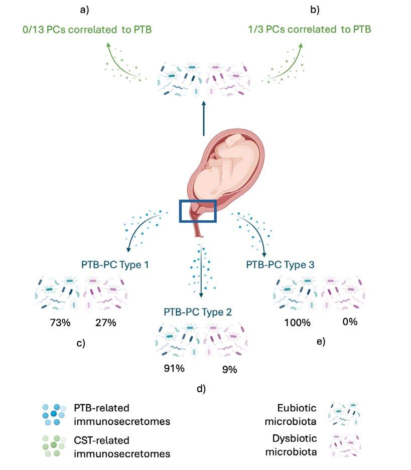
66 Cervico-vaginal Immunosecretomes, Rather than Community State Type IV, are Associated with Preterm Labour
Peón AN et al.
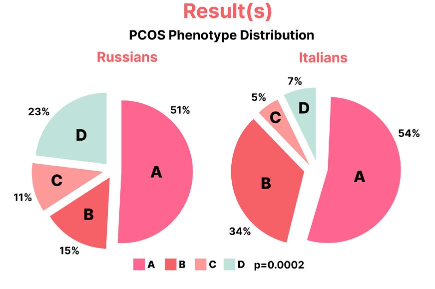
69 Polycystic Ovary Syndrome Features: The Role of Ethnicity
Bettikher O et al.
72 Combined Reproductive and Multiple Endocrinopathies in a Single Case
Pkhaladze E et al.
74 Determinants of Unplanned Pregnancy Among Antenatal Care Attendees in the Primary Healthcare Centres at King Abdulaziz Medical City, Jeddah, Saudi Arabia
Razaz Wali
77 Metabolomic Markers of Neurotransmitter-Related Metabolism in Women with Ovarian Endometriomas: The Role of the Kynurenine Pathway and Changes During Progestin Therapy
Dubrovina S et al.
79 Trabecular Bone Score in Women with Premature Ovarian Insufficiency and Correlation with Bone Mineral Density
Kulshrestha V et al.
Tommaso Simoncini
Andrea R. Genazzani
Roberta Diaz Brinton
Basil C. Tarlatzis
Peter A. Chedraui
Aims and Scope
EMJ Reproductive Health is an open-access, peer-reviewed eJournal committed to helping elevate the quality of practices in reproductive health globally by informing healthcare professionals on the latest research in the field. EMJ Reproductive Health endeavours to increase knowledge, stimulate discussion, and contribute to a better understanding of current issues around reproductive health.
The journal is six weeks after the International Society for Gynecological Endocrinology (ISGE) Congress, and features highlights from this congress, alongside interviews with experts in the field, as well as in-depth features on congress sessions.
EMJ Reproductive Health also publishes peer-reviewed research papers, review articles, and case reports relevant to the field. In addition, the journal welcomes the submission of features and opinion pieces intended to create a discussion around key topics in the field and broaden readers’ professional interests. The journal is managed by a dedicated editorial team that adheres to a rigorous double-blind peer-review process, maintains high standards of copy editing, and ensures timely publication.
EMJ Reproductive Health focuses on topics that are relevant to healthcare professionals in the field. We do not publish veterinary science papers or laboratory studies that are not linked to patient outcomes. We have a particular interest in topical studies that advance knowledge and inform of coming trends affecting clinical practice in the field.
Further details on coverage can be found here: www.emjreviews.com
Editorial Expertise
EMJ is supported by various levels of expertise:
• Guidance from an Editorial Board consisting of leading authorities from a wide variety of disciplines.
• Invited contributors who are recognised authorities in their respective fields.
• Peer review, which is conducted by expert reviewers who are invited by the Editorial team and appointed based on their knowledge of a specific topic.
• An experienced team of editors and technical editors.
Peer Review
On submission, all articles are assessed by the editorial team to determine their suitability for the journal and appropriateness for peer review.
Editorial staff, following consultation with either a member of the Editorial Board or the author(s) if necessary, identify three appropriate reviewers, who are selected based on their specialist knowledge in the relevant area.
All peer review is double blind. Following review, papers are either accepted without modification, returned to the author(s) to incorporate required changes, or rejected.
Editorial staff have final discretion over any proposed amendments.
Submissions
We welcome contributions from professionals,
consultants, academics, and industry leaders on relevant and topical subjects. We seek papers with the most current, interesting, and relevant information in each therapeutic area and accept original research, review articles, case reports, and features.
We are always keen to hear from healthcare professionals wishing to discuss potential submissions, please email: editorial.assistant@emjreviews.com
To submit a paper, use our online submission site: emj.kriyadocs.com
Submission details can be found through our website: www.emjreviews.com/contributors/authors
Reprints
All articles included in EMJ are available as reprints (minimum order 1,000). Please contact hello@emjreviews.com if you would like to order reprints.
Distribution and Readership
EMJ is distributed through controlled circulation to healthcare professionals in the relevant fields across Europe.
Indexing and Availability
EMJ is indexed on DOAJ, the Royal Society of Medicine, and Google Scholar®; selected articles are indexed in PubMed Central®
EMJ is available through the websites of our leading partners and collaborating societies. EMJ journals are all available via our website: www.emjreviews.com
Open Access
This is an open-access journal in accordance with the Creative Commons Attribution-Non Commercial 4.0 (CC BY-NC 4.0) license.
Congress Notice
Staff members attend medical congresses as reporters when required.
This Publication
Publication Date: April 2026 Online ISSN: ISSN 2059-450X
All information obtained by EMJ and each of the contributions from various sources is as current and accurate as possible. However, due to human or mechanical errors, EMJ and the contributors cannot guarantee the accuracy, adequacy, or completeness of any information, and cannot be held responsible for any errors or omissions. EMJ is completely independent of the review event (ISGE 2026) and the use of the organisations does not constitute endorsement or media partnership in any form whatsoever. The cover photo is of Rome, Italy, the location of ISGE 2026.
Niamh Holmes, Roli Omamuli, Alexander Perkins, Katrina Thornber, Aleksandra Zurowska
Reporter
Anna Caldwell
Creative Director
Tim Uden
Design Manager
Stacey White
Senior Designers
Tamara Kondolomo, Owen Silcox
Designers
Shanjok Gurung, Fabio van Paris
Junior Designers
Fraser Hoey, Helena Spicer, Caleb Wylie
Head of Marketing
Stephanie Corbett
Business Unit Lead
Kelly Byrne
Commercial Director
Oz Desai
Chief Executive Officer
Justin Levett
Chief Commercial Officer
Dan Healy
Founder and Chairman
Spencer Gore
Welcome
Dear Readers,
Welcome to this special issue, featuring our comprehensive coverage of the 40th Anniversary International Society of Gynecological Endocrinology (ISGE) 2026 Congress. This milestone event brought together physicians and scientists from around the world to discuss the latest advancements in women's health and gynaecological endocrinology.
In this issue, we present a review of the key highlights from ISGE 2026, alongside two features exploring early menopause and brain health, and endometriosis care. Discover the complex clinical and biological changes occurring across this critical life stage and gain an in-depth overview of the background and current treatments for endometriosis across the lifespan.
Complementing this, we are pleased to include five exclusive interviews with Andrea R. Genazzani, Tommaso Simoncini, Basil C. Tarlatzis, Peter A. Chedraui, and Roberta Diaz Brinton. They share their perspectives on evolving topics in gynaecological endocrinology, from hormonal health and the reproductive window, to emerging therapeutic approaches and future directions in the field.
Alena Sofieva Editorial Co-ordinator
Editorial enquiries: editor@emjreviews.com
Sales opportunities: salesadmin@emjreviews.com
Permissions and copyright: accountsreceivable@emjreviews.com
Reprints: info@emjreviews.com
Media enquiries: marketing@emjreviews.com
ISGE 2026
ISGE 2026 ultimately demonstrated a field that is both expanding in scale and evolving in scope
Congress Review
Review of the International Society of Gynecological Endocrinology (ISGE) Congress 2026
THIS YEAR, Rome, Italy, welcomed the International Society of Gynecological Endocrinology (ISGE) Congress 2026, marking a significant moment of renewed momentum and expansion for the field. With participation nearing 3,000 delegates and over 750 scientific contributions submitted, this meeting reflected both the scale of engagement and the growing global relevance of gynaecological endocrinology.
In the opening ceremony, Tommaso Simoncini, President of the ISGE, highlighted the transformation of the ISGE from a smaller, specialist meeting to a truly international congress. Once hosted in more intimate settings, including earlier editions in mountain locations and later across major global cities, the Congress has evolved into a large-scale scientific forum attracting participants from across Europe, Asia, the USA, Africa, and Oceania. Reflecting on this trajectory, Simoncini emphasised the importance of in-person exchange, noting that the ability to travel, collaborate, and share knowledge once again represents a defining strength of the current meeting.
This year’s Congress also marked a clear turning point following the disruption of recent years. Previous editions were significantly impacted, including the cancellation of the Florence meeting at the onset of the COVID-19 pandemic, followed by smaller-scale or transitional formats. In contrast, ISGE 2026 demonstrated a strong resurgence, with attendance figures approaching pre-pandemic growth
trajectories and signalling renewed confidence in international scientific gatherings.
A defining feature of the Congress was the scale and diversity of its scientific programme. It included an extensive range of sessions, with over 30 symposia organised by affiliated and partner societies, representing a wide spectrum of regional, national, and specialist groups. The programme also incorporated a broad range of educational formats, from plenary sessions and symposia to workshops and discussion-based sessions, designed to encourage both knowledge exchange and practical engagement.
The volume of submitted research was particularly notable. With more than 750 abstracts presented, ISGE 2026 recorded one of the highest levels of scientific contribution in its history. A large proportion of these submissions came from earlycareer researchers, showing strong engagement from the next generation of clinicians and scientists. This emphasis was
further supported by dedicated scholarship initiatives, which enabled younger participants, including trainees and earlystage investigators, to attend and present their work. These programmes, alongside reduced registration opportunities for contributors, reflect a broader commitment to accessibility and inclusivity within the Society.
International representation was another key strength of the Congress. While European participation remained prominent, there was clear expansion in contributions from regions including Asia, the USA, and Africa. Discussions throughout the Congress highlighted both regional differences in practice and shared challenges, particularly in translating emerging evidence into clinical care across varied healthcare systems.
Beyond its scientific content, the Congress reflected a broader shift in the field of
gynaecological endocrinology. As new concepts emerge and longstanding assumptions are re-evaluated, there is increasing emphasis on integrating scientific advances into clinical practice. The discussions at ISGE 2026 stressed the importance of addressing this gap, ensuring that innovation translates into tangible improvements in patient care.
ISGE 2026 ultimately demonstrated a field that is both expanding in scale and evolving in scope. With growing international participation, increasing scientific output, and a strong focus on collaboration and education, the Congress reaffirmed its position as a key meeting point for clinicians and researchers worldwide. As the specialty continues to develop, the importance of platforms such as ISGE in facilitating global exchange, supporting the next generation, and advancing patientcentred care remains clear.
ISGE 2026 demonstrated a strong resurgence, with attendance figures approaching pre-pandemic growth trajectories
Optimising Transdermal Oestradiol Therapy in Menopause: Dose, Serum Levels, and Clinical Outcomes
MENOPAUSAL hormone therapy remains the most effective treatment for vasomotor symptoms, genitourinary syndrome of menopause, and the prevention of bone loss and fractures. However, real-world evidence indicates that uptake remains below 10%, highlighting a persistent gap between efficacy and clinical use. Optimisation of treatment, including dose selection and delivery method, is therefore essential to improve outcomes, and this was explored in an analysis presented at the ISGE Congress 2026.1
This analysis brings together evidence from two pivotal Phase III RCTs evaluating a transdermal oestradiol hydroalcoholic gel formulation, alongside findings from the KEEPS study, to better define oestradiol serum levels associated with meaningful symptom relief.
A total of 567 patients were included in the intent-to-treat populations of the Phase III studies. Daily administration of oestradiol gel at doses of 1.25 g and 2.5 g was assessed and compared with outcomes observed using a transdermal patch delivering 50 µg of oestradiol per day.
In the first trial (n=216), both gel doses led to substantial reductions in the frequency of moderate-to-severe hot flashes, with mean values decreasing from approximately 10 episodes per day at baseline to 2.8 and 2.0 with the 1.25 g and 2.5 g doses, respectively, corresponding to reductions of 73% and 82%. In comparison, placebo was associated with a smaller reduction, from 11.0 to 5.2 episodes per day (53%).
In the second trial (n=351), similar improvements were observed, with hot flash frequency decreasing from baseline values of 11.7 and 11.8 to 2.9 and 2.3 with the 1.25 g and 2.5 g doses, respectively. Outcomes in the active comparator group showed a reduction from 10.9 to 1.3 episodes per day with the transdermal patch.
The proportion of patients achieving a clinically meaningful response, defined as experiencing only mild or no hot flashes, increased progressively across all treatment arms. By Week 12, response
rates reached 28.6%, 34.4%, and 47.6% with escalating gel doses (0.625 g, 1.25 g, and 2.5 g), and 47.7% in the transdermal patch group, demonstrating a clear dose–response relationship and comparable efficacy between the highest gel dose and patch therapy.
Overall, the 2.5 g oestradiol gel dose consistently demonstrated superior clinical benefit compared with the 1.25 g dose, achieving circulating oestradiol levels of approximately 60 pg/mL, which were associated with optimal symptom control. These findings support an individualised approach to menopausal hormone therapy, in which treatment is guided by target oestradiol levels to maximise relief of menopausal symptoms while also contributing to long-term benefits in bone and cardiovascular health.
The 2.5 g oestradiol gel dose consistently demonstrated superior clinical benefit compared with the 1.25 g dose
Lower FSH Linked to Adverse Glucose Metabolism in Postmenopausal Women
NEW RESEARCH presented at the ISGE Congress 2026 provides emerging evidence that follicle-stimulating hormone (FSH) may play a role in metabolic regulation beyond its established reproductive function. The findings offer relevant insights for clinicians who are managing the long-term health of postmenopausal women.2
Menopause is associated with increased cardiometabolic risk, including insulin resistance and Type 2 diabetes. While FSH is traditionally viewed as a marker of ovarian function, the identification of FSH receptors in extra-ovarian tissues such as the liver and adipose tissue has prompted investigation into its potential metabolic effects.
This retrospective study evaluated 82 naturally postmenopausal women (mean age: 65.2 years) not receiving hormone therapy. Researchers examined the relationship between circulating FSH levels and key metabolic parameters, including fasting
glucose, insulin, glycated haemoglobin (HbA1c), lipid profile, and insulin resistance as measured by the Homeostatic Model Assessment for Insulin Resistance (HOMA-IR) index.
FSH levels were not significantly associated with lipid abnormalities. However, a clear relationship emerged with glucose metabolism. Women with Type 2 diabetes had significantly lower FSH concentrations compared with those with insulin resistance or normal glucose metabolism (44.3 IU/mL versus 60.6 IU/mL and 69.4 IU/mL, respectively; p=0.045). In addition, participants in the lowest FSH quartile
Women with Type 2 diabetes had significantly lower FSH concentrations compared with those with insulin resistance or normal glucose metabolism
A modest but significant inverse correlation was observed between FSH and fasting insulin levels (r=–0.30; p=0.03), which persisted after adjustment for confounders. This association was more pronounced among women more than 6 years beyond menopause (r=–0.34; p=0.016), suggesting that the metabolic relevance of FSH may increase over time following the menopausal transition.
These findings indicate that lower circulating FSH levels are associated with impaired glucose metabolism in postmenopausal women, particularly in the late postmenopausal phase. FSH could serve as an additional biomarker in metabolic risk stratification, although further prospective studies are needed to clarify causality and underlying mechanisms.
Ongoing Menopausal Symptoms Drive Declines in Women’s Quality of Life
PERSISTENT sleep disturbances and vasomotor symptoms (VMS), such as hot flashes and night sweats, are strongly associated with poorer health-related quality of life (HRQoL) in women undergoing menopause, according to new longitudinal findings from the SWAN study presented at the ISGE Congress 2026.3
The study analysed data collected between 1999–2008, including baseline and 10 annual follow-up visits. Two groups of participants were examined: 1,176 women who consistently reported on sleep disturbances, and 1,159 who reported on VMS.
Sleep disturbances were defined as experiencing issues such as waking multiple times per night, difficulty falling asleep, or early waking at least 3–4 nights per week over the past 2 weeks. VMS were defined as hot flashes or night sweats occurring on at least 1–5 days within the same timeframe.
Researchers assessed HRQoL using three subscales from the Short Form Health Survey (SF-36): role limitations due to physical health, role limitations due to emotional problems, and energy/ fatigue. Higher scores indicate better quality of life.
On average, participants reported sleep disturbances at 6.8 visits and VMS at 5.8 visits over the study period. Both symptoms were negatively correlated with HRQoL across all domains. Sleep disturbances showed slightly stronger correlations, particularly with energy/fatigue. Statistical modelling revealed that each additional visit at which sleep disturbances were reported corresponded to a decrease of 0.94 points in role limitations due to physical health, 0.75 in
role limitations due to emotional problems, and 0.95 in energy/ fatigue scores. Similarly, each additional report of VMS was linked to declines of 0.70, 0.64, and 0.44 points, respectively.
Importantly, other factors also played a significant role. High anxiety levels and lower income were among the strongest predictors of reduced HRQoL across all measures, highlighting the multifactorial nature of wellbeing during menopause.
The findings underscore the cumulative impact of persistent menopausal symptoms over time. Rather than being transient inconveniences, ongoing sleep disturbances and VMS may lead to meaningful declines in physical, emotional, and energy-related aspects of daily life.
The authors emphasise the importance of regular monitoring and proactive management of these symptoms. Improved communication between women and healthcare providers, alongside targeted interventions, may help mitigate the long-term effects on quality of life.
Rather than being transient inconveniences, ongoing sleep disturbances and VMS may lead to meaningful declines in physical, emotional, and energy-related aspects of daily life
Dydrogesterone May Reduce Dysmenorrhoea in Endometriosis While Supporting Fertility Goals
NEW DATA presented at the ISGE Congress 2026 suggest that dydrogesterone may be an effective and well-tolerated treatment option for patients with endometriosis, particularly those wishing to preserve fertility.4
In a retrospective follow-up study from China, dydrogesterone was associated with significant improvements in dysmenorrhoea, while cyst size remained stable and no significant adverse effects on liver function, kidney function, or blood lipids were observed.
The study included 247 patients with endometriosis who had received dydrogesterone for more than 6 months at Women’s Hospital, School of Medicine, Zhejiang University, Hangzhou, China, between 1st January 2018–30th September 2024. Among these, 153 patients had dysmenorrhoea.
Pain severity, assessed using the visual analogue scale (VAS), was significantly reduced over time, with statistically significant differences reported at baseline, 1 month, 3 months, and 6 months, regardless of whether patients received dydrogesterone on a Day 5–24 or Day 15–24 regimen (p<0.0001).
When the regimens were analysed separately, significant improvements in VAS scores were seen in both groups. In the Day 5–24 group, which included 67 patients, reductions were significant across all assessed time points (p<0.001). In the Day 15–24 group, which included 66 patients, improvements were also significant at baseline, 1 month, 3 months, and 6 months (p<0.01).
No significant differences were observed in changes in maximum cyst diameter or cyst volume between the two treatment schedules at baseline, 3 months, and 6 months (p>0.05). The authors therefore concluded that dydrogesterone may help control symptoms and maintain cyst stability, rather than reduce cyst size.
They also reported no significant effects on liver function, kidney function, or blood lipid levels, supporting the tolerability of treatment in this cohort.
Overall, these findings suggest that dydrogesterone may offer a useful treatment approach for patients with endometriosis who require symptom control and also wish to retain the opportunity for natural conception. As this was a retrospective single-centre study, the findings should be interpreted within the context of the study design.
Dydrogesterone may offer a useful treatment approach for patients with endometriosis who require symptom control and also wish to retain the opportunity for natural conception
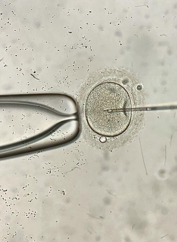
PGT-A Associated with Improved IVF Pregnancy Outcomes
PREIMPLANTATION
genetic testing for aneuploidy (PGT-A) may improve pregnancy outcomes in assisted reproductive technology by enabling more accurate embryo selection, according to a study presented at the ISGE Congress 2026.5
Assisted reproductive technology has transformed infertility treatment, but selecting viable embryos remains critical to success. PGT-A identifies chromosomally normal (euploid) embryos, aiming to improve implantation rates, reducing miscarriage risk, and increasing overall IVF efficiency.
Embryo selection using genetic testing may improve IVF efficiency by enabling fewer transfers to achieve pregnancy
Researchers conducted a retrospective comparative study of 225 patients undergoing IVF, analysing outcomes in those who received PGT-A versus those who did not. All patients underwent ovarian stimulation using a gonadotrophin-releasing hormone antagonist protocol, followed by oocyte retrieval and blastocyst transfer. In the PGT-A group, embryos were tested using next-generation sequencing.
Among the PGT-A group (n=110), 116 embryos were transferred, with 53.6% achieving pregnancy, compared with 35.7% in the non-tested group, where 220 embryos were transferred (mean: 1.91 per patient). PGT-A was associated with higher live birth rates and lower miscarriage risk, with miscarriage rates of 6.8% in the PGT-A cohort versus 21.9% in the non-tested group. Furthermore, 48.2% of transfers resulted in live birth, compared with 27.8% without testing.
Most patients in the PGT-A group underwent single embryo transfer, while more embryos per patient were transferred in the non-tested group, highlighting that embryo selection using genetic testing may
improve IVF efficiency by enabling fewer transfers to achieve pregnancy.
However, the retrospective design and selected patient population (aged 35 years and under) may limit generalisability. Cost considerations and individual patient characteristics and preferences should also inform PGT-A use. Overall, while PGT-A appears to be associated with improved IVF outcomes, further large-scale studies are needed to refine patient selection and assess long-term benefits.
Decreased Risk of Venous Thromboembolism with Body-Identical Oestrogens
NEW EVIDENCE presented at the ISGE Congress 2026 has demonstrated that bodyidentical oestrogens are associated with a lower risk of thrombosis compared to other hormonal contraceptives.6
Venous thromboembolism (VTE) remains a significant global health concern and is known to be associated with the use of hormonal contraceptives. To address this, researchers conducted a living network metaanalysis, designed to be updated every 3 years to incorporate emerging evidence and maintain the relevance and accuracy of findings.
The objective was to systematically evaluate the risk of VTE associated with hormonal contraceptives (including combined oestrogen–progestin and progestin-only formulations) in women aged over 18 years, compared with non-users or users of alternative contraceptive methods.
A comprehensive literature search was performed in September 2024 and updated in October 2025 using Medline and Embase databases. Clinical studies assessing VTE risk among users of hormonal contraceptives versus non-users or users of other contraceptives were identified. Study selection followed rigorous screening procedures, and data extraction was conducted using standardised methodologies. Statistical analyses were performed using a randomeffects model.
All ethinyl oestradiol-based combined contraceptives were associated with a significantly increased risk of VTE
Preliminary results identified 85 studies through abstract and full-text screening, of which 12 cohort studies and 17 case–control studies were included in the primary quantitative analysis. Unadjusted analyses demonstrated that, compared with non-use, all ethinyl oestradiolbased combined contraceptives were associated with a significantly increased risk of VTE, with ethinyl oestradiol/desogestrel combinations showing the highest excess risk.
In contrast, progestin-only contraceptives were associated with a risk comparable to nonuse and did not demonstrate statistically significant differences, except for injectable medroxyprogesterone acetate, which was associated with an increased risk.
Body-identical oestrogencontaining combined oral contraceptives did not show a statistically significant difference in VTE risk compared with the desogestrel-only pill. These findings were consistent across both cohort and case–control analyses.
Overall, while ethinyl oestradiolbased hormonal contraceptives have been widely used for decades, emerging evidence suggests that body-identical oestrogens may be associated with a lower risk of thrombosis.
Pre-conception Androgens Predict Pregnancy Outcomes in PCOS
A
NEW study presented at the ISGE Congress 2026 has shown that pre-conception androgen levels may help predict both fertility treatment requirements and pregnancy outcomes in females with polycystic ovary syndrome (PCOS).7
PCOS is the most common endocrine–metabolic condition affecting females of reproductive age and is frequently associated with anovulation, subfertility, and infertility. While assisted reproductive technologies such as IVF are widely used, identifying which patients are most likely to require intervention or experience complications has remained a clinical challenge.
In this retrospective cohort study, researchers analysed 160 females with confirmed PCOS who achieved at least one live birth beyond 23 weeks of gestation. A total of 232 pregnancies were evaluated to explore how pre-conception androgen profiles and mode of conception influenced obstetric outcomes.
The findings revealed that elevated preconception androstenedione levels were significantly associated with an increased risk of miscarriage. In contrast, lower levels of dehydroepiandrosterone sulphate and androstenedione were strongly linked to the need for IVF, suggesting that reduced androgen activity may reflect impaired natural fertility potential in this population. Additionally, a lower luteinising hormone/ follicle-stimulating hormone ratio was associated with a greater likelihood of requiring assisted reproductive interventions.
The study also identified important differences in pregnancy outcomes based on mode of conception. Pregnancies achieved through IVF were associated with higher rates of preterm birth and Caesarean section compared with spontaneous conceptions. However, there were no significant differences in the incidence of preeclampsia or gestational diabetes between the two groups.
These findings highlight the potential role of pre-conception hormonal profiling in guiding fertility management for females with PCOS. By identifying patients at higher risk of miscarriage or those more likely to require IVF, clinicians may be able to personalise treatment strategies and optimise prepregnancy management.
The study was conducted at a single centre and included only females who achieved a live birth, which may limit generalisability to those who are unable to conceive or experience pregnancy loss. Nevertheless, the results provide important real-world evidence supporting the integration of hormonal markers into clinical decision-making.
Overall, the study suggests that preconception androgen levels could serve as valuable predictors of both fertility treatment needs and obstetric risks, offering a step towards more individualised care in PCOS.
By identifying patients at higher risk of miscarriage or those more likely to require IVF, clinicians may be able to personalise treatment strategies
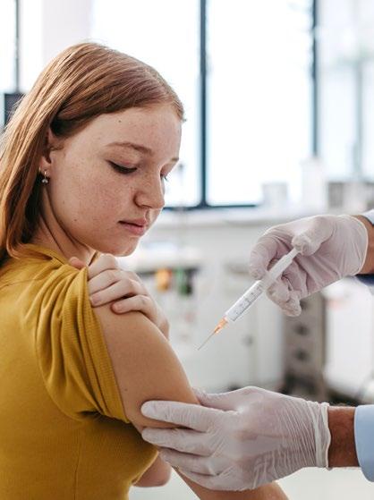
HPV Vaccination Gaps and High Dysplasia Rates Identified in Patients with Müllerian Anomalies
NEW RESEARCH from a tertiary healthcare centre, presented at the ISGE Congress 2026, highlights concerning gaps in human papillomavirus (HPV) vaccination uptake and cervical cancer screening practices among patients with Müllerian anomalies (MA), alongside unexpectedly high rates of cervical dysplasia in this population.8
MAs, congenital variations of the female reproductive tract affecting up to 6.7% of females, present unique anatomical and clinical challenges. Despite recommendations for HPV vaccination to prevent HPV-related cancers, real-world uptake and screening adequacy in this group have remained poorly understood.
In this retrospective crosssectional study, researchers analysed electronic medical records from 220 patients aged 9–36 years with MAs between 2012–2025. Data collected included demographics, MA subtype, HPV vaccination counselling and uptake, and cervical screening outcomes such as cytology, HPV co-testing, colposcopy, and histology.
Bicornuate uterus was the most common anomaly (45.0%), followed by septate uterus (20.9%) and uterus didelphys (9.1%). The mean age at diagnosis was 22.7 years. Just over half of patients (53.9%) were offered and accepted HPV vaccination, while 5.9% declined and a notable 40.2% had no documented counselling. Among those vaccinated, most (75.4%) completed the full vaccine series.
Of the 201 patients with a cervix, 89.1% underwent Pap smear screening. However, 29.1% had abnormal cytology results, most commonly low-grade squamous intraepithelial lesions (11.2%) and atypical squamous cells of undetermined significance with high-risk HPV positivity (7.3%). Among 114 patients who underwent HPV co-testing, 25.4% tested positive for high-risk HPV.
Screening practices were also inconsistent in patients with anatomical complexity. Among those with uterus didelphys, only 65% had Pap smears performed on both cervices. Of the 51 patients with abnormal cytology, just over half (54.9%) underwent colposcopy.
Histological analysis revealed cervical intraepithelial neoplasia (CIN) Grade I in 21.4%, CIN II in 17.9%, and CIN III in 14.3% of cases assessed. More than half of patients with CIN II or III required ablative or excisional treatment.
The high proportion of patients without documented HPV counselling, alongside incomplete screening in anatomically complex cases, suggests missed opportunities for early intervention
The findings point to both a substantial burden of cervical dysplasia and clear gaps in preventive care. The high proportion of patients without documented HPV counselling, alongside incomplete screening in anatomically complex cases, suggests missed opportunities for early intervention.
While limited by its retrospective design and reliance on documentation within electronic records, this study is, to the authors’ knowledge, the first to examine HPV vaccination and cervical dysplasia in a large cohort of patients with MAs.
These results underscore the need for improved education, standardised vaccination counselling, and tailored cervical cancer screening strategies for this population. The authors highlight that future prospective studies will be critical to inform evidence-based guidelines, similar to those developed for other highrisk or special populations.
Pulsatile GnRH Therapy Restores Fertility in Hypothalamic Amenorrhoea
A REAL-WORLD study presented at the ISGE Congress 2026 has shown that pulsatile gonadotropin-releasing hormone (GnRH) therapy is highly effective in restoring ovulation and achieving pregnancy in females with hypothalamic amenorrhoea (HA).9
HA is a condition characterised by disruption of the hypothalamic–pituitary–ovarian axis, leading to cessation of menstruation, anovulation, and infertility. It is often associated with factors such as low energy availability, stress, or excessive exercise, and can significantly impact reproductive health. Although pulsatile GnRH therapy is recommended as a first-line treatment because it mimics natural hormonal physiology, it has remained underutilised in routine clinical practice.
In this retrospective, single-centre cohort study, researchers evaluated the real-world effectiveness and safety of subcutaneous pulsatile GnRH therapy in 69 females aged 20–40 years with confirmed HA and a desire for pregnancy. Participants were treated between 2013–2025 using an infusion pump delivering individualised GnRH doses.
The findings demonstrated a remarkably high ovulation restoration rate of 95.7% (66/69), confirming the therapy’s ability to re-establish normal reproductive function. The clinical pregnancy rate reached 81.2% (56/69), with a median time-to-pregnancy of 117 days, indicating relatively rapid fertility restoration following treatment initiation.
Pregnancy outcomes were also favourable. Among the 56 pregnancies, there were 46 singleton live births, six ongoing pregnancies, and four miscarriages.
By closely replicating natural endocrine rhythms, the treatment achieves high rates of ovulation, singleton pregnancy, and live birth
Notably, no multiple pregnancies were reported, a key advantage compared with other fertility treatments that carry higher risks of multiple gestations. Furthermore, all infants demonstrated normal development at follow-up, and no severe adverse events were observed, supporting the therapy’s strong safety profile.
The study’s retrospective design and singlecentre setting may limit generalisability, and the absence of a comparator group prevents direct comparison with alternative fertility treatments. However, the consistency of outcomes across a substantial number of treatment cycles strengthens the reliability of the findings.
Overall, this study reinforces pulsatile GnRH therapy as a highly effective, physiological, and safe approach for fertility restoration in females with HA. By closely replicating natural endocrine rhythms, the treatment achieves high rates of ovulation, singleton pregnancy, and live birth, highlighting its potential for broader clinical adoption in reproductive medicine.
IBD and Endometriosis Link Highlights Diagnostic Overlap
INFLAMMATORY bowel disease (IBD) and endometriosis frequently coexist, with new findings highlighting overlapping symptoms and the need for heightened clinical suspicion, according to data presented at the ISGE Congress 2026.10
In this retrospective observational study, researchers evaluated premenopausal women with IBD who underwent 2D and 3D transvaginal ultrasound between January 2021–January 2025. Among 111 patients with IBD, endometriosis or adenomyosis was identified in 65 cases, representing a prevalence of 58.6%. The study compared these individuals with two control groups consisting of patients with IBD without endometriosis or adenomyosis and women with endometriosis or adenomyosis without IBD, matched for age and BMI.
The findings confirm that endometriosis is highly prevalent among patients with IBD, reinforcing the importance of recognising co-existing inflammatory conditions in this population. Patients with both conditions commonly presented with characteristic gynaecological symptoms, suggesting overlapping inflammatory mechanisms.
Patients with co-existing IBD and endometriosis reported significantly higher rates of dysmenorrhoea, dyspareunia, and heavy menstrual bleeding compared with those with IBD alone. Reported frequencies were
Patients with co-existing IBD and endometriosis reported significantly higher rates of dysmenorrhoea, dyspareunia, and heavy menstrual bleeding compared with those with IBD alone
dysmenorrhoea (90.8% versus 65.2%; p=0.0009), dyspareunia (63.1% versus 32.6%; p=0.001), and heavy menstrual bleeding (61.5% versus 37.0%; p=0.01).
When compared with women with endometriosis without IBD, those in the study group demonstrated a higher prevalence of bowel symptoms, particularly diarrhoea (32.3% versus 15.9%; p=0.004). In addition, adenomyosis was more common (75.4% versus 61.5%; p=0.04), alongside increased involvement of the left uterosacral ligament (p=0.03). Conversely, ovarian endometriosis and posterior deep infiltrating lesions were more frequently observed in patients without IBD.
These findings suggest that IBD should be suspected in women with endometriosis who present with bowel symptoms, especially diarrhoea. Equally, clinicians managing patients with IBD should consider the high likelihood of concurrent endometriosis in the presence of gynaecological pain symptoms.
The frequent co-existence of adenomyosis and site-specific pelvic involvement points towards shared inflammatory pathways between the two conditions. A multidisciplinary approach is therefore essential to reduce diagnostic delays and optimise reproductive health outcomes in this patient population.
References
1. Piette P et al. Optimization of transdermal estradiol gel and micronised progesterone for menopausal hormone therapy. Abstract. ISGE Congress, 4-6 March, 2026.
2. Barbagallo F et al. Association between follicle stimulating hormone levels and metabolic health in postmenopause. Abstract. ISGE Congress, 4-6 March, 2026.
3. Dinkel-Keuthage C et al. Persistent burden of sleep disturbances and vasomotor symptoms in women experiencing menopause: associations with health-related quality of life. Abstract. ISGE Congress, 4-6 March, 2026.
4. Zhang X. The clinical efficacy of dydrogesterone in the treatment of endometriosis: a follow-up study. Abstract. ISGE Congress, 4-6 March, 2026.
5. Makharadze M. Effect of PGT-A on IVF outcomes in women under 35: a retrospective comparative study. Abstract. ISGE Congress, 4-6 March, 2026.
6. Raskin L et al. Assessment of the risk of venous thromboembolism of combined hormonal contraceptives or progestinonly contraceptives in women over 18 in comparison with non-users or any other contraceptive. Abstract. ISGE Congress, 4-6 March, 2026.
7. Stercel O et al. The role of preconception androgen levels and mode of conception in the obstetric outcomes of women with polycystic ovary syndrome. Abstract. ISGE Congress, 4-6 March, 2026.
8. Stevenson J. Prevalences of HPV vaccination and cervical dysplasia among patients with Müllerian anomalies at a tertiary healthcare center. Abstract. ISGE Congress, 4-6 March, 2026.
9. Xiong Y et al. Pulsatile GnRH therapy in fertility restoration of hypothalamic amenorrhea. Abstract. ISGE Congress, 4-6 March, 2026.
10. Federica Iacobini et al. When an inflammatory bowel disease (IBD) should be suspected in patients with endometriosis? Abstract. ISGE Congress, 4-6 March, 2026.
ISGE 2026 recorded one of the highest levels of scientific contribution in its history
Early Menopause and Brain Health: From Clinical Risk to Neuroscience
AT THIS year’s International Society of Gynecological Endocrinology (ISGE) Congress 2026, held in Rome, Italy, the symposium ‘The Troubled Journey of the Menopausal Transition’ brought together leading experts to explore the complex clinical and biological changes occurring across this critical life stage. Chaired by Antonio Cano, University of Valencia, Spain; and Mark Brincat, University of Malta, Msida, the session examined the menopausal transition through multiple lenses, from cardiometabolic risk and musculoskeletal health to brain ageing and hormone therapy optimisation.
Panagiotis Anagnostis, Aristotle University of Thessaloniki, Greece, examined whether early menopause represents a distinct clinical entity from premature ovarian insufficiency (POI), while Roberta Brinton, University of Arizona, Tucson, USA, provided insights into the neurobiological impact of the menopausal transition and its implications for hormone therapy initiation timing.
EM VERSUS POI: CLINICAL OVERLAP AND DISTINCTION
The average age of menopause is 50–51 years. While menopause occurring more than two standard deviations below the average age (i.e., before 40 years) has been widely, though somewhat arbitrarily, classified as 'premature',1,2 loss of ovarian function before the age of 40 is more specifically termed POI, a condition affecting approximately 1% of women.3 Diagnostic criteria for POI include disordered menstrual cycles for ≥4 months, alongside elevated folliclestimulating hormone >25 IU/L (on at least one occasion).3
If menopause occurs between 40–45 years of age, it is considered early menopause (EM).³ This condition is relatively common, affecting up to 10% of postmenopausal women.³
Some of the well-recognised long-term health consequences of POI are an increased risk of cardiovascular disease, osteoporosis
and associated fracture risk, cognitive dysfunction, and a significant reduction in both quality of life and life expectancy.4-6
Regarding the type of menopause, there are currently no clear conclusions on whether surgical versus natural menopause differentially affects long-term outcomes.7
Long-Term Health Risks in POI
Compared to EM
However, we must address the question: do women with EM demonstrate the same long-term health risks as women with POI, particularly regarding cardiovascular disease?
Women with POI are at increased risk of both fatal and non-fatal cardiovascular disease, mainly driven by coronary heart disease and stroke.7 In women with EM, hazard ratios appear to converge; though to a lesser extent, they remain significant across these outcomes. Recent evidence from Anagnostis’s group confirms that these women are indeed at increased risk.8
Understanding Cardiovascular Risk
Why does this happen? One explanation is the higher prevalence of cardiovascular risk factors in this population. A meta-analysis conducted in 2019 showed that women with EM have a 12% increased risk of Type 2 diabetes, while those with POI have a 50% increased risk.7
These findings have been replicated in later studies.9,10 Arterial hypertension is also increased by around 10% in women with EM.9 This association was not significant in POI, possibly due to oestrogen therapy.10
Importantly, non-coronary outcomes such as heart failure and atrial fibrillation have been highlighted in meta-analyses, showing that both EM and POI are associated with increased risks.10 These are underrecognised issues that should be considered.
In summary, there is substantial evidence that women with EM are at increased risk of fatal and non-fatal cardiovascular disease, primarily due to coronary heart disease. This is largely driven by increased cardiovascular risk factors, particularly diabetes and hypertension, alongside noncoronary outcomes, such as heart failure and atrial fibrillation.5
Women with EM are at increased risk of fatal and non-fatal cardiovascular disease
Osteoporosis and Fracture Risk
What about the musculoskeletal system?
Data from the Australian Longitudinal Study on Women’s Health (ALSWH) show that women with POI have a 2.5-fold increased risk of osteoporosis.11 Women with EM also have a significantly increased risk, though to a lesser extent, while those with late menopause show reduced risk.11
However, findings are not entirely consistent. The Canadian study did not show increased osteoporosis risk in early menopause, though it remained elevated in POI.12 Regarding fractures, Panagiotis and colleagues’ analysis of approximately 500,000 menopausal women demonstrated that EM is associated with a 36% increased fracture risk compared to both women with menopause before 45 years of age and those with menopause at around 50 years of age.13 These findings have been replicated in subsequent studies.14
Additional analyses show that women with EM or POI have an increased risk
of osteoporosis and fractures, with an estimated risk of around 1.4. Risk factors include increasing age, lower BMI, and comorbidities.14 EM and POI are also recognised as independent risk factors in tools such as FRAX® (University of Sheffield, UK) highlighting the importance of assessing secondary osteoporosis.1,13
Role of Sarcopenia in Fracture Risk
Is fracture risk solely due to reduced bone mineral density (BMD)? The answer is no. Sarcopenia also plays a role.15 While categorical data are limited, Panagiotis’s group found reduced muscle mass in women with EM, with marginal effects on muscle strength, and more significant findings in POI, including reduced physical performance.15
Frailty, a broader concept encompassing both sarcopenia and overall health, is also increased, with approximately a two-fold higher risk in both EM and POI.16
Rationale for Using High Oestrogen Doses
Turning to hormone replacement therapy (HRT), in women with POI, recommended oestrogen doses, 100 µg/day transdermal or 2–4 mg/day oral oestradiol, are approximately twice the conventional dose, combined with appropriate progestogen in women with an intact uterus.4,17 While specific recommendations for EM are lacking, a pragmatic approach is to follow the same principles.
Why should we use higher oestrogen doses? The rationale for this is to restore physiological oestrogen levels (50–100 pg/mL), achievable with transdermal 17β-estradiol 100 µg/day or oral 17β-estradiol 2–4 mg/day.17-19 Evidence suggests that conventional HRT may be insufficient to prevent bone loss in POI, whereas higher doses can restore BMD to normal levels.18,19 Limited cardiovascular data indicate that higher oestrogen doses may reduce carotid intima-media thickness.18,19 This approach supports physiological levels and bone health, with potential cardiovascular benefit.18
Safety and Efficacy of HRT: Evidence from Clinical Studies
HRT is recommended until at least the average age of natural menopause, unless contraindicated.2,4 While it is generally considered safe, there is a lack of evidence from RCTs assessing the cardiovascular and bone effects of HRT in women with POI or EM.8
However, evidence from both retrospective and prospective cohort studies provides insight into the protective effects of HRT on the cardiovascular and musculoskeletal systems in women with POI or EM. For example, in a USA-based study, mortality was increased in women undergoing bilateral oophorectomy; however, this was not observed in those receiving HRT.20
Another study demonstrated a 35% reduction in cardiovascular mortality in women with surgical EM who received HRT.21 Similarly, a large Korea-based study showed that the risk of ischaemic stroke and all-cause mortality was increased in women with POI or EM who did not receive HRT, but not in those treated for more than 5 years.22
Although data on the effects of HRT on bone health in women with POI and EM are currently limited, there is substantial evidence that HRT significantly reduces fracture risk in postmenopausal women. Meta-analysis of 28 RCTs, involving over 33,000 participants, concluded that HRT significantly reduces the risk of total, hip, and vertebral fractures, with the most pronounced benefit (a 45% reduction) observed in women under the age of 60 years.23
Furthermore, another Korea-based study reported a 20% reduction in the risk of sarcopenia among postmenopausal women receiving HRT for more than 13 months.24
Panagiotis concluded that it is reasonable to recommend oestrogen therapy for women with EM, emphasising that this is not simply menopausal hormone therapy (MHT), but true HRT that helps reduce multiple longterm health risks.8
Suboptimal Long-Term Use of HRT
However, overall HRT use remains suboptimal.14 Although around 70% of women initiate HRT, only one in four use it for more than 5 years, and just 6% for over 10 years.14 This likely contributes to the persistent burden of cardiovascular and fracture risk.
Future Perspectives
Future research should focus on longitudinal and randomised studies to determine optimal oestrogen dosing for women after the age of 45 years, as well as the role of serum oestradiol monitoring, and management strategies following HRT discontinuation. Limited data suggest that agents such as alendronate or raloxifene may help maintain BMD after HRT cessation.
Panagiotis proposed a unified approach, advocating for the management of women with POI or EM using similar principles. This strategy may facilitate earlier and more appropriate use of HRT; however, optimal regimens and long-term outcomes remain to be established.
MENOPAUSAL TRANSITION AND BRAIN HEALTH: METABOLIC AND IMMUNE REPROGRAMMING
Brinton opened the talk by stating that “women are at a two-fold greater risk of developing Alzheimer’s disease. Worldwide, there are currently one billion menopausal women who share a common critical period in brain ageing: the perimenopause to menopause transition.” They went on to add that menopause represents a critical period, just like adolescence and early development.
The perimenopause to menopause transition, while often considered a transition in reproductive capacity, is in fact a transition that occurs in the brain.25 When you look at the symptoms of perimenopause and menopause, many have nothing to do with reproduction and everything to do with the brain. For example, sleep disturbance, depression, and cognitive complaints are all related to changes occurring in the brain.25
Role of Oestrogen in Brain Energy Metabolism
Oestrogen is a key regulator of glucose metabolism in the brain. It promotes glucose uptake, its metabolism, and ultimately the generation of ATP in the mitochondria.25-30 Brinton often refers to oestrogen as the ‘Queen of Darwin’, as it leaves nothing to chance, from glucose uptake to energy production. The brain is the most energetically demanding organ in the body.
Metabolic Reprogramming in the Brain
The decline in oestrogen availability activates a starvation response in the brain. Oestrogen supports approximately 20–25% of glucose uptake and metabolism, and its loss triggers the brain to utilise auxiliary fuels, as if it were starving. These auxiliary fuels are ketone bodies, which can provide energy for ATP generation.
While this is beneficial, the brain, being the most lipid-rich organ, begins to catabolise its own lipids, specifically white matter, to generate these ketone bodies. This leads to a loss of myelin density and impaired synaptic conduction. Additionally, the integrity of axonal structure is compromised. Lipidomic analyses confirmed that white matter lipids are converted into ketone bodies to sustain energy requirements.25
Brinton sought to characterise the complex process of metabolic reprogramming of the brain following the decline in oestrogen levels. They found out that initially, there is a decline in glucose metabolism, followed by a transient increase in the use of amino acids as alternative fuel sources. This diverts amino acids away from their role in synaptic protein formation.25-32
Subsequently, mitochondrial efficiency declines, shifting towards a phenotype that produces less energy and more heat. There is also an increase in fatty acid β-oxidation, necessary for ketone body production, and a rise in ketone levels.25-32
This process generates myelin fragments, which are taken up by microglia, the brain’s immune cells. This triggers a strong immune
response. Microglia present these fragments on their surface, and T cells from the peripheral immune system recognise them as foreign, suggesting an autoimmune-like response. Interestingly, 95% of individuals after the age of 50 years diagnosed with multiple sclerosis are women.25-33
Hot Flashes and Mitochondrial Heat Production
Brinton’s group has also investigated whether this metabolic reprogramming is linked to menopausal hot flashes: “Our findings suggest that the catabolism of white matter and the uptake of myelin fragments by microglia induce a mitochondrial burst. The loss of oestrogen leads to inefficient mitochondria that generate both ATP and heat, and this heat production may contribute to the experience of hot flashes.” Brinton also shared for the first time that “the generation of menopausal hot flashes, characterised by the release of heat, is likely associated with a microglial respiratory burst, during which lipids are taken up and converted into myelin fragments.”
Sequence of Biological Changes During Menopause
The first sign of this transition appears to be activation of the innate immune system, followed by reduced glucose metabolism and increased amino acid utilisation. This is followed by a mitochondrial deactivation and heat generation, increased fatty acid oxidation, and rising ketone levels. Both innate and adaptive immune systems become activated, which may explain the increased prevalence of autoimmune diseases during menopause.25-28,30-32,34-36
MHT: Timing of Intervention and Impact on Neurodegenerative Risk
When MHT is introduced early in this process, oestrogen can help sustain brain health; however, it does not reverse established disease.25,28,31 In clinical practice, women typically seek MHT when symptoms such as hot flashes arise, by which point significant neuroendocrine changes have already occurred.
The generation of menopausal hot flashes, characterised by the release of heat, is likely associated with a microglial respiratory burst
The brain at this stage differs markedly from that seen earlier in the transition, and differs again once the endocrinological ageing process is complete. As a result, the response to MHT varies depending on the timing of initiation. This likely contributes to the ongoing controversy and challenges surrounding MHT.25,28,31
Brinton and colleagues examined the impact of MHT on the risk of neurodegenerative diseases in a retrospective analysis of 379,352 women.37 MHT was associated with a reduced risk of Alzheimer’s disease, Parkinson’s disease, dementia, multiple sclerosis, and amyotrophic lateral sclerosis. Longer duration of therapy was associated with greater risk reduction, with an overall 50–60% decrease in the risk of Alzheimer’s disease.38
However, the timing of MHT initiation is crucial. When started during the menopausal transition, it reduces
References
1. ESHRE Guideline Group on POI et al. ESHRE Guideline: management of women with premature ovarian insufficiency. Hum Reprod. 2016;31(5):926-37.
2. Hamoda H et al. Menopause and cardiovascular disease: a review of the evidence. Best Pract Res Clin Endocrinol Metab. 2024;38(2):101823.
3. Panay N et al. Menopause and MHT in 2024: addressing the key controversies – an International Menopause Society White Paper. Climacteric. 2024;27(5):441-57.
4. Panay N et al. Global Consensus Statement on the use of testosterone therapy for women. Climacteric. 2024;27(6):510-20.
5. Anagnostis P et al. Menopauseassociated risk of cardiovascular disease. Endocr Connect. 2021;10(12):R302-11.
6. Karavitou E et al. Menopause and cardiovascular disease: current perspectives. Diagnostics. 2022;12(12):3097.
7. Anagnostis P et al. Early menopause and premature ovarian insufficiency are associated with increased risk of type 2 diabetes: a systematic review and meta-analysis. Eur J Endocrinol. 2019;180(1):41-50.
Alzheimer’s disease risk. However, initiation after the transition may increase risk. Brinton and colleagues hypothesise that this may relate to oestrogen’s effects on microglial activity.
CONCLUSION
The menopausal transition is a complex and critical stage with significant implications for long-term health. A clear message emerged across the presentations: early recognition and timely intervention are essential, particularly for reducing cardiovascular, musculoskeletal, and neurological risks. The speakers emphasised that menopause should be viewed as a dynamic process rather than a single event, requiring a personalised and evidence-based approach to care.
8. Anagnostis P et al. Is early menopause a different entity from premature ovarian insufficiency? Clin Endocrinol (Oxf). 2025;102(1):67-74.
9. Anagnostis P et al. Early menopause is associated with increased risk of arterial hypertension: a systematic review and meta-analysis. Maturitas. 2020;135:74-9.
10. Liu X et al. Premature ovarian insufficiency and risk of heart failure: a systematic review and meta-analysis. Maturitas. 2023;176:107784.
11. Xu X et al. Age at natural menopause and development of chronic conditions and multimorbidity: results from an Australian prospective cohort. Human Reproduction. 2020;35(1):203-11.
12. Shea AK et al. The association between primary ovarian insufficiency and osteoporosis in the Canadian Longitudinal Study on Aging. Menopause. 2021;28(6):693-98.
13. Anagnostis P et al. Menopauseassociated risk of fracture: a survival analysis of the UK Biobank and a meta-analysis of observational studies. Endocrine. 2019;63(2):213-24.
14. Jones AR et al. Bone health in women with premature ovarian insufficiency/early menopause: a 23-year longitudinal analysis. Human Reproduction. 2024;39(5):1013-22.
15. Divaris E et al. Early menopause and premature ovarian insufficiency may increase the risk of sarcopenia: a systematic review and meta-analysis. Maturitas. 2023;175:107782.
16. Kojima G et al. Earlier menopause is associated with higher risk of incident frailty in community-dwelling older women in England. J Am Geriatr Soc. 2022;70(9):2602-9.
17. Panay N et al. International Menopause Society recommendations on the use of testosterone for women. Climacteric. 2020;23(5):426-46.
18. Popat VB et al. Bone mineral density in estrogen-deficient young women: a 12-month randomized double-blind placebo-controlled trial of replacement therapy. J Clin Endocrinol Metab. 2014;99(9):3418-26.
19. Ostberg JE et al. A randomized crossover study of the effects of oral and transdermal ethinylestradiol and estradiol on the growth hormoneinsulin-like growth factor I axis in Turner's syndrome. Clin Endocrinol (Oxf). 2007;66(4):557-64.
20. Rocca WA et al. Survival patterns after oophorectomy in premenopausal women: a population-based cohort study. Lancet Oncol. 2006;7(10):821-8.
21. Rivera CM et al. Increased cardiovascular mortality following early bilateral oophorectomy. Menopause. 2009;16(1):15-23.
22. Lee GB et al. Association between premature menopause and cardiovascular diseases and all-cause mortality in Korean women. J Am Heart Assoc. 2023;12:e030117.
23. Zhu L et al. Effect of hormone therapy on the risk of bone fractures: a systematic review and meta-analysis of randomized controlled trials. Menopause. 2026;23:461-70.
24. Kim SW, Kim R. The association between hormone therapy and sarcopenia in postmenopausal women: the Korea National Health and Nutrition Examination Survey, 20082011. Menopause. 2020;27(5):506-11.
25. Brinton RD et al. Perimenopause as a neurological transition state. Nat Rev Endocrinol. 2015;11(7):393-405.
26. Yao J et al. White matter lipidomics: implications for neurologic outcomes. PNAS. 2009;106(34):14601-6.
27. Ding F et al. Early decline in glucose transport and metabolism precedes shift to ketogenic phenotype in
female Alzheimer's mice. Front Aging Neurosci. 2013;5:8.
28. Yin F et al. The perimenopausal aging transition in the female rat brain: decline in bioenergetic systems and compensatory responses. Neurobiol Aging. 2015;36(7):2282-95.
29. Mishra A et al. Estrogen regulation of glucose metabolism in the brain: implications for cognitive health. Science. 2020;12(12):101829.
30. Wang Y et al. 17b-estradiol promotes glucose metabolism in the brain via the estrogen receptor-a/PI3K pathway. Sci Rep. 2020;10:8529.
31. Klosinski LP et al. White matter lipids as a ketogenic fuel supply in aging female brain: implications for Alzheimer's disease. EBioMedicine. 2015;2(12):1888-904 .
32. Mishra A, Brinton RD. Inflammation: bridging age, menopause and APOEε4 genotype to Alzheimer's disease. Front Aging Neurosci. 2018;10:312.
33. Yin F, Brinton RD. Metabolic decline and the risk for Alzheimer's disease in women. Personal Med Psychiatry. 2017;1(2):36-45.
34. Yin F et al. Estrogen regulation of glucose metabolism and mitochondrial function: therapeutic implications for Alzheimer’s disease. PNAS. 2008;105(34):14664-9.
35. Pandalai S et al. Metabolomic profiling of the perimenopausal transition: identifying a molecular signature of reproductive senescence. Metabolomics. 2015;11(4):1012-24.
36. Mishra A et al. The white matter of the brain: a fuel source for the perimenopausal brain. Science. 2017;356(6342):101829.
37. Kim T, Brinton RD. The impact of menopause and oestrogen on brain ageing and Alzheimer’s disease risk. Alzheimer’s Dement (N Y). 2021;13(7):e12198.
38. Nerattini I et al. Systematic review and meta-analysis of the effects of menopause hormone therapy on risk of Alzheimer’s disease and dementia. Front Aging Neurosci. 2023;15:1260427.
Endometriosis Care: One Size Doesn’t Fit a Lifetime
A SESSION titled ‘Endometriosis and Adenomyosis’ at the International Society for Gynecological Endocrinology (ISGE) Congress 2026 offered an in-depth overview of the background and current treatments for endometriosis throughout the lifespan, including medical treatment, surgery, and assisted reproduction, with a focus on how to personalise treatment according to the patient’s symptoms, age, and desire for pregnancy. The session was chaired by Katerina Exacoustos, University of Rome Tor Vergata, Italy; and Gabriele Merki, University Hospital of Zürich, Switzerland.
TREATING SYMPTOMS
Felice Petraglia, University of Florence, Italy, started with a short summary of the evolution of medical treatments, providing an overview of the two classic options: surgery and drugs. Petraglia stressed the importance of treating endometriosis according to the symptoms, as no drug or guideline will work for everybody. The first step should be to distinguish between patients with pain but no desire for pregnancy and patients with infertility who have a desire for pregnancy.
First-Line Hormonal Treatments
If the patient does not wish to get pregnant, hormonal drugs are the first line of treatment. Compared to 20 years ago, Petraglia explained, we now have a much wider range of drugs available, each working towards the primary goal of blocking menstruation.1,2 Crucially, these hormonal drugs are effective for different endometriosis phenotypes, including endometrioma, superficial peritoneal endometriosis, and deep infiltrating endometriosis.
The first-line treatment in endometriosis consists of progestins, which have an antiestrogenic effect and induce endometrial decidualisation. The most modern example of this is
dienogest, but other options such as medroxyprogesterone acetate, noretindrone acetate, and danazol, are also available, and levonorgestrel intrauterine devices or progestogenonly pills are commonly used. Oral gonadotropin-releasing hormone antagonists represent a new option for long-term treatment of endometriosis.
The first-line treatment in endometriosis consists of progestins, which have an antiestrogenic effect and induce endometrial decidualisation
Their advantages include rapid reversibility, immediate luteinising hormone and folliclestimulating hormone suppression, and dose-dependent oestrogen suppression, and they are used in combination with add-back treatments such as oestradiol and norethindrone acetate. Combined oral contraceptives remain an option, and their use is effective in reducing dysmenorrhoea and pain symptoms; however, it is recommended to use the lowest possible dose (15/20 mg ethinylestradiol or 1.5 mg oestradiol) and to take these continuously.1,2 Petraglia highlighted the lack of randomised double-blind studies when it comes to
hormonal drug use in endometriosis; however, observational studies have suggested that hormonal drugs can be used long term, and can reduce endometriosisassociated pain significantly for up to 9 years of follow-up.3
Petraglia went on to present a few clinical cases, stressing the importance of being open to change when treatments do not have the desired effect. For example, a 20-year-old with endometriomas, dysmenorrhoea (visual analogue scale [VAS] 9), and dyspareunia (VAS 6) was given dienogest for 12 months. While this treatment provided good control of pain symptoms and amenorrhoea, it reduced libido, prompting a switch to desogestrel, which suited the patient’s need better while minimising side effects.
Surgical Treatment
Petraglia stressed that for a patient with pain and no desire for pregnancy, the first-line treatment should always consist of hormonal drugs with or without anti-inflammatory drugs. If medical treatment fails, however, surgery should be considered, in which case fertility
preservation should occur. He stated that surgery remains a very valid option, and should always be considered in cases of obstructive uropathy, symptomatic bowel stricture, resistance to medical therapy after multiple failures, absolute contraindications for medical therapy, or suspicion of cancer.
Assisted Reproductive Technology
European Society of Human Reproduction and Embryology (ESHRE) guidelines state that in cases of infertility associated with endometriosis, assisted reproductive technology can be performed, especially if tubal function is compromised, if there is male factor infertility, in case of low Endometriosis Fertility Index (EFI), and/ or if other treatments have failed.4 Research has suggested that in patients with endometriosis, deferred embryo transfer could increase the success rate.5 This technique, which involves collecting ovocytes, making the embryos, then postponing implantation, has been associated with significantly higher cumulative ongoing pregnancy rates.
Summary
Petraglia concluded that endometriosis should always be treated according to symptoms and age. In an adolescent with no desire for pregnancy, healthcare professionals should proceed with hormonal treatment, and if this fails, follow up with surgery after fertility preservation. In patients of reproductive age with a desire for pregnancy and/or associated uterine/reproductive disorders, assisted reproductive technology and surgery should be considered, followed by hormonal treatment to prevent recurrence. In a premenopausal patient with no desire for pregnancy and systemic comorbidities, one should consider multidimensional treatments and hormonal treatment, although some patients will prefer surgery.
TREATMENT IN THE PERIMENOPAUSE AND POSTMENOPAUSE
Tevfik Yoldemir, Marmara University, Istanbul, Türkiye, provided an overview of treatment for patients in perimenopause and postmenopause. Yoldemir explained that while endometriosis mainly occurs in the reproductive age, there is a 2–4% chance of occurrence postmenopause. The most likely symptoms of endometriosis change throughout the years, as the woman ages, and postmenopause, they are more likely to present with non-menstrual pain and dyspareunia, rather than menstrual pain.6 It is important to note that women with endometriosis are more likely to have either natural or surgical menopause early, compared to women without endometriosis.7 Furthermore, due to the wide range of symptoms that endometriosis can cause, many of which overlap with other conditions, Yoldemir reminded the audience that healthcare professionals should always consider differential diagnoses, such as pelvic floor tension, pudendal
neuralgia, and coccydynia, which may need alternative treatments.8
Hormonal Treatment
To manage symptoms, Yoldemir stated that hormonal contraception should be offered until the age of 50 years, after which hormonal treatments should be considered. However, he stressed the importance of assessing cardiovascular and cancer risk before prescribing hormonal treatment, placing the patient in one of three categories, low, intermediate, or high risk, with associated indications for, cautions with, and contraindications to hormone therapy.9 This is especially crucial due to the increased risk of thyroid, ovarian, and breast cancer in this population.
Surgical Treatment
In postmenopausal women, surgery is indicated for symptomatic relief or managing complications from pelvic adhesions, endometriomas, and deep infiltrating endometriosis; anatomical concerns such as ureteral or bowel obstruction due to endometriosis lesions; the presence of pelvic pain resistant to medical therapy or development of complications such as ovarian torsion or rupture; identification of suspicious adnexal masses to rule out malignancy; and refractory urinary symptoms such as haematuria, recurrent urinary tract infections, or hydronephrosis secondary to ureteral involvement by endometriosis.10
FERTILITY-SPARING SURGERY
Stefano Angioni, University of Cagliari, Italy, concluded the session with a presentation on fertility-sparing endometriosis surgery. While 25–40% of women struggle with infertility, this number rises to 30–50% in women with endometriosis, with research showing a lower monthly fecundity as well as a lower live birth rate.11 A few pathogenic mechanisms are believed to contribute to this number, including ovulation, gamete transport, disordered myometrial contractions, and endometrial receptivity.12 Furthermore, the occurrence of endometriomas can entail spacecompression effects, adverse changes in blood flow, and local inflammation, which reduces the amount of functional ovarian tissue available.
For Stage I–II endometriosis, some data have suggested a small but slight improvement in live birth rates after laparoscopic ablation of endometrial implants. The technique used during surgery seems uninfluential;13 however, Angioni stressed the lack of randomised trials supporting these data.
25–40%
of women struggle with infertility, this number rises to 30–50% in women with endometriosis
In case of endometriomas, surgery is indicated, and can include drainage, drainage plus gonadotropin-releasing hormone antagonists, fenestration and coagulation, stripping, three-step treatment, laser vaporisation of the cystic wall, combined treatment, or laparoscopic sclerotherapy. Two RCTs have confirmed a benefit dependant on excision technique,14,15 while other studies have suggested an increased risk of recurrence with vaporisation or coagulation.16,17
Angioni stressed that an important consideration is the impact that surgery can have on ovarian reserve. Ovarian damage can occur depending on the type of surgery, number of surgery, size of cyst and age of the patient, method of bleeding control, bilaterality, and surgical experience, and caution should be used to minimise this damage and maintain ovarian reserve.
He concluded that surgery should only be performed in patients with persistent pain on medical treatment, or functional problems, using an approach that is as minimally invasive as possible, aiming to minimise the decrease of ovarian reserve, and personalised according to age, symptoms, and pregnancy desire.
CONCLUSION
The recurring theme throughout these presentations was the importance of personalised treatment. Each presenter supported the idea that ‘one-size-fits-all’ is not an effective way to treat endometriosis, and that the symptoms and wishes of the patient should always be taken into consideration. Finally, as highlighted throughout this session, there is still a lack of double-blind randomised trials for endometriosis treatment, highlighting a great unmet need in this field.
References
1. Vercellini P et al. Medical treatment of endometriosis-related pain. Best Pract Res Clin Obstet Gynaecol. 2018;51:68-91.
2. Clemenza S et al. From pathogenesis to clinical practice: emerging medical treatments for endometriosis. Best Pract Res Clin Obstet Gynaecol. 2018;51:92-101.
3. Maionara A et al. Evaluation of long-term efficacy and safety of dienogest in patients with chronic cyclic pelvic pain associated with endometriosis. Arch Gynecol Obstet. 2024;309(2):589-97.
4. European Society of Human Reproduction and Embryology (ESHRE). ESHRE Guideline Endometriosis. 2022. Available at: https://www.eshre.eu/guideline/ endometriosis. Last accessed: 23 March 2026.
5. Bourdon M et al. The deferred embryo transfer strategy improves cumulative pregnancy rates in endometriosisrelated infertility: a retrospective matched cohort study. PLoS One. 2018;13(4):e0194800.
6. Fuldeore MJ et al. Prevalence and symptomatic burden of diagnosed endometriosis in the United States: national estimates from a cross-sectional survey of 59,411 women. Gynecol Obstet Invest. 2017;82(5):453-61.
7. Chung HF et al. Association between endometriosis and type and age of menopause: a pooled analysis of 279 948 women from five cohort studies. Hum Reprod. 2025;40(6):1210-9.
8. Raheem M et al. Endometriosis during peri-menopause and post-menopause: a review of the literature. J Clin Med. 2025;14(22):8067.
9. Cho L et al. Rethinking menopausal hormone therapy: for whom, what, when, and how long? Circulation. 2023;147(7):597-610.
10. Dave D et al. Clinical management of endometriosis in menopause: a narrative review. Medicina (Kaunas). 2024;60(8):1341.
11. Bulletti C et al. Endometriosis and infertility. J Assist Reprod Genet. 2010;27(8):441-7.
12. Elizur SE et al. Endometriosis and infertility: pathophysiology, treatment strategies, and reproductive outcomes. Arch Gynecol obstet. 2025;312(4):1037-48.
13. Tulandi T et al. A study of nerve fibers and histopathology of postsurgical, postinfectious, and endometriosisrelated adhesions. Obstet Gynecol. 1998;92(5):766-8.
14. Beretta P et al. Randomized clinical trial of two laparoscopic treatments of endometriomas: cystectomy versus drainage and coagulation. Fertil Steril. 1998;70(6):1176-80.
15. Alborzi S et al. A prospective, randomized study comparing laparoscopic ovarian cystectomy versus fenestration and coagulation in patients with endometriomas. Fertil Steril. 2004;82(6):1633-7.
16. Vercellini P et al. Coagulation or excision of ovarian endometriomas? Am J Obstet Gynecol. 2003;188(3):606-10.
17. Hart RJ et al. Excisional surgery versus ablative surgery for ovarian endometriomata. Cochrane Database Syst Rev. 2008;2:CD004992.
Breaking the Silence: Insomnia Disorder During Menopausal Transition
This symposium took place on 4th March 2026, as part of the 40th Anniversary International Society of Gynaecological Endocrinology (ISGE) Congress held in Rome, Italy
Support: The publication of this article was supported by Idorsia.
Chairpeople: Tommaso Simoncini,1 Zoe Schaedel2
Speakers: Petra Stute,3 Rosalia Silvestri,4 Zoe Schaedel2
1. University of Pisa, Italy
2. Brighton and Hove Federation Menopause Clinic, UK
3. Inselspital, Bern, Switzerland
4. University of Messina, Italy
Disclosure:
Simoncini has received consulting fees from Abbott, Astellas, Bayer, Besins Healthcare, Gedeon Richter, Estetra, and Novo Nordisk; and speakers’ honoraria from Abbott, Applied Medical, Gedeon Richter, Idorsia, Intuitive Surgical, Sérélys, Shionogi, and Theramex. Schaedel has received speaker and advisory fees from Astellas, Bayer, Besins, Exeltis, Idorsia, Orion, and Theramex; and is a director of Myla Health and The Good Sleep Clinic. Stute has received fees for lectures and participation in advisory boards in the past 5 years from Idorsia, Bionorica, Hexal, Besins Healthcare, Bayer, Jenapharm, Theramex, Gedeon Richter, Exeltis, and Astellas, and uses the fees to finance her research projects in Inselspital and her non-profit organisation, www. menoqueens.com. Silvestri has been a scientific consultant for Idorsia, Polifarma SpA, Italfarmaco SpA, Bioprojet Italia SRL, and Vivisol Srl.
Acknowledgements: Medical writing assistance provided by Priya Venkatesan, freelance science writer, London, UK.
Disclaimer:
This article summarises the key insights and findings shared during the symposium and summarises the views expressed by the symposium speakers. The opinions expressed in this article belong solely to the named speakers.
The UK prescribing information for QUVIVIQ
(25 mg and 50 mg daridorexant) is available here
The European Medicines Agency (EMA) prescribing information for QUVIVIQ (25 mg and 50 mg daridorexant) is available here
The Canada product monograph for QUVIVIQ (25 mg and 50 mg daridorexant) is available here (English) and here (French).
Switzerland prescribing information for QUVIVIQ (25 mg and 50 mg daridorexant) can be found here
Israel prescribing information for QUVIVIQ (25 mg and 50 mg daridorexant) can be found here.
China prescribing information for QUVIVIQ (25 mg and 50 mg daridorexant) is available here.
Hong Kong prescribing information for QUVIVIQ (25 mg and 50 mg daridorexant) is available here.
Japan prescribing information for QUVIVIQ (25 mg and 50 mg daridorexant) is available here
The US prescribing information for QUVIVIQ (25 mg and 50 mg daridorexant) is available here
US patient information for QUVIVIQ (25 mg and 50 mg daridorexant can be found here
Please refer to the appropriate Summary of Product Characteristics (SmPC) before prescribing. Licensing and availability may vary by country.
Please refer to your local SmPC and regulations to report any adverse events. Adverse events can also be reported here.
The symposium featured presentations from Tommaso Simoncini, Professor of Obstetrics and Gynaecology, Department of Clinical and Experimental Medicine, University of Pisa, Italy; Zoe Schaedel, a UK General Practitioner and British Menopause Society (BMS) Menopause Specialist, Brighton and Hove NHS Menopause Clinic, UK; Petra Stute, Director of the Menopause Center, Department of Obstetrics & Gynaecology, Inselspital Bern, Switzerland; and Rosalia Silvestri, Associate Professor of Neurology, University of Messina School of Medicine, Italy.
The faculty explored insomnia disorder as a prevalent, often overlooked disease during the menopausal transition. Its impact on quality of life and long-term health outcomes is profound, making it essential for clinicians to understand the burden and explore effective solutions.
Simoncini and Schaedel opened the session by describing the differences between insomnia disorder and ‘poor sleep’, highlighting how insomnia disorder can lead to longterm adverse consequences, including increased occurrence of health conditions such as cardiovascular disease and diabetes, and psychiatric disorders and neurological diseases, including dementia and Alzheimer’s disease. Stute addressed the occurrence of insomnia disorder specifically in menopausal women, highlighting this as a standalone disease occurring in 26–57% of women transitioning through menopause, with severe cognitive and cardiometabolic impacts on patients. Stute also underlined the challenges around managing the disease, including the fact that most menopausal guidelines do not distinguish between insomnia disorder and other sleep disturbances, highlighting a consequent management gap. Silvestri focused on treatment challenges to bridge this gap, including the need to initiate pharmacological treatment of insomnia disorder if first-line cognitive behavioural therapy is not effective, and addressing common comorbidities to be considered when making treatment decisions, such as obstructive sleep apnoea (OSA), restless leg syndrome, and nocturia. Finally, Schaedel presented data on targeted pharmacotherapeutic solutions for insomnia disorder during the menopausal transition, focusing on orexin receptor antagonists, such as daridorexant, to directly address the state of hyperarousal underlying insomnia disorder.
PHARMA
Introduction
Approximately 26–57% of women undergoing menopausal transition experience insomnia disorder,1-3 a chronic disease that negatively impacts women’s wellbeing and is associated with cognitive deficits and an increased risk of psychiatric, neurological, and cardiometabolic disorders.4,5 Although menopause hormone therapy (MHT) can address common vasomotor symptoms (e.g., hot flushes and night sweats) associated with the oestrogen decline in perimenopause/ menopause that may disrupt sleep,6,7 not all women with menopausal insomnia report vasomotor symptoms,8 treating vasomotor symptoms does not always improve sleep, and, importantly, MHT is not indicated for the treatment of insomnia disorder. This article discusses the importance of viewing insomnia disorder during the menopause as a standalone disease rather than simply a menopausal symptom, and the need for effective treatments for insomnia to reduce both the short- and long-term health impacts in women.
Setting the Stage: Understanding Insomnia Disorder During Menopausal Transition
The Burden of Insomnia Disorder
The symposium started with a short movie telling the story of a mid-40s, Berlinbased researcher and mother of three who lives with insomnia disorder and shares the impact it has on her physical and mental health, daytime functioning, work, and relationships with family. Following this moving story, Simoncini and Schaedel highlighted that insomnia disorder, a chronic, frequent inability to maintain sufficient sleep and impairment of daytime functioning, clearly delineated from ‘poor sleep’, is associated with several serious health conditions, including depression,9 cardiovascular diseases such as hypertension,10 and metabolic disorders, including Type 2 diabetes.11 Around 6–10% of adults in the overall population meet the criteria for insomnia disorder. The disorder
is characterised by a difficulty falling asleep and/or staying asleep occurring three or more times per week that has been ongoing for at least 3 months and has negatively impacted an individual’s daytime functioning.
Insomnia Disorder During Menopausal Transition
The speakers highlighted that insomnia disorder is particularly prevalent during menopausal transition, where 40–90% of women experience insomnia symptoms,1,5 and up to 57% meet criteria for clinical insomnia disorder.1,2 Yet, as they noted, “the distinction between sleep disturbances […] and insomnia disorder is not always clear in women’s health literature.”
This distinction matters: midlife women often require targeted treatments to address the drivers of disrupted sleep during menopause and to mitigate the substantial physical, occupational, and quality-of-life burdens associated with insomnia.
The
Hidden Burden: Unmasking the Impact of Insomnia Disorder During Menopausal Transition
Insomnia Impacts Quality of Life During Menopause
Stute presented data from an observational cross-sectional study12 of 480 women aged 45–65 years presenting to a specialised menopause unit. Nearly 40% of the cohort reported that insomnia had a negative impact on their quality of life. Using the 16-item Cervantes-Short Form (C-SF) scale, which assesses quality of life across menopause-related health, mental health, sexuality, and relationship domains, insomnia emerged as a stronger detractor of quality of life in the menopause domain than night sweats, hot flashes, or fatigue.
On a 0–5 scale (0–1: low impact; 2–3: moderate; 4–5: high), insomnia scored 2.76 (SD: 2.1), compared with 2.53 (SD: 1.6) for night sweats, 2.48 (SD: 1.6) for hot flashes, 2.27 (SD: 1.7) for fatigue, and 2.83 (SD: 1.7)
for dry skin. These findings underscore insomnia as a significant and often underappreciated contributor to reduced quality of life among midlife women.
Sleep Maintenance is the Most Common Sleep Difficulty During Menopause
In the 7-year longitudinal SWAN study3 of 3,045 White, African American, Chinese, Japanese, and Hispanic women aged 42–52 years not receiving MHT, 26% of premenopausal or perimenopausal women reported sleep maintenance difficulties, with symptoms experienced at least three times per week. This prevalence increased to over 40% among women in the late menopausal transition.
Across all stages assessed, premenopause, early menopausal transition, late menopausal transition, and postmenopause, difficulty maintaining sleep was the most commonly reported sleep problem, surpassing both sleep onset difficulties and early morning awakenings. Transition stages were determined using standard bleeding pattern criteria.
Stute further emphasised the broad health consequences of insomnia disorder. Daytime fatigue was identified as the most frequent symptom,13 alongside notable impairments in daytime functioning. These included cognitive difficulties, such as memory issues and reduced motor control; emotional impacts, including irritability and distress; and behavioural consequences, such as increased food intake contributing to weight gain.
Many Factors are Associated with Sleep Disturbances During Menopause
Hormonal changes, vasomotor symptoms, and stressful life challenges, including psychosocial and socioeconomic factors,5 are three key factors associated with sleep disturbances during menopausal transition, explained Stute. Declining oestrogen levels disrupt vasomotor control and normal thermoregulation, causing hot flashes and night sweats, which can significantly impair both the quantity and quality of sleep.6,7
However, not all menopausal sleep problems can be explained by vasomotor symptoms alone.8 In a cross-sectional study involving 34 women (mean age: 50.4 years; SD 2.7) and a total of 63 nights of polysomnographic monitoring, researchers found that only 27% of awakenings were associated with hot flashes.14 Decreased levels of oestrogen also have other physiological effects that impact sleep, including decreased melatonin production,15 increased nocturnal levels of cortisol,16 reduced serotonin activity,6 and increased orexin signalling.17,18
Stute emphasised that clinicians should therefore consider factors beyond vasomotor symptoms when evaluating sleep disturbances during the menopausal transition.
Important to Distinguish Between General Sleep Disturbances and Insomnia Disorder
Many menopausal guidelines do not clearly distinguish between general sleep disturbances and insomnia disorder, creating a gap in targeted management for women experiencing persistent insomnia during menopausal transition.19,20 According to the British Menopause Society (BMS) Guidelines,21 menopause symptoms causing sleep disruptions should first be addressed by managing contributing symptoms. Recommended options include MHT, with evidence that micronised progesterone may offer superior sleep benefits compared with other progestogens, as well as nonhormonal therapies, such as selective serotonin reuptake inhibitors or other agents that reduce vasomotor symptoms, and menopause-specific cognitive behavioural therapy (CBT).
If sleep disturbances persist despite menopause-focused treatment, clinicians are advised to check specifically for insomnia disorder, characterised by difficulty initiating or maintaining sleep, early awakening, and daytime impairment. For confirmed insomnia, the BMS recommends evidence-based therapies such as CBT for insomnia (CBTI), and, when medication is appropriate, options
include melatonin for short-term use, antidepressants in selected cases, or daridorexant, a dual orexin receptor antagonist (DORA) licensed for the treatment of insomnia disorder.
However, in view of the bidirectional relationship between menopause symptoms and insomnia disorder, and the increased risk of comorbid conditions such as depression and OSA, Stute emphasised that clinicians should routinely screen for insomnia disorder during menopausal transition. Recognising and treating insomnia disorder as a serious, standalone condition may help reduce the risk of future health consequences in midlife women.
Bridging the Gap: Navigating Treatment Challenges and Patient Needs During Menopausal Transition
Sleep Disturbances During Menopause and the Development of Insomnia Disorder
Silvestri highlighted that sleep disruption frequently emerges or worsens during menopausal transition, even among women who previously slept well. One longitudinal study5 of 60 premenopausal women, assessed at baseline and 6 years later, demonstrated a clear decline in sleep quantity and maintenance: total sleep time decreased and wakefulness after sleep onset increased, even after adjustment for vasomotor symptoms, BMI, and mood. These findings underline that hormonal changes accompanying menopausal transition can exert long-lasting effects on sleep architecture, regardless of whether women initially identify sleep as a problem.
The evolution from occasional disturbed sleep to clinically significant insomnia disorder can be explained by the established 3P model, which Silvestri described in the menopausal context.22 Predisposing factors, including neuroticism, depression, and obsessive personality traits, plus circadian irregularities related to age and hormonal fluctuations, lead
to a premorbid likelihood of insomnia. Precipitating factors such as age-related poor health, stress, pain, and vasomotor symptoms can provoke acute insomnia. If these disturbances persist, perpetuating hyperarousal, negative mood, maladaptive sleep habits, and continued vasomotor symptoms can perpetuate sleep disruption and shift women into insomnia disorder. These trajectories reinforce that menopause-related sleep disturbances arise from multiple interacting factors, with vasomotor symptoms explaining only a proportion of the overall burden.
Insomnia Disorder is an Independent Disorder During Menopausal Transition
Building on these observations, Silvestri emphasised the importance of recognising insomnia disorder as an independent clinical condition during the menopause and not merely a symptom of menopause. Although the treatment of vasomotor symptoms can improve sleep to some extent, these effects are only partial, and MHT provides, at best, only a modest benefit for insomnia. Importantly, MHT is not indicated for insomnia disorder.23,24 In contrast, CBTI is effective even if vasomotor symptoms persist,23 and is the first-line treatment for adults with insomnia of all ages, regardless of comorbidities.
When is Pharmacological Treatment Recommended for Insomnia Disorder?
Silvestri also outlined how treatment decisions may differ depending on the phenotype of insomnia.
The first phenotype,25 characterised by normal sleep duration and the absence of physiological hyperarousal, is generally associated with an anxious ruminative profile, normal stress system activity, and no increased cardiometabolic risk. In these cases, CBTI remains the cornerstone of management, and pharmacological therapy is frequently unnecessary. The second phenotype involves physiological hyperarousal accompanied by short sleep duration. This form is associated with
a biological vulnerability to insomnia, impaired neurocognitive functioning, and a heightened risk of cardiovascular morbidity and mortality. In such patients, CBTI alone is often insufficient,25 and pharmacological therapy is typically required as part of an individualised treatment plan (Figure 1).26
It is therefore important to recognise insomnia disorder as an independent disease and to actively treat insomnia disorder whenever it presents.26 Recognising insomnia disorder as a discrete condition means treating it whenever it occurs. While CBTI is the recommended first-line therapy, it may not always be available or adequate. In these situations, pharmacotherapy becomes necessary.
Treatments recommended in the 2023 European Insomnia Guideline26 include DORAs such as daridorexant, which act by blocking orexin-1 and orexin-2 receptors to
reduce hyperarousal. DORAs are currently the only pharmacological class granted the highest-grade (A) recommendation for short, medium, and, in certain cases, long-term treatment. Benzodiazepines and benzodiazepine receptor agonists are recommended only for short-term use of 4 weeks or less because of their potential for tolerance and dependence.27 Melatonin receptor agonists can be used in patients older than 55 years, with evidence for benefit including a meta-analysis of 24 studies in 1,173 postmenopausal women showing improvements in subjective sleep quality.28 The 2023 guideline also acknowledges that sedating antidepressants may be used off-label for brief periods of no more than 4 weeks, though clinicians must carefully consider contraindications and should remember that this class is not approved for the treatment of insomnia disorder.
Pharmacological treatment is recommended if CBTI is not effective
The European Insomnia Guidelines 2023
Recommendations for treatment DORA (daridorexant)
• Short term (≤4 weeks) (A)
• Up to 3 months (A)
• Longer term in certain cases (A)
Prolonged-release melatonin
In patients >55 years
• Longer term in some cases up to 3 months (B)
• Short term (≤4 weeks) (A)
• Longer term in certain cases (off-label use), either daily or preferably intermittently (B)
• Short term (≤4 weeks) at low doses (off-label use); contraindications should be carefully considered (B)
• Longer term in certain cases, if without comorbidities: at low doses (off-label use) (B)
Clinical treatment algorithm for insomnia disorder.
Silvestri also noted that insomnia disorder during menopause is frequently accompanied by other medical conditions and may worsen or contribute to their development. The relationship between insomnia and these comorbidities is often bidirectional, affecting psychiatric disorders such as depression and alcohol misuse, as well as cardiometabolic conditions.29-31 Restless legs syndrome is approximately twice as common in women over 40 years as in men,32 and is frequently observed alongside insomnia during menopausal transition. Sleep-disordered breathing also increases in prevalence: in a cohort of 589 premenopausal and menopausal women, the odds of five or more apnoea or hypopnoea events per hour rose to 2.6 in postmenopause, and the odds of 15 or more events per hour rose to 3.5.33 Nocturia is another common condition in this demographic, affecting 34% of women over 40 years in a broader cross-sectional analysis.34
OSA is a particularly important comorbidity given its frequent co-occurrence with insomnia disorder. Approximately 30–50% of people with OSA experience comorbid insomnia symptoms,35 and 30–35% of individuals with insomnia disorder are estimated to have underlying OSA. This co-presentation, referred to as Co-morbid Insomnia and Sleep Apnoea (COMISA), is characterised by fragmented sleep, non-restorative rest, daytime impairment, and worsening quality of life. Managing COMISA typically involves a dual approach using CBTI alongside continuous positive airway pressure,36 although adherence to continuous positive airway pressure is often poor. Although hormonal changes may influence sleep-disordered breathing, there is no clear evidence supporting MHT as a treatment for OSA. The use of sedative sleep medications such as benzodiazepines requires caution, as they may depress respiratory function37 or contribute to daytime drowsiness.38 In contrast, DORAs, including daridorexant and lemborexant, as well as slow-release melatonin, have demonstrated good tolerability and benefit in patients with COMISA,39-41 offering
alternative strategies when CBTI is inadequate.
Targeted Solutions: Daridorexant Insights in Insomnia Disorder During Menopausal Transition
Orexin and Hyperarousal: A Mechanistic Target in Menopausal Insomnia
Schaedel began by underscoring that understanding insomnia requires an appreciation of the central role of orexin in regulating wakefulness. Orexin, a relatively recently discovered neuropeptide produced in the lateral hypothalamus, functions as a master switch controlling the transition between wake and sleep.42 When orexin levels are high, they stimulate multiple wake-active neurons and maintain alertness, an effect entirely appropriate during the day. At night, the wake-promoting system ordinarily quietens, allowing sleep-active neurons to predominate in a flipflop mechanism that enables stable sleep. In insomnia disorder, however, this balance is disrupted, not because the sleep system is weak, but because the wake system remains overactive, driven by elevated orexin signalling and resulting in the persistent hyperarousal that typifies the condition. During the menopausal transition, declining oestrogen may further heighten orexin activity, contributing both to insomnia and to symptoms such as vasomotor instability.43 Daridorexant, a DORA, directly targets this overactive wake system by blocking the two orexin receptors, thereby reducing hyperarousal and facilitating sleep.44 This mechanism has been validated over 2 decades of research in animal and human studies, demonstrating that inhibiting orexin signalling induces sleep in a physiologically coherent way.
Efficacy and Safety of the DORA Daridorexant in Insomnia Disorder
Schaedel presented the results of the large Phase III programme evaluating daridorexant, published in 2022.45 More
than 1,800 adults with insomnia disorder across 18 countries were enrolled and randomised to receive either daridorexant at different doses or placebo for 3 months. Objective sleep outcomes were assessed using overnight polysomnography in sleep laboratories, capturing EEG-derived sleep architecture, eye movements, muscle tone, ECG, and oxygen saturation to characterise sleep stages and continuity. Alongside these objective measures, participants recorded twice daily electronic sleep diaries, providing subjective assessments of their sleep experience and the daytime consequences of insomnia.
A clear dose–response relationship was observed for efficacy, with 50 mg demonstrating greater benefit than 25 mg while maintaining a similar tolerability profile, making it the recommended dose for most patients. After 3 months of treatment, daridorexant 50 mg reduced the time needed to fall asleep, with a decrease in latency to persistent sleep of 34.8 minutes (95% CI: –38.1–-31.5; p<0.0001), and improved sleep maintenance by reducing wake after sleep onset by 29.4 minutes (95% CI: –33.4–-25.4; p<0.0001; Figure 2). The safety profile of daridorexant was favourable, with adverse events comparable across treatment and placebo groups and no evidence of a dose-related increase in adverse events.
Patients completing the initial 3-month treatment period were offered the opportunity to enrol in a 40-week extension study46 to assess long-term outcomes. Improvements in sleep measures and daytime functioning were observed for up to 1 year.46 Improvements in selfreported total sleep time (sTST) continued during the extension period: at Week 12 of the extension, participants receiving daridorexant 50 mg reported a placeboadjusted mean increase of 20.4 minutes in sTST (95% CI: 4.2–36.5). Improvements in sTST were numerically greater compared with placebo and remained statistically significant at Week 36 (p=0.038). These findings indicate that improvements in sleep measures with daridorexant were sustained with long-term treatment.
Daridorexant Improved Daytime Functioning in Patients with Insomnia Disorder
A strength of the daridorexant Phase III programme, as highlighted by Schaedel, was the systematic assessment of daytime functioning, an outcome often omitted from insomnia trials despite being central to patients’ lived experience. The Insomnia Daytime Symptoms and Impacts Questionnaire (IDSIQ) was used to evaluate the effect of daridorexant on daytime performance across domains, including sleepiness, cognitive functioning, mood, and physical activity. Lower scores on the IDSIQ correspond to reduced daytime impairment.
In the extension study,46 patients receiving daridorexant 50 mg showed clinically meaningful and sustained improvements in daytime functioning.47 At Weeks 12, 24, and 36, IDSIQ total scores decreased by –9.3 (95% CI: –15.1–-3.6), –9.5 (95% CI: –15.4–-3.5), and –9.1 (95% CI: –15.6–-2.7), respectively, compared with placebo (Figure 3). These changes reflect clinically meaningful improvements in key areas of daytime functioning measured by the IDSIQ, including alertness/cognition, mood, and sleepiness, with effects maintained for up to 1 year.
Daridorexant Improved Sleep in Patients with Insomnia and Comorbid OSA, Nocturia, or Mental Health Disorders
Schaedel went on to discuss several patient groups particularly relevant to clinical practice in menopausal women, beginning with those experiencing comorbid OSA. OSA frequently coexists with insomnia disorder during the menopausal transition, forming the phenotype known as COMISA. In a post-hoc analysis of the Phase III programme, daridorexant 50 mg improved key sleep outcomes in a subgroup with mild COMISA, reducing the time taken to fall asleep, decreasing nocturnal wakefulness, and increasing subjective total sleep time. Additionally, despite longstanding concerns about the use of hypnotics in patients with OSA, daridorexant did not increase daytime sleepiness or produce any signals of
Mean
Figure 2: Sleep induction and maintenance outcomes in daridorexant Phase III trials.45
Month 3
50 mg (n=310)
*Statistically significant versus placebo after multiplicity adjustment.
†LSM change from baseline (95% CI).
Mean changes from baseline in sleep induction and WASO times.
Adapted from Mignot et al.45
Sleep maintenance Wake after sleep onset
Month 3 80 20
Month 1
Placebo (n=310)
LSM: least squares mean; SEM: standard error of the mean; WASO: wake after sleep onset.
respiratory compromise.39,48 These findings reinforce that targeting the orexin system can improve insomnia symptoms in COMISA without adversely affecting safety.
Another important subgroup comprised patients with comorbid nocturia (defined as three or more voids per night for at least 1 month), a highly prevalent issue among menopausal women, often driven by a bidirectional relationship between sleep disruption and nocturnal awakenings. In a double-blind, placebo-controlled, crossover study of daridorexant 50 mg over 4 weeks,49 improvements in both sleep and nocturia were observed. Daridorexant increased sTST by almost an hour and reduced the number of nocturnal voids, with changes perceived as clinically meaningful. It also extended the time to the first nocturnal void by approximately 30 minutes.
Schaedel also highlighted women with comorbid mental health disorders, another group in whom insomnia symptoms are common and often intertwined with mood
disturbance. In a real-world, longitudinal study from Italy that included patients with unipolar or bipolar depression, many of whom were taking antidepressants, daridorexant improved insomnia severity at both 1 month and 3 months. Symptoms of depression measured in parallel also improved over time. These findings echo larger studies demonstrating that effectively treating insomnia can support improvement in mood symptoms, which is particularly relevant given the increased prevalence of depressive episodes during the menopausal transition.
Safety and Tolerability
Across all analyses, daridorexant was well tolerated. Rates of adverse events were similar to placebo, with no excess somnolence, increase in falls, or narcolepsy-like events. Long-term data from the extension study revealed no new safety signals. Daridorexant has
Daridorexant
3: Sustained daytime functioning (IDSIQ) improvements over 1 year.46,47
Impact on daytime functioning IDSIQ total score*
Double-blind over 3 months
Extension over 40 weeks
Clinically meaningful change
p=0.034†
RO
Mean change from baseline in
Improvement was clinically meaningful IDSIQ mood, sleepiness, and alert/cognition also significantly improved
*Mean of IDSIQ total scores, at study timepoints in patients who entered the extension study from Trial 1 and received daridorexant 50 mg or placebo.
†Two-sided p values shown are versus placebo, calculated using the linear-effects model for repeated measures. p values are for descriptive purposes only. Grey bar indicates the 7-day placebo run-out period. Baseline refers to the baseline of the confirmatory study. p-value versus placebo.
Adapted from Kunz et al.46 and Phillips-Beyer et al.47
IDSIQ: Insomnia Daytime Symptoms and Impacts Questionnaire; RO: run-out.
not been associated with dependence, withdrawal symptoms, rebound insomnia or any evidence of abuse potential, and no increase in suicidality was observed compared with placebo. These features distinguish daridorexant from earlier classes of sleep medications, and are of particular importance when considering long-term management of insomnia disorder.
In Women Aged 47–55 years, Daridorexant Improved Sleep and Daytime Functioning
Schaedel then summarised findings from a post-hoc analysis of a Phase III study focusing on women aged 47–55 years
with insomnia disorder,50 a demographic that likely includes most peri-and early postmenopausal participants. Over 3 months of treatment, daridorexant 50 mg numerically decreased the number of minutes spent awake after sleep onset and decreased the time to fall asleep compared with placebo, with no evidence of deviation from the overall study population. A visual analogue scale measuring morning sleepiness showed an improvement in morning sleepiness, denoting the lack of next-morning residual effects with daridorexant. Daridorexant was well tolerated in this demographic, with the most frequently reported adverse events of nasopharyngitis and headache occurring at
Figure
IDSIQ total score
Daridorexant 50 mg (n=310)
Placebo (n=310)
similar rates to placebo (four women [11%] versus five [13%] and three women [9%] versus one [3%], respectively).
Finally, Schaedel mentioned that antagonising orexin may not only improve insomnia, but could also have potential benefits for vasomotor symptoms. Given evidence in rats that oestrogen decline increases orexin signalling and may heighten sympathetic activity,51 reducing orexin-driven hyperarousal may theoretically attenuate hot flashes as well as improve sleep. This hypothesis will be explored in a Phase IV study52 in North America comparing daridorexant with digital CBTI and trazodone in peri- and postmenopausal women.
Conclusion
This symposium emphasised the importance of recognising insomnia disorder in menopausal transition as a distinct, chronic disease, rather than a secondary menopausal symptom. Speakers emphasised
the need for proactive identification and targeted management to avoid long-term health consequences and reduce the substantial burden on quality of life, daytime functioning, and overall wellbeing.
While CBTI remains the recommended firstline therapy, the discussions underscored that it is often unavailable or insufficient for many women. Within this context, DORAs, such as daridorexant, were discussed as one of the pharmacological approaches supported by evidence from clinical studies and reflected in current guideline recommendations for patients who require pharmacological treatment.
The data presented, covering efficacy, safety, and outcomes in relevant menopausal subgroups, illustrated how addressing hyperarousal through orexin pathway modulation can improve both nighttime symptoms and daytime functioning. These insights contribute to a broader understanding of how targeted therapies may fit into the overall management strategy for insomnia disorder in women undergoing menopausal transition.
Adverse events should be reported. For UK based HCPs, reporting information and forms can be found at https://yellowcard.mhra.gov.uk/
For HCPs based outside of the UK, adverse events should be reported at: https://poweredbyscience.idorsia.com/contact/ae-reporting
References
1. Flückiger O et al. Women's selfassessment of quality of life and menopausal symptoms: an online survey of 26,000 women in Germanspeaking countries. Int J Environ Res Public Health. 2025;22(10):1502.
2. Ohayon MM. Severe hot flashes are associated with chronic insomnia. Arch Intern Med. 2006;166(12):1262-8.
3. Kravitz HM et al. Sleep disturbance during the menopausal transition in a multi-ethnic community sample of women. Sleep. 2008;31(7):979-90.
4. Medic G et al. Short- and longterm health consequences of sleep disruption. Nat Sci Sleep. 2017;9:151-61.
5. Baker FC et al. Sleep and sleep disorders in the menopausal transition. Sleep Med Clin. 2018;13(3):443-56.
6. Russell JK et al. The role of estrogen in brain and cognitive aging. Neurotherapeutics. 2019;16(3):649-65.
7. Haufe A et al. The role of ovarian hormones in the pathophysiology of perimenopausal sleep disturbances: a systematic review. Sleep Med Rev. 2022;66:101710.
8. Joffe H et al. Evaluation and management of sleep disturbance during the menopause transition. Semin Reprod Med. 2010;28(5):404-21.
9. Vgontzas AN et al. Insomnia with short sleep duration and mortality: the Penn State cohort. Sleep. 2010;33(9):1159-64.
10. Vgontzas AN et al. Insomnia with objective short sleep duration is associated with a high risk for hypertension. Sleep. 2009;32(4):491-7.
11. Liu J et al. Assessing the causal role of sleep traits on glycated hemoglobin: a mendelian randomization study. Diabetes Care. 2022;45(4):772-81.
12. Fasero M et al. Women with low quality of life by Cervantes-short form scale choose menopausal hormone therapy. Eur J Obstet Gynecol Reprod Biol. 2020;252:43-9.
13. Fortier-Brochu E et al. Relations between sleep, fatigue, and healthrelated quality of life in individuals with insomnia. J Psychosom Res. 2010;69(5):475-83.
14. de Zambotti M et al. Magnitude of the impact of hot flashes on sleep in perimenopausal women. Fertil Steril. 2014;102(6):1708-15.e1.
15. Jehan S et al. Sleep, melatonin, and the menopausal transition: what are the links? Sleep Sci. 2017;10(1):11-18.
16. Cohn AY et al. Effects of sleep fragmentation and estradiol decline on cortisol in a human experimental model of menopause. J Clin Endocrinol Metab. 2023;108(11):e1347-57.
17. Kim HJJ et al. Estradiol-dependent hypocretinergic/orexinergic behaviors throughout the estrous cycle. Psychopharmacology (Berl). 2023;240(1):15-25.
18. Dorsey A et al. Neurobiological and hormonal mechanisms regulating women's sleep. Front Neurosci. 2021;14:625397.
19. Lumsden MA et al. European Society of Endocrinology clinical practice guideline for evaluation and management of menopause and the perimenopause. Eur J Endocrinol. 2025;193(4):G49-81.
20. Rees M et al. The essential menopause curriculum for healthcare professionals: a European Menopause and Andropause Society (EMAS) position statement. Maturitas. 2022;158:70-7.
21. British Menopause Society. Managing sleep disturbance during the menopause transition. 2025. Available at: https://thebms.org.uk/publications/ tools-for-clinicians/managing-sleepdisturbance/. Last accessed: 30 March 2026.
22. Spielman AJ et al. A behavioral perspective on insomnia treatment. Psychiatr Clin North Am. 1987;10(4):541-53.
23. Proserpio P et al. Insomnia and menopause: a narrative review on mechanisms and treatments. Climacteric. 2020;23(6):539-49.
24. Santoro N et al. Longitudinal changes in menopausal symptoms comparing women randomized to low-dose oral conjugated estrogens or transdermal estradiol plus micronized progesterone versus placebo: the Kronos Early Estrogen Prevention Study. Menopause. 2017;24(3):238-46.
25. Vgontzas AN, Fernandez-Mendoza J. Insomnia with short sleep duration: nosological, diagnostic, and treatment implications. J Sleep Med Clin. 2013;8(3):309-22.
26. Riemann D et al. The European insomnia guideline: an update on the diagnosis and treatment of insomnia 2023. J Sleep Res. 2023;32(6):e14035.
27. Krystal AD et al. The assessment and management of insomnia: an update. World Psychiatry. 2019;18(3):337-52.
28. Treister-Goltzman Y, Peleg R. Melatonin and the health of menopausal women: a systematic review. J Pineal Res. 2021;71(2):e12743.
29. Khurshid KA. Comorbid insomnia and psychiatric disorders: an update. Innov Clin Neurosci. 2018;15(3-4):28-32.
30. Miller MB et al. Insomnia treatment in the context of alcohol use disorder: a systematic review and meta-analysis. Drug Alcohol Depend. 2017;181:200-7.
31. Javaheri S, Redline S. Insomnia and risk of cardiovascular disease. Chest 2017;152(2):435-44.
32. Manconi M et al. When gender matters: restless legs syndrome. Report of the "RLS and woman" workshop endorsed by the European RLS Study Group. Sleep Med Rev. 2012;16(4):297-307.
33. Young T et al. Menopausal status and sleep-disordered breathing in the Wisconsin Sleep Cohort study. Am J Respir Crit Care Med. 2003;167(9):1181-5.
34. Hsu A et al. The burden of nocturia among middle-aged and older women. Obstet Gynecol. 2015;125(1):35-43.
35. Sweetman A et al. Comorbid insomnia and sleep apnea: assessment and management approaches. Sleep Med Clin. 2022;17(4):597-617.
36. Janssen HCJP et al. Management of insomnia in sleep disordered breathing. Eur Respir Rev. 2019;28(153):190080.
37. Mason M et al. Effects of opioid, hypnotic and sedating medications on sleep-disordered breathing in adults with obstructive sleep apnoea. Cochrane Database Syst Rev. 2015;7: CD011090.
38. FDA. AMBIEN (zolpidem tartrate). Highlights of prescribing information. 2022. Available at: https://www. accessdata.fda.gov/drugsatfda_docs/ label/2022/019908s40s044s047lbl. pdf. Last accessed: 30 March 2026.
39. Lettieri CJ et al. The effects of daridorexant on patients with comorbid insomnia disorder and untreated mild obstructive sleep apnoea: a post hoc subgroup analysis of a phase 3, randomised clinical trial. J Sleep Res. 2026;35(2):e70135.
40. Cheng JY et al. A randomized, double-blind, placebo-controlled, crossover study of respiratory safety of lemborexant in moderate to severe obstructive sleep apnea. J Clin Sleep Med. 2024;20(1):57-65.
41. Motlaq TM et al. Effect of melatonin on insomnia and daytime sleepiness, in patients with obstructive sleep apnea and insomnia (COMISA): a randomized double-blinded placebocontrolled trial. J Pharm Health Care Sci. 2024;10(1):25.
42. Muehlan C et al. The orexin story and orexin receptor antagonists for the treatment of insomnia. J Sleep Res. 2023;32(6):e13902.
43. Federici LM et al. Hypothalamic orexin's role in exacerbated cutaneous vasodilation responses to an anxiogenic stimulus in a surgical menopause model. Psychoneuroendocrinology. 2016;65:127-37.
44. Levenson JC et al. The pathophysiology of insomnia. Chest. 2015;147(4):1179-92.
45. Mignot E et al. Safety and efficacy of daridorexant in patients with insomnia disorder: results from two multicentre, randomised, double-blind, placebocontrolled, phase 3 trials. Lancet Neurol. 2022;21(2):125-39.
46. Kunz D et al. Long-term safety and tolerability of daridorexant in patients with insomnia disorder. CNS Drugs. 2023;37(1):93-106.
47. Phillips-Beyer A, et al. Meaningful within-patient change on the Insomnia Daytime Symptoms and Impacts Questionnaire (IDSIQ): analysis of phase III clinical trial data of daridorexant. Pharmaceut Med. 2023;37(4):291-303.
48. Boof M-L et al. Assessment of the effect of the dual orexin receptor antagonist daridorexant on various indices of disease severity in patients with mild to moderate obstructive sleep apnea. Sleep Med. 2022;92:4-11.
49. Lederer K et al. A randomised crossover trial of daridorexant for the treatment of chronic insomnia and nocturia. J Sleep Res. 2025;34(6):e70002.
50. Schaedel Z et al. Efficacy and safety of daridorexant for the treatment of insomnia disorder in women of menopausal transition age: insights from a randomized controlled trial. Maturitas. 2026;206:108821.
51. Dri et al. Are orexin antagonists capable of improving both insomnia and vasomotor symptoms in menopausal women? CNS Spectrums. 2025;30(1):e95.
52. Brigham and Women's Hospital. Comparing digital therapy, trazodone, and daridorexant for menopauserelated insomnia symptoms (CELESTE). NCT07136415. https:// clinicaltrials.gov/study/NCT07136415.
ISGE 2026
Abstract Reviews
Drawing on insights from the International Society of Gynecological Endocrinology (ISGE) 2026 Congress, these abstract reviews spotlight notable new advancements and key areas of focus in the field of gynaecological endocrinology.
The Personalised Approach for MHT Selection According to Women’s Androgen Status During Menopausal Transition
Author: *Svetlana O. Dubrovina1
1. Rostov State Medical University, Ministry of Health of Russia, Rostov-on-Don, Russia *Correspondence to s.dubrovina@gmail.com
Disclosure: The author has declared no conflicts of interest.
The choice of menopausal hormone therapy (MHT) is traditionally guided by the need to alleviate climacteric symptoms by correcting the deficiency of oestrogens and progesterone. However, androgens are equally fundamental for maintaining metabolic, sexual, cognitive, and overall systemic health. Age-related decline in androgen levels, as well as individual variability in androgen status, may influence treatment response and long-term outcomes. Emerging research suggests that certain MHT regimens may differentially affect androgen profile, particularly sex hormone-binding globulin (SHBG) and free/total testosterone levels. The aim of this study was to evaluate the potential effects of fixed-dose oral MHT containing oestradiol/dydrogesterone (1/10 mg) on serum androgen-related hormonal parameters in perimenopausal women, compared with transdermal oestradiol 0.1% plus oral dydrogesterone 10 mg or non-hormonal management.1
MATERIALS AND METHODS
This was a prospective, multicentre, randomised, open, comparative study
conducted across three parallel treatment groups. A total of 148 perimenopausal women were enrolled. Inclusion criteria were: perimenopausal state with amenorrhoea for at least 4 months, presence of climacteric symptoms, and absence of contraindications to MHT. Participants were randomised to one of the following: oral oestradiol/dydrogesterone, transdermal oestradiol plus oral dydrogesterone, or nonhormonal therapy (control group).
Serum hormonal parameters, including SHBG and total testosterone, were assessed at baseline and at 6- and 12-month follow-up visits. Statistical analysis included within-group and between-group comparisons with a significance threshold of p<0.05.
RESULTS
The analysis confirmed the relevance of considering androgen status in the selection of MHT. Absolute hyperandrogenism is relatively uncommon in peri- and postmenopausal women (prevalence ≤10%), whereas relative hyperandrogenaemia is more frequently encountered. SHBG measurement may serve as a practical approach to evaluating androgen status and predicting hormonal response to therapy.
Data from the multicentre trial demonstrated a statistically significant increase in SHBG levels in the group receiving fixed-dose oral oestradiol/dydrogesterone. SHBG increased at 6 months (48.3 nmol/L) and further at 12 months (50.32 nmol/L) versus baseline (p<0.01; Figure 1). In contrast, the transdermal MHT group and nonhormonal control group showed no significant changes over time.
Despite the rise in SHBG, total testosterone levels remained stable in the oral MHT group throughout the 12-month observation
*p<0.01.
E2: oestradiol; MHT: menopausal hormone therapy; SHBG: sex hormone-binding globulin.
period (0.82 nmol/L versus 0.147 nmol/L, respectively; p=0.413), suggesting preservation of endogenous androgen levels. No other significant changes in hormonal profile were observed in any study arm.
These findings align with previous reports indicating that MHT regimens containing dydrogesterone do not induce a decline in testosterone levels, unlike some other progestogens.
CONCLUSION
Age-related androgen decline may contribute to functional impairments affecting multiple systems. Therefore, selecting an MHT regimen that preserves
androgen balance is of clinical importance. Dydrogesterone, a highly selective and biologically similar progestogen, demonstrated no suppressive effect on baseline androgen concentrations when used as part of oral MHT. These results support dydrogesterone-containing fixed-dose combinations as a rational therapeutic option for perimenopausal women, particularly when maintaining endogenous androgen levels is desirable.
Reference
1. Dubrovina S. The personalized approach for MHT selection according to women’s androgen status during menopausal transition. P.017. ISGE Congress, 4-6 March, 2026.
Figure 1: The dynamics of SHBG level in the oral MHT and transdermal MHT group.
Cardiovascular Outcomes of Menopause Hormone Therapy Initiated in Women Aged ≥60 Years, or ≥10 Years Post-menopause: A Systematic Review of the Literature
Authors: *Nancy Yaneth Contreras Garza,¹ José Antonio Dávila Rivas,¹ Carlos Félix Arce,¹ María Elizabeth Fraustro Ávila,¹ Celina Salas Castro,¹ Sergio Alejandro Villarreal González,¹ Grecia Villa Cruz,¹ Belinda Maricela Contreras Garza,2 José Luis Neyro Bilbao3,4
1. Tecnológico de Monterrey, Escuela de Medicina y Ciencias de la Salud, Mexico
2. Departamento de Neumología, UMAE Hospital Cardiología No.34, IMSS, Monterrey, Mexico
3. Academia de Ciencias Médicas de Bilbao, Spain
4. Universidad a Distancia de Madrid, Spain *Correspondence to nancy.contrerasg@tec.mx
Disclosure: The authors have declared no conflicts of interest.
Acknowledgements: The authors would like to acknowledge the Department of Menopause at Hospital Zambrano Hellion, Tecnológico de Monterrey, Mexico, for providing the academic setting in which project follow-up meetings and discussions contributing to the development of this work were conducted.
Menopause hormone therapy (MHT) remains the most effective treatment for vasomotor symptoms and other manifestations of the climacteric. MHT also improves bone metabolism and urogenital health. However, its cardiovascular safety profile remains debated, particularly when therapy is initiated later in the menopausal transition.1,2
The ‘timing hypothesis’ proposes that cardiovascular outcomes associated with MHT depend on the interval between menopause onset and initiation of therapy.3 According to this hypothesis,
initiation closer to menopause may confer cardiovascular protection, whereas later initiation may be associated with increased cardiovascular risk.4 Following publication of the Women’s Health Initiative (WHI) trials, clinical practice shifted toward a more cautious approach to hormone therapy, particularly in women aged ≥60 years, or ≥10 years since menopause onset.3,4
Current menopause society guidelines recommend individualised decision-making in these patients, although the evidence supporting these recommendations remains limited.5 The study presented at the International Society of Gynecological Endocrinology (ISGE) Congress aimed to evaluate cardiovascular outcomes associated with late initiation of MHT through a systematic review of RCTs.1
MATERIALS AND METHODS
A systematic review of RCTs was conducted to evaluate cardiovascular outcomes associated with late initiation of MHT. A comprehensive literature search was performed across PubMed, Embase, SciELO, and the Cochrane Library from database inception–November 2023.
RCTs evaluating systemic hormone therapy in healthy women aged ≥60 years or those ≥10 years post-menopause were included. Eligible interventions comprised oestrogenonly therapy, combined oestrogen–progestogen therapy, and tibolone.
Primary cardiovascular outcomes included coronary heart disease events, stroke, venous thromboembolism, and cardiovascular mortality. Study selection, data extraction, and risk-of-bias assessment were performed independently and in duplicate following PRISMA methodology and using the Cochrane Risk of Bias tool (RoB2) for randomised trials.6,7
RESULTS
Nine RCTs involving 36,051 participants met the inclusion criteria.1 Six studies were derived from analyses of the WHI trials, while three were independent randomised studies evaluating different hormone therapy regimens.3,4 The mean follow-up duration across studies was 7.2 years. The main characteristics and cardiovascular findings of the randomised trials included in this review are summarised in Table 1.
Across WHI-derived analyses, oestrogenonly therapy and combined oestrogen–progestogen therapy did not demonstrate a statistically significant increase in cardiovascular risk among women aged ≥60 years.3,8 Hazard ratios for coronary heart disease, stroke, and cardiovascular mortality were generally comparable between hormone therapy and placebo groups. Long-term follow-up analyses of WHI cohorts also showed no increase in cardiovascular mortality associated with oestrogen therapy.8
However, findings from non-WHI trials showed some variation depending on the hormonal regimen evaluated. Tibolone therapy was associated with an increased risk of stroke in older postmenopausal women in one randomised trial.9 Additionally, the WISDOM trial
reported increased rates of venous thromboembolism among women receiving combined oestrogen–progestogen therapy, while other cardiovascular outcomes did not reach statistical significance.8
CONCLUSION
Available randomised evidence suggests that late initiation of MHT is not consistently associated with a statistically significant increase in cardiovascular risk. Nevertheless, the evidence remains heterogeneous and largely derived from secondary analyses of WHI trials.3,8
Differences in hormone formulations, doses, treatment duration, and patient characteristics across studies may influence cardiovascular outcomes.5 Furthermore, all trials evaluated oral oestrogen regimens, leaving an important evidence gap regarding other routes of administration, such as transdermal therapy.2
Given the multifactorial nature of cardiovascular risk, clinical decisionmaking should incorporate individualised risk assessment, including baseline cardiovascular risk factors, rather than relying solely on time since menopause onset. Study
No statistically significant increase in CHD or cardiovascular mortality WHI follow-up analyses
Non-WHI trials
Tibolone trial
Combined oestrogen–progestogen therapy Oral
Tibolone Oral
No increase in cardiovascular or all-cause mortality
Higher rates of venous thromboembolism reported in some trials
Increased risk of stroke in older postmenopausal women
Table 1: Summary of randomised trials evaluating cardiovascular outcomes of late initiation of MHT.
Current randomised evidence does not demonstrate a statistically significant increase in cardiovascular risk associated with initiating MHT in women aged ≥60 years, or ≥10 years post-menopause.1 However, the limited number of randomised trials and variability among hormone regimens highlight the need for further research evaluating contemporary hormone therapy formulations, lower doses, and alternative routes of administration.
References
1. Garza NYC et al. Cardiovascular outcomes of menopause hormone therapy initiated in women aged ≥60 years or ≥10 years post-menopause: a systematic review of the literature. Abstract 14844. ISGE Congress, 4-6 March 2026.
2. Genazzani AR et al. Hormone therapy in the postmenopausal years: considering benefits and risks in clinical practice. Hum Reprod Update. 2021;27(6):1115-50.
3. Rossouw JE et al. Postmenopausal hormone therapy and risk of cardiovascular disease by age and years since menopause. JAMA. 2007;297(13):1465-77.
4. Manson JE et al. Menopausal hormone therapy and long-term mortality. JAMA. 2017;318(10):927-38.
5. Faubion SS et al. The 2022 hormone therapy position statement of the North American Menopause Society. Menopause. 2022;29(7):767-94.
6. Moher D et al. Preferred reporting items for systematic reviews and meta-analyses: the PRISMA statement. Ann Intern Med. 2009;151(4):264-9.
7. Sterne JAC et al. RoB 2: a revised tool for assessing risk of bias in randomized trials. BMJ. 2019;366:l4898.
8. Vickers MR et al. Main morbidities recorded in the women’s international study of a long duration oestrogen after menopause (WISDOM): a randomised controlled trial of hormone replacement therapy in postmenopausal women. BMJ. 2007;335(7613):239.
9. Cummings SR et al. The effects of tibolone in older postmenopausal women. N Engl J Med. 2008;59(7):697-708.
Phenotypic and Functional Characterisation of Primary Endometriotic Stromal Cells for In Vitro Model Development
Authors: *Maja Novak Pušić,1 Maruša Godler,1 Tamara Dokmanović,1 Helena Ban Frangež,2 Tea Lanišnik Rižner1
1. Institute of Biochemistry and Molecular Genetics, Faculty of Medicine, University of Ljubljana, Slovenia
2. Department of Obstetrics and Gynecology, University Medical Centre, Ljubljana, Slovenia
*Correspondence to maja.pusic@mf.uni-lj.si
Disclosure: All the authors declare receiving support for the present manuscript from Slovenian research agency grants –Z3-4522 and Programme group P3-0499.
Endometriosis is a chronic gynaecological disorder affecting approximately 190 million women of reproductive age. It is characterised by the growth of endometriallike tissue outside the uterus. Despite its high prevalence, the aetiology and pathophysiology of endometriosis remain poorly understood, contributing to delayed diagnosis and limited treatment options. Although primary endometriotic cell cultures are valuable research models, they often lack comprehensive phenotypic and purity profiling. This study aims to thoroughly characterise primary endometriotic stromal cells that could serve as relevant in vitro models and as a basis for establishing a novel immortalised endometriotic cell line.1
MATERIALS AND METHODS
Endometriotic lesion tissue samples (n=18) were collected under a standardised protocol at the Department of Gynaecology, University Medical Centre Ljubljana,
Slovenia. The lesions included ovarian endometriomas (n=3), peritoneal lesions (n=14), and rectovaginal septum lesions (n=1). Primary endometriotic stromal cells were isolated using two enzymatic digestion protocols: (1) collagenase Type I and DNase I, and (2) collagenase Type I, dispase, and DNase I. All primary cultures were screened for mycoplasma contamination. Cell proliferation and population doubling time were determined by cell counting using a live-cell imaging system (Lionheart FX, Agilent Technologies, Santa Clara, California, USA), while migratory capacity was assessed using the same imaging platform. Further characterisation included analysis of oestrogen-related gene expression using qPCR and immunocytochemical staining for vimentin, cytokeratin, and PAX2. Additionally, the ability of cells to form 3D spheroids was assessed, and their hormonal responsiveness to oestradiol (E2) and medroxyprogesterone acetate (MPA) was evaluated using a live-cell imaging system.
RESULTS
Cultures from four patients failed to proliferate after cell isolation and were excluded from further analysis. The remaining cells maintained >90% viability through passage 5, and all tested negative for mycoplasma. No significant differences in growth kinetics were observed between cells isolated using the two digestion protocols (p=0.84; Mann-Whitney test) or between cells obtained during the proliferative and secretory phases of the menstrual cycle (p=0.43; Mann-Whitney test). Migratory capacity assessed by wound healing assay showed that most cultures achieved full wound closure within 48–72 hours, and migration kinetics were not affected by the digestion method (p=0.43; Mann-Whitney test) or menstrual cycle phase of the donor (p=0.73; MannWhitney test).
Both proliferation and migration analyses revealed inter-individual variability, indicating patient-specific cellular behaviour despite similar group averages. Hormonal profiling by qPCR demonstrated significant downregulation of ESR1 (p<0.0001, one-way ANOVA) and a reduced ESR1/ESR2 ratio (p<0.005, one-way ANOVA) in primary cells compared to normal endometrium, confirming retention of disease-specific characteristics in vitro. All primary cells exhibited strong vimentin expression and were negative for cytokeratin (antibody recognising cytokeratins 1, 4, 5, 6, 8, 10, 13, 18, and 19), consistent with a stromal phenotype. Additionally, all primary cultures expressed the endometrial marker PAX2, confirming their endometrial origin. Primary cultures formed 3D spheroids within 24 hours, which were stabilised by Day 5, with morphology and roundness varying between patient-derived samples. Endometriotic stromal cells showed
heterogeneous responses to E2 and MPA, with reduced E2-induced growth linked to lower ESR1 expression and variable MPA effects associated with reduced PGR, suggesting progesterone resistance.
CONCLUSION
The authors established well-characterised primary endometriotic stromal cell cultures with high viability, migratory capacity, and defined phenotype. These cultures provide a reliable in vitro model for endometriosis research and a basis for generating a novel immortalised endometriotic stromal cell line.
Reference
1. Novak Pušić M et al. Phenotypic and functional characterization of primary endometriotic stromal cells for in vitro model development. Abstract 15202. ISGE Congress, 3-6 March, 2026.
Frequency of Genitourinary Syndrome of Menopause Symptoms in Chinese-Speaking Sex Workers Attending an Urban Sexual Health Clinic in Sydney, Australia
Authors: *Eva Jones,1 Rosalind Foster,2,3 Arthur Wong,2,3 Maggie Ma,3 Luming Xu,3 Kerrie Jordan,4 Timothy R. Broady5
1. Department of Medicine, University of New South Wales, Sydney, Australia
2. Kirby Institute, University of New South Wales, Sydney, Australia
3. Sydney Sexual Health Centre, Sexual Health Service, Australia
4. Sex Worker Outreach Project, Sydney, Australia
5. Centre for Social Research in Health, University of New South Wales, Sydney, Australia
*Correspondence to evabethanymarilyn.jones@health.nsw.gov.au
Disclosure: The authors have declared no conflicts of interest. Ethical approval was obtained from the South East Sydney Local Health District Ethics Committee 2024/ETH01270.
Acknowledgements: The authors would like to thank the participants of this study.
Keywords: Menopause, occupational health, public health, sexual health, sex workers.
Sydney Sexual Health Centre (SSHC) is a publicly funded sexual health clinic for clients from key priority populations who are seen free of charge, regardless of citizenship or health insurance status. SSHC provides a dedicated sexual health clinic for Chinese-speaking sex workers and works closely with a key community partner, the Sex Workers Outreach Project (SWOP), an organisation led by and for sex workers.
More than half the of sex workers attending the Chinese language clinic are aged 45 years and over, and many report vaginal dryness/painful sex.1,2 The authors postulated that sex workers are especially likely to be impacted professionally by
genitourinary syndrome of menopause (GSM) and undertook a research project to further explore this issue.
MATERIALS AND METHODS
This cross-sectional survey was community co-designed with SWOP, with input from clinic clients and healthcare workers. Clinic clients were eligible to participate if they were Chinese-speaking, cisgender females, 45 years or older, and currently sex workers. Participants were invited to complete an anonymous online survey in simplified Chinese, which assessed demographic information, use of contraception, menstruation patterns, work satisfaction, impact of GSM symptoms on ability to work, comfort engaging with GSM related care and information, Personal Wellbeing Index (PWI) scale items, Modified Greene Climacteric Scale (GCS), and questions from the GSM Symptoms and Vaginal Treatment Acceptability Questionnaire (GSM-SVTAQ). Participants were compensated with 20 AUD for their time.
RESULTS
Seventy-three eligible patients participated in the study. Median age was 54 years (interquartile range: 50–58; range: 45–63). The majority of participants were either post-menopausal (47 participants; 68.1%) or peri-menopausal (13 participants; 17.8%).
Of these perimenopausal and postmenopausal patients, 49 (83.1%) reported symptoms consistent with GSM. Of those with GSM symptoms, 45 (91.8%) reported any vulvovaginal symptoms, 43 (87.8%) any urinary symptoms, and 43 (87.8%) any sexual symptoms.
The five most commonly reported GSM symptoms were loss of libido, followed
by loss of lubrication, vaginal dryness, nocturia, and dyspareunia.
CONCLUSION
To the authors' knowledge, this is the first study to describe the prevalence of menopause and GSM in sex workers, highlighting a need for culturally sensitive, accessible healthcare services to address their menopausal health concerns.
Findings from this study inform the development of a new menopause service for sex workers, which will be evaluated
through a cohort study measuring the effects of this service on client symptoms, quality of life, and ability to work.
References
1. Jones E et al. Frequency of genitourinary syndrome of menopause symptoms in Chinese speaking sex workers attending an urban sexual health clinic in Sydney, Australia. Abstract ID15539. ISGE Congress, 4-6 March 2026.
2. Zhou YZ et al. Genitourinary syndrome of menopause in Chinese-speaking sex workers attending an urban Australian sexual health clinic: a quality improvement project. Sexual Health. 2025;22(5):SH25077.
Fertility Preservation in Young Oncology Patients: Reproductive Outcomes Across Cancer Types
Authors: *Kseniia Kondrashkina,1 Alexandra Axenova2
1. Pirogov Russian National Research Medical University, Moscow, Russia
2. I.M. Sechenov First Moscow State Medical University, Russia *Correspondence to skm9710@yahoo.com
Disclosure: The authors have declared no conflicts of interest.
Acknowledgements: The authors would like to thank all participants and researchers whose work contributed to this meta-analysis.
With improving cancer survival rates, fertility preservation has become essential for reproductive-age women facing oncologic treatment.1,2 Cancer therapies often compromise ovarian function, creating an urgent need for effective preservation strategies.3
This systematic review and meta-analysis synthesises evidence on egg/embryo cryopreservation and ovarian tissue cryopreservation (OTC) across cancer types, providing evidence-based guidance for clinical decision-making.4
MATERIALS AND METHODS
A comprehensive systematic review and meta-analysis was conducted following PRISMA guidelines. The search strategy encompassed PubMed, Embase, Cochrane Library, and relevant conference proceedings up to December 2024.
Eligibility criteria included observational studies and clinical trials reporting fertility preservation outcomes in women under 45 years of age with various cancer diagnoses. Studies comparing fertility sparing approaches (egg/embryo cryopreservation, OTC) with or without comparator groups were included if they reported reproductive outcomes (live birth, clinical pregnancy), hormonal outcomes (menstrual resumption, ovarian reserve markers), or counsellingrelated data.
Two independent reviewers performed study selection, data extraction, and quality assessment using standardised tools. Extracted information included cancer type, patient age, preservation method, assisted reproductive technology utilisation, follow-up duration, reproductive outcomes, hormonal parameters, and counselling content. Statistical analysis involved random-effects meta-analysis where feasible, with outcomes reported as pooled odds ratios or risk ratios with 95% CIs. Heterogeneity was assessed using I² statistics, and subgroup analyses were conducted by preservation method and cancer type. Sensitivity analyses tested the robustness of findings.
RESULTS
Egg/embryo cryopreservation demonstrated consistent feasibility across cancer types, with higher confidence in live birth data for breast and haematologic cancers where patients pursued assisted reproductive technology prior to adjuvant therapy.1,5,6 Clinical pregnancy rates and live birth outcomes varied by cancer type and patient age.2,5 Time to first pregnancy posttreatment ranged from several months to several years, influenced by subsequent treatment and ovarian reserve.
OTC showed potential for restoration of ovarian function and subsequent pregnancies, particularly when immediate
cancer treatment was not delayed.6,7 Spontaneous pregnancies were reported following successful OTC procedures.6 However, live birth and time-to-pregnancy estimates were less precise than for egg/embryo cryopreservation due to smaller sample sizes and longer follow-up requirements.2,6
Hormonal implications revealed resumption of menses and ovarian function in a substantial proportion of patients, with variations by chemotherapy exposure, patient age, and cancer type. Markers of ovarian reserve (anti-Müllerian hormone) generally declined post-treatment, but showed variable recovery after successful preservation and pregnancy attempts.8
Counselling and decision-making studies highlighted the importance of multidisciplinary counselling, including realistic success rates, potential delays to cancer therapy, and psychosocial considerations.9 No consistent signal of increased cancer recurrence attributable to fertility preservation strategies was observed across included studies, though data remain limited.1,10,11
CONCLUSION
Fertility preservation represents a viable option for young patients with cancer. Egg/ embryo cryopreservation currently enjoys the strongest evidence base for subsequent fertility and live birth across several cancer types,1,5 while OTC offers meaningful possibilities for ovarian function restoration with growing evidence.6 Individualised, multidisciplinary counselling incorporating cancer type, planned therapies, patient age, and personal fertility goals remains paramount. High-quality prospective studies with standardised reporting are needed
to refine clinical recommendations and optimise patient-centred decision-making.
References
1. White R et al. Fertility preservation, its effectiveness and its impact on disease status in premenopausal women with breast cancer: a systematic review and meta-analysis. Eur J Obstet Gynecol Reprod Biol. 2023;287:8-19.
2. Bewtra C, Acharya N. Preservation of fertility in cancer patients: a narrative review. Cureus. 2023;15(10):e47910.
3. Boutas I et al. Breast cancer and fertility preservation in young female patients: a systematic review of the literature. Clin Pract. 2023;13(6):1413-26.
4. Kondrashkina K, Axenova A. Fertility preservation in young oncology patients: reproductive outcomes across cancer types. Poster 186. ISGE Congress, 4-6 March, 2026.
5. Dolmans MM et al. Utilization rates and results of long-term embryo cryopreservation before gonadotoxic treatment. J Assist Reprod Genet. 2015;32(8):1233-7.
6. Fraison E et al. Live birth rate after female fertility preservation for cancer or haematopoietic stem cell transplantation: a systematic review and metaanalysis of the three main techniques; embryo, oocyte and ovarian tissue cryopreservation. Hum Reprod. 2023;38(3):489-502.
7. Jensen AK et al. Outcomes of transplantations of cryopreserved ovarian tissue to 41 women in Denmark. Hum Reprod. 2015;30(12):2838-45.
8. Dittrich R et al. Pregnancies and live births after 20 transplantations of cryopreserved ovarian tissue in a single center. Fertil Steril. 2015;103(2):462-8.
9. Zaami S et al. Fertility preservation in female cancer sufferers: (only) a moral obligation? Eur J Contracept Reprod Health Care. 2022;27(4):335-40.
10. Arecco L et al. Safety of fertility preservation techniques before and after anticancer treatments in young women with breast cancer: a systematic review and meta-analysis. Hum Reprod. 2022;37(5):954-68.
11. Erden M et al. Utility and outcomes of ovarian tissue cryopreservation and transplantation for gynecologic cancers: a systematic review and meta-analysis. Obstet Gynecol. 2024;144(4):481-92.
The Cytokine Profile of the Endometrial Fluid in Women with Primary Infertility
1. Department of Obstetrics and Gynecology, Nicolae Testemitanu State University of Medicine and Pharmacy, Chișinău, Moldova *Correspondence to mihaelaburac@gmail.com
Disclosure: The authors declare no conflicts of interest.
The endometrium functions as an active immunological organ, where cytokines modulate local inflammatory responses essential for implantation.1,2 A subtle equilibrium between pro- and antiinflammatory mediators, such as ILs, is required for synchronised embryo–endometrial dialogue. Alterations in this cytokine milieu may compromise endometrial receptivity and contribute to primary infertility, even in the absence of structural or hormonal abnormalities.3 The objective of this work was to assess the endometrial cytokine profile of the endometrial fluid in women with primary infertility.
MATERIALS AND METHODS
A prospective cohort study was conducted at the Department of Obstetrics and Gynecology, Nicolae Testemitanu State University of Medicine and Pharmacy, Chișinău, Moldova. The protocol of this study was approved by the Research Ethics Committee of Nicolae Testemitanu State University of Medicine and Pharmacy (number 79/62 of 26.04.17). Patients signed informed consent for participation in the research.
The study included 96 patients, divided into two groups. The study group (L1) included 48 patients with the established diagnosis of primary infertility, and the control group (L0) included 48 fertile patients.
Endometrial fluid samples were collected during the proliferative phase of the menstrual cycle using a Pipelle® de Cornier (CooperSurgical, Trumbull, Connecticut, USA) endometrial suction curette.
The main outcomes were concentrations of IL-1β, IL-8, IL-10, and IL-4 in endometrial fluid.
RESULTS
The level of IL-1β was considerably increased in the L1 group: the mean IL-1β value was 1,044.7±871.5 pg/mL, with a median of 679.3 pg/mL. In the L0 group, the mean level was 345.3±491.8 pg/mL, with a median of 210.5 pg/mL, and a range between 16.5–3,150.8 pg/mL (p<0.001). For IL-4 in the L1 group, the mean value was 220.9±100.1 pg/mL, with a median of 201.8 pg/mL and values ranging from 92.8–415.6 pg/mL. In the L0 group, the mean was 197.5±90.1 pg/mL, with a median of 188.0 pg/mL and a range between 56.6–420.7 pg/m (p=0.3). In contrast, IL-8 showed significantly higher levels in the L1 group. The mean IL-8 level was 992.2±844.4 pg/mL, with a median of 665.6 pg/mL and values ranging from 141.9–3,528.4 pg/mL. In the L0 group, the mean value was 648.3±623.9 pg/mL, with a median of 473.3 pg/mL and a range between 126.5–3,401.6 pg/mL (p=0.014). For IL-10, an anti-inflammatory cytokine, no significant differences were observed between the two groups. In the L1 group, the mean IL-10 level was 186.1±30.7 pg/mL, with a median of 182.4 pg/mL and values ranging from 122.4–254.3 pg/mL. In the L0 group, the mean level was 186.9±34.3 pg/mL, with a median of 181.0 pg/mL and a range between 136.5–257.5 pg/mL (p=0.9).
CONCLUSION
In this study, the authors concluded that the levels of proinflammatory cytokines in the endometrial fluid, such as IL-1β and IL-8, were higher in patients with primary infertility than in the control group. Therefore, the levels of anti-inflammatory cytokines (IL-10 and IL-4) did not show any statistically significant differences between the groups.
Endometrial immune status in patients with primary infertility is characterised by a marked predominance of the Th1 cytokine profile, supporting significant levels of proinflammatory cytokines.
The results of this study may be a first step in the development of diagnostic tests for pro- and anti-inflammatory cytokines in patients with infertility.
References
1. Burac M et al. The cytokine profile of the endometrial fluid in primary infertility women. Abstract 163. ISGE Congress, 4-6 March, 2026.
2. Gholiof M et al. The female reproductive tract microbiotas, inflammation, and gynecological conditions. Front Reprod Health. 2022;DOI:10.3389/ frph.2022.963752.
3. Burac M et al. Dysregulation of immune response in the endometrium of primary infertility patients. Medicus. 2026;1(79):6-12.
Primary Ovarian Insufficiency as an Early Sentinel in Autoimmune Polyglandular Syndrome Type 2
1. Department of Endocrinology, USMF “Nicolae Testemitanu”, Chișinău, Moldova
*Correspondence to stela.vudu@usmf.md
Disclosure: The authors have declared no conflicts of interest. Informed consent was obtained from the patient for the publication of this case report.
Autoimmune polyendocrine syndrome Type 2 (APS-2) is a rare disorder characterised by progressive multi-glandular dysfunction, classically involving primary adrenal insufficiency, autoimmune thyroid disease, and Type 1 diabetes, and may occur alone or in association with other autoimmune disorders such as coeliac disease, alopecia, vitiligo, premature ovarian insufficiency (POI), and pernicious anaemia.1-3
Most causes of POI remain undefined; however, it is estimated that anywhere from 4–30% of cases are autoimmune in origin,4 having a strong relationship with other autoimmune conditions, primarily with autoimmune thyroid disease, followed by adrenal autoimmune disorders.5 In the context of APS-2, autoimmune POI is underestimated but clinically significant, with major consequences for fertility and long-term health.
CASE REPORT
The authors report the case of a 54-yearold woman with a complex form of APS-2. Autoimmunity first manifested at the age of 36 years as chronic autoimmune
thyroiditis with hypothyroidism, with thyroid peroxidase antibodies 282 IU/mL and thyroid-stimulating hormone level 7 mIU/mL. Levothyroxine replacement therapy was initiated, achieving good clinical and biochemical control, as confirmed by thyroid stimulating hormone levels. At the age of 39 years, despite adequate treatment, the patient developed secondary amenorrhea, which was ultimately attributed to autoimmune POI, representing a key sentinel event in the course of her disease.
Several years later, at the age of 54 years, the patient presented with classical features of primary adrenocortical insufficiency, hypotension, fatigue, mucocutaneous hyperpigmentation, and increased craving for salt, raising clinical suspicion for Addison’s disease, which was confirmed by biochemical findings showing elevated adrenocorticotropic hormone levels and low serum cortisol concentration, hyponatraemia, and hyperkalaemia. Hormone replacement therapy was started with hydrocortisone and fludrocortisone, with improvement of clinical and biochemical results, serum sodium 141 mEq/mL and serum potassium 4.84 mEq/mL.
Subsequently, symptoms of hyperglycaemia developed in the context of adrenal hormone replacement therapy and weight gain, initially considered steroid diabetes, with C peptide value within the normal reference values. Antidiabetic therapy with metformin was initiated; however, glycaemic control progressively deteriorated. Based on the patient’s medical history and the coexistence of autoimmune disorders, it became necessary to exclude alternative causes of hyperglycaemia. Therefore, glutamic acid decarboxylase antibodies were assessed, and their positivity (glutamic acid decarboxylase 140 IU/mL) confirmed the diagnosis of Type 1 diabetes.
This case demonstrates the progressive and multifaceted nature of APS-2. The early appearance of autoimmune POI, predating other endocrine failures by over a decade, underlines the importance of recognising secondary amenorrhea in young women as a potential marker of autoimmune disease (Figure 1). Raising awareness among endocrinology and gynaecology teams regarding the diagnosis of POI and the assessment of an autoimmune substrate could be life-changing for the patient, potentially preventing an adrenal crisis, which may be fatal.
CONCLUSION
APS-2 is a progressive, heterogeneous condition requiring long-term integrated management. Autoimmune POI may be the earliest clinical clue and should prompt active screening for associated endocrinopathies, especially measure of anti-21-hydroxylase antibodies and adrenal cortex autoantibodies, to rule out Addison’s disease.6
Early recognition and personalised treatment strategies, encompassing hormone replacement, fertility counselling, and careful metabolic management, are critical for improving outcomes.
References
1. Vudu S et al. Primary ovarian insufficiency as an early sentinel in autoimmune polyglandular syndrome type 2. P118, ISGE Congress, 4-6 March, 2026
2. Yao Z et al. Delayed diagnosis of the full triad autoimmune polyendocrine syndrome type 2 with adrenal crisis: a case report and literature review. Front Immunol. 2025;16:1563629.
3. Gonciarz M et al. Delay in diagnosis of autoimmune polyendocrine syndrome type 2 as a consequence of misinterpretation of gastrointestinal symptoms. Case Rep Gastrointest Med. 2022;2022:1-3.
4. Silva CA et al. Autoimmune primary ovarian insufficiency. Autoimmun Rev. 2014;13(4-5):427-30.
5. Szeliga A et al. Autoimmune diseases in patients with premature ovarian insufficiency—our current state of knowledge. Int J Mol Sci. 2021;22(5):2594.
6. Webber L et al.; European Society for Human Reproduction and Embryology (ESHRE) Guideline Group on POI. ESHRE guideline: management of women with premature ovarian insufficiency. Hum Reprod. 2016;31(5):926-37.
Figure 1: Autoimmune POI as a diagnostic red flag prompting screening for additional autoimmune diseases and APS.
Transcriptomic Stratification of Advanced Primary Endometrial Tumours According to Mismatch Repair Status
Authors: *Marija Gjorgoska,1 Tea Lanišnik Rižner1
1. Institute for Biochemistry and Molecular Genetics, Faculty of Medicine, University of Ljubljana, Slovenia
*Correspondence to marija.gjorgoska@mf.uni-lj.si
Disclosure: Gjorgoska has received the ISGE full congress package for attending their 40th Congress. Lanišnik Rižner has received grants J3-2535, J3-60065, and programme group funding P3-0449 from the Slovenian Research Agency, awarded to her institution.
Endometrial cancer (EC) is one of the most common gynaecological malignancies in high-income countries and is associated with poor outcomes in advanced stages.1-3 The introduction of immune checkpoint inhibitors has substantially improved survival in patients with advanced tumours with mismatch repair deficiency (MMRd) and high microsatellite instability (MSI-H).4,5 However, most ECs are mismatch repair–proficient (MMRp) and microsatellite stable (MSS),6,7 and these tumours derive limited benefit from immunotherapy.4,8,9 This represents a major unmet clinical need and underscores the importance of identifying alternative therapeutic targets and prognostic biomarkers for patients with advanced MMRp-MSS disease.
MATERIALS AND METHODS
The authors analysed clinical and transcriptomic data from The Cancer Genome Atlas Uterine Corpus Endometrial Carcinoma (TCGA-UCEC) cohort using the
TCGABiolinks package in RStudio (Posit, Boston, Massachusetts, USA). Patients with primary advanced-stage EC and complete clinical, molecular, and RNA sequencing data were included. Tumours were classified according to mismatch repair and molecular subtype status. Differential gene expression analysis between MMRp-MSS and MMRdMSI-H tumours was performed using DESeq2 library. Associations between gene expression levels and diseasespecific survival (DSS) within the MMRpMSS subgroup were assessed using Cox proportional hazards models. In the primary analysis, the MMRp-MSS group included tumours from the POLE-mutant, TP53altered, and no-specific-molecular-profile subtypes. A prespecified sub-analysis was conducted, excluding POLE-mutant tumours, to evaluate the robustness of findings.
RESULTS
Of 545 patients in the cohort, 139 had advanced-stage disease and met the inclusion criteria, including 27 with MMRdMSI-H tumours and 112 with MMRpMSS tumours. There were no significant differences in DSS between the two groups. Differential expression analysis identified 974 genes that were significantly different between MMRp-MSS and MMRdMSI-H tumours. Of these, 268 genes were significantly associated with DSS within the MMRp-MSS subgroup. Higher expression of genes involved in immune system activation and inhibition of Wnt signalling pathways was associated with improved survival. In contrast, increased expression of genes linked to oncogenic signalling, immune evasion, developmental reprogramming, cellular plasticity, neuronallike differentiation, and extracellular matrix remodelling was associated with poorer outcomes. The sub-analysis excluding POLEmutant tumours yielded consistent results.
CONCLUSION
In conclusion, advanced MMRp-MSS ECs exhibit a transcriptional landscape distinct from that of MMRd-MSI-H tumours. Within the MMRp-MSS subgroup, specific gene expression programmes are associated with clinical outcomes, highlighting biologically relevant pathways linked to both favourable and adverse prognosis. These findings provide insight into the molecular heterogeneity of advanced EC and identify potential therapeutic targets for patients with MMRp-MSS disease, a group with limited response to current immunotherapy approaches. The authors’ results support the development of molecularly stratified treatment strategies and may inform future efforts to improve outcomes in this high-risk population.
References
1. Gjorgoska M, Lanišnik Rižner T. Comparative transcriptomic profiling of MMR- deficient and MMR-proficient advanced endometrial cancers identifies therapeutic vulnerabilities in MMRproficient tumors. Abstract 15043. ISGE Congress, 4-6 March, 2026.
2. Global Cancer Observatory. Cancer tomorrow: predictions of the future cancer incidence and mortality burden worldwide up until 2050. 2025. Available from: https://gco.iarc.who.int/tomorrow. Last accessed: 1 February 2026.
3. Creasman WT et al. Carcinoma of the corpus uteri. Int J Gynaecol Obstet. 2006;95(Suppl 1):S105-43.
4. Mirza Mansoor R et al. Dostarlimab for primary advanced or recurrent endometrial cancer. N Engl J Med. 2023;388(23):2145-58.
5. Westin SN et al. Durvalumab plus carboplatin/ paclitaxel followed by maintenance durvalumab with or without olaparib as first-line treatment for advanced endometrial cancer: the phase III DUO-E Trial. J Clin Oncol. 2024;42(3):283-99.
6. Kelkar SS et al. Treatment patterns and realworld clinical outcomes in patients with advanced endometrial cancer that are non-microsatellite instability high (non-MSI-high) or mismatch repair proficient (pMMR) in the United States. Gynecol Oncol Rep. 2022;42:101026.
7. Pina A et al. Endometrial cancer presentation and outcomes based on mismatch repair protein expression from a population-based study. Int J Gynecol Cancer. 2018;28(8):1624-30.
8. Eskander RN et al. Pembrolizumab plus chemotherapy in advanced or recurrent endometrial cancer: overall survival and exploratory analyses of the NRG GY018 phase 3 randomized trial. Nat Med. 2025;31(5):1539-46.
9. Powell MA et al. Overall survival in patients with endometrial cancer treated with dostarlimab plus carboplatin-paclitaxel in the randomized ENGOT-EN6/GOG-3031/RUBY trial. Ann Oncol. 2024;35(8):728-38.
Endocrine Disruptors, Steroid Hormones, and Their Role in Gestational Diabetes
Authors: *Michaela Svojtková,1 Lucie Kolátorová,1 Tereza Škodová,1 Daniela Vejražková,2 Kateřina Anderlová,3,4 Patrik Šimják,3 Karolína Adamcová,3 Antonín Pařízek,3 Jana Vítků1
1. Department of Steroids and Proteofactors, Institute of Endocrinology, Prague, Czechia
2. Department of Molecular Endocrinology, Institute of Endocrinology, Prague, Czechia
3. Department of Gynecology, Obstetrics and Neonatology, First Faculty of Medicine, Charles University and General University Hospital, Prague, Czechia
4. 3rd Department of Medicine – Department of Endocrinology and Metabolism, First Faculty of Medicine, Charles University and General University Hospital, Prague, Czechia
*Correspondence to msvojtkova@endo.cz
Disclosure: Vítků, Vejražková, Svojtková, Škodová, and Kolátorová received support for the present study from the Ministry of Health of the Czech Republic (MHCR–RVO project; Institute of Endocrinology–EÚ 00023761), with payment to the institution. The other authors have declared no conflicts of interest.
Acknowledgements: The authors would like to thank Luboslav Stárka for his professional guidance and introduction to the topic of endocrine disruptors.
The growing use of substances that affect the endocrine system (endocrine disruptors [ED]), together with evidence of their role in the development of Type 2 diabetes (T2D), has led to the hypothesis that EDs may contribute to the rising incidence of gestational diabetes (GD).1-4
The aim of the authors’ study was to describe exposure to selected EDs in women diagnosed with GD compared to a control group and to identify changes in the steroid spectrum in relation to ED exposure and GD.1
MATERIALS AND METHODS
Ninety-seven pregnant women were involved in the study (55 with diagnosed GD [fetuses: 30 male and 25 female] and 42 healthy controls [fetuses: 15 male and 27 female]). The authors collected maternal blood two times during pregnancy, at delivery, 3 days after delivery, and 6 months after delivery, and collected mixed fetal cord blood5 after delivery. In all samples, steroids (nine C21 steroids, nine androgens, three oestrogens, and their conjugates) and EDs (bisphenols A, S, F, P, AF, AP, methylparaben, ethylparaben, propylparaben, butylparaben, benzylparaben, and their conjugates) were analysed using liquid chromatographytandem mass spectrometry.6,7 Data were statistically analysed using the Mann–Whitney test, with fetal sex considered.8
RESULTS
The differences between women with GD and controls were as follows: women carrying a male fetus had a lower level of conjugated ethylparaben and butylparaben, as well as lower conjugated androstenedione, conjugated progesterone, conjugated and free 5α-dihydroprogesterone, pregnenolone, 11-deoxycortisol, 11-deoxycorticosterone, and higher 11-ketotestosterone.1 Meanwhile, there were no differences in women carrying a female fetus.1 Important results were found in cord blood: the authors demonstrated the shift from bisphenol A to alternative bisphenols, compared to the authors’ previous study, where no alternative bisphenols were found in cord blood.9 In the cord blood of girls (those who were offspring of women with GD), there were higher levels of conjugated butylparaben and lower levels of dehydroepiandrosterone and oestradiol. In the cord blood of boys (those who were offspring of women with GD), there
Figure 1: Conjugated bisphenol S levels in mixed umbilical blood (male fetus) detected in both newborn groups.
0
Conjugated BPS (ng/mL)
The red cross indicates the mean value. Significantly, there were higher levels of BPS in the group whose mothers had GD compared to the control group.
GD: gestational diabetes; BPS: bisphenol S.
were higher levels of bisphenol A and bisphenol S1 (Figure 1).
CONCLUSION
The authors demonstrated the occurrence of ED not only in pregnant women, but also in their offspring, where a higher level was found in the offspring of women with GD. The authors also observed alterations in steroid levels in women with GD carrying a male fetus,1,9 some of which have been described in the literature in male patients with T2D.10
References
1. Svojtková M et al. Endocrine disruptors, steroid hormones and its role in gestational diabetes mellitus. Abstract. ISGE Congress, 4-6 March, 2026.
2. Alonso-Magdalena P et al. Bisphenol A exposure during pregnancy disrupts glucose homeostasis in mothers and adult male offspring. Environ Health Perspect. 2010;118(9):1243-50.
3. Ehrlich S et al. Endocrine disruptors: a potential risk factor for gestational diabetes mellitus. Am J Perinatol. 2016;33(13):1313-8.
4. Yan D et al. Endocrine-disrupting chemicals and the risk of gestational diabetes mellitus: a systematic review and meta-analysis. Environ Health. 2022;21(1):53.
5. Pašková A et al. Steroid metabolome in the umbilical cord: is it necessary to differentiate between arterial and venous blood? Physiol Res. 2014;63(1):115-26.
6. Kolatorova Sosvorova L et al. Determination of selected bisphenols, parabens and estrogens in human plasma using LC-MS/MS. Talanta. 2017;174:21-8.
7. Vitku J et al. Development and validation of LC-MS/ MS method for quantification of bisphenol A and estrogens in human plasma and seminal fluid. Talanta. 2015;140:62-7.
8. Adamcová K et al. Steroid hormone levels in the peripartum period – differences caused by fetal sex and delivery type. Physiol Res. 2018;67(Suppl 3):S489-97.
9. Kolatorova L et al. Prenatal exposure to bisphenols and parabens and impacts on human physiology. Physiol Res. 2017;66(Suppl 3):S305-15.
10. Allaoui G et al. Longitudinal assessment of classic and 11-oxygenated androgen concentrations and their association with type 2 diabetes mellitus development: the Tromsø study. Acta Diabetol. 2024;61(7):847-57.
Is There a Relationship Between Age at Menarche and the Development of PCOS?
Author: *Charmila Ayyavoo1,2
1. Aditi Hospital, Trichy, India
2. Department of Obstetrics & Gynecology, Dhanalakshmi Srinivasan Medical College, Perambalur, India
*Correspondence to dr.charmila@gmail.com
Disclosure: The author has declared no conflicts of interest.
Keywords: Age at menarche, BMI, childhood obesity, early menarche, menarche timing, polycystic ovary syndrome (PCOS).
Epidemiological studies1-3 have reported that polycystic ovary syndrome (PCOS) is associated with an early adrenarche, early menarche, and rapid pubertal progression. The determinants of early menarche are childhood obesity, genetic influences, mother's age at menarche, overall health, endocrine disruptors, social environment (e.g., stress), and early adrenarche.
This study was planned to identify whether age at menarche in a random sample of patients attending a clinic had a relationship with the development of PCOS in adult life. If a relationship was identified, it would help to screen adolescents with early menarche for the development of PCOS later, as this disease has medical and metabolic implications throughout a woman's life. There are many overlaps in the diagnosis of PCOS in adolescence, as the symptoms and signs overlap with normal puberty. This can lead to underevaluation and under-treatment until reproduction is desired. If an association can be identified between age at first
menstrual bleed and the development of PCOS, it will have multiple health benefits.
MATERIALS AND METHODS
It was a cross-sectional study conducted from December 2022–January 2023 at Aditi Hospital, Trichy, India, in 200 women attending the out-patient department. Sampling was a convenience sampling in women attending the fertility and adolescent clinic. A questionnaire was provided with queries on demography, marital status, age at menarche, menstrual history and patterns, and history of oral contraceptives usage. The women were evaluated for weight, height, BMI, waist and hip circumference, hirsutism, acne, and an ultrasound was done for diagnosing polycystic ovarian morphology. The association between the age at menarche and the development of PCOS was studied using the statistical analytical method of χ2 for linear trends in proportions.
RESULTS
PCOS prevalence was highest in the 15–25-year age group (39.2%), followed by 25–35 years (38.0%) and 35–45 years (37.5%). In the study, age at menarche did not correlate with the development of PCOS. The p value was 0.14642 and the linear trend had no significance. The correlation of BMI with the development of PCOS was studied. The odds ratio was 3.3 and 95% CI was 1.765–6.2879. The p value was 0.0001 and was statistically significant. A higher BMI corelated with an increasing risk of developing PCOS in the study. Many studies on early menarche and the development of PCOS have given conflicting results over the years. Further research is needed to identify a correlation.
CONCLUSION
This study aimed to evaluate the relationship between age at menarche and the development of PCOS. No association was identified between age at menarche and PCOS risk. However, a clear correlation was observed between higher BMI and an increased likelihood of developing PCOS. These findings highlight the potential importance of early lifestyle interventions, suggesting that improvements in diet and physical activity at younger ages may help reduce the risk of PCOS onset.
References
1. Ayyavoo C. Is there a relationship between age at menarche and the development of PCOS? ID 14827. ISGE Congress, 4-6 March 2026.
2. Ma Y et al. Age at menarche and polycystic ovary syndrome: a Mendelian randomization study. Int J Gynecol Obstet. 2023;162(3):1050-6.
3. Manthey C et al. Age at menarche and its potential role in early detection of hyperandrogenic polycystic ovary syndrome. Am J Biol Anthropol. 2025;186(4):e70046.
Cervico-vaginal Immunosecretomes, Rather than Community State Type IV, are Associated with Preterm Labour
Authors: *Alberto N. Peón,1,2 Arised G. Juárez-Reyes,1 Itzel Meza-Hernández,1 Roque D.
Licona-Meníndez1,2
1. Sociedad Española de Beneficencia, Pachuca de Soto, Mexico
2. Hospital Español de Pachuca, El Palmar, Mexico
*Correspondence to investigacion@benepachuca.com
Disclosure: This study was supported in its entirety by Sociedad Española de Beneficencia, Pachuca (grant number SEB2025inv) and the Hospital Español de Pachuca, Mexico (grant number HE2025inv).
Acknowledgements: The authors of this work would like to thank Sociedad Española de Beneficencia, Pachuca; and Hospital Español de Pachuca, Mexico, for the technical and financial support for this work. The students involved in the publication are grateful for the scholarships they received from both institutions. Furthermore, a special acknowledgment must be made to the team behind the creation of the March of Dimes Database for Preterm Birth Research, as the present work is based upon such database.
Pregnancy has been considered an immunological paradox due to the need for a perfect balance between immune surveillance/defense to avoid infection of the maternal-fetal interface, and immune tolerance to the fetus to prevent early rejection of the semi-allogenic product.1 It has been shown that, if this balance leans towards one of these ends, preterm birth (PTB) can occur, which is a leading cause for infant mortality and disability worldwide.2 For instance, pioneering studies show that the rate of positive amniotic fluid cultures is 21.6% in PTB cases,3 but
increases to 75% when preterm pre-labour rupture of membranes occurs.4 On the other hand, enhanced local T helper 1-type inflammation, either sterile or infectionrelated, has also been found to strongly associate with PTB.5
In this context, a four-step ascending process for microbial infections has been proposed, where an alteration of the vaginal microbiota is the first step for chorioamnionitis.6 This way, eubiotic microbiota is thought to protect from both infection and inflammation,7 an idea that has been cemented through recent research that shows enhanced humoral and innate inflammation in Lactobacillus spp depleted patients, and that such changes are associated with PTB.8 Nonetheless, the immunologic mechanisms associated with both eubiotic and dysbiotic states are only beginning to be understood, and the authors believe that further studies investigating the microbiome-derived effects on the immune response are needed to fully comprehend this phenomenon.
Thus, in the present study, the authors aimed to describe the community state type (CST)-associated immunosecretomes in order to understand the protective mechanisms of eubiotic microbiota (CST Types I, II, III, and V) and the pathophysiological immune mediators that are produced in dysbiotic (CST IV) subjects.
MATERIALS AND METHODS
A Luminex® immunoassay (Diasorin, Saluggia [Vercelli], Italy) was used to quantify cytokines in the cervico-vaginal samples of 133 subjects at risk of PTB, whereas 16S ribosomal RNA sequencing was used to classify the samples into the CSTs.8 The authors next studied the cytokine-secretion patterns from the cervico-vaginal samples by means of a principal component (PC) analysis
for each CST. Moreover, the authors studied the correlation of such PCs with PTB by calculating the odds ratios (OR). Finally, the authors characterised the immunosecretomes of all the subjects who presented PTB, disregarding their CST status.
RESULTS
In the CST IV patients, the authors found only one of three PCs that correlated with PTB (OR: 18.53; 95% CI: 2.8–202; p=0.0008) that was composed of high levels of IL-5, TNF-α, granulocyte-macrophage colonystimulating factor (GM-CSF), IL-1β, IL-4,
Figure 1: Pre-term birth correlates with distinctive inflammatory profiles, independent of the cervico-vaginal microbiota.
When PCs were studied in subjects with different CST: A) 13 PCs were found in eubiotic subjects, but neither one correlated to PTB; whereas B) only one of the three PCs found in dysbiotic subjects correlated to PTB. When PCs were studied in patients who presented with PTB, three distinct immune-secretion patterns were found: C) the first one was found to be mostly related (73%) to eubiotic microbiota; D) as well as the second (91%); and E) the third was only found in relation eubiotic states (100%).
CST: community state type; PC: principal component; PTB: preterm birth.
IL-8, interferon-γ (IFN-γ), and IL-10, but low IL-2, IL-6, and IL-18. No other CST-related PC was associated with PTB. Interestingly, the same PC was found when all patients presenting PTB were studied, and 73% of such subjects presented an eubiotic vaginal microbiome (32% CST I, 12% CST II, and 29% CST III), while only 27% belonged to the CST IV class. The other two PTBassociated PCs were rich in IL-4, IL-8, IL-10, IL-2, IL-5, and GM-CSF (for PC2), or IFN-γ, IL-2, IL-5, and TNF-α (for PC3). These two immunosecretomes are also associated with varied CSTs (43% CST I, 26% CST II, 22% CST III, and 9% CST IV for the PC2, and 89% CST I and 11% CST V for the PC3; Figure 1).
Finally, a PC-regression showed that only IFN-γ, IL-10, TNF-α, GM-CSF, IL-18, and IL-6 are significantly associated with PTB.
CONCLUSION
These results suggest that the aforementioned immunosecretomes, rather than CST status, enhance the risk for PTB. Moreover, the immune response during pregnancy, and therefore the PC status, does not appear to be CST determined. Other factors may be involved in the regulation of inflammation during pregnancy, and further research is needed to
understand both the origin and the clinical implications of such findings.
References
1. Juárez-Reyed AG et al. Cervico-vaginal immunosecretomes, rather than community statetype-IV are associated with preterm labor. Abstract 14817. ISGE Congress, 4-6 March, 2026.
2. Chavan AR et al. The inflammation paradox in the evolution of mammalian pregnancy: turning a foe into a friend. Curr Opin Genet Dev. 2017;47:24-32.
3. Romero R et al. Infection and labor. V. Prevalence, microbiology, and clinical significance of intraamniotic infection in women with preterm labor and intact membranes. Am J Obstet Gynecol. 1989;161(3):817-24.
4. Romero R et al. Intraamniotic infection and the onset of labor in preterm premature rupture of the membranes. Am J Obstet Gynecol. 1988;159(3):661-6.
5. Jameel S et al. Circulating levels of cytokines (IL-6, IL-10 and TGF- β) and CD4(+) CD25(+) FOXP3(+) Treg cell population in recurrent pregnancy loss. Reprod Biol. 2024;24(1):100842.
6. Gomez-Lopez N et al. The immunobiology of preterm labor and birth: intra-amniotic inflammation or breakdown of maternal-fetal homeostasis. Reproduction. 2022;164(2):R11-45.
7. Saraf VS et al. Vaginal microbiome: normalcy vs dysbiosis. Arch Microbiol. 2021;203(7):3793-802.
8. Chan D et al. Microbial-driven preterm labour involves crosstalk between the innate and adaptive immune response. Nat Commun. 2022;13(1):975.
9. Sirota M et al. Enabling precision medicine in neonatology, an integrated repository for preterm birth research. Sci Data. 2018;5:180219.
Polycystic Ovary Syndrome Features: The Role of Ethnicity
1. Almazov National Medical Research Center, Saint Petersburg, Russia
2. Fomin Clinic, Saint Petersburg, Russia
3. Department of Clinical and Experimental Medicine, University of Pisa, Italy
4. Health Science Interdisciplinary Center, Sant'Anna School of Advanced Studies, Pisa, Italy
*Correspondence to ophelia.bettikher@gmail.com
Disclosure: Simoncini serves as President of the International Society for Gynecological Endocrinology (ISGE). The other authors have declared no conflicts of interest.
Polycystic ovary syndrome (PCOS) is a highly heterogenic multifactorial disorder. Genetic, environmental, and cultural aspects, as well as eating habits, impact the various symptoms of PCOS. Understanding ethnic differences in PCOS can lead to a more personalised detection and management of this condition.1,2
The objective is to study ethnic differences in the phenotype and characteristics in two cohorts of patients with PCOS at two expert university medical centres in Italy and Russia.
MATERIALS AND METHODS
In a cross-sectional study, the authors analysed two retrospective cohorts of patients from Italy and Russia. All patients met the Rotterdam criteria (2003) for diagnosis and were between 20–41 years of age. They visited the outpatient departments of both clinics.
PATIENTS
The authors included 111 Italians and 94 Russians with PCOS and analysed clinical characteristics like height, weight, BMI, waist circumference, and age of menarche. They compared the structure of the phenotype and hyperandrogenic features (clinical and biochemical). Patients were age-matched: 25.4±4.7 years old.
The main outcome measures were clinical and biochemical characteristics of PCOS, and the phenotype structure in two different ethnic cohorts.
RESULTS AND DISCUSSION
The Russian cohort had lower BMI (23.4 [16.1–48.2] versus 25 [17.6–54.7] kg/m2; p=0.02) and waist circumference (71.0 [61–97] versus 83.0 [62–140] cm; p=0.0006), larger height (167.5 [154–185] versus 165.0 [143–180] cm; p=0.001), and a higher age of menarche (13.0 [9–16] versus 12.0 [9–17] years; p=0.0004) compared to the Italian cohort.
Half of the patients in both cohorts had phenotype A: 51% in Russians and 54% in Italians. The relationship between the rest of the types differed significantly: phenotype B was prevalent in 15% of the Russian cohort and 34% of the Italian cohort, phenotype C was prevalent in 11% of Russians and 5% of Italians, and phenotype D was prevalent in 23% of Russians and 7% of Italians (p=0.00023; Figure 1).
It is considered that the prevalence of hyperandrogenemia is similar among different races and ethnicities,2 but the authors' study has proved the opposite. They have found the difference not only in clinical hyperandrogenism but also in biochemical markers.
The ratio of patients with hirsutism (using the national approved Ferriman–Gallwey
PCOS: polycystic ovary syndrome.
cutoff score) is higher in Italians compared to Russians in hyperandrogenic phenotypes (HA): 88% in Italians versus 49% in Russians (p<0.00001). This was in accordance with biochemical HA markers: free androgen index (FAI; 6.24 [0.74–35.10] versus 4.00 [0–25.6]; p=0.01), total testosterone (2.08 [0–4.51] versus 1.76 [0.2–5.7] nmol/L; p=0.03), and 17 OH progesterone (1.35 [0.37–8.36] versus 1.04 [0.10–6.32] nmol/L; p=0.002).
The reasons for the difference in hyperandrogenic features in various ethnicities are believed to be connected to different 5α‐reductase activity (with difference in clinical but not biochemical HA), to the binding capacity of androgens to androgen receptors (with difference in clinical but not biochemical HA), and to the rate of obesity (positively correlated with 5α‐reductase activity, sex hormone binding globulin, and insulin resistance).2 None of these reasons can be fully applied to the authors' results. Although total testosterone, FAI, and hirsutism were positively correlated
with BMI, the difference in the rate of obesity and excess body weight in the groups cannot explain the entire difference in HA characteristics between the groups. Nevertheless, it can be connected to the 17β‐hydroxysteroid dehydrogenase and 3β‐hydroxysteroid dehydrogenase activity, which needs further study.2
CONCLUSION
There are some differences in the phenotypic aspects of PCOS in different ethnic groups.
The ratio of patients with HA phenotypes (A+B+C) and hirsutism, as well as androgen levels, is higher in Italian than in Russians, which cannot be fully explained by the impact of higher BMI in Italians. The Russian cohort is characterised by a higher age of menarche compared to the Italian cohort.
Future research should aim to know the impact of these differences on metabolic
Figure 1: PCOS phenotype distribution in two cohorts: Italian and Russian.
and infertility characteristics, as well as psychological characteristics. Nationwide comparative studies will pave the way for more national-specific clinical practice guidelines and better patient management.
References
1. Bettikher O et al. PCOS features: the role of ethnicity. Abstract 15550. ISGE Congress, 4-6 March, 2026.
2. Baba T. Polycystic ovary syndrome: criteria, phenotypes, race and ethnicity. Reprod Med Biol. 2025;24:e12630.
Combined Reproductive and Multiple Endocrinopathies in a Single Case
Authors: *Elisabed Pkhaladze,1 Vitali Vashakidze,2 Basa Gegeshidze,3 Ana Pruidze,1 Sopo Javelidze,4 Tea Khurodze5
1. Department of Endocrinology, David Tvildiani Medical University, Tbilisi, Georgia
2. Department of Gynecology, Caucasus International University, Tbilisi, Georgia
3. Department of Radiology, Caucasus International University, Tbilisi, Georgia
4. Department of Endocrinology, Ken Walker International University, Tbilisi, Georgia
5. Department of Endocrinology, Caucasus International University, Tbilisi, Georgia
*Correspondence to liza.pkhaladze@gmail.com
Disclosure: The authors declare no conflicts of interest.
Polycystic ovary syndrome (PCOS) is a prevalent endocrine disorder that leads to anovulatory infertility. PCOS is commonly linked to insulin resistance, substantially elevating the risk for metabolic disturbances, such as Type 2 diabetes, dyslipidaemia, and cardiovascular disease.1,2 Pregnant women with PCOS are at a higher risk for gestational diabetes, which can result in fetal macrosomia and complications, such as neonatal hypoglycaemia and long-term metabolic sequelae.3,4
While the Rotterdam criteria for PCOS diagnosis require the exclusion of hyperprolactinaemia and hypothyroidism, these conditions may coexist with PCOS and further contribute to anovulation and polycystic ovarian morphology.2 Notably, prolactinoma and Hashimoto’s thyroiditis have been implicated in increasing the risk of pregnancy-related complications.5,6
CASE REPORT
A 31-year-old female with a 2-year history of infertility was initially diagnosed with PCOS. During her infertility workup, hormonal analysis revealed significantly elevated serum prolactin concentrations, prompting further evaluation. MRI of the pituitary gland revealed a macroadenoma. Additional endocrine assessment confirmed isolated hyperprolactinaemia, with no other pituitary hormone abnormalities identified. The patient was started on cabergoline to reduce prolactin levels and shrink the pituitary macroadenoma. After 18 months of treatment, radiological analysis confirmed a reduction in tumour size.
The patient was additionally diagnosed with Hashimoto’s hypothyroidism and started on levothyroxine therapy. Upon achieving hormonal normalisation, the patient conceived. Cabergoline was maintained until 9 weeks of gestation. Her concomitant medical conditions exerted no detrimental impact on glucose tolerance or fetal development.
Upon early postpartum follow-up, given her history of pituitary adenoma, which may undergo progression due to pregnancyrelated hormonal changes, comprehensive postpartum surveillance was undertaken. Re-evaluation of serum prolactin levels demonstrated significantly elevated values beyond the expected postpartum range, and a follow-up MRI revealed adenoma enlargement. Findings indicated the need for reassessment by ophthalmology and re-evaluation of pituitary hormones. Cabergoline therapy was reinitiated at a dose of 1.5 mg weekly; however, hyperprolactinemia persisted despite a reduction in prolactin levels. Therefore, the dose was raised to 2.5 mg per week.
The patient exhibited insulin resistance characterised by impaired fasting glucose and hyperinsulinaemia. Despite being on therapeutic doses of metformin, she
experienced progressive weight gain, indicating suboptimal metabolic control.
Given her medical history, early gestational diabetes screening was conducted at 12 weeks during her second pregnancy. Acknowledging her diagnosis, the patient underwent continuous metabolic assessment throughout her pregnancy, allowing for proactive monitoring, personalised counselling, and timely glycaemic control to improve outcomes for both mother and fetus. After implementing lifestyle changes, the patient’s glucose levels stayed within normal ranges, and home readings were consistent. Fetal ultrasonography indicated growth about a week ahead of gestational age. The patient ultimately delivered at 38 weeks, a healthy neonate weighing 3.7 kg and measuring 51 cm.
Cabergoline therapy was promptly reinitiated in the postpartum period to prevent the recurrence of pituitary adenoma enlargement after being discontinued during pregnancy.
CONCLUSION
This case emphasises the challenges in diagnosing and managing concomitant endocrine disorders in reproductive-age women. Standard guidelines often fall
short in the presence of comorbidities, underscoring the importance of individualised management and multidisciplinary care. Early detection and intervention are critical for improving outcomes and preventing complications.
References
1. Pkhaladze E et al. Combined reproductive and multiple endocrinopathies in a single case. Abstract 115. ISGE Congress, 4-6 March 2026.
2. Teede HJ et al. Recommendations from the 2023 International Evidence-based Guideline for the Assessment and Management of Polycystic Ovary Syndrome. J Clin Endocrinol Metab. 2023;108(10):2447-89.
3. Palomba S et al. Pregnancy complications in women with polycystic ovary syndrome. Hum Reprod Update. 2015;21(5):575-92.
4. Toulis KA et al. Risk of gestational diabetes mellitus in women with polycystic ovary syndrome: a systematic review and a meta-analysis. Fertil Steril. 2009;92(2):667-77.
5. Petersenn S et al. Diagnosis and management of prolactin-secreting pituitary adenomas: a Pituitary Society International Consensus Statement. Nat Rev Endocrinol. 2023;19(12):722-40.
6. Garber JR et al. Clinical practice guidelines for hypothyroidism in adults: cosponsored by the American Association of Clinical Endocrinologists and the American Thyroid Association. Endocr Pract. 2012;18(6):988-1028.
Determinants of Unplanned Pregnancy Among Antenatal Care Attendees in the Primary Healthcare Centres at King Abdulaziz Medical City, Jeddah, Saudi Arabia
Author: *Razaz Wali1-3
1. Ministry of National Guard-Health Affairs, Department of Family Medicine, Jeddah, Saudi Arabia
2. King Abdullah International Medical Research Center, Jeddah, Saudi Arabia
3. King Saud Bin Abdulaziz University for Health Sciences, College of Medicine, Jeddah, Saudi Arabia
*Correspondence to dr_razazwali@hotmail.com
Disclosure: The author has declared no conflicts of interest.
Keywords: Determinants, London Measure of Unplanned Pregnancy (LMUP), risk factors, Saudi Arabia, unintended pregnancy, unplanned pregnancy.
Unplanned pregnancy remains a major public health issue worldwide, with well-described associations with delayed antenatal care, adverse maternal psychological outcomes, and increased health-system costs.1-3 Although data from Saudi Arabia show substantial regional variation in prevalence, evidence from the western region, particularly Jeddah, has been limited.4,5 Understanding pregnancy intention and its determinants in local antenatal populations is essential to guide family planning services, postpartum counselling, and pregnancy spacing initiatives aligned with international reproductive health priorities and Saudi Vision 2030.6 The present study was first presented at the International Society of Gynecological Endoscopy (ISGE) Congress 2026.7
MATERIALS AND METHODS
A descriptive cross-sectional study was conducted between September 2022–July 2023 among pregnant women aged 18–50 years attending routine antenatal clinics at King Abdulaziz Medical City (KAMC), Jeddah, Saudi Arabia. Using convenience sampling, 346 participants completed a self-administered electronic questionnaire. Pregnancy intention was measured using a modified Arabic version of the London Measure of Unplanned Pregnancy (LMUP), a validated multidimensional tool categorising pregnancies as planned, hesitant/ ambivalent, or unplanned (mistimed or unwanted at conception).8 Descriptive statistics summarised participant characteristics and pregnancy intention. χ2 testing explored bivariate associations, followed by multivariable logistic regression to identify independent determinants of unplanned pregnancy. Statistical significance was set at p<0.05.
RESULTS
The mean participant age was 29.4±5.3 years. Planned pregnancy accounted for 63.0% of participants, while 19.4% reported hesitancy and 17.6% reported an unplanned pregnancy, indicating that more than onethird (37.0%) experienced unintended or ambivalent pregnancy intention (Figure 1).7
Bivariate analysis showed strong associations between unplanned pregnancy and short interpregnancy interval (<2 years; 47% versus 25%; p<0.001), perceived inappropriate timing (p<0.001), thoughts/ actions toward pregnancy termination (p<0.001), and contraceptive-related problems, including incorrect/inconsistent use or side effects (p=0.007), as well as unmet contraceptive needs (p=0.03).
In multivariable modelling, key independent determinants were short interpregnancy interval (<2 years; adjusted odds ratio [AOR]: 2.15; 95% CI: 1.34–3.47), inadequate contraceptive support (AOR: 1.76; 95% CI: 1.11–2.81), and perceived mistiming (AOR: 3.24; 95% CI: 2.05–5.11). In contrast, maternal age, income, and marital status did not show significant associations after adjustment in this cohort.
CONCLUSION
This study demonstrates that unintended or hesitant pregnancy intention remains common among antenatal care attendees in Jeddah. Importantly, reproductive history and contraceptive-related factors were stronger predictors than socio-demographic characteristics, consistent with global and regional evidence indicating that pregnancy spacing, contraceptive access, and method adherence are key determinants of pregnancy intention.9,10
The findings highlight persistent gaps in contraceptive counselling, unmet family planning needs, and challenges related to contraceptive misuse or side effects; factors known to contribute to unintended pregnancy and adverse maternal outcomes.3 Increasing awareness and accessibility of emergency contraception, alongside tailored counselling that aligns reproductive intentions with method choice, may help reduce the burden of unplanned pregnancy. Strengthening postpartum family planning pathways and proactive identification of women at risk due to short birth spacing are also warranted.
These results support ongoing efforts to enhance reproductive health services within Saudi Arabia and align with international commitments to universal access to sexual and reproductive healthcare.6 By targeting modifiable determinants through integrated antenatal and primary care interventions, healthcare systems can better support women’s reproductive autonomy and improve maternal health outcomes in line with Saudi Vision 2030 priorities.
Figure 1: Prevalence of planned, hesitant, and unplanned pregnancies among antenatal clinic attendees at King Abdulaziz Medical City, Jeddah, Saudi Arabia (N=346).
References
1. Sedgh G et al. Intended and unintended pregnancies worldwide in 2012 and recent trends. Stud Fam Plann. 2014;45(3):301-14.
2. Bearak J et al. Unintended pregnancy and abortion by income, region, and the legal status of abortion, 1990–2019. Lancet Glob Health. 2020;8(9):e1152-61.
3. Gipson JD et al. The effects of unintended pregnancy on infant, child, and parental health: a review of the literature. Stud Fam Plann. 2008;39(1):18-38.
4. Soliman A et al. Reproductive health and neonatal consequences of unintended childbearing among Saudi women. J Nurs Educ Pract. 2014;5(1):115-22.
5. Alsafar FA et al. Prevalence of unplanned pregnancy and its psychological effect among pregnant females in Eastern Province, Saudi Arabia. Int J Med Dev Ctries. 2022;6(10):1251-60.
6. World Health Organization (WHO). SDG Target 3.7: Sexual and reproductive health. 2023. Available at:
https://www.who.int/data/gho/data/themes/topics/ indicator-groups/indicator-group-details/GHO/sdgtarget-3.7-sexual-and-reproductive-health. Last accessed: 12 February 2026.
7. Wali R. Determinants of unplanned pregnancy among antenatal care attendees in the primary healthcare centers at King Abdulaziz Medical City, Jeddah. Abstract P245. ISGE Congress, 4–6 March, 2026.
8. Barrett G et al. Conceptualisation, development, and evaluation of a measure of unplanned pregnancy. J Epidemiol Community Health. 2004;58(5):426-33.
9. Yaya et al. Prevalence and determinants of unintended pregnancy in sub-Saharan Africa: a systematic review and meta-analysis. BMJ Glob Health. 2018;3(2):e000674.
10. Jalali R et al. Prevalence of unwanted pregnancy in Iranian women: a systematic review and metaanalysis. Reprod Health. 2019;16:133.
Metabolomic Markers of Neurotransmitter-Related Metabolism in Women with Ovarian Endometriomas: The Role of the Kynurenine Pathway
and Changes During Progestin Therapy
Authors: *Svetlana Dubrovina,¹ Yuliya Berlim,2 Anna Aleksandrina,3 Elena Butenko,4 Marina Vovkochina,5 Mikhail Kukes,6 Diana Bogunova1
1. Rostov State Medical University of the Ministry of Health of Russia, Rostov-on-Don, Russia
2. Bashkir State Medical University, Ufa, Russia
3. Municipal Emergency Care Hospital, Rostov-on-Don, Russia
4. Southern Federal University, Rostov-on-Don, Russia
5. Municipal Hospital No. 6, Rostov-on-Don, Russia
6. International Association of Clinical Pharmacologists and Pharmacists, Rostov-on-Don, Russia
*Correspondence to s.dubrovina@gmail.com
Disclosure: Kukes serves in a leadership and/or fiduciary role for the International Association of Clinical Pharmacologists and Pharmacists. The other authors have declared no conflicts of interest.
Endometriosis, including ovarian endometriomas, is associated with chronic inflammation, pelvic pain, fatigue, and affective symptoms. Metabolites of the kynurenine cascade (tryptophan catabolism) are of particular interest as potential non-invasive biomarkers reflecting immune activation and neurometabolic shifts. This study aimed to characterise neurotransmitter-related metabolomic features (selected amino acids and kynurenine-pathway metabolites) in women with ovarian endometriomas compared with controls, to assess changes
during progestin therapy, and to explore associations between metabolite dynamics and clinical symptom severity.1
MATERIALS AND METHODS
A case-control study was performed, including women with ovarian endometriomas (Group I-A; n=60) and women without endometriosis (controls: Group II-B; n=30). Plasma metabolite concentrations were assessed at baseline (time point 1) and after a course of hormonal therapy with progestins (time point 3). The metabolomic panel included kynurenic acid, xanthurenic acid, picolinic acid, quinolinic acid, glycine, glutamine, tryptophan, and tyrosine. Clinical outcomes were evaluated using PainDETECT (pain), the Multidimensional Fatigue Inventory (MFI-20; fatigue), the Hospital Anxiety and Depression Scale (HADS; anxiety/ depression), and the Gastrointestinal Symptom Rating Scale (GSRS; gastrointestinal symptoms). Associations between changes in metabolites and changes in clinical scores were analysed using Spearman correlation.
RESULTS
At baseline, women with ovarian endometriomas showed significantly higher plasma kynurenic acid compared with controls (values are presented as median [IQR]; 1.82 [1.13– 2.31] versus 1.05 [0.84–1.80] μmol/L; p=0.001) and higher glycine (238.04 [221.59–270.14] versus 203.61 [99.75–250.43] μmol/L; p=0.008). Xanthurenic acid demonstrated a non-significant trend towards group differences (p=0.063), whereas picolinic acid, glutamine, tryptophan, tyrosine, and quinolinic acid did not differ significantly between groups at baseline.
Following progestin therapy (time point 3), partial normalisation of several metabolites was observed in Group I-A: kynurenic acid decreased to 1.46 (0.79–1.89) μmol/L (p<0.001), glycine decreased to 184.02 (105.32–292.60) μmol/L (p=0.004), picolinic acid decreased to 0.72 (0.49–1.42) μmol/L (p=0.004), and glutamine decreased to 499.31 (394.84–572.61) μmol/L (p=0.016). Xanthurenic acid increased to 0.54 (0.33–0.78) μmol/L (p=0.018).
Clinically, after 3 months of therapy, gastrointestinal symptoms improved significantly (GSRS: 36.5 [27–44] → 29 [20–38]; p<0.001). No significant changes were detected for pain (PainDETECT: 7 [6–10] → 7 [3–12]; p=0.80) or fatigue (MFI20: 37.5 [30–57] → 39 [30–58]; p=0.77).
A statistically significant increase in depressive symptoms was observed (HADS depression: 3.5 [1.8–6.0] → 5.0 [3–8.3]; p=0.017).
In correlation analysis, changes in picolinic acid were associated with changes in fatigue (Δpicolinic acid versus ΔMFI-20: ρ=+0.28; p=0.03). Borderline trends were observed for pain (ρ=+0.25; p=0.06) and depression (ρ=−0.24; p=0.06).
FUTURE RESEARCH AND LIMITATIONS
Prospective studies in larger, wellcharacterised cohorts with standardised progestin regimens are needed to
validate these findings. Expanding the biomarker panel to include inflammatory cytokines and vitamin B6 status may clarify mechanistic links between immune activation, kynurenine-pathway shifts, and neuropsychological symptom dynamics.
CONCLUSION
Women with ovarian endometriomas demonstrate metabolomic evidence of kynurenine-pathway activation (elevated kynurenic acid) and altered amino acid profile (elevated glycine) at baseline. Progestin therapy is associated with partial normalisation of several metabolites; however, short-term clinical benefit appears most pronounced for gastrointestinal symptoms, while pain and fatigue may remain unchanged. Notably, depressive symptoms may increase during therapy, highlighting the importance of monitoring mental health outcomes. Picolinic acid may represent a candidate marker related to fatigue dynamics and warrants further investigation.
Reference
1. Dubrovina S. The personalized approach for MHT selection according to women’s androgen status during menopausal transition. P.017. ISGE Congress, 4-6 March, 2026.
Trabecular Bone Score in Women with Premature Ovarian
Insufficiency and Correlation with Bone Mineral Density
1. Department of Obstetrics and Gynaecology, AIIMS, New Delhi, India
2. Department of Endocrinology and Metabolism, AIIMS, New Delhi, India
3. Department of Radiodiagnosis, AIIMS, New Delhi, India
*Correspondence to drvidushi.kul@gmail.com
Disclosure: The authors have declared no conflicts of interest.
Keywords: Bone health, bone mineral density (BMD), premature ovarian insufficiency (POI), trabecular bone score (TBS), vertebral fracture assessment (VFA).
Oestrogen deficiency associated with premature ovarian insufficiency (POI) results in increased osteoclastic activity leading to decreased bone density, earlier onset osteoporosis, and increased fracture risk. Hence, evaluation of baseline bone health is recommended for all women diagnosed with POI, including adolescents. Conventionally, bone health is assessed by a dual-energy X-ray absorptiometry (DEXA) scan, which quantitatively analyses bone mineral density (BMD). However, bone strength depends not only on bone density, but also on bone microarchitecture. Trabecular Bone Score (TBS) evaluates bone microarchitecture by textural analysis of lumbar spine DEXA images, with only a few studies in POI.1-3 Already existing fractures can be detected by Vertebral Fracture Assessment (VFA). The objective of this study was to assess bone health by TBS in women with POI and how it correlates with BMD.
MATERIALS AND METHODS
This was a prospective, cross-sectional study undertaken from April 2024–October 2025 after obtaining ethical approval. The trial was registered under Clinical Trials Registry India (CTRI). Women with POI, meeting all following criteria, were enrolled: age <40 years; primary amenorrhoea or secondary amenorrhoea for at least 4 months; and elevated follicle-stimulating hormone (FSH) >25.0 IU/L on two occasions at least 4 weeks apart. Exclusion criteria included history of surgery on tubes/ovaries, post radiation, post chemotherapy, history of chronic renal disease, untreated thyroid disorders, and corticosteroids/ anticonvulsant treatment.
All eligible subjects underwent a DEXA scan to measure BMD, TBS, and VFA. BMD was measured at lumbar spine (L1–L4) and left hip. TBS was measured at lumbar spine (L1–L4) using TBS iNsight™ (Medimaps Group, Geneva, Switzerland) software installed in the DEXA machine. Images at T6–L4 levels were used for VFA.
DEXA scan T-scores of >–1.0, –1.0–-2.5, and <-2.5 indicated normal bone density, osteopenia, and osteoporosis, respectively. TBS scores of >1.35, 1.2–1.35, and <1.2 reflected dense trabeculae with good bone microarchitecture, partially degraded microarchitecture, and degraded microarchitecture with high susceptibility to vertebral fracture, respectively. VFA could detect any wedge, biconcave, or crush fractures.
RESULTS
A total of 50 women with POI were enrolled: seven had primary amenorrhoea and 43 had secondary amenorrhea. Mean age was 25.47 years. Only 11 (22%) had normal bone density, 16 (32%) had osteopenia, and 23 (46%) women were osteoporotic.
bone mineral density; POI: primary ovarian insufficiency; TBS: Trabecular Bone Score.
Among the 11 women with normal BMD, illustrated by pink in Figure 1, who would be labelled as having good bone health, only 6/11 (54.5%) had good microarchitecture, and the other 5/11 (45.5%) had a TBS of 1.2–1.35. None had a TBS <1.2. In 16 women with osteopenia,, illustrated by purple in Figure 1, three, 11, and two women, respectively, had a TBS <1.2, 1.2–1.35, and >1.35. Thus, 68.7% of women had a TBS of 1.2–1.35, indicating that degradation has started, only 12.5% maintained good architecture and 18% had a TBS <1.2. Among the 23 women who were osteoporotic, illustrated by gray in Figure 1, none had a TBS score of >1.35. Respectively, 18 and five women had a TBS of 1.2–1.35 and <1.2.
Overall, only 6/50 (12%) women with POI had good bone health, in terms of both BMD and TBS. Figure 1 shows correlation between TBS and BMD. VFA-detected fractures were present in 3/50 (6%) women with POI. Of these three women, one had osteoporosis and two had osteopenia; none had a TBS of >1.35 and two had a TBS of <1.2.
CONCLUSION
TBS is a complimentary tool in the evaluation of bone health, as it assesses bone architecture and compliments fracture risk evaluation by BMD on DEXA scans. The combination of BMD, TBS, and VFA has the potential for early detection of poor bone health in women with POI. Early diagnosis of poor bone health may alleviate long-term consequences through timely counselling, lifestyle modifications, and intervention.
References
1. Kulshrestha V et al. Trabecular bone score (TBS) in women with primary ovarian insufficiency (POI) and correlation with bone mineral density. Abstract 15584. ISGE Congress, 4-6 March, 2026.
2. Dhakate M et al. Bone mineral density, vertebral fractures and trabecular bone score in primary ovarian insufficiency. J Endocrinol Inv. 2023;46:1865-74.
3. Samad N et al. Abnormal trabecular bone score, lower bone mineral density and lean mass in young women with premature ovarian insufficiency are prevented by oestrogen replacement. Front Endocrinol. 2022;13:860853.
Figure 1: Distribution of women with POI according to TBS and BMD, N=50.
Normal BMD Osteopenia Osteoporosis
Stay at the Forefront of Modern Medicine
High-level perspectives. Global experts. Essential updates.
Join us for conversations with the minds shaping healthcare's future. Gain the distilled insights you need to lead in your field and make maximum impact.
Jonathan Sackier: Non Executive Director & CMO, AiM Medical Robotics, Florida, USA
Saranya Ravindran: Paediatric Emergency Medicine Registrar, Imperial College Healthcare NHS Trust
Catherine Glass: Associate NHS GP and Senior Appraiser, NHS England
Congress Interviews
This year, we had the pleasure of speaking with several leading voices in gynaecological endocrinology at the International Society of Gynecological Endocrinology (ISGE) 2026 Congress. Tommaso Simoncini, President of ISGE, reflects on his career journey and the evolving understanding of hormone therapy, particularly its role in cardiovascular health and menopause care. Andrea R. Genazzani, Founder and Executive Director of ISGE, shares his vision of endocrinology across the female lifespan and the central role of hormones in health and disease.
We also hear from Roberta Diaz Brinton, Director of the Center for Innovation in Brain Science, Tucson, Arizona, USA, who explores the intersection between menopause and Alzheimer’s disease, and the future of precision approaches to brain health. Basil C. Tarlatzis, Past President of the European Society of Human Reproduction and Embryology (ESHRE), discusses reproductive ageing and fertility preservation, while Peter A. Chedraui, Universidad Espíritu Santo, Samborondón, Ecuador, offers a global perspective on menopause, cardiometabolic risk, and culturally responsive care. Together, they reflect on the latest advances presented at ISGE 2026 and the future of women’s health worldwide.
Featuring: Tommaso Simoncini, Andrea R. Genazzani, Roberta Diaz Brinton, Basil C. Tarlatzis, and Peter A. Chedraui
Tommaso Simoncini
Professor of Obstetrics and Gynaecology, University of Pisa; Head of the Obstetrics and Gynaecology Unit, University Hospital of Pisa, Italy; President, International Society of Gynaecological Endocrinology (ISGE)
Could you begin by outlining your professional journey and how your early scientific training shaped your focus on hormonal biology within obstetrics and gynaecology?
I started as a medical student and, as often happens, the early steps of your career are shaped by the people you meet, sometimes purely by chance. I happened to meet my mentor, Andrea Riccardo Genazzani, University of Pisa, Italy, who had just come to my university. He was an incredibly bright and inspiring person, and
that encounter drew me into obstetrics and gynaecology, particularly gynaecological endocrinology.
From the beginning, I was very interested in research. I had the opportunity to spend a long period abroad studying the molecular actions of steroid receptors in vascular cells. At the time, researchers were beginning to investigate why hormonal therapies and the hormonal changes associated with menopause influence cardiovascular health and disease.
Those were really pioneering years. I was fortunate to work in a fantastic laboratory at Brigham and Women’s Hospital (BWH), as part of Harvard Medical School, Boston, Massachusetts, USA. I spent 2–3 years there doing basic research, essentially training as both a molecular biologist and a gynaecologist, while working closely with cardiologists in a multidisciplinary environment.
That experience shaped my professional trajectory. Later, I returned to Italy, joined the University of Pisa, and gradually developed my career there, first as a trainee and then as a practising physician and academic.
Q2 Did you enjoy combining scientific research with clinical practice?
Yes, very much. I think that combination was the real added value of my career.
It is very clear that oestrogens have profound protective effects on the cardiovascular system
Of course, as you move forward professionally and take on more responsibilities, administrative duties and leadership roles tend to take up more of your time, but, for nearly 20 years, I had the privilege of doing both basic research and clinical work simultaneously.
I established a research laboratory in my department that remains very active today, with technicians, students, and young investigators. I was constantly moving between the laboratory and the clinic, which created a very stimulating professional environment.
Q3
Over the years, how has your perspective on hormone therapy evolved, particularly regarding cardiovascular risk and endothelial function?
Interestingly, my perspective has not changed very much; what has changed is the general perception within the medical community.
The scientific evidence has been remarkably consistent. It is very clear that oestrogens have profound protective effects on the cardiovascular system. This applies to both men and women, although women experience a dramatic shift in oestrogen levels at a specific point in life: menopause.
From an evolutionary perspective, oestrogens developed to support the enormous cardiovascular adjustments required during pregnancy. Pregnancy demands substantial changes in blood
flow and vascular function, and oestrogens play a central role in facilitating those changes.
When women lose oestrogen at menopause, the cardiovascular system is affected. Endothelial function changes significantly, and this contributes to the increase in cardiovascular disease risk observed after menopause, eventually reaching levels similar to those seen in men.
We now know that steroid receptors, including oestrogen, progesterone, and androgen receptors play key roles in cardiovascular biology. Endothelial cells are one of their most important targets, and the presence or absence of these hormones can substantially alter endothelial function.
Q4
Would you say that hormone replacement therapy reduces cardiovascular risk after menopause?
Yes, there is clear evidence that it does. This evidence comes
not only from basic science and animal studies, but also from clinical research.
We have many biomarkers that allow us to assess vascular function, such as measures of vessel dilation, blood pressure regulation, and endothelial activity. These biomarkers consistently show that oestrogen replacement improves vascular function.
Even the clinical trials that generated controversy around hormone therapy show that, when used appropriately, oestrogen therapy reduces cardiovascular risk rather than increasing it.
However, that does not mean hormone therapy should automatically be used in every menopausal woman. Medicine requires clear therapeutic goals.
Cardiovascular risk is multifactorial. It depends on genetics, lifestyle, and comorbidities. Lifestyle interventions, such as maintaining a healthy weight, exercising regularly, and controlling blood
pressure, remain the cornerstone of cardiovascular prevention.
Hormone therapy can play an important role, particularly for women who have symptoms or who may benefit from it, but it should not replace healthy lifestyle measures.
Q5
Translating laboratory findings into clinical practice is often challenging. What have been the key barriers in bringing mechanistic insights into routine patient care?
This is a complex issue. Interestingly, the main difficulty is not translating laboratory findings to patients, but rather translating them to other physicians.
Clinicians in fields such as cardiology or oncology are often well placed to understand molecular and biomarker data, but modern medicine tends to simplify information excessively. Many physicians simply do not have the time or energy to explore the underlying evidence in detail.
A good example is the reaction to the Women's Health Initiative (WHI). When those results were published, many people focused on the headlines rather than the full data.
For more than 20 years, we have been discussing and clarifying the findings, pointing out that hormone therapy is not harmful for the heart when used appropriately. The evidence supporting this is extensive and entirely consistent with basic science research.
Another challenge is that women’s health has historically received less research investment compared with areas such as oncology, cardiology, or diabetes. These fields attract significant pharmaceutical investment, whereas preventive approaches in women’s health have often been neglected.
Yet hormone therapy is one of the few treatments shown to prevent cardiovascular events and Type 2 diabetes in healthy women. That is an incredibly powerful effect, but it remains underutilised.
Q6
As President of the International Society of Gynaecological Endocrinology (ISGE), what has stepping into this role meant to you personally, and what vision do you hope to advance during your tenure?
It has been a great honour. I have worked with this society for many years, and it has grown into a truly global organisation.
We now have around 40 national or regional societies affiliated with ISGE, which gives us a very broad international presence.
My vision is that education should be the central mission of the society. Around the world,
there is still a tremendous need for education in gynaecological endocrinology, reproductive medicine, and menopause.
Topics such as menopause or hormonal physiology are often covered only superficially
In many countries, physicians receive little or no formal training in these areas. Some regions lack even the most basic hormonal therapies. The educational needs vary widely across the world, and our responsibility is to help meet those needs by providing appropriate training and resources.
Q7 Do you think current training programmes adequately cover gynaecological endocrinology?
Training varies greatly between countries, but in many places it is insufficient.
In the USA, for example, it has been estimated that many obstetrics and gynaecology residents receive little or no formal teaching on menopause.
In general, training programmes focus heavily on obstetrics, while functional gynaecology receives less attention. Topics such as menopause or hormonal physiology are often covered only superficially.
There is an enormous need for better education in this field, and addressing that gap is one of our priorities.
Q8 Which emerging themes or research presented at the ISGE 2026 Congress do you believe will have the greatest impact on clinical practice in the coming years?
This is a very exciting time for the field.
For about two decades, there were relatively few major pharmacological innovations in reproductive endocrinology. Treatments for ovulation induction, contraception, and menopause were well established, but there were limited new developments.
Now, we are seeing renewed interest. New therapeutic approaches are being developed for conditions such as endometriosis and other functional disorders that significantly affect women’s quality of life.
We are also revisiting fundamental concepts, including the neuroendocrine control of reproduction and metabolism. Understanding how the brain regulates ovarian function is opening new possibilities for treatment.
Technological advances, including big data and AI, are also likely to transform the field. Although these approaches have already made major impacts in areas such as oncology, they are only beginning to influence reproductive medicine.
I am hopeful that, in the coming years, we will see significant innovations that improve women’s health globally, ideally with treatments that are accessible and affordable, rather than limited to high-income settings.
Andrea R. Genazzani Professor of Obstetrics and Gynaecology, Department of Clinical and Experimental Medicine, University of Pisa, Italy; Past President, International Society of Gynecological Endocrinology (ISGE)
Q1You are playing a key role at this year’s International Society of Gynecological Endocrinology (ISGE) Congress. What was your vision in shaping the scientific programme?
My vision was that gynaecological endocrinology touches every moment of life, from the fertilisation of the oocyte to the death of the individual. In women, hormones are a fundamental part of not only the reproductive system, but also the function and development of the entire body. Every organ expresses oestrogen, progesterone, and androgen receptors. Every cell in the body is influenced by hormones.
to that protection. They also help the body adapt to stress, both acute and chronic, which can dramatically affect health and reproductive capacity.
When you think of all this together, you can understand why it is essential to have a meeting like this: to exchange knowledge across disciplines and to understand the full impact of endocrinology on the female body.
Q2 From your leadership perspective, where is gynaecological endocrinology heading over the next 5–10 years?
Over the next 5–10 years, we have several priorities.
Nature has protected the female body in remarkable ways, and hormones are central to that protection
We must also think about adolescence and the dramatic changes that occur throughout this period of life. These changes do not only affect gonadal function, but also personality development, body development, and the way an individual grows into adulthood. Hormones influence the capacity to reproduce and the way a woman thinks, lives, feels, and develops her identity.
That is why our society must bring together not only gynaecologists and endocrinologists, but also paediatricians, neurologists, psychiatrists, psychologists, and experts in metabolism, bone health, gut function, and cardiovascular medicine. Every system in the body is influenced by hormones.
Without hormonal support, a woman could not face pregnancy, delivery, or the physiological stresses that come with these events. Nature has protected the female body in remarkable ways, and hormones are central
First, we must better address adolescence and early female development, making these transitions easier, safer, and more protected. Second, we must continue improving contraception, making sexuality and conception safer. Through contraception and hormonal therapies, we can also help protect women from the later development of disease.
We must remember that the female body evolved for a very different life pattern: early menarche, early pregnancy, repeated pregnancies, breastfeeding, and then the natural end of fertility. Today, society has changed completely, but the biology has not changed at the same pace.
One very important point is that fertility declines years before menopause. Women often think, ‘Menopause happens around 50 years of age, so I still have plenty of time’, but that is not correct. Fertility may be lost
7–8 years before menopause, and if menopause comes early, fertility may end much sooner than expected. For example, if menopause happens at age 50, fertility may already be markedly reduced in the early 40s. If menopause occurs at the age of 40 years, fertility may decline significantly in the early thirties. The problem is that no woman knows exactly when her menopause will occur, and prediction is difficult.
We must encourage women to achieve their reproductive goals within the optimal reproductive window. However, for that to happen, society must also support them. Women must have the possibility to work, to live fully, and to become pregnant without being penalised.
Another major issue is that menopause itself has changed in meaning, because women now live so much longer. Around 200
It is only in the last century that women began living well into their 60s, 70s, 80s, and beyond. In Italy, for example, the average life expectancy for women is now close to 88 years. This means that in just one century, we have effectively discovered postmenopausal life.
After menopause, women lose hormonal protection. You cannot change the genes or regenerate ovarian function once menopause has occurred, so we have had to learn how to help women live longer and better in this phase of life.
Women must have the possibility to work, to live fully, and to become pregnant without being penalised
That is why we discovered that hormone therapy can protect women, not only the genital tract and sexuality, but also the heart, the bones, and the brain. Hormone therapy can help a woman maintain her femininity, her mobility, her cognitive function, and her quality of life for decades after menopause.
Unfortunately, the Women’s Health Initiative (WHI) damaged the field for more than 25 years. People became convinced that hormone therapy increased the risk of myocardial infarction, stroke, and cancer. While that was not the case, universities stopped teaching hormone therapy properly for years. Doctors were trained to view menopause as something ‘natural’ that women should simply accept, but menopause is natural in the same way that infection is natural; it does not mean we should not treat it when treatment can improve lives.
Q3 At this Congress, which emerging topic do you think deserves the most attention?
It is difficult to choose one single topic, because the programme is extremely broad. If you look through it, you will find major topics, including contraception, endometriosis, adenomyosis, abnormal uterine bleeding, adolescence and development, ageing, pelvic organ changes, gynaecological cancers, hormonesensitive cancers such as breast cancer, menopause, fertility, and pregnancy protection.
Each person will be drawn to the area closest to their own expertise. I am particularly interested in the effects of hormones on the brain and other tissues, so I naturally follow those sessions.
Q4 What areas do you think the Congress should prioritise in the future?
Contraception, menopause, pregnancy protection, reduction of pre-eclampsia, reduction of
recurrent pregnancy loss, and the prevention of unnecessary abortion through better reproductive care.
Q5
Having witnessed decades of evolution in the field, what gives you the greatest optimism, and what concerns you the most?
My concerns have changed over time. In the earlier years, many diseases, such as endometriosis, were poorly recognised. We had to define and characterise them properly as part of female reproductive and endocrine health.
Then, we focused greatly on the development of contraception, both to prevent unwanted pregnancies and to reduce abortion. We are not ‘pro-abortion’; we want to protect women so they can have a healthy and satisfying sexual life without unnecessary risk or complication.
Now, we are moving beyond efficacy alone and focusing more on safety. For example, in
contraception we have learned that only a few women, such as those with polycystic ovary syndrome or hyperandrogenism, truly need strong anti-androgenic approaches. For most women, we should be moving towards more natural oestrogens and well-selected progestogens that provide contraception without harmful androgenic effects.
In menopause, we have also learned that the old idea of oral treatment is not necessarily the best approach. If possible, we should use more physiological routes, especially transdermal oestrogen, which more closely matches the body’s normal circulation and avoids some liverrelated effects.
Another important development is the use of natural progesterone, not only to protect the endometrium, but because progesterone has major effects on the brain and nervous system. It plays a role in myelination and neural protection in ways that synthetic alternatives may not replicate as well.
This, to me, is a major direction for the future: more physiological, more natural, and more targeted hormonal treatment across the lifespan. In some cases, systemic treatment also needs to be combined with local therapy; for example, local oestrogen or androgen treatment at the vaginal level, to preserve vaginal function and quality of life.
Q6 If you had to summarise the core message of this Congress in one sentence, what would it be?
Hormones are essential to protect human life, maintain human health, and make the transmission of life possible.
Roberta Diaz Brinton Director, Center for Innovation in Brain Science; Regents Professor of Pharmacology and Pharmaceutical Sciences, University of Arizona, Tucson, USA
Q1Could you begin by telling us about your professional journey and how you first became interested in women’s brain health and Alzheimer’s disease?
My journey began when I was a postdoctoral fellow at The Rockefeller University, New York, USA. I had the remarkable opportunity to be a basic science observer on a clinical trial led by Howard M. Fillit, who was then an Assistant Professor and Associate Physician at Rockefeller, investigating the effects of oestrogen in women diagnosed with Alzheimer’s disease.
There was a clear rationale for the study. Oestrogen promotes the activity of choline acetyltransferase, which generates acetylcholine. At that time, the degeneration of cholinergic neurons in the basal forebrain was one of the earliest recognised hallmarks of Alzheimer’s pathology. These neurons produce acetylcholine, which is a neurotransmitter involved in learning and memory.
In that moment, that one woman with Alzheimer’s transformed this neuroscientist’s journey. She could not remember me for 30 seconds, and I have remembered her for over 30 years.
That moment changed the course of my scientific career and set me on a lifelong path to understand why women are at greater risk for Alzheimer’s and to develop therapies that could prevent and even cure this disease.
Q2
You continue to attend meetings such as the International Society of Gynaecological Endocrinology (ISGE) Congress. What draws you to clinically focused meetings as a neuroscientist?
ISGE creates an innovative environment that encourages strategic innovative thinking
Through that trial, I came to know one of the participating women. She was an Adlerian psychologist and a wonderful storyteller. I would walk with her on the Rockefeller campus, and she would regale me with stories about the intellectual rivalries between Jung, Adler, and Freud.
One evening I walked her back to her room at the hospital. I bid her good night, closed the door, waited about 30 seconds, and then knocked and entered again. I asked her “Do you remember me?” Her reply was “I am so sorry, should I?”
I come to ISGE for two broad reasons. First, the ISGE Congress is an opportunity to interact with clinicians. These are the people on the front lines of medicine. They are the ones making clinical decisions every day that affect women’s health. Their knowledge and their challenges bring perspective that can inform our scientific research and our clinical trials. Medicine, especially around transitions such as menopause, is not linear. The complexity of human biology means that progress requires collaboration. None of us have all the answers.
Because of the interaction between scientists and clinicians, ISGE creates an innovative environment that encourages strategic innovative thinking and advances innovative solutions to challenges in women’s health.
Q3
Your research focuses on why women are disproportionately affected by Alzheimer’s disease. Why is the female brain such an important area of investigation?
Two-thirds of all people living with Alzheimer’s disease are women. If we want to prevent or treat this disease effectively, we must understand women’s brain health.
Studying the female brain has revealed important biological insights, particularly around hormonal transitions such as the menopause. These transitions profoundly affect brain metabolism and neurological function. Interestingly, research in women has also advanced our understanding of the male brain. In men, testosterone can be converted to oestrogen within the brain. When testosterone production is suppressed, for example through certain medical treatments, the risk
of Alzheimer’s disease increases. Studying the female brain therefore helps illuminate broader mechanisms of brain ageing.
Q4
You mentioned a therapeutic currently being investigated in clinical trials. Could you describe the approach behind its development?
One of the therapeutics we are developing for Alzheiner’s is based on allopregnanolone, a neurosteroid designed to stimulate regenerative mechanisms in the brain. This programme is currently in a Phase II clinical trial supported by the National Institute on Aging (NIA) within the National Institutes of Health (NIH).
More than 20 years ago, when we first proposed the idea of regenerating the Alzheimer’s brain, regenerative medicine was not yet a well-established concept in neurology. Yet, the NIA supported
the idea and encouraged us to pursue it.
Our goal is to activate the brain’s intrinsic regenerative capacity. The Alzheimer’s brain retains survival mechanisms that persist even as the disease progresses. If we can activate those mechanisms, we may be able to restore function.
Q5
Another area of your research involves PhytoSERM (NEUTherapeutics, Tucson, Arizona, USA). What is the concept behind this approach?
PhytoSERMs are plant-derived selective oestrogen receptor modulators designed to support both brain health and breast health.
Evidence suggests that menopausal hormone therapy can reduce the risk of Alzheimer’s disease and other neurological conditions. However, many women hesitate to use hormone therapy because
of concerns about breast cancer. Instead of trying to persuade women to accept therapies they are uncomfortable with, we listened to those concerns.
Our research identified oestrogen receptor β as a particularly promising target. Activating this receptor in the brain promotes pathways associated with cognitive resilience, while in breast tissue it inhibits normal breast cell proliferation and also inhibits breast cancer cell proliferation and migration.
These two functions of oestrogen receptor β which is activated by PhytoSERMs has the potential to promote brain health while sustaining breast health. While our plan is to market PhytoSERM as an over-the-counter neutraceutical, we are developing PhytoSERM with all the scientific and clinical rigor of a pharmaceutical, because, in the end, our goal is provide women with an alternative that sustains brain health while protecting breast health.
Q6
Following the publication of the Women’s Health Initiative, there was widespread concern about hormone therapy. How has our understanding evolved since then?
The Women’s Health Initiative was conducted with rigorous scientific intent and a genuine concern for women’s health. However, one critical factor was not fully understood: the impact of age on response to menopausal hormone therapy.
In that trial, hormone therapy was initiated in women in their 60s and 70’s, long after the menopausal transition had occurred. We now understand that the brain undergoes substantial metabolic and inflammatory changes during menopause. Introducing hormone
therapy after these processes are activated does not provide any benefit and in some women can induce harm.
Today, we recognise the importance of the timing hypothesis, which proposes that initiating hormone therapy closer to the menopausal transition may produce very different outcomes compared with starting treatment later. Science evolves through this process; each generation of research informs the next.
Q7
From a neuroscience perspective, how should clinicians think about hormone therapy and brain health?
If a woman is still experiencing menopausal symptoms such as hot flashes, she is likely still within the transition window where hormone therapy may have neurological benefits. However, once the transition has fully completed and symptoms have resolved, initiating hormone therapy purely for brain health becomes less clear.
This is where clinical expertise becomes critical. Every woman has a unique biological profile, including genetics, metabolic health, inflammation, and overall risk factors. We are actively working on creating the future of menopausal treatment by bringing precision medicine strategies to menopausal hormone therapy. Our goal is to provide women and their clinicians menopausal hormone therapy that is both personal and precise.
Q8
Some of your analyses have explored how managing multiple risk factors affects disease progression. What have these studies shown?
We analysed large datasets supported by the NIA, including the National Alzheimer’s Coordinating Center and the Alzheimer’s
Disease Neuroimaging Initiative. We found that individuals who were receiving treatment for multiple Alzheimer’s risk factors, including metabolic disease, hypertension, inflammation, and lipid dysregulation, experienced a delay of roughly 10 years in disease progression compared with those who were not receiving treatment. This did not cure Alzheimer’s disease, but it significantly delayed the loss of independence. That additional decade of independent living is profoundly meaningful for patients and their families.
Q9 Looking ahead, how do you hope the conversation around menopause and brain health will evolve over the next decade?
In addition to their clinical assessments, physicians will have access to genetic data, metabolic biomarkers, and inflammatory profiles to guide treatment decisions. I believe the future lies in precision menopausal hormone therapy. Many different hormone formulations exist with different routes of administration. What we need now is the ability to match the right therapy to the right patient at the right time.
I return to the unique position that ISGE holds in the women’s health space. ISGE brings together innovators from the scientific and clinical domains of women’s health where they convene, challenge, and expand each other’s knowledge and understanding. Ultimately, ISGE is creating an international innovation incubator to advance women’s health across the lifespan.
Basil C. Tarlatzis
Professor Emeritus of ObstetricsGynaecology and Human Reproduction, Aristotle University of Thessaloniki, Greece; Chairman of the Scientific Board, FIVI Fertility & IVF Center; President, European Board and College of Obstetrics and Gynaecology (EBCOG); Past President, European Society of Human Reproduction and Embryology (ESHRE)
Q1At this year’s International Society of Gynecological Endocrinology (ISGE) Congress, which development in reproductive endocrinology or fertility treatment do you believe is most likely to influence clinical practice?
Congresses are important opportunities to connect with researchers, discuss new developments, and share knowledge. This Congress has highlighted developments that will influence practice. We are seeing several important advances that are already beginning to change how we work; for example, there are new medications being introduced.
Traditionally, many fertility medications were given through injections. Now we are seeing alternatives, such as oral medications and other delivery systems, being developed. These changes will likely reduce the need for injections and make treatments easier for patients.
I also believe that developments occurring at the intersection of reproductive endocrinology and reproductive medicine will drive many of the most important changes in the coming years.
Q2 In many countries, women are delaying pregnancy and starting families later. From a clinical perspective, which factors most strongly determine reproductive outcomes later in life?
Age is the most important factor. It is very clear from demographic data that the age at which women have their first child has increased significantly. In previous generations, women often completed their families by their late twenties or early thirties. Today, many women over 30 years old are having their first child, and sometimes even later. As a result, many women come to fertility clinics in their late thirties or early forties, when fertility has already declined significantly. This creates a major challenge for clinicians.
Menopause usually occurs around the age of 50 years, but fertility begins to decline much earlier. The decline starts in the early thirties, becomes more significant in the mid-thirties, and accelerates further after women reach 40 years. The issue is that many women believe that because menopause happens around the age of 50 years, they remain fertile until then. Unfortunately, that is not the case.
Q3
It is very clear from demographic data that the age at which women have their first child has increased significantly
One solution that has been discussed is egg freezing. What is your view on that and what age would you generally recommend for egg freezing?
I am generally in favour of egg freezing, because it can support women’s reproductive autonomy. Women today often delay childbearing for many understandable reasons, such
as education, careers, financial stability, and social factors. Egg freezing allows them to preserve their fertility earlier in life and potentially use those eggs later.
Women need to understand that freezing eggs at 40–42 years of age is usually too late
The important point is education. Women need to understand that freezing eggs at 40–42 years of age is usually too late. By that time, both the number and quality of eggs are already significantly reduced.
For this reason, timing is critical. Ideally, egg freezing should be considered between about 20–30 years of age, when egg quality is generally at its best. After around 34–35 years of age, the number of oocytes decreases, and the risk of chromosomal abnormalities begins to increase more significantly.
In practice, many women may undergo one cycle of egg retrieval and, if the number of eggs collected is insufficient, they may do a second cycle. Having around 15 frozen eggs can provide reasonable chances for success later.
Q4
We know that ovarian ageing varies significantly between individuals. From a biological standpoint, what aspects are still not fully understood?
There are several things we still do not fully understand. For example, in men, sperm production continues throughout life. In women, however, the number of eggs is fixed before birth. After birth, no new eggs are produced.
Women gradually lose eggs over time. We do not fully understand the precise mechanisms behind this process. To give a rough idea: at birth, a girl has around 1.2–1.4
million follicles. By puberty, this number drops to roughly 700,000. The number continues to decline over time until menopause.
Another key issue is egg quality. As women age, the proportion of chromosomally abnormal eggs increases. We understand that this happens, but the exact molecular mechanisms are still not fully known. If we could understand those mechanisms better, it might one day allow us to develop treatments to slow ovarian ageing.
Q5 Do you think public awareness of reproductive ageing is adequate?
No, I think awareness is still very limited. Many women who come to my clinic are surprised when they hear about these changes. They simply have never been told about them.
We now use certain markers to estimate ovarian reserve. One of the most widely used is antiMüllerian hormone. If anti-Müllerian hormone levels are very low, it suggests that the ovarian reserve is reduced. However, even with these markers, education is still extremely important.
Governments can support families by creating conditions that make raising children easier
Education about reproductive health should ideally begin much earlier, even in school. Young people should learn not only about contraception and sexually transmitted infections, but also about fertility and reproductive ageing. That knowledge allows people to make informed decisions later in life.
Q6 If you could change one aspect of fertility care or fertility education globally, what would have the greatest impact?
Education about the realities of human reproduction. Women and men should understand how fertility changes with
life, how to avoid unintended pregnancies, and how to make informed decisions about family planning. This knowledge allows individuals and couples to decide for themselves what they want.
Q7
Another topic discussed frequently is decreased birth rates. How significant is this decline?
Declining birth rates are a major demographic issue, especially in Europe. Countries such as Greece, Italy, Spain, and South Korea are experiencing very low fertility rates.
Assisted reproduction can help, but it cannot solve the problem alone. For example, in Greece, around 6–7% of annual births result from assisted reproduction. In Denmark, it is closer to 9–10%. While this contributes additional births, it does not fully address the broader demographic trend.
Q8 What role should policymakers play?
Governments can support families by creating conditions that make raising children easier. For example: affordable housing, stable employment, childcare support, and workplace policies that support parents. Countries like Sweden have implemented systems where childcare facilities are widely available, even near workplaces. These policies can make a significant difference.
Q9
From the discussions in your congress session, what is the single most important message clinicians should take back to their practice?
Once again, it is education. We must educate both women and men about leading a healthy reproductive life, the biological realities of fertility, the importance of family planning, and sexual health. Once people have accurate information, they can decide for themselves what they want to do. Our role is not to impose decisions,
Peter A. Chedraui
Senior
Researcher,
Postgraduate School of Health, Research Center, Universidad Espíritu Santo, Samborondón, Ecuador
Q1To begin with, could you tell us about your professional journey and what initially drew you to the field of gynaecological endocrinology and menopause research?
My career in gynaecology began around 30 years ago with a 3-year residency in gynaecology and obstetrics in Ecuador, after which I qualified as a specialist.
with Tommaso Simoncini, University of Pisa, and his group, collaborations that still continue today.
High FSH levels may contribute to inflammatory pathways and cardiometabolic risk
Although I did not complete a formal fellowship, I undertook several courses in maternal–fetal medicine at New York University Langone Health, USA, and also trained for a period in a maternal–fetal unit in the KU Leven Faculty of Medicine, Belgium. My early career was strongly oriented towards obstetrics. I became director of a high-risk pregnancy, labour, and delivery unit, a role I held for almost 18 years.
At the same time, while working at the Hospital Gineco-Obstétrico Enrique C. Sotomayor, Guayaquil, Ecuador, I collaborated with the Hospital Director, and we published mainly in obstetrics and perinatal medicine. We collaborated with colleagues from Yale School of Medicine, New Haven, Connecticut, USA, and I still maintain those contacts today, as we continue developing research related to menopause and gynaecological endocrinology.
During that period, we began conducting menopause research, which is when I connected with Andrea R. Genazzani from the University of Pisa, Italy. I first engaged with him in 1999, so that collaboration now spans around 27 years. Over time, we carried out research together and published
Later, when we moved to a new hospital with updated infrastructure, I transitioned to outpatient practice, seeing women who were pregnant and addressing general gynaecological issues, while continuing research and data collection. At the same time, I directed a molecular laboratory within my prior university that later developed into an institute.
After retiring from the hospital and my prior university, I was invited to join the university where I currently work. Their focus was clear: research visibility. Publications indexed in databases, such as Scopus, generate citations, and those citations contribute to university rankings and accreditation. In that sense, producing research output benefits the institution directly.
Around 6 years ago, Genazzani stepped down from his position as Editor-in-Chief of the official journal of the International Society of Gynecological Endocrinology (ISGE) and asked me to take on a greater role. I had been a very active editorial board member, regularly reviewing manuscripts, contributing papers, and supporting colleagues in publishing. That level of participation matters, because sometimes editorial board members are not active.
Over time, I also gained recognition across Latin America and internationally through work with the International Menopause Society (IMS) and participation on
several boards. That exposure came gradually through years of clinical work, research, and collaboration.
Q2 Your research has extensively explored the cardiometabolic and quality-oflife consequences of menopause. In your view, what aspects of menopausal health remain most under-recognised in routine clinical practice today?
One area I often highlight is the link between obesity, hot flashes, inflammation, and cardiovascular risk.
Back in 2006, when I began presenting lectures on cardiometabolic risk during the menopausal transition and postmenopause, our group had already started studying this relationship. We screened postmenopausal women for metabolic syndrome using validated instruments and noticed that women with metabolic syndrome, particularly those with obesity, experienced more hot flashes and menopausal symptoms.
At the time, the connection was not widely recognised. We began publishing on the association between obesity, hot flashes, and cardiovascular risk. Our findings suggested that women with obesity or metabolic syndrome have a pro-inflammatory state, with higher levels of inflammatory markers, such as IL-6, TNF-α, and C-reactive protein (CRP), as well as lower nitric oxide levels.
This led us to propose that hot flashes might act as a surrogate marker of inflammation and cardiovascular risk.
More recently, attention has turned to follicle-stimulating hormone
(FSH). The ‘hot topic’, so to speak, is that hot flashes may not be driven solely by low oestrogen, but also by elevated FSH. High FSH levels may contribute to inflammatory pathways and cardiometabolic risk.
Another important issue is that some women who are still classified as premenopausal, because they have regular cycles, may already experience vasomotor symptoms. If a woman reports regular menses but also hot flashes, she likely has hormonal fluctuations causing elevated FSH. In those cases, she may effectively be entering the perimenopausal phase and should be evaluated and treated accordingly.
We currently have a paper under preparation examining this phenomenon using data from our Latin American research network. Another emerging area we are studying is menopause and the
workplace, which is also becoming a major topic of interest.
Q3
You have contributed to numerous studies evaluating symptom burden and health risks in midlife women. Which tools or clinical approaches do you find most useful for assessing menopausal patients?
Two instruments are particularly useful.
The first is the Menopause Rating Scale (MRS). It provides a total symptom score, indicating overall symptom intensity, but it also divides symptoms into domains or subscales. This is extremely helpful in clinical practice.
For example, a 59-year-old woman might have a high total MRS score. However, when examining the domains, you may find that her symptoms are mainly urogenital rather than vasomotor.
This helps guide treatment decisions more precisely.
This domain-based approach is also valuable in research. For example, we are currently analysing a large cohort studying statin use in postmenopausal women. Initially, we observed that women taking statins reported more symptoms. A reviewer asked us to analyse the MRS not only by total score, but also by domain. When we did that, we found that the difference was driven mainly by psychological symptoms rather than vasomotor symptoms. This kind of analysis helps clinicians interpret symptoms more accurately.
The second instrument I use frequently is the six-item version of the Female Sexual Function Index (FSFI). The original 19-item questionnaire is too long and can be burdensome for patients. The shorter version allows rapid assessment of sexual function domains, such as desire, orgasm, and pain.
It does not provide a definitive diagnosis, but it helps identify which domain may require attention. In some cases, patients may need referral for counselling, psychology, or sexology.
At our university, we are trying to establish a service that integrates clinical care with research. Ideally, clinicians in this service would both treat patients and collect research data.
Other tools, such as cognitive assessments or sarcopenia measures, are used in research, but in routine practice the MRS and the short FSFI are very practical.
Q4As you have conducted research across different populations and regions, how important is it to consider cultural and geographic differences when studying menopausal health?
It is extremely important. Much of our research has focused on Latin America through the Red Latinoamericana de Investigación
The 40th ISGE Congress in Rome, Italy, was very successful, with approximately 3,000 participants
del Climaterio (REDLINC), a network studying menopause across the region.
Our work increasingly considers menopause from a holistic perspective, including symptoms, sexuality, mental health, nutrition, and psychosocial factors.
One of my long-term goals is to create a specialised menopause care model within my university that could serve as a reference for Latin America. In many developed countries, hospitals have dedicated menopause or gynaecological endocrinology units. In Latin America, such structures are rarely part of standard hospital systems or training programmes.
Another challenge is cultural perception. In Europe, women may have no hesitation about visiting a ‘menopause unit’. In Latin America, the term itself can carry stigma, with women associating it with ageing. Therefore, services may need to be framed differently, perhaps around metabolic health, endocrinology, or comprehensive women’s health.
The model I envision would involve multidisciplinary assessment, directing women to appropriate specialists
depending on their primary concern, whether urogenital symptoms, psychological issues, sexual dysfunction, or cardiometabolic risk.
We have also observed geographic differences. For example, women living at high altitude appear to report more severe symptoms than those living at lower altitude or on the coast. This may relate to oxygen levels and their influence on ovarian function or FSH secretion, although more research is needed.
These regional and cultural factors are central to understanding menopausal health.
Q5
Let’s discuss menopausal hormone therapy. What do you think clinicians should take away from the current evidence base?
In Latin America, fewer than 10% of women use menopausal hormone therapy.
Although recent regulatory changes may influence perceptions elsewhere, I do not believe these changes will significantly increase hormone therapy use in Latin America. Many women have longstanding concerns about hormone therapy, and those fears are difficult to change.
Even if doctors accept new evidence, patients may remain hesitant. Many women simply say they are afraid and prefer not to take hormones. That perception is deeply rooted.
For that reason, we are currently collecting data to understand how both doctors and patients perceive hormone therapy following the recent regulatory developments. Once we have the results, we will have a clearer understanding of
whether attitudes are changing and can therefore propose regional strategies to improve the use of this important therapeutic option.
Q6
Reflecting on the recent ISGE 2026 Congress, which emerging topics in gynaecological endocrinology will have the greatest impact in the coming years?
Scientifically, menopause was an important theme, including treatment, cardiovascular risk, and symptom management. Other important topics included polycystic ovary syndrome, endometriosis, and broader areas of gynaecological endocrinology, such as amenorrhoea.
It was encouraging to see the Congress becoming more focused on endocrine aspects of women’s health rather than surgical topics that are sometimes less directly related to the field.
The 40th ISGE Congress in Rome, Italy, was very successful, with approximately 3,000 participants. Because of this success, the society has decided to organise regional congresses between the main meetings. In 2027, we will hold a regional congress in Guayaquil, Ecuador for Latin America, and another regional meeting will take place in India, under the leadership of Shantha Kumari, Yashoda Hospital, Hyderabad, India.
These meetings aim to bring key topics from the main Congress to regional audiences, particularly those who were unable to attend the European meeting.
Q7
Looking ahead, what research priorities should the field focus on over the next decade?
Several priorities stand out. First, we need better identification of women who are still classified as premenopausal but already experience menopausal symptoms. These women may require earlier evaluation and treatment.
Second, we should improve early screening for cardiometabolic risk, hypertension, dementia risk, and cancer risk during midlife.
Lifestyle interventions will also remain critical. Research into cost-effective strategies such as diet, exercise, and other behavioural interventions can have a major impact on long-term health (for women and men).
Another key question is how menopausal hormone therapy will evolve globally. Uptake may differ between developed and developing countries depending on cultural attitudes, healthcare infrastructure, and education.
Finally, we must focus on building healthcare systems capable of screening women earlier for chronic disease risk and implementing preventive strategies. Early intervention and lifestyle modification are costeffective approaches that can significantly improve long-term health outcomes for women.