Roser Torra and Serhan Tuğlular share the latest updates from ERA 2026
Editor’s Pick:
Retrospective Cohort Study of Long-Term Outcomes Associated with Severe Hyponatraemia
Angela YeeMoon Wang
Academic Medical Centre; Senior Consultant, Department of Renal Medicine, Singapore General Hospital, Singapore
Congress Features Review of the European Renal Association (ERA) Congress 2026, 3rd–6th June
Open Your Mind in Nephroprotection: Controversies and Perspectives
Helena Bradbury
Sex Differences Across the Cardiorenal Continuum: Implications for Hypertension, CKD, and Kidney Replacement Therapy
Bertie Pearcey
Assisted Peritoneal Dialysis: A Missed Opportunity in Integrated Care for Patients on Dialysis
Serum Alkaline Phosphatase Levels Predict AKI in Paediatric Sepsis: A Multicentre Retrospective Study
Gurevich E et al.
Arterial Hypertension and Renal Involvement in Paediatric Patients Affected by Neurofibromatosis
Neto OF et al.
Wilson
Sousa
68 Editor's Pick: Retrospective Cohort Study of Long-Term Outcomes Associated with Severe Hyponatraemia
Connellan D et al.
78 Severe Methaemoglobinaemia and Acute Kidney Injury Following Intracavernosal Injection of a Recreational Alkyl Nitrite Product: A Case Report Anoopkishore Chidambaram
82 Enterococcal Emphysematous Pyelonephritis in a Patient with Diabetes Presenting as Acute Pancreatitis: Two Pathologies in One Patient Bansal M et al.
Editorial Board
Editor-in-Chief
Dr Angela Yee-Moon Wang
Duke-NUS Medical School, Singapore
Angela Yee-Moon Wang has a strong and prestigious track record in clinical and academic nephrology, having served at the Department of Medicine, Queen Mary Hospital, the University of Hong Kong, SingHealth Duke-NUS Academic Medical Centre, and Department of Renal Medicine, Singapore General Hospital. Wang is a world recognised for her research in cardiovascular-kidney-metabolic health, trials and innovations in biomarker research for personalised care.
Prof Wolfgang Jelkmann
Former Professor of Physiology, University of Lübeck, Germany
Dr Sanjay Agarwal
Marengo Asia Group of Hospitals, India
Prof Sebastjan Bevc
University of Maribor, Slovenia
Prof Harun Ur Rashid
Kidney Foundation Hospital and Research Institute, Bangladesh
Dr Ahmed Akl Mansoura University, Egypt
Dr Juliette Hadchouel
Hôpital Tenon, France
Prof Maurizio Salvadori
Careggi University Hospital Florence, Italy
Aims and Scope
EMJ Nephrology is an open access, peer-reviewed eJournal committed to all aspects of renal function and disease to help elevate the quality of nephrology healthcare.
The journal is published annually, six weeks after the European Renal Association (ERA) Congress, and features highlights from this congress, alongside interviews with experts in the field, reviews of abstracts presented at the congress, as well as in-depth features on congress sessions. EMJ Nephrology also covers advances within the clinical and pharmaceutical arenas by publishing sponsored content from congress symposia, which is of high educational value for healthcare professionals. This undergoes rigorous quality control checks by independent experts and the in-house editorial team.
EMJ Nephrology also publishes peer-reviewed research papers, review articles, and case reports in the field. In addition, the journal welcomes the submission of features and opinion pieces intended to create a discussion around key topics in the field and broaden readers’ professional interests. The journal is managed by a dedicated editorial team that adheres to a rigorous double-blind peer-review process, maintains high standards of copy editing, and ensures timely publication.
EMJ Nephrology endeavours to enhance knowledge, stimulate discussion, and contribute to a better understanding of renal diseases. Our focus is on research that is relevant to all healthcare professionals in the field. We do not publish veterinary science papers or laboratory studies not linked to patient outcomes. We have a particular interest in topical studies that advance knowledge and inform of the coming trends affecting clinical practice in nephrology.
Further details on coverage can be found here: www.emjreviews.com
Editorial Expertise
EMJ is supported by various levels of expertise:
• Guidance from an Editorial Board consisting of leading authorities from a wide variety of disciplines.
• Invited contributors who are recognised authorities in their respective fields.
• Peer review, which is conducted by expert reviewers who are invited by the Editorial team and appointed based on their knowledge of a specific topic.
• An experienced team of editors and technical editors.
Peer Review
On submission, all articles are assessed by the editorial team to determine their suitability for the journal and appropriateness for peer review.
Editorial staff, following consultation with either a member of the Editorial Board or the author(s) if necessary, identify three appropriate reviewers, who are selected based on their specialist knowledge in the relevant area.
All peer review is double blind. Following review, papers are either accepted without modification, returned to the author(s) to incorporate required changes, or rejected.
Editorial staff have final discretion over any proposed amendments.
Submissions
We welcome contributions from professionals, consultants, academics, and industry leaders on relevant and topical subjects. We seek papers with the most current, interesting, and relevant information in each therapeutic area and accept original research, review articles, case reports, and features.
We are always keen to hear from healthcare professionals wishing to discuss potential submissions, please email: editorial.assistant@emjreviews.com
To submit a paper, use our online submission site: https://emj.kriyadocs.com/submissions/submit/emj/emj/login
Submission details can be found through our website: www.emjreviews.com/contributors/authors
Reprints
All articles included in EMJ are available as reprints (minimum order 1,000). Please contact hello@emjreviews.com if you would like to order reprints.
Distribution and Readership
EMJ is distributed through controlled circulation to healthcare professionals in the relevant fields across Europe.
Indexing and Availability
EMJ is indexed on DOAJ, the Royal Society of Medicine, and Google Scholar®; selected articles are indexed in PubMed Central®
EMJ is available through the websites of our leading partners and collaborating societies. EMJ journals are all available via our website: www.emjreviews.com
Open Access
This is an open-access journal in accordance with the Creative Commons Attribution-Non Commercial 4.0 (CC BY-NC 4.0) license.
Congress Notice
Staff members attend medical congresses as reporters when required.
This Publication Launch Date: 2013 Frequency: Yearly Online ISSN: 2053-4248
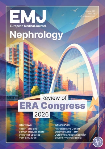
All information obtained by EMJ and each of the contributions from various sources is as current and accurate as possible. However, due to human or mechanical errors, EMJ and the contributors cannot guarantee the accuracy, adequacy, or completeness of any information, and cannot be held responsible for any errors or omissions. EMJ is completely independent of the review event (ERA 2026) and the use of the organisations does not constitute endorsement or media partnership in any form whatsoever. The cover photo is of Glasgow, UK, the location of ERA 2026.
Helena Bradbury, Katrina Thornber, Aleksandra Zurowska
Senior Editorial
Co-ordinator
Bertie Pearcey
Editorial Co-ordinators
Jess Nicholson, Alena Sofieva
Editorial Assistants
Niamh Holmes, Nonyelum Okonkwo, Roli Omamuli, Josh Warren Lister
Creative Director
Tim Uden
Design Manager
Stacey White
Senior Designers
Tamara Kondolomo, Owen Silcox
Designers
Shanjok Gurung, Fabio van Paris
Junior Designers
Fraser Hoey, Helena Spicer, Caleb Wylie
Marketing Director
Stephanie Corbett
Project Manager
Alex Hills
Chief Executive Officer
Justin Levett
Chief Commercial Officer
Dan Healy
Founder and Chairman
Spencer Gore
Welcome
Dear Readers,
We are thrilled to welcome you to the 2026 issue of EMJ Nephrology, which features coverage from this year’s European Renal Association (ERA) Congress, held in Glasgow, UK. Focusing on ‘Game Changers in Nephrology’, the event explored cutting-edge innovations across gene therapy, transplantation, vasculitides, acute kidney injury, chronic kidney disease, and both hypertensive and diabetic kidney disease.
Within our congress review, you will find multiple abstract summaries highlighting key breaking research from the event, including future perspectives on risk stratification in kidney disease. Additionally, this issue showcases multiple groundbreaking reviews of abstracts presented at ERA 2026, highlighting an assortment of topics that range from arterial hypertension in paediatric patients to the use of drug-dosing decision tools in primary care. We also present interviews with experts in the field, who discuss the latest developments in IgA nephropathy and robotic kidney surgery, as well as exclusive insights from prominent figures in the ERA association.
Our peer-reviewed content includes fascinating research spotlighting long-term outcomes for severe hyponatraemia, as well as two unique cases of severe methaemoglobinaemia and enterococcal emphysematous pyelonephritis.
We would like to take this opportunity to thank our Editorial Board, authors, peer reviewers, and interviewees for their support and critical contributions that have helped bring this issue to life.
We hope you enjoy reading.
Niamh Holmes Editorial Assistant
Contact us
Editorial enquiries: editor@emjreviews.com
Sales opportunities: salesadmin@emjreviews.com
Permissions and copyright: accountsreceivable@emjreviews.com
Reprints: info@emjreviews.com
Media enquiries: marketing@emjreviews.com
Stay at the Forefront of Modern Medicine
High-level perspectives. Global experts. Essential updates.
Join us for conversations with the minds shaping healthcare's future. Gain the distilled insights you need to lead in your field and make maximum impact.
Jonathan Sackier: Non Executive Director & CMO, AiM Medical Robotics, Florida, USA
Saranya Ravindran: Paediatric Emergency Medicine Registrar, Imperial College Healthcare NHS Trust
Catherine Glass: Associate NHS GP and Senior Appraiser, NHS England
Foreword
Dear Readers,
It is my pleasure to welcome you to the latest issue of EMJ Nephrology. I would like to extend my sincere appreciation to our authors, peer reviewers, and Editorial Board members, whose expertise and dedication continue to support the publication of high-quality, clinically relevant content.
This edition features a diverse collection of original research, case reports, and expert interviews, alongside highlights from the 63rd European Renal Association (ERA) Congress 2026. The Congress brought together the global nephrology community to explore advances, challenges, and emerging opportunities transforming kidney care.
ERA 2026 showcased key themes shaping modern nephrology, including precision medicine, AI, chronic kidney disease prevention, and novel therapeutic approaches. The meeting provided a platform for sharing cutting-edge research and discussing future directions in clinical practice, with a record number of over 3,000 abstracts submitted, reflecting continued innovation and engagement across the field.
This issue presents research exploring long-term outcomes associated with severe hyponatraemia, alongside two case reports highlighting complex clinical presentations involving acute kidney injury and rare renal complications.
We are also delighted to feature interviews with leading experts. Jonathan Barratt discusses advances in glomerular renal medicine, Erdem Canda explores robotic renal care, and our ERA 2026 Congress interviews with Serhan Tuğlular and Roser Torra provide insights into the key discussions and future priorities shaping nephrology.
Thank you for your continued readership and support. We hope this issue provides valuable insights and supports excellence in research and patient care.
The meeting provided a platform for sharing cutting-edge research and discussing future directions in clinical practice, with a record number of over 3,000 abstracts submitted
Dr Angela Yee-Moon Wang
Clinical Professor, Medicine Academic Clinical Programme, SingHealth Duke-NUS Academic Medical Centre; Senior Consultant, Department of Renal Medicine, Singapore General Hospital, Singapore
ERA 2026
The atmosphere throughout the Congress embodied the theme, ‘Open Your Mind’, encouraging attendees to embrace innovation
THE QUAINT city of Glasgow provided the backdrop for Europe’s premier nephrology meeting, the 63rd annual European Renal Association (ERA) Congress. Held from 3rd–6th June 2026 at the iconic Scottish Event Campus, the Congress brought together a record number of delegates from around the world. The venue, which has previously hosted major entertainment events, international conventions, and exhibitions, reflected the scale and ambition of this year’s meeting. The atmosphere throughout the Congress embodied the theme, ‘Open Your Mind’, encouraging attendees to embrace innovation, collaboration, and fresh perspectives in nephrology.
Giuseppe Palladino, Research, Development and Innovation Director at ERA, opened the ceremony by welcoming delegates and introducing Kate Stevens, Local Congress President, and Rosa Torra, ERA President.
In her opening remarks, Stevens celebrated Glasgow’s unique character and heritage, referencing the well-known phrase, ‘People Make Glasgow’, which reflects the warmth, friendliness, and hospitality of the city’s communities. She also highlighted Glasgow’s rich scientific legacy, noting the contributions of Thomas Graham, the Scottish chemist whose pioneering work on diffusion laid the foundations for modern dialysis and earned him recognition as the ‘father of dialysis’. Stevens further acknowledged Joseph Lister, whose development of antiseptic surgical techniques transformed healthcare practice worldwide and has saved countless lives. She also recognised the historic achievements of James McCune Smith, who studied at the University of Glasgow and
became the first African American to obtain a medical degree.
Stevens went on to discuss ERA’s commitment to community engagement and sustainability through its congress legacy project. Initiatives included support for the Glasgow Children’s Hospital Charity and the Kidney and Bee Adventure, a programme designed to encourage local children to engage with nature, while learning about kidney health and environmental stewardship.
Delegates were then welcomed by Bailie Marie Garrity, a long-serving member of Glasgow City Council, who further emphasised the city’s enduring contribution to kidney care and medical innovation. Drawing parallels between the values of Glasgow and the ERA community, Garrity highlighted their shared commitment to fairness, inclusion, and opportunity for all. She noted that the Congress theme, ‘Open Your Mind’, reflects
the importance of curiosity, innovation, and collaboration in addressing current and future healthcare challenges.
ERA President Rosa Torra then delivered the Presidential Address, expressing her gratitude to the nephrology community for its continued engagement with and support of the association. She noted that the high level of participation at ERA reflects a collective commitment to shaping the future of nephrology. Torra also encouraged delegates to embrace the spirit of ‘challenging their thinking’ by critically evaluating emerging evidence and new developments in the field. She remarked that, rather than a lack of information, the modern challenge lies in effectively interpreting and applying the vast amounts of data now available to clinicians and researchers. Torra concluded by thanking everyone involved in organising the congress, including staff, committee members, and partner organisations.
Before the scientific programme commenced, Torra invited Serhan Tuğlular, Chair of the ERA 2026 Scientific Committee, to the stage to receive a diploma in recognition of her contributions to the congress.
The ceremony then celebrated the recipients of the ERA 2026 awards. Manuel Praga Terente, Complutense University, Madrid, Spain, received the ERA Award for Outstanding Clinical Contributions to Nephrology. Robert Unwin, University College London, UK, received the ERA Award for Outstanding Basic Science Contributions to Nephrology. Mehmet Kanbay, Koç University Hopsital, Istanbul, Türkiye, was honoured with the ERA Award for Research Excellence in Nephrology, while Danilo Fliser, Saarland University, Saarbrücken, Germany,
received the ERA Award for Outstanding Contribution to the Society.
The ERA Award for Excellence in Sustainable Nephrology was presented to Groupe Néphrologie Verte (Société Francophone de Néphrologie, Dialyse et Transplantation), recognising its leadership in promoting environmentally sustainable kidney care.
Among the early-career award recipients, Verónica Miguel Herranz, RWTH Aachen University Hospital, Germany, received the Rosanna Gusmano Award for Young Investigators in Basic Science, while Michael Balzer, University of Kiel, Germany, was presented with the Stanley Shaldon Award for Young Investigators in Translational Science. Rik Olde Engberink, Amsterdam University Medical Center; and Michele Eisenga, University Medical Center Groningen, the Netherlands, received the Eberhard Ritz Award for Young Investigators in Clinical Science. Katharina Artinger, Medical University of Graz, Austria, was honoured with the Young Nephrologist ERA Council Award.
The opening ceremony also recognised Rosanna Coppo, University of Turin, Italy, who received Honorary Membership of the ERA in recognition of her longstanding contributions to nephrology and the association.
This year’s Opening Ceremony concluded with a vibrant celebration of Scottish culture, beginning with an exuberant pipe band performance that showcased the musical heritage of the host nation and provided a memorable close to the evening’s proceedings.
Pre-dialysis Exercise May Protect the Heart Across
Haemodialysis Sessions
A RANDOMISED crossover trial suggests that a single bout of exercise performed before haemodialysis can reduce dialysis-induced cardiac injury during not only the same treatment session, but also the subsequent session, according to findings presented at ERA 2026.1 These results may support the concept of exercise-induced cardiac preconditioning.
Researchers enrolled 26 patients receiving maintenance haemodialysis and compared two conditions: a 30-minute exercise
or vascular refilling, mechanisms traditionally thought to explain the cardioprotective effects of intradialytic exercise.
These results may support the concept of exercise-induced cardiac preconditioning
TTV Trajectories May Help Identify Rejection and Infection Risk After Kidney Transplantation
MONITORING
changes in torque teno virus (TTV) levels may help predict rejection and opportunistic infections during the first months after kidney transplantation, according to research presented at ERA 2026.
Achieving the right balance of immunosuppression following transplantation remains challenging, with excessive treatment increasing infection risk and insufficient treatment raising the likelihood of rejection. TTV has emerged as a potential biomarker of overall immunosuppressive burden, but most studies have focused on population-wide thresholds rather than individual patient patterns.
Researchers from Queen Mary University of London and Royal London Hospital, UK, prospectively followed 268 kidney transplant recipients, measuring TTV levels from transplantation to 24 weeks post-transplant.
TTV levels increased during the first 12 weeks before plateauing, but substantial variation was observed between patients. Importantly, interval-to-interval increases in TTV were associated with a significantly lower risk of biopsy-proven rejection. In contrast, absolute TTV levels were not associated with rejection risk.
Higher TTV levels were linked to a greater likelihood of opportunistic infections. Each one-log increase in TTV was associated with a 57% increase in the odds of opportunistic infection, including BK virus and cytomegalovirus infections. No significant association was observed with bacterial infections.
The findings suggest that individual TTV trajectories may provide more clinically useful information than fixed universal thresholds. Rather than relying solely on absolute TTV values, monitoring how levels change over time could help clinicians assess immunosuppressive burden and identify patients at risk of either rejection or opportunistic infection.
The authors concluded that TTV shows promise as a patient-specific biomarker in the early post-transplant period and highlighted the need for randomised multicentre trials evaluating TTV-guided immunosuppression strategies.
The findings suggest that individual TTV trajectories may provide more clinically useful information than fixed universal thresholds
57%
Each one-log increase in TTV was associated with a 57% increase in the odds of opportunistic infection
MAYO Clinic Chronicity Score was independently associated with long-term kidney transplant outcomes and may enhance risk stratification beyond established pathological classification systems, according to new research analysing kidney allograft biopsies, which was presented at ERA 2026.3
Current transplant pathology classifications place greater emphasis on active inflammation than chronic injury, despite chronic histological damage being irreversible and strongly linked to graft outcomes. Researchers investigated whether the Mayo Clinic Chronicity Score could provide a standardised approach to assessing chronic injury in kidney allograft biopsies.
The study evaluated 359 kidney allograft biopsies, with chronic lesions scored across four tissue compartments: glomerular sclerosis, interstitial fibrosis, tubular atrophy, and arteriosclerosis. The primary endpoint was progression to end-stage kidney disease within 5 years. Survival analyses and adjusted Cox regression models accounted for age, sex, estimated glomerular filtration rate, and proteinuria at the time of biopsy.
Results showed that higher Mayo Clinic Chronicity Score grades were independently associated with progression to end-stage kidney disease beyond 5 years. This association remained significant regardless of diagnostic category in both crude (p<0.0001) and adjusted analyses (p=0.02).
Among the individual histological components, interstitial fibrosis and tubular atrophy emerged as the strongest predictor of graft survival, demonstrating significant associations in both crude (p<0.0001) and adjusted analyses (p<0.003). Glomerular sclerosis was also associated with outcomes in unadjusted analyses (p=0.0013), although this relationship was attenuated after adjustment (p=0.2). Arteriosclerosis showed a non-significant trend towards poorer outcomes (p=0.079).
The composite Mayo Clinic Chronicity Score demonstrated the highest predictive accuracy at earlier time points, achieving area under the curve values of 0.836 at 12 months and 0.816 at 36 months.
At 60 months, interstitial fibrosis and tubular atrophy alone showed the strongest predictive performance, with an area under the curve of 0.746. Combining interstitial fibrosis and tubular atrophy with either glomerular sclerosis or arteriosclerosis further improved long-term predictive ability, with area under the curve values ranging from 0.745–0.770. In contrast, glomerular sclerosis and arteriosclerosis alone became less informative over time.
The findings represent the first systematic application of the Mayo Clinic Chronicity Score in kidney allograft biopsies and suggest the tool is practical, reproducible, and clinically relevant. The authors conclude that incorporating chronic injury assessment using the Mayo Clinic
At 60 months, interstitial fibrosis and tubular atrophy alone showed the strongest predictive performance, with an area under the curve of 0.746
Study Reveals that EKFC and CKD-EPI Yield Similar KFRE Performance
SWAPPING the newer European Kidney Function Consortium (EKFC) equations for established Chronic Kidney Disease Epidemiology Collaboration (CKD-EPI) ones makes little difference to how well the Kidney Failure Risk Equation (KFRE) predicts kidney failure, a large Swedish study reported, though EKFC sharpens accuracy when cystatin C is used.4
The KFRE guides European clinicians on when to refer patients to nephrology and prepare for kidney replacement therapy. It was built on CKD-EPI-based estimated glomerular filtration rate (eGFR). As centres increasingly adopt the EKFC equations, which match or outperform CKD-EPI, it is unclear whether substituting them disturbs the tool's reliability.
The team used the Stockholm CREAtinine Measurements (SCREAM) project, a population-based cohort exceeding three million residents. They identified adults who, between 2011–2021, had creatinine and cystatin C measured the same day plus an adjacent albuminuria test. Two- and fiveyear kidney failure risks were computed with the four-variable KFRE, covering eGFR, albuminuria, age, and sex. eGFR came from creatinine, cystatin C, or their combination, via CKD-EPI or EKFC. The final sample comprised 27,125 individuals (median age: 75 years; 45% women).
Kidney replacement therapy occurred in 620 people by 2 years and 1,265 by 5 years, while
4,214 and 8,287 respectively died without reaching kidney replacement therapy. KFRE discrimination was excellent across every equation and filtration marker, with areas under the curve spanning 0.95–0.97. For creatinine-based eGFR, EKFC and CKD-EPI delivered nearly identical calibration at both horizons. When cystatin C or the creatininecystatin C combination was used, EKFC produced better KFRE calibration than CKDEPI. Net benefit at the KDIGO-recommended thresholds for guiding nephrology care was similar at 2 and 5 years, irrespective of the equation applied.
The results demonstrate that, in a northern European system, estimating creatininebased eGFR with EKFC or CKD-EPI does not impact the KFRE's predictive performance or clinical utility, reassuring clinics adopting the newer equations.
Kidney replacement therapy occurred in 620 people by 2 years and 1,265 by 5 years
Cystatin C eGFR Improves Chronic Kidney Disease Risk Stratification
CYSTATIN C estimated glomerular filtration rate (eGFR) may improve chronic kidney disease risk stratification by identifying patients at greater risk of mortality and hospitalisation who are not detected using creatinine-based eGFR, according to new findings from a large population-based analysis.5
Researchers analysed data from 372,454 participants in the UK Biobank to investigate whether cystatin C-based eGFR could identify individuals with chronic kidney disease at increased risk of adverse outcomes compared with conventional creatinine-based measures.
Participants were categorised according to chronic kidney disease status using both methods. During a median follow up of 12.53 years, there were 23,470 deaths and 113,378 first hospitalisation episodes.
The analysis showed substantial differences in the number of participants identified as having chronic kidney disease. Creatininebased eGFR exclusively identified 2,584 individuals, whereas cystatin C eGFR alone identified 11,691 participants.
Among both the full cohort and participants eligible for chronic kidney disease screening, those identified exclusively through cystatin C eGFR consistently experienced higher rates of all-cause mortality and first hospitalisation.
In contrast, researchers found no consistent associations between adverse outcomes and participants classified as having no chronic kidney disease or those identified solely through creatinine-based eGFR.
Importantly, the relationship between cystatin C eGFR defined chronic kidney
disease and adverse outcomes remained evident among participants without albuminuria and across body mass index extremes. These findings suggest that cystatin C based assessment may provide prognostic information beyond traditional markers currently used in clinical practice.
The investigators also evaluated cohorts reflecting current eligibility criteria for disease modifying chronic kidney disease therapies. Within these groups, participants identified as having chronic kidney disease only through cystatin C eGFR experienced at least double the rates of mortality and hospitalisation compared with those identified exclusively using creatinine-based eGFR.
The authors concluded that reliance on creatinine-based eGFR alone may systematically exclude higher risk populations from future chronic kidney disease trials. The findings support the potential role of cystatin C eGFR in improving risk stratification and informing participant selection in future studies.
These results highlight the possibility that incorporating cystatin C eGFR into chronic kidney disease assessment frameworks could enable more accurate identification of patients at greatest risk of adverse clinical outcomes.
Mortality Patterns Offer Insight into the Obesity Paradox
ADULTS aged 80 years and above with chronic kidney disease (CKD) may have better survival outcomes when their BMI falls within the overweight range, according to an observational study presented at ERA 2026.6
The study explored the so-called ‘obesity paradox’ in the oldest-old population with CKD, a population often characterised by multimorbidity and age-related changes in body composition that have been largely overlooked in previous research.
Researchers analysed data from 1,267 adults aged 80 years or older with CKD, representing an estimated 4.2 million people in the USA. During a median followup period of 4.8 years, 795 deaths were recorded, 320 of these being cardiovascular.
Using restricted cubic spline analyses and adjusted Cox proportional hazards models, the investigators assessed relationships between BMI and all-cause, cardiovascular, and non-cardiovascular mortality. The analyses accounted for a range of factors including demographic characteristics, smoking status, comorbidities, kidney function measures, and laboratory values.
Results showed a U-shaped association between BMI and cardiovascular mortality, while non-cardiovascular mortality decreased as BMI increased. The analyses indicated that the BMI associated with the most favourable cardiovascular survival fell within the overweight range.
Among participants whose BMI exceeded the point associated with the lowest cardiovascular risk, each one-unit increase in BMI was linked to a 7% higher risk of
cardiovascular mortality. However, each additional BMI unit was associated with a 4% lower risk of non-cardiovascular mortality. Compared with overweight individuals, those with a BMI of 35 kg/m2 or higher had a significantly greater risk of cardiovascular death (HR: 1.81; 95% CI: 1.15–2.86).
The authors suggest that the obesity paradox in the oldest-old CKD population may be largely explained by lower rates of non-cardiovascular mortality among individuals with higher BMI, as higher BMI appeared protective for non-cardiovascular mortality.
As an observational study, a cause-andeffect relationship cannot be established. However, the results indicate that an overweight BMI may represent the most favourable range for survival in very elderly people with CKD and could help inform future guidance for this growing patient population.
The BMI associated with the most favourable cardiovascular survival fell within the overweight range
Air Pollution Linked to CKD Progression in Portugal
A LARGE Portuguese cohort study, presented at ERA 2026, suggests that exposure to air pollution, particularly fine particulate matter (PM2.5) and ozone (O3), may contribute to chronic kidney disease (CKD) development and progression, even in regions with generally good air quality.7
The IMPA-R-PT study analysed data from 23,620 adults followed for a median of 6.7 years. Participants lived within 2 km of air quality monitoring stations, allowing researchers to assess long-term exposure to pollutants including PM2.5, PM10, nitrogen dioxide (NO2), sulphur dioxide (SO2), and O3.
CKD prevalence in the cohort was 33.8%, with an incidence rate of 25.2 cases per 1,000 person-years. Patients with CKD had significantly higher exposure to PM2.5, PM10, and O3 than those without kidney disease. After adjustment for demographic and clinical factors, PM2.5, O3, and the PM10/ PM2.5 ratio remained independently associated with CKD prevalence.
Higher pollutant exposure was also linked to faster kidney function decline. Patients classified as rapid progressors experienced greater exposure to PM2.5, SO2, and O3, while those who progressed to advanced CKD (Stage 5) had significantly higher levels of PM2.5 and O3 exposure than the rest of the cohort.
Notably, PM2.5 emerged as an independent predictor of incident CKD in adjusted analyses, with each increase in average PM2.5 concentration associated with a 15% higher risk of developing CKD. Elevated levels of PM10, SO2, NO2, and O3 were also associated with increased CKD incidence, while a higher PM10/PM2.5 ratio appeared protective.
The authors note that air quality was rated good or very good on more than 90% of study days, suggesting that adverse renal effects may occur even at pollutant concentrations that meet current regulatory standards. The findings add to growing evidence that chronic exposure to air pollution may contribute to kidney disease through inflammatory and oxidative stress pathways and highlight potential implications for environmental health policy.
Myeloid
HIF-2α Deficiency Linked to Renal Fibrosis and Immune Dysregulation
DISRUPTION of myeloid hypoxia-inducible factor-2 α (HIF-2α) may drive renal fibrosis through altered communication between immune cells, according to research presented at ERA 2026.8
Renal fibrosis is a key contributor to chronic kidney disease progression, but the mechanisms linking hypoxia, immune dysfunction, and tissue scarring remain incompletely understood. While HIF-2α is known to play important roles in kidney biology, anaemia, and cancer, its influence on immune cell interactions in kidney disease has been less clear.
To investigate this, researchers generated a mouse model lacking HIF-2α specifically in myeloid cells and compared these animals with healthy control mice. The team examined kidney function, immune cell behaviour, and markers of fibrosis to determine how loss of myeloid HIF-2α affected renal health.
The investigators observed spontaneous and early development of renal fibrosis in mice lacking myeloid HIF-2α. By 3 months of age, affected animals showed evidence of kidney dysfunction, including increased serum creatinine levels and alterations in urinary biomarkers. Histological analysis revealed prominent glomerular sclerosis, while molecular studies demonstrated increased expression of genes associated with fibrosis, inflammation, and kidney injury.
Further analysis identified significant changes in immune cell behaviour. Macrophages isolated from younger mice displayed a pro-inflammatory profile, while those from older animals shifted towards a more anti-inflammatory phenotype. Despite this apparent change, the cells
remained metabolically hyperactive, exhibiting increased glycolysis and oxidative phosphorylation.
Proteomic analysis highlighted neutrophil degranulation as one of the most strongly enriched pathways in the kidneys of HIF-2α-deficient mice. Consistent with this finding, researchers identified hyperactive neutrophils with immunosuppressive and anti-apoptotic characteristics in the bone marrow.
Importantly, signalling molecules released by both HIF-2α-deficient macrophages and neutrophils promoted a profibrotic response in cultured mesangial cells, increasing expression of genes associated with tissue scarring and cellular transformation.
The findings suggest that abnormal communication between macrophages and neutrophils may be a key driver of fibrosis development. According to the authors, loss of myeloid HIF-2α promotes excessive neutrophil recruitment and activation within the kidney, accelerating tissue injury and disease progression.
These results identify a previously unrecognised role for myeloid HIF-2α in regulating immune res
ponses within the kidney and provide new insight into how hypoxia-related pathways contribute to renal fibrosis. The researchers suggest that targeting this immune crosstalk could represent a novel therapeutic strategy for slowing the progression of chronic kidney disease.
The investigators observed spontaneous and early development of renal fibrosis in mice lacking myeloid HIF-2α
Extracellular Vesicles Show Potential to Block Immune-Driven
Acute
Kidney Injury in Sepsis
NEW FINDINGS presented at ERA 2026 redefine the biology of sepsisassociated acute kidney injury (SA-AKI), identifying monocytes intracellular complement (complosome) activation as a key driver of organ damage and a promising target for intervention.9
Building on their earlier discovery that reduced podocyte-derived cluster of differentiation 35-positive extracellular vesicles (CD35-EVs) are associated with poor outcomes in SA-AKI, the researchers investigated whether these vesicles play a functional role in disease progression in cecal ligation and puncture mice. The study reveals that CD35-EVs act as natural regulators of monocyte complement activity, blocking a newly identified intracellular pathway that drives organ injury.
Monocyte-tracking and molecular assays were used to trace the original of renal component depositions, analyse organ infiltration, complosome activation, and CD35-EV-complosome interaction. Injury pathways driven by infiltrating monocytes were defined via multi-omics as well validation of the functional necessity of complosome through genetic and transplantation models. Monocytes from sepsis patients were also collected to assess the correlation between complosome activation and the severity of organ injury.
CD35-EV administration attenuated renal and multi-organ injury, improved survival, and limited monocyte infiltration. Injury was driven by infiltrating monocytes exhibiting intracellular complosome activation linked to
mitochondrial C5a receptor 1 signalling and glycolytic reprogramming that promoted migration. CD35-EVs were internalised by monocytes, where they neutralised intracellular C3b and suppressed alternative pathway activity, reversing metabolic and chemotactic programming. Monocytespecific C3 deletion reproduced these effects and eliminated additional CD35-EV benefit, confirming pathway specificity. In patients, complosome activation correlated with disease severity.
This study identifies intracellular complosome activation in monocytes as a key driver of SA-AKI and multiorgan injury. It further demonstrates the therapeutic potential of extracellular vesicles as nanocarriers for targeted delivery of CD35, disrupting complosomedependent monocyte chemotaxis and tissue infiltration. Collectively, these findings establish a mechanistically defined EVbased intervention strategy and provide a foundation for targeting intracellular complement in inflammatory disease. This reshapes current understanding of complement biology, expanding it beyond extracellular pathways and positioning the complosome as a tractable target for precision immunomodulation in sepsis and related organ dysfunction.
CD35-EV administration attenuated renal and multi-organ injury, improved survival, and limited monocyte infiltration
PPIs Linked to Fewer Upper GI Bleeds in Patients on Haemodialysis
NEW DATA presented at ERA 2026 has demonstrated that proton pump inhibitors (PPI) cut the risk of upper gastrointestinal bleeding by nearly 40% in patients on haemodialysis on oral anticoagulants.10
Patients on haemodialysis carry a high risk of upper gastrointestinal bleeding (UGIB), and that danger climbs once an oral anticoagulant is added. PPIs are widely prescribed, but their ability to avert bleeding in these patients has never been firmly established.
The investigators emulated a target trial using the French Renal Epidemiology and Information Network (REIN) registry, linked to the national health database. Eligible adults on haemodialysis had started an oral anticoagulant between January 2012–December 2023, with no PPI dispensed in the prior 3 months. They were followed for 12 months or until a first UGIB event, death, kidney transplantation, dialysis cessation, anticoagulant discontinuation, or loss to follow-up. The primary outcome, a first UGIB event, was identified through 10th revision of the International Classification of Diseases (ICD-10) hospital discharge codes. Risk in PPI users versus non-users was estimated with inverse probability of treatment weighting and a cause-specific Cox model, plus an as-treated secondary analysis. Of 8,205 eligible patients, 7,503
were included, 2,323 starting a PPI the same day as their anticoagulant.
Over a median follow-up of 1 year, 145 patients experienced a first UGIB event, with melena most common at 38% of cases. After weighting, the 12-month cumulative incidence of UGIB was slightly lower among PPI users than non-users. The risk reduction was statistically significant, with a weighted hazard ratio of 0.62 (95% CI: 0.41–0.95).
The as-treated secondary analysis of PPI initiation versus no initiation gave a closely matching weighted hazard ratio of 0.63 (95% CI: 0.39–0.99). Subgroup and sensitivity analyses, though underpowered, aligned with the main result.
The authors concluded that PPIs are linked to a lower risk of upper gastrointestinal bleeding after patients on haemodialysis begin oral anticoagulation, supplying rare evidence for a protective approach in this high-risk group. They cautioned that PPIs carry their own potential for serious harm, urging clinicians to review such prescriptions regularly rather than indefinitely.
References
1. Josse M et al. Biphasic cardioprotection by pre-dialysis exercise in hemodialysis patients: a randomized controlled trial. Abstract 2891. ERA Congress, 3-6 June, 2026.
2. Chakraborty R et al. Individualised torque teno virus trajectories predict rejection and opportunistic infection early after kidney transplantation. Abstract 114. ERA Congress, 3-6 June, 2026.
3. Berti GM et al. Five-year prognostic performance of the Mayo Clinic Chronicity Score in kidney transplant biopsies. Abstract 1381. ERA Congress, 3-6 June, 2026.
4. Créon A et al. Predictive performance and clinical utility of the kidney failure
risk equation using EKFC versus CKDEPI estimated GFR. Abstract 1377. ERA Congress, 3-6 June, 2026.
5. Tabinor M et al. Risk stratification in chronic kidney disease: outcomes using cystatin-C and creatinine based eGFR. Abstract 912. ERA Congress, 3-6 June, 2026.
6. Yang C et al. The obesity paradox is primarily driven by decreased noncardiovascular mortality risk among the oldest old with chronic kidney disease. Abstract 1362. ERA Congress, June 3-6, 2026.
7. Laranjinha I et al. Air pollution and chronic kidney disease progression in Portugal - IMPA-R-PT study (impact of air pollution on renal health in Portugal). Abstract 1201. ERA Congress, 3-6 June, 2026.
8. Barrenechea-Barrenechea JA et al. Myeloid HIF-2α: a key player in immune crosstalk and renal fibrosis. Abstract 442. ERA Congress, 3-6 June, 2026.
9. Li N et al. CD35-EVs ameliorate sepsis-associated AKI and multiorgan injury by regulating monocyte complosome activation. Abstract 1784. ERA Congress, 3–6 June, 2026.
10. Laville S et al. Proton pump inhibitors reduce upper gastrointestinal bleeding in hemodialysis patients initiating oral anticoagulation: a nationwide registry study. Abstract 637. ERA Congress, 3-6 June, 2026.
Open Your Mind in Nephroprotection: Controversies and Perspectives
AT THE 63rd Congress of the European Renal Association (ERA), experts from across the globe gathered to spotlight major advances and discuss pressing issues in the field of nephrology. This educational session, chaired by Samar Abd ElHafeez and Marcin Adamczak, Department of Nephrology, Transplantation and Internal Medicine, Medical University of Silesia, Katowice, Poland, featured three insightful talks centred around nephroprotection in chronic kidney disease (CKD).
FRUIT- AND VEGETABLE-RICH DIET IN PATIENTS WITH CKD
Juan-Jesus Carrero, Professor of Kidney Epidemiology, Department of Medical Epidemiology and Biostatistics, Karolinska Institutet, Stockholm, Sweden, opened the session by discussing the impact of a fruit- and vegetable-rich diet in patients with CKD. His talk assessed the evidence supporting the inclusion of dietary recommendations in recent clinical guidelines.1,2
A plant-based diet is a broad term encompassing any eating pattern in which animal-based foods are consumed in moderation, and a greater proportion of food choices are plant-based. As explained by Carrero, this umbrella term includes vegan, vegetarian, dietary approaches to stop hypertension (DASH), Mediterranean, healthy eating, and Okinawan diets. Carrero subsequently debunked several widespread myths, including that plantbased diets cause nutrient deficiencies, hyperphosphatemia, and hyperkalaemia.
Firstly, several studies have confirmed that vegetarian and vegan diets do not contribute to amino acid deficiencies,3 and this has also been demonstrated specifically in CKD in both clinical trials4 and observational studies.5 Regarding the second myth, that plant-based diets cause hyperphosphatemia, this has likewise been disproven. For example, in patients with Stage 4 CKD, plant-based diets resulted in lower serum phosphate concentrations and lower urinary phosphate excretion.6 Finally, Carrero cited several studies demonstrating a weak, if any, association between dietary and circulating potassium levels, reaffirming that diet is only one contributing factor to serum potassium concentrations.7
Carrero went on to emphasise that restricting organic potassium (K+) sources fails to recognise that potassium absorption and distribution are influenced by the intake of other nutrients. For example, in patients with CKD, consuming potassium together with glucose favours intracellular potassium deposition, whilst alkali promotes both intracellular potassium
Increasing plant protein intake has been shown to improve renal plasma flow and reduce the fractional clearance of albumin
deposition and renal potassium excretion. In addition, fibre improves faecal potassium excretion by increasing stool volume and alleviating constipation.
So, what can be done? Firstly, as noted by Carrero, increasing plant protein intake has been shown to improve renal plasma flow and reduce the fractional clearance of albumin.8 Secondly, he advocated for greater fibre consumption because of its well-established benefits in lowering cardiovascular risk and regulating blood glucose levels. Finally, greater consumption of plant foods increases dietary alkali intake, which has been shown to preserve glomerular filtration rate, decrease bone resorption, improve insulin sensitivity, and reduce sarcopenia.9
SHOULD WE TREAT MILD METABOLIC ACIDOSIS IN PATIENTS WITH CKD?
Marcin Adamczak, subsequently took the stage to discuss the controversial issue of treating mild metabolic acidosis in patients with CKD.
Metabolic acidosis in CKD is defined as an acid-base disorder diagnosed when the venous plasma or blood bicarbonate concentration is lower than 22 mmol/L in the absence of signs of respiratory alkalosis, and occurs in nearly 20% of patients with CKD.10
Each 1 mEq/L increase in bicarbonate was associated with a 3% reduction in the risk of reaching a renal endpoint, with the strongest association observed in patients with betterpreserved kidney function 3%
The main causes of metabolic acidosis are decreased tubular HCO3− reabsorption and insufficient HCO3− production during ammoniagenesis in the distal tubule.
As highlighted by Adamczak, metabolic acidosis can directly impair quality of life through reductions in skeletal muscle mass and strength, cognitive impairment, and sexual dysfunction in men.
Adamczak then discussed the association between metabolic acidosis and CKD progression, highlighting a prominent observational study.11 This study evaluated serum bicarbonate levels as a risk factor for renal outcomes, cardiovascular events, and mortality in 3,939 participants with CKD Stages 2–4. Over a median followup of 3.9 years, lower serum bicarbonate levels were independently associated with a higher risk of CKD progression. Each 1 mEq/L increase in bicarbonate was associated with a 3% reduction in the risk of reaching a renal endpoint, with the strongest association observed in patients with better-preserved kidney function (estimated glomerular filtration rate: >45 mL/min/1.73 m²). Similar associations have also been reported in adolescents.12
How Do We Treat Metabolic Acidosis in Patients with CKD?
Several studies have found that fruit- and vegetable-enriched diets increase plasma bicarbonate concentrations and possess
nephroprotective properties.13,14 Examples include apples, apricots, oranges, peaches, pears, raisins, strawberries, and carrots.
The second treatment option is oral sodium bicarbonate supplementation; however, this remains somewhat controversial. Adamczak discussed two key studies: the UBI study15 and the BICARB study.16 The UBI study was a randomised trial in patients with CKD Stages 3–5 and metabolic acidosis that found oral sodium bicarbonate significantly reduced kidney disease progression, dialysis initiation, and all-cause mortality compared with standard care. Sodium bicarbonate was generally well tolerated and did not significantly increase blood pressure, body weight, or hospitalisation rates. In contrast, the BICARB study found that oral sodium bicarbonate did not improve physical function, kidney function, or quality of life in older adults with advanced CKD and metabolic acidosis compared with placebo. Furthermore, bicarbonate treatment was associated with more adverse events and higher healthcare costs, suggesting that it is unlikely to be a cost-effective intervention in this population.
Due to the conflicting results of these pivotal studies, the question remains unresolved. Adamczak therefore advocated for more high-quality data, citing a 2024 meta-analysis of eight RCTs involving 2,037 patients with CKD.17 The analysis concluded that sodium bicarbonate treatment significantly improved estimated glomerular filtration rate.
Finally, Adamczak discussed sodium zirconium cyclosilicate for patients with hyperkalaemia. This selective, non-absorbed potassium binder promotes potassium elimination through the gastrointestinal tract. Studies have shown that it can also reduce the incidence of metabolic acidosis.18
In conclusion, as noted by Adamczak, the treatment of mild metabolic acidosis (serum bicarbonate 18.0–21.9 mmol/L) in patients with CKD remains controversial due to the lack of definitive placebo-controlled clinical trial data.
ALDOSTERONE ACTIVITY REDUCTION IN PATIENTS WITH CKD
Katherine R. Tuttle, Executive Director for Research at Providence Inland Northwest Health and Professor of Medicine at the University of Washington, Spokane, USA, delivered the final presentation of the session, focusing on the role of aldosterone activity reduction in patients with CKD. Tuttle explored the growing recognition of aldosterone as a key driver of kidney and cardiovascular disease progression and discussed emerging therapeutic strategies aimed at mitigating its harmful effects. The presentation highlighted the increasing importance of targeting mineralocorticoid receptor overactivation as part of a comprehensive nephroprotective approach.
Several studies have found that fruit- and vegetable-enriched diets increase plasma bicarbonate concentrations and possess nephroprotective properties
She opened her talk by discussing baxdrostat, a first-in-class aldosterone synthase inhibitor used in combination with other therapies to treat treatmentresistant hypertension. In a pivotal 2023 study, patients with treatment-resistant hypertension, defined as having blood pressure ≥130/80 mmHg despite treatment with three or more blood pressurelowering agents, including a diuretic, experienced significant reductions in blood pressure when baxdrostat was added to standard-of-care therapy.19 In the Phase 2 Advance-HTN trial, the aldosterone synthase inhibitor lorundrostat significantly reduced 24-hour ambulatory systolic blood pressure compared with placebo in patients with uncontrolled or treatmentresistant hypertension despite receiving multiple antihypertensive medications. After 12 weeks, lorundrostat lowered systolic blood pressure by an additional 6.5–7.9 mmHg versus placebo, although treatment was associated with an increased incidence of hyperkalaemia, with potassium levels exceeding 6.0 mmol/L in 5–7% of treated participants.20
Tuttle highlighted a Phase 2 trial evaluating the aldosterone synthase inhibitor vicadrostat (BI 690517) in 714 adults with CKD, with or without Type 2 diabetes, receiving stable ACE inhibitor or ARB therapy.21 Participants were randomised to receive varying doses of vicadrostat alone or in combination with empagliflozin, with the primary endpoint being the change in urinary albumin-to-creatinine ratio at 14 weeks. The study demonstrated dosedependent reductions in albuminuria, supporting aldosterone synthase inhibition as a promising strategy for reducing residual cardiorenal risk in patients with CKD.
References
1. Ikizler TA et al. KDOQI clinical practice guideline for nutrition in CKD: 2020 update. AJKD. 2020;76(3):S1-107.
2. Kidney Disease: Improving Global Outcomes (KDIGO) CKD Work Group. KDIGO 2024 clinical practice guideline for the evaluation and management of chronic kidney disease. Kidney Int. 2024;105(4S):S117-314.
Tuttle then discussed emerging data for vicadrostat, highlighting a Phase 2 study showing dose-dependent suppression of plasma aldosterone levels in patients with CKD and proteinuria.22 Maximal aldosterone suppression was observed at Week 14 and was sustained after treatment cessation, while cortisol levels remained unchanged, demonstrating selective inhibition of CYP11B2 without affecting glucocorticoid production. These findings further support vicadrostat as a promising strategy for reducing aldosterone-mediated kidney damage.
Additionally, she discussed the reported lower risks of oedema and hyperkalaemia with empagliflozin in patients with Type 2 diabetes and CKD.23
Finally, looking to the future, Tuttle highlighted the EXPLORE-CKD Phase 2b crossover trial, which met its primary endpoint. Lorundrostat 25 mg once daily achieved a 9.3 mmHg reduction in systolic blood pressure and a 7.5 mmHg placeboadjusted reduction at 4 weeks (p=0.0024).
CONCLUSION
The session highlighted both the progress made and the ongoing debates in nephroprotection, from challenging longstanding dietary restrictions to refining the management of metabolic acidosis and targeting aldosterone-driven kidney injury. Together, these discussions underscored a growing shift towards more personalised, evidence-based strategies aimed at slowing CKD progression and improving long-term patient outcomes.
3. Rizzo NS et al. Nutrient profiles of vegetarian and nonvegetarian dietary patterns. J Acad Nutr Diet. 2013;113(12):1610-9.
4. Soroka N et al. Comparison of a vegetable-based (soya) and an animalbased low-protein diet in predialysis chronic renal failure patients. Nephron. 1998;79(2):173-80.
5. Kandouz S et al. Reduced protein bound uraemic toxins in vegetarian kidney failure patients treated by haemodiafiltration. Hemodial Int. 2016;20(4):610-17.
6. Moe SM et al. Vegetarian compared with meat dietary protein source and phosphorus homeostasis in chronic kidney disease. Clin J Am Soc Nephrol. 2011;6(2):257-64.
7. Gritter M et al. Rationale and design of a randomized placebocontrolled clinical trial assessing the
renoprotective effects of potassium supplementation in chronic kidney disease. 2018;140(1):48-57.
8. Kontessis P et al. Renal, metabolic and hormonal responses to ingestion of animal and vegetable proteins. Kidney Int. 1990;38(1):136-44.
9. Carrero JJ et al. Plant-based diets to manage the risks and complications of chronic kidney disease. Nat Rev Nephrol. 2020;16(9):525-42.
10. Raphael KL. Approach to the treatment of chronic metabolic acidosis in CKD. Am J Kidney Dis. 2016;67(4):696-702.
11. Dobre M et al. Association of serum bicarbonate with risk of renal and cardiovascular outcomes in CKD: a report from the Chronic Renal Insufficiency Cohort (CRIC) study. Am J Kidney Dis. 2013;62(4):670-8.
12. Harambat J et al. Epidemiology of chronic kidney disease in children. Pediatr Nephrol. 2012;27(3):363-73.
13. Goraya N et al. Dietary acid reduction with fruits and vegetables or bicarbonate attenuates kidney injury in patients with a moderately reduced glomerular filtration rate due to
14. Mahboobi S et al. Effects of dietary interventions for metabolic acidosis in chronic kidney disease: a systematic review and meta-analysis. Nephrol Dial Transplant. 2025;40(4):751-67.
15. Di lorio BR et al. Treatment of metabolic acidosis with sodium bicarbonate delays progression of chronic kidney disease: the UBI Study. J Nephrol. 2019;32(6):989-1001.
16. BiCARD study group. Clinical and cost-effectiveness of oral sodium bicarbonate therapy for older patients with chronic kidney disease and lowgrade acidosis (BiCARB): a pragmatic randomised, double-blind, placebocontrolled trial. BMC Med. 2020;18(1):91.
17. Yang TY et al. Sodium bicarbonate treatment and clinical outcomes in chronic kidney disease with metabolic acidosis: a meta-analysis. Clin J Am Soc Nephrol. 2024;19(8):959-69.
18. Roger SD et al. Sodium zirconium cyclosilicate increases serum bicarbonate concentrations among patients with hyperkalaemia: exploratory
analyses from three randomized, multidose, placebo-controlled trials. Nephrol Dial Transplant. 2021;36(5):871-83.
19. Laffin LJ et al. Aldosterone synthase inhibition with lorundrostat for uncontrolled hypertension: the targetHTN randomized clinical trial. JAMA. 2023;330(12):1140-50.
20. Laffin LJ et al. Lorundrostat efficacy and safety in patients with uncontrolled hypertension. N Engl J Med. 2025;392(18):1813-23.
21. Tuttle KR et al. Efficacy and safety of aldosterone synthase inhibition with and without empagliflozin for chronic kidney disease: a randomised, controlled, phase 2 trial. Lancet. 2024;403(10424):379-90.
22. Gashaw IA et al. Pharmacodynamics of vicadrostat for aldosterone synthase inhibition in patients with CKD. Eur J Endocrinol. 2026;194(1):46-57.
23. Tuttle KR et al. Safety of empagliflozin in patients with type 2 diabetes and chronic kidney disease: pooled analysis of placebo-controlled clinical trials. Diabetes Care. 2022;45(6):1445-52.
Sex Differences Across the Cardiorenal Continuum: Implications for Hypertension, CKD,
AT THE 63rd European Renal Association (ERA) Congress, held in Glasgow, UK, from 3rd–6th June 2026, the session ‘Open Your Mind to Sex Differences in Hypertension, Diabetes and Chronic Kidney Disease’ explored how biological sex influences blood pressure regulation, cardiovascular disease, and kidney replacement therapy (KRT) across the spectrum of chronic kidney disease (CKD). Chaired by Juan F. Navarro-González, University Hospital Nuestra Señora de Candelaria, Santa Cruz de Tenerife, Spain; and Giuseppe Grandaliano, Università Cattolica del Sacro Cuore, Rome, Italy, the symposium highlighted emerging mechanistic insights alongside growing evidence that sex-specific differences remain insufficiently recognised in current clinical practice and research.
SEX DIFFERENCES IN BLOOD PRESSURE REGULATION
Opening the session, Thenral Socrates, University Hospital Basel, Switzerland, described sex differences in blood pressure regulation as fundamental biological processes driven by interactions between sex chromosomes, sex hormones, the renin–angiotensin–aldosterone system (RAAS), the sympathetic nervous system, immune pathways, and kidney-specific mechanisms.
Blood Pressure Across the Lifespan
Longitudinal cohort data demonstrate distinct blood pressure trajectories in men and women. Premenopausal women generally have lower blood pressure than age-matched men, but blood pressure begins to rise more steeply from the third decade of life.1 Following menopause, hypertension becomes more prevalent in women than men, affecting approximately 75% of women over 60 years of age. Importantly, women also appear to develop cardiovascular disease at lower blood
pressure thresholds than men, suggesting equivalent blood pressure values may confer different cardiovascular risk between the sexes.1
Following menopause, hypertension becomes more prevalent in women than men, affecting approximately 75% of women over 60 years of age
Renal and Hormonal Mechanisms
Socrates highlighted several kidneyspecific mechanisms underlying these observations. For example, women exhibit sex-specific regulation of renal sodium transporters, contributing to greater salt sensitivity, particularly after menopause. Oestrogen also modulates the RAAS through multiple pathways, exhibiting a protective effect before menopause, with loss of oestrogen protection leading to increased RAAS activation, endothelial dysfunction, arterial stiffness, and sympathetic nervous system activation.
Emerging pathways, including G protein-coupled oestrogen receptors, Klotho signalling, epigenetic regulation, and renal olfactory receptors, may further contribute to sex-specific blood pressure regulation and represent promising targets forfuture investigation.
One particularly striking physiological finding was that women aged 20–39 years have substantially higher aldosterone concentrations and lower renin levels than men despite lower blood pressure, resulting in a physiologically higher aldosterone-torenin ratio. Women also exhibit a greater aldosterone response to angiotensin II.2 Socrates suggested these findings may ultimately require sex-specific diagnostic thresholds when investigating disorders such as primary hyperaldosteronism in younger women.
Clinical Implications for Hypertension Management
Oestrogen provides broad cardiovascular protection by enhancing endothelial function, increasing nitric oxide bioavailability, modulating the RAAS, and promoting subcutaneous rather than visceral fat deposition. In contrast, androgens
promote vasoconstriction, sympathetic activation, and visceral adiposity. Following menopause, loss of these protective effects contributes to chronic vascular remodelling and renal fibrosis.
These biological differences translate into distinct clinical phenotypes. Women are more likely to develop microvascular disease, heart failure with preserved ejection fraction, and stroke, whereas men more commonly develop obstructive coronary artery disease and heart failure with reduced ejection fraction. Pharmacokinetic differences also influence treatment. Women experience higher rates of angiotensin-converting enzyme (ACE) inhibitor-induced cough, electrolyte disturbances with diuretics, and peripheral oedema with calcium channel blockers, while lower doses of some beta blockers may achieve blood pressure control comparable to that seen with approximately double the dose in men.
Despite these differences, current hypertension guidelines recommend identical blood pressure targets for men and women. Socrates concluded by encouraging greater female representation in clinical trials, alongside studies specifically
designed to evaluate sex-specific therapeutic strategies. She finished by reminding the audience: “We should strive to have future research projects keeping these knowledge gaps in mind.”
CARDIOVASCULAR DISEASE IN CKD
Lucia Del Vecchio, Azienda Socio Sanitaria Territoriale Lariana, Como, Italy, examined how CKD fundamentally alters the established relationship between sex and cardiovascular risk.
Loss of the Female Cardiovascular Advantage
Women in the general population experience lower cardiovascular risk and longer life expectancy than men. However, contemporary registry data indicate that CKD disproportionately increases cardiovascular risk in women. Although women continue to have lower overall mortality than men, the excess risk associated with CKD is consistently greater across all-cause mortality (6.84% in females versus 5.17% in males) and myocardial infarction (5.89% versus 3.81%), as well
as stroke, heart failure, and composite cardiovascular outcomes.3
The impact becomes even more pronounced in advanced kidney disease. Young women receiving dialysis or kidney transplantation lose substantially more years of life expectancy relative to the general population (44.1 years) than men of the same age (37.1 years), highlighting the profound cardiovascular burden associated with kidney failure.4
Why Does CKD Affect Women Differently?
Del Vecchio outlined several potential contributors, as CKD accelerates hormonal changes, reduces oestrogen signalling, and promotes vascular stiffness, inflammation, anaemia, and exposure to uraemic toxins. Female-specific factors, including hypertensive disorders of pregnancy, gestational diabetes, autoimmune disease, and premature menopause, may further increase cardiovascular risk.
Analysis of the FINE-HEART pooled analysis showed that women with diabetes, heart failure, and CKD carried a greater burden of advanced cardiovascular-kidney-
metabolic syndrome despite receiving fewer evidence-based therapies. Del Vecchio suggested that recognising this mismatch between disease burden and treatment intensity represents an important opportunity to improve care.5
Women aged 20–39 years have substantially higher aldosterone concentrations and lower renin levels than men despite lower blood pressure
Inflammation and Sex-Specific Risk
Women also appear particularly susceptible to microvascular dysfunction, potentially contributing to conditions including myocardial infarction with non-obstructive coronary arteries, heart failure with preserved ejection fraction, stroke, and hypertensive disorders of pregnancy.6
Inflammation may represent another important mechanism. Analyses of large population cohorts demonstrated higher inflammatory biomarker levels in women, with female sex conferring an inflammatory burden comparable to established cardiovascular risk factors such as smoking.7
Anaemia may also have sex-specific consequences. In the CKD-REIN cohort, both women demonstrated a greater increase in risk of major adverse cardiovascular events (MACE) than men with lower haemoglobin concentrations (≤12 g/dL). Interestingly, women were also observed to have a greater MACE risk at higher haemoglobin concentrations (≥14 g/ dL), representing a U-shape curve, whereas men showed a more linear association, suggesting that both low and high haemoglobin concentrations may increase cardiovascular risk in women.8
Encouragingly, recent evidence suggests sodium-glucose cotransporter-2 inhibitors and glucagon-like peptide-1 receptor agonists provide comparable cardiovascular benefit in women and men. However, women remain underrepresented in cardiovascular outcome trials, limiting evidence for several other therapeutic strategies.9
SEX DIFFERENCES IN KIDNEY REPLACEMENT THERAPY
Closing the session, Vianda S. Stel, ERA Registry, Amsterdam, the Netherlands, presented data from the ERA Registry examining sex differences across KRT in Europe.
Epidemiology and Access to Care
Approximately 60% of patients commencing KRT are men and 40% are women. Although CKD prevalence is generally higher in women, kidney function declines more rapidly in men, resulting in greater progression to kidney failure and higher KRT incidence.10
Registry data showed that diabetes remains the leading cause of kidney failure in both sexes, while the burden of KRT continues to increase across Europe, rising more rapidly in men than women.
Within individual countries, however, the distribution of dialysis and kidney transplantation is remarkably similar between the sexes.11 Although more men receive kidney transplants overall because more men progress to kidney failure,10 Stel noted that barriers to transplant waitinglist access may still exist, particularly for older women.
Outcomes and Ongoing Care Gaps
Five-year survival after dialysis remained below 50% in both sexes, while survival following a first kidney transplant approached 90%, with women retaining only a slight survival advantage.10
Importantly, registry findings suggest that disparities may begin well before KRT. Population studies from Sweden12 and Wales13 indicate that women are less likely to receive a diagnosis of CKD, undergo
References
1. Ji H et al. Sex differences in blood pressure trajectories over the life course. JAMA Cardiol. 2020;5(3):19-26.
2. Solanki P et al. Age- and sex-specific reference ranges are needed for the aldosterone/renin ratio. Clin Endocrinol (Oxf). 2020;93(3):221-8.
3. Tian Z et al. Chronic kidney disease confers increased excess risks of mortality and cardiovascular events in women compared with men. Nephrol Dial Transplant. 2026;41(5):898-908.
4. Balafa O et al. Sex disparities in mortality and cardiovascular outcomes in chronic kidney disease. Clin Kidney J. 2024;17(3):sfae044.
repeat kidney function or albuminuria testing, or be referred to a nephrologist. Such findings suggest that differences in disease progression alone cannot explain observed disparities, highlighting opportunities to improve earlier diagnosis and management.
CONCLUSION
Across all three presentations, a consistent message emerged: biological sex influences every stage of cardiorenal disease, from blood pressure regulation and cardiovascular risk to CKD progression and access to KRT. Yet clinical guidelines, diagnostic thresholds, and many treatment strategies continue to adopt largely uniform approaches.
The speakers emphasised that improving outcomes will require more than simply increasing female representation in clinical trials. A better understanding of sex-specific physiology, more equitable access to diagnosis and treatment, and studies specifically designed to evaluate sex-based therapeutic strategies are needed to translate biological insights into meaningful improvements in patient care.
As precision medicine continues to evolve, recognising sex as a clinically relevant biological variable may represent an important step towards more personalised management of hypertension, cardiovascular disease, and CKD.
5. Pabon MA et al. Sex differences in cardiovascular–kidney– metabolic syndrome: the FINE-HEART pooled analysis. Eur Heart J. 2026;DOI:10.1093/eurheartj/ehag162.
6. O’Hara M. Examining the disproportionate burden of microvascular disease in women. Curr Atheroscler Rep. 2025;27(1):65.
7. Ray KK et al. Factors associated with elevated high-sensitivity C-reactive protein levels in individuals with atherosclerotic cardiovascular disease in the USA and the UK. Eur J Prev Cardiol. 2026;33(5):643-52.
8. Le Gall L et al. A longitudinal analysis of haemoglobin levels and major
9. Zawadka M et al. Sex differences in trial representation and the cardiovascular effectiveness of newer glucose-lowering agents in patients with and without Type 2 diabetes: a systematic review and meta-analysis of cardiovascular outcome trials. Diabetes Obes Metab. 2026;28(3):2183-202.
10. Boenink R et al. The ERA Registry Annual Report 2022: epidemiology of kidney replacement therapy in Europe, with a focus on sex comparisons. Clin Kidney J. 2024;8(2):sfae405.
11. Chesnaye NC et al. Differences in the epidemiology, management
and outcomes of kidney disease in men and women. Nat Rev Nephrol. 2024;20(1):7-20.
12. Swartling O et al. Sex differences in the recognition, monitoring, and
management of CKD in health care: an observational cohort study. J Am Soc Nephrol. 2022;33(10):1903-14.
13. Mayne KJ et al. Sex and gender differences in the management
of chronic kidney disease and hypertension. J Hum Hypertens. 2023;37(8):649-53.
Assisted Peritoneal Dialysis: A Missed Opportunity in Integrated Care for Patients on Dialysis
Author: *Frank Ward1
1. Consultant Nephrologist, Tallaght University Hospital, Dublin, Ireland *Correspondence to francis.ward@tuh.ie
Disclosure: The author has disclosed no conflicts of interest.
Keywords: Assisted peritoneal dialysis, integrated dialysis care, patient-centred outcomes.
AT THE EUROPEAN Renal Association (ERA) 2026 Congress, Irma Tchokhonelidze, Tbilisi State Medical University, Georgia, presented a discussion on the importance of considering an integrated approach to dialysis care.
INTRODUCTION
With the increasing international burden of chronic kidney disease and its related care, the provision of optimal dialysis care is becoming increasingly challenging. The International Society of Nephrology (ISN) advocates for a goal-directed patient journey and an integrated approach to dialysis care, with patient choice in treatment decisions being strongly emphasised.1,2 The SONG initiative similarly highlights that patient outcomes cannot solely be reflected by biochemical markers or even patient survival, but that dialysis modality selection must provide positive patient-centred outcomes such as quality of life, flexibility of therapy, and patient autonomy.3,4 Tchokhonelidze framed her discussion in terms of ‘What should the approach to dialysis modality selection be?’, highlighting the importance of how the timing and combination of dialysis modalities can bestow significant benefits for patients.
The particular focus is on maintaining patients’ quality of life benefits relating to home dialysis modalities, without
compromise on clinical outcomes. NadeauFredette et al.5-7 have demonstrated in Canadian and ANZDATA cohorts that an integrated home dialysis model of peritoneal dialysis (PD), followed by timely direct transition to home haemodialysis (HHD) when PD is failing, has superior survival and lower risk of transfer to in-centre dialysis compared to initial treatment with HHD, when controlled for dialysis vintage and despite a higher burden of comorbidity in the PD exposed patients. The expected drivers of this benefit are PD-related preservation of residual renal function and deferring the need for vascular access. The speaker also highlighted that PD can be a longer-term dialysis modality in selected PDOPPS countries where transplantation rates are lower, for example, a median PD vintage of 3.2 years in Japan versus 1.7 years in the UK.8
OPTIMISING HOME DIALYSIS INTEGRATION
Maximising home dialysis uptake through a strategic and structured approach, as described by the International Home
Dialysis Consortium, has clear lifestyle benefits for patients and improved clinical outcomes, such as a lower risk of acute hospitalisation after the long in-centre HD intra-dialytic gap.9,10 There is also the favourable healthcare economics and environmental sustainability compared to in-centre HD to consider. Unfortunately, global uptake of home dialysis remains low, with New Zealand being the sole country reporting >30% home dialysis utilisation.11 Missed opportunities are occurring in patients with a stated preference for home therapies, with one Canadian study of 1,845 incident patients reporting that 320 (17.3%) patients were ‘missed’ and very few patients had an absolute contraindication to both PD and HHD.12 Furthermore, many patients are being recorded as unsuitable for home dialysis without clear contraindications being recorded or without being suitably assessed. Given a choice with shared decision making, a high proportion of this cohort will opt for home dialysis.13 Tchokhonelidze then moved on to discuss the key barriers to home dialysis utilisation, divided into person-related, centrerelated, and system-related barriers.14 In reality, absolute contraindications to home dialysis are considered rare, and
relative contraindications are reported to be more dependent on provider variability in experience, prejudice, enthusiasm, and available support for patients and their carers.15 The Canadian START initiative demonstrated clearly that with resources and a structured framework, home dialysis providers can positively impact the number of patients on dialysis treated with PD within 6 months of dialysis initiation.16 The speaker concluded that “registry and cohort studies demonstrate comparable or better outcomes when home therapies are used in integrated pathways, yet many patients miss opportunities for home dialysis and the data […] highlight that system, centre, and patient factors determine who gets offered home therapies.”
Unfortunately, global uptake of home dialysis remains low, with New Zealand being the sole country reporting >30% home dialysis utilisation
ASSISTED PERITONEAL DIALYSIS: A MISSED OPPORTUNITY
One of the key issues raised by Tchokhonelidze for the wider nephrology community in many jurisdictions to consider was the person-related barrier of ‘insufficient social and family support’, which can be framed through the lens of access to an assisted PD (asPD) programme. A high proportion of patients with no absolute contraindication to PD, particularly the elderly and more frail patients with chronic kidney disease, have physical, cognitive, or behavioural issues that preclude them carrying out many of the daily tasks required for selfcare PD.17 Patients with available family or partner support may get assistance to be safely offered PD, although PD caregiver burnout in this setting is unfortunately high and risks technique failure.18,19 For those without community supports, in-centre HD becomes the inevitable default, and the many medical, psychosocial, and economic benefits of PD are forfeited. Community asPD programmes are in operation in many countries globally, varying from large national or regional government-funded programmes in France or Canada, to smaller single centre programmes in the UK, Denmark, and Brazil funded through trusts, research grants, or private entities.20 These programmes vary in terms of the type of staff conducting the care, the location and duration of care provided (long-term versus respite care), and the number and setting of daily visits. In addition to the obvious quality of life benefits for patients who can undergo home dialysis, there is a strong economic argument for providing asPD, with reports in France and Canada showing cost-saving with asPD versus the default in-centre HD.21,22 Studies have shown that the availability of asPD can increase incident PD use and also reduce the risk of prevalent PD transfer to in-centre HD.23,24 Nurse-assisted PD is also associated with reduced technique failure compared to family-assisted PD and a protective effect from PD-related peritonitis.25,26 The enlightening European Assisted PD survey in 2022 reported on the wide variety of asPD programmes in operation, with valuable personal and national clinician
experience, data on the types of PD assistants employed, funding models, and the barriers to growth in 13 countries.27
Of these, there were six countries where family or private-carer asPD was in use, but with no formal public health-system funding or reimbursement for asPD (Austria, Finland, Ireland, Italy, Portugal, and Spain). The lead authors lament the overall lack of development in asPD services since the early reports of its implementation in Europe and propose a call to action focusing on key recommendations such as improving education of patients and providers on the benefits of PD, establishing national funding programmes for asPD, expanding the pool of PD assistants to non-healthcare professional staff with a standardised training curriculum, and defining clear protocols for establishing asPD programmes in renal units.
Allowing more patients to access asPD will confer to them clinical and quality of life benefits of home dialysis
CONCLUSION
The call to action to make access to asPD a reality is strongly in line with the conclusions of Tchokhonelidze’s discussion on the benefits of an integrated approach in the care of patients on dialysis. Specifically, allowing more patients to access asPD will confer to them clinical and quality of life benefits of home dialysis, improve dialysis vintage, and reduce the demand for incentre HD to an ever-increasing cohort of patients. Providing timely and equitable access to asPD, even in countries with already well established home dialysis programmes, will require strong clinical leadership and sustained engagement with national health authorities to provide appropriate funding, staffing, and training.
References
1. Harris DCH et al.; Working Groups of the International Society of Nephrology’s 2nd Glob-al Kidney Health Summit. Increasing access to integrated ESKD care as part of universal health coverage. Kidney Int. 2019;95(4S):S1-33.
2. Davison SN et al. Conservative kidney management and kidney supportive care: core components of integrated care for people with kidney failure. Kidney Int. 2024;105(1):35-45.
3. Manera KE et al.; SONG-PD Workshop Investigators. Establishing a core outcome set for peritoneal dialysis: report of the SONG-PD (Standardized Outcomes in Nephrology-Peritoneal Dialysis) Consensus Workshop. Am J Kidney Dis. 2020;75(3):404-12.
4. Evangelidis N et al. Developing a set of core outcomes for trials in hemodialysis: an international Delphi survey. Am J Kidney Dis. 2017;70(4):464-75.
5. Nadeau-Fredette AC et al. Clinical outcome of home hemodialysis in patients with previous peritoneal dialysis exposure: evaluation of the integrated home dialysis model. Perit Dial Int. 2015;35(3):316-23.
6. Desbiens LC et al. Outcomes of integrated home dialysis care: results from the Canadian Organ Replacement Register. Am J Kidney Dis. 2024;83(1):47-57.e1.
7. Nadeau-Fredette AC et al. Outcomes of integrated home dialysis care: a multi-centre, multi-national registry study. Nephrol Dial Transplant. 2015;30(11):1897-904.
8. Lambie M et al.; PDOPPS Steering Committee. Variation in peritoneal dialysis time on therapy by country: results from the Peritoneal Dialysis Outcomes and Practice Patterns
Study. Clin J Am Soc Nephrol. 2022;17(6):861-71.
9. Blake PG et al. Peritoneal dialysis and the process of modality selection. Perit Dial Int. 2013;33(3):233-41.
10. Tennankore KK et al. Home versus in-center dialysis and day of the week hospitalization: a cohort study. Kidney360. 2021;3(1):103-12.
11. United States Renal Data System. 2025 USRDS Annual Data Report. 2025. Available at: https://usrds-adr. niddk.nih.gov/2025. Last accessed: 22 June 2026.
12. Poinen K et al. Quantifying missed opportunities for recruitment to home dialysis therapies. Can J Kidney Health Dis. 2021;8:2054358121993250.
13. Heaf J et al. Choice of dialysis modality among patients initiating dialysis: results of the Peridialysis study. Clin Kidney J. 2020;14(9):2064-74.
14. Brown EA et al. Towards a better uptake of home dialysis in Europe: understanding the present and looking to the future. Clin Kidney J. 2024;17(Suppl 1):i3-12.
15. Lambie M, Davies S. An update on absolute and relative indications for dialysis treat-ment modalities. Clin Kidney J. 2023;16(Suppl 1):i39-47.
16. Quinn RR et al. The impact of the Starting dialysis on Time, At home on the Right Thera-py (START) project on the use of peritoneal dialysis. Perit Dial Int. 2025;45(1):26-34.
17. Oliver MJ et al. Impact of contraindications, barriers to self-care and support on incident peritoneal dialysis utilization. Nephrol Dial Transplant. 2010;25(8):2737-44.
18. Zhang R et al. Burden in primary family caregivers caring for uremic patients on mainte-nance peritoneal dialysis.
Perit Dial Int. 2020;40(6):556-62.
19. Kang A et al. Evaluating burden and quality of life among caregivers of patients receiving peritoneal dialysis. Perit Dial Int. 2019;39(2):176-80.
20. Oliver MJ et al. Assisted peritoneal dialysis: position paper for the ISPD. Perit Dial Int. 2024;44(3):160-70.
21. Giuliani A et al. Worldwide experiences with assisted peritoneal dialysis. Perit Dial Int. 2017;37(5):503-8.
22. Maierean SM, Oliver MJ. Health outcomes and cost considerations of assisted peritoneal dialysis: a narrative review. Blood Purif. 2021;50(4-5):662-6.
23. Boyer A et al. Impact of the implementation of an assisted peritoneal dialysis service on peritoneal dialysis initiation. Nephrol Dial Transplant. 2020;35(9):1595-601.
24. Lobbedez T et al. Is assisted peritoneal dialysis associated with technique survival when competing events are considered? Clin J Am Soc Nephrol. 2012;7(4):612-8.
25. Guilloteau S et al. Impact of assisted peritoneal dialysis modality on outcomes: a cohort study of the French Language Peritoneal Dialysis Registry. Am J Nephrol. 2018;48(6):425-33.
26. Benabed A et al. Effect of assistance on peritonitis risk in diabetic patients treated by peritoneal dialysis: report from the French language peritoneal dialysis registry. Nephrol Dial Transplant. 2016;31(4):656-62.
27. Brown EA et al. Availability of assisted peritoneal dialysis in Europe: call for increased and equal access. Nephrol Dial Transplant. 2022;37(11):2080-9.
ERA 2026
Abstract Reviews
Drawing on insights from the European Renal Association (ERA) Congress 2026, these abstract reviews showcase notable new research in the field of nephrology.
Serum Alkaline Phosphatase Levels Predict AKI in Paediatric Sepsis: A Multicentre
Alkaline phosphatase (ALP) is a membranebound glycoprotein, expressed in different tissues and playing several physiologic roles, including bacterial endotoxin detoxification through lipopolysaccharide dephosphorylation.1 In adult patients with sepsis, a potential association between changes in ALP levels and acute kidney injury (AKI) was found, and treatment with exogenous ALP has been shown to improve renal function in patients with severe sepsis and septic shock complicated by AKI.2-5 No similar data exist in paediatric patients. The authors aimed to investigate the association between serum ALP levels and disease severity, including AKI occurrence in paediatric patients with sepsis.6
MATERIALS AND METHODS
This retrospective study of Kineret, a computerised database of four Israeli Government hospitals (Barzilai, Poria, Hilel Yafe, Bnei Zion), included paediatric patients (age: 0–18 years) hospitalised between 1st January 2000–1st June 2024 with ICD-9 sepsis-related diagnoses. Demographic, clinical, and laboratory data were retrieved. Serum ALP levels were adjusted to sex and age-normal values and described, for unification between age groups and sexes, as standard deviation scores (SDS).
RESULTS
A total of 809,952 hospitalisations of paediatric patients were screened; 710 records comprised the study group. The median (interquartile range [IQR]) age was 17 (10–38) months, and 53% were males. Median (IQR) hospital stay length was 4 (3–6) days. Mean minimal ALP level was 177±76 U/L, minimal median (IQR) ALP SDS was −1.69 (−2.32–−0.98). A total of 164 patients (23%) developed AKI. Compared to non-AKI patients, patients with AKI were older (median age [IQR]: 19 [9–56] versus 16 [10–33] months; p<0.001), and had more hypoalbuminaemia and leukopenia (55% versus 10% [p<0.001], and 14.6% versus 3% [p<0.001], respectively). Minimal ALP levels were lower in the AKI group, both in absolute values (mean±SD: 156±83 U/L versus 183±73 U/L; p<0.001), and ALP SDS (median [IQR]: −2.02 [-2.60–−1.27] versus −1.59 [−2.22–−0.90]; p=0.017). Hospital stay was longer in the AKI compared to the non-AKI group (median [IQR]: 7 [4–12] versus 4 [3–5] days; p<0.001).
CONCLUSION
ALP was below average in paediatric patients with sepsis. Moreover, it was significantly
lower in the AKI versus non-AKI group. ALP can serve as an additional marker of sepsis severity. Further prospective studies are needed to confirm these findings.
References
1. Koyama I et al. Alkaline phosphatases reduce toxicity of lipopolysaccharides in vivo and in vitro through dephosphorylation. Clin Biochem. 2002;35(6):455-61.
2. Pickkers P et al. Alkaline phosphatase for treatment of sepsis-induced acute kidney injury: a prospective randomized double-blind placebocontrolled trial. Crit Care. 2012;16(1):R14.
3. Liu S et al. Association between alkaline phosphatase to albumin ratio and mortality among patients with sepsis. Sci Rep. 2024;14(1):3170.
4. Hümmeke-Oppers F et al. Innovative drugs to target renal inflammation in sepsis: alkaline phosphatase. Front Pharmacol. 2019;10:919.
5. Pickkers P et al. Effect of human recombinant alkaline phosphatase on 7-day creatinine clearance in patients with sepsis-associated acute kidney injury: a randomized clinical trial. JAMA. 2018;320(19):1998-2009.
6. Gurevich E et al. Serum alkaline phosphatase levels predict AKI in pediatric sepsis: a multicentre retrospective study. ERA26-RES-418. ERA Congress, 3-6 June, 2026.
Arterial Hypertension and Renal Involvement in Paediatric Patients Affected by Neurofibromatosis
Authors: Oreste Ferra Neto,1 Pedro Arango Sancho,2 Ana Cristina Aguilar Rodriguez,2 Victor Lopez Baez,1 Romina Soledad Escalante,1 Yolanda Calzada,1 Álvaro Madrid,1 Héctor Salvador 2
1. Hospital Sant Joan de Déu, Barcelona, Spain
2. Hospital Sant Joan de Déu and Pediatric Cancer Center Barcelona (PCCB), Barcelona, Spain
*Correspondence to oresteneto@icloud.com
Disclosure: The authors have declared no conflicts of interest.
Arterial hypertension and renal involvement are increasingly recognised but still underdiagnosed complications in children and adolescents with neurofibromatosis.1,2 Neurofibromatosis type 1 (NF1) is caused by pathogenic variants in the NF1 gene on chromosome 17q11.2, encoding neurofibromin, a negative regulator of the RAS/MAPK pathway.3,4 Loss of neurofibromin function promotes abnormal vascular proliferation, intimal hyperplasia, medial thinning, and elastic lamina disruption, predisposing to arterial stenosis, aneurysmal change, and renovascular disease.4,5 Hypertension in NF1 may be either essential or secondary, with renal artery stenosis, middle aortic syndrome, phaeochromocytoma/paraganglioma, and coarctation of the aorta among the most important described secondary causes.3,6,7 This study aimed to evaluate the incidence, aetiology, and severity of hypertension and renal manifestations in a single-centre paediatric neurofibromatosis cohort.
MATERIALS AND METHODS
A single-centre retrospective cohort study was conducted at Hospital Sant Joan de Déu, Barcelona, Spain, including 475 paediatric patients with neurofibromatosis followed between July 2015–July 2025. Most patients had NF1 (94.6%), with a smaller proportion with NF2-related schwannomatosis. Data on blood pressure measurements, ambulatory blood pressure monitoring (ABPM), renal function, and imaging findings were collected. Hypertension was defined and classified according to current paediatric guidelines. ABPM was performed in clinically indicated cases and was also used to detect masked hypertension and to characterise blood pressure patterns. Secondary causes of hypertension were systematically investigated, including renovascular imaging when clinically warranted.
RESULTS
Of the 475 patients evaluated, 7.78% showed renal involvement and 4.21% had arterial hypertension.8 Kidney function was generally preserved, although clinically relevant findings included haematuria, proteinuria, and occasional chronic kidney disease. Hypertension was detected at a median age of 12.5 years.8 Of hypertensive cases, 70% were identified by ABPM, underscoring the diagnostic value of outof-office blood pressure assessment in this setting.8 The most frequent cause of hypertension was pharmacological, followed by primary (essential) hypertension and vascular causes.8 Four patients had renal artery stenosis, and two required vascular intervention.8 Generalised systolic-diastolic hypertension was the most common ABPM pattern.8 Target organ injury, including microalbuminuria and mild ventricular hypertrophy, was present in a subset of patients.8 Cardiovascular disease-related findings in this cohort are also consistent with the broader literature describing
Patient demographics (n=475)
ABPM characterisation and severity
ABPM Analysis
Stage Il (25–30%)
Stage I (30–40%)
Elevated blood pressure (5–10%)
Aetiology (causes of aHTN, n=20) Clinical impact and management
~45% pharmacological triggers
A plurality of cases are linked to medication use, such as anti-VEGF therapies or methyiphenidate for ADHD. Target organ injury
Identified complications among patients include microalbuminuria (3 patients) Pharmacological management
The value of ABPM: 24-hour ambulatory blood pressure monitoring is highlighted as an essential tool for the accurate characterisation and follow-up of aHTN in patients with NF. Left ventricular hypertrophy (1 patient)
ACEi/ARB dominance: 70% of treated patients were prescribed ACEis or angiotensin Il receptor blockers.
Calcium channel blockers: 40% of patients required calcium channel blockers for blood pressure management. ~35% primary (essential) hypertension Early-onset essential hypertension occurs frequently within
NF1 as a condition with substantial cardiovascular risk (Figure 1).9
CONCLUSION
Hypertension in NF1 should not be considered an incidental finding, but rather part of a broader vascular phenotype with potential consequences for long-term morbidity.1,6,10 These findings reinforce the need for systematic blood pressure surveillance in paediatric neurofibromatosis, ideally beginning in early childhood and continuing through adolescence.3,6 Any confirmed hypertension should prompt structured evaluation for secondary causes, including renovascular imaging when clinically indicated, particularly in children with severe, early-onset, or difficult-to-control hypertension.3,6,10 Additional contributors such as chronic pain, anxiety, stimulant drugs, and anti-
vascular endothelial growth factor therapies may increase blood pressure burden and complicate clinical interpretation.2,3,7,11
Ambulatory blood pressure monitoring is particularly useful not only for diagnosis, but also for phenotyping hypertension severity, detecting masked hypertension, and guiding follow-up.8 A multidisciplinary approach, including paediatric nephrology, oncology, cardiology, neurology, and clinical genetics, is appropriate for early recognition of vascular complications, prevention of target organ damage, and individualised treatment.1,2,8 Early detection by regular blood pressure assessment and ABPM may improve long-term vascular and renal outcomes in this population.8
References
1. Singh E et al. Aorto-renal dysplasia in childhood: the overlap of neurofibromatosis type 1 and
Figure 1: Arterial hypertension in paediatric patients with neurofibromatosis.
2. Gutmann DH et al. Neurofibromatosis type 1. Nat Rev Dis Primers. 2017;3:17004.
3. Norton KK et al. Expression of the neurofibromatosis I gene product, neurofibromin, in blood vessel endothelial cells and smooth muscle. Neurobiol Dis. 1995;2:13-21.
4. Legius E et al. Revised diagnostic criteria for neurofibromatosis type 1 and Legius syndrome: an international consensus recommendation. Genet Med. 2021;23:1506-13.
5. Sivasubramanian R, Meyers KE. Hypertension in children and adolescents with Turner syndrome, neurofibromatosis 1, and Williams syndrome. Curr Hypertens Rep. 2021;23:18.
6. Loponen N et al. Hypertension in NF1: a closer look at the primacy of essential hypertension versus secondary causes. Mol Genet Genomic Med. 2024;12(1):e2346.
7. Izzedine H et al. Renal involvement in genetic neurocutaneous syndromes. Clin Nephrol. 2024;102(6):351-69.
8. Ferra Neto O et al. Arterial hypertension and renal involvement in pediatric patients affected by neurofibromatosis type 1 and 2. Infrequent Or Underdiagnosed?. Abstract. ERA Congress, 3-6 June, 2026.
9. Friedman JM et al. Cardiovascular disease in neurofibromatosis 1: report of the NF1 Cardiovascular Task Force. Genet Med. 2002;4(3):105-11.
10. Dubov T et al. High prevalence of elevated blood pressure among children with neurofibromatosis type 1. Pediatr Nephrol. 2016;31:131-6.
11. Kaas B et al. Spectrum and prevalence of vasculopathy in pediatric neurofibromatosis type 1. J Child Neurol. 2013;28(5):561-9.
Improving Medication Safety in Primary Care Using an Electronic Drug-Dosing Decision Support Tool for Chronic Kidney Disease: An Implementation Study
Authors: *Jo-Anne Wilson,1 Katie Halliday,1 Natalie Ratajczak,1 Marisa Battistella,2 Karthik K. Tennankore,1 Penelope Poyah,1 Steven Soroka,1 Keigan More,1 Cynthia Kendell,1 Jaclyn Tran,3 Maneka Sheffield,3 Daniel Rasic4
1. Dalhousie University, Halifax, Canada
2. University of Toronto, Canada
3. Nova Scotia Health Authority, Halifax, Canada
4. Virtual Hallway, Halifax, Canada
*Correspondence to Jo-Anne.Wilson@dal.ca
Disclosure: Wilson has received support for the present manuscript through Research Nova Scotia New Health Investigator Grant, Nova Scotia Health Research Fund, and Mitacs Accelerate Program; grants or contracts from Bayer Canada; payment or honoraria for lectures, presentations, speakers bureaus, manuscript writing, or educational events from Bayer Canada and Diabetes Canada; and support for attending meetings and/or travel from Dalhousie University. Halliday has received support for the present manuscript through the Mitacs Accelerate Program. Tennankore has received consulting fees from Otsuka Canada for CME development, speaking, and consultancy work, and Virtual Hallway for speaking and consultancy work; has an unpaid CJKHD deputy editor, CORR board president, and CNTN executive committee role; and owns equity in React Neuro (private firm). Soroka has received payments or honoraria for ADPKD lectures from Otsuka. Tran is the Co-chair of the NS Education Committee Canadian Society of Healthcare-Systems Pharmacy (volunteer). Sheffield has received an honorarium from Otsuka for moderating an education session. Rasic declares receiving support for the present manuscript (e.g., funding, provision of study materials, medical writing, article processing charges, etc.) from Virtual Hallway (Founder). The other authors have declared no conflicts of interest.
Acknowledgements: In memory of G. Rodrigues, with gratitude for his contributions.
Chronic kidney disease (CKD) affects 10% of the global population.1,2 Medication dosing errors in people with impaired kidney function are common and can lead to serious harm. Community pharmacy-based interventions may help improve medication safety in this population.3,4 An electronic drug-dosing and decision-support kidney (eDoseCKD) intervention integrated 30 validated algorithms into a digital health platform to support dosing.5 This study aimed to implement and evaluate the effectiveness and safety of the eDoseCKD intervention in primary care pharmacies over a 6-month period.
MATERIALS AND METHODS
Using a mixed-methods design guided by the Reach, Effectiveness, Adoption, Implementation, and Maintenance (RE-AIM) framework, the intervention was evaluated across 12 urban and rural pharmacies. Adults (≥18 years) with kidney disease were eligible if prescribed an intervention medication requiring kidney-based dose adjustment; participants received educational materials on medication safety. Reach was assessed via the at-risk kidney population and participation rates. Effectiveness was measured by the number of medication changes, unexpected adverse events, and patient and pharmacist satisfaction assessed using a 5-point Likert survey. Adoption was measured by site and pharmacist participation, implementation fidelity by consistency of intervention use, and maintenance at 3 months post-dose change and tool use at 6 months.
RESULTS
Eighteen pharmacists from six urban and six rural pharmacy sites participated. All
were female (mean±SD age: 41±11 years), and over half had greater than 15 years of experience. Eighty-six of 91 participants at-risk of kidney disease (57% female; mean±SD age: 78±7.5 years; mean±SD eGFR: 32±10.7 mL/min) consented and were enrolled, yielding a 94.5% participation rate. Ninety-three medication adjustments were made, most commonly metformin, sitagliptin, gabapentinoids, rivaroxaban, apixaban, rosuvastatin, and famotidine. Dose reductions worsened symptoms in two patients with gabapentin. Overall mean±SD patient and pharmacist participant satisfaction was 4.66±0.58 and 4.83±0.3, respectively. Adoption was high, with only two sites and three pharmacists declining. The intervention was implemented as intended in all but four people. All but one adjustment persisted at 3 months, with all pharmacists using the tool at 6 months.
CONCLUSION
The eDoseCKD intervention demonstrated high reach, adoption, acceptability, implementation fidelity, and short-term maintenance across urban and rural pharmacies. The intervention was effective in identifying and implementing kidneybased medication dose adjustments, with minimal adverse effects, supporting eDoseCKD as a safe, feasible, and scalable tool for community pharmacy practice.
References
1. Wilson JW et al. Improving medication safety in primary care using an electronic drug-dosing decision support tool for chronic kidney disease: an implementation study. Abstract 787. ERA Congress, 3-6 June, 2026.
2. GBD 2023 Chronic Kidney Disease Collaborators. Global, regional, and national burden of chronic kidney disease in adults, 1990-2023, and its attributable risk factors: a systematic analysis for the Global Burden of Disease Study 2023. Lancet. 2025;406(10518):2461-82.
3. Tran J et al. Community pharmacists’ perspectives on assessing kidney function and medication dosing for patients with advanced chronic kidney disease: a qualitative study using the theoretical domains framework. Can Pharm J (Ott). 2023;156(5):272-81.
4. Wilson JA et al. Medications for community pharmacists to dose adjust or avoid to enhance prescribing safety in individuals with advanced chronic kidney disease: a scoping review and modified Delphi. BMC Nephrol. 2024;25(1):386.
5. Halliday K et al. Improving medication safety and prescribing of higher-risk medications in individuals with chronic kidney disease: a validation study. Can Pharm J (Ott). 2025;158(5):312-25.
Caregiver Acceptance-Focused Coping Buffers Anxiety Transmission in Haemodialysis Dyads
Authors: Helena Sousa,1 Ana João Santos,1 Serafim Guimarães,2 Ana Ventura,3 Oscar Ribeiro,1 *Daniela Figueiredo4
1. RISE-Health, Department of Education and Psychology, University of Aveiro, Portugal
2. Medical Sciences Department, University of Aveiro, Portugal
3. Nefrodouro, Centro Médico do Douro, S.A., São Paio de Oleiros, Portugal
4. RISE-Health, School of Health Sciences, University of Aveiro, Portugal
*Correspondence to daniela.figueiredo@ua.pt
Disclosure: The authors have received support for this work from the Foundation for Science and Technology (FCT; Ref. 2023.14453.PEX). The authors have declared no other conflicts of interest.
Haemodialysis is associated with substantial psychological burden for both patients and their family caregivers. Preliminary findings of this study suggest that caregiver acceptance-focused coping may help buffer the spillover of anxiety within haemodialysis dyads.¹
Cumulative evidence shows the interdependence of emotional wellbeing in patient-caregiver dyads, in which psychological strain experienced by one member may negatively affect the other.² Such bidirectional processes may contribute to cycles of shared distress and maladaptive psychosocial adjustment to chronic illness.2 Acceptance-focused coping, characterised by the ability to experience difficult emotions and circumstances without excessive avoidance or psychological resistance,3 has previously been associated with greater psychological flexibility and improved psychosocial adjustment among family caregivers in
kidney failure.⁴ Nevertheless, its role in haemodialysis dyadic adaptation has not been fully explored.
This study examined whether caregiver acceptance-focused coping moderated the relationship between patient anxiety and caregiver anxiety and burden, as well as its effects on patient quality of life.
MATERIALS AND METHODS
This was a cross-sectional study involving 68 haemodialysis patient–caregiver dyads, of which 44% were couples. Patients and caregivers completed a set of validated health-related self-report measures assessing anxiety symptoms using the Hospital Anxiety and Depression Scale-Anxiety (HADS-A) subscale and quality of life using the WHO Quality of Life-BREF (WHOQOL-BREF) questionnaire. Caregivers also completed the Zarit Burden Interview (caregiver burden) and the acceptance subscale of the Brief COPE. Statistical analyses were performed using the PROCESS macro (Model 58) in Statistical Package for the Social Sciences (SPSS; IBM Corporation, Armonk, New York, USA) to test a moderated parallel mediation model, with all predictors mean-centred before analysis.
RESULTS
Higher patient anxiety was directly associated with poorer patient quality of life (B=−1.124; p=0.023), while caregiver acceptance-focused coping moderated the relationship between patient anxiety and caregiver anxiety (B=0.378; p=0.019), suggesting a buffering effect within the dyad. This interaction indicated that caregivers with higher acceptance-focused coping scores appeared less susceptible to the emotional impact of patient anxiety. Indirect effects of patient anxiety on patient quality of life through caregiver
anxiety or caregiver burden were not statistically significant. Figure 1 illustrates the moderated mediation model of the relationship between patient anxiety, caregiver anxiety and burden, and patient quality of life.
CONCLUSION
Findings suggest that acceptance-focused coping may operate primarily at the caregiver level by mitigating emotional strain
associated with haemodialysis caregiving and buffering the interpersonal transmission of anxiety within patient-caregiver dyads. Acceptance-based interventions targeting both patients and caregivers, such as acceptance and commitment therapy, may therefore offer a promising avenue for reducing shared distress and supporting adaptive psychosocial functioning within haemodialysis dyads. Such dyadic approaches may be particularly relevant in haemodialysis settings, where caregiver burden and distress frequently
Figure 1: Moderated mediation model of the relationship between patient anxiety, caregiver anxiety and burden, and patient quality of life.
remain underrecognised.5 Although large longitudinal studies are needed to clarify these bidirectional psychological pathways further, the present findings reinforce the clinical relevance of integrating family caregivers into psychosocial interventions and also support familycentred models in nephrology practice.
References
1. Sousa et al. The buffering effects of caregiver acceptance-focused coping in haemodialysis dyads: preliminary insights of a dyadic study. Abstract 1953. ERA Congress, 3-6 June, 2026.
2. Lyons KS, Lee CS. The theory of dyadic illness management. J Fam Nurs. 2018;24(1):8-28.
3. Hayes SC et al., Acceptance and commitment therapy: an experiential approach to behaviour change (1999), New York: Guilford Press.
4. Sousa H et al. Purpose in life among haemodialysis caregivers: links with adaptive coping, caregiver burden, and psychological distress. Stress Health. 2024;40(5):e3460.
5. Sousa H et al. The hemodialysis distress thermometer for caregivers (HD-DT-C): development and testing of the psychometric properties of a new tool for screening psychological distress among family caregivers of adults on hemodialysis. Qual Life Res. 2024;33:1513-26.
Congress Interviews
EMJ is delighted to present exclusive interviews with two distinguished members of the Congress Committee, Roser Torra, President of the European Renal Association (ERA); and Serhan Tuğlular, Chair of the Scientific Committee, ERA, who share their insights into the latest developments in nephrology and discuss the future direction of the ERA and the specialty.
Featuring: Roser Torra and Serhan Tuğlular
Roser Torra
President of the European Renal Association (ERA); Nephrology Department, Fundació Puigvert, Barcelona; University Autònoma Barcelona, Spain
Nephrology seemed to me to be the perfect medical specialty. It brings together many different areas: immunology, genetics, chronic disease, acute care, and also technology
Could you share how you became interested in nephrology and how your focus on genetic kidney diseases developed?
I became interested in nephrology quite early during medical school. I knew that I preferred medical specialties over surgical ones, but at that time, I felt that internal medicine was perhaps too broad for me. I wanted a specialty with depth, complexity, and a strong scientific basis.
Nephrology seemed to me to be the perfect medical specialty. It brings together many different areas: immunology, genetics, chronic disease, acute care, and also technology, particularly dialysis and transplantation. It is also a specialty in which patients are often very complex, so they come to us not only for kidneyrelated problems, but for many other medical issues.
My interest in genetic kidney diseases developed naturally from
this. These diseases are often rare, complex, and underdiagnosed, and they affect not only the patient but also the whole family. Very early in my career, I realised that improving diagnosis and care for these patients could have a major impact. That has been one of the main driving forces of my professional life.
Q2 What have been the most significant milestones in your research career so far?
I have devoted most of my career to inherited kidney diseases, and I feel proud of having contributed both clinically and scientifically to this field.
One important milestone was my early work in autosomal dominant polycystic kidney disease, helping to better define the clinical differences between PKD1 and PKD2. Later, I became very involved in improving the diagnosis of genetic kidney diseases, including the use of next-generation
sequencing. We published one of the first papers on the use of next-generation sequencing for PKD1, which was technically very challenging at that time.
More recently, I have been involved in developing new tools, including AI-based approaches, to support the diagnosis of inherited kidney diseases. I am also very proud of my work in Alport syndrome, particularly in autosomal dominant Alport syndrome, which has historically been less recognised.
In addition, I have worked on other rare diseases such as Fabry disease, tuberous sclerosis complex, cystinosis, and many others. Overall, I think my main contribution has been to help bring rare genetic kidney diseases into routine nephrology practice, making them more visible and better understood.
Q3
How has the field of genetic nephrology evolved since you first began working in it?
The field has evolved enormously. When I started, genetic diagnosis was very limited. In many cases,
we could only perform linkage analysis, which was timeconsuming, required several affected family members, and was not useful for many patients.
The arrival of next-generation sequencing completely changed the field. Suddenly, we were able to study many genes at the same time, diagnose patients much more accurately, and identify conditions that had previously been missed or misclassified.
This has changed not only diagnosis, but also clinical care. Today, a genetic diagnosis can guide prognosis, treatment decisions, family counselling, reproductive counselling, and living donor assessment. It also helps us avoid unnecessary tests, including kidney biopsies in selected cases.
Another major change is that genetic nephrology is no longer a niche area. It is becoming part of everyday nephrology. Many patients with chronic kidney disease (CKD), glomerular disease, cystic disease, or kidney failure of unknown cause may have an underlying genetic disorder. The challenge now is to make genetic testing accessible,
to interpret results correctly, and to ensure that nephrologists are trained to use this information in clinical practice.
Q4
What are the main barriers to the implementation of precision medicine worldwide?
I think the biggest barrier to the implementation of precision medicine worldwide is the lack of resources in many countries.
We often say that next-generation sequencing has made genetic testing more accessible and more democratic. This is true to some extent because the technology has become faster and cheaper than it was in the past. However, it is still not a reality for many patients around the world. Even within Europe, access to genetic testing is not equal across countries or healthcare systems.
The same is true for access to new treatments. Most of the drugs developed for rare diseases are orphan drugs and are extremely expensive. Even when they are approved by regulatory agencies, reimbursement is not always available, or it may take a long time. This means
that patients with the same disease may have very different opportunities depending on where they live.
So, for me, the challenge is not only scientific. We have made enormous progress in understanding diseases and developing targeted therapies, but we also need to ensure equitable access to diagnosis, expertise, and treatment. Otherwise, precision medicine will only benefit a small proportion of patients, and this would be a major failure.
Q5 What are the biggest unmet needs in rare kidney disease research and care?
One of the biggest unmet needs in rare kidney disease research and care is the difficulty of conducting clinical trials. Because these diseases have a low prevalence, it is often very challenging to recruit enough patients, design adequately powered studies, and generate robust evidence.
Even when a clinical trial is successful, another major challenge appears, like access to treatment. New drugs for rare diseases are often highly expensive. This makes their prescription difficult in many lowand middle-income settings, but it
can also be a challenge in highincome countries. Sometimes the treatment is not available to all patients who might benefit, but only to a very specific subgroup, depending on regulatory approval, reimbursement policies, or local criteria.
Diagnosis is another important unmet need. Although genetic testing and specialist expertise have improved enormously, they are still not available everywhere. Many patients remain undiagnosed or are diagnosed very late, which delays appropriate care, family counselling, and access to potential therapies.
So, for me, the main unmet needs are better access to diagnosis, more feasible clinical trial models for rare diseases, and more equitable access to effective treatments once they become available.
Q6 Are there specific areas, such as CKD progression, transplantation innovation, or acute kidney injury care, where you think Europe could lead globally?
Yes, I think Europe has a very strong opportunity to lead globally in several areas of nephrology.
One of them is CKD progression. Europe has excellent clinical research groups, strong registries, and very good collaborative networks. This gives us the possibility to generate high-quality real-world evidence, to better understand disease progression, and to improve early detection and prevention strategies.
Another important area is rare and genetic kidney diseases. Europe has a long tradition of collaboration in rare diseases, with reference networks, expert centres, patient organisations, and international registries. This is a field where Europe can clearly lead, not only in research, but also in improving diagnosis and access to specialised care.
Transplantation is also a key area. Europe has outstanding transplant programmes, strong expertise in immunology, organ allocation, living donation, and long-term follow-up. I think Europe can lead in making transplantation more accessible, safer, and more personalised, including better donor and recipient matching, improved management of immunosuppression, and innovation in organ preservation and allocation.
My main contribution has been to help bring rare genetic kidney diseases into routine nephrology practice, making them more visible and better understood
Finally, I think Europe can also contribute significantly to the integration of AI and digital tools in kidney care. But this has to be done carefully, with strong ethical standards, good data governance, and a clear focus on improving outcomes for patients.
So yes, I believe Europe can lead globally, especially if we continue to work collaboratively across countries, disciplines, and institutions.
Q7
How has the European Renal Association (ERA) evolved during your time in leadership roles, particularly in terms of scientific strategy, research coordination, education, and collaboration across Europe?
I think ERA has evolved enormously in recent years. One of the most important changes is that ERA has become a much stronger, more inclusive, and more connected community.
We now have around 30,000 members, which is a very important achievement. But beyond the number itself, what I find most valuable is the feeling of community among our members. ERA is not only a scientific association: it is a place where nephrologists, researchers, nurses, allied health professionals, young professionals, working groups, committees, and national societies can work together with a shared purpose.
In terms of scientific strategy, I think we have made a strong effort to give more visibility and responsibility to the Working Groups and Committees. They represent the real scientific expertise of ERA, and they are now more active, more connected with each other, and more involved in shaping ERA’s scientific priorities.
Education has also evolved very significantly. Of course, the Congress remains our flagship educational and scientific event, and this year in Glasgow, UK, we had more than 10,600 registrations, which is extraordinary. But education is now much broader than the Congress. We have expanded educational activities throughout the year, with courses, webinars, science meetings, and initiatives that reach many different groups within the kidney community.
Research coordination has also become more important. ERA is increasingly acting as a platform that connects people, ideas, registries, working groups, and national societies. This is essential if we want to address the big challenges in nephrology, from CKD prevention to kidney replacement therapies, rare diseases, AI, and sustainability.
Overall, I think ERA has become more open, collaborative, inclusive, prestigious, and ambitious. And perhaps most importantly, it has become a real community. A strong community, built by its members, who are working together to improve renal care across Europe and beyond.
Q8Looking ahead, how do you see ERA changing over the next 5–10 years in response to developments such as precision medicine and digital health?
I think ERA will have to evolve together with the profound changes that are already transforming medicine. Precision medicine, digital health, and AI will become increasingly embedded in our daily clinical practice. This is not something for the distant future; it is already happening.
At ERA, we are setting up an AI Committee because we believe that AI and digital tools will have a major impact on nephrology. They may help us with diagnosis, risk prediction, treatment decisions, patient monitoring, and the use of large datasets. But this also means that we have a responsibility to guide our community on how to use these tools properly.
For me, one of the most important roles of ERA will be education. Not only education on new technologies, but also education in critical thinking. This was, in a way, the spirit of our last Congress in Glasgow: challenge your thinking. Do not believe everything that is said. Do not trust incomplete data. Do not confuse correlation with causation. And do not apply information blindly without understanding the patient in front of you.
Precision medicine should not mean simply using more data or more technology. It should mean using all the available information to make better decisions for a particular patient. This includes clinical data, genetics, biomarkers, imaging, patient preferences, and context.
So, over the next 5–10 years, I see ERA becoming even more important as a platform for education, collaboration, and responsible innovation. We need to help nephrologists embrace new tools, but also remain critical, thoughtful, and patient-centred.
Serhan Tuğlular
Chair of the Scientific Committee, European Renal Association (ERA); Division of Nephrology, Marmara University School of Medicine, Istanbul, Türkiye
Advances in genetic testing and our understanding of inherited kidney diseases have substantially improved our ability to identify the underlying causes of CKD
Could you briefly describe your career path in nephrology and what has shaped your clinical focus most strongly?
I completed my fellowship in Nephrology at Marmara University School of Medicine, Istanbul, Türkiye, in 1999. My primary clinical and academic interests have been kidney transplantation and vasculitis. However, over the years, living and practicing in an earthquake-prone country such as Türkiye strongly shaped my professional focus. Recurrent natural disasters compelled me to devote increasing attention to disaster nephrology, including the management of patients with crush syndrome, disaster organisation and logistics, and the care of patients receiving kidney replacement therapies during emergencies. Educational activities aimed at the prevention and treatment of crush syndrome have also become an important component of my work. These experiences have significantly influenced both my clinical practice and broader perspective on nephrology care.
Q2
What have been the most significant changes you’ve observed in nephrology practice over the course of your career?
Nephrology has undergone remarkable changes. One of the most significant developments has been the recognition of chronic kidney disease (CKD) as a major global health problem. Earlier in my career, CKD was often under-recognised despite its considerable burden. Today, it is widely acknowledged as an
increasing public health challenge associated with substantial cardiovascular risk, morbidity, and healthcare costs.
This is also an encouraging and exciting time for nephrology. For many years, our ability to alter the progression of CKD was limited. We now have far more effective tools to slow disease progression and improve patient outcomes. In addition to renin angiotensin system (RAS) inhibitors, the introduction of therapies such as sodium-glucose cotransporter 2 (SGLT2) inhibitors, non-steroidal mineralocorticoid receptor antagonists, and glucagonlike peptide 1 (GLP-1) agonists, together with more comprehensive and evidence-based approaches to kidney protection, has significantly changed the therapeutic landscape.
Another major area of progress has been genetics. Advances in genetic testing and our understanding of inherited kidney diseases have substantially improved our ability to identify the underlying causes of CKD, allowing more precise diagnoses and, increasingly, more individualised approaches to patient care.
In parallel, nephrology has evolved from a largely supportive specialty to one with increasingly targeted and evidence-based therapies, particularly in areas such as glomerular diseases and vasculitis. Advances in immunosuppressive strategies and transplant medicine have substantially improved patient and graft outcomes, making kidney transplantation safer and
more successful than it was at the beginning of my career.
Another important change has been the growing recognition of acute kidney injury (AKI) as a major and potentially preventable clinical problem, accompanied by advances in critical care nephrology and kidney replacement therapies. Technological improvements in dialysis and more multidisciplinary models of care have further enhanced both survival and quality of life for patients with kidney disease.
From my own perspective, practicing in Türkiye has also highlighted another important evolution in nephrology: the increasing importance of disaster preparedness and disaster nephrology. Natural and human-made disasters, including earthquakes, armed conflicts, climate change, and broader environmental challenges, have become an increasingly important focus of nephrology practice. These events underscore the need for nephrologists not only to provide clinical care but also to contribute to preparedness, organisation, and coordinated responses for vulnerable kidney patients.
More recently, AI and digital technologies have emerged as promising areas in nephrology, with potential applications in risk prediction, diagnostic support, personalised medicine, and the optimisation of clinical decisionmaking and patient care.
Overall, nephrology today is more technologically advanced, evidence-driven, and collaborative than when I began my career. Importantly, we now have greater opportunities not only to treat kidney disease but also to prevent progression and improve
long-term outcomes, while still maintaining the strong patientcentred foundation that defines our specialty. I would strongly encourage young physicians to pursue nephrology, a specialty that is intellectually stimulating, rapidly evolving, and deeply rewarding.
Q3 What do you think are the defining themes of the European Renal Association (ERA) Congress this year, and why are they important now?
I believe the defining theme of ERA this year, ‘Open Your Mind, Challenge Your Thinking’, is both timely and highly relevant. Nephrology is evolving rapidly, with major advances in therapeutics, genetics, precision medicine, digital health, and AI. In such a dynamic environment, it is essential that we remain open to new ideas, question established paradigms, and continuously reassess how we deliver patient care.
This theme also reflects the importance of interdisciplinary collaboration and global perspectives. The challenges we face today, including the growing burden of CKD, health inequities, environmental threats, and the impact of natural and human-made disasters on kidney care, cannot be addressed through traditional approaches alone.
For me, ERA’s theme also carries an educational message. Progress in medicine depends not only on scientific discovery but also on intellectual curiosity, critical thinking, and the willingness to learn from different experiences and healthcare settings. This mindset is particularly important now, as nephrology enters an era with unprecedented diagnostic and therapeutic opportunities.
Q4
What is one of the most interesting findings or studies presented this year that has particularly caught your attention?
What really stood out to me was the full results of the FIND-CKD trial. What makes it so significant is that finerenone, a drug we have come to know well in the diabetic CKD setting through FIDELIO and FIGARO, has now demonstrated meaningful benefit in non-diabetic CKD, which is a population where we have been largely standing still therapeutically for a long time.
The trial is the largest Phase III study ever conducted in this population, with over 1,500 patients across a broad spectrum of underlying kidney diseases, from hypertensionrelated nephropathy to glomerular disorders, all on background RAS blockade. That heterogeneity strengthens the message, because the benefit appeared consistent across subgroups.
On the efficacy side, finerenone met its primary endpoint by meaningfully slowing the annual rate of eGFR decline compared to placebo. While this difference may seem modest in absolute terms, it translates into clinically relevant kidney function preservation over time. Perhaps equally important was the 23% relative risk reduction in the composite cardiovascular-kidney endpoint, which included hard outcomes like kidney failure, major eGFR decline, heart failure hospitalisation, and cardiovascular death. From a safety perspective, hyperkalaemia occurred more frequently with finerenone, as expected, but serious consequences, such as discontinuation or hospitalisation, were rare. AKI rates were comparable between arms, which is also reassuring.
Taken together, FIND-CKD opens a real conversation about expanding finerenone's role and, more broadly, about how we approach the large proportion of patients with CKD whose disease has nothing to do with diabetes.
Q5How do you see AI influencing the early detection and diagnosis of AKI in clinical practice?
AKI remains one of the most consequential and yet potentially preventable complications we face in hospital medicine. The challenge has always been that by the time AKI is detectable by conventional criteria, such as a rise in serum creatinine and a drop in urine output, the kidney injury is already established, sometimes significantly so. This is precisely where AI has the potential to change the game.
What AI-based systems can do, which clinicians simply cannot at scale, is continuously synthesise large volumes of routine clinical data, like trends in creatinine, fluid balance, medication exposures, vital signs, and comorbidity profiles, as well as identify patterns
that precede overt AKI by hours. Several predictive models have now demonstrated the ability to flag at-risk patients well before conventional diagnostic thresholds are crossed, giving us a window for intervention that we previously did not have.
The work coming out of electronic health record-linked systems, including early experience from large NHS trusts, has shown that these alerts can be clinically actionable, which has prompted a review of nephrotoxic medications, optimisation of fluid status, or escalation of monitoring. The key lesson from early implementations is that the algorithm is only as useful as the clinical response it triggers. Alert fatigue is a real risk, and poorly designed systems can increase workload without improving outcomes.
Looking ahead, I think the more sophisticated shift will be away from binary alert systems toward continuous risk stratification tools that integrate biomarkers, genomic data, and real-time physiological parameters to give a dynamic, personalised AKI risk profile. We are also beginning to see AI
applied not just to detection but to aetiology, helping differentiate, for instance, pre-renal from intrinsic causes, or identifying specific patterns consistent with druginduced nephrotoxicity. The honest caveat is that most of the evidence to date comes from retrospective validation studies or single-centre implementations. What we still need, and what I hope the next few years will bring, are prospective, multicentre trials demonstrating that AI-guided AKI management actually improves patient outcomes, not just detection rates. That distinction matters enormously if we are to justify widespread adoption and build genuine clinical trust in these tools.
Q6
Nephrology practice can vary significantly across countries. What do you see as the key drivers of these differences?
This is something I think about quite a lot, because the variation is striking, not just between high- and low-income settings, but within regions we might assume are relatively homogeneous, including across Europe.
The most obvious driver is resource availability. Access to renal replacement therapies, such as dialysis and transplantation, remains profoundly unequal globally. In many parts of subSaharan Africa and South Asia, most patients who develop kidney failure simply have no access to any form of long-term dialysis. The tragedy is that AKI, which is highly prevalent in those settings often driven by infectious and obstetric causes, goes untreated not because of a lack of clinical recognition but because the infrastructure does not exist. Meanwhile, in highincome countries, the debate is about optimising modality choice and expanding home therapies. We are, in a sense, having entirely different conversations.
But even among well-resourced healthcare systems, practice variation persists, and here the drivers are more nuanced. Differences in clinical guidelines and their adoption play a role.
Kidney Disease: Improving Global Outcomes (KDIGO) provides a global framework, but national societies and payers often adapt or selectively implement recommendations, sometimes for legitimate reasons, sometimes reflecting inertia. Referral patterns
differ considerably too; late referral to nephrology remains a problem in many systems, often rooted in how primary care is structured and how CKD is prioritised in general practice.
Epidemiology itself is a legitimate driver of practice difference that I think deserves more recognition. The predominant causes of CKD are not uniform; diabetic nephropathy dominates in some populations, IgA nephropathy in others, hypertensive disease in others still. Glomerulonephritis patterns vary by geography and ethnicity. A nephrology service calibrated to one disease burden will naturally look different from one shaped by another.
There is also the underappreciated influence of pharmaceutical access and reimbursement. The transformative therapies of the last decade, such as SGLT2 inhibitors and finerenone, are not equally available across countries. Regulatory approval, health technology assessment decisions, and reimbursement policies mean that a patient in one country may have access to evidence-based kidney-protective therapy that a patient elsewhere simply cannot obtain, despite identical clinical need. This creates a growing equity problem that the nephrology
community needs to engage with more loudly.
Closely related to this is the striking disparity in access to genetic testing and rare kidney disease diagnostics. Conditions such as autosomal dominant polycystic kidney disease, Alport syndrome, or primary hyperoxaluria are not rare in the consulting room, yet the availability of comprehensive genomic testing, which can transform diagnostic precision, inform prognosis, and increasingly guide therapy, varies enormously across healthcare systems. This means that in many countries, patients go undiagnosed or misdiagnosed for years, missing windows for diseasemodifying intervention.
Finally, I would highlight workforce and training structures. The density of nephrologists per population varies enormously, and so does the scope of practice expected of general internists or primary care physicians in managing CKD. In settings with limited specialist numbers, task-shifting and nurseled models have emerged out of necessity. These measures, in some cases, have proven highly effective, offering lessons that better-resourced systems could learn from rather than dismiss.
The honest conclusion is that while some variation reflects legitimate adaptation to local context and disease patterns, much of it reflects inequity, in funding, in access, and in the political prioritisation of kidney disease. Closing those gaps requires not just clinical innovation but advocacy, and I think that is increasingly part of what it means to be a nephrologist today.
Q7
How does your role within ERA help translate the latest scientific research into updated guidelines and wider European initiatives?
I should be clear that my specific role within ERA is as Scientific Chair, which means my primary responsibility has been coordinating and shaping the scientific programme for the Congress, curating the sessions, selecting the science, and ensuring that what is presented reflects both the cutting edge of research and the questions that matter most to clinicians in practice.
In that sense, the translation I contribute to is perhaps more immediate than the longer journey from evidence to guideline. By bringing together the right studies, the right debates, and the right speakers, including late-breaking trials like FIND-CKD this year, the Congress itself becomes a mechanism for dissemination. When a nephrologist leaves Glasgow having heard results presented and debated by the investigators themselves, that shapes how they think about their practice, often before any guideline update has been written.
There is also something to be said for the role the scientific programme plays in setting the agenda. The topics we choose to highlight, the controversies we
choose to ventilate, and the gaps we choose to draw attention to all feed into the wider conversation within the ERA community, including those colleagues who are more directly involved in guideline development and European initiatives. In that way, the scientific programme and the broader work of the organisation are in constant dialogue, even if my contribution sits more at the upstream end of that process.
Q8What advice would you give to clinicians working in rapidly evolving or resourcelimited environments?
This is probably the question I find most humbling, because the honest answer requires acknowledging that much of what we discuss at congresses like ERA, like the new therapies, the precision diagnostics, and the AI-driven tools, is simply not the reality for a large proportion of the world's nephrologists and their patients. Giving advice across that gap requires a degree of humility. That said, I think there are a few things I genuinely believe, having seen practice in different settings.
The first is to master what you have before lamenting what you do not. The fundamentals of nephrology, such as careful clinical assessment, blood pressure control, judicious use of RAS blockade, early identification of reversible causes of AKI, and thoughtful fluid management remain enormously powerful, regardless of setting. The evidence tells us that a substantial proportion of CKD progression and AKI-related mortality is preventable with interventions that do not require expensive technology. In resource-limited environments, rigorous application of the basics probably saves more lives than any single novel therapy.
The second piece of advice is to stay connected to the evidence, even when you cannot immediately apply it. The landscape is changing fast. Developments such as SGLT2 inhibitors, finerenone, and new options in glomerular disease are emerging, and understanding what the evidence shows matters, even if access is currently limited. Advocacy for better access starts with knowing what your patients are missing. Clinicians who are scientifically current are better placed to make the case to health systems, payers, and policymakers for what their patients need.
Third, and perhaps less obvious, is do not underestimate what you can contribute to the global knowledge base. Resource-limited settings often carry disease burdens, such as infectious-related AKI, tropical nephropathies, and genetic diseases that are less commonly recognised in wealthier settings. These diseases are profoundly underrepresented in the literature. Participating in registries, contributing to collaborative research, and documenting what you see is not a luxury reserved for academic centres in high-income countries; it is genuinely valuable and the nephrology community needs it.
Finally, on the human side, protect your clinical curiosity and resist burnout. Rapidly evolving or resource-constrained environments can be exhausting in different ways; one overwhelms you with change, the other with limitation. In both, the clinicians who sustain themselves longest and serve their patients best tend to be those who remain intellectually engaged, who seek out peers and communities (ERA exists precisely for this), and who hold onto the reasons they chose nephrology in the first place.
Interviews
EMJ is pleased to introduce two leaders in nephrology: Erdem Canda from Koç University, Istanbul, Türkiye, and Jonathan Barratt from the University of Leicester, UK. Canda shares cutting-edge advances in robotic surgery for nephrology, while Barratt provides expert insights into developments in the field of IgA nephropathy.
Featuring: Erdem Canda and Jonathan Barratt
Erdem Canda
Department of Urology, School of Medicine, Koç University, Istanbul; Director, RMK AIMES Training Centre, Istanbul, Türkiye
Your work has been at the forefront of robotic surgery. How have advances in robotics changed outcomes for patients undergoing kidney procedures over the last decade?
Robotic surgery has been increasingly applied to kidney tumours for more than 20 years. During this period, surgical techniques have changed in parallel to the advancements in the surgical robotic systems used.
Examples might include:
• Thinner and more ergonomic robotic arms with smaller instruments, used with a
With the technologic advantages of the surgical robotic systems, we can better
preserve normal kidney tissue
better technology, leading to microdissection with selective vascular clamping and zero ischaemia.
• Better 3D vision due to higherquality lenses, and the ability to use indo-cyanine green (ICG) with fluorescence imaging to identify perfusion of the mass and kidney.
• The ability to use endoscopic ultrasound to identify the location of particularly endophytic masses.
• The ability to use virtual reality (VR) images as a guide during robotic surgery.
• In addition to multiport surgical robotic technology, we now also have single port systems that have their own advantages.
In summary, these recently developed and still developing technologies have increased the quality and efficiency of the robotic surgical technology used, leading to better identification of kidney tumour location, better excision of the tumour with clear
surgical margins, less injury to the normal kidney tissue, and less ischaemia of the normal kidney, ultimately leading to better surgical and oncologic outcomes.
Q2
How do robotic techniques compare with conventional surgery in terms of recovery, renal preservation, and long-term outcomes?
Conventional open surgery is rarely applied nowadays because robotic surgery is increasingly being used due to the advantages of being minimally invasive, decreased complications, and decreased bleeding and need for blood transfusions. Thesefactors also lead to less pain, and thus fewer painkillers, faster recovery, shorter duration of hospital stay, and better cosmetic results.
The cost seems to be a disadvantage; however, when these advantages are taken into account, overall cost is much less than expected. In addition, we now have many robotic systems from different countries available, each with similarities and superiorities to each other, with commercially cheaper models because of the competition.
With the technologic advantages of the surgical robotic systems, we can better preserve normal kidney tissue and better remove the kidney tumour with less damage, less ischaemia, and better oncologic outcomes (i.e., with selective clamping, using endoscopic ultrasound and ergonomic smaller robotic instruments with a better 3D vision) for the long term.
With this multidisciplinary approach, we can offer better diagnosis, treatment, follow-up, and preventive measures
Q3
Partial nephrectomy has become a key strategy for preserving kidney function in patients with renal tumours. How has robotic surgery improved the precision of nephron-sparing procedures?
As the robotic systems that we have been using over the 20-year period have significantly improved
and will definitely be better in the future, this gives the surgeon the opportunity to better see, identify, and remove the kidney tumour with clear surgical margins, leading to better oncologic, surgical, and functional outcomes. Nowadays, kidney tumours are identified with a smaller size due to the advancements of radiological imaging and regular check-up programmes, leading to removal of the tumour rather than the kidney.
Q4 What role do you think multidisciplinary collaboration between nephrologists, urologists, oncologists, and surgeons plays in optimising outcomes for patients?
Dealing with kidney tumours is a team effort between urologists, nephrologists, oncologists, pathologists, radiologists, nuclear medicine physicians, and even genetic physicians. Therefore, with this multidisciplinary approach, we can offer better diagnosis, treatment, followup, and preventive measures. Collaboration with a nephrologist is important, as a kidney that has had a surgical injury (i.e., due to robotic partial excision of the tumour) will require postoperative follow-up by the nephrologist following surgical healing, and
patients with a solitary kidney who underwent kidney tumour excision are particularly important. As patients with kidney tumours are generally over 50 years old, an important part of this population has co-existing hypertension, cardiovascular disease, and diabetes, which all have a negative impact on kidney function.
Q5
AI is rapidly entering surgical practice. Where do you see AI having the greatest impact in robotic renal surgery over the next 5–10 years?
As in all fields, AI is increasingly being used and applied in urology. I think AI could play an important role in surgical planning by evaluating patient data from pre-operative imaging with patient characteristics, combined with the experience of the surgeon, which might suggest the type of robotic surgical approach needed, i.e., transperitoneal versus retroperitoneal; the type of surgical robotic system used, i.e., multiport versus single port; and the type of robotic instruments used, i.e., using endoscopic ultrasound, ICG, or VR tumour navigation, to achieve better outcomes.
Q6 Do you believe remote robotic surgery could eventually improve access to specialist renal procedures globally?
Telesurgery and telementoring are increasing being tested and used, with particular success in the last 2–3 years due to the increased availability of 5G technology, which enables negligible transmission delays (less than 0.035 seconds) that the human eye cannot identify. Transcontinental telesurgeries were even performed successfully with an over 12,000 km distance between the surgeon
and the patient. Therefore, I think that this will be the future of robotic surgery, not only in urology, but in almost all robotic surgical specialities. With this technology, an experienced robotic surgeon can help a colleague who is located on the other side of the world and who is less experienced to successfully and safely complete a kidney tumour removal procedure, and both the patient and the surgeon would benefit at the end. However, governments need to establish global guidelines by collaborating with medical specialities to use this technology safely and efficiently, taking into account medico-legal and ethical precautions.
Telesurgery and telementoring are increasing being tested and used, with particular success in the last 2–3 years due to the increased availability of 5G technology
Q7
In your experience, what are the most important factors when balancing oncological control with preservation of kidney function?
In surgical oncology, our aim is to completely remove the malignant tumour and preserve as much healthy tissue as we can while causing the least amount of harm to patients. With robotic surgery, we can achieve all of these targets. We can perform a minimally invasive robotic approach by using 8 mm-sized
robotic instruments that will lead to less patient injury and by using magnified 3D vision with ICG application; using endoscopic ultrasound to identify the borders and location of the tumour in the kidney; and applying selective clamping and using VR tumour navigation, with which we can completely remove the tumour and preserve normal kidney tissue with less injury. In addition, we can send this removed tumour to intraoperative pathological evaluation to make sure that it was removed completely.
In summary, we currently have all the technology that we require to achieve our goal of completely removing a kidney tumour with maximum preservation of the remaining kidney function, and I love using these technologies.
Q8 What advice would you give to young clinicians interested in pursuing robotic surgery and innovation in kidney care?
The future generation will definitely be better than us, and we will give our full support to train them. I think they will help us a lot in using, for instance, AI technology, to improve our daily robotic surgery practice. This will be a collaboration between generations that will lead to better patient outcomes. I would suggest that young urologists who are interested in robotic surgery should collaborate with experienced robotic urologists, as well as engineers and biomedical engineers, who will speed up their adaptation to the surgical techniques that we currently apply in robotic kidney surgery and also help them create new ideas for the future, particularly in using AI.
Jonathan Barratt Professor of Renal Medicine, University of Leicester; Honorary Consultant Nephrologist, Leicester General Hospital, Leicester, UK
What first drew you to nephrology, and how did your interest in IgA nephropathy develop?
At medical school, you rotate randomly around different wards, and my interest started when I was placed on nephrology, as I really enjoyed it. I liked the culture of the ward, I liked the consultants I worked with, and I found the subject area very interesting. I also liked the way that you would see a patient, take a history, and examine them. For the area that I'm most interested in, which are the glomerular diseases, the patient would have a kidney biopsy and then I'd be able to see what was going on inside their kidney, allowing me to relate what was happening in the kidney to the symptoms the patient was suffering from, the signs they had, and understand from a very basic perspective which immunological and pathophysiological processes were happening in the kidneys. So, I very much enjoy the bridge from basic science and physiology through to clinical medicine.
Q2
How has the field of nephrology changed since you started, and what excites you most about where it’s heading?
If we think about glomerular disease, very little was happening for most of my career. In fact, it's all happened in the last 5 years, to be quite honest, and I'm almost close to retirement. So nothing much happened for 20, 25 years of my consultant career. We had some really important changes related to how we measure how a drug works in clinical trials, particularly in the disease I'm interested in, IgA nephropathy, which meant that clinical trials could suddenly be feasible, deliverable, and affordable.
That attracted pharmaceutical companies who were willing to invest in the area, because they could invest in a trial that would deliver a result relatively quickly and could get their drug approved. So we've seen an absolute explosion in clinical trials of new therapies, not only in IgA nephropathy, but many other rare kidney diseases.
That has been predominantly driven by the fact that the drug regulators have changed how they assess whether a drug works in kidney disease by allowing earlier surrogate endpoints to be used. And that's tied in with the explosion in advances in basic science and the omics revolution that has allowed us to study kidney tissue in far more precise ways and better understand the pathophysiology of disease.
Q3 What do you see as the most important recent breakthroughs in understanding IgA nephropathy pathogenesis?
I think we've got a lot of data from genome-wide association studies, which have been global collaborations that have identified many different genetic risk alleles that are associated with IgA nephropathy and that has really pointed us to the underlying pathophysiology, which is at its heart, a dysregulation of the mucosal immune system, resulting in an excess of pathogenic IgA entering the circulation. And we've got a lot of really interesting studies from a variety of places across the globe, looking at the structure of that pathogenic IgA, how those immune complexes form, and what happens in the kidney tissue. We're seeing great collaboration globally, sharing of samples, and validation of results across populations, which is really helping us understand the underlying pathophysiology.
The other thing that's really changed is a better understanding of the natural history, identifying that IgA nephropathy does cause significant amounts of kidney failure, which has raised this disease up the agenda as an unmet need. If we can manage it, it will have a massive impact on the lives of young people. And
again, that's been through global collaboration and sharing of data through the International IgA Nephropathy Research Network.
IgA nephropathy does cause significant amounts of kidney failure, which has raised this disease up the agenda as an unmet need
and monitor the response. What we don't have at the moment is a very easy way of giving a patient a drug and then knowing whether it's truly working, other than looking at proteinuria and glomerular filtration rate, and they're very blunt tools. Each of these drugs is very expensive, and each of these drugs have side effects, so we really want to find the right drug for the right patient.
Q5 Are there any specific biomarkers that you think will transform early diagnosis and monitoring?
Q4 Despite advances, what are the biggest unanswered questions that still frustrate clinicians and researchers?
I think the big challenge now is that in the USA, we have five approved therapies, with at least another three before the end of the year. And then we're probably going to double the total number over the next couple of years. The challenge is finding the right drug for the right patient, at the right time in the natural history of the disease.
Because at the moment, we just do not have biomarkers that I can measure in my clinic that can help me decide which patient is suitable for which drug. We're still using the same biomarkers we used when I was at medical school, which are serum creatinine and proteinuria. So we do need new biomarkers, and the hope is that over the next 5–10 years, we will have a panel of biomarkers, probably from urine and blood, that will help us better define disease activity, and better define which pathways are active, which will allow us to choose the right drug for the right patient
We don't have anything that's validated yet. The Phase III clinical trials all have blood and urine biorepositories, and are all looking at specific biomarkers to see if they add value above what we do already. For instance, the complement therapies are looking at complement fragments in the urine that may be emerging. The B cell-directed therapies are looking at galactose-deficient IgA1 and other immunoglobulin targets, and we really don't have any data at the moment to say any of these add value and are worth the expense of measuring in clinical practice.
Q6 Are there any therapies in the pipeline that you believe could be truly game-changing?
The ones that we don't have Phase III data for that I think are interesting are the CD38 depleters, so mezagitamab and felzartamab. From their early phase data, these drugs look like they could give patients a treatment-free holiday while maintaining disease control, but we need to prove that. And then we have newer therapies that actually degrade the IgA without affecting the immune system, and
therefore potentially have less of a longer-term immunosuppressive effect, but they're in very early stages. So those are the two sets of therapies that are untested at the moment, in terms of having any Phase III data, but we've certainly got exciting data for the proliferation-inducing ligand and B-cell activating factor class of drug. We've got data for the complement inhibitors as well, so I think there's lots of opportunity for new treatments for our patients. We've got two in the NHS at the moment, with more to come, hopefully. One of the big challenges is that for one of those drugs that we trailblazed in the UK, the company have decided not to bring it to the UK. That's both for complement 3 glomerulopathy and for IgA nephropathy, and so that's very disappointing. It's something healthcare systems are going to have to try and face in terms of whether the price of these drugs are truly cost effective.
Q7
How has your research influenced the way you personally manage patients with IgA nephropathy?
I'm involved in all the clinical trials and I help write the Kidney Disease Improving Global Outcomes (KDIGO) guidelines, so I try and do exactly what the guidelines say. I try and diagnose as early as possible, I have a low threshold to treat, and I have a very high target I want to achieve in terms of control of disease. I utilise the drugs I have available in the UK, and, wherever possible, I get my patients into clinical trials.
Q8 How important has international collaboration been in advancing IgA nephropathy research?
We have the International IgA Nephropathy Research Network, which has a research group that has facilitated sharing of clinical data, and has led to the
generation of the International IgA Nephropathy Prediction Tool. It also led to the generation of the Oxford classification for how we classify kidney biopsies. It is doing more work looking at how you can use risk prediction tools and treatment response tools to look at identifying patients who are going to do better or worse with particular treatments.
At the moment, we are looking at collaborating to gain and collect even more clinical data, so that we can look more clearly at the kind of questions we've just been discussing, which is can we identify the right patient for the right treatment? So, it is a global collaborative that is collecting a lot of clinical data from populations around the world, bearing in mind it's really important to understand that IgA nephropathy behaves very differently in different patient populations. And so, we need that breadth of data to understand and compare populations with IgA.
Q9Do you think we will see a cure for IgA nephropathy in the foreseeable future?
I think we will see a way of controlling this disease. The genetic data tell us that patients have a genetic predisposition to mucosal immune system dysregulation, and we can't cure genetic changes, but we can control them. What we're seeing with the new therapies is that we can control this disease to the point that patients do not lose kidney function. Now, how we can do that with a minimal amount of drugs, we don't know yet. But I do believe we will be able to control this disease and prevent kidney failure in the long term for most patients.
The biggest challenge we face now is actually finding the
patients early enough, because most patients in the UK, and in fact in most countries, present when they've lost at least half their kidney function already. Because it's an asymptomatic disease, most of the time patients are just identified through luck and having a urine dipstick or a blood pressure check for a completely unrelated reason. So, one of the challenges we're
What we're seeing with the new therapies is that we can control this disease to the point that patients do not lose kidney function
going to face now we have the treatments is going out and finding these patients, so we can start treatment when they have good kidney function, and therefore give them the best chance of avoiding kidney failure in their lifetime.
Q10What advice would you give to early-career researchers entering nephrology today?
I would say it is the most exciting time to enter nephrology. We have fantastic opportunities across a full range of kidney diseases, utilising the most cutting-edge technologies to really understand the pathophysiology of disease, identify new drug targets, and really make a difference for patients by identifying new ways to treat kidney disease and prevent kidney failure.
Glomerulonephritis: Early Identification, Management,
GN is a group of immune-mediated disorders affecting the glomeruli and is a leading cause of CKD and ESRD worldwide. classified as either primary, originating within the kidney (such as IgAN or membranous nephropathy), or secondary, systemic condition (such as lupus or vasculitis).
Primary:
• PSGN: immune complex GN after streptococcal infection; usually self-limiting in children.
• IgAN: most common primary GN worldwide; may be secondary to liver disease, infections, or autoimmune disease.1
• MN: common adult nephrotic syndrome; often PLA2R antibody-mediated.
• Diabetic and hypertensive major non-inflammatory kidney disease.4
• Infection-associated GN: GN triggered by infections.
• Monoclonal gammopathy-associated linked to plasma cell or
Progression
Early diagnosis and intervention are crucial to prevent GN progression to CKD. Renal biopsy is the gold standard for subtype classification; however, it is typically performed in specialist centres, making timely referral essential.5,6
Last accessed: 3. KDIGO 2021 Clinical practice guideline for the management of glomerular https://kdigo.org/wp-content/uploads/2024/05/KDIGO-2021-Glomrular-Diseases-Guideline_English_2024-Chapter-Updates.pdf. Last accessed: 1 April 2025. 4. Dattani R, McAdoo S. Medicine. 2019;47(10):644-8.
Management, and Treatment Updates
worldwide. GN is broadly secondary, occurring as part of a
Major Complications and Risk Factors
Complications:
• AKI: rapid loss of kidney function, especially in crescentic/RPGN forms (e.g., ANCA, anti-GBM).7
GN secondary to systemic biopsy classification
crescentic GN with systemic autoantibody-mediated GN, often with haemorrhage.3 hypertensive glomerulopathy: non-inflammatory causes of chronic GN: immune complex infections. gammopathy-associated GN: GN or B cell disorders.
• CKD and ESRD: progressive kidney function decline can occur across all GN types.3
• Cardiovascular disease: Increased long-term risk linked to chronic inflammation and hypertension.3
• Histological severity: crescents, fibrosis, and sclerosis predict poorer prognosis.
• Delayed immunosuppression: worse renal outcomes in immune-mediated GN (e.g., LN, AAV).
Treatment Updates in Specific Subtypes
IgAN:
• First-line: blood pressure control and RAAS inhibition.
• Immunosuppression guided by proteinuria and progression risk.
• Sparsentan reduced proteinuria in the PROTECT trial and gained FDA approval in 2023.8
AAV:
• Induction: rituximab or cyclophosphamide plus corticosteroids.
• Avacopan achieved steroid-sparing remission with improved safety in the ADVOCATE trial.
• Maintenance therapy is typically rituximab.9
MN:
• First-line: RAAS blockade and risk stratification using proteinuria and anti-PLA2R titres.
• Rituximab is increasingly preferred for moderate/high-risk or refractory disease.
• Emerging therapies include obinutuzumab and belimumab.
LN:
• Induction: mycophenolate or cyclophosphamide, followed by maintenance immunosuppression.
• Belimumab improved renal outcomes in the BLISS-LN trial.
• Voclosporin is approved with mycophenolate and low-dose steroids.8
Anti-GBM disease:
• Standard therapy: plasma exchange, high-dose corticosteroids, and cyclophosphamide.
• Emerging: antigen-specific Treg therapies under investigation for targeted immune suppression.
5. Kazi AM, Hashmi MF, Glomerulonephritis [Internet] (2023) Treasure Island: StatPearls. Available at: https://www.ncbi.nlm.nih.gov/books/NBK560644/. Last accessed: 23 March 2025.
6. Han X et al. Ann Transl Med. 2019;7(11):234.
7. Gleeson PJ et al. NDT. 2023;38(11):2464-73.
8. Yau K et al. Can J Kidney Health Dis. 2023;10:20543581231191839.
9. Eggenhuizen PJ et al. Kidney Int. 2025;107(4):751-6.
Retrospective Cohort Study of Long-Term Outcomes Associated with Severe Hyponatraemia
Editor's Pick
This article highlights explores the findings of a retrospective cohort study investigating the long-term outcomes associated with severe hyponatraemia, the most common electrolyte abnormality among hospital inpatients. The study provides valuable insight into the clinical burden of this condition, demonstrating the significant morbidity and mortality associated with severe hyponatraemia. The findings highlight the importance of effective recognition, management, and ongoing monitoring of these patients to improve long-term outcomes.
Angela Yee-Moon Wang
Medicine Academic Clinical Programme, SingHealth Duke-NUS Academic Medical Centre; Department of Renal Medicine, Singapore General Hospital, Singapore
Authors: *David Connellan,1 Emma-Kate Lally,1 Damien Kelly,1 Louise Dervan,1 Victoria Cooper,1 Blathnaid Keyes,1 Brendan Byrne,1 Graham Lee,1 Denise Sadlier 1
1. Mater Misericordiae University Hospital, Eccles Street, Dublin, Ireland *Correspondence to davidjconnellan@gmail.com
Disclosure: The authors declare no conflicts of interest.
Acknowledgements: The authors would like to thank the Mater Misericordiae University Hospital Departments of Nephrology and Clinical Biochemistry for their participation in this study. The authors would also like to thank the Clinical Audit and Effectiveness Committee for their support of the project.
Erratum: This article was originally published online on 10th July 2026. An erratum has since been issued and can be viewed here.
Abstract
Hyponatraemia is the most common electrolyte abnormality among hospital inpatients. This retrospective cohort study examined demographic characteristics, diagnostic investigations, clinical management, and outcomes of patients admitted with severe hyponatraemia (serum sodium ≤120 mmol/L) over an 18-month period.
A total of 202 patients were admitted with severe hyponatraemia. Hypertonic saline (HS) was administered to 25.2% of patients, and was associated with rapid correction (>8 mmol/L/24 hours) in 31.4% of recipients. The overall rate of rapid correction was 28.2%.
18-month mortality was 40.1%, rising to 50% among patients with no correction at 24 hours. Hypertonic saline use was associated with reduced mortality (27.5% versus 44.4%; p=0.03). No patients died from hyponatraemic encephalopathy, or from complications associated with rapid correction. Readmission within 6 months was common (31.2%), with recurrent hyponatraemia in 60.3% of these cases. One case of osmotic demyelination syndrome occurred in association with rapid correction.
This study demonstrates the poor prognosis associated with severe hyponatraemia, with substantial morbidity and mortality observed at 18 months.
Key Points
1. Severe hyponatraemia was associated with an 18-month mortality of 40.1%, rising to 50% among patients who failed to show any sodium increase within the first 24 hours. Hospital readmissions, frequently with recurrent hyponatraemia, and new long-term care needs, were also common.
2. No deaths were attributed to hyponatraemic encephalopathy or complications of rapid correction, indicating that excess mortality is driven by the underlying cause of hyponatraemia rather than the condition itself.
3. Long-term morbidity and mortality related to severe hyponatraemia remain under-recognised, and it should be considered a marker of poor prognosis reflecting serious underlying disease.
INTRODUCTION
Hyponatraemia is the most common electrolyte abnormality among hospital inpatients, with clinical manifestations ranging from asymptomatic disease to life-threatening conditions.1 Its aetiology is diverse and is often classified according to the clinician’s assessment of extracellular fluid volume status. Severe hyponatraemia may present with neurological features such as confusion, seizures, and coma, collectively referred to as ‘hyponatraemic encephalopathy’, which requires urgent treatment with hypertonic saline (HS).2 However, rapid correction of chronic hyponatraemia carries the risk of osmotic demyelination syndrome (ODS), a rare but serious neurological disorder.2
ODS, formerly known as central pontine myelinolysis, was first described by Adams et al.3 in 1959 as a neurological condition manifesting as pseudobulbar palsy and quadriplegia occurring in chronic alcoholics. Liver disease, alcohol use, hypokalaemia, and malnutrition are known risk factors for ODS, with rapid correction of chronic hyponatraemia also proposed in the 1980s.4 Sterns et al.5 in 1986 suggested that ODS is a preventable complication of overly
rapid correction of chronic hyponatraemia based on observations of eight patients with hyponatraemia admitted over a 5-year period who had a worsening neurological condition after rapid correction. Incidence of ODS in a large multicentre study of 22,858 patients with hyponatraemia was 0.05%, with a frequency of rapid correction of 17.7%, although this population also included patients with mild hyponatraemia (plasma sodium [pNa]<130 mmol/L).6
Mortality associated with hyponatraemia has been well documented. In 1984, Baran et al.7 reported a mortality rate of 27% among hospitalised patients with hyponatraemia, with highest mortality at 64% in those with central nervous system symptoms related to factors other than hyponatraemia. A more recent longer-term prospective study observed a substantial 12-month mortality of 20.6% in patients with pNa less than 125 mmol/L. Rehospitalisation incidence in this group was also high at 56.2%, with recurrent hyponatraemia in 42.7%.8 Mortality rates exceeding 50% have been observed in some populations with pNa less than 120 mmol/L after a median follow-up of 431 days.9
Despite these findings, the reported mortality rates in severe hyponatraemia remain variable, and the cause of death is often unrelated to hyponatraemia itself. Chawla et al.10 observed only three deaths over a 12-year period that were directly attributed to hyponatraemic encephalopathy, with highest inpatient mortality associated with severe progressive illness such as sepsis or multiorgan failure. Underlying medical comorbidities associated with a high Charlson’s risk index are more likely to be associated with death in hyponatraemic patients, rather than severity of hyponatraemia itself.11
Analogous patterns are seen in other conditions. For example, hip fractures are associated with a well-recognised 1-year mortality of around 30%.12 The leading causes of death in this group include circulatory disease, dementia, and malignancy, suggesting that poor prognosis reflects the underlying frailty and comorbidities predisposing to fracture, rather than the fracture itself.13
The primary aim of this study is to compare management strategies and correction rates in a population with severe hyponatraemia, and to evaluate their impact on long-term outcomes, particularly 18-month mortality.
METHODS
Data from inpatients with severe hyponatraemia (serum sodium ≤120 mmol/L) between 1st February 2022–31st July 2023 were retrospectively analysed. Cases were identified from laboratory records of all hyponatraemia events during this 18-month period. Both Emergency Department and inpatient cases were included. Duplicate entries were removed, but readmission events were documented. Outpatient cases and likely spurious sodium readings were excluded. A total of 202 patients were included in the final analysis.
Demographic data, diagnostic evaluations, therapeutic interventions, and sodium correction rates were reviewed. Correction rates were based on pNa 24 hours after the
preceding measurement, with rapid correction defined according to hospital protocol as an increase >8 mmol/L within 24 hours.
Long-term outcomes, including readmission (within 6 months) and 18-month mortality rates, were recorded. Discharge destination was included if a new long-term care requirement arose during the index hospital admission.
Subgroup analyses focused on specific management strategies (e.g., HS use or management in critical care environment), rapid correction (>8 mmol/L in 24 hours), and extreme hyponatraemia (serum sodium ≤110 mmol/L). Categorical variables were compared using Pearson’s χ2 test or Fisher’s exact test where expected cell counts were <5. Statistical significance was defined as p<0.05.
RESULTS
Cohort Characteristics
Among 202 patients, the median age was 68 years (range: 16–95 years), with a balanced sex distribution (50.5% male). Common aetiologies included hypovolaemia (22.3%), syndrome of inappropriate antidiuretic hormone (21.8%), and hypervolaemia (17.3%). Alcohol-related hyponatraemia accounted for 12.4% of cases, while 6.4% (n=13) had extreme hyponatraemia (pNa ≤110 mmol/L). Mixed aetiology was observed in 8.9%, which was defined as presence of more than one concurrent mechanism of hyponatraemia, as diagnosed by the treating physician. Demographic characteristics of the study population are displayed in Table 1
True hyponatraemia (serum osmolality <285 mmol/kg) was observed in 78.2%, while pseudohyponatraemia was rare (3.4%; n=7).
Diagnostic work-up revealed high urine osmolality (>300 mmol/kg) in almost 50%, while urine osmolality was low (<100 mmol/ kg) in only 5%. Urine sodium <20 mmol/L and urine sodium >40 mmol/L was noted in 41.1% and 25.7%, respectively. Urine diagnostic studies (urine sodium or urine osmolality) were tested in 176 patients
Table 1: Demographic characteristics.
(87%). Thyroid dysfunction and adrenal insufficiency were uncommon contributors. While syndrome of inappropriate antidiuretic hormone was commonly diagnosed (21.8%), a culprit medication was documented in only four patients, all of whom were prescribed selective serotonin reuptake inhibitors, serotonin and norepinephrine reuptake inhibitors, or carbamazepine/ oxcarbazepine. Medication-related hyponatraemia was also observed in patients prescribed thiazide diuretics, resulting in cessation of this drug in six patients, along with loop diuretics in four patients, and mineralocorticoid receptor antagonists in two patients.
MANAGEMENT
HS was administered to 25.2% of patients, primarily in critical care settings (92.2%). Rapid sodium correction (>8 mmol/L/24 hours) occurred in 31.4% of HS patients versus 27.2% of non-HS patients (p=0.56), while safe correction rates (≤8 mmol/L/24 hours) were also similar between these groups (39.2% versus 34.4%; p=0.54).
Sodium correction rates by HS use are displayed in Table 2
Table 3 demonstrates clinical characteristics, aetiology, management, and outcomes according to hyponatraemia correction rate in the first 24 hours of diagnosis only. Group 1 did not increment, or had a decline in pNa in the first 24 hours of diagnosis, whilst Groups 2 and 3 had slow (1-8 mmol/L/24 hours) or rapid (>8 mmol/L/24 hours) correction, respectively.
Rapid correction of pNa in any 24 hour period in the first 72 hours of diagnosis was observed in 28.2%, with clinical characteristics detailed in Table 4. Notably, mortality was lower in those who experienced rapid correction (26.3% versus 45.5%; p=0.02).
Patients with extreme hyponatraemia (pNa ≤110 mmol/L) were more likely to require critical care management (92.3% versus 44.4%; p=0.001), and receive HS (84.6% versus 21.2%; p<0.001). An increased frequency of alcohol-related causes was observed in this group, though not reaching statistical significance (23.1% versus 11.1%; p=0.63).
In addition to use of HS, other treatment strategies included use of intravenous
Table 2: Sodium correction rates by hypertonic saline use.
Table 3: Clinical characteristics, aetiology, management, and outcomes according to hyponatraemia correction rate in the first 24 hours of diagnosis.
crystalloids in 102 patients (50.5%), and fluid restriction in 91 patients (45%). Loop diuretics were prescribed in 35 patients (17.3%), and sodium chloride tablets in 33 (16.3%). Prescription of multiple therapies (e.g., fluid restriction and loop diuretic) was
Table 4: Rapid (>8 mmol/L/24 hours) versus slow correction (1–8 mmol/L/24 hours) of sodium after 72 hours.
Outcomes:
common, and many patients had changes to therapy during their treatment course. Rescue strategies for rapid correction of plasma sodium included intravenous hypotonic fluid use, typically with 5% dextrose solution, or administration of desmopressin. Despite the incidence of rapid correction of 28.2%, the frequency and choice of rescue strategy use was not accurately recorded.
Outcomes
The overall 18-month mortality was 40.1% (n=81). As displayed in Table 3, the highest frequency of mortality was observed among patients with no correction at 24 hours (Group 1, 50%), followed by those with slow correction (Group 2, 38.9%) and rapid correction (Group 3, 30%), though not reaching statistical significance (p=0.26). Mortality was similar in the extreme hyponatraemia group at 23.1%
compared to 41.3% in the pNa 111–120 mmol/L group (p=0.25).
Of patients who died during their index hospitalisation with hyponatraemia, hyponatraemic encephalopathy or complications of hyponatraemia correction were not documented as cause of death in any cases. Common causes of death were malignancy in 35 patients (43.2%), decompensated cirrhosis in seven (8.6%), and decompensated heart failure in seven (8.6%).
Patient outcomes associated with use of HS are displayed in Table 5. Mortality was lower among patients who were treated with HS compared to non-HS management (27.5% versus 44.4%; p=0.03), but overall re-admission rates (25.5% versus 33.1%; p=0.31) were similar. The readmission rate within 6 months was 31.2%, with recurrent hyponatraemia in 60.3% of these cases. Re-admission with hyponatraemia
LTC: long-term care; Na: sodium.
was observed more frequently among patients who were previously treated with HS (84.6% versus 54%; p=0.04). Longterm care requirements arose in 8.4% of patients, reflecting the chronic impact of severe hyponatraemia.
One case of ODS occurred in association with rapid correction in a 68 year-old male admitted with symptomatic hyponatraemia of 113 mmol/L, with past medical history notable for significant alcohol consumption. This patient was managed with intravenous isotonic saline and did not receive HS. An increase in plasma sodium of 7 mmol/L was observed in the first 24 hours, 10 mmol/L in the second 24 hours, and 6 mmol/L further between hours 48–72. MRI was consistent with ODS, and he made a full neurological recovery.
DISCUSSION
Severe hyponatraemia management requires a balance between timely correction and avoidance of complications like osmotic demyelination. Many patients require management in a critical care environment, and the observed rapid correction rate of 28.2% in this cohort highlights the need for close monitoring.
HS appears beneficial when judiciously applied, with a lower mortality rate observed in this cohort. Incidence of rapid or safe correction was similar in HS recipients compared to nonHS management. Readmission with hyponatraemia was commonly observed, and often with severe hyponatraemia again.
HS has emerged as a treatment standard in cases of symptomatic hyponatraemia to prevent neurological complications such as hyponatraemic encephalopathy, with a goal to increase pNa by 4–6 mmol/L in the first 4 hours.14 The HS administration rate of 25.5% in cases of severe hyponatraemia observed in this cohort reflects underutilisation of HS in the authors’ centre. Correction limits are suggested to reduce the risk of iatrogenic brain injury secondary to rapid correction, and desmopressin is proposed as a mechanism to prevent water diuresis.15 Varying approaches of proactive (in anticipation of water diuresis), reactive (in response to water diuresis), or rescue (rapid correction has already occurred) strategies to desmopressin administration have been described, but a clear consensus has not been reached.15
MacMillan et al.16 retrospectively analysed 1,450 admissions with severe hyponatraemia and compared correction strategies according to desmopressin
Table 5: Outcomes according to use of hypertonic saline.
HS: hypertonic saline; Na: sodium.
use, concluding that a reactive approach resulted in improved safe sodium correction. In contrast to this, AlShanableh et al.17 observed improved rates of overcorrection in patients treated with a proactive desmopressin approach along with HS administration, compared to a reactive or rescue approach in a smaller observational study of patients with severe hyponatraemia. Many experts still propose a proactive strategy to desmopressin use, along with HS administration, which may reduce the incidence of overcorrection in severe hyponatraemia at risk for ODS.15,18,19
A limitation of the authors’ study was that rescue strategies such as use of hypotonic fluids or desmopressin were not recorded. This was also a single centre, retrospective, observational analysis of HS prescribing practices, whereas prospective data collection would be beneficial. Several subgroup analyses were limited by small sample sizes and sparse event frequencies, reducing statistical power, and the heterogenous presentations of hyponatraemia represent challenges in clearly classifying disease aetiology.
The most striking observation in this study was the 18-month mortality of 40.1% among patients with severe hyponatraemia, rising to 50% in those who failed to demonstrate any increment in sodium within the first 24 hours. However, no patients in the study died of hyponatraemic encephalopathy, or from complications associated with rapid correction. This suggests that the underlying aetiology of hyponatraemia,
References
1. Burst V. Etiology and epidemiology of hyponatremia. Front Horm Res. 2019;52:24-35.
2. Adrogué HJ et al. Diagnosis and management of hyponatremia: a review. JAMA. 2022;328(3):280-91.
3. Adams RD et al. Central pontine myelinolysis: a hitherto undescribed disease occurring in alcoholic and malnourished patients. AMA Arch Neurol Psychiatry. 1959;81(2):154-72.
4. Verbalis JG et al. Diagnosis, evaluation, and treatment of hyponatremia: expert panel
particularly those conditions predisposing to slower or more difficult correction, may drive excess mortality rather than the correction process itself.
Patients with the most profound hyponatraemia (pNa ≤110 mmol/L) had reduced mortality, though not reaching statistical significance. Alcohol- and medication-related hyponatraemia were common in this subgroup, which may reflect a population with fewer significant medical comorbidities. By contrast, hypervolaemic hyponatraemia was more common in patients who failed to correct at 24 hours or corrected slowly. Seethapathy et al.20 showed that correction rates below 6 mmol/L per 24 hours are associated with increased 30-day mortality, particularly among patients with cirrhosis, congestive cardiac failure, or metastatic cancer, which may explain this observation.20
In conclusion, this study recognises severe hyponatraemia as a prognostic marker for poor outcomes. In addition to long-term morbidity and mortality, severe hyponatraemia is associated with recurrent hospitalisation and long-term care needs. Mortality reached 50% at 18 months in some subgroups, emphasising the importance of recognising severe hyponatraemia as a marker of underlying disease severity and poor prognosis. HS has emerged as a treatment standard in management of this condition, and the reduced mortality observed in the authors’ cohort is further evidence to support this practice.
recommendations. Am J Med. 2013;126(10 Suppl 1):S1-S42.
5. Sterns RH et al. Osmotic demyelination syndrome following correction of hyponatremia. N Engl J Med. 1986;314(24):1535-42.
6. MacMillan TE et al. Osmotic demyelination syndrome in patients hospitalized with hyponatremia. NEJM Evid. 2023;2(4):EVIDoa2200215.
7. Baran D et al. The outcome of hyponatremia in a general hospital population. Clin Nephrol. 1984;22(2):72-6.
8. Winzeler B et al. Long-term outcome of profound hyponatremia: a prospective 12 months follow-up study. Eur J Endocrinol. 2016;175(6):499-507.
9. Krummel T et al. Prognosis of patients with severe hyponatraemia is related not only to hyponatraemia but also to comorbidities and to medical management: results of an observational retrospective study. BMC Nephrol. 2016;17(1):159.
10. Chawla A et al. Mortality and serum sodium: do patients die from or with hyponatremia? Clin J Am Soc Nephrol. 2011;6(5):960-5.
11. Kang SH et al. Is the sodium level per se related to mortality in hospitalized patients with severe hyponatremia? Clin Nephrol. 2012;77(3):182-7.
12. Downey C et al. Changing trends in the mortality rate at 1-year post hip fracture - a systematic review. World J Orthop. 2019;10(3):166-75.
13. Panula J et al. Mortality and cause of death in hip fracture patients aged 65 or older: a population-based study. BMC Musculoskelet Disord. 2011;12:105.
14. Sterns RH et al. The treatment of hyponatremia. Semin Nephrol. 2009;29(3):282-99.
15. Rondon-Berrios H et al. Hypertonic saline for hyponatremia: meeting goals and avoiding harm. Am J Kidney Dis. 2022;79(6):890-6.
16. MacMillan TE et al. Outcomes in severe hyponatremia treated with and without desmopressin. Am J Med. 2018;131(3):317.e1-317.e10.
17. AlShanableh Z et al. Plasma sodium correction rates in patients with severe hyponatremia treated with hypertonic saline with and without proactive desmopressin. Kidney360. 2025;6(9):1462-71.
18. MacMillan TE et al. Desmopressin to prevent rapid sodium correction in severe hyponatremia: a systematic review. Am J Med. 2015;128(12):1362.e15-24.
19. Phekoo A et al. Use of the desmopressin clamp in hyponatremia: smooth sailing or turbulent waters? Kidney360. 2025;6(9):1430-2.
20. Seethapathy H et al. Severe hyponatremia correction, mortality, and central pontine myelinolysis. NEJM Evid. 2023;2(10):EVIDoa2300107.
Severe Methaemoglobinaemia and Acute Kidney Injury Following Intracavernosal Injection of a Recreational Alkyl Nitrite Product: A Case Report
Author: *Anoopkishore Chidambaram1
1. Emergency Department, Queen Alexandra Hospital, Portsmouth Hospitals University NHS Trust, UK *Correspondence to Anoopkishore.chidambaram3@nhs.net
Disclosure: The author has declared no conflicts of interest. No specific funding was received for this work. Written informed consent for publication was obtained from the patient and is held by the author. The author was responsible for clinical care, data collection, literature review, manuscript drafting, and final approval of the submitted version.
Poppers (alkyl nitrites) are commonly used recreational vasodilators that are typically inhaled. Severe methaemoglobinaemia is a recognised complication of nitrite exposure, but life-threatening toxicity from unconventional routes of administration is rarely described. A 27-year-old man presented with profound cyanosis and persistently low oxygen saturation approximately 2 hours after self-injecting an estimated 10–20 mL of an alkyl nitrite product into the base of the penis, based on the patient’s report. Co-oximetry confirmed severe methaemoglobinaemia (41.7%) with metabolic acidosis, and examination demonstrated ischaemic priapism with a large scrotal haematoma. He was treated with high-flow oxygen, intravenous fluids, and two doses of methylene blue (2 mg/kg each), resulting in resolution of cyanosis and normalisation of oxygen saturation. Renal function was mildly impaired at presentation despite previously normal renal function, then progressed rapidly to Stage 3 acute kidney injury with oliguria on repeat testing the following morning, requiring critical care admission and urgent haemodialysis. Renal function fully recovered by 2-week follow-up. This case underlines the need to consider methaemoglobinaemia in patients with persistent cyanosis despite oxygen therapy, particularly where there is a clear saturation gap between pulse oximetry and arterial oxygen tension. It also demonstrates the risk of significant multiorgan toxicity when alkyl nitrites are used via non-inhalational routes.
Key Points
1. This report describes severe methaemoglobinaemia (41.7%) with dialysis-requiring acute kidney injury after patient-reported intracavernosal injection of an alkyl nitrite product. It shows that parenteral nitrite exposure may lead to rapid severe systemic toxicity.
2. Methaemoglobinaemia should be considered when cyanosis does not improve with oxygen therapy, especially where pulse oximetry readings remain low despite relatively preserved arterial oxygen tension. Early co-oximetry and prompt methylene blue treatment are essential.
3. Intracavernosal injection may precipitate ischaemic priapism and local haematoma, which can complicate management. Early multidisciplinary involvement from emergency, critical care, nephrology, and urology teams helps optimise outcomes, and harm-reduction advice should be offered.
INTRODUCTION
Poppers, a colloquial term for volatile alkyl nitrites, are widely used recreational substances because of their rapid vasodilatory and smooth muscle relaxing properties. Most exposures occur via inhalation, but serious toxicity can occur, including methaemoglobinaemia and systemic hypotension.1-3 Although oral ingestion and other atypical exposures have been reported, parenteral exposure remains uncommon, with only isolated reports of intravenous nitrite misuse.4 Methaemoglobinaemia can be challenging to recognise because cyanosis persists despite oxygen therapy, and pulse oximetry may be discordant with arterial oxygen tension (the saturation gap).5,6
Acute kidney injury following nitrite exposure is rarely reported, but severe cases requiring haemodialysis have been described.7 This report describes lifethreatening methaemoglobinaemia with rapidly progressive acute kidney injury following intracavernosal injection of an alkyl nitrite product, with concurrent ischaemic priapism.
CASE DESCRIPTION
A 27-year-old man with no known past medical history presented to the emergency department by ambulance approximately 2 hours after self-injecting an estimated 10–20 mL of an alkyl nitrite product into the base of the penis, based on the patient’s report. The patient described the injected product as ‘amyl nitrite’; however, the product container was not available for inspection or analytical testing, and the precise alkyl nitrite composition could not be confirmed. He stated that the injection was performed to enhance
sexual experience and denied coingestants. Previous blood tests performed approximately 1 month earlier had demonstrated normal renal function.
On arrival, the patient was alert and fully orientated but appeared markedly cyanotic. Observations showed blood pressure of 170/85 mmHg, heart rate of 135 beats per minute (sinus tachycardia), respiratory rate of 20 breaths per minute, and temperature of 36.8 °C. Pulse oximetry remained persistently low despite high-flow oxygen delivered via a non-rebreather mask. Chest examination revealed clear breath sounds bilaterally.
The documented local findings were penile swelling, painful rigid erection, and scrotal haematoma at the injection site. No further description of cutaneous chemical injury was available in the clinical record. Peripheral perfusion was otherwise adequate and no focal neurological deficit was identified.
Initial venous blood samples appeared dark chocolate brown. Arterial blood gas analysis on high-flow oxygen demonstrated pH: 7.21; PaCO₂: 41 mmHg; bicarbonate: 14 mmol/L; base excess: −12 mmol/L; lactate: 4.5 mmol/L; and PaO₂: 308 mmHg. Co-oximetry confirmed a methaemoglobin level of 41.7%.
Renal function was mildly impaired at presentation and worsened on repeat testing the following morning, consistent with Stage 3 acute kidney injury with oliguria classified according to Kidney Disease Improving Global Outcomes (KDIGO) criteria. Other biochemical parameters, including markers of muscle injury, were within normal limits. CT imaging of the kidneys, ureters, and bladder showed no evidence of obstruction or structural renal abnormality.
High-flow oxygen and intravenous crystalloid resuscitation were initiated immediately. Intravenous methylene blue was given at 2 mg/kg, with partial improvement in oxygen saturation. A second 2 mg/kg dose was administered after reassessment due to incomplete response, in keeping with published guidance for severe or refractory cases.5 Following the second dose, cyanosis resolved and oxygen saturation normalised.
Urgent urology review was requested. Corporal aspiration was attempted but did not achieve adequate decompression. A suprapubic catheter was inserted to secure urinary drainage and permit accurate fluid balance monitoring.8-10
The patient was transferred to critical care and required urgent intermittent haemodialysis for worsening metabolic acidosis and oliguria. Over the subsequent week, urine output improved and renal function recovered. At 2-week outpatient follow-up, renal function had normalised and renal replacement therapy was no longer required.
The patient received harm-reduction counselling and was offered referral to substance misuse services. Poppers maculopathy and the need to report visual symptoms were discussed.11
DISCUSSION
Methaemoglobinaemia arises when the iron in haem is oxidised from the ferrous (Fe²⁺) to the ferric (Fe³⁺) state, impairing oxygen delivery despite adequate ventilation.5 Clinically, it presents with cyanosis that does not resolve with supplemental oxygen and a saturation gap where pulse oximetry underestimates oxygenation compared with arterial PaO₂.5,6 Alkyl nitrites are established causes of acquired methaemoglobinaemia in recreational settings.1,2,6
In this case, intracavernosal injection likely permitted rapid systemic absorption of the alkyl nitrite product, resulting in a severe methaemoglobin level of 41.7% and clinically significant functional hypoxia.4,5
Unless analytical testing of the product is performed, it is more accurate to refer to such products as alkyl nitrites, as commercially available products may contain butyl nitrite, isobutyl nitrite, or other alkyl nitrites rather than amyl nitrite specifically.12,13
Methylene blue is first-line therapy for clinically significant methaemoglobinaemia. It acts as an electron carrier to accelerate the nicotinamide adenine dinucleotide phosphate (NADPH)-dependent reduction of methaemoglobin back to functional haemoglobin. An initial dose of 1–2 mg/kg intravenously is recommended, with repeat dosing considered if response is inadequate.5,14
Severe acute kidney injury following nitrite exposure is uncommon but has been reported, including dialysis-requiring cases.7,15 In this patient, normal baseline renal function 1 month earlier and the subsequent full recovery support an acute, reversible process. The rapid progression between presentation and the following morning is consistent with acute tubular injury, plausibly mediated by combined oxidative stress from nitrite exposure and renal hypoxia from impaired oxygen delivery.7,15
Pigment-related renal injury typically involves renal vasoconstriction, intratubular cast formation, and direct tubular toxicity from haem proteins.16 Although there was no evidence of rhabdomyolysis on available testing, severe oxidative haem protein stress may contribute to tubular injury in profound methaemoglobinaemia.15,16
The mechanism of priapism in this case is uncertain. Alkyl nitrites are vasodilators and have previously been described in the context of priapism treatment; therefore, a direct pharmacological causal link should be interpreted cautiously. In this case, the more plausible explanation is local injectionrelated injury, including direct cavernosal trauma, irritant chemical exposure, and haematoma, causing impaired venous outflow and low-flow priapism.8-10
It is worth noting that alkyl nitrite liquids are documented to be highly irritant and potentially corrosive. Cases of chemical
burns and mucosal injury following direct liquid contact have been reported in the literature. This is relevant in cases of non-inhalational exposure, where local tissue damage at the site of contact may compound systemic toxicity.3,17
Non-inhalational routes of exposure may be associated with a higher risk of severe systemic toxicity. Clinicians should specifically enquire about the route and product used, and ask about visual symptoms in view of reported cases of poppers maculopathy.11
A limitation of this report is that the product was identified from the patient’s history and was not available for analytical confirmation. Commercial ‘poppers’ products may contain
References
1. Hunter L et al. Methaemoglobinaemia associated with the use of cocaine and volatile nitrites as recreational drugs: a review. Br J Clin Pharmacol. 2011;72(1):18-26.
2. Movio G et al. Methaemoglobinaemia due to alkyl nitrites in a patient with suspected traumatic injuries. BMJ Case Rep. 2023;16(5):e255131.
3. White J, Elliott RA. A 10-year retrospective review of exposures to volatile nitrites reported to the Victorian Poisons Information Centre. Clin Toxicol (Phila). 2025;63(1):32-36.
4. Reisinger A et al. Lessons of the month 3: intravenous poppers abuse. Clin Med (Lond). 2020;20(2):221-3.
5. Cortazzo JA, Lichtman AD. Methemoglobinemia: a review and recommendations for management. J Cardiothorac Vasc Anesth. 2014;28(4):1043-7.
6. Wilkerson RG. Getting the blues at a rock concert: a case of severe methaemoglobinaemia. Emerg Med Australas. 2010;22(5):466-9.
different alkyl nitrites or mixtures, so the exposure is best described as a recreational alkyl nitrite product rather than confirmed amyl nitrite.12,13
CONCLUSION
This case demonstrates that intracavernosal administration of an alkyl nitrite product may lead to severe methaemoglobinaemia and rapidly progressive acute kidney injury requiring haemodialysis, alongside local complications such as ischaemic priapism and scrotal haematoma. Early recognition of methaemoglobinaemia, prompt methylene blue treatment, and coordinated multidisciplinary management are important to optimise patient outcomes.
7. Peng T et al. Nitrite induced acute kidney injury with secondary hyperparathyroidism: case report and literature review. Medicine (Baltimore). 2018;97(8):e9889.
8. European Association of Urology (EAU). EAU guidelines on sexual and reproductive health: priapism. Available at: https://uroweb.org/ guidelines/sexual-and-reproductivehealth/chapter/priapism. Last accessed: 10 February 2026.
9. Muneer A, Ralph D. Guideline of guidelines: priapism. BJU Int. 2017;119(2):204-8.
10. Kovac JR et al. A pathophysiology based approach to the management of early priapism. Asian J Androl. 2013;15(1):20-6.
11. Rewbury R et al. Poppers: legal highs with questionable contents? A case series of poppers maculopathy. Br J Ophthalmol. 2017;101(11):1530-4.
12. Makarewicz NS et al. Assessing popper purity: implications for the regulation and recreational use of alkyl nitrites. Psychoactives. 2024;3(3):400-10.
13. Advisory Council on the Misuse of Drugs (ACMD). Alkyl nitrites (“poppers”): updated harms assessment and consideration of exemption from the Psychoactive Substances Act 2016. 2024. Available at: https://www.gov.uk/government/ publications/alkyl-nitrites-acmdexemption-consideration. Last accessed: 10 February 2026.
14. Skold A et al. Methemoglobinemia: pathogenesis, diagnosis, and management. South Med J. 2011;104(11):757-61.
15. Qureshi AH et al. Methemoglobinemia induced acute kidney injury. Open Urol Nephrol J. 2013;6:36-41.
16. Giuliani KTK et al. Pigment nephropathy: novel insights into inflammasome-mediated pathogenesis. Int J Mol Sci. 2019;20(8):1997.
17. Moret F et al. The untimely popping phial: poppers as an unusual cause of skin corrosion of the thigh. Case Rep Emerg Med. 2020;DOI:10.1155/2020/2058624.
Enterococcal Emphysematous Pyelonephritis in a Patient with Diabetes Presenting as Acute Pancreatitis: Two Pathologies in One Patient
Emphysematous pyelonephritis (EPN) constitutes a rare, gas-producing necrotising infection of the kidney. It primarily occurs in individuals with uncontrolled diabetes. It is mostly caused by Gram-negative Enterobacteriaceae like Escherichia coli or Klebsiella pneumoniae. Here, the authors report an unusual case of EPN due to Enterococcus species in a 56-year-old woman with diabetes who displayed symptoms suggestive of acute pancreatitis. Symptoms included epigastric and right upper quadrant pain, vomiting, and diarrhoea. Initial clinical and biochemical evaluation supported a diagnosis of acute pancreatitis, which was complicated by sepsis and acute kidney injury. A contrast-enhanced CT scan was performed as part of routine evaluation for pancreatitis severity and it incidentally revealed intrarenal gas consistent with EPN. Urine culture confirmed Enterococcus species. The individual was managed conservatively with broad spectrum intravenous antibiotics initiated empirically and later tailored according to culture sensitivity, along with strict glycaemic control. Her renal function and inflammatory markers normalised, and she was discharged by Day 9 in stable condition. This case highlights the importance of considering concurrent intra-abdominal pathologies in patients with diabetes. The authors also highlight the utility of imaging in uncovering atypical infections. This case underscores the importance of a multidisciplinary approach in managing such complex medical presentations.
Key Points
1. This case highlights the need for broad differential diagnosis in patients with diabetes who present with abdominal pain, because multiple overlapping pathologies may coexist.
2. This case report describes an unusual dual presentation of acute pancreatitis with incidental early-stage emphysematous pyelonephritis due to Enterococcus, i.e., a rare causative pathogen.
3. Early imaging and antibiotic therapy, guided by culture, can prevent complications and eliminate the need for surgical intervention in complex diabetic presentations.
INTRODUCTION
Emphysematous pyelonephritis (EPN) is a highly uncommon and fatal necrotising infection of the renal parenchyma.1-3 It is characterised by gas production within the renal tissues, collecting system, or perinephric space. It occurs in patients with poorly controlled diabetes and is typically caused by gas-producing Gram-negative bacilli, e.g., Escherichia coli, Klebsiella pneumoniae, and Proteus mirabilis
Prompt diagnosis and intervention are critical, because the condition carries high morbidity and mortality if left untreated.4-6
The classical presentation includes fever, flank pain, and pyuria, but atypical or overlapping clinical scenarios can complicate timely recognition in patients with diabetes and multiple comorbidities.3 The Enterococcus species is usually associated with uncomplicated urinary tract infections. They are rarely implicated in EPN and are infrequently reported in the literature as causative pathogens in this condition.7
The authors present a rare and challenging diagnostic case of EPN caused by Enterococcus spp. in a patient with poorly controlled Type 2 diabetes, who initially presented with signs and symptoms indicative of acute pancreatitis, acute kidney injury, and sepsis. Thorough diagnostic evaluation and imaging can aid in uncovering coexistent or unsuspected intra-abdominal infections.
CASE PRESENTATION
A 56-year-old woman presented to the Emergency Department with a 6-day history of epigastric and right upper quadrant abdominal pain, vomiting, and diarrhoea. The abdominal pain was of moderate-to-
severe intensity, continuous in nature, and radiating to the back. Pain intensity was partially relieved by assuming a forwardbending posture. She reported a history of burning micturition and fever 2 weeks prior, suggestive of a urinary tract infection, which was managed symptomatically without documented investigations or antibiotic therapy, potentially predisposing her to the current condition. Her past medical history included Type 2 diabetes, hypertension, and hypothyroidism, but she was poorly compliant with her medications. On examination, she was conscious with a Glasgow Coma Scale (GCS) score of E4V5M6, febrile, and haemodynamically stable.8 Physical examination indicated reduced breath sounds in the right infrascapular region without added sounds, and tenderness in the epigastric region without peritoneal signs. There were no focal neurological deficits.
Initial laboratory evaluation showed marked leukocytosis (total leukocyte count: 26,920 /mm³ with neutrophilia), elevated serum creatinine (2.5 mg/dL), and blood urea nitrogen (175 mg/dL). This indicated acute kidney injury (Kidney Disease Improving Global Outcomes [KDIGO] Stage 2). Urine output was closely monitored and remained preserved throughout the clinical course, consistent with a nonoliguric presentation. In addition to renal involvement, the patient was diagnosed with acute pancreatitis according to the Revised Atlanta Criteria, based on characteristic epigastric pain and significantly elevated pancreatic enzymes (serum amylase and lipase were 494 U/L and 2,316 U/L, respectively).9 Severity assessment indicated mild-to-moderate disease, as there was no evidence of persistent organ failure or local complications. A thorough aetiological evaluation excluded common causes, including alcohol use,
gallstones, recent endoscopic retrograde cholangiopancreatography, and exposure to any offending medications. In this context, the aetiology remained idiopathic after exclusion of common causes (Table 1). Glycated haemoglobin was 13.9% and indicated poor glycaemic control; procalcitonin was markedly elevated (7.33 ng/mL) and suggested a systemic infection. The chest X-ray did not contribute significantly to findings, highlighting limitations of early bedside imaging. CT imaging was performed to assess the severity and aetiology of pancreatitis. It indicated peripancreatic fat stranding and incidentally also detected gas in the right renal parenchyma (Figure 1). This confirmed EPN Class 2 according to Huang and Tseng classification.2 Urinalysis showed glycosuria and pyuria; urine culture yielded Enterococcus species with a colony count of >10⁵ CFU/mL, consistent with significant bacteriuria and sensitive to linezolid, ampicillin, vancomycin, and teicoplanin (Table 2). Species-level identification could not be performed due to laboratory limitations. Blood cultures were obtained as part of the sepsis workup and showed no growth.
The patient was managed conservatively with a multidisciplinary approach. She was kept nil per oral initially with nasogastric decompression. Empirical therapy with piperacillin–tazobactam was initiated at a renal-adjusted dose of 2.25 g every 6 hours, and subsequently switched to intravenous ampicillin 2 g every 12 hours according to culture sensitivity, with dosing adjusted for renal function. This was then followed by oral linezolid. Glycaemic control was achieved with a basal-bolus insulin regimen, and thyroxine (75 μg once daily) was continued. Acute kidney injury was managed conservatively with intravenous fluids, strict input-output monitoring, and avoidance of nephrotoxic agents. Renal replacement therapy was not required. A soft, low-protein, low-fat oral diet was resumed on Day 5. By Day 9, the patient’s symptoms resolved and her laboratory parameters normalised. She was discharged in stable condition on oral linezolid 600 mg twice daily for 7 days, along with oral antidiabetic medications (oral linagliptin
5 mg once daily and oral metformin 500 mg twice daily) and thyroxine. At 4-week follow-up, the patient remained asymptomatic. Laboratory evaluation demonstrated complete normalisation of renal function, with serum creatinine returning to baseline levels. Glycaemic control was also maintained on standard oral antidiabetic therapy.
This case emphasises the importance of early imaging in patients with diabetes with abdominal pain, since it can uncover coexisting pathologies such as EPN, even in the absence of overt urinary symptoms.
DISCUSSION
The pathogenesis of EPN involves complex interactions between impaired host defences, high tissue glucose levels, and pathogenic bacterial metabolism.10,11 In uncontrolled diabetes, hyperglycaemia and worsened leukocyte function create a conducive environment for infection. Bacteria such as E. coli and K. pneumoniae produce gas via mixed acid fermentation of glucose and yield CO2 and hydrogen. These gases accumulate within the renal parenchyma and perinephric tissues. Enterococcus, however, is not a typical gas-forming organism, as it produces lactic acid via fermentation, generating minimal gas compared to typical organisms. Its presence in EPN has rarely been reported in the setting of hyperglycaemia and impaired tissue perfusion.12,13 Disease progression usually begins with focal parenchymal infection and can quickly evolve into diffuse necrosis and perinephric extension. Radiologically, this is indicated in the Huang and Tseng classification,2 which categorises disease based on the anatomical distribution and extent of gas within the renal parenchyma and adjacent structures. Severe forms are often associated with septic shock, multiorgan dysfunction, and high mortality. Early recognition and prompt intervention are therefore crucial, because progression from localised infection to widespread necrosis can occur within days in patients who are immunocompromised.
Table 1: Baseline laboratory parameters at presentation.
The initial evaluation summarised in this table is suggestive of severe bacterial sepsis complicated by acute kidney injury and acute pancreatitis, in the context of poorly controlled diabetes, accompanied by significant electrolyte disturbances, thrombocytopenia, and marked inflammatory dysregulation.
In the authors’ case, the patient was diagnosed with acute pancreatitis, sepsis, and acute kidney injury. It was during the radiological evaluation for pancreatitis that emphysematous changes in the right kidney were incidentally detected on CT, which demonstrated gas confined to the renal
parenchyma without evidence of extrarenal extension, corresponding to Class 2 EPN. This is typically associated with a relatively favourable prognosis and is often amenable to conservative management. Imaging can aid in uncovering concurrent and clinically silent pathologies in high-risk
CT abdomen (axial view) showing peripherally enhancing collection with gas pocket in the right renal parenchyma and perinephric fat stranding, consistent with Class 2 emphysematous pyelonephritis according to Huang and Tseng classification.2
patients. The patient’s initial presentation was severe, but timely initiation of broadspectrum intravenous antibiotics, glycaemic control, and supportive care led to full clinical recovery without the need for surgical intervention or renal replacement therapy.14,15 The use of culture-directed therapy against Enterococcus spp. proved effective in this case.
In Class 2 EPN, literature supports that early-stage disease can often be managed
conservatively: Somani et al.16 demonstrated successful outcomes with medical therapy alone, while Kapoor et al.17 and Chen et al.18 reported favourable survival in patients receiving antibiotics and supportive care. The classification-based approach described by Huang and Tseng2 further reinforces that patients with Class 1–2 EPN, in the absence of haemodynamic instability, may not require invasive intervention. The authors’ case exemplifies this principle, with complete resolution achieved through
Figure 1: Contrast-enhanced CT abdomen showing Class 2 emphysematous pyelonephritis.
The patient exhibits negative serologies for HBsAg, anti-HCV, and HIV, while urine analysis demonstrates significant glycosuria and pyuria, with Enterococcus species bacteriuria confirmed by culture, superimposed on uncontrolled diabetes.
EC: epithelial cells; HBsAg: hepatitis B surface antigen; HCV: hepatitis C virus; HPF: high-power field; RBC: red blood cells.
culture-directed antibiotics, glycaemic optimisation, and supportive care.
This case highlights the diagnostic challenges in patients with diabetes presenting with abdominal pain, where overlapping pathologies may coexist. The initial presentation suggested acute pancreatitis, but contrast-enhanced CT incidentally revealed early-stage EPN. Reports that describe the coexistence of EPN and acute pancreatitis are extremely limited in the literature. The involvement of Enterococcus as the aetiologic agent in EPN is even rarer. Minimal urinary symptoms and concurrent acute kidney injury further complicated timely recognition. Early imaging and culture-directed antibiotic
therapy enabled successful conservative management, preventing progression and avoiding surgical intervention. This case adds to the limited body of evidence and supports conservative management in selected patients with early-stage or incidentally detected EPN, even in the presence of systemic complications.16,17 It indicates the importance of a combinative approach in the assessment and management of patients with diabetes who present with abdominal pain, e.g., when multiple intra-abdominal pathologies may coexist or may mimic each other. This rare combination is presented in the hope of raising clinical awareness and to promote early recognition and personalised management in such complex presentations.15
Table 2: Microbiological and serological investigations.
References
1. Ubee SS et al. Emphysematous pyelonephritis. BJU Int. 2011;107(9):1474-8.
2. Huang JJ, Tseng CC. Emphysematous pyelonephritis: clinicoradiological classification, management, prognosis, and pathogenesis. Arch Intern Med. 2000;160(6):797-805.
3. Falagas ME et al. Risk factors for mortality in patients with emphysematous pyelonephritis: a meta-analysis. J Urol. 2007;178(3)880-885.
4. Mahesan T et al. Emphysematous pyelonephritis: a review of a rare condition. Curr Bladder Dysfunct Rep. 2015;10:207-11.
5. Kumar N et al. An uncommon cause of postpartum renal failure – bilateral emphysematous pyelonephritis. Ren Fail. 2009;31(2):180-3.
6. Kalloo A, Buscaglia JM (eds.), Complicated Cases in GI (2009) 1st edition, Thorofare: SLACK Incorporated.
7. Ma LP et al. Emphysematous pyelonephritis: six case reports and review of literature. World J Clin Cases. 2022;10(10):3268-77.
8. Sternbach GL. The Glasgow Coma Scale. J Emerg Med. 2000;19(1):67-71.
9. Banks PA et al. Revised Atlanta classification of acute pancreatitis. Gut. 2013;62:102-11.
10. Eswarappa M et al. Emphysematous pyelonephritis: case series from South India. Kidney Int Rep. 2018;3(4):950-5.
11. Pontin AR, Barnes RD. Current management of emphysematous pyelonephritis. Nat Rev Urol. 2009;6:272–9.
12. Tsu JHLet al. Emphysematous pyelonephritis: an 8-year retrospective review across four acute hospitals. Asian J Surg. 2013;36(3):121-5.
13. Uruc F et al. Emphysematous pyelonephritis: our experience in managing these cases. Can Urol Assoc J. 2015;9(7-8):E480-3.
14. Grozel F et al. Bilateral emphysematous pyelonephritis resolving to medical therapy: demonstration by US and CT. Eur Radiol. 1997;7(6):1044-6.
15. Ansari S et al. Emphysematous pyelonephritis: radiological perspective. Bangladesh J Med Sci. 2013;12(1):93-5
16. Somani BK et al. Is percutaneous drainage the new gold standard in the management of emphysematous pyelonephritis. Evidence from a systematic review. J Urol. 2008;179(5):1844-9.
17. Kapoor R et al. Predictive factors for mortality and need for nephrectomy in patients with emphysematous pyelonephritis. BJU Int. 2010;105(7):986-9.
18. Chen MT et al. Percutaneous drainage in the treatment of emphysematous pyelonephritis: 10-year experience. J Urol. 1997;157(5):1569-73.