Interviews: HIV at a Crossroads: Innovation and Inequity Infographic:
Clark Russell and Daniel Pan discuss antimicrobial resistance, diagnostics, surveillance, and the future of infectious diseases care
Review of the European Society of Clinical Microbiology and Infectious Diseases (ESCMID) Global Congress 2026, 17th–21st April 2026
Rajeshwar Reddy Kasarla Professor, Microbiology Department, Malla Reddy Institute of Medical Sciences, Hyderabad, India Congress Features
Microbiome Modulation as a Determinant of Cancer Therapy Response: Highlights from ESCMID 2026
Cristina Royo-Cebrecos
Chronic Viral Hepatitis at ESCMID 2026: From Molecular Biomarkers to Clinical Practice
Ivana Hockicková
Profile of High-Volume Antibiotic Prescribers: A Population-Based Study from the French National Health Data System, 2023–2024
Desmars H et al.
Domain-Aware Versus Machine Learning Imputation for Sparse Antimicrobial Susceptibility Data
Mutisya F et al.
Differential Expression of miRNAs Involved in Shock Are Able to Characterise Mortality
Rebollo-Mato I et al.
StackPred: AI-Boosted AMR Phenotype Prediction for Multiple Species and Antimicrobial Agents
Welling J et al.
Genomic Surveillance in a Low- or Middle-Income Country: Convergent Colistin Resistance in Epidemic Klebsiella pneumoniae (ST11, ST147) in Peshawar, Pakistan
Maria Khan
How Do We Define 'Appropriate Antimicrobial Use' In One Health? A Global Delphi Survey
Scarborough R et al.
Investigation of Enterococcus Colonisation Impact on Clostridioides difficile Disease Severity
Mai A et al.
Addressing Prescribing Behaviour to Reduce Broad-Spectrum Antibiotic Use
Chorro-Mari V et al.
A Multi-Seasonal Mixed-Method Point Prevalence Study of Antibiotic Prescription Patterns in a Tertiary Healthcare Facility in India
Modgil V et al.
"ESCMID Global reaffirms itself not only as a platform for cutting-edge science, but as a unifying force in an increasingly complex world"
Find out more about our Microbiology & Infectious Diseases content
Dimitrios Kontoyiannis Edward Holmes
Filomeen Haerynck
Florian Wagenlehner 79
Senjuti Saha 83
HIV at a Crossroads: Innovation and Inequity
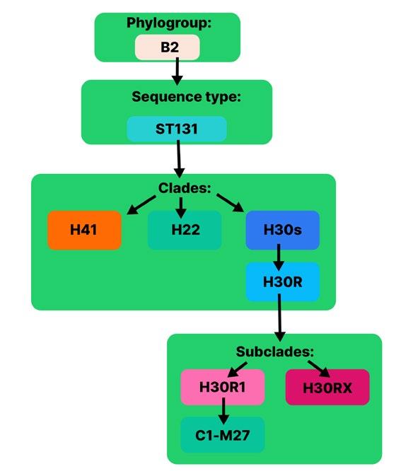
Editor's Pick: Review of Multifactorial Drivers of the Global Dissemination of Multidrug-Resistant Escherichia coli Sequence Type 131
Gul S et al.
Use of Terminal Smear and PCR to Detect Mycobacterium genavense in Patients with Advanced HIV: A Case Series and Review of the Literature
Iroegbu U et al.
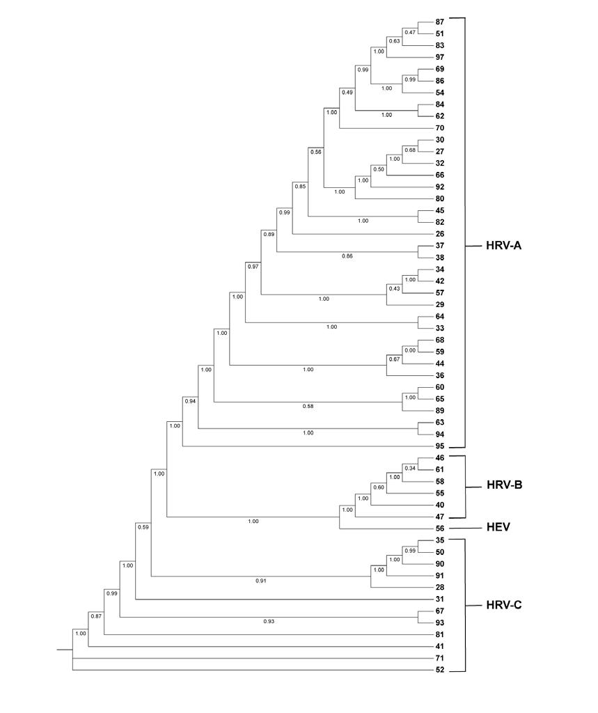
Genetic Diversity of Human Rhinovirus Among Hospitalised Paediatric Patients in Santa Fe, Argentina, 2010–2011
Peralta RD et al.
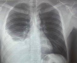
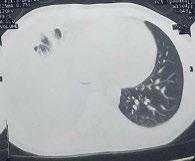
Microbiologically Confirmed Pleural Tuberculosis in an Adult Male: A Case Report and Review of Diagnostic Approaches
Agenge ET et al.
Editorial Board
Prof Jens Lundgren
Editor-in-Chief
Prof Rajeshwar Reddy Kasarla
Professor, Microbiology Department, Malla Reddy Institute of Medical Sciences, Telangana, India
Rigshospitalet, University of Copenhagen, Denmark
Prof David Fisman
University of Toronto, Canada
Dr Ali Elbeddini
University of Ottawa, Canada
Dr Emilio Bouza
Hospital Gregorio Marañón, Spain
Dr Mohammad Nazish
Farwaniyah Hospital, Kuwait
Dr Muge Cevik
University of St Andrews, UK
Dr Oliver Grundmann
University of Florida, USA
Dr Smilta Shevade
Millennium Path Lab, India
Dr Rahul Garg
All India Institute of Medical Sciences-Raipur, India
Prof Manisha Gupta
Super Specialty Cancer Institute and Hospital, India
Dr Sanjay Bhattacharya
Fakhruddin Medical College, India
Dr Hisham Elkhayat
Theodor Bilharz Research Institute, Egypt
Dr Daniel Pan
University of Leicester, UK
Aims and Scope
EMJ Microbiology & Infectious Diseases is an open access, peer-reviewed ejournal committed to publishing the highest quality medical research concerning all aspects of the prevention, diagnosis, and management of infectious diseases in humans.
The journal is published annually, six weeks after the European Society of Clinical Microbiology and Infectious Diseases (ESCMID) Congress, and features highlights from this congress, alongside interviews with experts in the field, reviews of abstracts presented at the congress, as well as in-depth features on congress sessions. The journal also covers advances within the clinical and pharmaceutical arenas by publishing sponsored content from congress symposia, which is of high educational value for healthcare professionals. This undergoes rigorous quality control checks by independent experts and the in-house editorial team.
EMJ Microbiology & Infectious Diseases also publishes peerreviewed research papers, review articles, and case reports in the field. In addition, the journal welcomes the submission of features and opinion pieces intended to create a discussion around key topics in the field and broaden readers’ professional interests. The journal is managed by a dedicated editorial team that adheres to a rigorous double-blind peer-review process, maintains high standards of copy editing, and ensures timely publication.
EMJ Microbiology & Infectious Diseases endeavours to increase knowledge, stimulate discussion, and contribute to a better understanding of infectious diseases. Our focus is on research that is relevant to all healthcare professionals in the field. We do not publish veterinary science papers or laboratory studies not linked to patient outcomes. We have a particular interest in topical studies that advance knowledge and inform of coming trends affecting clinical practice in microbiology and infectious diseases.
Further details on coverage can be found here: www.emjreviews.com
Editorial Expertise
EMJ is supported by various levels of expertise:
• Guidance from an Editorial Board consisting of leading authorities from a wide variety of disciplines.
• Invited contributors who are recognised authorities in their respective fields.
• Peer review, which is conducted by expert reviewers who are invited by the Editorial team and appointed based on their knowledge of a specific topic.
• An experienced team of editors and technical editors.
Peer Review
On submission, all articles are assessed by the editorial team to determine their suitability for the journal and appropriateness for peer review.
Editorial staff, following consultation with either a member of the Editorial Board or the author(s) if necessary, identify three appropriate reviewers, who are selected based on their specialist knowledge in the relevant area.
All peer review is double blind.
Following review, papers are either accepted without modification, returned to the author(s) to incorporate required changes, or rejected.
Editorial staff have final discretion over any proposed amendments.
Submissions
We welcome contributions from professionals, consultants, academics, and industry leaders on relevant and topical subjects. We seek papers with the most current, interesting, and relevant information in each therapeutic area and accept original research, review articles, case reports, and features.
We are always keen to hear from healthcare professionals wishing to discuss potential submissions, please email: editorial.assistant@emjreviews.com
To submit a paper, use our online submission site: https://emj.kriyadocs.com/submissions/submit/emj/emj/login
Submission details can be found through our website: www.emjreviews.com/contributors/authors
Reprints
All articles included in EMJ are available as reprints (minimum order 1,000). Please contact hello@emjreviews.com if you would like to order reprints.
Distribution and Readership
EMJ is distributed through controlled circulation to healthcare professionals in the relevant fields across Europe.
Indexing and Availability
EMJ is indexed on DOAJ, the Royal Society of Medicine, and Google Scholar®; selected articles are indexed in PubMed Central®.
EMJ is available through the websites of our leading partners and collaborating societies. EMJ journals are all available via our website: www.emjreviews.com
Open Access
This is an open-access journal in accordance with the Creative Commons Attribution-Non Commercial 4.0 (CC BY-NC 4.0) license.
Congress Notice
Staff members attend medical congresses as reporters when required.
All information obtained by EMJ and each of the contributions from various sources is as current and accurate as possible. However, due to human or mechanical errors, EMJ and the contributors cannot guarantee the accuracy, adequacy, or completeness of any information, and cannot be held responsible for any errors or omissions. EMJ is completely independent of the review event (ESCMID 2026) and the use of the organisations does not constitute endorsement or media partnership in any form whatsoever. The cover photo is of Munich, Germany, the location of ESCMID 2026.
Simple and rapid next‑generation sequencing for microbiology and infectious disease research
In the fast‑moving field of clinical microbiology, faster sequencing workflows help deliver insight to scientists when it matters.
Clinical microbiology research requires fast, reliable insights, particularly for emerging and re-emerging infectious diseases, antimicrobial resistance (AMR), and complex infections.
Traditional methods, while essential, can be time-intensive and limited in resolution. Next-generation sequencing (NGS) enables high-resolution pathogen characterization, genomic epidemiology, and detection of resistance and virulence markers.
The MiSeq i100 Series is designed to make these capabilities more accessible. By combining faster run times with simplified workflows and integrated data analysis, it enables laboratories to move from sample to insights in hours rather than weeks. This supports near-real-time genomic insights, iterative study designs, and faster dissemination of findings.
• Same‑day results. Sequence in as little as 4 hours, up to 4× faster than the original MiSeq¹
• Easy setup. Ready in under 20 minutes with three simple steps¹
• Onboard DRAGEN™ analysis. Powerful secondary analysis built into the instrument
• No cold chain required. Reagents shipped and stored at room temperature¹, reducing logistical complexity and costs
Editorial Director
Andrea Charles
Editor
Sean Boyle
Managing Editor
Darcy Richards
Senior Copy Editor
Noémie Fouarge
Copy Editors
Meghan Garcka, Sarah Jahncke
Editorial Leads
Helena Bradbury, Katrina Thornber, Aleksandra Zurowska
Editorial Co-ordinators
Jess Nicholson, Bertie Pearcey, Alena Sofieva
Editorial Assistants
Niamh Holmes, Josh Warren Lister, Roli Omamuli, Alexander Perkins
Reporter
Anna Caldwell
Head of Marketing
Stephanie Corbett
Creative Director
Tim Uden
Design Manager
Stacey White
Senior Designers
Tamara Kondolomo, Owen Silcox
Designers
Shanjok Gurung, Fabio van Paris
Junior Designers
Fraser Hoey, Helena Spicer, Caleb Wylie
Senior Business Unit Lead
Nabihah Durrani
Chief Executive Officer
Justin Levett
Chief Commercial Officer
Dan Healy
Founder and Chairman
Spencer Gore
Welcome
Dear Readers,
I am pleased to introduce the 2026 issue of EMJ Microbiology & Infectious Diseases, featuring highlights from the European Society of Clinical Microbiology and Infectious Diseases (ESCMID) Global Congress 2026, held in Munich, Germany. This year’s Congress once again brought together the international infectious diseases community, with a renewed focus on antimicrobial resistance, advanced diagnostics, pandemic preparedness, and health equity, priorities that continue to define the field’s most urgent challenges.
Our coverage includes a comprehensive Congress review, alongside interviews with two ESCMID Young Investigator Award recipients, offering insights into the perspectives shaping the next generation of leaders. An array of abstract reviews explores timely and practice-relevant developments, with a strong emphasis on antimicrobial stewardship. Expert-written features further reflect key ESCMID sessions, including the role of the microbiome in cancer, advances in chronic viral hepatitis, and evolving approaches to antimicrobial resistance health policy.
This issue also brings together a range of original, peer-reviewed research, including work on the genetic diversity of human rhinovirus, two case reports highlighting diagnostic complexities in mycobacterial disease, and a review of the drivers underpinning multidrug-resistant Escherichia coli
We are also pleased to present interviews with five leading experts across mycology, urology, virology, immunology, and genomics, capturing the breadth of contemporary infectious diseases research. Finally, this edition features an infographic on the current landscape of HIV care, framed by the ongoing tension between rapid innovation and persistent inequity.
Ada Enesco, Editorial Lead
Editorial enquiries: editor@emjreviews.com
Sales opportunities: salesadmin@emjreviews.com
Permissions and copyright: accountsreceivable@emjreviews.com
Reprints: info@emjreviews.com
Media enquiries: marketing@emjreviews.com
Foreword
Dear Colleagues,
It is a pleasure to present the 2026 edition of EMJ Microbiology & Infectious Diseases. This issue brings together insights from the European Society of Clinical Microbiology and Infectious Diseases (ESCMID) Global Congress 2026 in Munich, Germany, alongside a range of peer-reviewed research, expert features, and interviews that reflect the breadth of activity across our field.
This year’s congress highlighted several key priorities, including the ongoing fight against antimicrobial resistance, the need to advance diagnostics in resourcelimited settings, and the importance of global surveillance for emerging and re-emerging infections. These themes are carried throughout the issue, from EMJ’s independent coverage of ESCMID Global to selected abstract reviews and expertled features on topics such as antibiotic prescription patterns and OneHealth approaches to antimicrobial resistance.
We are also pleased to include interviews with five leading experts, Dimitrios Kontoyiannis, Edward Holmes, Senjuti Saha, Filomeen Haerynck, and Florian Wagenlehner, each of whom delivered keynote sessions at ESCMID Global 2026, offering their perspectives across a wide range of disciplines.
In addition, a timely infographic explores the current landscape of HIV care, a field that continues to sit at the intersection of rapid innovation and persistent inequity.
Finally, our peer-reviewed articles cover a range of clinically relevant topics, including original research on the genetic diversity of human rhinovirus in a paediatric population, a review of the key drivers of multidrugresistant Escherichia coli, and more.
I would like to thank all authors, reviewers, and contributors for their work in bringing this issue together. I hope you find it both useful and informative.
This year’s congress highlighted several key priorities, including the ongoing fight against antimicrobial resistance
Rajeshwar Reddy Kasarla Professor, Microbiology
Department, Malla Reddy Institute of Medical Sciences, Hyderabad, India
ESCMID 2026
ESCMID Global reaffirms itself not only as a platform for cutting-edge science, but as a unifying force in an increasingly complex world
ESCMID’s growing community now includes more than 14,500 members worldwide, with over a third based outside Europe
Review of the European Society of Clinical Microbiology and Infectious Diseases (ESCMID) Global Congress 2026
SPRING, Munich, Germany, hosted the 36th European Society of Clinical Microbiology and Infectious Diseases (ESCMID) Global Congress from 17th–21st April, bringing together a global community of infectious diseases experts at a time marked by geopolitical tension, rising healthcare costs, and eroding public trust in science.
In his opening address, ESCMID President Robert Leo Skov acknowledged the ongoing global conflicts and divisions shaping the healthcare landscape, as well as the enduring impact of misinformation following the COVID-19 pandemic, particularly around vaccines. Coupled with funding cuts affecting scientific research, Skov warned that, “in this climate of growing uncertainty and distrust, science itself is under attack.” He framed the current moment as a dual battle against infectious diseases and misinformation.
Skov outlined a clear path forward rooted in several key priorities. First, he emphasised the need to double down on scientific research, particularly in vaccinology. Reflecting this commitment, Maheshi Ramasamy, University of Oxford, UK; and Joint Committee on Vaccination and Immunisation, has been appointed as ESCMID’s first Vaccines Subcommittee Director. At the same time, ESCMID continues to broaden its scientific reach, launching five new study groups spanning AI and digitalisation, mobile genetic elements and plasmids, urinary tract infections, sexually transmitted infections, and maternal and child health. With 37 study groups now active, the society reflects the scale
and importance of international scientific collaboration. Efforts to tackle antimicrobial resistance (AMR) have also intensified, including the co-founding of the Global AMR Innovators Conference.
Equally important is the need to bridge science and policy. ESCMID has joined forces with organisations such as the Infectious Diseases Society of America (IDSA), the American Society for Microbiology (ASM), and the AMR Action Fund in calling on the G7 to adopt a One Health diagnostics compact, aimed at improving access to diagnostics, strengthening stewardship, and accelerating research and development. At the European level, the society contributes to advisory work for the Health Emergency Preparedness and Response Authority, while its AMR Science Policy Forums support alignment between national strategies and global commitments.
Global collaboration remains a defining feature of the society. ESCMID’s growing community now includes more than 14,500 members worldwide, with over a third based outside Europe and 14% in low- and middle-income countries. The ESCMID Local Champions initiative, now
comprising 100 representatives across 88 countries, continues to strengthen regional engagement and knowledge exchange. Skov emphasised that progress in infectious diseases is built incrementally through shared achievements over time. He closed by calling for unity and collective responsibility in the face of future challenges.
The opening ceremony also celebrated excellence across the field. The ESCMID Award for Outstanding Contributions in Infection was presented to Leonard Leibovici, Tel Aviv University, Israel, for his pioneering work in neutropenia and sepsis. The Award for Excellence in Science went to Nicholas Day, Director of the Mahidol Oxford Tropical Medicine Research Unit, Bangkok, Thailand, recognising more than 3 decades of research in low-resource settings.
The Lifetime Achievement Award honoured Murat Akova of Hacettepe University School of Medicine, Türkiye, whose work has shaped the understanding of infections in immunocompromised patients and clinical ethics.
Finally, ESCMID Young Investigator Awards were presented to Sarah Delliere, Université Paris Cité and Hôpital Saint-Louis, France; Iris K. Minichmayr, Medical University of Vienna, Austria; Clark D. Russell, University of Edinburgh Centre for Inflammation Research in the Institute for Regeneration and Repair, UK; Daniel Pan, University of Leicester and University Hospitals of Leicester, UK; and Grace O. Androga, Malawi Liverpool Wellcome Programme, Blantyre, Malawi, highlighting the next generation of leaders in infectious diseases.
As the Congress unfolds, ESCMID Global reaffirms itself not only as a platform for cutting-edge science, but as a unifying force in an increasingly complex world. Read on for key insights from this year’s meeting, and be sure to follow next year’s coverage of ESCMID Global 2027.
In this climate of growing uncertainty and distrust, science itself is under attack
High-Dose Influenza Vaccine Shows Consistent Benefit in Immunosuppressed Older Adults
FINDINGS from a prespecified analysis of the DANFLU-2 trial, presented at ESCMID Global 2026, provide important real-world evidence on influenza vaccine effectiveness in immunosuppressed older adults, a population at heightened risk of severe outcomes and often reduced vaccine responsiveness.1
This pragmatic, registry-based, randomised trial included 332,438 adults aged ≥65 years across the 2022/23–2024/25 influenza seasons. Participants were randomised 1:1 to receive either high-dose inactivated influenza vaccine (HD-IIV) or standard-dose vaccine (SD-IIV).
A total of 14,315 individuals (4.3%) met predefined criteria for immunosuppression, with 7,187 assigned to HD-IIV and 7,128 to SD-IIV. Baseline characteristics were well balanced between groups, although immunosuppressed participants had a higher burden of comorbidities and consistently higher event rates.
For the primary endpoint (hospitalisation for influenza or pneumonia), HD-IIV and SD-IIV showed comparable effectiveness across immunosuppression strata. However, HD-IIV was associated with fewer influenza-related hospitalisations overall, with a relative vaccine effectiveness (rVE) of 42.1% (95% CI: −16.5–72.3%) in immunosuppressed participants and 43.9% (95% CI: 26.5–57.3%) in nonimmunosuppressed individuals.
Reductions were also observed in cardiorespiratory hospitalisations, with rVE estimates of 12.3% (95% CI: −2.2–24.7%) in immunosuppressed participants and 5.1% (95% CI: 0.5–10.7%) in those without immunosuppression.
Notably, a potential differential effect emerged for cardiovascular hospitalisations, where HD-IIV was associated with a greater reduction among immunosuppressed individuals (rVE: 26.7%; 95% CI: 6.3–42.9%) compared with non-immunosuppressed participants (rVE: 5.8%; 95% CI: −0.1–11.4%).
Across all endpoints, immunosuppressed participants experienced higher absolute rates of hospitalisation, highlighting their vulnerability despite vaccination.
Overall, these findings suggest that while high-dose and standard-dose influenza vaccines offer broadly similar protection against the primary endpoint, HD-IIV may provide additional benefits in reducing influenza-related and cardiorespiratory hospitalisations, with a possible enhanced effect on cardiovascular outcomes in immunosuppressed patients.
4.3
14,315 individuals (4.3%) met predefined criteria for immunosuppression, with 7,187 assigned to HD-IIV and 7,128 to SD-IIV %
IgG4 Rise Linked to Repeated SARS-CoV-2 Exposure
RESEARCH presented at ESCMID Global 2026 suggests that repeated SARS-CoV-2 vaccination and infection drive a sustained shift in antibody responses, characterised by increasing IgG4 levels.2
The study analysed a cohort of healthcare workers with multiple immunological exposures (≥6 vaccination or infection events), using longitudinal serum samples to assess antibody subclasses, neutralisation capacity, and Fc-mediated functions. Individuals who were infectionnaïve prior to vaccination developed a distinct immune profile from the third mRNA dose onwards, marked by a sharp and persistent rise in anti-spike IgG4. Those with hybrid immunity showed a similar trend, although more gradual.
IgG4 levels continued to increase with successive exposures, including against nucleocapsid protein following breakthrough infections, indicating broader class switching beyond spike-specific responses. Despite this shift, IgG4 appeared to have minimal impact on Fc effector functions, as its depletion did not significantly alter antibody-dependent cellular cytotoxicity.
Notably, while correlations between IgG1–3 and neutralisation of newer variants weakened, IgG4 maintained a stable association, suggesting a potential role in cross-reactive immunity.
Individuals who were infectionnaïve prior to vaccination developed a distinct immune profile from the third mRNA dose onwards
Overall, the findings indicate that the sequence and frequency of SARSCoV-2 exposures shape long-term antibody profiles, with IgG4 emerging as a consistent feature of repeated immunisation. The clinical implications of this shift, whether beneficial or potentially dampening protective immunity, remain to be determined.
Global Surveillance Identifies Diverse Nairoviruses in Ixodes Ticks
RESEARCHERS at ESCMID Global 2026 reported that Ixodes ticks harbour substantial viral diversity across countries and host species, including known nairoviruses and previously unidentified variants, highlighting the value of early surveillance to detect emerging vector-borne pathogens as climate change reshapes disease spread.3
Research has shown that climate change is impacting ecological systems, animal behaviour, vector distribution, and microbial equilibria. These changes enable disease vectors to spread into new regions, with concerns rising that emerging viruses may spread unnoticed. Researchers within the ARBO-WATCH preparedness network sought to investigate the epidemiogenetics of members of the Nairoviridae in ticks collected globally.
Researchers designed a pan-orthonairovirus hemi-nested reverse-transcriptase-PCR targeting a 347 bp region of the RNAdependent RNA polymerase gene. The assay was applied to RNA extracted from single ticks or pooled samples collected from vertebrate hosts or the environment. Positive samples underwent Sanger sequencing, while full genomes were generated through metagenomics. Maximum-likelihood phylogenetic analyses were then performed across the Nairoviridae family. Overall, 1,916 ticks from 14 species were analysed from 11 countries spanning six continents. Completion numbers were the full tested sample set, with all positive pools sequenced.
Fifteen Ixodes pools tested positive, containing five viruses from four genera. Sulina virus was identified in a Danish pool with nucleotide identity of 96.3–97.3% versus reference strains. Taggert virus was detected in Ixodes uriae from Antarctica with 93.8% identity. A relative of South Bay virus was found in Mongolian Ixodes persulcatus with 74.7% identity. Two Grotenhout virus variants were identified in Ixodes ricinus from Denmark, Italy, and Poland, with identities of 96.3–98.6%. Most notably, two variants representing a novel genus were discovered in 14 of 31 Danish Ixodes hexagonus ticks, equivalent to 45.2%. Their bisegmented genomes were fully sequenced and named Esrum virus 1 and 2. Full RNA-dependent RNA polymerase sequences showed less than 50% identity to reference viruses.
Researchers concluded that Ixodes ticks harbour substantial viral diversity across countries and host species. Early findings suggest strong tick species specificity, which may help model future spillover and emergence pathways. Continued international surveillance could improve early warning systems, guide ecological risk assessment, and strengthen preparedness for future vector-borne threats.
1,916 ticks from 14 species were analysed from 11 countries spanning six continents
A NEWLY characterised clinical strain of Klebsiella pneumoniae is raising concerns after demonstrating non-susceptibility to the antibiotic combination meropenem–durlobactam, a therapy designed to overcome resistance in some of the most difficult-to-treat infections, as shown by results presented at ESCMID Global 2026.4
Carbapenem-resistant Klebsiella pneumoniae (CRKP) is recognised as a major global health threat, often associated with high mortality in hospital settings. Durlobactam, a diazabicyclooctane β-lactamase inhibitor, has been developed to restore the activity of β-lactam antibiotics such as meropenem by blocking enzymes like KPC and OXA-48 carbapenemases that degrade them. However, new findings suggest that resistance mechanisms beyond β-lactamase activity may undermine this approach.
In this study, researchers analysed a CRKP strain isolated from a bile sample. Laboratory testing revealed extremely high minimum inhibitory concentrations for both meropenem–durlobactam and sulbactam–durlobactam, indicating poor susceptibility despite the presence of the inhibitor. Genomic sequencing showed that the strain belonged to sequence Type 11 and capsular Type 64, and carried the carbapenemase gene blaKPC-2.
Importantly, the strain also possessed several virulence factors, including genes linked to aerobactin and yersiniabactin production, as well as regulators associated with a hypermucoid phenotype. These features suggest that the bacterium may not only be resistant, but also potentially more capable of causing severe disease.
Further analysis identified structural changes in the bacterial outer membrane that may explain the reduced antibiotic susceptibility. The lamB gene, which encodes a porin responsible for the uptake of small molecules, including certain antibiotics, was truncated. Loss of LamB function is known to reduce antibiotic entry into bacterial cells. In addition, the strain lacked the gene encoding AsmA, a protein involved in assembling another key porin, OmpK35. Disruption of these membrane channels likely limits drug penetration, reducing the effectiveness of meropenem even when protected by durlobactam.
The findings highlight a growing challenge in antimicrobial resistance: even novel inhibitor combinations may fail when multiple resistance mechanisms converge. Researchers emphasise the need for continued surveillance and deeper investigation into non-enzymatic resistance pathways, particularly those affecting membrane permeability.
As new therapies are introduced to combat multidrug-resistant pathogens, this study underscores the adaptability of CRKP and the importance of staying ahead of emerging resistance mechanisms.
LC16m8 Vaccine Generates Strong Immunity but Limited Mpox Coverage
A NEW study presented at ESCMID Global 2026 has shown that
the replicating smallpox vaccine LC16m8 generated strong and durable antibody responses in high-risk populations during mpox vaccine deployment in Colombia, although cross-protection against circulating mpox strains appeared limited.5
The research addressed a critical evidence gap regarding the immunogenicity of LC16m8 outside Japan, particularly in populations at elevated risk of mpox infection, including people living with HIV. Understanding how effectively this vaccine stimulates immune responses is essential for informing outbreak preparedness and vaccination strategies.
Investigators conducted a hybrid study combining a randomised delayed vaccination trial with an observational cohort across three centres in Bogotá, Colombia. Adults aged 18–50 years at increased risk of mpox infection were enrolled, including individuals receiving antiretroviral therapy, pre-exposure prophylaxis users, and those reporting high-risk sexual behaviours. A predefined immunogenicity sub-study analysed neutralising antibody responses in 60 participants, with serum samples collected at baseline and at 14, 30, and 180 days following vaccination.
Findings showed that neutralising antibody responses against the vaccine strain increased rapidly after immunisation. Geometric mean titres rose from 9.8 at baseline to 359.8 at Day 14 and peaked at 2220.9 at Day 30 before declining to
899.4 at Day 180, remaining well above baseline levels. Seroconversion rates were consistently high, reaching 100.0% at Day 14, 98.3% at Day 30, and 100.0% at Day 180, demonstrating robust and sustained homologous immune responses.
In contrast, cross-neutralising responses against the mpox virus clade IIb strain were modest. Antibody titres increased only slightly, from 8.9 at baseline to 23.2 at Day 30, and fell to 12.6 at Day 180. Correspondingly, seroconversion peaked at 33.9% before declining to 12.1% at 6 months, indicating limited cross-reactivity.
The authors noted that while LC16m8 elicited strong immune responses against its own strain, the comparatively weak neutralisation of mpox virus highlights challenges in assessing cross-protection using current assays.
Overall, the findings supported the immunogenic potential of LC16m8 in highrisk populations, including people living with HIV, but underscored the need for further research to better understand its effectiveness against circulating mpox strains and to optimise vaccination strategies in outbreak settings.
mRNA Embecovirus Spike and HE Antigens Induce Cross-Reactive Antibody Responses
PRECLINICAL data presented at ESCMID Global 2026 provide important insights into next-generation vaccine strategies targeting embecoviruses, a clinically relevant and evolutionarily diverse subgroup of betacoronaviruses that includes human seasonal pathogens, such as OC43 and HKU1, as well as animal reservoir viruses.6
The study evaluated mRNA-based immunogens encoding codon-optimised spike and haemagglutinin esterase (HE) proteins from multiple embecoviruses, including OC43, HKU1, human enteric coronavirus (HECV), and murine hepatitis virus (MHV). Using lipid nanoparticledelivered mRNA vaccines, BALB/c mice were immunised in a prime-boost regimen, and sera were assessed for cross-reactive binding and neutralising activity across a phylogenetically diverse panel of human and animal embecoviruses.
Overall, the vaccine constructs were immunogenic but elicited distinct antigenspecific patterns of cross-reactivity. OC43 and HECV spike proteins generated broad heterotypic antibody binding responses against multiple animal reservoir spikes, although cross-reactivity was reduced against HKU1 and MHV. In contrast, the MHV spike induced more limited responses, with weak or negligible cross-reactivity to OC43 and HKU1.
HE antigens showed a divergent profile. OC43 and HECV HE proteins produced cross-reactive binding responses broadly similar to their spike counterparts, but did not bind MHV targets. Notably, MHV HE induced narrower antibody breadth compared with spike. Despite these differences, HE antigens demonstrated functional relevance in neutralisation assays: OC43 and HECV HEs generated broader and more potent heterotypic neutralisation, including activity against porcine coronavirus, whereas MHV HE was non-neutralising.
In contrast, OC43 and HECV spike proteins induced strong homotypic
neutralising responses but more limited heterotypic neutralisation. MHV spike retained homotypic neutralisation capacity, while HKU1 spike and HE failed to elicit detectable antibody responses under the study conditions.
These findings highlight important antigenspecific differences in breadth and function of immune responses induced by embecovirus proteins delivered via mRNA platforms. Importantly, the data identify HE as an underexplored antigen capable of mediating cross-reactive neutralisation, supporting its potential inclusion in broadly protective betacoronavirus vaccine strategies.
The study provides crucial preclinical evidence to inform the rational design of pan-betacoronavirus vaccines aimed at improving antigenic coverage across human and zoonotic reservoirs.
Using lipid nanoparticle-delivered mRNA vaccines, BALB/c mice were immunised in a prime-boost regimen
Updated COVID-19 Vaccines Elicit Strong Responses in At-Risk Groups
INTERIM data, presented at ESCMID Global 2026, suggest that updated mRNA vaccines continue to strengthen immune protection against COVID-19 in at-risk populations.7
COVID-19, caused by the SARS-CoV-2 virus, remains a significant health concern, particularly for older adults and individuals with underlying conditions who are more likely to experience severe outcomes. As the virus continues to evolve genetically, vaccines are periodically updated to better match circulating variants and maintain protection.
Interim data from two ongoing clinical studies evaluating variant-targeted vaccines show encouraging immune responses. These Phase IIIb/IV open-label studies assessed updated formulations designed to target the LP.8.1 variant of COVID-19. Participants included adults aged 65 years and over, as well as individuals aged 12–64 years with at least one risk factor for severe illness.
Across both studies, 660 participants aged 65 years and older and 662 participants aged 12–64 years with at least one risk condition for severe COVID-19 were enrolled. Researchers measured neutralising antibody levels at baseline (Day 1) and again at Day 29 post-vaccination to assess immune response.
Results showed a substantial rise in antibody levels following vaccination, with increases ranging from 15.4- to 53.0-fold. These findings indicate that updated vaccines can effectively stimulate the immune system against the targeted variant, with responses exceeding the fourfold increase predefined as the study’s primary immunogenicity objective. No serious adverse events were reported during this interim analysis, supporting a favourable safety profile in the populations studied.
While the interim results are descriptive and based on early data from a limited subset of participants, longer-term outcomes, including durability of protection and realworld effectiveness, are still being evaluated.
These results highlight the potential of updated mRNA vaccines to maintain protection as SARS-CoV-2 continues to evolve, particularly for those most vulnerable to severe disease. Future analyses could help clarify how these immune responses translate into clinical protection and inform future vaccination strategies.
A NEWLY identified bacteriophage, P49, shows promise as a broadspectrum therapeutic candidate against drug-resistant bacteria according to recent research, presented at ESCMID Global 2026, investigating alternatives to antibiotics for Klebsiella pneumoniae (KP) infections.8
KP is a major cause of hospital-acquired infections, and many phages that target it are highly specific, often limited to a single capsule type. This narrow host range has restricted their clinical usefulness. To address this, researchers isolated phage P49 from wastewater using KP strains resistant to existing phages (P04 and P40).
Laboratory testing revealed that P49 effectively lysed mutant KP strains that were resistant to other phages, forming clear plaques that ranged from 1.0–2.0 mm in diameter, though it could not infect the original non-resistant strain. Electron microscopy showed that P49 has a long, noncontractile tail typical of certain doublestranded DNA phages. It also demonstrated strong biological properties, including stability across a range of temperatures and pH levels, rapid adsorption to host cells, and a high burst size, all desirable traits for therapeutic use.
Genomic analysis showed that P49 has a genome of approximately 114 kb and, importantly, does not carry genes associated with antimicrobial resistance or virulence, supporting its safety profile. Phylogenetic analysis placed P49 within the genus Epseptimavirus, closely related to phages that infect Salmonella and Escherichia coli.
Notably, P49 exhibited an unusually broad host range. Beyond targeting resistant KP mutants, it was capable of lysing multiple bacterial species, including E. coli, Salmonella enterica, Kluyvera tianfuensis, and Enterobacter ludwigii. In growth inhibition experiments, P49 suppressed E. coli for up to 11 hours and other tested species for 16 to over 24 hours.
This cross-species activity is uncommon among phages and suggests potential for wider clinical application, particularly in treating mixed or difficult-to-diagnose infections involving multiple pathogens.
Overall, the findings position P49 as a strong candidate for further development in phage therapy, offering a potential tool against multidrug-resistant infections where current treatments are failing.
KP is a major cause of hospitalacquired infections, and many phages that target it are highly specific, often limited to a single capsule type
New Staphylococcus aureus Panel Captures Resistance and Virulence Diversity
A
GENETICALLY DIVERSE panel of 95 Staphylococcus aureus isolates has been developed to enhance and standardise antimicrobial resistance research, comprised of isolates collected from 18 countries, as stated in the abstract presentation at ESCMID Global 2026.9
S. aureus is a versatile pathogen responsible for infections ranging from skin and soft tissue disease to life-threatening sepsis. Due to its ability to acquire resistance to multiple antibiotics, it has been a persistent contributor to the global antimicrobial resistance crisis.
In response to this crisis, efforts have focused on providing the scientific community with well-characterised bacterial isolate panels for research. The panel was derived from 6,484 isolates collected across 18 countries over a 20-year period. A twostep selection process combined k-medoid clustering (to maximise genetic distance between isolates) with manual curation to retain strains with distinctive metadata, including date, country, and site, as well as major resistance and virulence biomarkers.
All 95 isolates underwent whole-genome sequencing using Oxford Nanopore (Oxford Nanopore Technologies, Oxford, UK) technology, alongside antibiotic susceptibility testing in a College of American Pathologists (CAP)-accredited laboratory.
The resulting panel included 69 sequence types, 74 S. aureus protein A types, and
six Staphylococcal cassette chromosome mec types. It included 47 methicillinsusceptible and 48 methicillin-resistant isolates, ensuring balanced representation of clinically relevant phenotypes.
Resistance profiling identified isolates with reduced susceptibility to linezolid, co-trimoxazole, and rifampin, amongst other clinically relevant antibiotics. One isolate was classified as extensively drugresistant, while four isolates remained fully susceptible to all antibiotics tested. A total of 55 distinct antimicrobial resistance and antiseptic/disinfectant resistance genes, as well as 35 characterised mutations, were detected across the panel.
Virulence analysis revealed 50 distinct virulence factors, including Panton–Valentine leukocidin in 25% of isolates and toxic-shock syndrome toxin-1 in 8.4%.
While not intended to directly guide treatment decisions, this standardised panel offers a reproducible platform for evaluating diagnostics, therapeutics, and surveillance tools. Limitations include its fixed size and curated selection, which may not capture all emerging resistance mechanisms.
Virulence analysis revealed 50 distinct virulence factors, including Panton–Valentine leukocidin in 25% of isolates and toxic-shock syndrome toxin-1 in 8.4%
Nirsevimab Shows Consistent Protection Against Severe RSV Outcomes Across Seasons
REAL-WORLD data presented at ESCMID Global 2026 provide important insights into the effectiveness of nirsevimab across successive respiratory syncytial virus (RSV) seasons in infants with bronchiolitis following its introduction in Italy in 2024.10
The study evaluated nirsevimab, a longacting monoclonal antibody targeting RSV, using a single-centre test-negative case–control design. Infants aged 1–12 months presenting with bronchiolitis to a tertiary paediatric emergency department (ED) in Milan, Italy, were included. Infants who were RSV-positive were classified as cases and those who were RSV-negative were classified as controls, with immunisation status assessed at admission. Vaccine effectiveness (VE) was estimated against RSV-associated ED-attended illness, RSV-related hospitalisation, and paediatric ICU (PICU) admission. Epidemiological outcomes from November–January in the 2025–2026 season were compared with the same period in 2024–2025.
Overall, VE estimates during the second RSV season varied by clinical severity and were associated with wide CIs due to reduced case numbers. VE was 19.3% (95% CI: −137.0–72.6) against RSV-associated ED-attended illness, 75.0% (95% CI: −33.7–95.3) against RSV-related hospitalisation, and 75.0% (95% CI: −1498.0–99.6) against PICU admission.
In parallel, notable reductions in bronchiolitis burden were observed across seasons.
ED visits for bronchiolitis decreased from 147 in 2024–2025 to 89 in 2025–2026 (−39.5%). RSV-positive ED visits declined from 63 to 33 (−47.6%; p<0.001), while RSV-negative visits decreased from 84 to 56 (−33.3%). Overall hospital admissions fell from 79 to 54. Among infants who were RSV-positive, hospitalisations decreased from 43 to 28 (−34.9%), although hospitalisation rates remained similar between seasons (68.3% versus 84.8%; p=0.08).
These findings highlight a gradient in observed effectiveness, with lower VE against ED-attended illness and higher VE against more severe outcomes, including hospitalisation and PICU admission. This pattern is consistent with stronger protection against severe disease and may also reflect selection and threshold effects inherent to ED-based test-negative designs, where vaccinated infants presenting to care may represent a higher-risk subgroup with differing healthcare-seeking and testing behaviours.
Collectively, these real-world data support the continued implementation of nirsevimab and underscore the importance of robust surveillance systems to inform RSV prevention strategies and policy decisions.
References
1. AM Reimer Jensen et al. High-dose vs standard-dose influenza vaccine effectiveness in immunosuppressed older adults: a prespecified analysis of the DANFLU-2 trial. Abstract 06036. ESCMID Global, 17-21 April, 2026.
2. Perisse T et al. History of vaccination and infection shapes the humoral response to SARS-CoV-2 through IgG4 class switching. Abstract 03143. ESCMID Global, 17-21 April, 2026.
3. Bianchi S et al. Identifying emerging vector-borne pathogens in a time of climate change, the ARBO-WATCH network. Abstract 08522. ESCMID Global, 17-21 April, 2026.
4. Zong Z et al. Carbapenem-resistant Klebsiella pneumoniae non-susceptible
to mero-penem/durlobactam. Abstract 09287. ESCMID Global, 17-21 April, 2026.
5. Hernandez J et al. Humoral immunogenicity of the replicating smallpox vaccine LC16m8 during mpox vaccine deployment in high-risk populations in Colombia. Abstract 09306. ESCMID Global, 17-21 April, 2026.
6. George C et al. mRNA delivered embecovirus spike and haemagglutinin esterase antigens elicit cross-reactive humoral immunity to divergent human and animal coronaviruses. Abstract 09328. ESCMID Global, 17-21 April, 2026.
7. Chalkias S et al. Effectiveness and safety of variant-updated COVID-19
vaccines. Abstract 09326. ESCMID Global, 17-21 April, 2026.
8. Yin X, Feng Y. The biological and genomic characteristics of a broadspectrum phage target Klebsiella pneumoniae. Abstract 09452. ESCMID Global, 17-21 April, 2026.
9. Vargas-Cuebas GG et al. A panel of global and genetically distinct Staphylococcus aureus isolates for research and development. Abstract 04666. ESCMID Global, 17-21 April, 2026.
10. Pisano EM et al. Consistency of nirsevimab effectiveness across successive RSV seasons in infants with bronchiolitis: a test-negative study. Abstract 09278. ESCMID Global, 17–21 April, 2026.
Microbiome Modulation as a Determinant of Cancer Therapy Response: Highlights from ESCMID 2026
Author: Cristina Royo-Cebrecos1,2
1. Internal Medicine Department, Hospital Nostra Senyora de Meritxell, Andorra Health Services (SAAS), Les Escaldes, Andorra
2. Research Department, Hospital Nostra Senyora de Meritxell, Andorra Health Services (SAAS), Les Escaldes, Andorra
*Correspondence to croyo@saas.ad
Disclosure: The author has declared no conflicts of interest.
THE ROLE of the microbiome as a modulator of cancer therapy response has become a key topic in recent oncology research. At the European Society of Clinical Microbiology and Infectious Diseases (ESCMID) 2026 Global Congress, emerging data highlighted how not only microbial composition, but especially microbial function, can influence outcomes in patients receiving immunotherapy and chemotherapy. In precision oncology, where treatments are tailored to tumour biology, the microbiome and diet are emerging as relevant and modifiable factors.
INTRODUCTION
Clinical outcomes in oncology remain highly heterogeneous, even among patients with similar molecular profiles. Despite major advances in targeted therapies, a significant proportion of patients do not achieve the expected benefit. This variability has led to increasing interest in host-related factors, particularly the gut microbiome, as contributors to treatment response.
This also raises a broader question: how can we reprogramme the immune system in patients with cancer beyond tumour-directed strategies? While oncology has traditionally focused on tumour-specific mechanisms, there is growing recognition that systemic factors, such as immunity, metabolism, and the microbiome, also play a key role.
The microbiome influences systemic immune responses, tumour microenvironment interactions, and drug metabolism. Importantly, its impact seems to go beyond the presence or absence of specific bacterial taxa. Functional outputs, particularly microbial metabolomic activity, are increasingly recognised as key determinants of response. Data presented at ESCMID Global 2026 reinforced this shift towards a more functional perspective.
MICROBIOME AND IMMUNOTHERAPY RESPONSE
There is now substantial evidence linking the gut microbiome to response to immune checkpoint inhibitors. Both preclinical and clinical data consistently link specific microbial profiles with improved antitumour immunity, particularly
through enhanced CD8+ T-cell activity.1 Differences in microbial composition between responders and non-responders have been consistently described, although no single ‘beneficial’ microbiome signature has been universally identified.2
How can we reprogramme the immune system in patients with cancer beyond tumour-directed strategies?
Faecal microbiota transplantation (FMT) provides some of the strongest evidence supporting a causal relationship. In patients with melanoma who were refractory to immunotherapy, transfer of microbiota from responders has led to clinical benefit in a subset of cases. The effect does not appear to depend on specific bacterial species alone, but rather on broader ecosystem characteristics.
Mechanistically, modulation of tumour immunogenicity appears relevant. Data presented by Markel et al. (Markel, unpublished data) suggested a potential link between microbiome modulation and tumour immune recognition, possibly involving pathways such as adenosine deaminase acting on RNA 1 (ADAR1)-mediated RNA editing, which may enhance T-cell recruitment and chemokine signalling. Despite these promising findings, several practical questions remain unresolved, including the optimal route of administration, the need for microbiome depletion prior to FMT, and how to maintain its effects over time.
ANTIBIOTICS AND DISRUPTION OF RESPONSE
Antibiotic use is common in patients with cancer, with up to one-third receiving antibiotics at the start of immunotherapy (often broad-spectrum agents), and rates of receiving antibiotics reach up to 31% during this period.3,4 It is consistently associated with reduced immune
checkpoint inhibitor efficacy, likely mediated through disruption of microbial diversity and function.3,4
Meta-analyses involving large patient cohorts have repeatedly shown worse outcomes in patients exposed to antibiotics close to the start of treatment, highlighting the importance of timing.5,6
These findings are further supported by large-scale analyses including over 46,000 patients across more than 100 studies, confirming worse outcomes in both immunotherapy and chemo-immunotherapy settings.7 However, not all antibiotics have the same impact, with broad-spectrum agents and combination regimens showing the most pronounced negative effects, likely due to greater microbiome disruption (Alves, unpublished data).
In the context of antibiotic exposure, dysbiosis may promote a more tolerogenic immune environment, alter immunometabolic pathways, and disrupt key microbial networks. Changes in bile acid metabolism, expansion of less favourable taxa such as Enterocloster spp., and altered signalling pathways may all contribute to impaired treatment response.8
Microbiome-based tools, such as TOPOSCORE, are beginning to translate these insights into clinically relevant strategies, enabling stratification of patients according to their likelihood of response to immunotherapy.9 These findings underscore the importance of careful antibiotic stewardship, favouring targeted approaches and limiting unnecessary exposure.10,11
BEYOND COMPOSITION: THE ROLE OF MICROBIOME FUNCTION
A key takeaway from ESCMID Global 2026 is the shift from focusing on microbial composition to understanding microbial function. While earlier studies aimed to identify specific bacterial taxa, current evidence suggests that metabolic activity may be more relevant.
This is illustrated by early-phase studies evaluating prebiotics such as camu camu, a polyphenol-rich source including castalagin.12 Despite minimal changes in
microbiome composition, improvements in clinical outcomes were observed, accompanied by significant shifts in metabolomic profiles. These findings support the concept that function, rather than taxonomy, drives response.
Among the pathways of interest, bile acid metabolism has emerged as an important immunomodulatory axis, with microbiome-driven alterations shown to impair tumour-specific T cell responses.13 Similarly, metabolites derived from tryptophan metabolism appear to influence treatment response.
MICROBIOME AND CHEMOTHERAPY
The microbiome also plays a role in chemotherapy, influencing both efficacy and toxicity. A key aspect is the presence of intratumoural bacteria, which challenges the traditional view of tumours as sterile environments.14
A key takeaway from ESCMID
Global 2026 is the shift from focusing on microbial composition to understanding microbial function
In pancreatic cancer, intratumoural bacteria are thought to originate from the gut, supporting the concept of microbial translocation.15 The microbiome can influence treatment response through several mechanisms, such as drug metabolism, immune modulation, and the production of bioactive metabolites.16 A well-known example is gemcitabine, which can be inactivated by bacteria within the tumour that express specific enzymes, ultimately leading to treatment resistance.17 This effect appears drug-specific and has not been clearly observed with other chemotherapies. Preclinical studies have shown that antibiotic treatment can restore gemcitabine efficacy, and this strategy is currently being explored in clinical trials such as the ongoing PRODIGE 106 PANORAMIX study.
Beyond these direct interactions, the microbiome has also been linked to tumour biology and clinical outcomes. In pancreatic cancer, both microbial signatures and metabolomic profiles have been associated with disease risk and prognosis.18
THERAPEUTIC MODULATION AND CLINICAL IMPLICATIONS
Therapeutic modulation of the microbiome is emerging as a promising strategy to improve cancer treatment. Among the most relevant studies reported this year, the FMT-LUMINate trial reported encouraging response rates in melanoma and nonsmall cell lung cancer, while in renal cell carcinoma, the randomised Phase II TACITO trial showed similar findings, supporting the potential benefit of microbiome modulation across tumour types.19,20
Multi-omics analyses suggest that these effects may be driven less by donor engraftment itself and more by the depletion of deleterious microbial taxa, although this requires confirmation in larger, controlled studies.20,21 Although results vary between studies, most data suggest a role for FMT in modulating response to immunotherapy. However, its implementation in routine practice remains limited by challenges related to scalability, standardisation, and regulatory complexity.
Importantly, commonly used over-thecounter probiotics do not appear to provide benefit and may even impair microbiome recovery and treatment outcomes. These findings highlight the need for more effective strategies to preserve the microbiome, including optimising antibiotic use and reducing unnecessary polypharmacy.22,23 In this context, antibiotic stewardship becomes particularly relevant in clinical practice. Emerging approaches, such as microbiomesparing antibiotics like lolamicin, have shown promising results by selectively targeting pathogens while preserving microbial diversity and maintaining antiprogrammed cell death protein 1 (PD-1) inhibitor activity.24
EXPERT PERSPECTIVE
Data from ESCMID 2026 further support the idea that the microbiome plays an important, although still not fully understood, role in shaping responses to cancer therapy. Beyond tumour-directed strategies, host-related factors, particularly microbiome function and diet, are increasingly recognised as modifiable elements that may help improve outcomes.
Across regions, including Europe and North America, current strategies are broadly aligned, with a shared interest in how the microbiome influences treatment response. There is also a clear shift towards studying microbial function rather than focusing only on taxonomy. At the same time, microbiome composition varies according to geography, diet, and environmental exposures, leading to differences across populations. While
References
1. Geva-Zatorsky N et al. Mining the human gut microbiota for immunomodulatory organisms. Cell. 2017;168(5):928-43.e11.
2. Reddy N et al. Killing cancer takes guts: lessons learned from the manipulation of gut microbiome and immunotherapy for the future of urothelial carcinoma. Oncoimmunology. 2026;15(1):2611458.
3. Routy B et al. Gut microbiome influences efficacy of PD-1-based immunotherapy against epithelial tumors. Science. 2018;359(6371):91-7.
4. Elkrief A et al. Antibiotics are associated with decreased progression-free survival of advanced melanoma patients treated with immune checkpoint inhibitors. Oncoimmunology. 2019;8(4):e1568812.
5. Derosa L et al. Microbiota-centered interventions: the next breakthrough in immuno-oncology? Cancer Discov. 2021;11(10):2396-412.
6. Zhou J et al. The impact of antibiotic use on clinical features and survival outcomes of cancer patients treated with immune checkpoint inhibitors. Front Immunol. 2022;DOI:10.3389/ FIMMU.2022.968729/PDF.
7. Elkrief A et al. Antibiotics are associated with worse outcomes in lung cancer patients treated with chemotherapy and immunotherapy. NPJ Precis Oncol. 2024;8(1):143.
8. Derosa L et al. Gut bacteria composition drives primary resistance to cancer immunotherapy in renal
Research is moving towards identifying functional profiles and potentially harmful microbial patterns
these variations are unlikely to change the underlying biological mechanisms, they may influence which microbial taxa or functions are most relevant in a given context.
Finally, it is becoming clear that there is no single definition of a ‘healthy’ microbiome. Instead, research is moving towards identifying functional profiles and potentially harmful microbial patterns. This more pragmatic, function-oriented approach may help address regional variability and support the development of microbiomebased strategies applicable across different settings.
9. Fidelle M et al. A microbiotamodulated checkpoint directs immunosuppressive intestinal T cells into cancers. Science. 2023;380(6649):eabo2296.
10. Derosa L et al. Custom scoring based on ecological topology of gut microbiota associated with cancer immunotherapy outcome. Cell. 2024;187(13):3373-89.e16.
11. Taplitz RA et al. Antimicrobial prophylaxis for adult patients with cancer-related immunosuppression: ASCO and IDSA Clinical Practice Guideline update. J Clin Oncol. 2018;36(30):3043-54.
12. Messaoudene M et al. A natural polyphenol exerts antitumor activity and circumvents anti-PD-1 resistance through effects on the gut microbiota. Cancer Discov. 2022;12(4):1070-87.
13. Varanasi SK et al. Bile acid synthesis impedes tumor-specific T cell responses during liver cancer. Science. 2025;387(6730):192-201.
14. Nejman D et al. The human tumor microbiome is composed of tumor type–specific intracellular bacteria. Science. 2020;368(6494):973-80.
15. Thomas RM, Jobin C. The microbiome and cancer: is the ‘oncobiome’ mirage real? Trends Cancer. 2015;1(1):24-35.
16. Lei W et al. Gut microbiota shapes cancer immunotherapy responses. NPJ Biofilms Microbiomes. 2025;DOI:10.1038/S41522-02500786-8.
17. Geller LT et al. Potential role of intratumor bacteria in mediating tumor resistance to the chemotherapeutic drug gemcitabine. Science. 2017;357(6356):1156-60.
18. Irajizad E et al. A blood-based metabolomic signature predictive of risk for pancreatic cancer. Cell Rep Med. 2023;4(9):101194.
19. Porcari S et al. Fecal microbiota transplantation plus pembrolizumab and axitinib in metastatic renal cell carcinoma: the randomized phase 2 TACITO trial. Nat Med. 2026;32(4):1316-24.
20. Duttagupta S et al. Fecal microbiota transplantation plus immunotherapy in non-small cell lung cancer and melanoma: the phase 2 FMT-LUMINate trial. Nat Med. 2026;32(4):1337-50.
21. Baruch EN et al. Fecal microbiota transplant promotes response in immunotherapy-refractory melanoma patients. Science. 2021;37(6529):602-9.
22. Spencer CN et al. Dietary fiber and probiotics influence the gut microbiome and melanoma immunotherapy response. Science. 2021;374(6575):1632-40.
23. Suez J et al. Post-antibiotic gut mucosal microbiome reconstitution is impaired by probiotics and improved by autologous FMT. Cell. 2018;174(6):1406-23.e16.
24. Muñoz KA et al. A gram-negativeselective antibiotic that spares the Gut Microbiome. Nature. 2024;630(8016):429-36.
Chronic Viral Hepatitis at ESCMID 2026: From Molecular Biomarkers to Clinical Practice
Author: *Ivana Hockicková1,2
1. Department of Infectology and Travel Medicine, Faculty of Medicine at Pavol Jozef Safarik University in Košice, Slovakia
2. Louis Pasteur University Hospital in Košice, Slovakia *Correspondence to ivana.hockickova@upjs.sk
Disclosure: Hockicková has received honoraria for lectures from Abbvie Inc., with payment made to the individual.
CHRONIC viral hepatitis remains a major global health burden, with ongoing challenges in both diagnosis and management despite significant therapeutic advances. In recent years, increasing attention has been directed towards translating mechanistic insights into clinically meaningful strategies, particularly in the context of hepatocarcinogenesis, viral reactivation, and emerging antiviral therapies. The session ‘Turning Mechanisms into Clinical Strategies in Chronic Viral Hepatitis’, presented at the European Society of Clinical Microbiology and Infectious Diseases (ESCMID) 2026 Global Congress, brought together a series of studies addressing these key aspects, with a strong emphasis on biomarker discovery, risk stratification, and real-world clinical practice.
NOVEL LONG NON-CODING
RNA BIOMARKERS IN HBVRELATED POST-TRANSPLANT HCC RECURRENCE
One of the most concerning complications of chronic hepatitis B virus (HBV) infection is hepatocellular carcinoma (HCC), with HBV itself representing a major driver of hepatocarcinogenesis. In the first presentation, delivered by Secil Aksoy,1 Near East University, Nicosia, Türkiye, the authors focused on HCC recurrence following liver transplantation. Building on a previous cohort of 132 HBV-related HCC cases, 20 patients with post-transplant HCC recurrence were selected for further analysis.1
The study aimed to identify novel prognostic biomarkers, specifically long non-coding RNAs (lncRNA), which regulate epigenetic and transcriptional processes associated with tumour progression. An in vitro HepG2 model with sodium taurocholate co-transporting polypeptide (NTCP) overexpression was used to investigate HBV infection, evaluating viral markers, tumourrelated behaviour, and the expression of selected lncRNAs and microRNA-21. HBV infection in NTCP-expressing cells was associated with increased proliferative and migratory capacity, along with upregulation of oncogenic lncRNAs (MALAT1, HULC, and PVT1) and microRNA-21, while SNHG16 expression remained unchanged.1 This model therefore represents a promising translational platform for the functional validation of prognostic biomarkers and
the development of antiviral or epigenetictargeted therapies. Notably, these findings highlight the growing relevance of non-coding RNA signatures as clinically meaningful tools that may help refine posttransplant risk stratification and guide more personalised surveillance strategies.
In the subsequent presentation, delivered by Ekta Gupta,2 Institute of Liver and Biliary Sciences (ILBS), New Delhi, India, the authors focused on distinct HBV mutational patterns in HCC.2 Similar to the previous study, the aim was to identify novel biomarkers by comparing mutational differences between patients with HCC and those with chronic hepatitis B (CHB) without malignancy. Mutations across different regions of the viral genome were evaluated as potential biomarkers of hepatocarcinogenesis. The study cohort included 10 patients with HCC (median age: 61 years; eight male) and seven patients with CHB (median age: 39.5 years; three male), with mutation frequencies assessed across 12 HBV genomic regions. Patients with HCC demonstrated significantly higher mutation frequencies in the reverse transcriptase and surface antigen regions compared with those with CHB, while the spacer region showed the highest overall mutation burden in both groups, albeit without statistical significance. Mutation hotspots in HCC predominantly involved the spacer, reverse transcriptase, surface antigen, and X protein regions, whereas in CHB they were more frequently observed in the spacer, RNase H, X protein, and reverse transcriptase regions. Notably, pre-core mutations were detected exclusively in patients with HCC, and immune escape mutations at position 120 were present in 60% of cases.2
Clinically, patients with HCC exhibited markedly higher liver stiffness and fibrosis scores, along with a higher prevalence of prior antiviral treatment. Overall, these findings indicate that distinct HBV mutational profiles, particularly involving the reverse transcriptase and surface antigen regions, may contribute to hepatocarcinogenesis through mechanisms related to viral replication and immune evasion. Importantly, integrating viral
mutation patterns with clinical parameters may improve risk stratification and surveillance strategies in patients with chronic HBV infection.
HBV MUTATIONAL PROFILING AS A TOOL FOR HEPATOCARCINOGENESIS RISK STRATIFICATION
In the third presentation, Italian authors addressed an important and increasingly discussed topic: the prophylaxis of HBV reactivation (HBVr) in patients receiving immunosuppressive therapy. HBVr is a serious complication, defined by a sudden increase or reappearance of viral replication (HBV DNA >100 IU/mL) or re-detection of hepatitis B surface antigen (HBsAg) in individuals with previously undetectable levels, or at least a 10-fold rise in HBV DNA compared with baseline.3 HBVr may lead to significant liver injury and, in severe cases, acute liver failure. Recent European Association for the Study of the Liver (EASL) guidelines stratify the risk of HBVr into high (>10%), intermediate (1–10%), and low (<1%) categories, based on the type of immunosuppressive therapy and the patient’s HBsAg/hepatitis B core antibody (anti-HBc) status. Historically, lamivudine was widely used for prophylaxis; however, it is no longer recommended due to its low barrier to resistance compared with tenofovir or entecavir, which are currently preferred.3
In a single-centre study, Giuseppe De Simone,4 Tor Vergata University of Rome, Italy, presented a cohort of 644 patients, including 515 with resolved HBV infection (HBsAg-negative/anti-HBc-positive) and 139 with indeterminate serological status (HBsAg-negative/anti-HBc-negative/ antibody to hepatitis B surface antigenpositive, vaccine-naïve).4 Notably, more than half of the cohort (55%) was classified as high risk for HBVr. A total of 97.7% of patients received prophylaxis against HBVr, predominantly lamivudine (91.4%). Prophylaxis was discontinued in a subset of patients after a median duration of 29 months, with subsequent reactivation events primarily occurring after withdrawal.
Overall, 50 viral episodes were identified, most commonly following discontinuation of prophylaxis, and predominantly in patients classified as high risk. HBV-breakthrough (defined as any detectable viraemia or HBsAg positivity during prophylaxis) was observed in 14 patients, occurring after a median of 11 months from the initiation of prophylaxis. HBV-reactivation after prophylaxis was observed in 31 patients, after a median of 7 months after discontinuation of therapy. HBVr without prophylaxis was observed in five cases. There were only two cases of hepatitis and no fatal events.4
Taken together, these findings suggest that while lamivudine prophylaxis was associated with a low rate of on-treatment breakthrough, the risk of HBVr remains significant after treatment discontinuation, underscoring the importance of prolonged post-prophylaxis monitoring. Notably, although current guidelines favour highbarrier agents such as tenofovir or entecavir, these real-world data indicate that lamivudine may still represent a pragmatic option in selected settings when combined with careful patient selection and structured follow-up. This presentation also prompted a broad discussion among the audience, with questions focusing on the optimal duration of prophylaxis, the frequency of monitoring, and the role of lamivudine. This highlights the need for future research to define the optimal duration of prophylaxis and post-treatment monitoring, as well as to identify novel biomarkers that could improve patient risk stratification and enable more individualised approaches to HBVr prophylaxis.
HCV REACTIVATION IN PATIENTS RECEIVING BTK INHIBITORS: A LOW BUT CLINICALLY RELEVANT RISK
In another presentation, authors from the University of Texas MD Anderson Cancer Center, Houston, USA, addressed an interesting and relatively underexplored topic: the potential for hepatitis C virus (HCV) reactivation in patients receiving Bruton's tyrosine kinase inhibitors (BTKI),
which are commonly used in the treatment of B cell lymphomas. While BTKIs are associated with an intermediate risk of HBVr, data regarding HCV reactivation (HCVr) remain limited and less clearly defined. Although HCVr is rarely fatal, it is still essential to screen all patients for anti-HCV antibodies prior to initiating immunosuppressive therapy, followed by HCV RNA testing in those with positive serology. The advent of highly effective direct-acting antivirals has made it possible to cure nearly all patients with chronic hepatitis C; however, drug–drug interactions remain a key challenge, particularly in the context of concomitant administration with BTKIs.5
In the presented retrospective study, patients with cancer who were infected with HCV and receiving BTKIs were screened for anti-HCV antibodies. Among a large screened population (140,020 patients), only a small subset of patients (36; 0.02%) with anti-HCV positivity undergoing BTKI therapy was identified.5 Most patients (29/36; 80.5%) had previously treated HCV, and, among those with active viraemia (6/36; 16.7%), the majority remained clinically stable during treatment, without hepatitis flare. HCV viral load tended to decrease during BTKI therapy, although complete viral suppression was not achieved. Median HCV RNA decreased from 6.11 log10 IU/mL before BTKI to 3.2 log10 IU/mL after BTKI. Notably, HCVr (increase in HCV RNA level >1 log10 IU/mL over baseline after starting cancer treatment) was rare, occurring in only one patient. This patient had lymphoma pre-treated with rituximab and ibrutinib followed by acalabrutinib for 2 months at the time of HCVr. During cancer treatment, alanine aminotransferase (ALT) levels remained persistently elevated and viral load tended to increase. Patient was successfully managed with direct-acting antiviral therapy without the need to interrupt BTKI treatment.5
In this first study evaluating the risk of HCVr in patients treated with BTKIs, findings suggest that BTKI therapy appears to be associated with a low risk of HCVr, although the limited sample size warrants
cautious interpretation. Nevertheless, they underscore the importance of systematic HCV screening and highlight that, when needed, direct-acting antiviral therapy can be safely and effectively co-administered without interrupting oncological treatment.
BULEVIRTIDE FOR CHRONIC HEPATITIS D: 12-MONTH REAL-WORLD EXPERIENCE FROM ROMANIA
In the final presentation of the session,6 George Sebastian Gherlan, Carol Davila University of Medicine and Pharmacy, Bucharest, Romania, presented 12-month real-world experience with bulevirtide treatment for chronic hepatitis D, which has been available in Romania since July 2024. The study included 58 adult patients who received bulevirtide at a dose of 2 mg administered subcutaneously. At baseline, patients had a mean age of 51.5 years and showed elevated liver enzymes (aspartate aminotransferase median: 67.5 IU/L; ALT median: 106.5 IU/L), moderate fibrosis (median: 10.6 kPa), and high viral load (median: 6.4 log10 IU/mL).6 While ALT levels did not decrease significantly after 1 month of treatment, a progressive and statistically significant reduction was
References
1. Aksoy S et al. NTCP-based HBV infection model to define ncRNA drivers of post-transplant HCC recurrence. Abstract O0491. ESCMID Global, 17-21 April, 2026.
2. Gupta E et al. Distinct hepatitis B virus mutational patterns in hepatocellular carcinoma patients vs chronic hepatitis B patients highlight potential biomarkers for cancer progression. Abstract O0492. ESCMID Global, 17-21 April, 2026.
observed from Month 2 onwards, with sustained improvement up to 12 months. In parallel, platelet counts and prothrombin index improved significantly over time, indicating better liver function. Viral load decreased at 6 and 12 months, with over half of patients achieving a ≥2 log reduction by 12 months. Liver stiffness also declined significantly, reflecting an improvement in fibrosis parameters. These findings are consistent with data from clinical trials, and the discussion also addressed concerns regarding potentially reduced treatment adherence related to the subcutaneous mode of bulevirtide administration.6
Taken together, the presented studies highlight the evolving landscape of chronic viral hepatitis management, where integration of molecular biomarkers, viral genetic profiling, and clinical parameters is becoming increasingly important. From improved risk stratification in HBV-related hepatocarcinogenesis, through optimisation of antiviral prophylaxis strategies, to emerging data on HBV and HCVr and novel therapies for hepatitis D, these findings underscore the shift towards more personalised and evidence-based care. Further research is warranted to refine these approaches and translate them into routine clinical practice.
3. European Association for the Study of the Liver (EASL). EASL Clinical Practice Guidelines on the management of hepatitis B virus infection. J Hepatol. 2025;83(2):502-83.
4. De Simone G et al. Prophylaxis for hepatitis B virus reactivation in patients undergoing immunosuppressive therapy: is there a continued role for lamivudine? Abstract O0493. ESCMID Global, 17-21 April, 2026.
5. Pasucci JA et al. Hepatitis C virus reactivation in cancer patients receiving Bruton tyrosine kinase inhibitors. Abstract O0494. ESCMID Global, 17-21 April, 2026.
6. Gherlan GS et al. Bulevirtide treatment of HBV–HDV coinfected patients: 12 months of real-life experience. Abstract O0495. ESCMID Global, 17-21 April, 2026.
ESCMID 2026
Abstract Reviews
Drawing on key findings presented at ESCMID Global 2026, the following abstract reviews spotlight the latest developments in antimicrobial resistance, stewardship, microbial genomics, and data-driven approaches shaping the future of microbiology and infectious diseases.
Profile of High-Volume Antibiotic Prescribers: A Population-Based Study from the French National Health Data System, 2023–2024
Monitoring antibiotic consumption is a key part of the strategy to fight antibiotic resistance, one of the major threats to global public health.1,2 In France, general practitioners (GP) are responsible for three-quarters of antibiotic prescriptions (AP).3 Given that prescribing behaviours represent one of the major determinants in this resistance emergence,4 Regional Centers of Antibiotic Therapy have been progressively set up since 2022 to support antibiotic prescribing, education, and antibiotic consumption monitoring.
The study aimed to describe GPs’ antibiotic prescribing practices in one French region with one of the lowest GP densities and identify factors associated with high prescribing rates.
MATERIALS AND METHODS
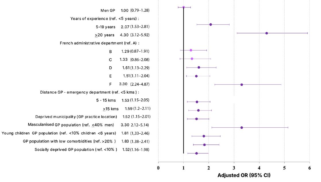
A cross-sectional study was conducted using the French National Health Data System (SNDS), which contains all comprehensive information on healthcare consumption for approximately 99% of the French population, covering both outpatient and inpatient care.5 GP consultations in primary care practices in the Centre-Val de Loire region in 2023–2024 were extracted. For each practitioner, the proportion of consultations resulting in systemic AP was estimated based on the dispensation codes (J01 ATC) and GPs were categorised as high-volume prescribers if >3rd quartile (11.5%; median 8.5%). GP features and practice population (patient characteristics attributed to each GP) associated with higher AP were estimated through a multivariable logistic regression model.
RESULTS
Over the 2-year period, 1,936 GPs (55.5% men; average age: 52.9±12.9 years) prescribed antibiotics in 10.1% of the 15 million consultations provided to 4 million patients (24.6% of patients). Concerning GPs, over two-thirds had 5 or more years of professional clinical experience (71%; 35.8% ≥20 years), and 59.8% were located ≥5 km from an emergency department (31.4% ≥15 km).
Regarding patients, 44.9% were men, 8.0% were under 6 years old, 23.5% had a chronic disease and/or disability, and 10.3% were in a precarious situation. Regarding prescriptions, antibiotics accounted
1: GP and patient population factors associated with being a major prescriber in Centre-Val de Loire, France, 2023–2024.
Masculanised population (2.12–5.14) (1.33-2.46) (1.38-2.41) (1.16-1.98)
GP: general practitioner; OR: odds ratio; ref: reference.
for 10.1% of consultations concerning 24.6% of patients.
The main prescriptions were amoxicillin (41.3%), macrolides (17.2%), and amoxicillin + clavulanic acid (14.5%). One quarter of GPs were high prescribers (n=491; 25.4%).
Factors associated with high prescription rates were linked to (a) patient population: young children (OR: 1.8 [95% CI: 1.3–2.5]), large male population >40% (3.3 [2.1–5.1]), socioeconomic deprivation (1.5 [1.2–2.0]), and fewer chronic diseases/disability (1.8 [1.4–2.4]; and (b) GP profile: ≥5 years’ experience with an increasing gradient (4.3 [3.1–5.9] if ≥20 years’ experience), emergency department ≥5 km with increasing gradient (1.6 [1.2–2.1] if ≥15km), GP practice in a socially deprived area (1.5 [1.2–2.0]), and practice department; whereas GP gender and access were not significantly associated to higher AP (Figure 1).
CONCLUSION
This large-scale study pinpoints GP and patient factors driving higher AP, highlighting the importance of targeted interventions to promote antibiotic stewardship to tackle antimicrobial resistance.
Real-world data obtained via the SNDS can provide actionable insights for Regional Centers of Antibiotic Therapy, supporting intervention initiatives and informing public health strategies. However, further analyses will be required to identify more accurate prescription behaviours according to spectrum, duration, and adequacy of prescription/diagnosis.
References
1. Desmars H et al. Profile of high-volume antibiotic prescribers: a population-based study from the French National Health Data System, 2023–2024. Abstract E0359. ESCMID Global, 17-21 April, 2026.
Figure
2. World Health Organization (WHO). Antimicrobial resistance. 2023. Available at: https://www.who.int/ news-room/fact-sheets/detail/ antimicrobial-resistance. Last accessed: 1 April 2026.
3. Santé Publique France. Consommation d’antibiotiques en secteur de ville en France 2014-2024. Available at: https://
www.santepubliquefrance.fr/resistanceaux-antibiotiques/rapportsynthese/ consommation-dantibiotiques-ensecteur-de-ville-en-france-2014-2024. Last accessed: 1 April 2026.
4. Harbarth S et al. Control of multiply resistant cocci: do international comparisons help? Lancet Infect Dis. 2001;1(4):251-61.
5. Maillard O et al. Use of the French National Health Data System (SNDS) in pharmacoepidemiology: a systematic review in its maturation phase. Therapie. 2024;79(6):659-69.
Domain-Aware Versus Machine Learning Imputation for Sparse Antimicrobial Susceptibility Data
Authors: *Fredrick Mutisya,1 Taïoh Yokoyama,1 Sana Boujaafar,1 Cyprien de Turckheim,1 Mathieu Raad1
1. SmartBiotic, Strasbourg, France
*Correspondence to fredrick.mutisya@smartbiotic.ai
Disclosure: Raad, de Turckheim, and Yokoyama have received support from SmartBiotic for the present manuscript. Mutisya, de Turckheim, Raad, and Yokoyama have received consulting fees from SmartBiotic. SmartBiotic is a software platform analysing local bacterial ecology of hospitals to develop tailored antibiotic therapy recommendations. Boujaafar has declared no conflicts of interest.
Acknowledgements: The authors would like to thank Pfizer and Vivli for open access to the Atlas dataset.
Antimicrobial susceptibility testing datasets frequently miss data in a structured way due to selecting testing practices. Handling these gaps with generic statistical imputation may violate well-established microbiological rules such as intrinsic resistance or non-reportable combinations. This study aims to compare domainaware completion based on established microbiology rules with several machine learning (ML)-based imputation strategies under controlled conditions.1
MATERIALS AND METHODS
The authors evaluated imputation strategies using the Atlas dataset 2022 (Pfizer, New York, USA) for levofloxacin, meropenem, and gentamicin, selected to represent different drug classes and resistance patterns. SmartBiotic’s (Montreal, Canada) rule engine
derived from the European Committee on Antimicrobial Susceptibility Testing (EUCAST) and Clinical and Laboratory Standards Institute (CLSI) expected resistance and susceptibility phenotypes was used for domain-aware completion. For validation, 20% of truly observed results were randomly masked per antibiotic, simulating missing completely at random conditions. Five reconstruction strategies of masked values were compared: global frequency imputation, ML with listwise deletion, ML with random undersampling, ML with synthetic minority oversampling technique (SMOTE), and a rulebased strategy. Outcomes were binarised (susceptible versus resistant/intermediate) and evaluated using accuracy, sensitivity, specificity, predictive values, F1 score, and Cohen’s kappa.
RESULTS
The authors’ inferred resistance rules added 75,730 new cells, mainly from intraspecies inference (75,729 cells), while intrinsic resistance rules augmented all 55,549 rows with 502,847 additional cells (11.2%). In the masking experiment, the domain rule completion demonstrated high performance for levofloxacin (accuracy: 95%; sensitivity: 92%; specificity: 96%), meropenem (91%/97%/90%), and gentamicin (86%/57%/96%). With imbalance handling, ML with SMOTE oversampling achieved moderate sensitivity improvement (levofloxacin: 58%; meropenem: 92%; gentamicin: 54%) over unbalanced ML (43%/90%/41%). Random undersampling produced balanced profiles but lower overall performance (66–72% across metrics). Frequency imputation yielded 0% sensitivity across all antibiotics despite acceptable accuracy (69–79%).
CONCLUSION
These findings suggest that antimicrobial susceptibility testing missingness should
first be addressed as a microbiological problem and only then as a statistical one. Rule-based systems predict only when applicable, yielding high specificity but variable coverage-depending sensitivity. ML required imbalance correction for meaningful resistance detection, with SMOTE oversampling offering optimal compromise. These results, consistent with recent ML approaches in AMR surveillance, support hierarchical imputation
with deterministic approach first, then imbalance-aware ML for residual gaps. Future validation work on these approaches should assess performance under real-world settings.
References
1. Mutisya F et al. Domain-aware versus machine learning imputation for sparse antimicrobial susceptibility data. Abstract O0401. ESCMID Global, 17-21 April, 2026.
Differential Expression of miRNAs Involved in Shock Are Able to Characterise Mortality
Authors: *I. Rebollo-Mato,1 A. García-Concejo,2 L. Polo-Sánchez,3 R. Prieto-Utrera,3 P. Martínez-de Paz,2 M. Álvarez-Bardón,1 J. Matesanz-Isabel,3 H. Gonzalo-Benito,3 R. Cobo-Zubia,4 E. Gómez-Sánchez,5 M. Martín-Fernández,1 E. Tamayo-Gómez5
1. University of Valladolid, Spain
2. Center for Biomedical Research Network on Infectious Diseases, Madrid, Spain
3. Institute of Health Sciences of Castile and León, Soria, Spain
4. Carlos III Health Institute, Madrid, Spain
5. University Clinical Hospital of Valladolid, Spain
*Correspondence to irina.rebollo22@uva.es
Disclosure: The authors have declared no conflicts of interest.
Postoperative shock, particularly septic shock, is a leading cause of circulatory failure and mortality in the ICU.1,2 Differentiating septic from non-septic postoperative shock remains a major clinical challenge due to overlapping haemodynamic profiles and the limited sensitivity of conventional clinical scores in detecting early molecular alterations.3,4 Extracellular vesicle-derived microRNAs (EV-miRNA) have emerged as promising biomarkers reflecting endothelial dysfunction, inflammatory activation, and immunometabolic dysregulation in shock states.5,6 This study aimed to characterise the EV-miRNome in postoperative shock and evaluate its diagnostic and prognostic utility.
METHODS
The authors conducted a multicentre prospective study including two independent cohorts of adult surgical patients: a discovery cohort (n=164) and a validation cohort (n=84). Patients were classified into postsurgical controls without shock and a shock cohort, including septic and non-septic postoperative shock. Only microbiologically confirmed septic shock cases were included. Plasma EV-miRNAs were isolated within 24 hours of shock diagnosis and profiled using small RNA sequencing. Differential expression analysis was performed using DESeq2 (Bioconductor; Buffalo, New York, USA), followed by Gene Ontology/ Kyoto Encyclopedia of Genes and Genomes enrichment, receiver operating characteristic-based prognostic modelling, quantitative PCR validation, and Cox regression for 90-day mortality.
RESULTS
Twenty-six EV-miRNAs were significantly dysregulated in shock patients compared to controls. miR-4488 and miR-3960 showed the strongest association with disease severity, correlating with Sequential Organ Failure Assessment (SOFA) score, haemodynamic impairment, and hepatic dysfunction. Individually, miR-4488 (area under the curve [AUC]: 0.786) and miR3960 (AUC: 0.775) showed moderate predictive ability for mortality, while their combined model significantly improved prognostic performance (AUC: 0.895), outperforming C-reactive protein, procalcitonin, leukocytes, creatinine, and clinical scoring systems. High expression of both miRNAs was independently associated with increased 90-day mortality in multivariate Cox analysis. Quantitative PCR validation confirmed their overexpression, achieving an AUC of 0.952 in the validation cohort.
DISCUSSION
Postoperative shock represents a heterogeneous clinical syndrome in which early discrimination between septic and non-septic aetiologies remains essential but challenging. Current diagnostic strategies rely on systemic inflammatory markers and physiological parameters that fail to capture underlying molecular heterogeneity, highlighting the need for more sensitive biomarkers.7 Extracellular vesicles act as mediators of intercellular communication and carry microRNAs reflecting endothelial injury, immune dysregulation, and metabolic reprogramming.8,9 In this context, EV-miRNAs provide a dynamic and minimally invasive window into disease pathophysiology.10,11
The authors’ findings identify a distinct EV-miRNA signature associated with postoperative shock. miR-4488 and miR-3960 were consistently linked to organ dysfunction and severity, suggesting their involvement in vascular integrity and systemic inflammatory pathways. Their combined predictive value significantly improved mortality stratification compared with standard biomarkers and clinical scores. These results support the role of EV-miRNAs as active contributors to shock pathophysiology rather than passive markers.
CONCLUSION
In conclusion, postoperative shock exhibits a specific EV-miRNA profile with strong diagnostic and prognostic implications. miR-4488 and miR-3960 emerge as robust biomarkers associated with endothelial dysfunction, immunometabolic activation,
and organ failure. Their integration into biomarker panels may improve early risk stratification and mortality prediction in postoperative patients who are critically ill.
References
1. Rebollo-Mato I et al. Differential expression of microRNAs involved in shock are able to characterise mortality. Abstract O0279. ESCMID Global, 17-21 April, 2026.
2. Singer M et al. The third international consensus definitions for sepsis and septic shock (Sepsis-3). JAMA. 2016;315(8):801-10.
3. Shankar-Hari M et al. Developing a new definition and assessing new clinical criteria for septic shock: for the Third International Consensus Definitions for Sepsis and Septic Shock (Sepsis-3). JAMA. 2016;315:775-87.
4. Meini J et al. Platelet-derived extracellular vesicles promote endothelial dysfunction in sepsis by enhancing neutrophil extracellular traps. BMC Immunol. 2023;24(1):22.
5. Xu J et al. Circulating plasma extracellular vesicles from septic mice induce inflammation via microRNA- and TLR7-dependent mechanisms. J Immunol. 2018;201(11):3392-400.
6. Moreno-Torres V et al. Better prognostic ability of NEWS2, SOFA and SAPS-II in septic patients. Med Clin (Barc). 2022;159(5):224-9.
7. Qiu G et al. Diagnostic potential of plasma extracellular vesicle miR-483-3p and Let-7d-3p for sepsis. Front Mol Biosci. 2022;9:814240.
8. Momen-Heravi F et al. Current methods for the isolation of extracellular vesicles. Biol Chem. 2013;394(10):1253-62.
9. García-Concejo A et al. Study on the diagnostic role of exosome-derived miRNAs in postoperative septic shock and non-septic shock patients. Crit Care. 2025;DOI:10.1186/s13054-025-05320-y.
10. Ye R et al. miR-150-5p in neutrophil-derived extracellular vesicles associated with sepsisinduced cardiomyopathy in septic patients. Cell Death Discov. 2023;9(1):19.
11. Schiavello M et al. Extracellular vesicles: new players in the mechanisms of sepsis- and COVID19-related thromboinflammation. Int J Mol Sci. 2023;24(3):1920.
StackPred: AI-Boosted AMR Phenotype Prediction for Multiple Species and Antimicrobial Agents
Authors: *Julian Welling,1,2 Miriam Balzer,1,2
Leah Consten,1 Stefan Bletz,3 Jan Buer,1 Valérie
Chapot,1 Dag Harmsen,4 Evelyn Heintschel von Heinegg,1 Alexander Mellmann,3 Wolfgang Pölking,3 Friederike Salhöfer,1 Frieder Schaumburg,5 Natalie Scherff,3 Niklas Wiesmann,5 Folker Meyer1,2
1. Department of Medicine, University of Duisburg-Essen, Germany
2. Department of Computer Science, University of Duisburg-Essen, Germany
3. Institute of Hygiene, University Hospital Münster, Germany
4. Ridom GmbH, Münster, Germany
5. Institute of Medical Microbiology, University Hospital Münster, Germany
*Correspondence to julian.welling@uk-essen.de
Disclosure: The authors have declared no conflicts of interest.
Acknowledgements: This study was funded by the federal state of North Rhine-Westphalia (AZ-Innovationsförderagentur NRW) under the grant EFRE-20800459.
Genome-based antimicrobial susceptibility testing is emerging as a promising alternative to culturebased methods, which remain timeconsuming despite being the clinical gold standard.1,2 Here, the authors present an extension of the stacked Random Forest framework, StackPredAMR (under revision), which originally covered 18 antimicrobial agents across three species. At the European Society of Clinical Microbiology and Infectious Diseases (ESCMID) 2026, the authors presented an extension of this framework, demonstrating its
straightforward scalability to additional species and antimicrobial agents for multi-agent antimicrobial resistance (AMR) prediction from genomic data.3
METHODS
The model was trained on BVBRC VITEK (bioMérieux, Marcy-l’Étoile, France) antimicrobial susceptibility testing data comprising more than 2,500 isolates from Escherichia coli, Klebsiella pneumoniae, Acinetobacter baumannii, and Enterobacter cloacae, covering up to 23 antimicrobial agents.4 Genomic input features were derived from the Comprehensive Antibiotic Resistance Database (CARD) annotations, and encoded as binary presence/absence of AMR genes.5 The approach builds on the original stackPredAMR architecture by combining individual Random Forest classifiers for each antimicrobial agent with a second-layer meta-model. This stacking strategy enables the model to capture cross-resistance patterns between antimicrobial agents while handling incomplete phenotype labels, a common limitation in AMR datasets.6 Compared to rule-based approaches, which rely on known resistance genes,7 machine learning methods can learn more complex patterns directly from genomic data.8
RESULTS
Model performance was evaluated using 5-fold cross-validation. For E. coli and K. pneumoniae, median major error and very major error rates across antimicrobial agents remained below 3%. Predictions for A. baumannii and E. cloacae, included as a proof of concept with a smaller sample size, were also successfully generated, highlighting the flexibility and extensibility of the framework (Figure 1).
Figure 1: Median error rates and sample counts across antibiotics for four bacterial species.
Median error rate (%)
Median ME and VME rates across antibiotics for four bacterial species, including their sample count. Error bars show 95% bootstrap CIs. The dashed line marks the 3% error threshold.
A. baumannii: Acinetobacter baumannii; E. cloacae: Enterobacter cloacae; E. coli: Escherichia coli; K. pneumoniae: Klebsiella pneumoniae; ME: major error; VME: very major error.
Further analysis of precision–recall performance in relation to effective sample size showed that reduced predictive performance occurred only for antimicrobial agents with limited and imbalanced data. Effective sample size, defined as the harmonic mean of resistant and susceptible isolates, decreased substantially under class imbalance, underlining the importance of sufficiently large and balanced datasets for robust resistance prediction.
CONCLUSION
Overall, this work demonstrates that extending StackPredAMR enables scalable and flexible multi-agent AMR prediction across bacterial species, even in the presence of incomplete phenotype data. Future work will focus on incorporating additional species and antimicrobial agents, integrating regulatory genomic features, and evaluating the approach on metagenomic data to support faster, culture-independent diagnostics.
References
1. Arnold A et al. How AI can help us beat AMR. NPJ Antimicrob Resist. 2025;3(1):18.
2. Su M et al. Genome-based prediction of bacterial antibiotic resistance. J Clin Microbiol. 2019;57(3):e01405-18.
3. Welling J et al. StackPred: AI boosted AMR phenotype prediction for multiple species and antimicrobial agents. ePoster E0487. ESCMID Global, 17-21 April, 2026.
4. Olson RD et al. Introducing the bacterial and viral bioinformatics resource center (BV-BRC): a resource combining PATRIC, IRD and ViPR. Nucleic Acids Res. 2023;51(D1):D678-9.
5. Alcock BP et al. CARD 2023: expanded curation, support for machine learning, and resistome prediction at the comprehensive antibiotic resistance database. Nucleic Acids Res. 2023;51(D1):D690-9.
6. Aytan-Aktug D et al. Prediction of acquired antimicrobial resistance for multiple bacterial species using neural networks. mSystems. 2020;5(1):e00774-19.
7. CRyPTIC Consortium et al. Prediction of susceptibility to first-line tuberculosis drugs by DNA sequencing. N Engl J Med. 2018;379(15):1403-15.
8. Anahtar MN et al. Applications of machine learning to the problem of antimicrobial resistance: an emerging model for translational research. J Clin Microbiol. 2021;59(7):e0126020.
Genomic Surveillance in a Low- or Middle-Income Country: Convergent Colistin Resistance in Epidemic Klebsiella pneumoniae (ST11, ST147) in Peshawar, Pakistan
Author: *Maria Khan1
1. Pathology Department, Peshawar Institute of Cardiology-MTI, Pakistan *Correspondence to kmaria22@hotmail.com
Disclosure: The author has declared no conflicts of interest.
The escalating crisis of antimicrobial resistance has positioned colistin as a ‘last-resort’ therapy for carbapenemresistant Klebsiella pneumoniae. 1-5 However, in low- and middle-income countries like Pakistan, unregulated antibiotic use and high infection burdens have accelerated the emergence of colistin resistance. This study provides a critical genomic snapshot of the molecular mechanisms and clonal distribution of colistin-resistant isolates in a tertiary care setting in Peshawar, Pakistan.
MATERIALS AND METHODS
Between January 2024–December 2025, 250 clinical K. pneumoniae isolates were collected. Following species identification via matrix-assisted laser desorption/ ionisation time-of-flight mass spectrometry, colistin susceptibility was assessed using broth microdilution. Whole-genome sequencing was performed on 35 resistant isolates (14.0%) to characterise sequence types (ST), resistomes, and specific colistin resistance determinants, including chromosomal mutations (e.g., mgrB) and plasmid-mediated genes (e.g., mcr-8).
RESULTS
Genomic surveillance revealed a highpressure resistance environment dominated by two international high-risk clones: ST11 (51.4%) and ST147 (20.0%). These lineages were predominantly associated with the carbapenemase gene blaNDM-1, highlighting an extensively drug-resistant profile (Table 1).
Resistance was primarily driven by chromosomal alterations in 85.7% of isolates, with mgrB inactivation via insertion sequences (ISKpn26 and IS5-like) being the most prevalent mechanism. Plasmidmediated resistance was identified in 14.3% of isolates, specifically the mcr-8 gene within the ST147 lineage. Notably, the study documented a critical evolutionary milestone: convergent resistance. Three ST147 isolates simultaneously harboured both plasmid-borne mcr-8 and chromosomal mgrB disruptions. This dualmechanism profile resulted in significantly higher minimum inhibitory concentration values (90–128 mg/L) compared to isolates with single resistance determinants.
CONCLUSION
The findings underscore a shift toward pan-drug resistance in Pakistan’s clinical settings. The convergence of independent resistance pathways within globally disseminated clones like ST147 poses a severe public health threat, as these strains are both highly fit and difficult to treat. The study advocates for an urgent expansion of regional genomic surveillance, stringent antimicrobial stewardship to preserve remaining polymyxins, and enhanced infection control protocols in high-risk units like the ICU to mitigate the spread of these ‘super-clones’.
Table 1: Genomic epidemiology and resistance profiles of colistin-resistant K. pneumoniae isolates.
Category Key findings
Prevalence Overall colistin resistance
Primary clones
Chromosomal
Data/mechanisms
14.0% (35/250 isolates)
Dominant sequence types ST11 (51.4%) and ST147 (20.0%)
Most frequent mechanism mgrB inactivation (85.7% of resistant isolates)
Plasmid-mediated Key resistance gene mcr-8 (found exclusively in ST147)
Convergent resistance
Combined mechanisms
mgrB+mcr-8 (MIC 32–128 mg/L)
Associated AMR Co-carried resistance blaNDM-1 (carbapenemase), blaCTX-M-15
1. Khan M. Genomic surveillance in an LMIC: convergent colistin resistance in epidemic Klebsiella pneumoniae (ST11, ST147) in Peshawar, Pakistan. Abstract E0846. ESCMID Global, 17-21 April, 2026.
2. Li P et al. Convergence of carbapenem resistance and hypervirulence in a highly-transmissible ST11 clone of K. pneumoniae: an epidemiological, genomic and functional study. Virulence. 2021;12(1):377-88.
3. Farzana R. A genomic approach to understanding the molecular epidemiology and clinical burden of multi-drug resistant Enterobacterale Infections in Bangladesh. 2020. Available at: https:// orca.cardiff.ac.uk/id/eprint/140464/5/Thesis_ Farzana_R_200421.pdf. Last accessed: 9 April 2026.
4. Wang X et al. Emergence of colistin resistance gene mcr-8 and its variant in Raoultella ornithinolytica Front Microbiol. 2019;10:228.
5. Főldes A et al. Characterization of carbapenemaseproducing Klebsiella pneumoniae isolates from two Romanian hospitals co-presenting resistance and heteroresistance to colistin. Antibiotics (Basel). 2022;11(9):1171.
How Do We Define 'Appropriate Antimicrobial Use' In One Health? A Global Delphi Survey
Authors: *Ri Scarborough,1,2 Courtney Ierano,1-3
Kirsten Bailey,1,2 Glenn Browning,1,2 Karin Thursky,1-3 Ruby Biezen,1,2 Brian Hur,2,4
Brendan McMullan,5,6 Leslie Dowson,1-3 Allegra Schermuly,7 Leanne Teoh,1,2 Laura Hardefeldt1,2
1. The University of Melbourne, Parkville, Australia
2. National Centre for Antimicrobial Stewardship, Melbourne, Australia
3. The Royal Melbourne Hospital, Parkville, Australia
4. University of Washington, Seattle, USA
5. University of New South Wales, Kensington, Australia
6. Sydney Children's Hospital, Australia
7. Monash University, Clayton, Australia
*Correspondence to ri.scarborough@unimelb.edu.au
Disclosure: Funding for this project was provided by the Australian Government via the Medical Research Future Fund (MRFF), Grant ID MRF2028452; Hur received no salary from this grant. Schermuly has received internal funding from the Monash School of Social Sciences to cover her registration at the ASA 2025 and TASA 2025 conferences to present different data from the same broad study as this manuscript, with payment to the author. McMullan has received an Australian Government Grant: NHMRC Investigator Grant, Grant ID 2008632, with payment to the institution; an honorarium for providing a paediatric infectious diseases lecture in the ALJESAL courses in 2024–2025, with payment to the author (later donated in full to the Sydney Children’s Hospitals Foundation); was a member of the DSMB for the PATRIC trial (unpaid); and is the Chair of the Australian and New Zealand Paediatric Infectious Diseases Network and Secretary of the World Society for Pediatric Infectious Diseases, both unpaid. Teoh has received a grant from the National Health and Medical Research Council (Investigator Grant 2016647); royalties for Drugs4dent in accordance with the University of Melbourne IP Policy (Drugs4dent is licensed to MIMS Australia); payment or honoraria for lectures, presentations, speakers bureaus, manuscript writing, or educational events from the Australian Dental Association, Australian Dental Association Victorian Branch, Australian Dental Association New South Wales, New Zealand Dental Association, and Royal Australasian College of Dental Surgeons; support for attending meetings and/or travel from the University of Melbourne; participated on the MIMS Clinical Advisory Board; and held a leadership or fiduciary role in the FDI World Dental Federation Preventing AMR and Infections Task Team.
The other authors have declared no conflicts of interest.
Acknowledgements: The authors would like to thank the antimicrobial stewardship experts from 44 countries who provided responses to the Delphi survey.
Keywords: Antimicrobial stewardship (AMS), antimicrobial use (AMU), One Health.
Many early antimicrobial stewardship (AMS) initiatives focused on reducing antimicrobial use (AMU). However, reducing AMU may cause harm. To avoid unintended consequences, the focus is now moving to increasing appropriateness of AMU. At the same time, there has been a shift from a single-context approach towards a unified, One Health AMS approach. However, the lack of an agreed definition of appropriate AMU between stakeholders representing animal, human, and environmental health remains a barrier to coordinated, One Health AMS implementation.
METHODS
The authors conducted a literature review and held four focus groups with diverse stakeholders to develop a list of proposed statements that could describe appropriate (systemic) AMU across all One Health contexts.1 The authors included these in a four-round, online Delphi survey circulated to AMS experts globally, whom the authors identified from their publications, professional networks, project partner organisations, and snowballing. Each statement was rated on a seven-point Likert scale (0–6); a score of four or more was considered agreement. Participants could also provide written feedback on each statement. Statements were included in the consensus only when they reached 80%
agreement. Those with <60% agreement in any round were considered rejected, and statements with 60–80% agreement were presented again in the subsequent round, until round three, with changes based on participant feedback. In the fourth round, the authors asked the feasibility of measuring each consensus item in the context with which the participant was most familiar.
RESULTS
The authors invited 391 AMS experts to participate, and 243 from 44 countries (62%) provided a response to at least one survey round. Twenty-two percent were from low- and middle-income countries, and 63% were in a position to influence their national AMR strategy. Sixty-three percent represented human health, 26% represented animal health, and 11% environmental health or ‘other’. In round one, 43 of 76 proposed statements (Table 1) reached 80% consensus, with a further eight statements reaching consensus over subsequent rounds. Consensus statements covered a wide range of topics, including who should receive systemic antimicrobials; when, why, and how these should be initiated;
and whether it should be for a therapeutic or preventative indication. The selection of drug, route, dosing, and duration were the other main topics; in general, participants agreed that relevant guidelines should be followed, where these are available. In the absence of guidelines, participants agreed that the narrowest spectrum or lowest importance drug should be used, by the safest effective route, and for the shortest duration to control the infection (or risk of infection, in the case of preventative use). The median proportion of respondents who felt that, in their context, it would be feasible to measure concordance with each statement was 62%, with feasibility almost always rated higher for human health contexts than animal health contexts. Statements that were rejected in the first round included: using antimicrobials for the purpose of growth promotion in livestock and using preventative antimicrobials for the purpose of avoiding economic loss.
CONCLUSION
The 51 consensus items from this global survey form a new foundation for further One Health discussion around appropriate AMU and unified measures of appropriate use.
Table 1: The 51 consensus statements regarding important features of appropriate AMU, with the survey question: when considering whether a systemic use of antimicrobials was appropriate, how important is the following?
was adequate evidence that the person/animal had an infection. Suffering and/or complications from the infection would likely be lessened with antimicrobials.
An equivalent (or better) health outcome was not achievable without systemic antimicrobial treatment.
Preventative AMU
There was potential for significant suffering without preventative antimicrobial use. There was potential for significant mortality without preventative antimicrobial use.
All practicable management options to control infection (e.g., moving animals/people away from risk) had already been implemented.
Consideration was given to the risks of preventative antimicrobial use, including AMR.
Table 1: The 51 consensus statements regarding important features of appropriate AMU, with the survey question: when considering whether a systemic use of antimicrobials was appropriate, how important is the following? (Continued).
Livestock-specific considerations
Initiation of AMU
The drug was not prohibited in the animal species in which it was used.
The drug choice and regimen ensured the animal product(s) complied with local and relevant export antimicrobial residue limits.
Person/animal/herd was under the care of a clinician (for livestock, a veterinarian had visited the farm at least once in the last year).
Person/animal/herd was examined by a clinician during the current episode.
Disease severity justified starting antimicrobials, rather than a 'watch and wait' strategy.
Where sepsis was suspected, treatment was initiated as soon as possible.
For surgical prophylaxis, timing of antimicrobial administration was concordant with current guidelines.
Where recommended in current guidelines, a sample was collected for culture and susceptibility testing.
The reason or indication for antimicrobial use was documented in medical record.
Patient or carer was meaningfully involved in the decision to use antimicrobials (emergencies excepted).
Patient or carer was given instructions (appropriate to their health literacy) on taking or administering the antimicrobials correctly, including when to stop.
Patient or carer was informed about potential adverse effects.
A date for review or cessation of antimicrobial treatment was set.
Drug Where treatment guidelines were available and the pathogen was unknown, drug choice was concordant with current guidelines for the known or presumptive diagnosis.
Empiric drug selection accounted for local epidemiology (e.g., antibiogram data) where available.
Where feasible, point-of-care testing was used to refine the empiric antimicrobial selection.
Where culture and susceptibility results were available, the results were used to guide drug choice.
Patient had no known allergy or other contraindications to the drug.
Drug penetrates the target tissue(s) adequately and remains active at the target tissue.
Where multiple drugs would be suitable, consideration was given to minimising adverse effects.
Where multiple drugs would be suitable, consideration was given to minimising antimicrobial resistance risk.
Where multiple drugs would be suitable, a drug with the narrowest spectrum or lowest importance rating was selected.
Where current guidelines recommended multi-agent therapy, a recommended combination was used.
For patients with compliance challenges receiving oral medication, consideration was given to palatability of available formulations.
Cost of drug was bearable for the bill payer(s). Payer can be the patient, animal owner, insurer, government, or other.
Table 1: The 51 consensus statements regarding important features of appropriate AMU, with the survey question: when considering whether a systemic use of antimicrobials was appropriate, how important is the following? (Continued).
Route
Dosing
Duration
Off-label use
Reasons to deviate from appropriate use principles
Route allowed drug to reach the site of infection at a concentration that will be effective.
Safest effective route for the patient at that time point.
Route concordant with current guidelines, where these were available.
Route as physically comfortable as possible for the patient (assuming there were multiple safe and effective routes).
Route convenient for the patient if self-administering (assuming there were multiple safe and effective routes).
Route convenient and safe for the carer if patient not administering (assuming there were multiple safe and effective routes for the patient).
Where guidelines were available, dosing regimen was concordant with current guidelines for the known or presumptive diagnosis.
Consideration was given to the actual or adjusted body weight of the patient.
Dosing regimen was adjusted for other relevant patient factors, e.g., renal insufficiency.
Dosing regimen was adjusted based on antimicrobial susceptibility results where dosedependent susceptibility was present.
Dosing regimen accounted for the site of infection.
Where guidelines were available, duration was concordant with current guidelines for the known or presumptive diagnosis.
Duration was only as long as required to ensure control of the infection (or for prophylaxis, only as long as required to manage infection risk).
Where appropriate, duration was adapted according to improvement in clinical signs/ symptoms and/or infection-specific biomarkers.
There was no registered antimicrobial for the species (or age group) and indication.
There was sufficient evidence that the extra-label use was safe and effective for that clinical situation.
The patient had a known, relative contraindication to the registered antimicrobial(s).
There was sufficient evidence that the product information (label) regimen is subtherapeutic (in livestock, provided that the withholding period is appropriately adjusted).
Recommended drug not available (e.g., supply chain issue).
1. Scarborough R et al. How do we define 'appropriate antimicrobial use' in One Health? A global Delphi survey. Abstract 01387. ESCMID Global, 17-21 April, 2026.
Investigation of Enterococcus Colonisation Impact on Clostridioides difficile Disease Severity
Authors: Alexander Mai,1 Adelaide Horvath,1 Khurshida Begum,1 Thomas Horvath,1-3
Kevin Garey,1 *Taryn Eubank1
1. Department of Pharmacy Practice and Translation Research, College of Pharmacy, University of Houston, Texas, USA
2. Department of Pathology and Immunology, Baylor College of Medicine, Houston, Texas, USA
3. Texas Children’s Microbiome Center, Department of Pathology, Texas Children’s Hospital, Houston, USA
*Correspondence to taeubank@cougarnet.uh.edu
Disclosure: This project was funded by an SIDP Early Career Investigator Grant and ACCP Foundation Junior Investigator Research Award, with payment to the institution. Mai has received support for the present manuscript through the UH SURF Award and UH PURS Award (stipend to support tuition during research), with payment to the author. Garey has received research grants from Acurx Pharmaceuticals and Paratek Pharmaceuticals, with payment to the institution. Horvath T. has received support for the present manuscript via an NIH ORIP S10 Shared Instrument grant, with payment to the institution; a Wellcome LEAP FORM grant and a Wellcome LEAP 1KD grant, with payment to the institution; honoraria from Cell Press STAR Protocols for service to the journal as an associate academic editor; equipment (LC/MS system) from Revvity for the duration of the validation experiments; and served as an Editorial Advisory Board Member for STAR Protocols. The other authors have declared no conflicts of interest.
Enterococcus colonisation with Enterococcus faecalis and Enterococcus faecium is a known risk factor for Clostridioides difficile infection (CDI).1-3 Enterococcus co-colonisation increases C. difficile virulence and toxin production through cross-feeding.4 However, current
disease severity definitions do not take into account the patients' microbiota. This study aimed to investigate the impact of Enterococcus colonisation on disease severity.
MATERIALS AND METHODS
This was a case-control study of adult patients hospitalised with CDI from two health systems (14 hospitals) in Houston, Texas, USA (2016–2025). Patients with severe CDI were matched to nonsevere patients (1:1) on age ±10 years and immunocompromised status. Stool samples were collected from hospitalised patients with CDI, and stool underwent DNA extraction for quantitative PCR of E. faecalis and E. faecium. Metabolomics were completed by liquid chromatographytandem mass spectrometry. Disease severity and CDI classification were defined according to the 2017 Infectious Diseases Society of America (IDSA)/Society for Healthcare Epidemiology of America (SHEA) clinical guidelines.5
RESULTS
A total of 190 patients (95 matches) with CDI were included (female: 54.7%; age >65 years: 60%; hospital-acquired CDI: 42%; CDI initial episode: 87%). Patients with Enterococcus spp. quantity >106 were designated as high colonisation; 61% of patients were highly colonised. The group with severe disease had a higher percentage of patients categorised as having high Enterococcus spp. colonisation, though not statistically significant (67.5% versus 58.7%; p=0.07). While high Enterococcus spp. colonisation trends with IDSA disease severity, it is highly possible that other microbiota members cross-feed with C. difficile. Metabolomics completed on 84 matches with sufficient stool showed that patients with severe disease had significantly higher amounts of
ornithine in the stool than patients with non-severe disease (6,739 ng/mL versus 4,270 ng/mL; p=0.02).
CONCLUSION
The gut microbiota is a diverse environment with many interspecies interactions. While a trend is observed between Enterococcus spp. colonisation and CDI disease severity, it is highly likely that other microorganisms contribute to this association. Future metagenomic and metabolomic analysis is warranted.
References
1. Mai A et al. Investigation of Enterococcus colonization impact on Clostridioides difficile disease severity. Poster E0154. ESCMID Global, April 17-21, 2026.
2. Bosnjak M et al. Multi-omics analysis of hospitalacquired diarrhoeal patients reveals biomarkers of enterococcal proliferation and Clostridioides difficile infection. Nat Commun. 2023;14(1):7737.
3. Berkell M et al. Microbiota-based markers predictive of development of Clostridioides difficile infection. Nat Commun. 2021;12(1):2241.
4. Smith AB et al. Enterococci enhance Clostridioides difficile pathogenesis. Nature.2022;611(7937):780-6.
5. McDonald LC et al. Clinical practice guidelines for Clostridium difficile infection in adults and children: 2017 update by the Infectious Diseases Society of America (IDSA) and Society for Healthcare Epidemiology of America (SHEA). Clin Infect Dis. 2018;66(7):e1-48.
Addressing Prescribing Behaviour to Reduce Broad-Spectrum Antibiotic Use
Authors: *Veronica Chorro-Mari,1 Amy Dalton,1 Selena Yan,1 Stephen Glass,1 Lisa White1
1. East Kent University Hospitals NHS Foundation Trust (EKHUFT), UK *Correspondence to veronica.chorro-mari@nhs.net
Disclosure: The authors have disclosed no conflicts of interest.
Acknowledgements: The authors would like to thank all antimicrobial stewardship (AMS) champions at EKHUFT.
To define a new AMS strategy that identifies AMS activities incorporating behaviourchange principles and assess its impact on broad-spectrum antibiotic prescribing across the Trust.4
METHODS
A series of AMS-focused activities was developed through regular multidisciplinary (MDT) meetings involving pharmacists, nurses, microbiologists, and clinicians. A prospective analysis was carried out over 2 consecutive years across adult inpatients in the authors’ 1,050-bed Trust. A series of AMS-focused activities was developed prospectively through regular MDT meetings involving pharmacists, nurses, microbiologists, and clinicians.
The main key drivers were grouped in five themes: 1) guideline and policy governance; 2) antimicrobial prescribing quality; 3) diagnostics; 4) in-patient and outcomefocused audits; 5) education and culture.
Prescribing behaviours were assessed in different stages, depending on audits conducted at admission, during inpatient stay, and at discharge.
Key activities explored included: guideline compliance; ward risk-based C. difficile key performing indicator-audits; documentation quality; timeliness of antimicrobial review; indication of antimicrobials with guidelines; guideline development and updates; optimisation of prescribing decisions; use of diagnostics prior to antibiotic initiation in acute medical units; surgical prophylaxis practice; AMS education delivery; and AMS policy revision.
RESULTS
Over 10 different activities were conducted involving different specialties. There was a reduction of BSA consumption over a 2-year period, plateauing the graph for consumption (stabilising a previously rising trend) with a 2.7% decrease in BSA and a 5.3% decrease in the Watch and Reserve category usage compared to the 2023 peak (Figure 1).
Figure 1: Total antibiotic versus ‘Watch and Reserve’ antibiotic consumption at EKHUFT in DDD/1,000 admissions (2014–2024; including FP10 data).
Total Watch and Reserve category abx usage Total abx usage
abx: antibiotics; DDD: daily defined dose; EKHUFT: East Kent University Hospitals NHS Foundation Trust.
There was greater awareness of AMS through MDT participation in audits and active communications.
Engagement with AMS education was mixed; training required targeted delivery and was not implemented as widely as initially planned. The AMS team took feedback from clinicians who wanted to see the impact on patients rather than daily defined dose data alone, so the authors used C.difficile infections as a measure to raise awareness amongst them.
CONCLUSION
A structured AMS strategy incorporating behavioural-change interventions can reduce BSA use and improve prescribing quality. Continued monitoring and reinforcement are required to sustain behaviour change and ensure long-term
impact. Pharmacy-led AMS teams play a crucial role in driving improvements when a clear strategy is implemented.
References
1. UK Health Security Agency. English surveillance programme for antimicrobial utilisation and resistance (ESPAUR) report. Available at: https:// www.gov.uk/government/publications/englishsurveillance-programme-antimicrobial-utilisationand-resistance-espaur-report. Last accessed: 25 November 2025.
2. Department of Health and Social Care. UK 5-year action plan for antimicrobial resistance 2024 to 2029. Available at: https://www.gov.uk/ government/publications/uk-5-year-action-planfor-antimicrobial-resistance-2024-to-2029. Last accessed: 25 November 2025.
3. British Society for Antimicrobial Chemotherapy. Behavioural science and AMR. Available at: https:// bsac.org.uk/behavioural-science-and-amr/. Last accessed: 25 November 2025.
4. Chorro-Mari V et al. Addressing prescribing behaviour to reduce broad spectrum antibiotic use. Poster E0236. ESCMID Global, 17–21 April, 2026.
A Multi-Seasonal Mixed-Method Point Prevalence Study of Antibiotic Prescription Patterns in a Tertiary Healthcare Facility in India
Authors: Vinay Modgil,1 *Sundeep Sahay,1,2
Arunima Mukherjee,2,3 Rashmi Surial,1 Oshin
Sinha,1 Sahil Kumar,1 Raman Chauhan,4 Sunil
Kumar Raina4
1. Society for Health Information Systems Programmes (HISP India), New Delhi, India
2. Department of Informatics, University of Oslo, Norway
3. Institute of Health and Society (HELSAM), University of Oslo, Norway
4. Department of Community Medicine, Dr. Rajendra Prasad Government Medical College (RPGMC), Kangra at Tanda, India
*Correspondence to sundeeps@ifi.uio.no
Disclosure: This work was conducted as part of the EquityAMR project (2021–2025), a collaborative research initiative between Norway and India on antimicrobial resistance and health equity, supported by the Research Council of Norway, with institutional and hospital collaboration support in Himachal Pradesh, India. The authors have declared no conflicts of interest.
Antimicrobial resistance is a growing global health concern, and inappropriate antibiotic use in hospitals is one of the key drivers of resistance, particularly in low- and middle-income countries (LMICs).1 Point prevalence surveys (PPS) are recommended by the WHO as an important tool to monitor antimicrobial use and guide antimicrobial stewardship programmes. However, in many LMICs settings, prescribing practices are influenced not only by clinical guidelines but also by diagnostic access, drug availability, and operational challenges within hospital systems.2-5 This study aimed to examine antibiotic prescribing patterns across different seasons and to understand
the behavioural and operational factors influencing antibiotic use and culture testing in a tertiary care hospital in northern India.6
MATERIALS AND METHODS
A multi-seasonal hospital-wide PPS was conducted across four seasons: autumn (November 2023), summer (April 2024), monsoon (August 2024), and winter (January 2025). Each phase involved 2 weeks of data collection following the WHO Global PPS methodology. Data were collected from five inpatient departments: Medicine, Surgery, Obstetrics-Gynaecology, Neonatal/Paediatric Intensive Care Units, and Adult Intensive Care Units. Quantitative data included antibiotic indications, routes of administration, and WHO AWaRe classification categories. These data were complemented by ward observations and inpatient follow-up to assess culture testing practices, antibiotic sensitivity testing (AST), and treatment modifications following laboratory results.
RESULTS
A total of 1,680 inpatients were surveyed across the four PPS rounds. Ceftriaxone was consistently the most prescribed antibiotic (30–33%), followed by piperacillin/tazobactam (9–20%) and doxycycline (8–16%). Azithromycin use declined sharply after the first survey phase, suggesting changes in prescribing behaviour over time. Use of higher-end antibiotics such as amikacin (6–9%) and meropenem (4–7%) remained relatively low. Empirical prescribing dominated antibiotic use, accounting for approximately 63–67% of prescriptions, while cultureguided therapy remained very limited (≤6%). More than 80% of antibiotics were administered parenterally, indicating a strong reliance on injectable therapy in inpatient settings. In terms of AWaRe
classification, watch antibiotics accounted for 46–56% of prescriptions, while access antibiotics accounted for 35–51%, and reserve antibiotics remained ≤4%. Prophylactic antibiotic use ranged from 24–30%, particularly in surgical wards, and combination therapy was common in intensive care units. Seasonal variation was observed in clinical diagnoses, with gastrointestinal conditions (20–22%) and chronic conditions (17–23%) being the most frequent overall, while respiratory
infections (3–9%) peaked during the monsoon and winter seasons (Table 1). Antibiotic modification following AST results occurred in only 4.7–6.5% of cases, indicating limited use of microbiology AST data in guiding therapy.
Qualitative observations from ward rounds and patient follow-up highlighted several systemic challenges influencing prescribing practices. These included stock-outs of oral antibiotics leading to higher use of
Antibiotic prescriptions
PPS: point prevalence survey.
Table 1: Seasonal variation in antibiotic prescriptions and clinical diagnoses across four PPS rounds.
Table 1: Seasonal variation in antibiotic prescriptions and clinical diagnoses across four PPS rounds (Continued).
Clinical diagnoses
category
injectable drugs, delays in culture sample collection and reporting, documentation gaps during patient transfers between wards, and limited antimicrobial stewardship activities. Together, these operational and behavioural factors contributed to continued reliance on broad-spectrum empirical antibiotic prescribing.
CONCLUSION
Overall, this multi-seasonal PPS demonstrated high empirical and broadspectrum antibiotic use, limited culturebased prescribing, and systemic barriers to effective antimicrobial stewardship. Strengthening diagnostic access and utilisation, ensuring consistent drug availability, and embedding multidisciplinary stewardship teams with real-time feedback mechanisms are essential steps to promote evidence-based antibiotic prescribing in resource-limited hospital settings and to support global efforts to combat antimicrobial resistance.
References
1. Laxminarayan R, Chaudhury RR. Antibiotic resistance in India: drivers and opportunities for action. PLoS Med. 2016;13:e1001974.
2. Abubakar U et al. Antibiotic use among hospitalized patients in Africa: a systematic review of point prevalence studies. J Racial Ethn Health Disparities. 2024;11:1308-29.
3. Afriyie DK et al. Antimicrobial point prevalence surveys in two Ghanaian hospitals. Antimicrob Resist Infect Control. 2020;9:191.
4. Ahoyo AT et al. Prevalence of nosocomial infections and anti-infective therapy in Benin. Antimicrob Resist Infect Control. 2014;3:17.
5. Amponsah OKO et al. Point prevalence survey of antibiotic consumption across three hospitals in Ghana. JAC Antimicrob Resist. 2021;3(1):dlab008.
6. Modgil V et al. A multi-seasonal mixed-method point prevalence study of antibiotic prescription patterns in a tertiary healthcare facility in India. E0232. ESCMID Global, 17-21 April, 2026.
Congress Interviews
EMJ spoke with leading early-career researchers to discuss the future of infectious diseases research, from macrophage host defence and host-directed therapies to viral transmission, infectiousness testing, and pandemic preparedness. Featuring insights from Young Investigator Awardees at the European Society of Clinical Microbiology and Infectious Diseases (ESCMID), these interviews explore how translational science, clinical medicine, and public health are shaping new approaches to antimicrobial resistance, respiratory virus transmission, and personalised infection management.
Featuring: Clark Russell and Daniel Pan
Clark Russell
NES/CSO Postdoctoral Clinical Lecturer, Infectious Diseases & Microbiology, University of Edinburgh, UK; Young Investigator Awardee, ESCMID Global 2026
Citation:
Studying immuneadaptive pathogen variants is a tool to cut through some of the complexity of host responses
How did you first become interested in infectious diseases and microbiology, and what path led you to this field?
I’ve been interested in infectious diseases and microbiology since I was a medical student, particularly after an intercalated Bachelor of Medical Sciences degree in infectious diseases.
My specific interest in Staphylococcus aureus and macrophages comes from a patient I saw as a medical student on a ‘medicine of the elderly’ rotation, which I still remember very clearly. They had Staphylococcus aureus bacteraemia, and I remember learning about the pathogenesis of this infection in the context of that patient, and how diverse it could be.
Staphylococcus aureus has the capacity to infect almost any organ in the body, and the idea that it
can do this by surviving inside phagocytes, the very cells meant to protect us, really stayed with me. Ever since that patient, I've been very interested in this area.
Q2
Your Young Investigator Award at the European Society of Clinical Microbiology and Infectious Diseases (ESCMID) Global 2026 recognises your work on macrophage antibacterial defence. What do you see as the key scientific findings that led to this recognition?
My interest in macrophage antibacterial defence lies in therapeutically augmenting it through host-directed therapies, as an alternative or an adjunct to conventional antibiotics, both in response to antibiotic resistance and to address intrinsic limitations of antibiotics, such as harm to the microbiome and failure to target intracellular bacteria.
A particular challenge in identifying targets for hostdirected therapies is the complexity of immune responses in vivo. There's a lot of redundancy and many layers of regulation. Identifying appropriate mediators to target is difficult, and that might be one of the reasons why there are so few examples of host-directed therapies in clinical practice.
So, we took a different perspective on this problem and tried to take a pathogen’s perspective on the host response.1 We aimed to test the hypothesis that macrophage antibacterial mechanisms that a successful pathogen variant has evolved to escape might be particularly important in host defence and could be good targets for hostdirected therapies.
We studied clinical isolates of bacteria using primary human macrophages and identified bacterial isolates that were resistant to macrophage intracellular killing. We then identified some of the
macrophage responses that differed during challenge with these resistant isolates and validated, both in vitro and in vivo, some of these responses.
Overall, this approach of taking a pathogen’s perspective allowed us to identify new mechanisms by which macrophages kill bacteria and to identify targets for hostdirected therapies, including repurposing an existing drug, an old antihistamine called clemastine.
I think what this study adds is that studying immune-adaptive pathogen variants is a tool to cut through some of the complexity of host responses.
Q3
Much of your research focuses on how macrophages kill Gram-positive bacteria like Staphylococcus aureus. What have we recently learned about these immune mechanisms that we didn’t understand a few years ago?
Staphylococcus aureus, Streptococcus pneumoniae, and enterococci are all major Gram-
positive pathogens. Classically, they're considered extracellular bacteria. However, more recently, the importance of their intracellular phase in disease pathogenesis has been recognised.
In particular, survival within professional phagocytes, cells that in health might be able to kill the bacteria, can fail in certain disease states. This intracellular survival is important in the pathogenesis of the diseases that they cause.
I think this represents a shift in the relative importance of extracellular versus intracellular defence mechanisms. In the past, intracellular defence was mainly thought to be important for pathogens like mycobacteria or intracellular fungi. That's the new perspective.
What remains to be established is what the key mechanisms are that should control the intracellular phase of these Gram-positive bacteria, how and when they fail, and most importantly, what can be done
about it to recalibrate host responses in infection.
Q4 In your ESCMID presentation, you link host-directed therapies with patient stratification in Staphylococcus aureus bacteraemia. Can you walk through the central idea of this approach and why it matters clinically?
Currently, in clinical practice, clinical trials, and studies of disease pathogenesis, Staphylococcus aureus bacteraemia, like other bacterial infections, is usually considered a single entity. In reality, it's intrinsically very heterogeneous.
There's variation in the host, like age, sex, comorbidities, prosthetic material, there's variation in the pathogen, and there's variation in the extent of infection in an individual.
The patient stratification element came from some studies where we used routine clinical data to reproducibly identify clinical subphenotypes of Staphylococcus
aureus bacteraemia.2,3 This has potential value in clinical practice for prognostication, and we identified potential differential treatment effects of an existing therapy.
However, this is based only on observable features (age, sex, comorbidity), so it's just the starting point. The next step is to understand how disease mechanisms differ between patients. What are the pathophysiologic differences between these subphenotypes?
That’s where the link to hostdirected comes in. It's likely that different patient groups there have different problems. Some patients might have defective intracellular killing; others might have issues with immunometabolism or abnormal platelet-thrombotic responses.
Unpicking these differences opens the door to mechanism-targeted, host-directed therapies. The specific approach I am interested in is augmenting antibacterial immune responses.
Q5Your research also explores why Staphylococcus aureus bacteraemia varies so widely between patients. What biological or clinical factors appear to drive that variability?
The studies in which we identified these subphenotypes were based on routine clinical data. These included patient factors like age, sex, and comorbidity, infection factors like the presence or absence of metastatic infection, whether the infection was community-acquired, and how the bacteria entered the bloodstream.
Using these variables, we identified five distinct subphenotypes of bacteraemia. The main factors driving the separation between sub-phenotypes were age, certain comorbidities, route of infection, and the presence or absence of dissemination (metastatic infection).
A limitation of these studies was the exclusive use of routine clinical variables. We didn't include biomarkers or experimental
assays such as readouts of immune function or pathogen virulence traits. So, the answer is constrained by the data available.
From a biological perspective, something that interests me is the impact of age. The biggest differences in survival between sub-phenotypes correlated very closely with age. Younger people, who predominantly acquire Staphylococcus aureus bacteraemia as a complication of injection drug use, can have a high burden of infection with metastatic spread, but, fortunately, they tend to have high survival rates. In contrast, elderly patients with multiple comorbidities have very poor survival rates.
I think the impact of age on mechanisms of host defence is relatively underexplored. Mechanisms aren't well understood but are increasingly important epidemiologically.
Q6 Looking beyond your own talk, are there particular sessions or themes at ESCMID Global this year that you’re particularly looking forward to?
I think one of the most exciting things for me, as someone interested in Staphylococcus aureus, will be Asha Bowen, University of Western Australia, Perth, Australia, presenting results from the adjunctive clindamycin arm of the Staphylococcus aureus Network Adaptive Platform (SNAP) trial, the large international platform trial. I’m really looking forward to that.
Part of my work is clinical as well as research, and I really enjoyed a session on the first day about interpreting antibiograms. It was very helpful in addressing
challenges related to new and emerging resistance mechanisms, as well as some of the newer drugs that are now available. That was an excellent session.
Q7
As a Young Investigator awardee, how important has mentorship been in your career so far, and what makes a good mentor in your experience?
Mentorship has been essential. I’ve been fortunate to have excellent mentors.
Some of the traits that make a good mentor include a willingness to challenge you and help you identify areas where you need to improve or develop. Another helpful trait is being able to step back and support pursuing your own interests, even if they diverge from theirs, so that you can develop some independence and create your own research niche.
Q8
For early-career researchers or clinicians attending ESCMID, what advice would you give on building a path that combines clinical work with research?
I can only speak from my own experience, and I recognise that I've been very fortunate to have protected research time through an External Quality Control of Diagnostic Assays and Tests (ECAT)/Wellcome PhD training fellowship and now through a lectureship from the Scottish Chief Scientist Office.
First, if you are in such a fortunate position to have protected, funded research time, it’s important to recognise how lucky you are, because it's a huge advantage in terms of achieving research productivity.
Second, identify specific skills that align with the questions you want to answer, and learn to do them very well, so that you have a unique selling point. Whether in data science, epidemiology, or wet lab science, identifying and learning specific research and technical skills is very important, and this often requires focusing on one area, rather than spreading yourself too thin.
Finally, collaborate and embrace team science. This has been essential for all my work. It's allowed me to work with brilliant people, learn a great deal, and it's also been fun. As a clinician scientist, where time is always pressured, having a network of collaborators is key to getting things done.
Q9
Finally, what’s next for you in terms of your research?
I am coming towards the end of my clinical training; I'll finish in around 1 year. I'm now planning an application for a research fellowship, which I hope to start after I finish my training.
I want to stay in the macrophage host defence field and continue working on Staphylococcus aureus as a model pathogen. I’m interested in investigating the mechanisms by which ageing increases both the risk of invasive bacterial infection and the risk of death in people who become infected.
This is a clear clinical observation and epidemiologically robust, but the underlying mechanisms remain unknown. As a result, there are currently no therapies to modify age-related susceptibility to infection, and that’s something I’d like to address.
References
1. Russell CD et al. Immune-adaptive pathogen variation reveals targetable mediators of gram-positive bacterial killing in macrophages. Sci Adv. 2026;12(9):eaea0375.
2. Swets MC et al. Reproducible identification of Staphylococcus aureus bacteremia clinical subphenotypes. Clin Infect Dis. 2025;DOI:10.1093/cid/ciaf655.
3. Swets MC et al. Clinical subphenotypes of Staphylococcus aureus bacteremia. Clin Infect Dis. 2024;79(5):1153-61.
Daniel Pan
Honorary Clinical Research Fellow, University of Leicester; Resident Physician (Specialist Registrar) in Infectious Diseases and General Internal Medicine, University Hospitals of Leicester NHS Trust, UK
Q1You were recently awarded a Young Investigator Award at the European Society of Clinical Microbiology and Infectious Diseases (ESCMID) Congress Global 2026. What does this recognition represent for you and your work?
I think the most important thing this award recognises is the nature of the award itself. It brings together young scientists from across the world, because ESCMID is the largest infection conference globally, and submissions are judged by senior scientists as well.
This is probably the most prestigious award you can receive at this stage of your career. Any award after this isn’t as competitive; this is extremely competitive.
then I know it is impactful, and it helps me believe in myself.
Q2
Could you describe the main scientific questions your research is addressing and the key findings that have emerged so far?
My work spans quite a wide range, from molecular virology to public health. It’s very translational. I’m a clinician by background, and I didn’t originally set out to do research.
During the pandemic, I was on the frontline as an infectious disease clinician. I was seeing a lot of patients, got infected myself, and ended up in intensive care. There was a strong, almost subconscious impetus to pursue this line of work.
Transmission requires three things: someone to be infectious, to emit a virus, and for a susceptible person to receive it
It also means that the work I’m doing is legitimate. That sounds strange, but when you’re doing the work, you’re just in your office or university, getting on with it; you don’t really think about these things. There are so many people doing really good work out there that it’s hard to put your own work into context.
You apply without expecting to get the award, and you just carry on. So it’s great to hear that your work is recognised at a much higher level. Pragmatically, it signals that this is important work; that I’m not just going down a rabbit hole, and that others may be interested in it too.
This helps me think about developing my career, moving towards independence, applying for grants, and targeting journals. If people think this is good work,
Leicester, UK, is a great place for infection training because it’s very ethnically diverse; there’s no ethnic majority. People often travel to visit family and bring infections back, so we see a wide range of cases.
During the pandemic, we noticed that ethnic minority groups were coming to the hospital more, going to intensive care more, and dying more. We were one of the first groups to raise concerns that ethnic minority groups were disproportionateply affected by COVID-19.
The question then became: why? We did work to disentangle this. What we found was that, at a population level, you’re more likely to be infected because you’re more likely to live in larger households, less likely to be able to isolate, and more likely to work in public-facing or key worker roles.
If more people are infected, then more people will be hospitalised and pass away, but that step often gets skipped. People see infection and mortality and assume a biological link. There may be some, but our large meta-analysis, including around 200 million people, showed that most of the effect was driven by increased risk of infection, not increased severity once infected.
This led us to focus on the infection itself. Transmission requires three things: someone to be infectious, to emit a virus, and for a susceptible person to receive it. So, we became interested in what makes someone infectious.
The tests we use, PCR or lateral flows, were designed to diagnose infection, not to measure infectiousness. Yet, we were using them to decide whether people could return to work or isolate. We showed that swabs are not very good at determining infectiousness at an individual level. That led to the idea that maybe we were sampling from the wrong place. A swab samples the nose, but not all the virus that is emitted into the air.
In Leicester, we developed a face mask sampling tool with strips that capture exhaled pathogens. The idea is that what you capture reflects what someone is breathing out, and therefore transmission risk.
We then conducted household transmission studies and showed that mask sampling was much better than swabs at predicting transmission. We also found that viral dynamics differ between nasal samples and exhaled breath.
This raises a broader point: depending on what you want to measure (diagnosis, disease severity, or transmission), you may need to sample different parts of the respiratory tract. This is a largely unexplored area and could become an entirely new field.
Q3
You’ve led large studies like BE-DIRECT and COVMASK, including measuring virus in exhaled breath. What has that work revealed about how respiratory viruses spread between people?
Those studies include a longitudinal household transmission study (COVMASK) and an immunological study in healthcare workers (BE-DIRECT).
For the household study, we needed to capture transmission at the right time. With COVID-19, transmission often occurs before symptom onset, so sampling people after they develop symptoms misses the key window.
To address this, I set up a system where healthcare workers who tested positive would immediately contact occupational health, which connected them to me. We recruited them on the same day. We also did public engagement, including at Leicester’s football stadium, to recruit participants early.
This allowed us to sample people before they became
symptomatic. We then sampled both them and their household contacts. By doing this early, we could observe transmission practically in real time.
We also applied very strict criteria: participants had to be partners sharing a bedroom with their contact throughout the study. This removed confounders like exposure duration. What remained were key factors: the contact’s immunity and how much virus the index case emitted.
This design allowed us to detect meaningful effects with smaller sample sizes. We found that higher levels of exhaled virus increased transmission risk, and mask sampling was a better predictor than swabs.
The BE-DIRECT study complements this. It is a longitudinal immunology study in healthcare workers. We found that healthcare workers are increasingly asymptomatic or mildly symptomatic, but still potentially infectious. We also found that around 50% had serological evidence of infection each year, suggesting ongoing transmission.
Together, these findings suggest that healthcare settings may benefit from better tools, such as mask sampling, to detect infectious individuals.
Q4Why has it been so difficult to move beyond PCR testing when trying to measure infectiousness?
There are several layers to this. First, there is a lot of misunderstanding. Terms like infectivity, infectiousness, and transmission are often used interchangeably, but they mean different things.
Infectivity refers to whether a virus can infect cells. Infectiousness is about the individual, whether they’re emitting a virus that can infect others. Transmission is the actual event of spread. We need to standardise these terms
Second, PCR measures viral RNA. It was never meant to measure infectiousness. It’s like a fingerprint: detecting RNA doesn’t mean the virus is still active or transmissible. You might find my fingerprint in a room, but that
doesn’t mean I’m still there.
Viral culture is closer to measuring infectivity, but it is technically difficult, resource-intensive, and not scalable. Lateral flow tests detect viral proteins and correlate somewhat with infectiousness, but again, they rely on nasal sampling rather than exhaled virus.
So, the issue is not just technical; it’s conceptual. We have been using the wrong proxies, and there is a broader misunderstanding of what infectiousness really is across different levels, from the public to clinicians to modellers.
Q5 In your ESCMID presentation, you ask whether we can develop better tests for infectiousness. What would a clinically useful test look like, and how might it change decisions around isolation or treatment?
A clinically useful test needs to accurately define when infectiousness starts and ends; that’s where current tests are weakest. It also needs to be scalable, simple, and acceptable.
PCR could still play a role, but we may need thresholds rather than just positive/negative results. And acceptability matters, especially with something like mask-based testing, because people have different views on masking depending on culture or politics.
Clinically, such a test could safely shorten isolation for patients or healthcare workers. From a public health perspective, it could help define contagious periods in new outbreaks.
Scientifically, it could improve transmission models. For example, models use the concept of ‘quanta’, the amount of virus needed to
infect someone, but currently estimate it from nasal viral load. Measuring exhaled virus directly could improve those models.
It could also transform clinical trials. Right now, vaccines and antivirals are assessed based on immune response or disease severity, not transmission. A reliable proxy for infectiousness could change that.
Q6 Your research helped identify ethnic disparities during COVID-19 and fed into WHO guidance. How should those lessons shape the way we approach future pandemics or respiratory infections?
Ethnicity is not just biology. It’s social, cultural, and environmental, and these factors all influence exposure risk.
Existing frameworks focus on chronic disease, where risk factors affect both disease onset and severity. But infectious diseases are different; the risk of infection and the risk of severe disease are distinct.
Our work with WHO helped highlight this. For infectious diseases, you need to focus on transmission and prevention, as well as severity. That distinction is crucial for future policy.
Q7
You’ve worked closely with policymakers. What did that experience teach you about the gap between scientific evidence and real-world decision-making?
As a clinician, I always think about whether research benefits patients.
It is easy to get stuck in purely mechanistic science. For example, understanding exactly where the exhaled virus originates in the
respiratory tract is interesting, but may not directly change policy.
From a policy perspective, what matters is whether something works and can be implemented. For example, with face mask sampling, the next step is testing it in real-world settings, like emergency departments. That is less intellectually driven, but it is where impact happens.
Q8
As a Young Investigator with a strong track record already, how important has mentorship and collaboration been, and what advice would you give to early-career researchers trying to have a real-world impact?
They’re essential, especially for multidisciplinary work. This kind of work can’t be done alone.
Collaborations often develop naturally. I didn’t actively seek out lots of mentors at the start. I did the work, met people, and collaborations grew organically. My advice is to keep an open mind and focus on doing good work. You don’t need forced networking; relationships develop naturally over time.
You also need to understand your role. You can’t be good at everything. For example, I’m not a mathematical modeller, but I understand it enough to bridge the gap between clinicians and modellers. That’s where my value lies.
Finally, not everyone needs to be a leader either. Good science is a team effort. My CV isn’t just mine; it reflects the work of many people. We all contribute to each other’s work.
Interviews
EMJ had the pleasure to speak with five leading experts across microbiology and infectious diseases, each bringing a unique perspective from their area of expertise. From advances in fungal diagnostics and viral evolution to breakthroughs in inborn errors of immunity, evolving strategies for managing urinary tract infections, and the expansion of genomic capacity in low-resource settings, these interviews explore both cutting-edge science and real-world clinical challenges. Collectively, they offer a timely overview of the innovations, collaborations, and global considerations shaping the future of infectious disease research and care.
Featuring: Dimitrios Kontoyiannis, Edward Holmes, Filomeen Haerynck, Florian Wagenlehner, and Senjuti Saha
Dimitrios
Kontoyiannis
Professor and Robert C. Hickey Chair in Clinical Care, Department of Infectious Diseases, The University of
You’ve had an extraordinary career spanning clinical care, research, and mentorship. Looking back, what first drew you to medical mycology?
Q2
While I see the value of these guidelines, we also have to acknowledge their limitations when translated to practice in the real-world
From the very beginning of my career as a postdoctoral research fellow, studying fungal infections in patients who are immunosuppressed has been fascinating in many aspects: pathophysiology, epidemiology, and management are very nuanced in mycology. It is more ‘art’ than ‘science’, and having a translational/clinical, patientcentric research programme has been intellectually stimulating. Most importantly, helping patients with those severe infections to continue on their cancer journey has been very rewarding.
Many of your landmark studies have shaped our understanding of invasive fungal infections, including epidemiology, risk factors, and outcomes of mucormycosis. How have these insights influenced current approaches to diagnosis and treatment in high-risk patient populations?
I have been in the field for over a quarter of a century. The more I have been immersed in clinical care of patients with various opportunistic mycoses, an activity which has been coupled with translational research, the more it has become apparent to me how, based on the level and type of immunosuppression, diagnostic certainty of infection and extent and aetiology of fungal infections necessitate ‘personalised’ approaches to risk
stratification and management. I think the epidemiological and autopsy studies at The University of Texas MD Anderson Cancer Center, Houston, USA, the risk factor analyses, the emphasis of timing of appropriate preemptive therapy on the outcome of specific mycoses, and our research on chronic toxicities of antifungals contributed to more patient-level decision making in clinical practice, both in the acute and longitudinal aspects of treatment.
Q3
Your work on the diagnosis and management of aspergillosis, including contributions to the Infectious Diseases Society of America (IDSA) practice guidelines, has been highly influential. How have these guidelines evolved over time, and what do you see as the next steps in improving care for patients with invasive aspergillosis?
With time, we came to appreciate the importance of guidelines in codifying standardised
diagnostic and treatment approaches to management, yet the heterogeneity and complexity of such various mycoses is significant. I think the major changes have been a shift from empiric to diagnosticdriven therapy, the incorporation of biomarkers, and the role of mould-active triazoles as primary therapy for aspergillosis, but many gaps remain (e.g., breakthrough infections on mouldactive prophylaxis, combination therapy evidence). We also have to acknowledge that many of the studies that lead to ‘A1-level recommendations’ are based on data from drug registration trials or studies that excluded more complicated patients, i.e., patients with multiple comorbidities, organ dysfunction, or drug interactions to reduce confounding factors, but we treat patients with ‘confounding’ conditions in real life, so, while I see the value of these guidelines, we also have to acknowledge their limitations when translated to practice in the real-world.
Host-directed strategies with various cellular and non-cellular immunotherapies have become a major emphasis of our research efforts in the last decade
Q4
Antifungal resistance remains a major challenge. Drawing on your research, which emerging strategies, whether novel drugs, combination therapy, or immunotherapy, show the most promise in overcoming resistant fungal infections?
I think antifungal resistance can be attacked on many levels. On the policy level, the One Health approach is mindful of and curtails the indiscriminate use of antifungals in the environment. In the healthcare setting, thoughtful diagnostic and therapeutic stewardship are key. On the patient level, with a combination of risk stratification, earlier diagnosis based on fungal biomarkers, and innovative approaches, such as new in-class agents, immunotherapy, and perhaps anti-virulence strategies (e.g., monoclonal antibodies against fungal toxins), the right intervention is chosen, so the selection pressure with the use of antifungals is less. In fact, host-directed strategies with various cellular and non-cellular immunotherapies have become a major emphasis of our research efforts in the last decade.
Q5
Mucormycosis continues to pose a serious threat, particularly in immunocompromised patients and in the context of COVID-19. Based on your studies, what are the most critical factors that determine patient outcomes, and how can clinicians intervene effectively?
The foundations of optimal management in mucormycosis are: thinking of the disease early and starting effective, preemptive anti-Mucorales therapy; early detection; staging of the disease; surgical resection of infected tissue; reversal of immunodeficiencies; and correction of metabolic abnormalities through multidisciplinary care. We have learned a lot about this devastating mycosis in the last 25 years: its evolving epidemiology, mortality trends, the fact that it is a common breakthrough infection to Aspergillusactive agents, the prognostic significance of neutrophil recovery, site and extent of infection as prognostic factors, the role of pre-emptive liposomal amphotericin B and its dosing, and the importance of glycaemia control in patients with diabetes.
Q6
Your lab has pioneered innovative models, from mini-host flies to murine and in vitro systems, to study fungal pathogenesis. How have these models advanced our understanding of fungal diseases in ways that clinical observation alone cannot?
Fungi are known to infect and kill invertebrates, such as fruit flies, when they lack innate immune responses. I have been amazed by the high concordance we see in fungal pathogenesis between flies and mammalian models. This allows us to ask bold questions in our mini-host fly model and validate in mice. For example, we did in vitro studies on the synergistic activity of calcineurin inhibitors with posaconazole against Mucorales in vitro, validated those observations as a first step in flies, and ultimately in a mouse model of mucormycosis. Also, our experimentation with pathophysiologically relevant, acute, and subacute murine models allowed us to dissect the pharmacokinetic/ pharmacodynamic behaviour of current antifungal drugs.
Q7 You’ve mentored many young investigators and shaped the next generation of mycologists. In such a highly specialised and rapidly evolving field, what qualities or approaches do you use?
I have no magic formula. I think a key ingredient is caring and adjusting to the individual’s specific skill sets, aspirations, and needs. For some, it is more technical, for others, it’s a bigger picture or life lessons. I like the ‘apprenticeship’ model of mentoring. Good mentors are mentors for life. They build communities of mentees and do not have difficulty admitting to and learning from their mistakes. Finally, as it is true for success everywhere, active listening, authenticity, humility, and leading by example are key ingredients to inspire people to do better.
Q8
Looking ahead, what developments in medical mycology, whether in diagnostics, therapeutics, or epidemiology, excite you most, and how do you envision their impact on patient care over the next decade?
I think mycology will be shaped by two opposing forces in the future. First, global warming will bring more fungal adaptation and more fungal cases, some in the context of epidemics postnatural disasters, and, as COVID19-associated mucormycosis showed, in a complex context of geoclimatic and populationbased risk. Second, in the cancer and chronic immunosuppression ecosystem, mycology will increasingly become a ‘niche’ area where immunosuppression will be harder to quantify, as less patients will receive cytotoxic chemotherapy and/or myeloablative transplants, but will
Good mentors are mentors for life. They build communities of mentees and do not have difficulty admitting to and learning from their mistakes
have complex immunosuppression by drugs targeting specific immune pathways and cellular therapies. In addition, the era of ‘omics’, although somewhat overhyped to date, will mature, and I hope to see these technologies better translated into clinical management of patients to improve risk stratification, better diagnostics, and allow for more precise assessments of effectiveness/ toxicity of antifungals. Finally, a more in-depth understanding of fungal pathophysiology and fungal immune responses will bring multimodal strategies that comprise combinatorial new interventions, as well as anti-virulence therapies and immunotherapy, in addition to new and important ‘first-inclass’ drugs. I expect the future of mycology to be exciting, with further improvements in outcomes of those difficult-totreat infections, all happening in the ever-changing and complex landscape of mycology research and clinical care.
Edward Holmes
National
Health and Medical Research Council (NHMRC)
Leadership
Fellow & Professor of Virology, School of Medical Sciences, University
of Sydney, Australia
All human viruses ultimately come from animals, and humans transmit viruses to animals too
What first drew you to virology, and how did your early experiences influence your focus on viral emergence and transmission?
In 1990, I finished my PhD on primate evolution and went to my postdoc in Davis, California, USA. It was meant to be on fruit fly genetics, but I wasn’t really into it. We drove a lot to San Francisco, California, USA. This was during the peak of the AIDS epidemic: there were no therapies that worked, and the number of deaths was very high. Seeing it close up, I thought this disease was a lot more important and interesting than working on fruit flies. I heard someone give a talk about looking at HIV sequence data and how the virus evolved, and I thought, I should be doing that instead. That experience was really formative.
Then I went to Edinburgh, UK, and worked on HIV. Edinburgh had a big HIV outbreak in the 80s and 90s, especially in communities with injecting drug use. HIV also really brought the study of disease emergence into
focus. People wanted to know where it came from. I realised that an evolutionary approach was natural for thinking about how viruses jump species and emerge. Essentially, every human virus ultimately comes from an animal reservoir at some point. It could have been long ago in our evolutionary past, such as herpesviruses, or recently, like COVID-19. Evolutionary biology has some fantastic tools to study this process.
Q2
Your research has significantly advanced our understanding of how RNA viruses jump from animals to humans. From your experience, what are the key factors that allow some viruses to make that leap while others stay confined to their animal hosts?
All human viruses ultimately come from animals, and humans transmit viruses to animals too, so it’s a two-way traffic. Perhaps the most important factor shaping the likelihood of emergence is how close the animals are to humans in evolutionary (i.e., phylogenetic)
terms. The closer they are, the more likely the virus can recognise and replicate in human cells. For example, HIV comes from chimpanzees, which are obviously very close to humans. So, the virus didn’t have to adapt much to infect humans. In contrast, we’re constantly exposed to plant viruses as part of our diet, but we don’t get infected. Most of our viruses have mammalian origins as they are biologically similar to us.
The virus also needs the right mechanisms to enter and exit cells. Cell receptors that are conserved between species are easier for viruses to exploit. The tissue the virus infects also matters: the virus has to replicate in the right location for transmission. For example, avian influenza virus preferentially replicates in the lower respiratory tract, which is good for disease but poor for transmission.
Finally, ecological and epidemiological factors matter. Even if a virus is genetically suited to humans, there must be enough susceptible hosts to sustain transmission. In a remote area with low population density, the virus may burn out as there are just not enough hosts to pass it on. In a large, dense city like Wuhan, China, even a poorly adapted virus can spread and adapt. All these factors (genetics, tissue tropism, and ecology) have to align for emergence. You can have the best adapted virus in the world, but it won’t spread without the right ecological context. HIV, for instance, emerged in central-west Africa in the early 20th century, but it didn’t spread widely until it reached cities like Kinshasa, Democratic Republic of the Congo.
Q3Your current projects leverage metagenomic and metatranscriptomic approaches to explore the virosphere. How do these technologies change our ability to predict or prevent the next viral emergence, and what are some surprising discoveries you’ve made using them?
Prediction is tough. It’s not like forecasting the weather. All the variables we’ve talked about have to align perfectly. And there are unknowns. But what we can do is focus on high-risk human–animal interfaces, where humans interact with wildlife. HIV emerged when humans logged forests in West Africa and encountered monkeys and apes. SARS 1 and SARS 2 emerged largely because of the wildlife trade. Stressed animals from multiple species
were brought into cities, often sick and shedding viruses onto each other, with humans handling them without personal protective equipment or health checks.
Metagenomics, particularly total RNA sequencing, allows us to see what viruses are circulating in these populations. We can identify viruses jumping between species, which are the ones we need to worry about. Coronaviruses, in particular, seem good at jumping hosts. Surveillance at live markets, abattoirs, or near bat roosts can give us a global system for early detection. However, this is being held back by politics. Open, free data sharing is essential, but geopolitical tensions and blame games make it harder. Technically, it’s completely doable. We have the tools; the challenge is collaboration and the political will.
Q4 You’re studying ancient pathogens to understand the spread of past pandemics, like plague and cholera. How can these historical clues inform how we respond to current emerging infections?
Ancient pathogens don’t tell us what will happen because human society is so different now. For instance, plague strains from the Black Death and Justinian’s plague are genomically very similar to modern strains. The high death toll then was most likely due to poor living conditions, not higher virulence.
What ancient DNA does give us is insight into how genetic factors have changed, or not, through time. We can see if the same types of mutations occur repeatedly through time when pathogens jump from animals to humans. It also shows the impact of human ecology: when humans became farmers and sedentary, disease exposure increased. Industrialisation, urbanisation, and now global trade all amplify disease spread. Ancient DNA,
therefore, provides a long-term perspective on how ecology and genetics interact in disease emergence.
Q5
Having studied SARSCoV-2 extensively, what do you think were the key evolutionary lessons from the COVID-19 pandemic regarding viral adaptation, transmission, and control measures?
I was surprised by how much SARS-CoV-2 evolved. Genetically, it was pretty predictable, but the phenotypic changes in fitness were immense. Omicron, for example, is thousands of times more infectious than the original Wuhan strain. The first virus wasn’t especially well adapted to humans, but in a dense population, it could spread enough to allow rapid adaptation.
Early on, virulence and transmissibility increased in parallel, which is unusual. Later, Omicron became better at infecting the upper respiratory tract, which increased transmission but decreased
lung-related disease, so virulence declined. Watching these traits evolve in real time was remarkable. Population density and ecological context were crucial. Even a virus that can infect cells well won’t spread without the right conditions.
Q6 Are there specific virus groups that you consider most likely to cause future pandemics?
Respiratory viruses are the biggest concern because they transmit easily, often before symptoms appear. The main groups for these viruses are: paramyxoviruses, like Hendra, Nipah, and measles; influenza viruses; and coronaviruses. Fortunately, paramyxoviruses are usually local and contained quickly. Influenza is perennial, and we’ve been worried about it for decades. But coronaviruses worry me most. They’ve appeared repeatedly in the past 20 years, jump species easily, and have incredible potential to cause another pandemic.
Industrialisation, urbanisation, and now global trade all amplify disease spread
Q7 In Australia, you are investigating emerging tick-borne diseases. What challenges do these pathogens pose in terms of detection, surveillance, and predicting their potential impact on human and animal health?
Globally, tick-borne diseases are becoming more recognised, especially in Asia and Europe. In western China, Xinjiang, Kazakhstan, and parts of Europe, the burden is high. In Australia, the challenge is figuring out what’s causing various illnesses linked to tick bites. Lyme disease definitely isn’t here; the pathogen doesn’t exist in Australian ticks. Despite extensive metagenomic studies, we haven’t found an infectious agent for the cases we do see. It may be an immune or tick toxin response instead.
Ticks are increasingly recognised as viral vectors. The good thing is, we now have tools that worked elsewhere. Modern metagenomics allows detection within 24 hours. But in Australia, despite applying these tools, we haven’t yet identified a clear pathogen for cases of tick-associated disease.
Q8
How can understanding the evolution of viral virulence and host range guide public health strategies for emerging infectious diseases, particularly in the context of zoonotic spillover events?
Virulence is complicated. It can increase, decrease, or stay the same over time. The key to public health is understanding the human–animal interface, where pandemics usually start. Limiting interactions with wildlife, through habitat protection, avoiding wildlife markets, and surveillance, is crucial. Detection and vaccines are feasible, but again, politics
often interfere. Open scientific collaboration is essential, yet data sharing has become harder since COVID-19. Climate change and habitat destruction will only increase risks if we don’t address these interfaces.
Q9
After over 35 years studying viral emergence, what keeps you excited about the future of this field, and where do you think the next big breakthroughs might come from?
The combination of genomics and AI is transformative.
Genomics provides absolutely enormous data sets (many human genomes worth per run), and AI can analyse them to identify new viruses, predict protein structures, viral functions, and risk. AI and genomics together give us unprecedented ways to understand viral diversity, evolution, and emergence like never before. That’s incredibly exciting.
The key to public health is understanding the human–animal interface, where pandemics usually start
Filomeen Haerynck
Associate Professor, Faculty of Medicine, Ghent University; Clinical Head of Center for Primary Immunodeficiency Ghent (CPIG); Principal Investigator, PID research laboratory, Ghent University Hospital, Belgium
Q1Your work combines frontline paediatric care with genetic immunology research. How does this dual perspective shape the questions you choose to investigate in primary immunodeficiency (PID)?
My journey has been a long one. I am a paediatric immunologist and principal investigator of a PID research laboratory, and this combination has allowed me to forge a unique path that integrates clinical expertise with pioneering research. Today, we increasingly refer to PID as inborn errors of immunity, which better reflects the underlying genetic basis of these disorders.
My interest began in the early 2000s, when I was treating patients with pulmonary disease and became increasingly involved in caring for patients with severe infectious diseases. Many of these patients were later found to have inborn errors of immunity. I then completed a fellowship in Hôpital Necker, Paris, France, where I gained a lot of experience treating patients with immune disorders, followed by a PhD focusing on chronic infectious and
Whole-exome and whole-genome sequencing have led to the identification of more than 500 genetic defects associated with inborn errors of immunity
inflammatory diseases. In 2017, I secured a university position and established an independent PID research laboratory.
This combination of clinical care and research has been particularly exciting because it allows me to focus on translational research: what we often describe as ‘from bedside to bench and back again’. In clinical practice, I encountered many patients in whom we could not identify the underlying molecular defect and, as a result, could not offer optimal treatment. Conducting research alongside clinical care enables us to address this gap.
Another key motivation for combining research with clinical work is the rapid expansion of knowledge in this field. Advances in next-generation sequencing, including whole-exome and wholegenome sequencing, have led to the identification of more than 500 genetic defects associated with inborn errors of immunity. When I began my career, only around 100 genes had been described. Today, new disease-causing genes are reported almost weekly, making it increasingly difficult for clinicians alone to stay fully informed about diagnosis and treatment. This was a major reason for establishing and leading a dedicated PID research laboratory.
In our laboratory, we work very closely with clinical colleagues. I am a paediatrician and primarily see children, but I also coordinate the Center for Primary Immunodeficiency Ghent (CPIG),
Earlier diagnosis and improved targeted treatments have significantly improved survival, allowing many children to reach adulthood
in Belgium. Together with adult immunologists, geneticists, specialists from other disciplines, and researchers involved in these disorders, we discuss patients on a weekly basis. When we cannot identify an underlying molecular defect through standard diagnostics, we perform additional functional assays in my research laboratory to pinpoint which immune pathways are affected.
When we identify a novel genetic variant, a major challenge is determining whether it is truly disease causing or merely coincidental. Proving causality is the reason this translational research laboratory was established. To give an idea of the timeline, during the past year we identified three novel genes associated with inborn errors of immunity. From the initial diagnosis to establishing a clear genotype–phenotype correlation, this process takes, on average, more than 3 years, even with high-throughput assays and close collaboration with partners at Ghent University; the Flemish Institute for Biotechnology, Ghent, Belgium; and international colleagues.
We also frequently encounter novel variants of uncertain significance. We call these Class 3 variants. Using detailed pathway analyses in our laboratory, we can determine whether these variants are pathogenic. We always start from the patient, design extensive functional experiments, and aim to prove causality. Once causality is established, we can move to the next crucial step: targeted treatment.
Inborn errors of immunity are clinically very heterogeneous. While many patients present with recurrent or invasive infections, others have predominantly non-infectious manifestations, such as immune dysregulation, autoimmunity, autoinflammation, or an increased risk of malignancy. I often say that these disorders are ‘more than meets the eye’. Because treatment depends entirely on the underlying molecular defect, identifying that defect is essential. Some patients require regular Ig infusions, others need haematopoietic stem cell transplantation, and for some, gene therapy is an option. Understanding the precise mechanism is therefore critical for appropriate management.
Q2 The CPIG now cares for over 2,000 patients. What have been the most significant challenges in building such a large, specialised centre, and how have these shaped patient outcomes?
In the early 2000s, we were following approximately 200 patients. The growth to over 2,000 patients reflects a major expansion, driven by close collaboration across disciplines. Establishing a truly multidisciplinary team was the greatest challenge.
Although I am an immunologist and pulmonologist, patients with inborn errors of immunity are also seen by dermatologists, rheumatologists, gastroenterologists, and many other specialists. Building a multidisciplinary team that included both paediatric and adult care was essential. Over the past 20 years, we have also seen a shift in patient demographics. Initially, around 80% of our patients were children, whereas today the cohort is approximately evenly split between paediatric and adult patients.
There are several reasons for this shift. Earlier diagnosis and improved targeted treatments have significantly improved survival, allowing many children to reach adulthood. In addition, increased awareness, driven by our multidisciplinary approach, has led to more diagnoses in adults. Although these are genetic disorders, symptoms do not always present in childhood; many patients first develop symptoms in their 30s or 40s.
I am very proud of our multidisciplinary team. Since 2015, the CPIG has been recognised as an international Jeffrey Modell Foundation Diagnostic and Research Center, reflecting excellence in both clinical care and research. In 2019, we also became a European Reference Network (ERN) centre for PID.
We have weekly multidisciplinary meetings involving clinicians, diagnostic laboratories, and research teams. This close integration is highly synergistic, improving diagnosis, enabling targeted treatment, and ultimately enhancing patient outcomes.
What are the main reasons for these genetic diseases not manifesting until adulthood?
In the majority of adult patients, we identify germline mutations. One explanation is that disease manifestation depends on exposure to specific microorganisms. During the COVID-19 pandemic, I was involved in an international consortium that showed that persons with previously uneventful medical histories could develop severe or fatal COVID-19 due to monogenic defects affecting Type I interferon pathways.
In other words, the timing of the first clinical manifestation may depend on when a person encounters a pathogen for which they have an impaired immune response. Other factors include epigenetic influences and modifying genes. Even within the same family, individuals carrying the same mutation can show very different phenotypes, a phenomenon known as incomplete penetrance. Monoallelic expression in specific cell types may also contribute to this variability.
Q3Your team has identified novel disease-causing mutations, including GTF3A and RC3H1. What do these discoveries tell us about the mechanisms underlying immune dysregulation?
Regarding GTF3A, our team, in close collaboration with Michaela Gack’s laboratory at the Cleveland Clinic Institute in Florida, USA, described the first human patients with biallelic GTF3A mutations. These patients presented with herpes simplex encephalitis. GTF3A encodes transcription Factor IIIA, which is essential for the transcription of 5S ribosomal RNA.
Although ribosomal RNA and pseudogenes, previously considered as ‘junk DNA’, have traditionally received less attention, we found that patients with GTF3A mutations had reduced transcription of a 5S ribosomal RNA pseudogene 141 (RNA5SP141) causing impaired innate immune response upon viral infections. Experimental studies showed that this pseudogene, rather than the virus itself, triggers innate immune signalling and Type I interferon responses during herpes simplex virus infection. This provides a novel insight into human antiviral immunity and demonstrates an unexpected role for ribosomal RNA and pseudogenes in innate immune defence. We have since identified additional patients and are exploring broader phenotypes, including potential roles in adaptive immunity.
With respect to RC3H1 (roquin-1), we were the first to describe biallelic mutations in humans associated with severe hyperinflammatory syndromes. While this pathway had been studied in mice, it had not previously been linked to human disease.
Subsequent identification of patients carrying monoallelic mutations, who presented with comparatively milder autoimmune phenotypes, suggests a gene dosage effect: biallelic mutations drive severe hyperinflammation, while a single mutantv allele is sufficient to predispose to autoimmune disease. Additional studies in our laboratory are ongoing to formally test this hypothesis. Studying rare disorders in this way also provides valuable insights into more common autoimmune and rheumatological diseases.
Do you see more monoallelic or biallelic mutations in inborn errors of immunity?
It is very heterogeneous. In inborn errors of immunity, we see biallelic, monoallelic, and X-linked mutations. Initially, these disorders were considered strictly Mendelian, but we now recognise that incomplete penetrance and variable expressivity are common. Notably, our work on RC3H1, and that of others on several genes previously described as autosomal recessive defects, shows that monoallelic variants can cause disease, challenging the traditional view that those individuals are asymptomatic carriers.
Q4How have advances in genetic and molecular diagnostics changed the way clinicians approach children with severe or unexplained infections?
In the past, when a child presented with a single invasive infection, such as invasive pneumococcal disease, clinicians might have considered this ‘bad luck’. As a paediatric immunologist, I never accept this explanation. Many children of the same age are exposed to Streptococcus pneumoniae,
yet only a small number develop severe disease. This strongly suggests an underlying inborn error of immunity.
Diagnosing an inborn error of immunity is a very long process. I often refer to it as a diagnostic odyssey. As mentioned earlier, even once a novel variant is identified, proving its causality takes an average of 3 years, and often closer to 3–5 years. By the time a patient reaches my clinic, they have usually already been seen by four or five other physicians over many years.
Worldwide, the average diagnostic delay for patients with inborn errors of immunity is 8–10 years, which is extremely long. During this period, patients suffer from recurrent or severe infections, irreversible organ damage, and significant psychological burden. Advances in genetic and molecular diagnostics are therefore crucial, not only for diagnosis, but also for guiding targeted treatment decisions and improving patient outcomes. Every child or adult who presents with invasive or recurrent infections should be screened for an inborn error of immunity. First, this helps prevent further invasive infections such as pneumococcal meningitis, which can lead to irreversible complications or death. Second, it allows for targeted treatment, as each of the 500 identified genetic defects has its own specific therapeutic approach.
Third, identifying the underlying genetic defect is essential for the family. It enables appropriate genetic counselling, assessment of siblings, and early identification of affected relatives. Our ultimate goal is to diagnose these children before they present with severe infections. While it is not feasible to screen for all
500 genetic defects, improved genetic diagnostics allow us to adapt management strategies. For example, if a patient carries a monogenic defect associated with increased risk of autoimmunity or malignancy, we will adjust followup and surveillance accordingly. Without a molecular diagnosis, clinicians must wait for symptoms to appear. Knowing the genetic defect allows us to anticipate complications and manage patients proactively rather than reactively.
Are there screening guidelines for at-risk children?
There are guidelines, including the widely used ‘10 warning signs’ for inborn errors of immunity in children and adults. These are mainly aimed at general physicians, as most patients are first seen in primary care. The guideline states that, if a patient presents with two or more of these warning signs, the patient should be referred to an immunologist.
However, these warning signs do not capture all presentations. For example, current guidelines suggest that more than one invasive infection should raise suspicion, but even a single invasive infection should prompt evaluation, in my view. A cornerstone of medical teaching said: "When you hear hoofbeats, think horses, not zebras," meaning common diagnoses should come first. But inborn errors of immunity demand the opposite instinct: consider rare diagnoses when the presentation does not fit common partners.
Q5Translating genetic discoveries into tangible clinical benefit remains a major challenge in rare immune disorders. What strategies have proven most effective in moving your laboratory findings into routine care?
Collaboration is key. In rare immune diseases, we may be studying only a single patient, making international collaboration essential. Translational research in rare diseases relies on sharing data, samples, and expertise across centres, both nationally and internationally, as well as sharing results through publications and presentations at international meetings, such as the European Society for Immunodeficiencies (ESID) and others. This culture of collaboration is one of the greatest strengths within this field.
The COVID-19 pandemic provides a powerful example. During this time, all borders between centres effectively disappeared. Laboratories and clinicians worked together
globally, sharing data and samples in real time. This unprecedented collaboration, led by Jean-Laurent Casanova and Helen Su through the COVID Human Genetic Effort, allowed researchers to demonstrate within 6 months that a significant proportion of patients with severe COVID-19 had monogenic defects affecting Type Iinterferon immunity.
This same collaborative approach underpins our work. When we identify a novel mutation, we present it at meetings and discuss it with colleagues. Often, other centres recognise similar patients, and we form international cohorts. Conversely, if I encounter a patient with a mutation outside my laboratory’s focus, I send samples to colleagues with the appropriate expertise. This continuous exchange between clinicians and research centres is essential for translating discoveries into clinical practice.
Future progress will depend on expanding our understanding of defects in non-coding regions, RNA genes, and somatic mutations
Q6Looking ahead, which emerging areas will most transform diagnosis and management of inborn errors of immunity?
Despite major advances in nextgeneration sequencing, we still fail to identify a molecular defect in approximately 60% of patients with suspected inborn errors of immunity. This shows that we are not yet at the end of the diagnostic journey and that we are now confronted with important limitations.
Future progress will depend on expanding our understanding of defects in non-coding regions, RNA genes, and somatic mutations. Over recent years, we have increasingly identified patients with somatic mutations causing inborn errors of immunity. These are known as phenocopies, which represent one of the most challenging and fascinating areas in the field.
Phenocopies include not only somatic mutations but also autoantibodies against cytokines that are essential for immune responses. COVID-19 again played a key role in highlighting the importance of autoantibodies against cytokines, particularly Type I interferons. Since then, many other infectious diseases have been linked to similar mechanisms. Expanding knowledge in this area will be critical for future diagnostics and treatment.
I want to end on a message of hope. More than 500 genetic defects have now been described, with over 200 identified in recent years alone. This rapidly expanding knowledge allows us to move towards increasingly personalised medicine. Not only can we better treat infectious susceptibility,
but we can also offer targeted therapies for patients with immune dysregulation.
In the past, patients often received broad immunosuppressive treatment with significant side effects. Today, identifying the precise molecular defect allows for more targeted therapy, such as through repurposing of drugs used in common rheumatological and autoimmune disorders.
Gene therapy, while currently available for only a few inborn errors of immunity, represents a rapidly advancing and promising frontier in the field. Overall, the diagnostic and therapeutic landscape for patients with inborn errors of immunity is evolving very rapidly.
Florian Wagenlehner Director, Clinic of Urology, Pediatric Urology, and Andrology, Justus Liebig University, Giessen, Germany
Urinary tract infections (UTI) remain one of the most common bacterial infections. How have recent trends in incidence and pathogen profiles changed the way clinicians should approach diagnosis and initial management?
The most important trend is the rise in resistance, especially in complicated UTIs
UTIs cover a very broad area; there isn’t just one type. Therefore, we try to better classify them. Traditionally, UTIs are divided into uncomplicated and complicated infections, but this is now evolving. The Infectious Diseases Society of America (IDSA), similarly to the European Association of Urology (EAU), has moved towards classifying UTIs in a new way. The EAU classifies UTI as either localised or systemic. Both can have or not have risk factors or complicating factors. This approach is more encompassing and allows for comparable cohorts in studies.
Using this newer classification, we can still observe trends in incidence and prevalence. These correlate with morbidity and age. As the population ages, we see a higher prevalence of UTIs. Severe infections, such as urosepsis, are also increasing, and primarily affect those over 65 years of age.
Changes in the pathogen spectrum are less pronounced. In infections without risk factors, Escherichia coli predominates. In patients with risk factors, other Enterobacteria and Gram-positive cocci, such as Enterococcus, are more frequent. Now, with microbiome research, we have more patient data, but we still do not know exactly what the urobiome means for pathophysiology and disease course.
The most important trend is the rise in resistance, especially in complicated UTIs. This trend is endemic and has persisted for many years. Resistance started with co-trimoxazole, about 2 decades ago, followed by fluoroquinolones, then cephalosporins. Even in Central Europe, including Germany, low levels of carbapenem-resistant bacteria are now observed. In other countries, such as Greece, Italy, and parts of Asia, these bacteria are already common. This significantly influences how clinicians manage these patients.
Q2
Antibiotic resistance in uropathogens is rising globally. Which resistance patterns are most concerning, and how should empiric therapy adapt to this evolving landscape?
Resistance to key antibiotics is the main concern, including fluoroquinolones, third-generation cephalosporins, and emerging carbapenem resistance. Carbapenem is particularly concerning as it drives mortality in severe UTIs.
In the next 2–3 years, we will have validated markers that can be applied in routine clinical practice
The problem with empiric therapy is that it creates a vicious cycle: there is a tendency to use broadspectrum antibiotics to cover potentially resistant pathogens. In Germany, resistance rates are around 25% for fluoroquinolones, 15% for cephalosporins, and less than 5% for carbapenems. This means that the majority of pathogens remain susceptible, yet broad-spectrum antibiotics are often used unnecessarily, which drives collateral damage and further resistance. Breaking this cycle is essential.
Q3
Chronic prostatitis and recurrent UTIs are notoriously difficult to treat. How has your research improved our understanding of their pathophysiology, and what strategies are proving most effective for long-term management?
These are two distinct entities, so I’ll address them separately.
Chronic prostatitis is classified, using the National Institutes of Health (NIH) system, into acute bacterial prostatitis, chronic bacterial prostatitis, and chronic pelvic pain syndrome. Less than 10% of patients with these symptoms have bacterial prostatitis at diagnosis, which means that 90% have a nonbacterial form. These patients do not need antibiotics at that stage, even if bacterial prostatitis was previously causative.
Diagnosis in these patients is often incomplete. At least a two-
glass test is needed to rule out contamination, though a threeglass or four-glass test is better. True chronic bacterial prostatitis is rare outside acute inflammation. Treatment is difficult because antibiotic penetration into the prostate is limited. Only lipophilic drugs, mainly fluoroquinolones, reach adequate concentrations. Most studies use fluoroquinolones, and increasing resistance poses a major challenge. Alternative antibiotics, such as fosfomycin, are often used off-label, but evidence is variable. Patients with chronic pelvic pain syndrome are even more difficult to treat due to varying phenotypes.
Recurrent UTIs are usually more common in females. In males, they are often catheter-associated. For recurrent cystitis in females, we try to avoid antibiotics where possible. Non-antibiotic strategies are evolving, though evidence is limited, and vaccines are not yet available. We have immune-modulating strategies and fimbrial-blocking agents, but the evidence is low. Treatments like cranberry or d-mannose are not entirely convincing, so recommendations differ.
Other strategies include urine disinfection, e.g., methenamine hippurate, where evidence is increasing. In postmenopausal women, local oestrogen therapy is recommended. Antibiotics are reserved for failure of these strategies, either as long-term therapy or for each infection episode.
Q4
Your work on host–pathogen interactions in pyelonephritis and complicated UTIs has revealed new insights. How can these findings inform more personalised approaches to patient care?
We are currently investigating this in depth in a Deutsche Forschungsgemeinschaft (DFG)-funded consortium called Bacterial Renal Infection and Defence (BARICADE). We are using proteomics, metabolomics, metagenomics, microbiome analysis, and immune phenotyping to study the activation of different immune cell populations. I think we will be able to identify signals that can help predict which patients are at higher risk of pyelonephritis, severe disease courses, or recurrent infections. For the time being, though, results have not yet fully unravelled.
This consortium involves a lot of basic research, and we already have findings from mouse models
that we are now translating into the clinic. Currently, I cannot pinpoint individual pathophysiological pathways, but I am confident that, in the next 2–3 years, we will have validated markers that can be applied in routine clinical practice.
Q5
Diagnostic tools, like the Acute Cystitis Symptom Score (ACSS), aim to streamline clinical decisions. How do you see symptom-based assessment and microbiology-driven diagnostics complementing each other in everyday practice?
We developed the ACSS for two reasons. One was to provide a low-threshold, diagnostically accurate tool for acute bacterial cystitis in everyday clinical practice. The other was to create a tool suitable for clinical and regulatory studies, because regulatory authorities, including the FDA and EMA, now include symptoms as a primary endpoint. We needed a way to objectively assess these symptoms.
We have shown that asking about individual symptoms alone is not accurate enough, and that graded symptom scoring is better. This tool can be used both in everyday practice and as a patient-related outcome measurement in clinical studies. It can be used for followup to assess the success or failure of different treatments, and to compare treatment arms.
The ACSS is currently translated and validated in almost 20 languages. It complements microbiology-driven diagnostics; it does not replace them. Additionally, point-of-care microbiological testing is developing rapidly, and I think this will be extremely important in the future for combining both approaches.
Q6
Developing novel antibiotics for UTIs faces challenges, including resistance, safety, and regulatory hurdles. From your experience, what strategies are most effective in bringing new therapies to patients?
The hurdles start even before the antibiotics come to market. Developing novel antibiotics is expensive, and for pharmaceutical companies, the reimbursement is relatively low. That’s because once these antibiotics are available, we intentionally restrict their clinical use to avoid overuse, which is important to prolong their effectiveness and prevent resistance.
A key strategy in the past 10 years has been to develop novel β-lactam antibiotics combined with new β-lactamase inhibitors. In the future, it would be helpful to develop entirely new classes of antibiotics, ideally not broadspectrum, but selective antibiotics, to protect the microbiome and reduce collateral damage.
The next important point is market access. Even if these novel antibiotics are developed, not all countries have equal access. For example, only about half of the new antibiotics are available on the European market. Political and regulatory measures are needed to incentivise pharmaceutical companies to make these drugs widely available.
Finally, once these novel antibiotics are on the market, we need strategies to avoid overuse, particularly in outpatient populations, where stewardship is more challenging than in hospitals. Prudently managing their use is critical to maintaining their effectiveness.
Q7Looking ahead, how do you envision molecular diagnostics, host immune profiling, and personalised medicine transforming the diagnosis and treatment of urogenital infections over the next decade?
We need a lot more diagnostics in general. Culture takes 48–72 hours, which is usually after antibiotics have already started, or sometimes even stopped. We need to know within the first 2–4 hours if bacteria are present, their classification, and their susceptibility. Point-of-care testing, including phenotypic investigations, is rapidly evolving and is one of the most important diagnostic strategies. They allow appropriate first treatment and support antimicrobial or diagnostic stewardship, helping to avoid unnecessary use of lastresort or broad-spectrum antibiotics.
The second important area is host immune profiling. Currently, we often only know if leukocytes are present or not. Ideally, we
would know which immune cell populations are present and whether they are activated. This is feasible; we already do it routinely for haematological patients. For more severe UTIs, detecting such signatures during the acute phase would allow stratification of patients by higher or lower risk. This drives personalised medicine.
Other techniques, such as metabolomics and proteomics, will likely develop in parallel. Proteomics at the point of care may be more difficult, but omics data overall will enhance our understanding and enable a more personalised view of the patient, guiding management.
This is the challenge for the next decade. These methods may become available, but we need to translate them from the bench to the bedside and incorporate them into routine clinical practice. Yes, they will cost more initially, but in the end, they will be cost-effective.
Omics data overall will enhance our understanding and enable a more personalised view of the patient
Senjuti Saha
Deputy Executive Director, Child Health Research Foundation (CHRF),
Dhaka, Bangladesh
I realised this was my opportunity to give back to the community that raised me and shaped who I am
Q1 Firstly, what motivated you to return to Bangladesh after your PhD and postdoctoral training, and how did that decision shape your approach to research in lowresource settings?
I grew up in a family of public health practitioners; both my parents were microbiologists, and I am extremely close to them. While I was living in Canada, where I did my undergraduate and PhD training, I spoke to them about microbiology every day: what they were doing in my father’s hospital lab and in my mother’s public health lab, where she did preclinical vaccine trials, etc.
Although my PhD was going very well and I worked in a comfortable, well-resourced lab, I felt I was not as happy as my parents. They did not have access to all the resources I did, and we were never very solvent financially, but they had a clear sense of joy, satisfaction, and contribution. I wanted to do something similar.
After my PhD, I decided to come to Bangladesh for a year ‘to try things out’ and started working with my father in his hospital lab. I quickly realised there was so much to learn. Despite having a PhD, I understood it would take me years just to learn how to work effectively in such resourcelimited environments, because it requires a completely different mindset and skillset.
As I started working and learning, I realised this was my opportunity to give back to the community that
raised me and shaped who I am. It felt much more rewarding, and I began to experience the kind of joy I had seen in my parents. The work involves constant problem solving; everything is harder and takes longer, but solving even a small problem (that would not be a problem in a high-resource lab) feels much more rewarding.
Professionally, I also like working with microbes, and Bangladesh, unfortunately, has a high burden of infections. For a microbiologist, this provides an important opportunity to learn about microbes, how they evolve, how they cause infections, and how we can prevent infections in the most disadvantaged children.
Q2Establishing a stateof-the-art genomics centre at the Child Health Research Foundation (CHRF) in Dhaka, Bangladesh was a major milestone. What were the biggest scientific and logistical challenges in creating this infrastructure, and how did you overcome them?
CHRF has worked on pathogens for a long time using basic microbiology and biochemistry, but we realised genomics was becoming increasingly important. As in many low-resource settings, our first entry into genomics was through large international collaborations, in which we shipped samples to high-resource settings that did the sequencing and analysis. Papers would be published, and we would often be middle authors.
We wanted to build local capacity. I had previously done my PhD in molecular genetics and was the first in my Canadian lab to do bacterial whole genome sequencing and data analysis. At CHRF, we began discussing how to set this up locally in Bangladesh.
The first logistical challenge was convincing donors of why it was important to invest in infrastructure in Bangladesh. It is cheaper in the short term to ship DNA to a lab that already has all the infrastructure and can sequence at scale. Donors, however, would have to fund the machine, the training, and the running costs for a smaller-scale facility, which would remain more expensive per sample for some time. So, I found myself constantly advocating for why local capacity matters and how it would pay off in the long term.
I was fortunate that, within 2 years, I had built relationships with decision-makers in donor and grant agencies who believed in this vision and agreed to support purchasing a machine and building
capacity. Our first experiments went very well.
I think what really proved the value of local capacity was COVID-19. When the pandemic hit, we could not ship samples out; flights stopped and laboratories abroad were occupied with their own samples. Because we had invested in sequencing capacity just a year or two earlier, we became the first nongovernmental lab in Bangladesh to start testing for COVID-19 and began sequencing immediately. We were able to sequence the first SARS-CoV-2 genome in the country very quickly. That success allowed us to move forward and overcome some early scepticism and logistical barriers.
However, many challenges remain. It is still more expensive to buy reagents in Bangladesh than in places like London or San Francisco. There are no direct suppliers; multiple intermediaries increase costs, and we lack tax exemptions. As a result, sequencing remains more expensive in Bangladesh, and, for
every project, I must again explain and advocate why we should not ship samples abroad.
Scientifically, we also face isolation. In Canada, I could attend seminars, meet scientists informally, and stay close to cutting-edge work. Here, we are a small group trying to do science with few principal investigators and limited opportunities to interact with other research groups. That isolation makes it hard to generate new ideas, stay current with the field, and maintain motivation, especially when access to publications is often restricted by paywalls and reagents ordered from abroad can take weeks or months to arrive.
We also struggle with brain drain. We train talented people, but many leave the country, because there are limited opportunities for higher education and research, as well as broader socio-economic challenges. So, we are constantly dealing with loss of human capital and the need to rebuild teams. These are some of the major logistical and scientific challenges.
We became the first non-governmental lab in Bangladesh to start testing for COVID-19 and began sequencing immediately
Q3 Your work on typhoid, paratyphoid, and Klebsiella antimicrobial resistance has informed both local and global health discussions. How do you see genomic data transforming infectious disease surveillance and policymaking in low- and middle-income countries (LMIC) over the next decade?
I see two major areas where genomic data can have impact: surveillance and activities beyond surveillance.
First, many countries, partly because of COVID-19, now know how to generate sequencing data. More affordable machines and kits are entering the market, and there is a growing recognition that sequencing is not ‘rocket science’. If you can run a polymerase chain reaction, you can run a sequencing experiment. Simple analytical pipelines are increasingly automated, and more students, even in countries like Bangladesh, are learning basic bioinformatics during their studies.
Second, and crucially, we need to ensure that genomic data feed into pharmaceutical innovation and core research and development in our own settings. At present, we are generating and analysing data, but often they are used elsewhere to design interventions, and we may or may not benefit from these. If we want our communities to benefit, science must be practised locally at the level of core research and development, whether for antibody development, vaccine development, or other interventions. At CHRF, we focus on preventing infections, and I hope that the genomics data we and others generate can increasingly inform innovations that are directly relevant to our environments.
Antimicrobial resistance (AMR) is another major area where genomics can help. AMR is a growing, persistent threat and is closely linked to the functioning of the healthcare system: overcrowded hospitals, limited resources, insufficient healthcare facilities, and workforce shortages all favour resistant bacteria. These structural problems cannot be fixed overnight, so resistant bacteria will continue to thrive.
Genomics can help by revealing the biology and pathways of transmission of resistant bacteria and resistance plasmids. For example, is transmission linked to specific hospitals, supply chains, ambulances transporting patients, or delivery settings for newborns? If we can use genomics to accurately trace where resistance is coming from, we can design much more targeted interventions to prevent infections and slow the spread of resistance.
Q4 You led the sequencing of Bangladesh’s first SARS-CoV-2 genome early in the pandemic. What lessons did that experience offer about the importance of rapid-response local capacity for global pathogen surveillance?
Our main lesson from COVID-19 is that the traditional ‘hub-andspoke’ model is unlikely to work in the long term. In many genomic and surveillance systems, a central ‘hub’ dictates how multiple ‘spoke’ laboratories or countries function.
As technology advances rapidly, it is unrealistic to expect one hub to keep up and then enable all the spokes to keep up as well. We have seen the consequences when large funders withdraw from hub-centred projects. Institutions and livelihoods built around that model can be severely disrupted. Similarly, some international surveillance programmes have
AMR is a growing, persistent threat and is closely linked to the functioning of the healthcare system
struggled when central funding or coordination ended. These systems assumed there would always be a strong hub.
We need to move towards an ‘empowered spoke’ model, in which local laboratories and institutions are capable and connected, and, instead of a single hub, there is an overarching monitoring and quality-control system. The alternative, completely decentralised ‘network-to-network’ model, where everyone does whatever they want, also carries risks, so some form of oversight is essential.
Ideally, we would have empowered local centres that collaborate with each other, supported by mechanisms that ensure ethical, equitable work; high-quality data; and proper evaluation. This applies both across countries and within institutions. Even within CHRF, our headquarters should not be the only decision-maker; all our labs need to be empowered to respond rapidly.
Q5You’ve described your vision as “science by and for the many.” How can global collaborations become more equitable, so that researchers and communities in LMICs are full partners in discovery and policy?
It is a difficult question, because existing models have produced important work but have also perpetuated inequities. One issue is the incentive structure in highresource institutions. Promotion, tenure, and grants often depend on being first or last author. Scientists in those institutions may genuinely want to invest time in capacity building, training others, building labs, and strengthening systems, but they are not rewarded for that work.
I believe we need to rethink how scientists are evaluated and incentivised, so that training, mentorship, and capacity building are recognised and rewarded. This is true not only in the Global North, but also in countries like Bangladesh. If one lab or institute is doing well, we should incentivise those scientists to help strengthen other labs and train others.
We in the Global South also have responsibilities. There should be much more South–South collaboration. Some institutes in the South have already figured out how to solve particular logistical or administrative challenges, including applying for international grants. There is a lot of knowledge that could be shared, and we do not do enough of that.
Finally, better education at the grassroots level is critical, with syllabi that are regularly updated, so that students learn current techniques and ways of thinking about problems. One of the biggest challenges in Bangladesh is that curricula do not keep pace with advances in life sciences. Students may learn about, say, metagenomics, but the field quickly moves on. Without ongoing updates, knowledge becomes outdated very fast.
Our main lesson from COVID-19 is that the traditional ‘hub-and-spoke’ model is unlikely to work in the long term
Climate change will affect both pathogens and human hosts
Q6Through your ‘Building Scientists for Bangladesh’ initiative, you’ve worked to expand access to scientific education and mentorship. What systemic barriers do you see young scientists facing in LMICs, and what approaches can help dismantle them?
I founded Building Scientists for Bangladesh in 2022, with the aim to build scientists who will work for Bangladesh in some capacity, whether from within the country or abroad. It has three streams.
Stream 1, ‘bringing science to people’, focuses on school students. We go to villages, set up science camps, bring microscopes and other tools, and allow students to engage with science and meet scientists.
Stream 2, ‘bringing people to science’, is also for school students, who come to our labs and experience how science is done in real research settings.
Stream 3, ‘developing skills and continuing education’, is for university students and professionals. Through these streams, more than a thousand trainees have already graduated from the programme.
The key challenge we are trying to address is the pace of scientific advancement. It is very hard for under-resourced laboratories and universities to keep up, both in terms of technology and training. Students may receive theoretical knowledge but often lack opportunities to develop practical analytical and research skills.
Our approach has been to use the infrastructure we already have, for example, our Genomic Centre, to complement university training. Our machines are not in use every day. When students who have learned about sequencing want hands-on experience, they can come and use our machines when they are free. Our colleagues show them how to operate the instruments, process samples, understand the resulting data, and then work with bioinformaticians to see how analysis is done.
This model shares existing infrastructure with universities to support broader development of students in Bangladesh so they can contribute to life sciences in the country. It is only one institution and one programme, but we hope it can grow and inspire similar efforts.
Q7 As technologies like single-cell genomics and metagenomics advance rapidly, how can we ensure that cutting-edge science remains both sustainable and accessible in LMICs?
One of the main barriers is cost. We have figured out many logistical aspects of importing reagents and equipment, but we pay very high prices because of multiple intermediaries, profit margins, and a lack of pricing regulation. For many research inputs, we effectively have no choice but to pay whatever is asked, because they are essential for our work.
Analyses of pricing show that costs for consumer electronics, like smartphones and televisions, have stabilised, because there is competition and these are not essential goods. In contrast,
prices for healthcare services and certain medicines have continued to rise, because they are essential and demand is inelastic. Where there has been control, it is usually because governments or global actors intervened and set rules that you cannot charge beyond a certain level for essential goods.
I think similar interventions are needed for key research inputs, particularly in areas like genomics and pandemic preparedness that have global implications. It should not be the case that a large, wellconnected institution in a highresource setting pays far less for the same sequencing kit than a small organisation, like CHRF in Bangladesh. National governments and global players should treat certain research tools as essential public goods and set fair pricing guidelines so that suppliers cannot charge dramatically different prices for the same product.
We have seen similar measures for some essential medicines and, during COVID-19, for items like masks when prices became unreasonable. It is a question of political will and collective action, but it is certainly possible.
Q8
Looking towards the future, which infectious threats or scientific innovations most excite you, and how do you envision aligning CHRF’s research priorities with the evolving challenges of global health?
At CHRF, we see three main threats to children’s health in Bangladesh. The first is respiratory viruses with pandemic potential. The second is AMR. The third is climate change, particularly in the context of Bangladesh as a lowlying delta with rising sea levels and salinity, and the impact this has on pregnant women, mothers, and children.
Climate change will affect both pathogens and human hosts.
We are aligning our work with all three, but I will focus on respiratory pathogens with pandemic potential. Pneumonia and other respiratory diseases remain the leading cause of death among children in Bangladesh and in many other low-resource settings across South Asia and sub-Saharan Africa. Historically, we have focused largely on the pathogen, such as pneumococcus or Haemophilus influenzae, and paid much less attention to the host and the environment.
Now, as we build infrastructure and expertise to study pathogens, we also want to understand
the host response. For many respiratory viruses, such as respiratory syncytial virus (RSV), there is little or no difference between the viruses circulating in Bangladesh and those in highincome settings. Yet, while nearly all children worldwide are exposed to RSV by the age of 4 years, about 97% of RSV-related deaths occur in countries like ours.
If the virus is the same, we need to understand why outcomes differ so dramatically. Beyond access to healthcare, we suspect roles of host genetics, environmental exposures, pollution, and nutrition. We want to investigate the mechanistic pathways that lead to worse outcomes.
This is challenging, because we have many microbiologists and clinicians but relatively few immunologists in Bangladesh. We have to train ourselves and our teams in immunology and adopt methods that allow us to study host responses in depth. Over the next few years, we hope to focus increasingly on this area, including using techniques like single-cell RNA sequencing and mammalian cell culture models to study the host alongside the pathogen. But I hope that through this work, we can demonstrate that the future of global health depends not only on where the data is generated, but also on where the discovery is led.
HIV is now largely preventable, chronic, and manageable, but progress is uneven across regions.
• 1.3M people acquired HIV in 2024, with ≈50% new infections in sub-Saharan Africa.1,2
• At least seven African countries have met the 95–95–95 targets, and 18 have virtually eliminated mother‑to‑child transmission.1
• Since 2010, globally:1 54%
AIDS‑related deaths New infections
Innovation can move us forward...
Long‑Acting Prevention and Treatment
Long‑acting PrEP reduces reliance on daily pills:
• Cabotegravir (every 2 months)3
• Lenacapavir (every 6 months)4
• Additional options like the dapivirine vaginal ring1 and on-demand PrEP5 allow people to choose what fits their lives
Long acting ART now includes cabotegravir + rilpivirine (every 2 months)6
HIV now stands at a crossroads
In Europe:
• 105,922 diagnoses in 2024 across 49 countries.2
• Overall slight decline, but 11 countries reported increases in diagnoses (2024 vs 2023).2
• Strong regional variation: highest rates in the East (27.2 per 100,000) vs West (5.9) and EU/EEA (5.3).2
References:
1. Joint United Nations Programme on HIV/AIDS (UNAIDS). 2025. Available at: https://www.unaids.org/sites/default/files/202507/2025-global-aids-update-JC3153_en.pdf. Last accessed: 23 April 2026.
2. European Centre for Disease Prevention and Control (ECDC). 2025. Available at: https://www.ecdc.europa.eu/en/publicationsdata/hivaids-surveillance-europe-2025-2024-data. Last accessed: 23 April 2026.
3. World Health Organization (WHO). 2022. Available at: https://www.who.int/publications/i/item/9789240054097. Last accessed: 23 April 2026.
4. World Health Organization (WHO). 2025. Available at: https://www.who.int/publications/i/item/9789240111608. Last accessed: 23 April 2026.
...but inequities block progress
• Funding gaps and uncertainty prevention, treatment, services, risking up to and 4M deaths by 2030.
• Around 9M people with HIV are Marginalised groups, including MSM, and trans people, have limited access services, and coverage has declined
• Stigma, discrimination, criminalisation away from testing and treatment
• In 2024, 120,000 children acquired receive effective treatment during
5. U.S. Centers for Disease Control and Prevention accessed: 23 April 2026.
6. National Institute for Health and Care Excellence chapter/1-Recommendations. Last accessed:
7. European AIDS Treatment Group (EATG 2025). treatment-looks-good-at-two-years/. Last accessed:
8. Eron JJ et al. Lancet HIV. 2024;11(3):e146-55.
9. Landovitz RJ et al. Nat Rev Microbiol. 2023;21(10):657-70.
HIV self testing, telemedicine, pharmacy access, peer-led care, and decentralised clinics are improving access and convenience1-2
Towards Remission and Cure
Current approaches are in early-stage (Phase 1–2) research:
• bNAbs target conserved parts of the virus and may support remission without daily ART.9
• ‘Shock and kill’ and ‘block and lock’ approaches explore ways to clear or permanently silence latent HIV.9
• Gene‑editing strategies (e.g., CRISPR) aim to cut or disable integrated HIV DNA9
How do we redirect the road?
• Domestic funding and innovative financing keep HIV services going and support affordable PrEP and treatment.
inequities progress
uncertainty threaten treatment, and community to 6M new infections 2030.1
still not on ART.1
MSM, PWID, sex workers, access to basic prevention declined in some countries.1
criminalisation, and violence drive people treatment and undermine adherence.1,2 acquired HIV because their mothers did not during pregnancy or breastfeeding.1
Prevention (CDC). Available at: https://www.cdc.gov/hiv/prevention/prep.html. Last Excellence (NICE). 2025. Available at: https://www.nice.org.uk/guidance/TA1106/ 23 April 2026.
2025). Available at: https://www.eatg.org/hiv-news/eacs-2025-weekly-oral-hivaccessed: 23 April 2026.
2023;21(10):657-70.
• Community‑led organisations maintain services, reach marginalised groups with PrEP and ART, and adapt care to people’s lives.
• Inclusive laws, human‑rights protections, and anti‑stigma efforts allow everyone to benefit from HIV innovation.
Abbreviations: ART: antiretroviral therapy; bNAbs: broadly neutralising antibodies; EEA: European Economic Area; M: million; MSM: men who have sex with men; PrEP: pre-exposure prophylaxis; PWID: people who inject drugs; vs: versus.
Review of Multifactorial Drivers of the Global Dissemination of Multidrug-Resistant Escherichia coli Sequence Type 131
Editor's Pick
Escherichia coli sequence type 131 has become one of the most clinically significant multidrug-resistant pathogens worldwide since its identification in 2008. This review by Gul S et al. unpacks the resistance, ecological, and epidemiological drivers behind its global spread, offering a critical framework for developing targeted interventions against this formidable clone.
1. Department of Biomedical Sciences, College of Medicine and Health, University of Birmingham, UK
2. Dudley Group NHS Foundation Trust, UK *Correspondence to muhammadshahid@nhs.net
Disclosure: The authors have declared no conflicts of interest.
Received: 25.08.25
Accepted: 15.04.26
Keywords: Antimicrobial resistance (AMR), Escherichia coli sequence type (ST131), global epidemiology, gut colonisation, plasmid-mediated transmission.
Over the past 3 decades, the global emergence of Escherichia coli sequence type 131 (ST131) has posed a significant public health concern, making it one of the most notable multidrug-resistant pathogens. ST131 is responsible for urinary tract infections and bloodstream infections (bacteraemia) that affect people of all ages worldwide. First identified in 2008, it has rapidly spread across various continents, demonstrating its remarkable ability to disseminate. Its success is attributed to a combination of antimicrobial resistance gene acquisition, efficient host colonisation, and transmission dynamics. Although not hypervirulent, ST131 outcompetes commensal E. coli in gut colonisation and persists asymptomatically in carriers, facilitating community and hospital spread. Key to its dominance are mobile genetic elements (e.g., incompatibility group F plasmids carrying extendedspectrum β-lactamase blaCTX-M-15) and clonal sub-lineages, such as H30-Rx, which combine resistance and virulence traits. However, the relative contributions of antimicrobial resistance, metabolic adaptation, and host factors remain a topic of debate. This review synthesises evidence on the evolutionary, ecological, and epidemiological drivers of ST131’s dissemination, highlighting gaps in understanding its persistence and transmission. Addressing these gaps is critical for developing targeted interventions against this multidrug-resistant clone.
Key Points
1. Over the last 3 decades, Escherichia coli sequence type 131 has become a prominent multidrug-resistant pathogen, causing a significant number of urinary tract and bloodstream infections worldwide, and it has rapidly spread across continents since its discovery.
2. Its success can be attributed to the acquisition of antimicrobial resistance genes, as well as its ability to efficiently colonise the gut, facilitate asymptomatic carriage, and transmit in both community and healthcare environments.
3. Although sequence type 131 is not classified as hypervirulent, it possesses advantages in resistance and colonisation; however, the specific contributions of antimicrobial resistance, metabolic changes, and host factors to its prevalence are still not fully understood, emphasising the necessity for focused research to guide control measures.
INTRODUCTION
Escherichia coli is a Gram-negative bacillus that constitutes a significant element of the human gut microbiome. Despite its normal presence in healthy individuals, it possesses pathogenic strains that can lead to a range of diseases, including diarrhoea, urinary tract infections (UTI), meningitis, and sepsis.1,2 The spread of multidrugresistant (MDR) E. coli is a significant public health concern worldwide.3-5 Based on infection site, virulence factors, and clinical manifestations, Peirano et al.6 categorise E. coli pathotypes into: 1) normal gastrointestinal flora of humans and animals; 2) E. coli causing gastrointestinal infections; and 3) extraintestinal pathogenic E. coli (ExPEC), leading to infections outside the gastrointestinal tract. Among these, the pandemic ExPEC sequence type (ST) 131 has emerged as one of the most successful MDR strains.7 For instance, in North America, approximately 50% of extended-spectrum β-lactamase (ESBL) producing and 20% of fluoroquinolone-resistant E. coli are linked to ST131.8
Moreover, it affects adult and paediatric populations globally and causes UTIs and bloodstream infections.7,9,10 For example, these organisms are responsible for an estimated annual incidence of up to 7% of communityacquired UTIs in the USA and globally. Among these cases, post-menopausal women are at risk of developing severe and drug-resistant forms of UTI.7 Additionally, there has been a notable increase in the faecal carriage of ST131 within asymptomatic individuals.11,12
For instance, studies on the prevalence of E. coli ST131 among ESBL-producing or fluoroquinolone-resistant E. coli isolates colonising the digestive tracts found notably high rates among nursing home residents, reaching 44% in Germany (2010–2011) and 98% in the UK (2005–2006), as well as among children in French daycare centres, with a rate of 44%.3,13-14
ST131 was first reported in 200815-17 and spread to nine countries across three continents, namely North America (Canada), Europe (France, Portugal, Spain, Switzerland), and Asia (India, South Korea, Kuwait, and Lebanon), by 2009.3,17,18 Recent research indicates that the prevalence of ST131 among human clinical E. coli isolates varies by geographic location and host group (overall rates ranging from 12.5% to nearly 30%).17,19-21 The emergence of these pathogens is linked to the acquisition of ESBL-producing genes, typically found on MDR plasmids, which make them resistant to many classes of antimicrobial drugs.19 They can also produce CTX-M-15 cephalosporinases.20 Among all E. coli isolates, ST131 accounts for approximately 12–30% of cases.17 In contrast, its prevalence among fluoroquinolone-resistant and ESBL-producing E. coli isolates is significantly higher, at approximately 70–80% and 50–60%, respectively.17,21
In addition to acquiring MDR genes,22,23 the spread of ST131 has been attributed to its transmission24 and enhanced colonisation abilities.24-26 Although ST131 is not typically associated with hypervirulence,27 it has
been observed to outcompete commensal gut E. coli in murine models and is an efficient gut coloniser.28 This literature review will examine how factors such as virulence, MDR gene acquisition, colonisation, and transmission influence the evolution and widespread dissemination of ST131 strains, as well as the interplay among these factors that contributes to their success as a global superbug.
EVOLUTION OF MDR SUBLINEAGES OF ST131
Recently, the prevalence of MDR E. coli strains has increased.6,17,29 In 2019, MDR E. coli caused just over 800,000 global deaths.6 E. coli is widely classified into phylogroups A, B1, B2, C, D, E, and F, and further categorised into STs.5 ST131 belongs to phylogroup B2 of E. coli,
with O25b:H4 as the most common serotype.30 It can be further subdivided into three clades (a monophyletic group) depending on the presence of various FimH alleles, namely H41, H22, and H30 (alternatively named A, B, and C in Petty classification),31,32 and further subdivided into subclades or clones (Figure 1).5,32,33
ST131 can transmit resistance across species and includes many globally prevalent MDR clones.34 The estimated emergence of its most recent common ancestor is around the mid-1800s.5,35 Resistant ST131-H30R has arisen from drug-susceptible ST131-H30S (alternatively named C0) in 1982.32 The former is divided into fluoroquinolone resistance (FQR) subclades ST131-H30R1 and ST131-H30Rx (Figure 1).32,36 ST131-H30R1 and ST131H30Rx (containing blaCTX-M-27 and blaCTX-M-15 genes, respectively) are reported to have
Figure 1: E. coli ST131 division into sub-lineages arranged as phylogroup, sequence type, clades, and subclades.
Adapted from Cummins et al.32
E. coli: Escherichia coli.
taken over ST131-H30R (possessing blaCTX-M-14), which was dominant in 2000.37,38 In 2016, subclone C1-M27 emerged in Japan and was soon found to have spread across three continents.5,37 This clade had unique genomic characteristics and was present in ST131 from Thailand, Australia, Canada, and the USA.39 In recent years, the rapidly evolving lineages of these MDR E. coli have been linked to increased international travel and excessive antibiotic use.26,29
Therefore, the rapid emergence and dissemination of lineages containing antibiotic resistance genes can be attributed to multiple factors. Important contributors such as international travel, interspecies transmission, and antibiotic overuse emphasise the urgent need for coordinated stewardship across human, animal, and environmental health.
THE INTERPLAY BETWEEN TRANSMISSION, COLONISATION, AND VIRULENCE, AND ITS INFLUENCE ON THE PROPAGATION OF ST131
The precise reason for the widespread dissemination of ST131 remains unclear; however, effective gut colonisation and its persistence and transmission may contribute to this phenomenon.24 A study conducted in Sweden identified ESBLproducing E. coli among travellers returning from Türkiye, Southeast Asia, India, and North Africa, which are recognised as regions with a high prevalence of this bacterial strain.12 The travellers were tested with rectal swabs collected before travel and around 12–14 days after return. Thirty-two percent of those who initially tested negative for ESBL-producing E. coli and used antibiotics during the trip tested positive upon return. This finding highlights the link between international travel to high-prevalence areas, antibiotic use, and colonisation.12
Moreover, for ExPEC to infect extraintestinal tissue, colonisation of the host intestines is necessary. Therefore, effectively colonising may significantly contribute to its successful spread.40
Studies demonstrate rectal colonisation among asymptomatic individuals.3 In a case study, asymptomatic household members were tested for gut colonisation after two young children experienced a UTI caused by ESBL-producing MDR ST131.24 Over a 19-week period, a total of three faecal samples were collected per person from seven asymptomatic household members, yielding eight different E. coli isolates. The most persistent strain identified was ST131-H30Rx, detected in five household members across all three faecal samples, consistent with the urine isolates from the patients. In contrast, seven other E. coli strains were consistently found in only one or two household members.24 Additionally, a study conducted in Seville, Spain, found that there was an increased risk of rectal colonisation and transmission in both hospital and community settings where there was a higher level of personto-person contact. This included groups such as elderly patients, catheterised patients, and those who require enhanced basic care or who were using proton pump inhibitors.25 Also, a study found that person-to-person transmission of E. coli ST131 was more prevalent among household members than hospitalised patients, with the carrier state persisting for up to 4 months.41 These findings indicate that increased transmission and colonisation through close contact among asymptomatic individuals and index patients can manifest across all age groups and in both community and hospital settings. This phenomenon contributes significantly to the dissemination of ST131 within various populations.
However, research regarding the virulence capability of ST131 shows conflicting evidence.40 A study in Northwest England found that ST131 did not exhibit hypervirulence compared to other uropathogenic E. coli strains.27 Although the ST131-H30Rx subclone was reported to have a higher virulence score in this study, the effect on clinical outcomes was not significant.27 Additionally, in another study, ST131 in a murine model was not associated with more severe subcutaneous sepsis than non-ST131.42 Similarly, Lavigne et al.43 found that ST131 was less virulent in animal
Table 1: Summary of key studies examining the virulence of E. coli ST131, their findings, and implications.21,27,28,42-50
Study/model
Clinical study in Northwest England27
ST131-H30Rx subclone analysis27
Murine model (subcutaneous sepsis)42
Caenorhabditis elegans and zebrafish embryo models43
General interpretation of virulence factor (fimH)44
Biofilm production studies45,46
Metabolic fitness studies (supportive)40,47
Metabolic fitness studies (contradictory)48,49
Mouse colonisation model28
General interpretation
Finding
ST131 not more virulent than other UPEC strains
Higher virulence score noted
ST131 is not associated with more severe sepsis than non-ST131 strains
ST131 is less virulent than non-ST131 strains
Virulence factors like fimH aid colonisation and gut adaptation, not infection
ST131 exhibits more efficient biofilm production
Implication
No evidence of hypervirulence in ST131
Did not lead to significantly worse clinical outcomes
No increased virulence was observed in the mouse infection model
Suggests lower virulence in alternative animal models
ST131 prevalence is likely due to enhanced colonisation rather than increased pathogenicity
May enhance gut colonisation compared to non-ST131
Higher metabolic fitness is linked to prolonged colonisation and transmission Suggests metabolic traits contribute to ST131's persistence
No significant metabolic advantage in ST131 over non-ST131
ST131 outcompetes commensal E. coli in the gut
ST131's prevalence is not due to increased pathogenicity21
Challenges the idea that metabolic fitness drives ST131 success
Persistence, not virulence, contributes to its clinical success
Likely driven by factors such as colonisation and persistence rather than direct disease causation50
E. coli: Escherichia coli; ST131: sequence type 131; UPEC: uropathogenic E. coli.
models using Caenorhabditis elegans and zebrafish embryos than non-ST131 strains. Therefore, it is evident from these studies that the prevalence of ST131 may not be attributable to its pathogenic capability (Table 1).21,27,28,42-50 Moreover, Mokady et al.44 reported that virulence factors such as fimH facilitated bacterial colonisation and
promoted adaptation to the gut environment rather than causing infection.
Also, more efficient biofilm production has been reported in ST131, possibly enhancing its gut colonisation ability compared to non-ST131 E. coli 45,46 According to Celebi et al.,46 E. coli biofilm production is a significant
virulence factor that contributes to antibiotic resistance. Biofilm production by the ST131 strain may shield the bacteria from high antibiotic levels, thereby promoting resistance even at low levels of exposure.46
Another important factor suggested by a few studies is metabolic fitness, which may explain the efficient transmission of ST131 by prolonging colonisation.40,47 Conversely, others found no significant difference in the metabolic fitness of ST131 compared with that of non-ST131.48,49 In one study, ST131 was found to outcompete the commensal E. coli in colonising the intestines of mice.28 Therefore, it is clear that the persistence of ST131 within the host contributes to its virulence rather than enhancing disease causation (Table 1).50
THE ROLE OF ANTIBIOTIC RESISTANCE IN ST131 SUCCESS
Numerous studies have demonstrated that antibiotic selection pressure has led to the emergence of successful antimicrobial resistance (AMR) strains. 51-53 A common characteristic shared by most ST131 isolates worldwide is resistance to both extended-spectrum cephalosporins and fluoroquinolones.3,16 The production of ESBLs and carbapenemases32 causes resistance to three or more classes of antibiotics,5 including β-lactam antibiotics such as penicillins, cephalosporins, and monobactams.32 Resistance to third generation cephalosporins and carbapenems is associated with the carriage of the CTX-M class of ESBLs and metallo-lactamases, respectively.17,54 H30-Rx has been associated with the spread of ESBL blaCTX-M-15 and ESBL blaCTX-M-14. 25 The emergence of clade C has been attributed to the acquisition of FQR through point mutations in DNA gyrase and DNA topoisomerase genes, which are the main drivers, in addition to acquisition of well-defined ExPEC virulence factors.5,33 These factors include adhesins, toxins, iron acquisition factors, lipopolysaccharides, polysaccharide capsules, and invasins, which are usually encoded on pathogenicity islands, plasmids, and other mobile genetic elements.55
Conversely, other studies have shown that the acquisition of MDR was not the main driving force shaping the distribution of ST131, suggesting instead that it was a negative frequency-dependent selection phenomenon (an evolutionary concept where the success of a trait or lineage decreases as it becomes common and increases when rare).5,34 However, many have regarded the ESBL phenotype and specific virulence genes as important factors contributing to their success.10,56,57 Johnson et al.58 observed that the ST131 H30 subclone of E. coli exhibited greater FQR and distinct mutations in gyrA, parC, and parE compared with other fluoroquinolone-resistant strains, suggesting a fitness advantage at low fluoroquinolone concentrations that may aid with their persistence.
AMR genes are located either on chromosomes or plasmids.32 A large MDR plasmid acquired and maintained by ST131 is associated with the carriage of resistance genes40 and their spread within and between the populations.32 The pandemic potential of ST131 is attributed to its MDR ability, which enables it to cause prolonged and more aggressive infections.57 Therefore, the success of E. coli ST131 appears to result from a combination of factors, including multidrug resistance, virulence traits, and evolutionary influences, such as negative frequency-dependent selection (Table 2).3,5,10,16,25,32-34,56-58 While resistance genes are crucial, various other factors also play a significant role in its worldwide dissemination.
IMPORTANCE OF MOBILE GENETIC ELEMENTS IN THE PREVALENCE OF ST131 RESISTANCE
Mobile genetic elements, including plasmids, transposons, bacteriophages, and genetic islands, such as pathogenicityassociated and resistance islands, play a key role in the acquisition of AMR in E. coli ST131.59 Mobile genetic elements encode for the synthesis of virulence factors like siderophores, aerobactin, and yersiniabactin (important for extra-intestinal
Table 2: Summary of conflicting evidence regarding whether multidrug resistance, virulence, or evolutionary dynamics are the key factors contributing to ST131's global success.3,5,10,16,25,32-34,56-58
Perspective
MDR as the main driver3,5,16,32,57
Virulence genes as key contributors5,33,56-58
H30-Rx subclone and ESBL spread25
Negative frequency-dependent selection5,34
Combination of MDR and virulence10,58
Evidence/claim
Success is linked to resistance to multiple antibiotics (ESBLs, fluoroquinolones, carbapenems)
Specific virulence factors enhance the fitness and pathogenicity of ST131
Spread of ESBL blaCTX-M-15 and ESBL blaCTX-M-14 is closely associated with the H30-Rx lineage
Suggests distribution is shaped more by negative frequency-dependent selection than MDR advantage
Synergy of resistance and virulence factors is seen as central to pandemic potential
ESBL: extended-spectrum β-lactamase; MDR: multidrug-resistant; ST131: sequence type 131.
colonisation), and P fimbrial tip adhesin variant PapGII (PapGII).59,60 PapGII has been associated with higher virulence and the causation of invasive infections through inflammation and renal tissue damage.61,62 A study conducted by Cuénod et al.62 identified PapGII as a major risk factor for progression from UTI to bacteraemia. Similarly, another study found that PapGII was present in isolates from patients with invasive infections, such as bacteraemia or pyelonephritis, whereas these genes were absent from non-invasive (cystitis) or asymptomatic UTI isolates.60
Remarkably, the evolution of ST131 has been transformed by the acquisition of MDR plasmids.50 E. coli ST131 isolates from various origins harbour plasmids that differ in incompatibility group (Inc), conjugative transfer, size, replicon types, and bla genes.3 However, the IncF group of plasmids (related to F-type Pili production) is commonly associated with ST131.63 They carry genes encoding ESBLs21 that contribute to their fitness
through determining virulence and AMR.50 Resistance to antibiotics, such as fluoroquinolones, macrolides, and aminoglycosides acquired through IncF plasmids, limits treatment options for infections caused by ST131.63 Additionally, they carry virulence-associated genes, such as: iron haemostasis and enterotoxin production; conjugation genes, for example traT, which encodes a conjugation transfer protein; and toxin-antitoxin systems that maintain plasmids in successive generations of these bacteria.64 In ST131, IncF plasmids utilise addiction systems and post-segregational killing to propagate and maintain themselves, thereby contributing to the spread of blaCTX-M-15 in ST131 isolates, even in the absence of antibiotics.40
Moreover, the acquisition of IncF plasmids led to the emergence of the prevalent H30-Rx subclone.65 Banerjee et al.22 found that H30-Rx isolates had higher virulence scores than other ST131 isolates due to the acquisition of blaCTX-M-15 containing plasmid, which may explain their increased
prevalence. Similarly, another study conducted on ST131 prevalence and characteristics among USA veterans found that additional virulence factors aggregated in ST131 as they developed FQR and acquisition of ESBLs, particularly blaCTX-M-15, 23 highlighting the role of plasmid-related virulence factors in H30-Rx subclone prevalence. Traditionally, plasmid carriage is associated with a fitness cost, including a decline in bacterial growth rate,50,66 due to plasmid-related cytotoxic effects67 and competition for translational resources between the plasmid and the host cell.68 However, many studies have demonstrated that plasmid carriage did not affect fitness in ST131.69-71 Schaufler et al.71 found that ESBL plasmid-containing strains showed enhanced biofilm formation but reduced motility, as nutrient use was more efficient; the bacteria did not need to move to nutrient-rich areas, resulting in reduced motility However, in another study, blaCTX-M-14 or blaCTX-M-15 containing ST131 had decreased virulence, particularly in biofilm formation, but no effect on fitness or growth rate compared with the parent ESBL-sensitive strain.69 Therefore, the existence of plasmids in ST131 mutually benefits both50 and may contribute to the success of ST131.
CONCLUSION
The rapid global dissemination of MDR ST131 is a significant public health concern, as numerous frontline antibiotics have
References
1. Köhler CD, Dobrindt U. What defines extraintestinal pathogenic Escherichia coli? Int J Med Microbiol. 2011;301(8):642-7.
2. Venturini Cet al. Diversity of P1 phage-like elements in multidrug resistant Escherichia coli. Sci Rep. 2019;9(1):18861.
3. Nicolas-Chanoine MH et al. Escherichia coli ST131, an intriguing clonal group. Clin Microbiol. 2014;27(3):543-74.
become ineffective, particularly in treating common conditions such as UTIs.21 While the specific factors contributing to its global prevalence remain somewhat unclear, evidence indicates that ST131 possesses a notable ability to colonise, persist,24,27 and transmit between hosts,41 even in the absence of traditional hypervirulence traits.47 Its ability to acquire and maintain MDR plasmids has facilitated the horizontal transfer of AMR genes across diverse populations,32 severely constraining treatment options.
To address this escalating threat, a deeper understanding of the host and microbe factors involved in the rapid global dissemination of MDR ST131 is required. Future research should focus on the gut microbiome’s influence on colonisation dynamics and strain competition, as these factors may significantly impact the persistence of ST131. Concurrently, innovative therapeutic approaches targeting colonisation, such as phage therapy and anti-adhesion strategies, could be explored to alleviate transmission. Furthermore, coordinated surveillance through a One Health approach is essential for monitoring the emergence and spread of ST131 across human, animal, and environmental reservoirs. A multidisciplinary effort combining microbiology, clinical research, and public health is critical to developing sustainable strategies to combat this high-risk clone.
4. Roca Iet al. The global threat of antimicrobial resistance: science for intervention. New Microbes New Infections. 2015;6:22-9.
5. McNally A et al. Diversification of colonization factors in a multidrugresistant Escherichia coli lineage evolving under negative frequencydependent selection. MBio. 2019;10(2):e00644-19.
6. Peirano G et al. CTX-M-producing Escherichia coli: history, molecular epidemiology and laboratory detection. Infect Drug Resist. 2025;10:18:6549-60.
7. Tchesnokova V et al. Gut resident Escherichia coli profile predicts the eighteen-month probability and antimicrobial susceptibility of urinary tract infections. medRxiv. 2024; DOI:10.1101/2024.04.05.24305377
8. Jafari A et al Emergence of Escherichia coli ST131 causing urinary tract infection in Western Asia: a systematic review and meta-analysis. Microb Drug Resist. 2020;26(11):1357-64.
9. Brolund A et al. Epidemiology of extended-spectrum β-lactamaseproducing Escherichia coli in Sweden 2007–2011. Clin Microbiol Infection. 2014;20(6):O344-52.
10. da Cruz Campos AC et al. Determining the virulence properties of Escherichia coli ST131 containing bacteriocinencoding plasmids using shortand long-read sequencing and comparing them with those of other E. coli lineages. Microorganisms. 2019;7(11):534.
11. Gurnee EA et al. Gut colonization of healthy children and their mothers with pathogenic ciprofloxacinresistant Escherichia coli. J Infect Dis. 2015;212(12):1862-8.
12. Vading M et al. Frequent acquisition of low-virulence strains of ESBLproducing Escherichia coli in travellers. J Antimicrob Chemother. 2016;71(12):3548-55.
13. Izdebski R et al. Clonal structure, extended-spectrum β-Lactamases, and acquired AmpC-type cephalosporinases of Escherichia coli populations colonizing patients in rehabilitation centers in four countries. Antimicrob Agents and Chemother. 2013;57(1):309-16.
14. Blanc V et al. Prevalence of daycare centre children (France) with faecal CTX-M-producing Escherichia coli comprising O25b:H4 and O16:H5 ST131 strains. J Antimicrob Chemother. 2014 1;69(5):1231-7.
15. Coque TM et al. Dissemination of clonally related Escherichia coli strains expressing extended-spectrum β-lactamase CTX-MEmerg. Infectious Dis. 2008;14(2):195.
16. Nicolas-Chanoine MH et al. Intercontinental emergence of Escherichia coli clone O25: H4-ST131 producing CTX-M-15. J Antimicrob Chemother. 2008;61(2):273-81.
17. Banerjee R, Johnson JR. A new clone sweeps clean: the enigmatic emergence of Escherichia coli sequence type 131. Antimicrobagents and Chemother. 2014;58(9):4997-5004.
18. Peirano G, Pitout JD. Molecular epidemiology of Escherichia coli producing CTX-M β-lactamases: the worldwide emergence of clone ST131 O25: H4. Int J Antimicrob Agents. 2010;35(4):316-21.
19. Dunn SJ, Connor C et al. The evolution and transmission of multidrug resistant Escherichia coli and Klebsiella pneumoniae: the complexity of clones and plasmids. Curr Opin Microbiol. 2019;51:51-6.
20. Johnson JR et al. Escherichia coli sequence type ST131 as the major cause of serious multidrug-resistant E. coli infections in the United States. Clin Infect Dis. 2010;51(3):286-94.
21. Whitmer GR et al. The pandemic Escherichia coli sequence type 131 strain is acquired even in the absence of antibiotic exposure.
PLOS Pathogens. 2019;15(12):e1008162.
22. Banerjee R et al. Molecular epidemiology of Escherichia coli sequence type 131 and its H30 and H30-Rx subclones among extendedspectrum-β-lactamase-positive and -negative E. coli clinical isolates from the Chicago region, 2007 to 2010. Antimicrob Agents and Chemother. 2013;57(12):6385-8.
23. Colpan A et al. Escherichia coli sequence type 131 (ST131) subclone H 30 as an emergent multidrug-resistant pathogen among US veterans. Cli Infect Dis. 2013;57(9):1256-65.
24. Madigan T et al. Extensive household outbreak of urinary tract infection and intestinal colonization due to extended-spectrum β-lactamase–producing Escherichia coli sequence type 131. Clin Infect Dis. 2015;61(1):e5-12.
25. Morales Barroso I et al. Intestinal colonization due to Escherichia coli ST131: risk factors and prevalence. Antimicrob Resist Infect Control. 2018;7:135.
26. ÖstholmBalkhed Å et al. Duration of travel-associated faecal colonisation with ESBL-producing Enterobacteriaceae-a one year follow-up study. PLoS One. 2018;13(10):e0205504.
27. Gibreel TM et al. Population structure, virulence potential and antibiotic susceptibility of uropathogenic Escherichia coli from Northwest England. J Antimicrob Chemother. 2012;67(2):346-56.
28. Vimont S et al. The CTX-M-15producing Escherichia coli clone O25b: H4-ST131 has high intestine colonization and urinary tract infection abilities. 2012;7(9):e46547.
29. Bevan ER et al Acquisition and loss of CTX-M-producing and nonproducing Escherichia coli in the fecal microbiome of travelers to South Asia. MBio. 2018;9(6):e02408-18.
30. Petty NK et al. Global dissemination of a multidrug resistant Escherichia coli clone. PNAS. 2014;111(15):5694-9.
31. Croxall G et al. Molecular epidemiology of extraintestinal pathogenic Escherichia coli isolates from a regional cohort of elderly patients highlights the prevalence of ST131 strains with increased antimicrobial resistance in both community and hospital care settings. J Antimicro Chemother. 2011;66(11):2501-8.
32. Cummins EA et al. The role of potentiating mutations in the evolution of pandemic Escherichia coli clones. Eur J Clin Microbiol Infect Dis. 2021;DOI:10.1007/s10096021-04359-3.
33. Stoesser Net al. Evolutionary history of the global emergence of the Escherichia coli epidemic clone ST131. MBio. 2016;7(2):e02162-15.
34. Kallonen T et al. Systematic longitudinal survey of invasive Escherichia coli in England demonstrates a stable population structure only transiently disturbed by the emergence of ST131. Genome Res. 2017;27(8):1437-49.
35. Sarkar Set al. Intestinal colonization traits of pandemic multidrug-resistant Escherichia coli ST131. J Infect Dis. 2018;218(6):979-90.
36. Pitout JD, DeVinney R. Escherichia coli ST131: a multidrug-resistant clone primed for global domination. F1000Research. 2017;DOI:10.12688/ f1000research.10609.1.
37. Matsumura Y et al. CTX-M-27-and CTX-M-14-producing, ciprofloxacinresistant Escherichia coli of the H 30 subclonal group within ST131 drive a Japanese regional ESBL epidemic. J Antimicrob Chemother. 2015;70(6):1639-49.
38. Merino I et al. Emergence of ESBLproducing Escherichia coli ST131C1-M27 clade colonizing patients in Europe. J Antimicrob Chemother. 2018;73(11):2973-80.
39. Matsumura Y et al. Global Escherichia coli sequence type 131 clade with blaCTX-M-27 gene. Emerg Infect Dis. 2016;22(11):1900-7.
40. Mathers AJ et al. The role of epidemic resistance plasmids and international high-risk clones in the spread of multidrug-resistant Enterobacteriaceae. Clin Microbiol Rev. 2015;28(3):565-91.
41. Torres E et al. Prevalence and transmission dynamics of Escherichia coli ST131 among contacts of infected community and hospitalized patients. Clin Microbiol Infect. 2018;24(6):618-23.
42. Johnson JR et al. Virulence of Escherichia coli clinical isolates in a murine sepsis model in relation to sequence type ST131 status, fluoroquinolone resistance, and virulence genotype. InfectImmun. 2012;80(4):1554-62.
43. Lavigne JP et al. Virulence potential and genomic mapping of the worldwide clone Escherichia coli ST131. PLoS One. 2012;7(3):e34294.
44. Mokady D et al. Virulence factors of septicemic Escherichia coli strains. Int J Med Microbiol. 2005;295(6–7):455-62.
45. Clermont O et al. The CTX-M-15producing Escherichia coli diffusing clone belongs to a highly virulent B2 phylogenetic subgroup. J Antimicrob Chemother. 2008;61(5):1024-8.
46. Celebi D et al. Evaluation of presence of clone ST131 and biofilm formation in ESBL producing and non-producing Escherichia coli strains. Mol Biol Rep. 2023 ;50(7):5949-56.
47. Gibreel TM et al High metabolic potential may contribute to the success of ST131 uropathogenic Escherichia coli. J Clin Microbiol. 2012;50(10):3202-7.
48. Alqasim A et al. Phenotypic microarrays suggest Escherichia coli ST131 is not a metabolically distinct lineage of extra-intestinal pathogenic E. coli. PLoS One. 2014;9(2):e88374.
49. Hussain A et al. Genotypic and phenotypic profiles of Escherichia coli isolates belonging to clinical sequence type 131 (ST131), clinical non-ST131, and fecal non-ST131 lineages from India. Antimicrob Agents Chemother. 2014;58(12):7240-9.
50. Pitout JD, Finn TJ. The evolutionary puzzle of Escherichia coli ST131. Infect Genet Evol. 2020;81:104265.
51. He M et al. Emergence and global spread of epidemic healthcareassociated Clostridium difficile. Nat Genet. 2013;45(1):109-13.
52. Holden MT et al. A genomic portrait of the emergence, evolution, and global spread of a methicillin-resistant Staphylococcus aureus pandemic. Genome Res. 2013;23(4):653-64.
53. Da Cunha V et al. Streptococcus agalactiae clones infecting humans were selected and fixed through the extensive use of tetracycline. Nat Commun. 2014;5(1):4544.
54. Peirano G et al Characteristics of NDM-1-producing Escherichia coli isolates that belong to the successful and virulent clone ST131. Antimicrob Agents Chemother. 2011;55(6):2986-8.
55. Sarowska J et al. Virulence factors, prevalence and potential transmission of extraintestinal pathogenic Escherichia coli isolated from different sources: recent reports. Gut Pathog. 2019;11(1):10.
56. Gonçalves LF et al. Multidrug resistance dissemination by extendedspectrum β-lactamase-producing Escherichia coli causing communityacquired urinary tract infection in the Central-Western Region, Brazil. J Glob Antimicrob Resist. 2016;6:1-4.
57. Downing T. Tackling drug resistant infection outbreaks of global pandemic Escherichia coli ST131 using evolutionary and epidemiological genomics. Microorganisms. 2015;3(2):236-67.
58. Johnson JR et al. Intensity and mechanisms of fluoroquinolone resistance within the H 30 and H 30Rx subclones of Escherichia coli sequence type 131 compared with other fluoroquinolone-resistant E. coli. Antimicrob Agents Chemother. 2015;59(8):4471-80.
59. Biggel M, Moons P. Convergence of virulence and antimicrobial resistance in increasingly prevalent Escherichia coli ST131 papGII+ sublineages. Commun Biol. 2022;5(1):1.
60. Biggel M et al. Horizontally acquired papGII-containing pathogenicity islands underlie the emergence of invasive uropathogenic Escherichia coli lineages. Nat Commun. 2020;11(1):5968.
61. Denamur E et al. Genome wide association study of human bacteremia Escherichia coli isolates identifies genetic determinants for the portal of entry but not fatal outcome. medRxiv. preprint doi: https://doi.org/1 0.1101/2021.11.09.21266136.
62. Cuénod A et al. Bacterial genomewide association study substantiates PAPGII of Escherichia coli as a major risk factor for urosepsis. Genome Med. 2023;15(1):89.
63. Carattoli A. Resistance plasmid families in Enterobacteriaceae. Antimicrob Agents Chemother. 2009;53(6):2227-38.
64. Lanza VF et al. Plasmid flux in Escherichia coli ST131 sublineages, analyzed by plasmid constellation
network (PLACNET), a new method for plasmid reconstruction from whole genome sequences. PLoS genetics. 2014;10(12):e1004766.
65. Peirano G, Pitout JD. Fluoroquinoloneresistant Escherichia coli sequence type 131 isolates causing bloodstream infections in a Canadian region with a centralized laboratory system: rapid emergence of the H 30-Rx sublineage. Antimicrob Agents Chemothery. 2014;58(5):2699-703.
66. Lenski RE, “The cost of antibiotic resistance—from the perspective of a bacterium,” Chadwick DJ, Goode J (eds.), Ciba Foundation Symposium 207 ‐ Antibiotic Resistance: Origins, Evolution, Selection and Spread: Antibiotic Resistance: Origins, Evolution, Selection and Spread: Ciba Foundation Symposium 207 [internet] (2007), New Jersey: John Wiley & Sons Ltd, pp.131-40. Available at: https://onlinelibrary.wiley.com/doi/ abs/10.1002/9780470515358.ch9. Last accessed: 21 April 2026.
67. Baltrus DA. Exploring the costs of horizontal gene transfer. Trends Ecol Evol. 2013;28(8):489-95.
68. Shachrai I et al Cost of unneeded proteins in E. coli is reduced after several generations in exponential growth. Molecular cell. 2010;38(5):758-67.
69. Shin J, Ko KS. Effect of plasmids harbouring blaCTX-M on the virulence and fitness of Escherichia coli ST131 isolates. Int J Antimicrob Agents. 2015;46(2):214-8.
70. Johnson TJ et al. Separate F-type plasmids have shaped the evolution of the H 30 subclone of Escherichia coli sequence type 131. MSphere. 2016;1(4):e00121-16.
71. Schaufler K et al. Carriage of extended-spectrum beta-lactamaseplasmids does not reduce fitness but enhances virulence in some strains of pandemic E. coli lineages. Front Microbiol. 2016;7:336.
Use of Terminal Smear and PCR to Detect Mycobacterium genavense in Patients with Advanced HIV: A Case Series and Review of the Literature
1. Department of Infection and Travel Medicine, St James University Hospital, Leeds Teaching Hospitals NHS Trust, UK
2. Department of Microbiology, St James University Hospital, Leeds Teaching Hospitals NHS Trust, UK
3. School of Medicine, Cardiff University, University Hospital of Wales, UK
4. Nuffield Centre for International Health and Development, University of Leeds, UK
*Correspondence to uchechika.iroegbu@nhs.net
Disclosure: Hall has received a grant from Gilead outside of the submitted work, paid to her institution. The other authors have declared no conflicts of interest.
Acknowledgements: The authors would like to acknowledge colleagues in the Department of Infection and Travel Medicine and Department of Medical Microbiology at Leeds Teaching Hospital NHS Trust, UK, for their role in the diagnosis and management of these cases. Iroegbu consented patients, wrote the main manuscript, did the literature review, and prepared all tables and figures. Brodie consented patients, gathered data from Leeds Teaching Hospital’s non-tuberculous mycobacteria cases in advanced HIV, and edited the manuscript. Travis provided information on the microbiology of Mycobacterium genavense and edited the manuscript. Brodie, Travis, Hall, and McGill edited the manuscript, and all authors reviewed the manuscript. Written consent was obtained from the next of kin of all three cases, as the cases were deceased at the time of this manuscript.
Mycobacterium genavense is a rare, slow-growing non-tuberculous mycobacterium that primarily affects immunocompromised individuals, particularly those with advanced HIV. Due to its fastidious growth requirements and prolonged incubation period, conventional culture techniques frequently fail to detect the organism, delaying diagnosis and treatment. Molecular diagnostics such as PCR and 16S ribosomal RNA sequencing may improve detection, but their use is not yet standardised.
The authors present three cases of M. genavense infection in patients with advanced HIV/ AIDS. All patients exhibited profound immunosuppression, with cluster of differentiation 4 (CD4) counts ranging from 1–82 cells/µL, and presented with prolonged fevers, pancytopenia,
and multi-system involvement including lymphadenopathy, hepatosplenomegaly, and/ or neurological symptoms. Conventional cultures remained negative in all cases despite extended incubation, with culture-negative specimens requiring diagnosis through terminal acid-fast bacilli smears and molecular methods (PCR). All three patients died. All cases had a delay in definitive diagnosis that contributed to late treatment initiation and poor outcomes.
A comprehensive literature review of 34 studies from 1992–2025 highlights the diagnostic challenges and variable outcomes of M. genavense infection in individuals who are HIVpositive. PCR and 16S ribosomal RNA sequencing offer the highest diagnostic yield. Macrolide-based multidrug therapy remains the cornerstone of treatment, often requiring prolonged durations. Mortality rates range from 28.6–44.0%, and survival is significantly influenced by antiretroviral therapy adherence and timing of antimycobacterial therapy.
M. genavense should be considered in patients who are HIV-positive, especially those with CD4 less than 100 cells/µL and persistent constitutional symptoms and negative mycobacterial cultures. Terminal smear and PCR can play a pivotal role in diagnosing culturenegative disseminated infections. Empiric antimycobacterial therapy should be considered early in such patients, and diagnostic protocols should include terminal smear and molecular testing before treatment cessation. Early diagnosis and prompt initiation of treatment may improve outcomes in this vulnerable population.
Key Points
1. Mycobacterium genavense is a rare, fastidious non-tuberculous mycobacterium affecting patients with advanced HIV. Routine cultures often fail, delaying diagnosis and treatment. Recognising its clinical relevance is essential to improving patient outcomes.
2. The authors present three fatal cases of M. genavense infection in advanced HIV, where diagnosis required terminal smear and PCR after repeated culture negativity. A literature review (1992–2025) contextualises these cases, highlighting diagnostic and therapeutic challenges.
3. In patients who are HIV-positive with cluster of differentiation (CD)4 <100 cells/µL and have persistent systemic symptoms with negative cultures, M. genavense should be suspected. Terminal smear and PCR are valuable diagnostic tools, and empiric antimycobacterial therapy should be initiated early to reduce delays and improve outcomes.
INTRODUCTION
Immunocompromised individuals are susceptible to mycobacterial infections. First identified in 1992, Mycobacterium genavense is a rare, slow-growing environmental non-tuberculous mycobacterium (NTM).1,2
M. genavense has been identified in various sources, including tap water, the intestinal tracts of healthy humans, and several animals, such as birds, rabbits, cats, and ferrets.3 There is no evidence of humanto-human transmission. In birds, it is the most frequently isolated mycobacterium, particularly in parrots and parakeets.3-5
Laboratory identification of M. genavense infection is achieved through traditional acid-fast bacilli (AFB) smear microscopy of blood, lymphatic tissue, and bone marrow samples and culture methods, as discussed below in more detail.
The most traditionally used smear for AFB, a characteristic of Mycobacterium species, is the Ziehl–Neelsen stain. Auramine-phenol is increasingly used and more sensitive for AFB compared to Ziehl–Neelsen, which is more specific.6 Terminal smear refers to a smear, either Ziehl–Neelsen or Auramine, performed on culture-negative
samples as a means of internal quality control. Terminal smear may be performed on blood, lymph node, and bone marrow samples. The terminal smear, although not a definitive diagnostic method, can provide an indication that mycobacteria are present, prompting further testing.7
The ideal medium for culturing M. genavense in primary cultures is Middlebrook 7H11, adjusted to an acidic pH of 6.2±0.2 and supplemented with charcoal and sheep blood. Under these conditions, all strains form colonies within 6–12 weeks, with colony counts nearly matching the initial bacterial inoculum.7,8 M. genavense requires liquid media, an acidic environment, elevated temperatures of around 45 °C, the addition of mycobactin J, and an incubation period of at least 3 months for successful growth. Due to its slow and challenging growth, molecular techniques are essential for definitive species identification. Additionally, susceptibility testing is particularly difficult because of its complex isolation process and the extended incubation time necessary for sufficient growth.7,9
In humans, infections can range from mild, nonspecific symptoms in otherwise healthy individuals to widespread disease in those who are immunocompromised. The clinical presentation closely resembles that of Mycobacterium avium complex infections, with symptoms and signs including fever, abdominal pain, diarrhoea, weight loss, lymphadenitis, hepatosplenomegaly, and progressive anaemia.10,11 Less frequently, the infection may involve the lungs, central nervous system, skin and soft tissues, or genital tract. Its affinity for the small intestine suggests that the digestive system could serve as a reservoir, with possible transmission occurring through oral or intestinal routes.12
Through examination of three cases managed in the unit, the authors highlight the role of terminal smear and molecular methods in diagnosing difficultto-detect M. genavense infections in immunocompromised patients, showcasing its effectiveness in overcoming the limitations of conventional culture-based approaches.
CASE PRESENTATIONS
Case 1
Case 1 was a 44-year-old male who had a very late diagnosis of HIV-1 infection, complicated with Pneumocystis jirovecii pneumonia (PJP) and progressive multifocal leukoencephalopathy (PML). He was treated empirically for disseminated mycobacterial infection, but the diagnosis of M. genavense was only confirmed posthumously from inhouse PCR of mycobacterial blood cultures.
The patient presented to hospital with new onset neurological deficit following a fall and head injury. Originally from Poland, he had no history of travel in the past 5 years and was living in the UK. He had no history of alcohol or recreational drug use, but smoked 20–30 pre-rolled cigarettes daily. He had exposure to cats and dogs and was married to a long-term male partner.
On admission, the patient exhibited isolated right lower limb weakness (Medical Research Council (MRC) scale 3/5 in all muscle groups), while right lower limb sensation remained intact. There were no other significant neurological deficits.
Initial diagnostic workup revealed that the patient was HIV-1 positive with cluster of differentiation (CD)4 82 cells/ µL (7% absolute count). The patient was diagnosed with PJP and PML secondary to advanced HIV and initiated on highdose co-trimoxazole for 21 days, along with antiretroviral treatment (ART) with emtricitabine and tenofovir disoproxil, and dolutegravir. As he had mild PJP, he did not receive steroids and was subsequently discharged home with appropriate outpatient follow-up.
The patient was re-admitted a week later with pyrexia and worsening neurology, having developed new confusion, left arm weakness, and hypotonia alongside preexisting lower limb weakness. Serial blood cultures, sepsis screen, repeated lumbar puncture, and imaging failed to identify new sources of infection (Supplementary Table 1). Over the following 2 months, the patient continued to deteriorate with
persisting fevers and became progressively comatose despite showing a virological response to ART. Serial blood cultures remained negative. In view of the persisting fevers, deranged liver function tests, and pancytopenia, he was eventually empirically treated for disseminated mycobacterial infection with clarithromycin 500 mg twice daily, ethambutol 800 mg once daily (OD), and rifabutin 300 mg OD. Rifabutin was chosen over rifampicin to avoid a clinically significant interaction that reduces dolutegravir concentrations.13 Additional consults were sought from neurology and haematology specialists, but, unfortunately, these did not offer new differentials. Pembrolizumab was considered for PML, but due to limited evidence and ongoing poor prognosis, it was felt that this would be unlikely to be beneficial. A summary of the clinical timeline and treatment received can be seen in Figure 1.
Due to continued decline despite maximum treatment, the patient was referred to palliative care for end-of-life care. Medications were rationalised accordingly and antimycobacterial treatment was discontinued. He passed away 2 weeks later. As is standard practice in the local departmental mycobacteriology laboratory, a terminal AFB smear was performed on a culture-negative mycobacterial blood culture after 6-week incubation as part of routine inhouse internal verification processes. In this case, microscopy was positive and prompted PCR, which identified M. genavense, suggestive of disseminated infection.
Case 2
Case 2 was a 36-year-old White British male with known advanced HIV infection (CD4: 30 cells/µL; 11% absolute count), whose treatment was complicated by
Figure 1: Timeline of case presentation and mycobacterial investigations highlighting difficulty in diagnosing M. genavense leading to delays in antimycobacterial treatment and poor prognosis (Case 1).
Mycobacterial cultures sent (negative)
Presented with new neurology following a fall
Mycobacterial cultures sent (negative)
Antiretrovirals commenced; discharged with diagnoses of PML and PJP
Mycobacterial cultures sent (negative)
Mycobacterial cultures sent (negative)
Days from initial presentation
New diagnosis of HIV
New diagnosis of PJP; co-trimoxazole commenced
Mycobacterial cultures sent (negative) Re-admitted with pyrexia of unknown origin
Anidulafungin stopped as pan-fungal PCR and histoplasma antigens negative; empirical MAI treatment commenced
Anidulafungin commenced due to positive histoplasma antibodies
Mycobacterial cultures sent (negative)
Empirical mycobacterial treatment stopped
M. genavense identified by PCR on Day 6 blood cultures following positive terminal smear
Post-humous
Patient passed away
Ongoing fevers with neurological decline; referred to palliative care
episodes of disengagement from care and who was diagnosed with disseminated M. genavense infection by PCR on bone marrow aspirate, but relapsed 1 year later and ultimately passed away. He presented with widespread lymphadenopathy, fevers, pancytopenia, and significantly deranged liver function tests. CT scan of the thorax, abdomen, and pelvis showed multiple abdominal pathological lymph nodes with necrotic components. He underwent endoscopic ultrasound-guided biopsy of the necrotic nodes, which was sent for standard bacterial and fungal cultures as well as mycobacterial cultures (Figure 2). Lymph node samples were smear positive for AFBs, prompting commencement of azithromycin 500 mg OD, rifabutin 300 mg OD, and ethambutol 600 mg OD, pending organism identification. His ART was also restarted and, following this, he made a clinical recovery with defervescence of fevers and was discharged with outpatient follow-up.
Multiple samples were culture-negative for mycobacteria after 8 weeks incubation on solid and liquid media. Due to the initial positive smear on the lymph node but negative culture, the Mycobacterial
Growth Indicator Tube (MGIT) liquid from the smear-positive lymph node underwent PCR to confirm the initial smear-positive AFB results at the end of incubation. M. genavense was detected by PCR.
A year later, the patient presented with fevers and a first history of seizure following disengagement with HIV treatment. CT and MRI scans showed a solitary lesion within the right temporal lobe with surrounding oedema. He was commenced on treatment for suspected toxoplasmosis but showed no improvement after 2 weeks of treatment. Whole body CT was repeated and showed that the pathological lymph nodes remained unchanged. A summary of the investigations performed during this admission and their results can be viewed in Supplementary Table 2. The patient’s mycobacterial blood cultures remained negative by culture but positive on terminal smear and PCR for M. genavense.
Due to ongoing fevers, he was also managed empirically for neutropenic sepsis with high-dose piperacillin-tazobactam, until bacterial infection was excluded. His
Figure 2: Endoscopic ultrasound of pathological necrotic abdominal nodes due to M. genavense (Case 2).
ART and anti-mycobacterial medication were also recommenced. Dieticians were also involved due to extreme malnutrition, and a supplementary nasogastric feeding regimen was recommended. Unfortunately, the patient declined medication and nutritional support. With the support of the inpatient psychiatry team, he was assessed as having capacity to make this decision. He self-discharged and unfortunately passed away a couple of weeks later, likely due to uncontrolled HIV, disseminated mycobacterial infection, and malnutrition.
Case 3
Case 3 was a 30-year-old man with vertically acquired HIV-1 infection who was admitted with pancytopenia and HIV wasting syndrome. First-line microbiological investigations were negative, and the diagnosis of disseminated NTM infection was made following positive AFB microscopy on bronchoalveolar lavage sample. However, multiple samples remained culture-negative, with the diagnosis of M. genavense infection being confirmed posthumously by PCR on both bronchoalveolar lavage samples and mycobacterial blood culture.
He presented with several weeks of progressive lethargy, profound fatigue, and worsening shortness of breath. He had a documented history of poor adherence to ART, marked by recurrent disengagement from care. He contacted his HIV care team due to escalating breathlessness and was subsequently admitted. On arrival, he appeared pale and malnourished, with a BMI of 17.3. Laboratory results revealed pancytopenia and severe immunosuppression, with a CD4 count of 1 cell/µL (1%) and an HIV viral load of 683,859 copies/mL. He required urgent transfusion for symptomatic anaemia (Supplementary Table 3).
On clinical examination, he was cachectic, with dry skin and bilateral pitting oedema. Cardiovascular and respiratory examinations were unremarkable. Abdominal examination revealed tenderness in the left upper quadrant, with percussion findings suggestive of splenomegaly.
There was no oral candidiasis or peripheral lymphadenopathy; however, poor dentition was noted. Neurological examination was initially normal. He was commenced on combination ART with bictegravir/ emtricitabine/tenofovir alafenamide and prophylactic treatment for opportunistic infections with fluconazole and cotrimoxazole. Due to persistent fever, he was empirically started on piperacillin–tazobactam and infection work-up commenced. A contrast-enhanced CT of the thorax, abdomen, and pelvis demonstrated hepatosplenomegaly with splenic infarcts and low-attenuation foci, diffuse subcutaneous oedema, ascites consistent with hypoalbuminaemia, and inflammatory pulmonary changes including bronchiectasis.
A diagnostic bronchoscopy was performed, yielding an AFB-positive smear. However, GeneXpert® (Cepheid, Sunnyvale, California, USA) Mycobacterium tuberculosis/rifampicin PCR and mycobacterial cultures were negative. Given ongoing pancytopenia, a bone marrow aspirate was obtained, revealing granulomatous inflammation. Microbiological cultures and PCR for mycobacteria, fungi, and other pathogens from the marrow were negative.
Considering these findings, empiric treatment for possible disseminated NTM infection was initiated with rifabutin 300 mg OD, ethambutol 800 mg OD, and azithromycin 500 mg OD. ART was subsequently adjusted to a regimen of dolutegravir and emtricitabine/tenofovir to mitigate potential drug–drug interactions with rifamycins.
He developed acute agitation without identifiable metabolic or structural cause and was too unsettled to undergo neuroimaging. He subsequently sustained a cardiac arrest. Despite full advanced life support measures and treatment of suspected reversible causes (including profound hyperkalaemia [K⁺ >8 mmol/L]), resuscitation efforts were unsuccessful after 40 minutes. Review of recent investigations showed no ECG abnormalities or corrected QT interval prolongation prior to the arrest.
Posthumously, the smear positive AFB from bronchoscopy was identified as M. genavense. Given his background, two mycobacterial blood cultures that were culture-negative on extended culture for 18 weeks underwent terminal smear and PCR, which also identified M. genavense Other microbiological and infectious disease workup remained unremarkable and is summarised in Supplementary Table 3. The patient’s death was attributed to advanced HIV complicated by disseminated M. genavense infection and HIV wasting syndrome, with contributions from severe immunosuppression, malnutrition, and multisystem involvement.
LITERATURE REVIEW
Method
A local database of all M. genavense cases in Leeds Teaching Hospital Trust, UK, was searched, and these were the only three cases identified as of December 2024.
A search was conducted on PubMed and using Elicit Research Assistant (Elicit, Oakland, California, USA) to identify cases of M. genavense infection in patients with HIV in the literature. The PubMed search used the terms ‘Mycobacterium genavense’, ‘HIV’, and ‘Human Immunodeficiency Virus’. Elicit used the question ‘Literature Review of Reported Cases of Mycobacterium genavense in Immunocompromised Patients with HIV, 1992–2025’ to search across over 126 million academic papers from the semantic scholar corpus. Five hundred papers most relevant to the query were retrieved. Additional references were identified from citations within these papers. These studies were cross-referenced with the PubMed search outcomes and duplicates were removed.
Studies were then screened and included if they met the following criteria:
• The study included patients with confirmed HIV infection
• M. genavense infection was confirmed through culture or molecular methods
• The study reported clinical presentation, diagnostic methods, and/or treatment outcomes
• The study design was a case report, case series, observational study, or systematic review
• Individual patient data could be extracted from the study
• The study involved human subjects rather than animal or in vitro experiments
• Published in English Language due to interpretive restrictions (non-English studies with English abstracts were reviewed, though the rest of the study in a different language was not appraised)
Results
A total of 33 publications, dated between 1992–2025, were identified through the search strategy. Including this present report, this yields a cumulative total of 34 reports reviewed in the literature. Of these, 19 are single-patient case reports, while the remainder consist of small case series, except for one meta-analysis encompassing 223 patients (171 who were HIV-positive and 52 who were HIV-negative; Supplementary Table 4).2
Two reports based on case series and retrospective observations reported frequency of symptoms, including fever in 75.0–87.0% and weight loss in 79.0–87.0% of cases;11,12 with other common findings of abdominal pain (71.0%),11 diarrhoea (44.0–62.5%),11,12 splenomegaly (43.0–71.0%),11,12 hepatomegaly (39.0–62.5%),11,12 lymphadenopathy (62.5%),11 and anaemia (72.0%).12
The analysis of the studies reviewed identified four main diagnostic methods for detecting M. genavense (Supplementary Table 4). Detection rates varied across these methods, with high detection rates reported for PCR14,15 and 16S rRNA sequencing,16,17 while culture using liquid media showed a low-to-moderate detection rate.18,19 AFB smear results were variable.20,21 No specific detection rates or quantitative comparisons
between the methods were found. Some studies noted that the sensitivity of AFB smears may depend on the sample type and bacterial load. Culture using liquid media can be challenging due to the slow growth of M. genavense. PCR and 16S rRNA sequencing offer higher sensitivity and specificity, but may require specialised equipment. Additionally, line probe assays were suggested to show promise for more rapid and accurate identification of M. genavense, though further validation in larger studies is needed.15
The authors’ analysis of the included studies identified five antimicrobial classes used in the treatment of M. genavense infection: macrolides, rifamycins, ethambutol, fluoroquinolones, and aminoglycosides.12,16,17,20-24 Each antimicrobial class was reported in at least two studies, totalling 10 study reports across the five classes. Two reports note treatment courses of at least 12 months;12,25 one study observed median survival of 263 days for treated patients versus 81 days12 for untreated patients, and another reported overall mortality between 28.6–44.0% with a 1-year survival rate of 72%.11,26 Treatment duration varied significantly across studies, ranging from several months to over 2 years.12,24,25 More recent studies tend to report more standardised approaches to treatment, often based on macrolide-containing regimens. }The largest study reported high overall mortality, but found that macrolide-containing regimens were associated with better survival.2
The diverse range of infecting species and clinical presentations makes it challenging to generalise about the prognosis and mortality rates of NTM infections.10 However, evidence indicates that survival rates among individuals with HIV and NTM infections have significantly improved with the introduction of ART. For instance, a global case series from 1995, conducted before the availability of ART, found that 76% of 54 individuals with M. genavense infections and HIV had died within 1 year, with no survivors beyond 21 months (Table 1).10-12,25 In contrast, a retrospective study involving 25 cases of M. genavense infection in individuals with HIV, spanning 19 centres in France after ART became available, demonstrated significantly lower mortality rates: 26% at 1 year and 50% at 5 years.10,12
Outcomes are influenced by several factors, including the timing of diagnosis and treatment initiation, the patient’s underlying immune status and degree of immunosuppression, the presence of other opportunistic infections, and the use of highly active antiretroviral therapy in patients who are HIV-positive.27 Some studies report cases of relapse after initial treatment success, emphasising the need for prolonged therapy and close monitoring.28 While limited data exist on long-term outcomes, some studies suggest successful treatment with no relapse after extended follow-up periods.25 However, treatment challenges remain, as some studies document difficulties in eradicating the infection, with persistent positive cultures or clinical symptoms despite prolonged therapy.28
Table 1: Summary of literature on treatment response and outcomes in M. genavense infections.11,12,25
The authors reviewed all patients with HIV and disseminated NTM disease from their own unit. The authors identified nine patients with NTM and HIV infection between 2016–2023. The only three patients to have died were the patients with M. genavense infection described here. This highlights the potential poor prognosis that is associated with M. genavense, which may be, in part, due to delays in diagnosis.
DISCUSSION
In the cases presented in this report, conventional culture techniques initially yielded negative results, underscoring the limitations of culture-based diagnostics of M. genavense in immunocompromised individuals. PCR, however, was able to detect M. genavense DNA in various specimens, including blood and lymph tissue, allowing for a definitive diagnosis. In the first case, PCR testing would not have been conducted if the posthumous terminal smear had not been performed and yielded a positive result.
The first two cases suggest a potential role for terminal AFB smears in culturenegative samples, with follow-up PCR testing in patients identified as high risk for M. genavense infection. As all three cases highlight, delays in mycobacterial treatment lead to poor outcomes. Terminal smear refers to smear looking for AFBs performed on culture-negative samples, and is regarded as a quality control method rather than a definitive diagnostic procedure.
In a prospective study conducted between 1994–1995, 750 blood culture bottles submitted for mycobacteria detection between 1st January 1994–16th August 1995 were incubated for 12 weeks.9 These samples were noted to have been taken from mainly people living with advanced HIV (CD4 <50 cells/µL). They were reviewed after the incubation period, and 68 had a growth index (GI) >10, indicating mycobacterial presence. Among 545 negative cultures (GI <10) over 12 weeks, Ziehl-Neelsen staining detected AFB in one, later identified as M. genavense via hsp65 PCR analysis. In six of 39 patients with positive cultures, growth occurred after more than 6 weeks (M. tuberculosis in one, M.
genavense in three, and Mycobacterium avium complex in two). The conclusion of this study was that, though extended incubation and systematic Ziehl-Neelsen staining increased M. genavense recovery from 5.0% to 14.5%, only three patients received antimycobacterial treatment due to late detection.9
Even though the terminal smear can be useful, it is typically less sensitive than more specialised methods like PCR or 16s rRNA sequencing. While the terminal smear may reveal the presence of AFB, it cannot differentiate between various species of mycobacteria. In some cases, the smear may be negative, especially if the bacterial load is low, as may be seen in disseminated infections in the immunocompromised.9 The authors' local standard operating procedure for identification of mycobacterial species follows a process of DNA extraction, amplification, and sequencing using 16S. In-depth mycobacterial analysis requires dedicated scientists and specialist equipment, which may only be available in specialised laboratories, and given that disseminated M. genavense is a rare disease, widespread application of these methods may not be practical.
Despite its advantages, as described in this report and the literature, PCR can occasionally yield false positive results or fail to distinguish between M. genavense and other closely related mycobacterial species. Cross-reactivity with other NTM species is a known issue, and further sequence analysis or specific probes may be required to confirm the species identity. Additionally, PCR can only detect the presence of bacterial DNA and does not provide information on the viability of the bacteria, which is important for understanding the clinical significance of the result.5,9
PCR may be performed on various sample types, including blood, tissue, fluid, and stool; however, there is no direct comparison in sensitivity and specificity of PCR of these different methods, and generally diagnosis of disseminated NTM in HIV infection requires a positive blood culture. There is no role for screening or early surveillance for disseminated NTM infections, as the investigations described are clinical diagnostic tests as opposed to screening tests. However,
in patients presenting with a consistent clinical syndrome and CD4 <100 cells/ µL, it is important to send at least two mycobacterial blood cultures and consider mycobacterial PCR if appropriate and locally available.
The diagnostic accuracy of quantitative PCR on stool has been investigated in M. tuberculosis complex, and results show it has comparable sensitivity to sputum Xpert® Ultra (Cepheid, Sunnyvale, California, USA; 94.8%; 89.1–98.1), and, thus, may be a valuable tool for the diagnosis of tuberculosis, particularly in patients unable to produce sputum, which is often the case in advanced HIV.29 However, the role of stool PCR for the diagnosis of disseminated NTM is not well understood, and there may be difficulties with low bacterial load and the presence of PCR inhibitors in stool, such as bile salts, haem, and polysaccharides, both resulting in false negative results.
The UK Standard for Microbiological Investigations (UK SMI) describes the role of real-time PCR in diagnosing M. tuberculosis infections and genetic mutations associated with drug resistance.7 Commercially available DNA line probe assays, which are sensitive and specific for identifying most mycobacteria, are available but limited due to misidentification of different strains such as Mycobacterium abscessus and Mycobacterium chelonae. At the time of writing, there is no recommendation in the UK SMI for
References
1. Baldolli A et al. Mycobacterium genavense infections in immunocompromised patients without HIV: case series of solid organ transplant patients and literature review. Open Forum Infect Dis. 2022;9(10):ofac498.
2. Wetzstein N et al. High overall mortality of Mycobacterium genavense infections and impact of antimycobacterial therapy: systematic review and individual patient data meta-analysis. J Infect. 2021;84(1):8-16.
3. Manarolla G et al. Avian mycobacteriosis in companion birds: 20-year survey. Vet Microbiol. 2009;133(4):323-7.
routine terminal smears or PCR in culturenegative mycobacterial samples.7
CONCLUSION
The authors' findings are consistent with previous studies that have shown the difficulty in diagnosing infections caused by difficult-to-grow mycobacteria. In suspected disseminated mycobacterial infection, empiric treatment should be considered in patients with compatible symptoms and signs after appropriate cultures have been taken, and treatment should not be discontinued prematurely whilst awaiting culture results. While culture remains gold-standard, consideration should be given to terminal smear, PCR (if terminal smear is positive), and histopathology to allow a timelier diagnosis and reduce delays in commencement of appropriate therapy if treatment has not been started empirically. The authors’ case series and literature review highlight the significant morbidity and mortality associated with disseminated M. genavense infection, thus timely diagnosis and treatment are of utmost importance. The case series and current local practice suggest that terminal smear and PCR could be considered as part of a standardised diagnostic pathway on all samples from patients with suspected disseminated mycobacterial infection and advanced HIV infection (CD4 <100 cells/µL) before stopping empiric treatment if cultures are negative.
4. Ullmann LS et al. Mycobacterium genavense infection in two species of captive snakes. J Venom Anim Toxins Incl Trop Dis. 2016;22:27.
5. De Lorenzi G, Kamphuisen K. Mycobacterium genavense infection in a domestic ferret (Mustela putorius furo). Top Companion Anim Med. 2018;33(4):119-21.
6. Kandikuppa RT et al. Mycobacterium genavense mycobacteremia in a host with advanced AIDS. J Clin Infect Dis Soc. 2024;2(1):25-7.
7. UK Standards for Microbiology Investigations. Investigation of specimens for Mycobacterium species. Available at: https:// www.rcpath.org/static/39286bfe4f8a-4ce0-8d21387c6f772c6c/
UK-SMI-B-40i73-Investigation-ofspecimens-for-Mycobacteriumspecies-October-2020.pdf. Last accessed: 1 April 2025.
8. Santos M et al. Mycobacterium genavense infections in a tertiary hospital and reviewed cases in non-HIV patients. Pathol Res Int. 2014;2014:371370.
9. Greub G et al. Detection of mycobacteria in blood cultures using the Bactec system: 6 weeks versus 12 weeks of incubation? Clin Microbiol Infect. 1998;4(7):401-4.
10. Nelson M et al. British HIV Association guidelines on the management of opportunis-tic infection in people living with HIV: non-tuberculous mycobacteria. Available at:
https://bhiva.org/wp-content/ uploads/2024/10/OI-guidelines-NTM. pdf. Last accessed: 1 April 2025.
11. Charles P et al. Mycobacterium genavense infections: a retrospective multicenter study in France, 1996-2007. Medicine (Baltimore). 2011;90(4):223-30.
12. Pechère M et al. Clinical and epidemiologic features of infection with Mycobacte-rium genavense. Swiss HIV Cohort Study. Arch Intern Med. 1995;155(4):400-4.
13. Liverpool HIV Interactions. HIV drug interactions. Available at: https://hivdruginteractions.org/checker. Last accessed: 1 April 2025.
14. Andre E et al. Immune reconstitution inflammatory syndrome due to Mycobacterium genavense in an HIV-infected patient: impact of rapid species identification by rpoB sequencing on clinical management. JMM Case Rep. 2015;2(6):DOI:10.1099/jmmcr.0.005007.
15. Trueba F et al. Rapid identification of Mycobacterium genavense with a new com-mercially available molecular test, INNO-LiPA mycobacteria v2. J Clin Microbiol. 2004;42(9):4403-4.
16. Berman SM et al. Mycobacterium genavense infection presenting as a solitary brain mass in a patient with AIDS: case report and review. Clin Infect Dis. 1994;19(6):1152-4.
17. Yan JJ et al. Disseminated Mycobacterium genavense infection in a patient with acquired immunodeficiency syndrome: first case report in Taiwan. J Formos Med Assoc. 1999;98(1):62-5.
18. Shafran SD et al. Disseminated Mycobacterium genavense infection in Canadian AIDS patients. Tuber Lung Dis. 1995;76(2):168-70.
19. Thomsen VO et al. Disseminated infection with Mycobacterium genavense: a challenge to physicians and mycobacteriologists. J Clin Microbiol. 1999;37(12):3901-5.
20. Nadal D et al. Invasive infection with Mycobacterium genavense in three children with the acquired immunodeficiency syndrome. Eur J Clin Microbiol Infect Dis. 1993;12(1):37-43.
21. Ogawa Y et al. [A case of disseminated Mycobacterium genavense infection in an AIDS patient. A case report and a review of the literature]. Kansenshogaku Zasshi. 2015;89(2):259-64.
22. Abe K et al. Duodenal Mycobacterium genavense infection in a patient with acquired immunodeficiency syndrome. Endoscopy. 2013;45(Suppl 2):E27-8.
23. Bessesen MT et al. Disseminated Mycobacterium genavense infection: clinical and microbiological features and response to therapy. AIDS. 1993;7(10):1357-61.
24. Rammaert B et al. Mycobacterium genavense as a cause of subacute pneumonia in patients with severe cellular immunodeficiency. BMC Infect Dis. 2011;11:311.
25. Mahmood M et al. Disseminated Mycobacterium genavense in the immunocompromised: a case series at Mayo Clinic Rochester. Open Forum Infect Dis. 2017;4(Suppl 1):S672-3.
26. Hoefsloot W et al. Mycobacterium genavense in the Netherlands: an opportunistic pathogen in HIV and nonHIV immunocompromised patients. An observational study in 14 cases. Clin Microbiol Infect. 2013;19(5):432-7.
27. Bourlon C et al. Mycobacterium genavense invading the bone marrow in an HIV-positive patient. Clin Case Rep. 2017;5(6):1043-5.
28. Manion M et al. M.genavense in the ART era: from persistent disseminated disease to severe disease. Open Forum Infect Dis. 2019;6(Suppl 2):S501-2.
29. Kay A et al. Performance of a stoolbased quantitative PCR assay for the diagnosis of tuberculosis in adolescents and adults: a multinational, prospective diagnostic accuracy study. Lancet Microbe. 2024;5(5):e433-41.
30. Serra FA et al. Sclerosing mesenteritis due to Mycobacterium genavense infection: a case report. Medicine (Baltimore). 2022;101(36):e30351.
31. Benchetrit A et al. Microsporum canis pseudomycetoma and disseminated Mycobacterium genavense infection in an HIV/AIDS patient, an unusual combination. Rev Argent Microbiol. 2024;56(3):217-20.
32. Gaynor CD et al. Disseminated Mycobacterium genavense infection in two patients with AIDS. Clin Infect Dis. 1994;18(3):455-7.
33. Heiken H et al. [Mycobacterium genavense infection in AIDS]. Dtsch Med Wochenschr. 2008;118(09):296300. (In German).
34. Hillebrand-Haverkort ME et al. Generalized Mycobacterium genavense infection in HIV-infected patients:
detection of the mycobacterium in hospital tap water. Scand J Infect Dis. 1999;31(1):63-8.
35. Hosoda T et al. Splenic infarction complicated with immune reconstitution inflammatory syndrome due to disseminated Mycobacterium genavense infection in a patient infected with human immunodeficiency virus. J Infect Chemother. 2019;25(12):1060-4.
36. Kirschner P et al. Bias of culture techniques for diagnosing mixed Mycobacterium genavense and Mycobacterium avium infection in AIDS. J Clin Microbiol. 1994;32(3):828-31.
37. Kyrilli A et al. Meningitis and splenic infarction due to disseminated Mycobacterium genavense infection in an HIV patient case report and review of the literature. Acta Clin Belg. 2013;68(3):220-2.
38. Murata Y et al. Acid-fast bacilli smear test of a blood culture sample for the diagnosis of disseminated Mycobacterium genavense infection: a case report. Int J STD AIDS. 2021;32(5):483-5.
40. Robalo Nunes T et al. Disseminated infection by Mycobacterium genavense in an HIV-1 infected patient. IDCases. 2020;21:e00926.
41. Rodríguez P et al. [Disseminated Mycobacterium genavense infection in patients with HIV infection. Description of 5 cases and review of the literature]. Enferm Infecc Microbiol Clin. 1996;14(4):220-6. (In Spanish).
42. Tortoli E et al. Mycobacterium genavense and Mycobacterium avium mixed infection in an AIDS patient. Clin Microbiol Newslet. 1995;17(22):175-6.
43. Tribuna C et al. Pulmonary Kaposi sarcoma and disseminated Mycobacterium genavense infection in an HIV-infected patient. BMJ Case Rep. 2015;DOI:10.1136/bcr-2015-211683.
44. Vergnaud M et al. Genital infection due to Mycobacterium genavense in a patient with AIDS. Clin Infect Dis. 1998;27(6):1531.
45. Vazquez E et al. Mycobacterium genavense: una causa infrecuente de lesión cerebral ocupante de espacio. Neurol Argent. 2023;15(3):198-202.
Genetic Diversity of Human Rhinovirus Among Hospitalised Paediatric Patients in Santa Fe, Argentina, 2010–2011
Authors: Rodrigo D. Peralta,1 María V. Vera Garate,2 Juan M. Rudi,2 Gabriela A. Nilva,2 Gabriela F. Kusznierz,2 *Juan P. Bustamante1,3
1. Facultad de Ingeniería, Universidad Nacional de Entre Ríos (UNER), Oro Verde, Argentina
2. Instituto Nacional de Enfermedades Respiratorias "Dr. Emilio Coni" (INER-ANLIS), Santa Fe, Argentina
3. Centro Interdisciplinario de Investigaciones en Ciencias de la Salud y del Comportamiento (CIICSAC, CONICET-UAP), Facultad de Ciencias de la Salud, Universidad Adventista del Plata, Entre Ríos, Argentina
*Correspondence to juanpablo.bustamante@uap.edu.ar
Disclosure: The authors have declared no conflicts of interest. The datasets used and/or analysed in the current study are available from the corresponding author upon reasonable request.
Background: Human rhinovirus (HRV) is classified into three different species (HRV-A, HRV-B, and HRV-C), and several studies have reported a close relationship between the species and the severity of the disease. In a previous investigation, a high circulation of HRV was detected in paediatric patients from the city of Santa Fe, Argentina, hospitalised with acute respiratory infection.
Methods: A cross-sectional, retrospective study was conducted, consisting of the analysis of nasopharyngeal aspirate samples from hospitalised paediatric patients of Santa Fe city, from March 2010–February 2011. Clinical-epidemiological characteristics of the study subjects (N=56) were described. To characterise the circulating HRV species, RNA extracted from the samples was sequenced in both directions using specific semi-nested primers and automated analytical methods, after complementary DNA synthesis and reverse transcription PCR amplification. The phylogenetic tree was constructed by the maximum likelihood method using PhyML, with a topology search based on nearest neighbour interchange. The observed distribution was compared with studies that used the same molecular marker to characterise HRV sequences worldwide.
Results: Results showed HRV-A as the predominant species (66.1%), followed by HRV-C (21.4%) and HRV-B (10.7%). Notably, HRV-C infections were significantly associated with a history of asthma (p=0.024), asthmatic crises at discharge (p=0.024), and longer hospitalisation times (p=0.026). Phylogenetic analysis also identified four potentially novel HRV-C sequences forming a distinct clade.
Conclusion: This investigation successfully characterised the genetic diversity of HRV circulating in a paediatric population hospitalised with acute respiratory infection in Santa Fe. The findings highlight the clinical relevance of HRV-C in paediatric populations and its potential association with severe outcomes.
Key Points
1. Human rhinovirus (HRV) is a leading cause of acute respiratory infections in children, yet species-level surveillance data from Latin America remain scarce, limiting regional understanding of circulating viral diversity and its clinical implications.
2. In a cross-sectional, retrospective study of 56 HRV-positive nasopharyngeal aspirates from paediatric inpatients in Santa Fe, Argentina (2010–2011), phylogenetic analysis was used to characterise circulating HRV species and assess associations with clinical outcomes.
3. HRV-A predominated (66.1%), but HRV-C was significantly associated with a prior history of asthma, asthmatic exacerbations at discharge, and longer hospitalisation times, supporting its role as a clinically relevant species in high-risk paediatric populations.
BACKGROUND
Human rhinoviruses (HRV), belonging to the genus Enterovirus and the Picornaviridae family, are spherical particles, nonenveloped and whose genome, a single linear molecule of positive sense RNA, is contained in an icosahedral symmetry capsid consisting of four viral proteins: VP1, VP2, VP3 and VP4. Thanks to the emergence of sequencing, which allowed the viral genome to be analysed in detail, variations in the structure of the capsid could be detected, particularly in VP1 and VP4/VP2, and this could also classify the HRV into one of the three already described species: HRV-A, HRV-B and HRV-C.1-3
Initially, it was thought that the only clinical manifestation associated with rhinovirus infection was the common cold, but this hypothesis was refuted with the development of molecular biology, which made it possible to detect the presence of this pathogen in infections of the lower respiratory tract4–7 and in patients with asthma.8-11 The relationship between the rhinovirus species and the severity of the disease has been reported in numerous research studies, where it has been concluded that the HRV-A and HRV-C
species are more infective and related to severe pathologies.12-14
An annual study conducted in the city of Santa Fe, Argentina, revealed a high percentage of hospitalisations for acute respiratory infections (ARI) associated with rhinovirus and a percentage of positivity for this virus close to 40%, making it the second most circulated virus, behind respiratory syncytial virus (RSV).15 Due to the importance of these results, this work aims to characterise the diversity of circulating HRV species and genetically defined HRV types in that population, and to establish possible associations between rhinovirus species and the severity of clinical symptoms observed in previously studied paediatric patients.
METHODS
Type Of Study and Study Subjects
A cross-sectional, retrospective study was conducted, consisting of the analysis of nasopharyngeal aspirate samples from an original population of 99 patients under 15 years of age who were hospitalised in three hospitals in Santa Fe city, between March
2010–February 2011, with a diagnosis of ARI at the time of admission. All samples underwent RNA extraction, amplification, and sequencing procedures. Following sequence filtering and quality assessment, 56 RNA sequences were considered suitable for inclusion in the final analysis.
The studied samples were obtained from an original population of HRV-positive samples, provided by the National Institute of Respiratory Diseases “Dr. Emilio Coni” (INER), Santa Fe, and processed and diagnosed by Rudi et al.15 using a nested reverse transcription PCR (RT-PCR) that is highly sensitive and specific for most known genetically defined HRV types.
Sample Collection
Regarding the samples from the source population of the study by Rudi et al.,15 each participating healthcare facility was responsible for proper sample collection by the laboratory staff of the same institution, as well as for their storage and shipment for processing and analysis. The samples were labelled with a unique identification number (ID) for each patient and transported using a triple packaging system at a temperature of 4 °C to the laboratory of INER in the city of Santa Fe for processing.
Nucleic acids were extracted using silica membrane columns and the QIAamp Viral RNA kit (QIAGEN, Venlo, the Netherlands), and analysed for HRV using an RT-PCR assay developed by Steininger et al.,16 which amplifies a 93 bp fragment located at the end of the 5′ noncoding region of the HRV genome, conserved among all virus serotypes.
HRV-positive samples analysed in this study were obtained from an original population for which the absence of other respiratory viruses (RSV, adenovirus, and parainfluenza, as well as influenza A and B) had been previously confirmed by Rudi et al.15 In their work, Rudi et al.15 detected viral antigens for RSV, adenovirus, and parainfluenza using the indirect immunofluorescence technique, employing specific monoclonal antibodies and goat anti-mouse IgG conjugated with fluorescein isothiocyanate. Detection of
influenza A and influenza B was carried out using real-time RT-PCR, following an inhouse protocol transferred from the National Reference Laboratory, targeting the M gene and the NS gene, respectively. Samples with cycle threshold (Ct) values below 38 were considered positive.15
Although samples from the original population were collected from March 2010–February 2011, and HRV circulated throughout the entire period, the months of highest viral circulation were March–May 2010 (autumn), August (winter), and October–November 2010 (spring), with positivity rates ranging from 39.3–51.4% throughout the period.
Sample Inclusion Criteria
From the initial set of 99 HRV-positive samples, those sequences showing ≥90% identity with reference HRV sequences in the NCBI nr database with BLAST algorithm17 were retained. Finally, 56 HRVpositive samples were included in the final analyses. This criteria is consistent with the HRV molecular classification, where sequence identity values of approximately 87–90% are commonly used to support genotype assignment and phylogenetic clustering, particularly in the VP4/VP2 and VP1 regions.18,19
The remaining samples (n=43) were excluded due to identity lower than the predefined threshold, low sequence quality, and/or insufficient genomic coverage, which may indicate non-specific amplification or unreliable sequence data. Given the high genetic diversity of HRV and the extensive nucleotide variability observed among circulating strains, the application of stringent sequence identity thresholds is particularly important to ensure reliable taxonomic assignment and to minimise the inclusion of non-specific or low-quality sequences.20
Although this filtering reduced the sample size, it ensured accurate species classification and robust phylogenetic inference, minimising the risk of misclassification bias in a highly heterogeneous RNA virus.
Clinical Characteristics of the Subjects
Following the implementation of these sequence quality and taxonomic reliability criteria, the resulting cohort of 56 HRVpositive cases was further examined to describe its demographic and clinical characteristics: 53.6% were male and 46.4% were female; 60% were younger than 6 months and more than 80% were younger than 5 years. In 77.0% (43) of the cases, no clinical history of risk factors or previous diseases was reported, whereas five cases had a history of prematurity, three of congenital heart disease, two of asthma, and one of malnutrition. The most frequent discharge diagnosis was bronchiolitis (35.7%), followed by pneumonia (25.0%) and bronchitis (14.3%).
Amplification and Sequencing of Extracts
RNA extracted from samples where the rhinovirus genome was previously detected, was used in this investigation. The typing was performed by amplifying and sequencing a 542 bp fragment of the viral genome containing the VP4/VP2 regions, using a reverse primer and external and semi-nested forward primers obtained from previous research.21
The complementary DNA synthesis was performed by RT-PCR using the OneStep RT-PCR kit (QIAGEN). The final reaction volume was 25 µL and contained 5 µL of buffer (5 X), 1 µL of deoxynucleoside triphosphates (10 µM), 2.5 µL of external forward and reverse primers (5 µM), 1 µL of enzyme mix OneStep (QIAGEN), 5 µL of RNA, and water to complete volume. The incubation steps for the reverse transcription process were initially 30 min at 50 °C, followed by 15 min at 95 °C and finally, 2 min at 95 °C. Then, 35 PCR cycles were carried out under the following conditions: 30 sec at 95 °C, 30 sec at 45 °C, and 30 sec at 72 °C. As a final step, a final extension was carried out at 72 °C for 5 min. The amplification of the final product was carried out in a second PCR, whose final reaction volume was 50 µL and which contained 5 µL of buffer (10 X) with MgCl2 (20 mM), 2 µL of deoxynucleoside triphosphates mixture (10 mM), 1 µL of
semi-nested forward and reverse primers (5 µM), 1 µL of DreamTaq FERMENTAS enzyme (Thermo Fisher Scientific Inc., Waltham, Massachusetts, USA; 5 U/µL), and 2 µL of the complementary DNA obtained in the previously performed PCR. The PCR product was visualised on an agarose gel (2%).
The concentrations of the amplicons were determined using the NanoDrop™ Lite spectrophotometer (Thermo Fisher Scientific inc.) and the corresponding dilutions were made to adjust them to an optimum final concentration for their sequencing (10–20 ng/µL). The sequencing was performed in both directions with the semi-nested forward and reverse primers using an ABI 3730XL Sequencer (Thermo Fisher Scientific Inc).
Phylogenetic Analysis
Trev22 was used to edit the 56 RNA sequences from their corresponding electropherograms, eliminating the ends that did not have a correct base calling (phred number <20). Then, a multiple sequence alignment was performed with MAFFT v7.40223 using the 56 sequences of interest. This alignment was edited with JalView24 deleting the misaligned ends. Finally, with the maximum likelihood method using PhyML,25 the phylogenetic tree was constructed, and using 1,000 replicate bootstraps as a branch technique (tree support or reliability), a topology search was chosen for the exchange of nearest neighbors (NNI: nearest neighbor interchange). The phylogenetic tree graph was generated with iTOL.26 The taxonomic assignment of the 56 RNA sequences was performed using BLAST and Genome Detective 27
HRV Distribution
This distribution was compared with studies that used the same molecular marker to characterize the Rhinovirus sequences in different locations of the world, taking into account that these studies are subsequent to the year 2008, when the HRV-C species was defined.
Table 1: Distribution of rhinovirus sequences in different populations around the world.
HRV: human rhinovirus.
The coefficient of variation28 was used to determine if the distribution percentages of the rhinovirus species in this study are similar to those performed by others in different locations, as observed in Table 1
GC Content
The GC content of each sequence was calculated for both the analysed sequences and the reference sequences with the geecee tool of the bioinformatic package EMBOSS. Then, the average GC content,
with its respective standard deviation, was obtained for each rhinovirus species.
Statistical Analysis
Statistical analysis of clinical and epidemiological data was performed to explore potential associations between HRV species and patient characteristics and outcomes. Fisher’s exact test was applied for categorical variables given the small subgroup sizes, and the Mann–Whitney U test was used for continuous variables. The student’s t-test or non-parametric MannWhitney U test, ANOVA tests, or Kruskal–Wallis test were used for continuous variables, when applicable. Any p values <0.05 were considered as statistically significant.
Ethical Considerations
The present study was reviewed and approved by the Provincial Bioethics Committee of the Province of Santa Fe (Provincial Registry No. 471/2015) on 18th August 2015. In addition, authorisation was obtained from the executive boards of the participating hospitals and from INER prior to the start of the study.
Data handling was conducted in accordance with Argentine National Law No. 25,326 on the Protection of Personal Data. All data were consolidated into a database without personal identifiers, using numerical coding.
RESULTS
Assignment of Species and Types
The construction of a phylogenetic tree (Figure 1), was used to define the assignment of the sequences analysed at the species and type level together with the search carried out with BLAST and Genome Detective.
All sequences were assigned at the species level: 37 sequences were assigned to the species HRV-A (66.1%), six to the species HRV-B (10.7%), 12 to the species HRV-C (21.4%), and one to the HEV species (1.8%). Moreover, at type level, six sequences
(10.7%) were not able to be assigned to a particular HRV type, three of them corresponding to the species HRV-A, one corresponding to the species HRV-B, and two to HRV-C clade.
The HRV-A strains for each genotype were as follows: A8 (1); A10 (3); A12 (4); A15 (1); A21 (1); A22 (1); A24 (4); A29 (2); A30 (1); A40 (3); A46 (2); A49 (1); A51 (2); A57 (1); A65 (2); A78 (1); A81 (2); A82 (2). The present HRV-B strains in each genotype were as follows: HRV-B3 (1); HRV-B14 (1); HRV-B83 (2); HRV-B91. Finally, the HRV-C strains for each genotype were as follows; HRV-C6 (1); HRV-C7 (3); HRV-C12 (1); HRV-C26 (1); HRV-C39 (1); HRV-C41 (1); HRV-C42 (1); HRV-C43 (1). Notably, six sequences could not be assigned to a specific HRV type despite successful species-level classification. These included three HRV-A sequences (ID 83, 89, and 97), one HRV-B sequence (ID 58), and two HRV-C sequences (ID 52 and 71), suggesting potential genetic divergence from currently recognised reference types.
Distribution of Rhinovirus Sequences in Different Locations
When comparing the estimated distribution percentage for each species (66.1% for HRV-A, 10.7% for HRV-B, and 21.4% for HRV-C) with findings from different locations worldwide, variations of 12.0%, 45.0%, and 19.0% were obtained for the HRV-A, HRV-B, and HRV-C species, respectively (Table 1).
GC Content
The GC content for the sequences of the species HRV-A was 40±1.6, for the species HRV-B was 41±1.1, and for the species HRV-C was 44±1.9.
Rhinovirus Species and Clinical Associations
Table 2 summarises the clinical and epidemiological characteristics of patients with a positive diagnosis of HRV, stratified according to the detected HRV species. Overall, no significant differences were observed in mean age or sex distribution
Figure 1: Phylogenetic tree of rhinovirus sequences distributed in the HRV-A, HRV-B, and HRV-C clades, along with a sample belonging to the HEV clade (n=56). The numbers on the leaves of the phylogenetic tree correspond to the ID of each RNA sequence.
among the groups. However, statistically significant differences were identified in some clinical variables. HRV-C infections were significantly associated with a prior history of asthma (Fisher’s exact test, p=0.024) and with an asthmatic crisis recorded at discharge (Fisher’s exact test,
p=0.024). Patients infected with HRV-C had a longer median hospitalisation time (12.4 days) compared to those infected with HRV-A or HRV-B (Kruskal–Wallis test, p=0.026). No significant differences were observed in age, sex distribution, oxygen requirement, fever, ICU admission,
HEV: human enterovirus; HRV: human rhinovirus.
Table 2: Clinical-epidemiological features of patients with a positive diagnosis of rhinovirus and acute respiratory infections.
*Statistically significant (p<0.05).
All analyses are exploratory and unadjusted for multiple comparisons.
Note: Percentages for sex and discharge diagnoses are calculated over the total per species group. Percentages for clinical history sub-categories are calculated over the total of each species group (n). Low-frequency categories (n≤2 in all groups) should be interpreted with caution.
or discharge diagnoses across the three species groups (all p >0.05; Table 2). All statistical analyses were exploratory and unadjusted for multiple comparisons. These associations should therefore be interpreted as preliminary and hypothesisgenerating, given the limited number of HRV-C cases (n=12) and the very small number of asthma-related observations (n=2), which preclude any causal inference or generalisable conclusions. In addition, both patients with a documented history of asthma in this cohort were infected with HRV-C. Although the number of cases is limited, this observation is consistent with the statistical associations described above and further supports a potential link between HRV-C infection and asthmarelated clinical outcomes.
DISCUSSION
Types Assignment
The presence of several sequences that could not be confidently assigned to currently recognised HRV types suggests a level of genetic divergence that may reflect the high evolutionary dynamics of rhinoviruses. This could be due to the exceptionally high nucleotide mutation rate that RNA viruses exhibit, which is also observable on a timescale comparable to the host immune system’s response capacity.46 This excellent adaptability and high genetic heterogeneity also lead to phenotypic variability in the population, where physical fitness is not uniformly distributed among all coexisting strains.47
The observed association between HRV-C infection and asthma-related variables is consistent with previous reports suggesting increased susceptibility of asthmatic
airways to HRV-C and a potential role of this species in triggering more severe exacerbations.48,49
Notably, four of the unassigned HRV-C sequences (IDs 52, 71, and two additional sequences forming a distinct clade in the phylogenetic tree) appeared to cluster separately from all currently recognised HRV-C types, which could suggest the presence of potentially novel HRV-C types in circulation. However, this interpretation must be expressed with caution. Formal designation of a novel HRV type requires meeting specific criteria established by the International Committee on Taxonomy of Viruses (ICTV) and described by McIntyre et al.,19 including sufficient pairwise nucleotide distance from all recognised types in the VP1 region and robust phylogenetic support. The VP4/VP2 fragment analysed in this study provides a useful initial classification tool, but is insufficient on its own for definitive novel type assignment. Full-length VP1 sequencing or whole-genome characterisation would be needed to substantiate this claim. Accordingly, these four sequences are best described as ‘putative novel types’ or ‘unclassifiable sequences warranting further characterisation’, rather than formally proposed novel HRV-C types.
GC Content
The GC composition is an important genomic factor that can be evolutionarily optimised for adaptation to multiple environmental constraints (such as ideal growth temperature).50
GC content is an identifying mark within an organism. In addition, as an important feature of the mating of the GC bases, their greater thermal stability is highlighted
by forming a triple hydrogen bridge, in comparison with the AT bases that form two hydrogen bonds. Even so, the thermal stability occurs not only because of the three hydrogen bonds that are formed between the GC bases, but also because of the quantity of these arranged consecutively.51
The observed GC content patterns indicate that HRV-C sequences tend to exhibit higher GC content compared to HRV-A and HRV-B, in agreement with previous reports.50,52 The GC content does not seem to be the most adequate strategy to differentiate Rhinovirus species since, despite having a clear distinction between HRV-A and HRV-C species versus HRV-C, it is very difficult to differentiate within this value at the species HRV-A and HRV-B for being very similar. On average, the sequences of the HRV-C species show a higher GC content than the sequences of the other two species.
Distribution of Rhinovirus Sequences in Different Locations
The relatively consistent distribution of HRV species across geographic regions suggests that global circulation patterns are largely conserved, with HRV-A typically predominating, followed by HRV-C and HRV-B. This observation aligns with previous large-scale analyses, supported by the review published by Esneau et al.,53 in 2022 where, when studying 31 articles, they observed that HRV-A species was the most detected overall (56% on average; minimum of 44% of detection and a maximum of 75.9% of detection). The next most detected species was HRV-C, with a minimum of 20% of subtypes belonging to this species and a maximum of 55% (the average was 34.5%).
Although 19% of HRV subtypes belong to HRV-B species, the maximum of detection was 18.2% in one study and 8.5% on average for this species.54
Limitations
Regarding the limitations of this study, it should be noted that the cross-sectional and retrospective design precludes causal inference; therefore, the observed
associations between HRV species and clinical outcomes should be considered exploratory and hypothesis-generating.
Second, the overall sample size was modest (n=56 after quality filtering), and subgroup sizes were particularly small, which limits the statistical power of subgroup comparisons and, therefore, these findings should be interpreted with caution.
Additionally, both the historical sampling period, as well as the duration (12 months) and the study setting (a single city), may not reflect current patterns of HRV circulation nor capture interannual and regional variation in viral diversity. In this regard, it would be of interest to replicate the study under current conditions, over a longer period and across different regions of the country.
Although HRV-positive samples were confirmed negative for RSV, adenovirus, parainfluenza, and influenza A and B, other potential viral or bacterial co-pathogens were not systematically evaluated. The possible presence of undetected coinfections represents a limitation of this study, as such agents could influence clinical severity; however, given the absence of systematic co-infection data, no conclusions regarding their confounding effect can be drawn.
Finally, it should be considered that this study predates the widespread adoption of more comprehensive sequencing approaches; although standard for HRV typing, there are limitations regarding the definitive assignment of novel types and full genomic characterisation.
CONCLUSION
This investigation successfully characterised the genetic diversity of HRV circulating in a paediatric population hospitalised with ARI in Santa Fe, Argentina. The analysis of 56 samples revealed that HRV-A was the predominant species (66.1%), followed by HRV-C (21.4%) and HRV-B (10.7%). This distribution aligns with global trends, showing that HRV-A is consistently the most
prevalent species worldwide, irrespective of geographical location, while HRV-B remains the least common.
The statistical analysis of clinical data suggested potential associations between HRV species and patient outcomes. In particular, HRV-C infections were more frequently observed in patients with a history of asthma (Fisher’s exact test, p=0.024) and in those who experienced asthmatic exacerbations at discharge (Fisher’s exact test, p=0.024). Additionally, patients infected with HRV-C showed a longer average hospitalisation time (12.4 days) compared to those with HRV-A or HRV-B (Kruskal–Wallis test, p=0.026). All analyses were exploratory and unadjusted for multiple comparisons.
Due to the relatively small overall sample size, limited subgroup sizes, and the crosssectional retrospective study design, along with the absence of adjustment for multiple comparisons, causal inferences cannot be established. In addition, the potential confounding effect of unmeasured coinfections cannot be ruled out, as only a limited set of respiratory viruses was assessed. Consequently, although these findings are consistent with previous literature suggesting greater clinical severity associated with HRV-C, particularly in vulnerable populations such as children with asthma, conclusions regarding its clinical impact should be interpreted with caution.
The observed GC content further distinguished HRV-C, which exhibited a significantly higher average GC content (44%) compared to HRV-A (40%) and HRV-B (41%), providing a genomic marker that corroborates the phylogenetic classification.
References
1. To KW et al. Rhinovirus - from bench to bedside. J Formos Med Assoc. 2017;116:496-504.
2. Casanova V et al. Antiviral therapeutic approaches for human rhinovirus infections. Future Virol. 2018;13:505-18.
3. Jacobs SE et al. Human rhinoviruses. Clin Microbiol Rev. 2013;26:135-62.
4. Royston L, Tapparel C. Rhinoviruses and respiratory enteroviruses: not as simple as ABC. Viruses. 2016;8:16.
In conclusion, although it would be valuable to compare these findings with more upto-date data on circulating strains, this study confirms the high circulation of HRV, particularly HRV-A and HRV-C, as significant pathogens in paediatric ARI in Santa Fe. The observed associations between HRV-C infection and asthma history, asthmatic exacerbations, and longer hospitalisation suggest a potential link with greater clinical severity.
Although these findings should be interpreted as exploratory, they support existing evidence and underscore the need for continued surveillance and further research to better characterise the clinical impact and pathogenesis of HRV-C, particularly in high-risk paediatric populations. Future studies incorporating larger sample sizes and prospective, multicentre designs, along with multivariable analyses, will be essential to validate and extend these findings and to more accurately assess the independent contribution of HRV species to clinical outcomes, while accounting for potential co-infections, ultimately informing clinical management strategies. In addition, studies incorporating viral load quantification and longitudinal designs will be essential to better define the relationship between HRV species, viral dynamics, and clinical outcomes. Finally, the application of more extensive sequencing approaches could deepen the understanding of genetic diversity and potential virulence determinants, particularly in HRV-C. In this regard, the identification of potentially novel sequences in this study highlights its contribution to the understanding of viral diversity and underscores the importance of continued genomic surveillance.
5. Lu X et al. Rhinovirus viremia in patients hospitalized with communityacquired pneumonia. J Infect Dis. 2017;216:1104-11.
6. Hara K et al. Clinical study concerning the relationship between communityacquired pneumonia and viral infection in northern Thailand. Intern Med. 2011;50:991-8.
7. Liu L et al. Association between rhinovirus wheezing illness and the development of childhood asthma: a meta-analysis. BMJ Open. 2017;7:e013034.
8. Gern JE. The ABCs of rhinoviruses, wheezing, and asthma. J Virol. 2010;84:7418-26.
9. Jackson DJ, Lemanske RF. The role of respiratory virus infections in childhood asthma inception. Immunol Allergy Clin North Am. 2010;30:513-22.
10. Sears MR. Epidemiology of asthma exacerbations. J Allergy Clin Immunol. 2008; 122:662-8 .
11. Pech M et al. Rhinovirus infections
change DNA methylation and mRNA expression in children with asthma. PloS One. 2018;13:e0205275.
12. Griggs TF et al. Rhinovirus C targets ciliated airway epithelial cells. Respir Res. 2017;18:84.
13. Demirkan E et al. [Genotypes of rhinoviruses in children and adults patients with acute respiratory tract infections]. Mikrobiyol Bul. 2017;51:350-60.
14. Lau SKP et al. Clinical features and complete genome characterization of a distinct human rhinovirus (HRV) genetic cluster, probably representing a previously undetected HRV species, HRV-C, associated with acute respiratory illness in children. J Clin Microbiol. 2007;45:3655-64.
15. Rudi JM et al. The role of rhinovirus in children hospitalized for acute respiratory disease, Santa Fe, Argentina. J Med Virol. 2015;87:2027-32.
16. Steininger C et al. Early detection of acute rhinovirus infections by a rapid reverse transcription-PCR Assay. J Clin Microbiol. 2001;39:129-33.
17. Altschul SF et al. J Basic local alignment search tool. J Mol Biol. 1990;215:403-10.
18. Simmonds P et al. Proposals for the classification of human rhinovirus species C into genotypically assigned types. J Gen Virol. 2010;91:2409-19.
19. McIntyre CL et al. Proposals for the classification of human rhinovirus species A, B and C into genotypically assigned types. J Gen Virol. 2013;94:1791-806.
20. Palmenberg AC et al. Sequencing and analyses of all known human rhinovirus genomes reveal structure and evolution. Science. 2009;324:55-9.
21. Franco D et al. High genetic diversity and predominance of Rhinovirus A and C from Panamanian hospitalized children under five years with respiratory infections. Virol J. 2012;9:257.
22. Bonfield JK et al. Trev: a DNA trace editor and viewer. Bioinforma Oxf Engl. 2002;18:194-5.
23. Katoh K et al. MAFFT multiple sequence alignment software version 7: improvements in performance and usability. Mol Biol Evol. 2013;30:77280.
24. Clamp M et al. The Jalview Java alignment editor. Bioinforma Oxf Engl. 2004;20:426-7.
25. Guindon S et al. New algorithms and methods to estimate maximumlikelihood phylogenies: assessing the performance of PhyML 3.0. Syst Biol. 2010;59:307-21.
26. Letunic I. Interactive Tree of Life
(iTOL) v6: recent updates to the phylogenetic tree display and annotation tool. Nucleic Acids Res. 2024;52:W78-82.
27. Vilsker M et al. Genome Detective: an automated system for virus identification from high-throughput sequencing data. Bioinformatics. 2019;35:871-3.
28. Kokosk S, Zwillinger D. CRC Standard Probability and Statistics Tables and Formulae, Student Edition (2000) 1st edition. Boca Raton: CRC Press.
29. Marcone DN et al. Genetic diversity and clinical impact of human rhinoviruses in hospitalized and outpatient children with acute respiratory infection, Argentina. J Clin Virol. 2014;61:558-64.
30. de Almeida MB et al. Rhinovirus C and respiratory exacerbations in children with cystic fibrosis. Emerg Infect Dis. 2010;16:996-9.
31. Miller EK et al. Human rhinovirus C associated with wheezing in hospitalised children in the Middle East. J Clin Virol. 2009;46:85-9.
32. Lau SKP et al. Clinical and molecular epidemiology of human rhinovirus C in children and adults in Hong Kong reveals a possible distinct human rhinovirus C subgroup. J Infect Dis. 2009;200:1096-103.
33. Prill MM et al. Severe respiratory illness associated with rhinovirus during the enterovirus D68 outbreak in the United States, August 2014-November 2014. Clin Infect Dis. 2018;66:1528-34.
34. L’Huillier AG et al. Molecular epidemiology of human rhinoviruses and enteroviruses highlights their diversity in Sub-Saharan Africa. Viruses. 2015;7:6412-23.
35. Zhao Y et al. Genotypic diversity and epidemiology of human rhinovirus among children with severe acute respiratory tract infection in Shanghai, 2013-2015. Front Microbiol. 2018;9:1836.
36. Morikawa S et al. Seasonal variations of respiratory viruses and etiology of human rhinovirus infection in children. J Clin Virol. 2015;73;14-9.
37. Park S et al. Human rhinoviruses in Korean with acute low respiratory tract infections. Open J Med Microbiol. 2015;5(4):237-45.
38. Richter J et al. Molecular epidemiology of rhinoviruses in Cyprus over three consecutive seasons. Epidemiol Infect. 2015;143:1876-83.
39. van der Linden L et al. A molecular epidemiological perspective of rhinovirus types circulating in Amsterdam from 2007 to 2012. Clin Microbiol Infect. 2016;22(12):1002.e9-14.
40. Martin EK et al. Molecular epidemiology of human rhinovirus infections in the pediatric emergency department. J Clin Virol. 2015;62:25-31.
41. Ahn JG. Clinical characteristics and cytokine profiles of children with acute lower respiratory tract infections caused by human rhinovirus. PLoS One. 2018;13(7):e0198624.
42. Li W et al. Genetic diversity and epidemiology of human rhinovirus among children with severe acute respiratory tract infection in Guangzhou, China. Virol J. 2021;18:174.
43. Sánchez-Ramos J et al. Genotypic diversity of human rhinovirus in children with pneumonia before and during the COVID-19 pandemic in Mexico. Pathogens. 2025;14:1236.
44. Jurkowicz M et al. Clinical characteristics, outcomes, and subtype diversity in hospitalized human rhinovirus (HRV) patients. PLoS One. 2025;20:e0335739.
45. Castañeda-Ribeyro A et al. High prevalence and clinical characteristics of respiratory infection by human rhinovirus in children from Lima-Peru during years 2009-2010. PloS One. 2022;17:e0271044.
46. Rice P et al. EMBOSS: the European Molecular Biology Open Software Suite. Trends Genet. 2000;16:276-7.
47. Drake JW. Rates of spontaneous mutation among RNA viruses. Proc Natl. Acad Sci U S A. 1993;90:4171-5.
48. Stich M et al. Collective properties of evolving molecular quasispecies. BMC Evol Biol. 2007;7:110.
49. Su YT et al. High correlation between human rhinovirus type C and children with asthma exacerbations in Taiwan. J Microbiol Immunol Infect. 2020;53:561-8.
50. Mikhail I et al. Asthma and viral infections: an intricate relationship. Ann Allergy Asthma Immunol. 2019;123:352-8.
51. Tapparel C et al. New complete genome sequences of human rhinoviruses shed light on their phylogeny and genomic features. BMC Genomics. 2007;8:224.
52. Yakovchuk P et al. Base-stacking and base-pairing contributions into thermal stability of the DNA double helix. Nucleic Acids Res. 2006;34:564-74.
53. Esneau C. Understanding rhinovirus circulation and impact on illness. Viruses. 2022;14(1):141.
54. Ashraf S et al. Biological characteristics and propagation of human rhinovirus-C in differentiated sinus epithelial cells. Virology. 2013;436:143-9.
Microbiologically Confirmed Pleural Tuberculosis in an Adult Male: A Case Report and Review of Diagnostic Approaches
1. Department of Medicine, Lagos State University Teaching Hospital, Nigeria
2. Olabisi Onabanjo University, Ago-Iwoye, Nigeria *Correspondence to agengeenawayon@gmail.com
Disclosure: The authors have declared no conflicts of interest. The patient provided informed consent for the publication of this case report and accompanying images.
Pleural tuberculosis is a common extrapulmonary manifestation of Mycobacterium tuberculosis, presenting with non-specific respiratory and constitutional symptoms. Diagnostic confirmation is often difficult due to the paucibacillary nature of pleural fluid. However, molecular tools such as the GeneXpert MTB/RIF assay (Cepheid, Sunnyvale, California, USA) have improved microbiological diagnosis. The authors present a case of pleural tuberculosis in a 37-year-old Nigerian male with microbiological confirmation and a review of diagnostic strategies.
Key Points
1. Pleural tuberculosis can occur with negative sputum studies, complicating early diagnosis.
2. GeneXpert MTB/RIF assay (Cepheid, Sunnyvale, California, USA) on pleural fluid enabled microbiological confirmation in this case. Elevated pleural fluid adenosine deaminase and lactate dehydrogenase also supported the diagnosis in the absence of culture growth.
3. The integration of clinical, biochemical, and molecular tools improves diagnostic accuracy in resource-limited settings like Nigeria.
INTRODUCTION
Tuberculosis remains a major public health issue globally, particularly in low- and middle-income countries. Nigeria ranks among the top 10 countries with the highest tuberculosis burden according to the WHO.1 Extrapulmonary tuberculosis accounts for up to 20% of cases in immunocompetent individuals, with pleural tuberculosis being the most common form.
Pleural tuberculosis often presents with unilateral exudative pleural effusion, chest pain, fever, and weight loss. Diagnosing pleural tuberculosis remains a challenge due to the low bacillary load in pleural fluid. Traditional diagnostic modalities like Ziehl–Neelsen staining and culture often yield poor sensitivity. Biochemical markers such as elevated adenosine deaminase (ADA) and lactate dehydrogenase (LDH) are supportive, while nucleic acid amplification tests like GeneXpert MTB/RIF (Cepheid, Sunnyvale, California, USA) have improved sensitivity and specificity for pleural tuberculosis.2-5
This report is informative, as it elucidates the need for a high index of suspicion, as well as corroboration of clinical findings and molecular testing, especially in the background of atypical pleural findings and negative sputum studies in resource-limited and tuberculosis endemic environments.
CASE PRESENTATION
A 37-year-old male truck driver presented with a 3-week history of dry cough, which later became productive of whitish, non-foul-smelling sputum. He reported associated high-grade intermittent fever, chills, rigors, pleuritic chest pain, weight loss, and night sweats. He had no history of contact with individuals with chronic cough, smoking, and exposure to biomass fuel, occupational dust, or poultry. No recent travel or unpasteurised milk intake was reported.
Physical examination revealed a patient who was alert and not in respiratory distress, afebrile, not pale, anicteric, and without finger clubbing, lymphadenopathy, or
pedal oedema. Respiratory exam showed a respiratory rate of 20 cycles per minute, and oxygen saturation level was 95% on room air, with reduced chest expansion on the right, central trachea, dull percussion note, reduced tactile fremitus and vocal resonance, and diminished breath sounds in the right mid and lower lung zones. Cardiovascular examination revealed a pulse rate of 75 bpm, blood pressure of 137/84 mmHg, and first and second heart sounds with no murmur. Abdominal examination was unremarkable.
INVESTIGATIONS
Initial laboratory investigations were done, revealing a markedly elevated erythrocyte sedimentation rate of 110 mL/hour, indicative of an inflammatory condition. Other haematological and biochemical parameters were within normal range. Microbiological screening for HIV I and II serology was negative. Sputum evaluation, including acid fast bacilli microscopy and GeneXpert MTB/ RIF assay, did not detect Mycobacterium tuberculosis. Sputum gram staining and bacteriologic culture showed no bacterial growth (Table 1).
A plain chest radiograph showed right-sided pleural effusion with a meniscus sign. Chest CT revealed massive right-sided pleural effusion with underlying consolidation, ground-glass opacities, and linear densities suggestive of atelectasis or fibrosis (Figure 1).
A diagnostic thoracocentesis was performed through an aseptic procedure, which yielded amber, cloudy pleural fluid. Pleural fluid analysis revealed features of exudative effusion with elevated ADA and LDH, and reduced glucose concentration. Elevated ADA is supportive of tuberculous cause, in corroboration with other findings. Cytological assessment showed an atypical feature of an absent white blood cell, which is unusual in pleural tuberculosis.
The pleural fluid gram staining and culture showed no bacterial growth; however, pleural fluid GeneXpert MTB/RIF detected M. tuberculosis (Table 2).
Table 1: Baseline laboratory investigations of the patient.
Figure 1: Radiological features of right-sided pleural tuberculosis demonstrating massive pleural effusion with associated parenchymal changes on chest radiograph and CT.
DIAGNOSIS
The diagnosis of pleural tuberculosis was made based primarily on the microbiological detection of M. tuberculosis in pleural fluid
via GeneXpert, supported by elevated ADA and LDH levels and the presence of a unilateral exudative pleural effusion in a symptomatic patient.
A) X-ray before thoracocentesis. B, C) CT scan of the upper and lower lobe of the lung window. D) Axial view of mediastinal window.
A B
C
D
Table 2: Pleural fluid biochemical, cytological, and microbiological analysis.
Pleural fluid analysis Result
Appearance
Volume
WBC
RBC
Gram stain and culture (bacteriologic)
Amber and cloudy
25 mL
0 cells/high powered field
Too numerous to count
No organisms seen; no growth after 48 hours
LDH 889 IU/L (elevated)
ADA 89 U/L (elevated)
GeneXpert (MTB/RIF)
Protein
M. tuberculosis detected
3.2 mg/dL
Glucose 60 mg/dL
ADA: adenosine deaminase; GeneXpert: Cepheid, Sunnyvale, California, USA; LDH: lactate dehydrogenase; RBC: red blood cells; WBC: white blood cells.
MANAGEMENT
The patient was commenced on standard anti-tuberculous therapy with a 2 month intensive phase of a daily dose of isoniazid 300 mg, rifampicin 600 mg, pyrazinamide 1,600 mg, and ethambutol 550 mg, followed by 4 months of continuation phase with a daily dose of isoniazid 300 mg and rifampicin 600 mg. Therapeutic thoracentesis was performed to relieve respiratory symptoms. The patient received adherence counselling and supportive care.
OUTCOME AND FOLLOW-UP
At 1-month follow-up, the patient showed significant clinical improvement, including resolution of fever and night sweats, weight gain, and reduced chest discomfort. He remained adherent to medications. He completed a course of anti-Koch, repeat chest radiograph showed resolution of pleural effusion, and erythrocyte sedimentation rate became normal.
DISCUSSION
Pleural tuberculosis is the second most common extrapulmonary tuberculosis,6 with a prevalence of 30% in tuberculosis endemic areas. Diagnosis is challenging, owing to its paucibacillary nature and nonspecific clinical characteristics.7 This case report highlights the multimodal diagnostic approach to a middle-aged Nigerian man with pleural tuberculosis in a high-burden area and resource-limited setting.
Pleural tuberculosis is due to an immunemediated response to the rupture of subpleural parenchymal focus into the pleural cavity.8 Typical clinical presentation is exudative unilateral pleural effusion, usually right-sided, alongside pleuritic chest pain, cough, and fever.7 Pleural fluid finding in tuberculosis commonly has an appearance of a straw-coloured exudate characterised by lymphocytic predominance on cytology, elevated pleural protein, high LDH, and reduced glucose in contrast to serum glucose.9 Pleural fluid biomarkers also have an important role in diagnosis, especially in resource-limited settings.
ADA, one of the commonly used pleural fluid biomarkers, has high positive predictive value in high-burden areas, with a Nigerian study reporting its presence in 85% of patients with pleural tuberculosis.10 A metaanalysis that analysed ADA at a cut-off of 40±4 IU/L, revealed a pooled sensitivity and specificity at 93% and 90%, respectively.11 Pleural ADA is not totally specific, considering that it could be confounded by rheumatological conditions and cancers.8 Pleural interferon gamma is another useful biomarker, and a meta-analysis demonstrated its superiority to ADA.12 However, ADA, which is cost effective, is a valuable diagnostic tool in resource-limited settings when analysed in combination with radiographic, mycobacteriologic, and clinical features.13 Novel biomarkers such as pleural fluid soluble fluid IL-2 receptor, lysozyme, IL-27, interferon gamma release assay, and Aptamer-Linked Immobilized Sorbent Assay (ALISA) have also been studied, with their varying sensitivity and specificity.9,14,15 Owing to its low bacillary load and associated diagnostic challenges, this has led to emerging evidence showing that transcriptional pleural fluid signatures, such as caspase recruitment domain family member 17 (CARD17), guanylate binding protein 2 (GBP2), and complement C1q subcomponent subunit B (C1QB), and potential proteomic biomarkers, such as LV218, have excellent profiles.16,17 However, most of these potential biomarkers were studied in the non-African population, thereby needing further validation in resource-limited settings such as the sub-Saharan African population.
The paucibacillary nature of pleural tuberculosis makes microbiological confirmation with mycobacterial culture and Zhiel–Neelsen staining difficult. Thoracoscopic-assisted pleural biopsy revealing granulomatous inflammation is considered the gold standard.3,9 However, in resource-constrained settings where funding might be an issue, the deployment of nucleic acid amplification tests such as GeneXpert RIF/MTB is of benefit.
Although having modest sensitivity and high specificity, as demonstrated in a meta-analysis, its use aligns with global recommendations such as the WHO.5
Pleural fluid findings of elevated protein, LDH, and detection of M. tuberculosis through GeneXpert MTB/RIF in the authors’ patient is consistent with studies.4,9 In contrast to several pieces of evidence showing lymphocytic predominance in pleural tuberculosis,4,9 the unusual absence of pleural white blood cells in this patient might be explained by a possible cytological assessment method in a high powered field, rather than the standard automated nucleated cell count with differentials. Arrigo et al.18 reported an advantage of automated nucleated cell count over manual methods of counting. However, his clinical presentation, with pleural fluid biochemical findings, and detection of Mycobacterium with high-specificity GeneXpert MTB/RIF support the diagnosis of pleural tuberculosis.
Follow-up revealed an improved response of the authors’ patient to the standard 6-month antitubercular therapy (2 months of isoniazid, rifampicin, pyrazinamide, and ethambutol followed by 4 months of isoniazid and rifampicin), as espoused in current recommendations.19 This further reinforces the possibility of the authors’ diagnosis and the need for timely intervention, even with limited investigation accessible in resource-limited settings and endemic areas.
The absence of a full biochemical profile, histological confirmation, and low sensitivity of nucleic acid testing is a limitation in these findings. However, this case emphasises the importance of a corroborating clinical assessment alongside modalities of pleural fluid analysis, biomarkers, and microbiological confirmation in diagnostic approaches in endemic areas with resource limitation. It demonstrates that the microbiological confirmation through GeneXpert and other modalities amidst atypical pleural fluid finding could help to prevent delayed diagnosis.
CONCLUSION
Pleural tuberculosis should be considered in patients with unilateral pleural effusion and constitutional symptoms, especially in endemic regions.
References
1. World Health Organization (WHO). Global tuberculosis report 2025. Available at: https://www.who. int/teams/global-programme-ontuberculosis-and-lung-health/ tb-reports/global-tuberculosisreport-2025. Last accessed: 22 April 2026.
2. Light RW. Update on tuberculous pleural effusion. Respirology. 2010;15(3):451-8.
3. Valdés L et al. Tuberculous pleurisy: a study of 254 patients. Arch Intern Med. 1998;158(18):2017-21.
4. Denkinger CM et al. Xpert MTB/RIF assay for diagnosis of extrapulmonary tuberculosis: a systematic review. Eur Respir J. 2014;44(2):435-46.
5. World Health Organization (WHO). WHO consolidated guidelines on tuberculosis. Module 3: diagnosis – rapid diagnostics for tuberculosis detection. 2021. Available at: https://iris.who.int/server/api/core/ bitstreams/b9125fa4-dcc0-418a9421-56c28275461d/content. Last accessed: 22 April 2026.
6. Lipman K et al. The diagnostic challenges of pleural tuberculosis: a case report. J Brown Hosp Med. 2024;3(1):91176.
GeneXpert MTB/RIF and ADA analysis are valuable tools that enable early and accurate diagnosis, even in sputumnegative cases. Timely microbiological confirmation facilitates prompt treatment initiation and improved outcomes.
7. Lo Cascio CM et al. Diagnosis of tuberculous pleural effusions: a review. Respir Med. 2021; DOI:10.1016/j.rmed.2021.106607.
8. Chiner E et al. Clinical and epidemiological features of tuberculous pleural effusion in Alicante, Spain. J Clin Med. 2021;10(19):4392.
9. McNally E et al. The tuberculous pleural effusion. Breathe (Sheff). 2023;19(4):230143.
10. Okonkwo RC et al. Diagnostic utility of adenosine deaminase in tuberculous pleural effusion in Nigerian patients. Niger J Clin Pract. 2018;21(4):465-71.
11. Aggarwal AN et al. Adenosine deaminase for diagnosis of tuberculous pleural effusion: a systematic review and meta-analysis. PLoS One. 2019;14(3):e0213728.
12. Aggarwal AN et al. Pleural fluid tumor necrosis factor for diagnosis of pleural tuberculosis: a systematic review and meta-analysis. Cytokine. 2021;141:155467.
13. Rabbi MF et al. Pleural fluid adenosine deaminase to serum C-reactive protein ratio for diagnosing tuberculous pleural effusion. BMC Pulm Med. 2023;23(1):349. Erratum in: BMC Pulm Med. 2023;23(1):380.
14. Aggarwal AN et al. Pleural fluid lysozyme as a diagnostic biomarker of pleural tuberculosis: a systematic review and meta-analysis. Lung India. 2022;39(5):428-36.
15. Yan Z et al. Pleural fluid soluble interleukin-2 receptor as a biomarker for the diagnosis of tuberculosis pleural effusion: a systematic review and meta-analysis. J Trop Med. 2022;DOI:10.1155/2022/4348063.
16. Corrêa RDS et al. Reanalysis and validation of the transcriptional pleural fluid signature in pleural tuberculosis. Front Immunol. 2024;14:1256558.
17. Ma Q et al. LDH and LV218 as biomarkers for diagnosing microbiologically positive tuberculous pleural effusions. Tuberculosis (Edinb). 2026;157:102745.
18. Arrigo C et al. The laboratory investigation of pleural fluids: an update based on the available evidence. Ann Clin Biochem. 2023;60(4):228-35.
19. Zhai K et al. Tuberculous pleural effusion. J Thorac Dis. 2016;8(7):E486-94.