Coronary Revascularisation in TAVR Candidates: Evidence and Implications
Congress Feature:
Co-pilot, Not Autopilot: A Practical Method for Using Large Language Models in Interventional Cardiology
Co-pilot, Not Autopilot: A Practical Method for Using Large Language Models in Interventional Cardiology
Desroche LM et al.
All You Need to Know About Vulnerable Plaque
Alexander Perkins
Pablo Sepúlveda Interventional Cardiologist, Division of Cardiovascular Diseases, Endovascular Therapy
Center, Catholic University of Chile, Santiago, Chile
Association of Inflammation and Extent of Coronary Artery Disease in Type 2 Diabetes and Acute Coronary Syndrome
Chai J et al.
Abbreviated Dual Antiplatelet Therapy After Percutaneous Coronary Intervention Among Patients with High Bleeding Risk
Singh M et al.
ChatGPT-Assisted
Can Help in the STEMI Activation Pathway: A Single-Centre Diagnostic Accuracy Study
Ashok
"The scale and reach of EuroPCR 2026 reflected the continued growth of the global interventional community."
65 Editor's Pick: Coronary Revascularisation in TAVR Candidates: The Evolving Evidence and Its Clinical Implications Ghazal and Sawaya
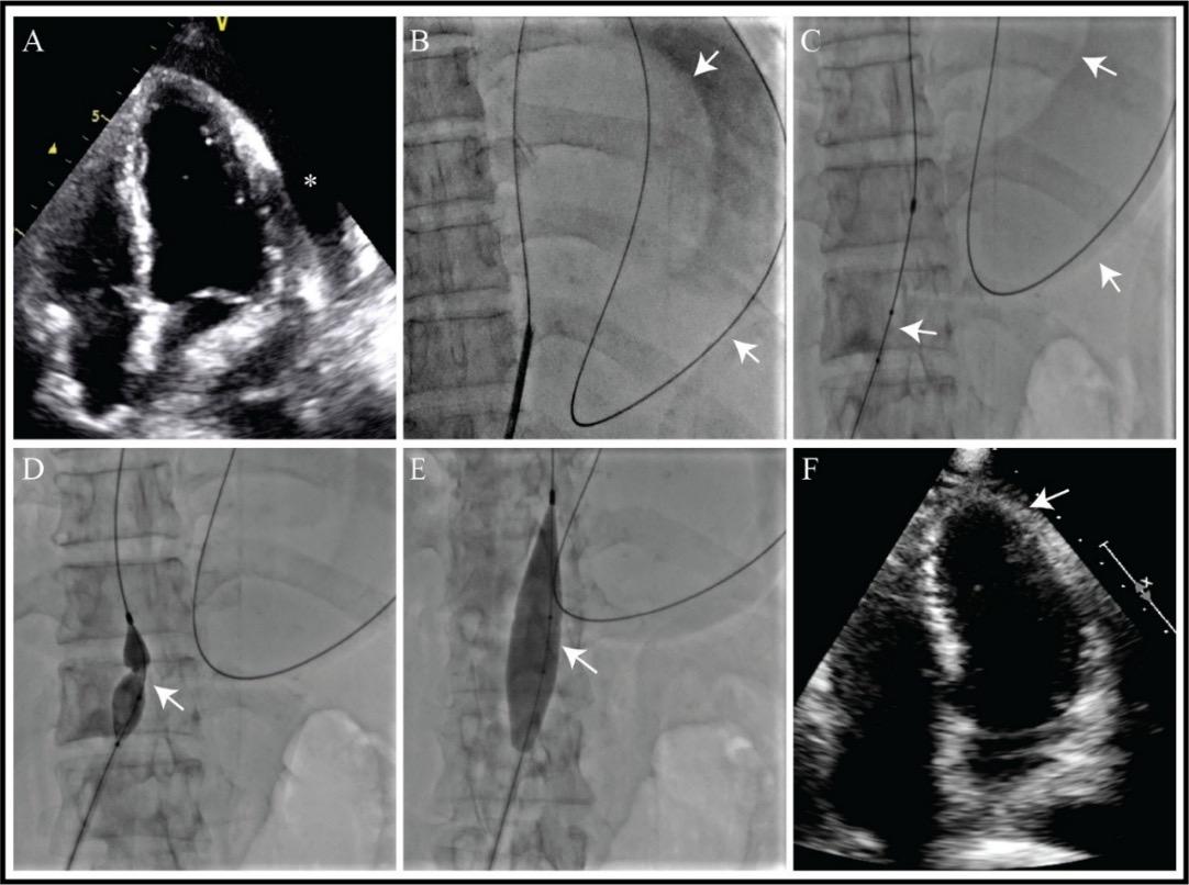
69 Percutaneous Balloon Pericardiotomy for Recurrent Haemodynamically Significant Malignant Pericardial Effusion: A Palliative Therapeutic Alternative Salzar C et al.
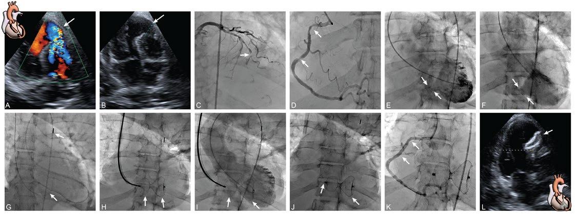
74 Emergency Transcatheter Closure of Post-Myocardial Infarction Ventricular Septal Rupture Using a Hyperion™ Occluder: A Multidisciplinary Case Report Salzar and Buitrago-Gomez
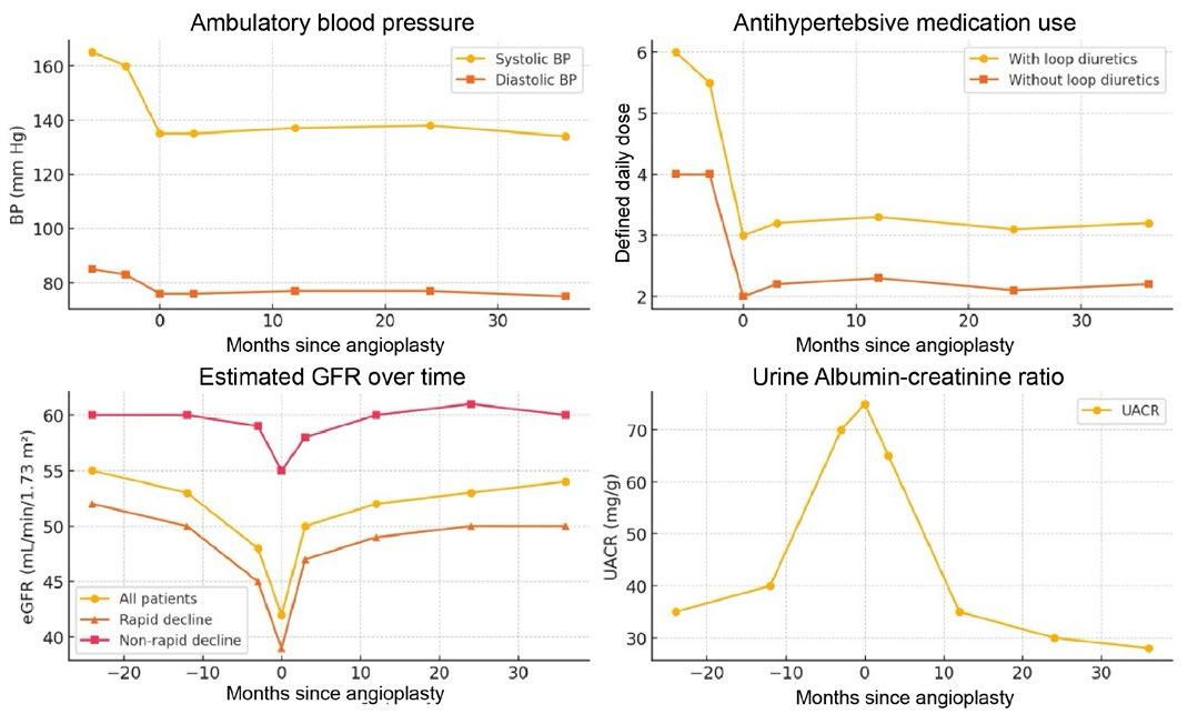
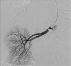
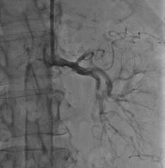
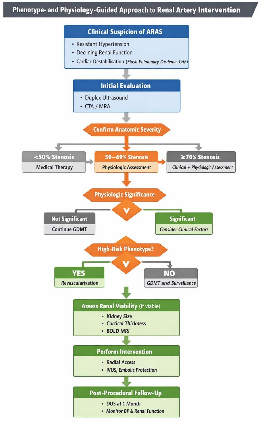
Renal Artery Intervention
Tafur and White
How Performance Metrics Influence Clinical Decisions
Lloyd W. Klein
Editorial Board
Editor-in-Chief
Dr Pablo Sepúlveda
Catholic University of Chile, Santiago, Chile
Interventional Cardiologist, Division of Cardiovascular Diseases, Endovascular Therapy Center, Catholic University of Chile, Santiago, Chile
European Prevention Center joint with Medical Center Düsseldorf (Grand Arc), Germany
Dr Sanjog Kalra
Einstein Medical Center Philadelphia, USA
Dr Aaron Kugelmass Baystate Health System, USA
Prof Eduard Margetic Clinical Hospital Center Zagreb, Croatia
Dr Gregory Pavlides
University of Nebraska Medical Center, USA
Prof Dr Rainer Wessely
Cologne and Fresenius University of Applied Sciences, Germany
Aims and Scope
EMJ Interventional Cardiology is an open access, peerreviewed ejournal committed to helping elevate the quality of practices in interventional cardiology globally by informing healthcare professionals on the latest research in the field.
The journal is published annually, six weeks after the EuroPCR Event, and features highlights from this event, alongside interviews with experts in the field, reviews of abstracts presented at EuroPCR, as well as in-depth features on sessions from this event. The journal also covers advances within the clinical and pharmaceutical arenas by publishing sponsored content from congress symposia, which is of high educational value for healthcare professionals. This undergoes rigorous quality control checks by independent experts and the in-house editorial team.
EMJ Interventional Cardiology also publishes peer-reviewed research papers, review articles, and case reports in the field. In addition, the journal welcomes the submission of features and opinion pieces intended to create a discussion around key topics in the field and broaden readers’ professional interests. The journal is managed by a dedicated editorial team that adheres to a rigorous double-blind peer-review process, maintains high standards of copy editing, and ensures timely publication.
EMJ Interventional Cardiology endeavours to increase knowledge, stimulate discussion, and contribute to a better understanding of practices in the field. Our focus is on research that is relevant to all healthcare professionals in this area. We do not publish veterinary science papers or laboratory studies not linked to patient outcomes. We have a particular interest in topical studies that advance knowledge and inform of coming trends affecting clinical practice in interventional cardiology.
Further details on coverage can be found here: www.emjreviews.com
Editorial Expertise
EMJ is supported by various levels of expertise:
• Guidance from an Editorial Board consisting of leading authorities from a wide variety of disciplines.
• Invited contributors who are recognised authorities in their respective fields.
• Peer review, which is conducted by expert reviewers who are invited by the Editorial team and appointed based on their knowledge of a specific topic.
• An experienced team of editors and technical editors.
Peer Review
On submission, all articles are assessed by the editorial team to determine their suitability for the journal and appropriateness for peer review.
Editorial staff, following consultation with either a member of the Editorial Board or the author(s) if necessary, identify three appropriate reviewers, who are selected based on their specialist knowledge in the relevant area.
All peer review is double blind. Following review, papers are either accepted without modification, returned to the author(s) to incorporate required changes, or rejected.
Editorial staff have final discretion over any proposed amendments.
Submissions
We welcome contributions from professionals, consultants, academics, and industry leaders on relevant and topical subjects. We seek papers with the most current, interesting, and relevant information in each therapeutic area and accept original research, review articles, case reports, and features.
We are always keen to hear from healthcare professionals wishing to discuss potential submissions, please email: editorial.assistant@emjreviews.com
To submit a paper, use our online submission site: https://emj.kriyadocs.com/submissions/submit/emj/emj/login
Submission details can be found through our website: www.emjreviews.com/contributors/authors
Reprints
All articles included in EMJ are available as reprints (minimum order 1,000). Please contact hello@emjreviews.com if you would like to order reprints.
Distribution and Readership
EMJ is distributed through controlled circulation to healthcare professionals in the relevant fields across Europe.
Indexing and Availability
EMJ is indexed on DOAJ, the Royal Society of Medicine, and Google Scholar®; selected articles are indexed in PubMed Central®
EMJ is available through the websites of our leading partners and collaborating societies. EMJ journals are all available via our website: www.emjreviews.com
Open Access
This is an open-access journal in accordance with the Creative Commons Attribution-Non Commercial 4.0 (CC BY-NC 4.0) license.
Congress Notice
Staff members attend medical congresses as reporters when required.
This Publication
Launch Date: 2013 Frequency: Yearly
Online ISSN: 2053-423X
All information obtained by EMJ and each of the contributions from various sources is as current and accurate as possible. However, due to human or mechanical errors, EMJ and the contributors cannot guarantee the accuracy, adequacy, or completeness of any information, and cannot be held responsible for any errors or omissions. EMJ is completely independent of the review event (EuroPCR 2026) and the use of the organisations does not constitute endorsement or media partnership in any form whatsoever. The cover photo is of Paris, France, the location of EuroPCR 2026.
Helena Bradbury, Katrina Thornber, Aleksandra Zurowska
Editorial Co-ordinators
Jess Nicholson, Bertie Pearcey, Alena Sofieva
Editorial Assistants
Niamh Holmes, Josh Warren Lister, Nonyelum Okonkwo, Roli Omamuli, Alexander Perkins
Creative Director
Tim Uden
Design Manager
Stacey White
Senior Designers
Owen Silcox, Tamara Kondolomo
Designers
Shanjok Gurung, Fabio van Paris
Junior Designers
Fraser Hoey, Helena Spicer, Caleb Wylie
Head of Marketing
Stephanie Corbett
Business Unit Lead
Kelly Byrne
Chief Executive Officer
Justin Levett
Chief Commercial Officer
Dan Healy
Founder and Chairman
Spencer Gore
Welcome
Dear Readers,
Welcome to the latest edition of EMJ Interventional Cardiology, featuring expert perspectives, clinical insights, and key developments from EuroPCR 2026 in Paris, France.
This year’s course centred on a theme central to interventional practice: complications. Through open discussion, case-based learning, and the sharing of real-world experience, the meeting reinforced a principle that has long defined EuroPCR: education saves lives. Reflecting the multidisciplinary nature of modern cardiovascular care, EuroPCR 2026 explored complex coronary intervention, structural heart disease, imaging, AI, and procedural planning.
In this issue, readers will find comprehensive congress coverage, including reviews of key sessions, late-breaking clinical trials, and notable research presented at the meeting. We also highlight EuroPCR’s award-winning abstracts and cases, showcasing emerging evidence and innovative approaches shaping the field.
Readers can gain insights from our interview with the EuroPCR Course Directors Nicolas Dumonteil and Thomas Cuisset, alongside PCRonline Editor-in-Chief Salvatore Brugaletta, who reflect on the meeting’s key themes, innovations, and future direction. Additional interviews with leading experts explore innovation, prevention, patient management, and the future of cardiovascular care.
This edition also includes a review of renal artery intervention, features on coronary revascularisation in transcatheter aortic valve replacement candidates and performance metrics in clinical decision-making, and two instructive case reports on ventricular septal rupture and recurrent malignant pericardial effusion.
We would like to thank our Editorial Board, authors, reviewers, interviewees, and contributors for their expertise, time, and continued support. We hope you find this edition informative, engaging, and valuable to your clinical practice.
Alexander Perkins Editorial Assistant
Editorial enquiries: editor@emjreviews.com
Sales opportunities: salesadmin@emjreviews.com
Permissions and copyright: accountsreceivable@emjreviews.com
Reprints: info@emjreviews.com
Media enquiries: marketing@emjreviews.com
Stay at the Forefront of Modern Medicine
High-level perspectives. Global experts. Essential updates.
Join us for conversations with the minds shaping healthcare's future. Gain the distilled insights you need to lead in your field and make maximum impact.
Jonathan Sackier: Non Executive Director & CMO, AiM Medical Robotics, Florida, USA
Saranya Ravindran: Paediatric Emergency Medicine Registrar, Imperial College Healthcare NHS Trust
Catherine Glass: Associate NHS GP and Senior Appraiser, NHS England
Foreword
Dear Colleagues,
It is my pleasure to introduce this latest edition of EMJ Interventional Cardiology, bringing readers comprehensive coverage and expert perspectives from EuroPCR 2026, held in Paris, France.
As one of the leading meetings in the cardiovascular calendar, EuroPCR continues to drive discussion around innovation, evidence-based practice, and the evolving future of patient-centred interventional care.
This year’s congress showcased the breadth and dynamism of the field, from complex coronary intervention and structural heart disease to advances in imaging, physiology, and multidisciplinary care. The programme reflected not only technological progress, but also the growing emphasis on clinical decision-making, procedural optimisation, and long-term patient outcomes.
Within this issue, readers will find coverage of many of the key themes and conversations emerging from EuroPCR 2026. Featured content includes a review of renal artery intervention, alongside an exploration of coronary revascularisation in transcatheter aortic valve replacement candidates and its evolving clinical implications. This edition also explores challenging clinical scenarios, including emergency transcatheter closure of postmyocardial infarction ventricular septal rupture and the use of percutaneous balloon pericardiotomy for recurrent malignant pericardial effusion.
Importantly, this issue also examines the broader realities shaping contemporary interventional practice. In a feature by Lloyd Klein, readers are invited to reflect on how performance metrics can influence clinical decision-making and patient care in modern healthcare systems.
In addition, readers will find a series of insightful interviews with leading voices in cardiology. We are delighted to feature conversations with Ramzi Khamis, Robert Kelly, and Ashok Seth, each offering unique perspectives on the intersection of intervention, prevention, innovation, and patient management. We also bring readers exclusive EuroPCR interviews with course directors Nicolas Dumonteil and Thomas Cuisset, as well as PCRonline Editor-in-chief Salvatore Brugaletta, who share their reflections on this year’s meeting and the future direction of the specialty.
EuroPCR continues to drive discussion around innovation, evidence-based practice, and the evolving future of patient-centred interventional care
Together, these contributions capture the spirit of EuroPCR 2026 and reflect the continued progress of interventional cardiology as a rapidly evolving and collaborative discipline.
I would like to extend my sincere thanks to all authors, reviewers, interviewees, and contributors whose expertise and dedication have made this issue possible. I hope readers find this edition both informative and engaging as we continue to advance innovation and excellence in interventional cardiology.
Pablo Sepúlveda
Catholic University of Chile, Santiago, Chile
EuroPCR 2026
Recognising that complications are encountered by all interventional cardiologists in daily practice, the directors emphasised the meeting's core message that "education saves lives"
Congress Review
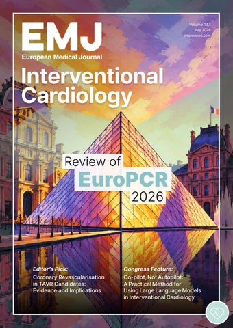
Review of EuroPCR 2026
Location: Paris, France
Date: 19th–22nd May 2026
Citation: EMJ Int Cardiol. 2026;14[1]:10-21. https://doi.org/10.33590/emjintcardiol/8605A680
THE ANNUAL world-leading course in interventional cardiology, EuroPCR, welcomed 12,147 participants to Paris, France, for a dynamic 4-day event at the Palais des Congrès. Bringing together interventional cardiologists, cardiac surgeons, imaging specialists, nurses, allied healthcare professionals, researchers, innovators, and industry representatives from around the globe, the course once again provided a unique platform for sharing expertise, advancing education, and improving patient care.
The scale and reach of EuroPCR 2026 reflected the continued growth of the global interventional community. This year’s programme featured more than 500 educational sessions, over 200 training workshops, 24 live cases transmitted from eight live centres, and 145 sessions streamed through the Course Platform. Supported by more than 1,100 faculty members, 1,500 presenters, 106 industry partners, and 76 international collaborations, the meeting delivered an extensive educational experience. The scientific programme was shaped by a record 3,494 submissions, highlighting the ongoing commitment of the community to innovation and evidence generation.
The Opening Ceremony was led by Course Directors Thomas Cuisset, APHM Hôpital La Timone Adultes, Marseille, France; Nieves Gonzalo, Hospital Clinico San Carlos, Madrid, Spain; Bernard Prendergast, Cleveland Clinic London, UK; and Nicolas Dumonteil, Clinique Pasteur, Toulouse, France, who joined live from Toulouse.
Together, they introduced the central theme, or ‘fil rouge’, of EuroPCR 2026: complications. Recognising that complications are encountered by all interventional cardiologists in daily practice, the directors emphasised the meeting’s core message that “education saves lives.” They highlighted the importance of openly sharing not only successes, but also challenges and complications, reinforcing EuroPCR’s longstanding culture of collective learning and continuous improvement.
Building on this theme, the directors outlined three principal objectives for the course: to better understand the mechanisms underlying complications, to improve strategies for their prevention, and to optimise their management when they occur. Ultimately, these goals are aimed at enhancing patient outcomes across the spectrum of cardiovascular interventions.
The ceremony also paid tribute to the pioneers whose vision helped shape EuroPCR into the world-renowned
course it is today. Jean Marco, founder of the original course established in 1989, was present during the ceremony and was recognised alongside Jean Fajadet, Clinique Pasteur, Toulouse, France; and William Wijns, The Lambe Institute for Translational Medicine and Curam, Galway, Ireland, for their enduring contributions to the field and to the EuroPCR community. The directors reflected on the importance of continuing the work of those who came before, while fostering a collaborative environment in which knowledge, experience, and expertise can be shared across generations of practitioners.
Ultimately, these goals are aimed at enhancing patient outcomes across the spectrum of cardiovascular interventions
Several important additions enriched the EuroPCR 2026 programme. A new Calcium Skills Lab offered participants a comprehensive exploration of calcified coronary artery disease, covering diagnosis, pathophysiology, imaging, treatment strategies, and complication management. A dedicated chronic total occlusion programme was also introduced, featuring live cases, abstract presentations, and case-based discussions focused
on chronic total occlusion interventions and their broader relevance to complex percutaneous coronary intervention practice. Innovation remained a central focus, with the launch of a new Innovation Track designed to create continuity between Innovators Day and the wider EuroPCR programme, encompassing advances in interventional technologies, imaging, and AI. Complementing this initiative, attendees were able to visit a dedicated AI Lab for the first time, providing hands-on opportunities to explore the growing role of AI in cardiovascular intervention.
Among the scientific highlights of the meeting were three major late-breaking trials, selected by the Course Directors for their potential to influence practice worldwide. These included studies examining coronary revascularisation in patients undergoing transcatheter aortic valve implantation, outcomes following left atrial appendage closure versus direct oral anticoagulants across different age groups in patients with atrial fibrillation, and longterm mortality following revascularisation for left main coronary artery disease.
Read on for key insights from EuroPCR 2026 and the latest advances shaping the future of interventional cardiology.
LAAC Shows Comparable Outcomes to DOAC Across Age Groups in AF
NEW data from a prespecified subgroup analysis of the CHAMPION-AF trial presented at EuroPCR 2026 showed that left atrial appendage closure (LAAC) demonstrated similar efficacy to direct oral anticoagulant (DOAC) therapy across different age groups in patients with atrial fibrillation (AF), while significantly reducing non-procedural bleeding events.1
AF is one of the most common cardiac rhythm disorders and is associated with a substantially increased risk of stroke. Oral anticoagulants remain the standard approach for stroke prevention, but longterm treatment can increase bleeding risk and may not be suitable for all patients. LAAC has emerged as an alternative strategy that mechanically seals the left atrial appendage, a major source of clot formation in AF.
Earlier this year, the main CHAMPION-AF trial reported that device-based LAAC was non-inferior to DOAC therapy for the composite endpoint of cardiovascular death, stroke, or systemic embolism, while offering superior safety for non-procedurerelated bleeding. This new subgroup analysis evaluated whether patient age influenced these outcomes.
The analysis included 1,915 patients younger than 75 years and 1,085 patients aged 75 years or older. Investigators compared the incidence of cardiovascular death, stroke, or systemic embolism over 3 years between patients treated with LAAC and those receiving DOAC therapy.
Results showed that the primary efficacy endpoint was similar between treatment groups regardless of age. In patients younger than 75 years, the hazard ratio (HR) for LAAC versus DOAC was 1.07 (95% CI: 0.67–1.71; p=0.7789). Among patients aged 75 years or older, the HR was 1.34 (95% CI: 0.85–2.12; p=0.2036), with no significant interaction between age and treatment effect.
36%
In patients younger than 75 years, bleeding risk was reduced by 36% with LAAC compared with DOAC therapy (HR: 0.64; 95% CI: 0.50–0.82; p<0.0001)
Importantly, LAAC was associated with significantly lower rates of non-procedural major and clinically relevant non-major bleeding in both age groups. In patients younger than 75 years, bleeding risk was reduced by 36% with LAAC compared with DOAC therapy (HR: 0.64; 95% CI: 0.50–0.82; p<0.0001). Similar findings were observed in older patients (HR: 0.68; 95% CI: 0.51–0.91; p=0.0002).
Although intention-to-treat analysis suggested a higher incidence of ischaemic stroke with LAAC, this difference was no longer significant among patients who received their assigned treatment as intended. Rates of disabling ischaemic stroke remained very low and comparable across treatment groups and age categories.
The findings suggest that age alone should not exclude otherwise suitable patients from consideration for LAAC. However, investigators noted that subgroup analyses have inherent limitations, including reduced statistical power, and emphasised that decisions between LAAC and DOAC therapy should continue to be individualised
Coronary Revascularisation May Improve Outcomes After TAVI
NOVEL data from a patient-level meta-analysis, presented at EuroPCR 2026, suggest that coronary revascularisation may offer a modest clinical benefit for patients with severe aortic stenosis and coronary artery disease undergoing transcatheter aortic valve implantation (TAVI).2
The analysis pooled individual participant data from four contemporary RCTs that evaluated percutaneous coronary intervention (PCI) against optimal medical treatment in patients undergoing TAVI. Although the trials differed in design, all addressed ongoing uncertainty surrounding the role of coronary revascularisation in this population and the most appropriate method for assessing coronary lesions.
A total of 1,050 patients were included. Among them, 439 underwent fractional flow reserve-guided PCI, 255 underwent angiography-guided PCI, and 356 received optimal medical treatment alone.
No difference was observed in net adverse clinical events between PCI and optimal medical treatment
The primary endpoint was major adverse cardiac events at 1 year, defined as all-cause death, myocardial infarction, any coronary revascularisation, and stroke. A co-primary endpoint of net adverse clinical events included major adverse cardiac events plus major bleeding.
Overall, PCI was associated with a lower risk of major adverse cardiac events compared with optimal medical treatment (hazard ratio [HR]: 0.70; 95% CI: 0.49–0.99). This reduction was primarily driven by a lower risk of repeat coronary revascularisation: (HR: 0.34; 95% CI: 0.14–0.80).
However, no difference was observed in net adverse clinical events between PCI and optimal medical treatment.
When analysed according to assessment strategy, fractional flow reserveguided PCI was associated with lower risks of both major adverse cardiac events (HR: 0.58; 95% CI: 0.37–0.91) and net adverse clinical events (HR: 0.68; 95% CI: 0.51–0.90) compared with optimal medical treatment.
In contrast, angiographyguided PCI showed no significant differences in either endpoint compared with optimal medical treatment. Rates of major bleeding were 8.2% with fractional flow reserve-guided intervention, 13.7% with angiography-guided intervention, and 12.6% with optimal medical treatment.
Overall, PCI was associated with a lower risk of major adverse cardiac events compared with optimal medical treatment (hazard ratio: 0.70; 95% CI: 0.49–0.99)
These data represent the most comprehensive patient-level assessment to date of coronary revascularisation in patients with severe aortic stenosis and coronary artery disease undergoing TAVI. The findings suggest that routine intervention may offer limited benefit, while a selective physiology-informed approach could improve clinical outcomes.
Prognostic Impact of IVUS-Guided Left Main PCI at 10 Years
RESEARCH
presented at EuroPCR 2026 has demonstrated a long-term analysis from the NOBLE trial examining whether intravascular ultrasound (IVUS) guidance influences outcomes following percutaneous coronary intervention (PCI) for left main coronary artery (LMCA) disease.3
The NOBLE trial enrolled 1,201 patients with unprotected LMCA disease across 36 centres in Northern Europe between 2008–2015. Patients were randomised to PCI or coronary artery bypass graft (CABG), with IVUS strongly recommended during PCI procedures. For this analysis, investigators compared outcomes among patients treated with CABG (n=574), PCI with post-procedural IVUS (n=443), and PCI without post-procedural IVUS (n=160). The primary endpoint was all-cause mortality at 10 years, while secondary endpoints included major adverse cardiac and cerebrovascular events (MACCE) at 5 years.
Propensity-adjusted Cox regression models were used to account for baseline differences.
Complete 10-year mortality follow-up was available for 98% of participants. In crude analyses, mortality was significantly higher among patients who underwent PCI without IVUS compared with both CABG and IVUS-guided PCI (33.9% versus 23.3% and 20.6%, respectively; p=0.0047). After adjustment, mortality remained comparable between IVUS-guided PCI and CABG (hazard ratio [HR]: 0.85; 95% CI: 0.68–1.15), whereas PCI without IVUS was associated with numerically higher mortality relative to CABG (HR: 1.33; 95% CI: 0.98–1.81; overall treatment effect: p=0.031).
For this analysis, investigators compared outcomes among patients treated with CABG (n=574), PCI with post-procedural IVUS (n=443), and PCI without post-procedural IVUS (n=160)
those undergoing PCI without IVUS had a 59% higher risk (HR: 1.59; 95% CI: 1.12–2.26; p=0.0025).
Among 224 patients with a core laboratory assessment of minimum stent area (MSA), median LMCA MSA was 13.5 mm2. No significant association was observed between larger post-procedural MSA and long-term mortality, suggesting that factors beyond stent expansion alone may contribute to the prognostic benefit associated with IVUS use.
Despite these mortality findings, CABG remained superior for the composite MACCE endpoint. Compared with CABG, patients treated with IVUS-guided PCI experienced a 52% higher risk of MACCE (HR: 1.52; 95% CI: 1.17–1.98), while
52%
Compared with CABG, patients treated with IVUS-guided PCI experienced a 52% higher risk of MACCE
These findings reinforce the growing evidence supporting intracoronary imaging during complex coronary interventions. While CABG continued to provide superior protection against MACCE, IVUS-guided PCI achieved comparable long-term survival, whereas omission of IVUS was associated with worse mortality outcomes. The results highlight the importance of routine imaging guidance when performing PCI in patients with unprotected LMCA disease, and further support IVUS as a key component of contemporary left main intervention.
TAVR Versus Surgical Aortic Valve Replacement in Patients on Dialysis
SELECTING the optimal aortic valve replacement strategy for patients receiving dialysis remains particularly challenging due to their elevated procedural risk and reduced life expectancy. A large real-world analysis comparing transcatheter aortic valve replacement (TAVR), bioprosthetic surgical aortic valve replacement (SAVR), and mechanical SAVR in patients with end-stage renal disease requiring dialysis was presented at EuroPCR 2026.4
Using data from the United States Renal Data System (USRDS), investigators conducted a retrospective analysis of patients undergoing isolated aortic valve replacement between 2012–2020. The study included 13,527 patients, comprising 8,948 who underwent TAVR, 2,938 who received a bioprosthetic surgical valve, and 1,641 who underwent mechanical SAVR. To minimise baseline differences between groups, inverse probability of treatment weighting was applied, achieving a good balance across most demographic and clinical characteristics.
TAVR was associated with clear advantages in the perioperative period. In both unmatched and weighted analyses, patients undergoing TAVR experienced significantly lower intraoperative mortality compared with either surgical approach. Following adjustment, intraoperative mortality was 2.8% with TAVR, compared with 6.4% for bioprosthetic SAVR and 6.5% for mechanical SAVR (p<0.01). TAVR recipients also demonstrated lower rates of prolonged mechanical ventilation and substantially shorter ICU stays, with a median ICU stay of just 1 day compared with 5 days for both surgical groups.
Despite these early benefits, long-term outcomes favoured surgery. Kaplan–Meier analyses revealed significantly higher all-cause mortality among patients treated with TAVR compared with either form of SAVR, a finding that remained significant after adjustment. Similar results were observed when kidney transplantation was included as a competing risk, suggesting that the survival
TAVR was associated with clear advantages in the perioperative period. In both unmatched and weighted analyses
advantage associated with surgery was robust across analytical approaches.
The investigators also evaluated cerebrovascular and bleeding outcomes. Although stroke rates initially appeared lower in the TAVR cohort, this difference was no longer significant after adjustment for baseline characteristics. In contrast, patients receiving mechanical valves experienced significantly higher bleeding rates than those treated with either TAVR or bioprosthetic SAVR, both before and after inverse probability of treatment weighting adjustment.
These findings highlight the complex balance between procedural safety and long-term durability when selecting valve replacement strategies in patients who are dependent on dialysis. While TAVR offered lower perioperative mortality, reduced respiratory complications, and shorter ICU stays, these short-term advantages did not translate into superior long-term survival. The authors suggest that TAVR may be most appropriate for patients with limited life expectancy or prohibitive surgical risk, whereas younger and fitter patients receiving dialysis may derive greater long-term benefit from surgical valve replacement.
wall strain in patients with ST-segment elevation myocardial infarction (STEMI) may help identify high-risk non-culprit lesions
associated with future target-lesion failure, according to a retrospective study presented at EuroPCR.5 The findings suggest that angiographyderived radial wall strain provides incremental prognostic value beyond standard anatomical and functional assessments.
The researchers aimed to evaluate the prognostic utility of angiography-derived radial wall strain in predicting adverse events in untreated mild-to-moderate non-culprit lesions in patients with STEMI. Radial wall strain is a novel index of plaque biomechanical deformation that reflects the mechanical stress experienced by coronary plaques.
The primary endpoint was target-lesion failure, defined as a composite of cardiac death, target-lesion myocardial infarction, and unplanned target-lesion revascularisation.
A total of 403 untreated non-culprit lesions from 243 patients with STEMI were included in the analysis. Over a median follow-up period of 3.9 years, target-lesion failure occurred in 33 lesions (8%).
Lesions with a maximum radial wall strain of ≥13% demonstrated a significantly higher incidence of target-lesion failure compared with lesions below
this threshold (26.0% versus 3.0%; p<0.001). Furthermore, maximum radial wall strain independently predicted target-lesion failure, with an adjusted hazard ratio of 1.46 (95% CI: 1.32–1.61; p<0.001).
The investigators also reported that radial wall strain showed good discriminative performance, with an area under the curve of 0.79 (95% CI: 0.69–0.89; p<0.001).
Importantly, radial wall strain provided significant incremental prognostic value over both quantitative coronary angiography stenosis assessment and Murray’s law-based quantitative flow ratio.
The authors concluded that radial wall strain may improve risk stratification in patients with STEMI by identifying nonculprit lesions at increased risk of future adverse events. They suggested this approach could help guide intensified secondary prevention strategies and support more personalised management following STEMI.
The findings suggest that angiographyderived radial wall strain provides incremental prognostic value beyond standard anatomical and functional assessments
Aorto-Ostial RCA CTO PCI: A High-Risk, Low-Success Subset
PERCUTANEOUS coronary intervention (PCI) of aorto-ostial right coronary artery (RCA) chronic total occlusions (CTO) is considered one of the most technical challenges in interventional cardiology practice. Outcomes are associated with significantly lower procedural success and higher rates of adverse events compared with non-ostial RCA CTOs. One of the largest contemporary analyses of this complex lesion subset to date, presented at EuroPCR 2026, evaluated outcomes from 26,374 patients undergoing RCA CTO PCI between 2016–2024.6
Of the patients evaluated, 2,850 presented with aorto-ostial lesions. Technical success was achieved in 79.6% of aorto-ostial RCA CTO procedures, a significantly lower rate than the 89.7% observed in non-aorto-ostial lesions (p<0.001). On multivariable analysis, an aorto-ostial location emerged as an independent predictor of procedural failure (adjusted odds ratio (OR): 1.77; p<0.001), reinforcing the unique anatomical and procedural constraints posed by these lesions.
Findings also pointed to a significantly higher incidence of major adverse cardiovascular events in the aorto-ostial cohort compared with non-ostial RCA CTO PCI (4.0% versus 2.7%; p=0.011).
Several lesion- and patient-specific characteristics were independently associated with technical failure in the aorto-ostial subgroup. These included a stumpless proximal cap (adjusted OR: 2.58), severe calcification (adjusted OR: 2.00), moderate-to-severe vessel tortuosity (adjusted OR: 2.24), and prior coronary artery bypass grafting (adjusted OR: 1.45).
During the presentation, the authors highlighted that current CTO scoring systems may not adequately capture the specific anatomical complexity of aorto-ostial lesions, and suggested that the development of a dedicated scoring approach could help improve procedural planning and risk stratification in this challenging subset.
Despite advances in contemporary CTO techniques, the absence of a clear proximal cap, extensive calcium burden and challenging vessel geometry continue to limit crossing success. Together, the findings underscore the importance of dedicated equipment, operator experience, and precise lesion assessment in patients presenting with aorto-ostial RCA CTOs.
Published data in this patient population remain limited. Hence, this study adds important large-scale evidence to the field with further characterisation of a lesion subset that continues to present significant procedural challenges in complex CTO PCI.
Technical success was achieved in 89.7%
of aorto-ostial RCA CTO procedures, a significantly lower rate than the observed in non-aorto-ostial lesions (p<0.001) 79.6%
Myocardial Bridging May Warrant Provocation Testing in Suspected INOCA
MYOCARDIAL bridging, a congenital anatomical variant in which a coronary artery travels through the myocardium rather than over its surface, has traditionally been regarded as benign, but emerging evidence presented at EuroPCR 2026 suggests it may contribute to abnormal coronary vasomotion and vasospastic symptoms in patients with ischaemia and no obstructive coronary artery disease (INOCA).7
The research assessed whether myocardial bridging independently associates with vasospastic angina in patients with INOCA undergoing invasive coronary assessment, finding that it more than doubled the risk of epicardial coronary spasm.
The authors analysed data from 1,118 consecutive patients enrolled in an international multicentre cohort study who underwent protocolised invasive coronary endotyping. All participants received coronary angiography, physiological assessment, and standardised intracoronary acetylcholine provocation testing. Myocardial bridging was identified angiographically, and the primary outcome was epicardial coronary spasm during provocation testing.
Myocardial bridging was identified in 11.3% patients overall. Among 1,088 evaluable patients, epicardial coronary spasm occurred in 17.4%. Rates of epicardial spasm were substantially higher in patients with myocardial bridging compared with those without bridging (31.1% versus 15.2%; p<0.001).
These findings support an association between myocardial bridging and vasospastic angina in patients with INOCA, with potential implications for routine clinical assessment across European clinics
cardiovascular risk factors were not independently significant.
These findings support an association between myocardial bridging and vasospastic angina in patients with INOCA, with potential implications for routine clinical assessment across European clinics. Clinicians who identify myocardial bridging during angiography may wish to consider coronary provocation testing in patients with suspected vasospastic symptoms, as recognition of an underlying vasomotor disorder could guide targeted therapy and improve symptom management.
In the fully adjusted regression analysis of 939 complete cases, myocardial bridging remained independently associated with epicardial coronary spasm, with an odds ratio of 2.59 (95% CI: 1.62–4.16; p<0.001). Average marginal effects analysis demonstrated that myocardial bridging increased the absolute probability of spasm by 12.7%. Notably, myocardial bridging emerged as the strongest predictor of epicardial spasm, while traditional
Rates of epicardial spasm were substantially higher in patients with myocardial bridging compared with those without bridging (31.1% versus 15.2%; p<0.001)
In terms of limitations, the study’s observational design prevents conclusions regarding causality, and myocardial bridging was identified angiographically rather than with advanced imaging modalities, which may underestimate prevalence. Nevertheless, the large multicentre cohort and standardised invasive testing strengthen the relevance of the findings to clinical practice.
Operator Adherence to OCT Guidance Linked to Lower MACE
NEW data from the ILUMIEN-V AERO study, presented at EuroPCR 2026, suggest that the clinical benefit of optical coherence tomography (OCT)-guided percutaneous coronary intervention (PCI) depends not only on the use of intracoronary imaging, but on how effectively operators act on the information it provides.8
This analysis evaluated whether adherence to OCT-guided decision-making was associated with improved clinical outcomes following PCI. ILUMIEN-V AERO is a prospective, multicentre observational study that was conducted across 17 centres in Germany and the UK, enrolling 708 patients with 761 lesions treated using OCT-guided PCI.
Investigators assessed operator performance using a 10-component adherence index covering key stages of OCT-guided PCI, including image quality,
Among 63 participating operators, the median adherence index was 64%. Highadherence operators treated 444 patients, while low-adherence operators treated 264 patients. Baseline lesion complexity and vessel characteristics were comparable between groups.
At 6 months, patients treated by highadherence operators experienced significantly lower rates of major adverse cardiac events, defined as myocardial infarction, target vessel revascularisation, or all-cause death. Major adverse cardiac events occurred in 1.8% of patients treated by high-adherence operators compared with 6.1% among those treated by low-adherence operators, corresponding to an adjusted hazard ratio of 0.31 (95% CI: 0.17–0.56; p<0.001).
Major adverse cardiac events occurred in 1.8% of patients treated by high-adherence operators compared with 6.1% among those treated by low-adherence operators, corresponding to an adjusted hazard ratio of 0.31 (95% CI: 0.17–0.56; p<0.001)
Notably, the greatest differences between groups were observed in adherence to OCT-guided landing-zone selection, achievement of post-PCI optimisation, and agreement with core laboratory assessment of final PCI results. Despite these outcome differences, minimum stent area was similar between groups, suggesting that procedural success was not explained by stent expansion metrics alone.
The findings indicate that active interpretation of OCT findings and subsequent procedural decision-making may be critical determinants of clinical benefit. According to the
References
1. Nielsen-Kudsk JE et al. Outcomes with left atrial appendage closure vs. direct oral anticoagulants in patients with atrial fibrillation across different age groups. Presentation. EuroPCR, 19-22 May, 2026.
2. Scarsini R et al. Coronary revascularisation in patients undergoing TAVI: an IPD meta-analysis of four randomised trials. Presentation. EuroPCR, 19-22 May, 2026.
The findings indicate that active interpretation of OCT findings and subsequent procedural decisionmaking may be critical determinants of clinical benefit
investigators, the results support the concept that successful OCT-guided PCI requires meaningful engagement with imaging data rather than simply performing OCT or targeting optimisation thresholds.
3. Nielsen HE et al. Prognostic impact of IVUS during left main PCI versus coronary artery bypass grafting at 10 years. Abstract A100640EH. EuroPCR, 19-22 May, 2026.
4. Tagliafierro M et al. Nationwide long-term outcomes of aortic valve replacement in US dialysis patients: TAVR vs SAVR. Abstract A96408MT. EuroPCR, 19-22 May, 2026.
5. Fezzi S et al. Prognostic implications of angiography-derived radial wall strain in non-culprit lesions of STEMI. Abstract A102596SF. EuroPCR, 19-22 May, 2026.
6. Skalidis I et al. Aorto-ostial RCA CTO PCI: technical outcomes and predictors of failure from ERCTO registry. Abstract A98233IS. EuroPCR, 19-22 May, 2026.
7. Sykes R et al. Myocardial bridge is associated with epicardial coronary spasm. Abstract A96845RS. EuroPCR, 19-22 May, 2026.
8. Nitin CM et al. Operator adherence to OCT-guided PCI metrics and outcomes: insights from ILUMIEN-V AERO. Abstract A102553NC. EuroPCR, 19-22 May, 2026.
Co-pilot, Not Autopilot: A Practical Method for Using Large Language Models in Interventional Cardiology
Authors: *Louis-Marie Desroche,¹ Christopher Cook,² ³ Thomas Modine⁴
1. Interventional Cardiology Unit, University Hospital of La Réunion, SaintDenis, France
2. The Essex Cardiothoracic Centre, Basildon, UK
3. Anglia Ruskin University, Cambridge, UK
4. Department of Cardiac Surgery, CHU de Bordeaux, Haut-Lévêque Hospital, France
*Correspondence to louis-marie.desroche@chu-reunion.fr
Disclosure:
Cook has served as a consultant for Edwards, Abbott, and Philips; and holds equity in Viz.ai and cerebria.ai. Modine reports receiving grants/ contracts and consulting fees from Abbott, Edwards, Medtronic, and Jenscare; advisory board participation for Abbott, Edwards, Medtronic, and Jenscare; and patents planned, issued or pending; leadership/fiduciary roles; and stock/stock options in Versa Vascular. Desroche has declared no conflicts of interest.
Acknowledgements: This feature is based on the AI Lab session ‘AI fundamentals for busy cardiologists’, presented at EuroPCR 2026, Paris, France.
Keywords:
AI, clinical decision support, Heart Team, interventional cardiology, large language models (LLM), prompt engineering.
Citation: EMJ Int Cardiol. 2026;14[1]:22-25. https://doi.org/10.33590/emjintcardiol/0I89LD76
WHEN A LARGE language model (LLM) gives a cardiologist a poor answer, it is not always the model that is the only problem. More often than we care to admit, it is the briefing (or ‘prompt’) that is poor. Put simply, the way we ask a question shapes the answer that we get, and most of us were never taught how to ask. This is not a fringe concern; these tools are already in daily use, whether that be for discharge letters, guideline checks, or translation. Indeed, the adoption of LLMs applied to everyday clinical tasks has proliferated faster than anyone has taught us to use them. Furthermore, the evidence base for the accuracy of LLMs does not seem to match the enthusiasm of its adopters. A large systematic review of 519 studies found that only 5% used real patient-care data; 44.5% tested examination-style knowledge and 84.2% addressed question-answering.1 Some LLMs reach passing or near-passing scores on selected cardiology board-style examinations; however, this reflects ‘exam’ evidence, not ‘bedside’ evidence. Accordingly, we have a tool that is already in widespread use, but with limited proof of appropriateness at the bedside, compounded by no shared method for using it optimally. This article offers one practical way to narrow that gap.
WHAT ACTUALLY IS A LLM, AND THE DISTINCTION THAT MATTERS
In simplified terms, a LLM is a very large ‘autocomplete’; the same predictive-text
mechanism that suggests the next word on your mobile phone, but scaled up tremendously. Trained on much of the public internet, LLMs have learned one task; specifically, given a sequence of words,
to predict probabilistically the next most likely word to follow. Therefore, contrary to many people’s belief or understanding, there is no medical ‘brain’ intrinsic to LLMs. Furthermore, there is no causal reasoning, no awareness of your patient, and unless it is connected to retrieval or external tools, its built-in knowledge is limited to what it was exposed to during its training. Scaled massively, that simple mechanism is impressive, indeed capable of passing cardiology board examinations, but fluency in language does not equate to accuracy of language.
For our specialty, one key distinction is decisive. Two different technologies travel under the umbrella label of ‘AI in cardiology’. Task-specific deep learning, the networks behind AI-ECG and AI-echocardiography, is regulatory-cleared and has been validated in large cohorts and randomised trials. For example, the AI-ECG screen for low ejection fraction was tested in a pragmatic randomised trial of 22,641 patients.2 However, LLMs have not been tested or validated anywhere near as robustly.
Indeed, to the authors’ knowledge, no LLMbased cardiology decision-support system
has yet been CE-marked or FDA-cleared for autonomous clinical decision-making. Acknowledging this, the EU AI Act classifies medical AI as ‘high-risk’, which mandates human oversight. As such, the clinician remains entirely responsible for the decision.
WHERE THE EVIDENCE STANDS: CO-PILOT, NOT AUTOPILOT
One of the few controlled signals in cardiology so far comes from a randomised comparison in which the same general cardiologists managed complex cardiomyopathy cases twice, once alone and once assisted by an LLM. Blinded subspecialists preferred the LLM-assisted assessments (46.7% versus 32.7%) and found fewer clinically significant errors (13.1% versus 24.3%).3 Crucially, the cardiologist stayed in the loop: the model was a co-pilot, and the autopilot was never tested.
The way we ask a question shapes the answer that we get, and most of us were never taught how to ask
But availability is not integration, and a randomised trial proves it. Physicians given access to a leading LLM scored no better than those using conventional resources (76% versus 74%; adjusted difference: 2 percentage points; 95% CI: -4–8), even though the model alone outscored both groups by 16 points.4 Worse, when an LLM is confidently wrong, clinicians can be dragged down with it, even AI-literate ones.5 The lesson is uncomfortable: the hardest skill is perhaps to overrule the model when it is confidently wrong.
THE LEVER IS CONTEXT, AND THE METHOD IS A BRIEFING
The conceptual shift of 2026 is from polishing the wording of a prompt to engineering the context around it. The most interventional-native evidence we have makes the point directly: across 20 complex coronary and structural cases scored by five blinded interventional cardiologists (600 evaluations), the same model scored 6.9/10 by default, 7.8 with web search enabled, and 7.7 with European guidelines supplied in the prompt.6 Same question, richer context, better-rated answers.
With one and the same model, how you brief it determines the answer you get, and thus how safely you can act upon it
A useful mental model is a brilliant Nobel laureate newly arrived at your hospital, who has read every textbook but has never met your patient, does not know your centre, and does not know your local pathways. That is the LLM. A prompt is, therefore, a clinical briefing; the same kind you give a fellow before a complex case. If you would not brief the laureate that way, do not brief the model that way.
The briefing has a three-pillar structure: frame, ground, and verify, each supported by evidence and undone by a concrete failure (Table 1).
Frame is how you formulate the question. Across 300 simulated hypertension cases, the model-and-prompt configuration shifted decision accuracy from 63% to 91%, and some poorly designed configurations even dragged physicians below their own baseline.7
Ground is the patient context you supply. Given structured Heart Team data on 150 patients with severe aortic stenosis, ChatGPT (OpenAI, San Francisco, California, USA) matched the actual Heart Team decision 77% of the time, and 90% for transcatheter implantation.8
Verify is the format that lets you check the reasoning. In a randomised study of radiologists, step-by-step LLM explanations improved diagnostic accuracy by 12.2 points compared with no LLM support, and by 7.2 points compared with a standard answer giving no explanation,9 because a reasoning chain you can see is one you can challenge.
Across all three pillars, the message is the same: with one and the same model, how you brief it determines the answer you get, and thus how safely you can act upon it. These levers also outlast any single model: framing the problem, grounding it in context, and demanding a verifiable format do not date.
A RESEARCH AGENDA FOR THE INTERVENTIONAL COMMUNITY
The honest limitation that runs through almost all of the aforementioned studies is that it is scenario-based, not bedside. Specifically, the interventional comparison6 used cases, not real patients; the hypertension7 and Heart Team8 studies were simulations or retrospective. A pragmatic randomised trial of LLM assistance in real interventional workflows has never been done. Herein lies the opportunity for the European interventional community. A multicentre, pragmatic trial, embedding an LLM in Heart Team preparation or peri-procedural decision support, with a human in the loop, retrieval grounded in local and European guidelines, and a documented audit trail consistent
Table 1: Frame, ground, verify: briefing the co-pilot.
Pillar What it means/what to ask yourself A plausible clinical failure when it is skipped
Frame Define the decision, the patient, and the objective before anything else. What decision, for whom, to what end, and could the premise itself be wrong?
Ground Supply the relevant context: comorbidities, current drugs, prior imaging, files, and your local and European pathways.
Verify Demand sources, assumptions, a counterargument, and a reasoning chain you can actually inspect and challenge.
Asking ‘which statin dose?’ when perhaps no statin is indicated. The model answers the premise it is given; it does not challenge it.
Omitting HIV infection on boosted antiretroviral therapy because it ‘felt irrelevant’, yet interactions and risk category change the LDL management.
Acting on a confident answer without checking which guideline, and which version, it rests on; especially dangerous where the recommendation has recently changed.
A practical three-step structure for any clinical query to a large language model. LDL: low density lipoprotein.
with the EU AI Act, would move us from exam evidence to the bedside evidence we actually need.
Until then, the message is simple. Use these tools for the tasks where they already help: knowledge retrieval, structuring reports, drafting discharge letters, and preparing the
References
1. Bedi S et al. Testing and evaluation of health care applications of large language models: a systematic review. JAMA. 2025;333(4):319-28.
2. Yao X et al. Artificial intelligenceenabled electrocardiograms for identification of patients with low ejection fraction: a pragmatic, randomized clinical trial (EAGLE). Nat Med. 2021;27(5):815-9.
3. O'Sullivan JW et al. A large language model for complex cardiology care. Nat Med. 2026;32(2):616-23.
4. Goh E et al. Large language model influence on diagnostic reasoning: a randomized clinical trial. JAMA Netw Open. 2024;7(10):e2440969.
5. Qazi I et al. Automation bias in large language model–assisted diagnostic
Heart Team. Always with review, and as a supervised component of care rather than an autonomous decision-maker.10 Treat the model as a co-pilot, never an autopilot, and learn to brief it: frame, ground, and verify. The model will change every 6 months; the discipline of asking the right question, and doubting the answer, is what lasts.
reasoning among physicians trained in AI literacy: a randomized clinical trial. NEJM AI. 2026;DOI:10.1056/ AIoa2501001.
6. Lauretti A et al. Performance of large language models in interventional cardiology: the ILLUMINATE blinded model-comparison study. J Invasive Cardiol. 2026;DOI:10.25270/ jic/25.00104.
7. Li Z et al. The effects of multitype prompt engineering for large language models in hypertension treatment decisions. npj Digit Med. 2026;DOI: 10.1038/s41746-026-02645-y.
8. Salihu A et al. A study of ChatGPT in facilitating Heart Team decisions on severe aortic stenosis. EuroIntervention. 2024;20(8):e496-503.
9. Spitzer P et al. The effect of medical explanations from large language models on diagnostic accuracy in radiology. NPJ Digit Med. 2026;9(1):333.
10. Desroche LM et al. Artificial intelligence in cardiovascular care for internal medicine: from promising algorithms to useful clinical services. Eur J Intern Med. 2026;DOI:10.1016/j. ejim.2026.106944.
Author:
All You Need to Know About Vulnerable Plaque
Alex Perkins, EMJ, London, UK
Citation: EMJ Int Cardiol. 2026;14[1]:26-29. https://doi.org/10.33590/emjintcardiol/Q28ZLI0F
AT EUROPCR 2026, the session ‘All We Need to Know About Vulnerable Plaque’, chaired by Salvatore Brugaletta, Hospital Clínic de Barcelona, Spain, explored one of the most important unresolved questions in contemporary interventional cardiology: can identifying vulnerable plaque help prevent future myocardial infarction, and if so, should these lesions be treated?
The session brought together experts in intracoronary imaging, preventive intervention, and vascular biology to explore three key issues: why vulnerable plaque matters, how it should be identified, and whether intervention offers benefit beyond intensive medical therapy. Throughout the discussion, a recurring theme emerged: although clinicians are becoming increasingly adept at identifying high-risk plaques, uncertainty remains regarding which lesions warrant treatment and how best to manage them.
IDENTIFYING THE VULNERABLE PLAQUE
Opening the session, Hector Garcia-Garcia, MedStar Washington Hospital Center, Washington, D.C., USA, focused on the characteristics that define vulnerable plaque and their relationship with future adverse cardiovascular events.
According to Garcia-Garcia, the most common pathway leading to acute coronary syndromes remains plaque rupture arising from a native plaque with high-risk morphological features. He argued that three characteristics are central to the identification of vulnerable plaque: large plaque burden, lipid-rich composition, and thin fibrous cap thickness.1
Among these features, plaque burden emerged as the strongest predictor of major adverse cardiovascular events.¹ Typically measured by delineating both the vessel wall and lumen and expressing the proportion occupied by plaque as a percentage, a plaque burden exceeding 70% was highlighted as a key marker of
vulnerability.1 Garcia-Garcia described intravascular ultrasound (IVUS) as the preferred technique for characterising these vulnerable, non-obstructive plaques.1
Lipid content was identified as the second most important predictor of future events, and can be assessed using near-infrared spectroscopy (NIRS) and optical coherence tomography (OCT). Findings from the Lipid-Rich Plaque study and PROSPECT II demonstrated that plaques with high lipid content identified by NIRS-IVUS are associated with increased rates of future coronary events.2,3
Thin fibrous cap thickness, primarily assessed using OCT, remains a more controversial marker. While studies have linked thin fibrous caps and lipid arcs exceeding 180° with adverse outcomes,
Plaque burden emerged as the strongest predictor of major adverse cardiovascular events
associations have not been entirely consistent across datasets.4,5
Small lumen area, typically below 4 mm2 , has also been associated with increased risk and can be assessed using IVUS, OCT, and coronary CT angiography.1 As GarciaGarcia noted, the field continues to debate which plaque characteristics provide the greatest prognostic value and how best to combine them for risk prediction.
BEYOND A SINGLE MARKER OF VULNERABILITY
Francesco Prati, San Giovanni Addolorata Hospital, Rome, Italy, expanded on this discussion by reviewing evidence from OCT-based studies, including CLIMA, COMBINE, and PECTUS, alongside NIRSIVUS investigations such as PROSPECT II and Lipid-Rich Plaque.
While Garcia-Garcia emphasised plaque burden as the strongest predictor of future events, Prati argued that fibrous cap thickness may be the most important marker of plaque vulnerability. Drawing on long-term follow-up from the COMBINE OCT-FFR study, he highlighted evidence that patients with OCT-defined thin-cap fibroatheroma experience substantially higher rates of death and myocardial infarction than those without these features.6
Similarly, findings from the CLIMA registry demonstrated worse outcomes among patients with a combination of adverse OCT characteristics, including thin fibrous cap, large lipid arc, macrophage infiltration, and small minimal lumen area.7
Prati proposed a hierarchy of risk markers, placing fibrous cap thickness first, followed by lipid-rich plaque, minimal lumen area, and inflammatory cell infiltration. However, rather than relying on any single feature, he advocated for combining multiple imaging markers, as explored in the CLIMA and INTERCLIMA programmes.7
Despite growing evidence linking vulnerable plaque characteristics to future events,
Prati acknowledged that no universal consensus currently exists regarding the morphological criteria that should define vulnerability. Whether individual features or combined phenotypes provide the most clinically useful approach remains an area of active investigation.
SHOULD VULNERABLE PLAQUES BE TREATED?
If identifying vulnerable plaque remains challenging, determining how to manage it may be even more controversial.
Elvin Kedhi, Royal Victoria Hospital, Montreal, Canada, argued that current physiology-guided strategies fail to identify many future myocardial infarctions because they focus primarily on ischaemia-producing lesions. He described vulnerable plaque as the true substrate of future coronary events, noting that thin-cap fibroatheroma has been associated with a five-fold increase in major adverse cardiovascular events despite the absence of demonstrable ischaemia.8
Referencing studies such as FAME II and ISCHEMIA, Kedhi argued that although ischaemia-guided revascularisation reduces myocardial infarction rates, many subsequent events continue to arise from intermediate, non-ischaemic lesions that would not typically be selected for intervention. In his view, using physiology alone to predict future infarction is akin to using "a butter knife as a screwdriver." For Kedhi, vulnerable plaque itself should become the primary target of preventive treatment.
This debate was illustrated by a clinical case presented by Josep Gomez-Lara, Bellvitge University Hospital, Barcelona, Spain. A 69-year-old man underwent successful primary percutaneous coronary intervention for an inferior ST-elevation myocardial infarction. Subsequent evaluation identified intermediate,
Absence of ischaemia does not necessarily equate to absence of future risk
non-culprit lesions in the left circumflex and left anterior descending arteries. Despite negative physiological assessment, OCT revealed vulnerable plaque characteristics, including plaque burden approaching 80%, minimal lumen area below 2.1 mm2, and fibrous cap thickness of 70 μm. Two years later, the patient was readmitted with a non-ST-elevation acute coronary syndrome caused by progression and rupture of one of these previously identified lesions.
Importantly, Gomez-Lara stressed that such cases remain the exception rather than the rule, with the majority of vulnerable plaques never progressing to clinical events. The case nevertheless highlighted a key message repeated throughout the session: absence of ischaemia does not necessarily equate to absence of future risk.
Kedhi reviewed several potential treatment approaches, including contemporary drug-eluting stents, drug-coated balloons, bioresorbable scaffolds, and emerging bioadaptor technologies. While modern
Inflammation remains one of the field’s major blind spots and an important driver of plaque progression
drug-eluting stents remain the benchmark for procedural safety and efficacy, he argued that long-term outcomes may be improved by bioadaptor devices.
However, the discussion that followed highlighted the uncertainty that continues to surround preventive treatment of vulnerable plaque. Audience members raised concerns regarding permanent implants, including restenosis, neoatherosclerosis, and devicerelated events, with drug-coated balloons being discussed as a potential alternative that avoids leaving material behind in the vessel.
Not all panellists shared the same enthusiasm for preventive intervention. During the discussion, Peter Libby, Mass General Brigham Hospital, Massachusetts, USA, argued that advances in medical therapy may ultimately prove as important as advances in device technology, cautioning against committing patients to permanent implants before stronger evidence becomes available.
THE FUTURE OF VULNERABLE PLAQUE
Looking ahead, Libby suggested that the next major advance in the field may come from better characterisation of plaque biology rather than plaque morphology
Vulnerable plaque is no longer simply a pathological concept; it is now visible in everyday clinical practice
alone. While advances in imaging have improved the identification of highrisk plaque features, he argued that inflammation remains one of the field’s major blind spots and an important driver of plaque progression.
In particular, Libby highlighted photoncounting CT as a potentially transformative technology, offering significantly improved tissue characterisation compared with current CT systems. Combined with emerging measures such as perivascular fat attenuation, these approaches may enable more precise assessment of coronary inflammation and plaque biology.
Gomez-Lara also highlighted the ongoing VULNERABLE trial, which aims to determine whether preventive treatment of highrisk, non-culprit plaques can reduce future cardiovascular events.9 Designed to evaluate whether prophylactic intervention can improve outcomes in lesions identified as vulnerable despite lacking functional significance, the study may provide some of the strongest evidence yet on whether identifying vulnerable plaque should alter clinical management. As evidence continues to emerge, clinicians may soon have greater
References
1. Garcia-Garcia HM et al. Advances in coronary imaging of atherosclerotic plaques. EuroIntervention. 2025;21(14):e778-95.
2. Waksman R et al.; LRP Investigators. Identification of patients and plaques vulnerable to future coronary events with near-infrared spectroscopy intravascular ultrasound imaging: a prospective cohort study. Lancet. 2019;394(10209):1629-37.
3. Erlinge D et al.; PROSPECT II Investigators. Identification of vulnerable plaques and patients by intracoronary near-infrared spectroscopy and ultrasound (PROSPECT II): a prospective
clarity regarding which plaques should be treated and which are best managed conservatively.
CONCLUSION
The session demonstrated that vulnerable plaque is no longer simply a pathological concept; it is now visible in everyday clinical practice. Imaging modalities, including IVUS, OCT, and NIRS, have enabled increasingly sophisticated identification of high-risk plaque features, while emerging technologies, such as photon-counting CT, promise further advances.
Yet visibility has outpaced certainty. Although vulnerable plaque characteristics are clearly associated with future adverse events, uncertainty remains regarding which features matter most and whether intervention should follow identification.
As summarised in the session’s closing learning points, the presence of a thin fibrous cap, wide lipid arc, narrow lumen, and inflammation should prompt clinicians to stop and think. What remains unclear is whether the optimal response is to treat, monitor, or leave the lesion alone. The answer will likely depend on the results of the next generation of vulnerable plaque trials.
natural history study. Lancet. 2021;397(10278):985-95.
4. Garcia-Garcia HM, Bass R. Optical coherence tomography and vulnerable plaque detection: how far are we willing to stray from true histology? Eur Heart J Cardiovasc Imaging. 2021; 22(12):1385-6.
5. Radu MD et al. Variability in the measurement of minimum fibrous cap thickness and reproducibility of fibroatheroma classification by optical coherence tomography using manual versus semi-automatic assessment. EuroIntervention. 2016;12(8):e987-97.
6. Fabris E et al. Long-term outcomes of patients with normal fractional flow reserve and thin-cap fibroatheroma.
EuroIntervention. 2023; 18(13):e1099-107.
7. Biccirè FG et al. Long-term prognostic impact of OCT-derived high-risk plaque features: extended follow-up of the CLIMA study. JACC Cardiovasc Interv. 2025;18(11):1361-72.
8. Kedhi E et al. Thin-cap fibroatheroma predicts clinical events in diabetic patients with normal fractional flow reserve: the COMBINE OCT-FFR trial. Eur Heart J. 2021;42(45):4671-9.
9. Gómez-Lara J et al. Treatment of functionally nonsignificant vulnerable plaques in multivessel STEMI: design of the VULNERABLE trial. REC Interv Cardiol. 2024;6(4):278-86.
EuroPCR 2026
Abstract Reviews
Drawing on research presented at EuroPCR 2026, these abstract reviews present some of the latest developments in the field of interventional cardiology. Topics covered include inflammation and coronary artery disease, abbreviated antiplatelet therapy after percutaneous coronary intervention in patients with high bleeding risk, and AI-assisted ECG interpretation for ST-segment elevation myocardial infarction detection.
Association of Inflammation and Extent of Coronary Artery Disease in Type 2 Diabetes and Acute Coronary Syndrome
Authors: *Jason Chai,1,2 Charlotte Lees,3 Antonio Berretta,1 Injung Jang,1 Nawshad Hayder,3 Katherine Boden,1 Andrew Lucking,2 Jeremy Tomlinson,4 Susan Shapiro,5 Adam Mead,3 Robin Choudhury1,2
1. Division of Cardiovascular Medicine, Radcliffe Department of Medicine, University of Oxford, UK
2. Oxford Heart Centre, Oxford University Hospitals NHS Trust, UK
3. MRC Weatherall Institute of Molecular Medicine, Oxford, UK
4. Oxford Centre for Diabetes, Endocrinology and Metabolism, UK
5. Oxford Haemophilia and Thrombosis Centre, Oxford University Hospitals NHS Trust, UK
*Correspondence to jason.chai@cardiov.ox.ac.uk
Disclosure: The authors have declared no conflicts of interest.
Citation: EMJ Int Cardiol. 2026;14[1]:31-33. https://doi.org/10.33590/emjintcardiol/S9J3058X
BACKGROUND AND AIMS
In a collaborative analysis of lipid-lowering therapy trials, high-sensitivity C-reactive protein (hsCRP) was a stronger predictor of cardiovascular risk than low-density lipoprotein cholesterol.1 Most participants in these trials had diabetes (76%), a condition associated with elevated inflammatory markers including hsCRP, IL-6, and IL-18, which predict cardiovascular outcomes.2-5 More complex coronary artery disease (CAD) in diabetes is also associated with adverse cardiovascular outcomes, whereas in non-complex disease, outcomes are similar to patients without diabetes.6
Therefore, the authors aimed to explore the relationship between inflammation, defined by established blood biomarkers, and CAD extent in Type 2 diabetes (T2D).7
MATERIALS AND METHODS
This was a single-centre study prospectively recruiting patients presenting with acute coronary syndrome. hsCRP was performed clinically upon presentation. Plasma was isolated at follow-up. hsCRP, IL-6, and IL-18 were measured using the Ella apparatus (ProteinSimple™ [BioTechne, Minneapolis, Minnesota, USA]) as per manufacturer. Invasive coronary angiography was analysed then scored using Synergy Between Percutaneous Coronary Intervention With Taxus and Cardiac Surgery (SYNTAX), blinded to diabetes status and biomarkers. CAD extent was dichotomised to high versus low SYNTAX using clinically established cutoffs (high >22, low ≤22).
Statistical analyses used t-test/analysis of variance or the Mann–Witney/Kruskal–Wallis test as appropriate, and multivariable linear regression was performed.
RESULTS
One hundred and sixteen participants consented to the study; 62 (53.4%) had T2D or pre-diabetes. The mean (±SD) age was 66±10.2 years. Ninety-nine participants had hsCRP measured clinically at median 0 (interquartile range: 0–1) days from admission. Plasma was isolated at followup, median 57 (44–72) days from discharge in 86 participants. CAD extent, defined by SYNTAX score, was similar between patients with and without T2D or prediabetes (mean: 24±11.9 versus 22±11.9; p=0.513). At admission, patients with T2D or pre-diabetes had higher hsCRP (median: 3.90 [1.4–8.0] versus 2.35 [1.1–4.4] mg/L; p=0.09).
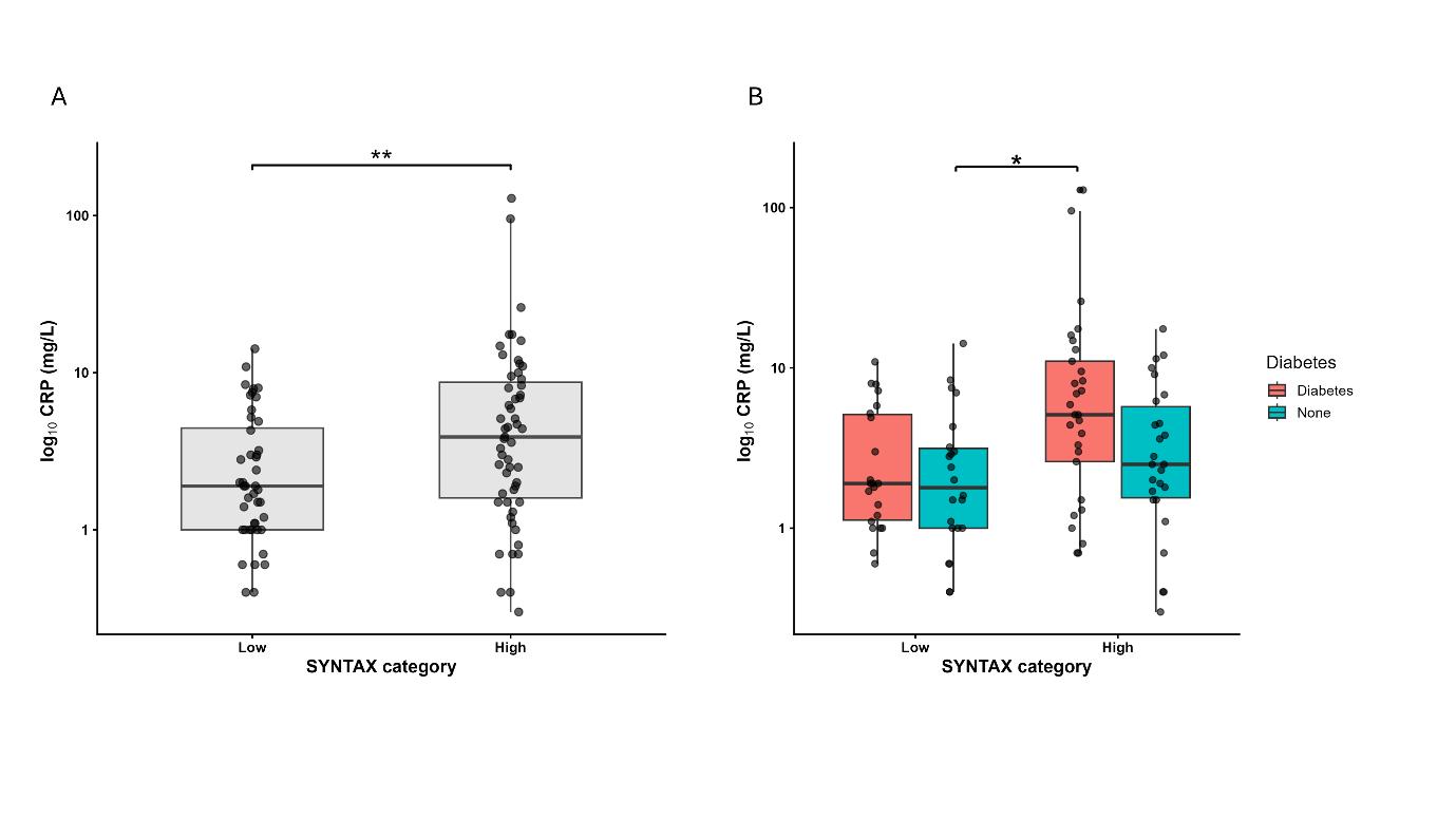
When participants were dichotomised by CAD extent, hsCRP was significantly higher in patients with high SYNTAX scores (3.9 [1.6–8.7] versus 1.9 [1.0–4.5]
mg/L; p=0.01) (Figure 1A) but more so in diabetes (5.1 [2.6–11.0] versus 2.5 [1.6-5.8] mg/L; p=0.052) (Figure 1B). Adjusting for age, sex, and BMI using multivariate linear regression, high SYNTAX score (β=0.32; p=0.002) and diabetes (β=0.25; p=0.01) were independently associated with higher hsCRP.
At follow-up, hsCRP was similar between participants with and without diabetes (median: 1.5 [0.9–3.6] versus 1.2 [0.5–5.0] mg/L; p=0.223). After adjustment for age, sex, BMI, and patients who had coronary artery bypass grafting, the association with hsCRP, CAD extent, and diabetes status, which was demonstrated at admission, was lost.
IL-6 and IL-18 were higher in patients with T2D or pre-diabetes (median: 4.64 [2.80–6.35] versus 3.38 [2.03–4.76] pg/mL; p=0.051 and 340 [266–422] versus 274
[224–369] pg/mL; p=0.032, respectively). After adjusting for covariates, diabetes was independently associated with IL18 (β=0.08; p=0.044), but there was no association between IL-6 and IL-18 with CAD extent.
CONCLUSION
The authors demonstrated that C-reactive protein (CRP) upon presentation with acute coronary syndrome, but not at follow-up, was associated significantly with extensive CAD and diabetes. Measurement of CRP during the acute presentation of CAD is often systematically excluded, and is primarily focused on the rehabilitation or baseline period. However, this study suggests that elevation of hsCRP acutely may identify a propensity to inflammation that is relevant to CAD.
* p value < 0.05
** p value < 0.01
CRP: C-reactive protein; hsCRP: high-sensitivity C-reactive protein; SYNTAX: Synergy Between Percutaneous Coronary Intervention With Taxus and Cardiac Surgery.
Figure 1: Relationship between hsCRP and SYNTAX score (A) stratified by diabetes status (B).
References
1. Ridker PM et al. Inflammation and cholesterol as predictors of cardiovascular events among patients receiving statin therapy: a collaborative analysis of three randomised trials. Lancet. 2023;401(10384):1293-301.
2. Kaptoge S et al. C-reactive protein concentration and risk of coronary heart disease, stroke, and mortality: an individual participant meta-analysis. Lancet. 2010;375(9709):132-40.
3. Lowe G et al. Circulating inflammatory markers and the risk of vascular complications and mortality in people with type 2 diabetes and cardiovascular disease or risk factors: the ADVANCE Study. Diabetes. 2014;63(3):1115-23.
4. Trøseid M et al. The role of interleukin-18 in the metabolic syndrome. Cardiovasc Diabetol. 2010;9(1):11.
5. Kaptoge S et al. Inflammatory cytokines and risk of coronary heart disease: new prospective study and updated meta-analysis. Eur Heart J. 2014;35(9):578-89.
6. Kedhi E et al. Impact of coronary lesion complexity on drug-eluting stent outcomes in patients with and without diabetes mellitus: analysis from 18 pooled randomized trials. J Am Coll Cardiol. 2014;63(20):2111-8.
7. Chai J et al. Association of inflammation and extent of coronary artery disease in type 2 diabetes and ACS. Abstract A100137JC. EuroPCR, 19-22 May, 2026.
Abbreviated Dual Antiplatelet Therapy After Percutaneous Coronary Intervention Among Patients with High Bleeding Risk
Authors: Manveer Singh,¹ *Etienne Puymirat,¹
Michel Zeitouni,2 Céline Lambert,3 Grégoire
Rangé;4 on behalf of FRANCE-PCI investigators
1. Cardiology Department, Hôpital Européen Georges-Pompidou, Paris, France
2. Cardiology Department, AP-HP Hôpital Universitaire Pitié-Salpêtrière, Paris, France
3. Biostatistics Unit, DRCI, CHU ClermontFerrand, France
4. Cardiology Department, Les Hôpitaux de Chartres, Le Coudray, France
*Correspondence to etienne.puymirat@aphp.fr
Disclosure: Rangé has received grants or contracts, consulting fees, and payment or honoraria for lectures, presentations, speakers’ bureaus, manuscript writing, or educational events from Abbott Vascular and Shockwave; and is the President of the France PCI Association. The other authors have declared no conflicts of interest.
Citation: EMJ Int Cardiol. 2026;14[1]:34-35. https://doi.org/10.33590/emjintcardiol/JTA9HZ6S
BACKGROUND AND AIMS
While dual antiplatelet therapy (DAPT) effectively reduces thrombotic and ischaemic complications following percutaneous coronary intervention (PCI), it is associated with an increased risk of bleeding, particularly among patients with high bleeding risk (HBR).1 Although recent guidelines advocate abbreviated DAPT (≤3 months) in this population, the uptake of these strategies in routine clinical practice remains insufficiently described.2-4 The authors sought to assess real-world prescription patterns of abbreviated DAPT after PCI according to bleeding risk status, and to identify factors associated with prolonged DAPT use in patients with HBR using the nationwide FRANCE-PCI registry.5
MATERIALS AND METHODS
This analysis included consecutive patients undergoing PCI for acute or chronic coronary syndrome between 2014–2023 in 56 French centres participating in the prospective FRANCE-PCI registry.6 Patients alive at 1 year with available DAPT duration data were included. HBR was pragmatically defined by the presence of at least one of the following criteria: age ≥75 years, chronic oral anticoagulation, prior stroke, or chronic kidney disease. Multivariable mixed-effects logistic regression analysis was performed to identify predictors of prolonged DAPT (>3 months) among patients with HBR.
RESULTS
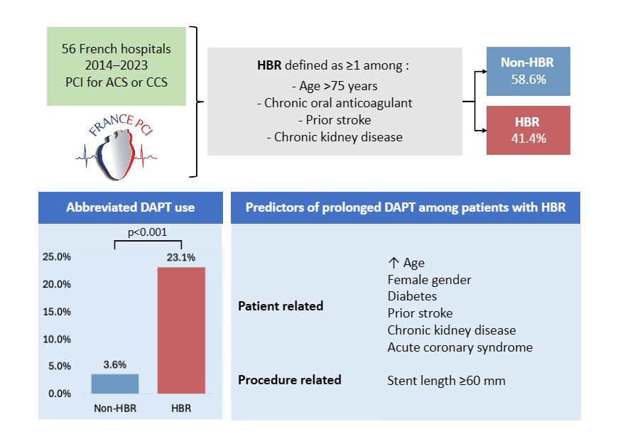
Among 115,992 patients included, 48,028 (41.4%) fulfilled HBR criteria. Despite guideline recommendations favouring shorter antiplatelet strategies in this population, abbreviated DAPT was prescribed in only 23.1% of patients with HBR compared with 3.6% of patients without HBR (p<0.001; Figure 1).
Within the HBR subgroup, several clinical and procedural characteristics remained independently associated with prolonged DAPT use, including older age (odds ratio [OR]: 1.02; 95% CI: 1.02–1.03), female sex (OR: 1.18; 95% CI: 1.11–1.25), diabetes (OR: 1.15; 95% CI: 1.08–1.22), prior stroke (OR: 1.54; 95% CI: 1.39–1.69), chronic kidney disease (OR: 1.24; 95% CI: 1.15–1.34), acute coronary syndrome presentation (OR: 1.52; 95% CI: 1.44–1.61), and total stent length ≥60 mm (OR: 1.18; 95% CI: 1.09–1.28). Over the study period, abbreviated DAPT use progressively increased among patients with HBR, although prolonged DAPT remained the predominant strategy overall.
CONCLUSION
In this large, contemporary, nationwide PCI registry, fewer than one in four patients with HBR received abbreviated DAPT, despite current guideline recommendations. DAPT duration
remained strongly influenced by markers of ischaemic and procedural complexity, underscoring the persistent challenges of balancing bleeding and ischaemic risks in routine clinical practice, and highlighting the gap between randomised trial evidence and real-world management.
1. Urban P et al. Defining high bleeding risk in patients undergoing percutaneous coronary intervention: a consensus document from the Academic Research Consortium for High Bleeding Risk. Eur Heart J. 2019;40(31):2632-53.
2. Byrne RA et al. 2023 ESC Guidelines for the management of acute coronary syndromes: developed by the task force on the management of acute coronary syndromes of the European Society of Cardiology (ESC). Eur Heart J. 2023;44(38):3720-826.
3. Valgimigli M et al. Dual antiplatelet therapy after PCI in patients at high bleeding risk. N Engl J Med. 2021;385(18):1643-55.
4. Mehran R et al. 3- or 1-month DAPT in patients at high bleeding risk undergoing everolimus-eluting stent implantation. JACC Cardiovasc Interv. 2021;14(17):1870-83.
5. Puymirat E et al. Real-world use of abbreviated dual antiplatelet therapy after PCI in high bleeding risk patients. Abstract A96111EP. EuroPCR, 19-22 May, 2026.
6. Rangé G et al. The France PCI registry: design, methodology and key findings. Arch Cardiovasc Dis. 2023;116(11):489-97.
Figure 1: Real-world use of abbreviated DAPT after PCI in patients with HBR.
ChatGPT-Assisted ECG Interpretation Can Help in the STEMI Activation Pathway: A Single-Centre Diagnostic Accuracy Study
1. Hospital Professor Doutor Fernando Fonseca, Amadora, Portugal
2. Faculdade de Medicina, Universidade de Lisboa, Lisbon, Portugal
3. Centro Cardiovascular da Universidade de Lisboa, Faculdade de Medicina, Universidade de Lisboa, Portugal
*Correspondence to ines.miranda@ulsasi.min-saude.pt
Disclosure: Mateus has received support for attending meetings and/or travel from A. Menarini, Tecnimede, Pfizer, Novartis, and Boehringer Ingelheim. Santos has received payment or honoraria for lectures, presentations, speaker’s bureaus, manuscript writing, or educational events from Shockwave; and support for attending meetings and/or travel from Abbott. The other authors have declared no conflicts of interest.
Acknowledgements: The authors thank the interventional cardiology and emergency teams at Hospital Professor Doutor Fernando Fonseca, Amadora, Portugal, for their contribution to patient care and data collection.
Citation: EMJ Int Cardiol. 2026;14[1]:36-37. https://doi.org/10.33590/emjintcardiol/91666QG5
BACKGROUND AND AIMS
Timely identification of ST-segment elevation myocardial infarction (STEMI) and prompt activation of the catheterisation laboratory team are determinant for patient outcomes. Current guidelines recommend a first-medical-contact-to-balloon time of less than 90 minutes for primary percutaneous coronary intervention,
yet real-world workflows are frequently hampered by diagnostic uncertainty and variability in ECG interpretation.1 Between 5–10% of emergent cath lab activations represent false positives,2 resulting in unnecessary risk and resource expenditure, while missed or delayed diagnoses carry substantial morbidity and mortality. Conventional 12-lead ECG analysis depends on operator experience and, even among cardiologists, inter-observer agreement for STEMI diagnosis is imperfect.3
Generative AI, and large language models in particular, have attracted growing interest in Cardiology. Prior studies evaluating earlier ChatGPT (OpenAI, San Francisco, California, USA) versions for ECG interpretation showed moderate overall accuracy, with the model performing best for normal tracings and poorly for acute ischaemic changes.4,5 The emergence of multimodal large language models has opened the possibility for direct visual analysis of ECG images to support clinical triage.
Dedicated AI ECG platforms have demonstrated high sensitivity and specificity on coronary occlusion detection,2 but require institutional integration and technical infrastructure. ChatGPT is accessible; however, there is few data stating it can meaningfully support the STEMI activation decision in a real-world clinical setting.
The authors’ aim was to assess the diagnostic accuracy of ChatGPT version 5.0 for ECG-based detection of acute coronary occlusion in a real-world STEMI activation cohort.6
MATERIALS AND METHODS
This single-centre retrospective study enrolled all consecutive coronary team (‘Via Verde Coronária’) STEMI activations at a Portuguese tertiary centre from
January–November 2025. Available ECG images were analysed using ChatGPT version 5.0, prompted with a single standardised question: “Based on this ECG, is there an occluded coronary artery?”
ChatGPT’s binary prediction (‘yes’/‘no’) was compared against the reference standard of invasive coronary angiography. Significant coronary artery disease was defined as stenosis >90% requiring urgent revascularisation.
RESULTS
A total of 220 STEMI activations were reviewed (mean age: 64±14 years; 30.9% female; comorbidities including hypertension in 63.2%, dyslipidaemia in 42.7%, and diabetes in 21.8%). ECGs were available for analysis in 176 cases (80%); the remaining 20% were unavailable due to external acquisition or technical issues. Of the 176 analysable patients, 152 (86%) had angiographically confirmed significant coronary stenosis.
ChatGPT correctly predicted significant stenosis in 126 of 152 cases (83%), with 26 missed occlusions: left anterior descending artery (n=10), left circumflex artery (n=5), right coronary artery (n=5), diagonal branch (n=2), obtuse marginal branch (n=2), and three-vessel disease (n=1). Among 24 patients without significant stenosis, 20 (83%) were correctly classified; four false positives were identified in patients with normal coronary arteries (n=2), coronary vasospasm (n=1), or undergoing repeat angiography (n=1).
Overall, diagnostic accuracy was 83.0%. Sensitivity was 82.9% and specificity 83.3%, with a positive likelihood ratio of 4.97. The positive predictive value (PPV) was high at
96.9%, compared with 84.4% for the human comparator, confirming strong rule-in capability. The negative likelihood ratio was 0.21 and the negative predictive value was 43.5%, indicating limited rule-out capability.
CONCLUSION
These results indicate a good overall performance, driven mainly by ChatGPT’s rule-in ability, with a high PPV, providing practical reassurance for clinicians uncertain about activating the interventional team or even the on-duty cardiologist. The negative predictive value was low (although limited by the high disease prevalence), and ChatGPT should not be used to exclude a coronary occlusion. The high PPV suggests further investigations are necessary to explore ChatGPT’s capability in similar settings, such as identifying non-STEMI patients with an acutely occluded artery.
References
1. Byrne RA et al. 2023 ESC Guidelines for the management of acute coronary syndromes. Eur Heart J. 2023;44(38):3720-826.
2. Herman R et al. AI-enabled ECG analysis improves diagnostic accuracy and reduces false STEMI activations: a multicenter U.S. registry. JACC Cardiovasc Interv. 2026;19(2):145-56.
3. Günay S et al. Comparison of emergency medicine specialist, cardiologist, and ChatGPT in electrocardiography assessment. Am J Emerg Med. 2024;80:51-60.
4. Çamkıran V et al. Artificial intelligence (ChatGPT) ready to evaluate ECG in real life? Not yet! Digit Health. 2025;11:20552076251325279.
5. Zhu L et al. Multimodal ChatGPT-4V for electrocardiogram interpretation: promise and limitations. J Med Internet Res. 2024;26:e54607.
6. Miranda I et al. ChatGPT-assisted ECG interpretation can help in the STEMI activation pathway. Abstract A95257IM. EuroPCR, 19-22 May, 2026.
Congress Interviews
EMJ is delighted to present this exclusive interview series from EuroPCR 2026, featuring insights from EuroPCR Course Directors Thomas Cuisset and Nicolas Dumonteil, alongside Salvatore Brugaletta, Editor-in-Chief of PCRonline and PCR Board member. These interviews delve into the future of interventional cardiology, exploring advances in structural heart interventions, imaging, AI, complex PCI, and continuous education, while highlighting the innovations and collaborative approaches shaping patient care worldwide.
Featuring: Salvatore Brugaletta, Thomas Cuisset, and Nicolas Dumonteil
Salvatore Brugaletta
Hospital Clínic i Provincial de Barcelona, Barcelona, Spain
We want to understand how AI can help us in realworld decision-making and patient care
Citation: EMJ Int Cardiol. 2026;14[1]:38-41. https://doi.org/10.33590/emjintcardiol/5T9QN757
Q1
As Editor-in-Chief of PCRonline and a member of the PCR Board, how do you see educational needs in interventional cardiology changing, and how did the EuroPCR 2026 Congress adapt to meet them?
That is a very important question because it is something we ask ourselves every year when we begin planning EuroPCR. To me, there are two main changes.
The first is the growing interest in structural procedures. Years ago, this was just beginning, but now we see more and more interventional cardiologists coming to EuroPCR specifically to learn about structural interventions.
The second major topic is AI. This year, AI was introduced much more broadly throughout EuroPCR, and I think many of us are very interested in understanding how AI can actually be applied
in daily clinical practice, not just theoretically, but practically. We want to understand how AI can help us in real-world decisionmaking and patient care.
Q2
Imaging has become increasingly central to decision-making in percutaneous coronary intervention (PCI). How do you see the integration of CT, intravascular ultrasound (IVUS), optical coherence tomography (OCT), and physiology reshaping the way operators approach complex coronary disease?
Imaging and physiology are fundamental. If we look back at coronary stents themselves, I think we have more or less reached the top in terms of device performance: there is not much more improvement to be made there. What we can improve is understanding when a stent should be implanted using physiology, and how we can implant it better in order to improve patient outcomes.
CT is extremely important for procedural planning, while IVUS and OCT are essential during PCI itself. Every interventional cardiologist should know not only how to use these technologies, but also how to take full advantage of them. It is not enough to simply open the box and use the pressure wire or imaging catheter: you need to understand how to extract the maximum value from these techniques.
This was highlighted by recent studies such as CHIP and OPTIMAL, presented at the American College of Cardiology (ACC) 2026 Scientific Sessions, which showed that simply using imaging is not always enough if you are not fully leveraging the information it provides.
At EuroPCR this year, there were again many small group sessions focused on imaging techniques, with greater emphasis on CT planning. I believe this is the future. Every operator should understand how to integrate these techniques into daily practice.
Q3 At EuroPCR 2025, there was significant focus on vulnerable plaque and myocardial infarction with non-obstructive coronary arteries. Do you think interventional cardiology is moving beyond simply treating obstructive disease towards a more preventive and mechanistic approach?
Yes, I do think so. With myocardial infarction with non-obstructive coronary arteries, imaging can help clinicians make the diagnosis, but for me, the more important evolving topic is vulnerable plaque. We are becoming increasingly interested not only in preventive drug therapies but also in whether preventive treatment of plaque, for example, with stenting or other interventional strategies guided by physiology or OCT, may help reduce future vulnerable events.
This year at EuroPCR, we had two dedicated sessions on vulnerable plaque. One focused on how to identify vulnerable plaque, while the other explored how to treat it. This field will become even more important over the next few years
because several major studies are ongoing.
The COMBINE INTERVENE trial, for example, is expected to present results later this year, possibly at Transcatheter Cardiovascular Therapeutics (TCT) 2026, and additional studies will follow over the next 1–2 years. These trials may help us understand whether there is a clear role for interventional cardiologists in treating vulnerable plaque before events occur
Q4 Your recent work has explored OCT-guided PCI and advanced calcium visualisation techniques. Which imaging innovations do you believe are closest to changing routine clinical practice?
That is a difficult question because it is hard to select only one imaging modality.
When we talk about calcium assessment, I think most of us would agree that OCT is superior to IVUS in some aspects, particularly for measuring calcium
thickness. That does not mean IVUS is not useful in calcified lesions, but OCT provides additional detail, and calcium thickness measurements can directly influence procedural strategy.
CT also plays an important role in planning procedures, although it cannot be used in real time during PCI.
Another interesting development is 3D stent reconstruction using angiography-based imaging. After stent implantation, a 300-degree rotational acquisition can create a 3D image of the stent and the calcified plaque behind it. In the future, we may even be able to acquire this information before stenting, simply by positioning a balloon within the lesion. That could allow us to understand calcium length, arc, and thickness in much greater detail before intervention. Some interesting data on this were presented by Nicolas Amabile, Cardiovascular Institute Paris Sud (ICPS), Massy, France, during EuroPCR.
Overall, I would say that whatever imaging technique you use, the key is und erstanding it well and knowing how to use it properly.
Q5 Complex multivessel disease and high-risk PCI remain major challenges. In your view, what are the most important factors in improving outcomes for these increasingly fragile patients?
When we treat these patients, we must think about the patient as a whole, not just the lesion. The lesion belongs to a patient, and often both are high risk.
As the population ages, we are treating increasingly complex and fragile patients compared with 10 years ago. Education is therefore essential in understanding how to manage these cases.
For high-risk PCI, you need preparation and experience. You need a plan A and a plan B. These are not procedures that everyone can simply perform routinely. Teamwork within the catheterisation lab is also extremely important.
Operators need to understand all available devices: not only imaging tools for PCI optimisation, but also plaque-modification technologies such as intravascular lithotripsy, rotational atherectomy, orbital atherectomy, and others. In addition, it is crucial to understand
when mechanical circulatory support is necessary.
High-risk PCI will become an even bigger focus in the future because of the ageing population and the increasing number of elderly patients undergoing procedures such as transcatheter aortic valve implantation, often with multiple comorbidities and highly complex anatomy.
Q6
PCRonline has become a year-round educational platform rather than simply a meeting resource. How important is continuous digital education in a field evolving as rapidly as interventional cardiology?
Education is always the key.
Whether you organise a congress, a course, a symposium, or a webcast, the goal is always to educate others. Science does not belong to one individual: it belongs to everyone. It would be a mistake for someone to keep a specific technique or experience only to themselves instead of sharing it with the wider community.
That philosophy really is the foundation of PCR and PCRonline.
When we treat these patients, we must think about the patient as a whole, not just the lesion
In addition to the congress itself, we now have educational journals, as well as textbooks and educational resources covering many different topics.
Another important development announced recently is an open access journal, where physicians around the world can share their experiences, regardless of whether they are based in Europe, Asia, or the USA.
The important thing is continuous education. Clinical practice may ultimately aim towards the same goal, but there are many different ways to approach it. Learning from different experiences allows
physicians to choose the strategy that works best for their own hospital and clinical reality.
Q7Looking back on EuroPCR 2026, what do you hope participants will take away from the meeting, not only scientifically, but also in terms of how they approach patient care and collaboration?
For me, the most important thing is always networking and returning home with the feeling that you have learned something new.
That learning may come from live cases, new devices, imaging techniques, poster sessions, or
discussions with colleagues. What I appreciate most about EuroPCR is not one single technology or topic, but the overall feeling that when you leave the meeting, you go back to your hospital thinking, ‘I learned something here, and now I will approach this differently’.
To me, that is the best possible takeaway from EuroPCR.
Science does not belong to one individual: it belongs to everyone
Citation: EMJ Int Cardiol. 2026;14[1]:42-44. https://doi.org/10.33590/emjintcardiol/T9REZL3I
Thomas Cuisset
APHM Hôpital La Timone Adultes, Marseille, France
Q1As Course Director of the European Association for Percutaneous Cardiovascular Interventions (EuroPCR), how would you describe the evolution of the meeting in recent years, and what continues to make it unique within the global interventional cardiology landscape?
I think it’s actually quite difficult to describe precisely how the meeting has evolved in recent years, but what has remained absolutely constant is its DNA. EuroPCR has always been a very practical meeting.
Q2 Looking ahead to EuroPCR 2026, what central themes or ‘fil rouge’ can participants expect this year?
Each year, we define a kind of ‘fil rouge’, a central theme that runs throughout the meeting. For 2026, we have chosen to focus on complications.
These three initiatives, the CTO track, the Calcium Skills Lab, and the physiology course, I think, are the main novelties attendees can look forward to
The core idea is to come together to share practice, not just theory, and to improve collectively by exchanging knowledge, experience, and skills. That focus on real-world practice, and on learning from one another, is what continues to define EuroPCR and makes it unique.
This includes both the prevention of complications and their optimal management when they occur. We strongly believe that while it is important to share successes, it is probably even more important to share difficulties and how they are overcome.
For us, that is where medical education truly has an impact and ultimately contributes to saving lives.
Q3
EuroPCR is known for its strong emphasis on education and live case learning. Are there any new formats, technologies, or session types being introduced in 2026 that attendees should be particularly excited about?
There are three key new elements I would highlight this year.
First, we are introducing a dedicated track on chronic total occlusion (CTO) interventions, which will be part of EuroPCR for the first time. There is a clear and growing interest in this area within the community, and it felt important to reflect that with a focused programme.
Second, we are launching a ‘Calcium Skills Lab’. As our patient population becomes increasingly elderly and frail, we are seeing more calcified lesions. Rather than addressing devices or imaging modalities separately, we wanted to create a dedicated environment to approach this challenge more integratively, combining imaging and treatment strategies.
Finally, just before the official opening of the meeting, a dedicated physiology course will be held. This course has been developed by pioneers in the field, and for the first time, it will be fully integrated into the EuroPCR Week, focusing on the use of physiology in coronary intervention.
These three initiatives, the CTO track, the Calcium Skills Lab, and the physiology course, I think, are the main novelties attendees can look forward to.
Q4Recent sessions you’ve been involved in have explored the intersection of TAVI and coronary artery disease. What are the key unresolved challenges in managing these increasingly complex patients?
I would actually say that the situation is becoming more reassuring, because things are getting clearer. Recent data, including trials presented at the American College of Cardiology (ACC) Annual Scientific Session, have confirmed what many of us suspected.
Performing percutaneous coronary intervention (PCI) systematically before transcatheter aortic valve implantation (TAVI) does not appear to provide significant benefit for most patients, and it may even expose them to additional risk, particularly bleeding related to dual antiplatelet therapy.
This has been a highly debated topic over the past decade, but we are now moving towards a more selective approach. In the past, many centres probably performed PCI before TAVI routinely. Current evidence suggests we need to be much more careful in selecting which patients truly benefit, as the overall advantage is limited and may, in some cases, be outweighed by harm.
EuroPCR is the place where physicians and the wider cardiovascular community come together from all over the world to share, learn, and discuss
Finally, just before the official opening of the meeting, a dedicated physiology course will
be held. This course has been developed by pioneers in the field, and for the first time, it will be fully integrated into the EuroPCR Week, focusing on the use of physiology in coronary intervention.
These three initiatives, the CTO track, the Calcium Skills Lab, and the physiology course, I think, are the main novelties attendees can look forward to.
This approach aims to strike a better balance between efficacy and safety, maintaining protection against ischaemic events while reducing the risk of bleeding complications.
Q5
You have contributed extensively to research in antithrombotic therapy. How do you see current strategies evolving, particularly in complex PCI and patients with high bleeding risk?
We increasingly recognise that many patients undergoing complex PCI, especially those with calcified lesions, are also elderly and therefore carry both high ischaemic and high bleeding risk.
In stable patients, advances in PCI optimisation, including newergeneration stents and the use of intracoronary imaging, now allow
us to safely shorten the duration of dual antiplatelet therapy in selected cases.
In patients with acute coronary syndrome, the situation is slightly different, because we are not only treating the PCI but also the underlying atherothrombotic event. Here, we are seeing growing evidence supporting a de-escalation strategy: starting with potent dual antiplatelet therapy in the early phase after PCI, then reducing intensity after 1 month or 6 weeks, either by switching the P2Y12 inhibitor or discontinuing aspirin.
Q6
What do you see as the biggest unmet needs in cardiovascular intervention today, and where should the field be focusing its efforts next?
There has already been tremendous progress in the field, which is very encouraging. But if I could ask for one major advance in the coming years, it would be further improvement in noninvasive diagnostic tools.
Coronary CT is already highly effective, and CT-based physiology is becoming increasingly valuable. In an ideal world, we should not need to bring patients to the cath lab purely for
diagnostic purposes. Diagnosis should be entirely non-invasive, and invasive procedures should be reserved for treatment.
In structural intervention, the need is different. We still require better, more dedicated devices for mitral and tricuspid valve disease, devices that are predictable, easy to use, and capable of delivering consistent and meaningful reductions in regurgitation.
Q7
Finally, if you had to summarise EuroPCR 2026 in one sentence, what would it be?
EuroPCR is the place where physicians and the wider cardiovascular community come together from all over the world to share, learn, and discuss, with the ultimate goal of improving their skills and, in turn, improving patient outcomes.
It is, in a way, the link between education and patient outcomes, through the continuous development of the physician.
Attendees will be able to visit a dedicated AI Lab, offering opportunities to better understand how AI may influence the future of cardiovascular intervention
Citation: EMJ Int Cardiol. 2026;14[1]:45-48. https://doi.org/10.33590/emjintcardiol/479T91H3
Q1
As the EuroPCR Course Director, how are you shaping the 2026 programme to better support clinicians managing increasingly complex structural heart patients?
Each year, we begin building the next edition of EuroPCR around September, always starting from the needs of the community. We gather insights through a large international survey distributed across our companion database, which includes more than 20,000 colleagues worldwide.
From this, we try to understand where the field is moving and what clinicians need most. In structural heart disease, it is clear that transcatheter aortic valve implantation (TAVI) has now become mainstream, with the majority of interventional cardiologists practicing it routinely, while mitral and tricuspid interventions are developing extremely quickly.
Our goal is, therefore, to ensure that the EuroPCR programme reflects these changes. We shape the sessions, live cases, and educational tracks around the real-world challenges clinicians are facing as structural interventions become increasingly complex.
Q2
What new features or educational highlights can attendees expect at EuroPCR 2026?
Based on the evolution of the field and feedback from the community, we wanted to introduce several new features into this year’s programme.
One major addition is the new Calcium Skills Lab, which will provide a comprehensive overview of calcified coronary artery disease, from diagnosis and pathophysiology through to imaging, treatment strategies, and complication management.
We have also launched a dedicated chronic total occlusions (CTO) programme, delivered in collaboration with experts from the EuroCTO Club. This will include live cases, abstract presentations, and case-based discussions focused on CTO interventions. Importantly, many of the techniques developed in CTO practice can also benefit operators managing other forms of complex percutaneous coronary intervention (PCI).
Another key development is the introduction of an Innovation Track designed to create continuity between Innovators Day and the wider EuroPCR programme. This will cover not only interventional innovation, but also advances in imaging and AI.
Nicolas Dumonteil
Clinique Pasteur, Toulouse, France
For the first time, attendees will also be able to visit a dedicated AI Lab, offering opportunities to better understand how AI may influence the future of cardiovascular intervention.
Q3
You have extensive experience as a live case operator across coronary and structural procedures. What makes live case learning such a powerful educational format at EuroPCR?
Interventional cardiology is a discipline built around procedures. Even if you work in a high-volume centre and have many years of experience, you will never encounter every possible anatomy, complication, or clinical scenario during your own career.
That is why live case education is so valuable. It gives colleagues the opportunity to observe another operator treating a patient in real time, which is fundamentally different from watching an edited recording.
During a live case, you see how someone adapts when the initial strategy does not work, how they move from plan A to plan B, and how complications are managed, or, ideally, anticipated and avoided altogether. That is the essence of live case learning.
Q4
TAVI is now moving into younger and lower-risk patients. How will EuroPCR 2026 address lifetime management, valve durability, and future coronary access in this population?
This is a crucial and very timely issue. Current guidelines and evidence now allow us to treat younger patients with longer life expectancy using TAVI, which is understandably appealing for many patients. Increasingly, patients come to the clinic asking specifically for a transfemoral valve procedure rather than surgery.
However, if a patient receives a TAVI valve at 70 or 71 years old, there is a strong possibility they will outlive that prosthesis and require
another intervention in the future. That means we must think carefully about lifetime management from the very first procedure.
If the initial valve selection or implantation strategy is not optimal, a future redo TAVI can become extremely challenging. We may face issues such as coronary obstruction risk, sizing limitations, or severe prosthesis mismatch.
At EuroPCR, our mission is therefore to educate both ourselves and the wider community on proper patient selection, procedural planning, and device choice. We also need to identify which patients should still be referred for surgery as a first intervention.
This topic is integrated throughout the structural programme, including live cases, case-based discussions, learning sessions, and CT hands-on training.
Q5 Your recent work has explored redo TAVI, valvein-valve procedures, and leaflet modification. What are the biggest technical and clinical challenges in treating patients who return for repeat valve intervention?
One of the major challenges is the risk of coronary occlusion, particularly in patients who received their first TAVI valve at a time when we did not yet fully understand the long-term implications of redo procedures.
In the past, valves were sometimes implanted in small aortic roots or in anatomies with low coronary takeoffs, which can create a ‘neo-skirt’ during a redo TAVI and threaten coronary perfusion.
However, an equally important and perhaps under-recognised issue is valve sizing. After the first TAVI implantation, the available internal space for a second valve becomes significantly reduced.
For example, a patient may initially have a 26 mm annulus, but after the first valve implantation, the effective internal diameter available for a second valve may be closer to 21 mm. Even if coronary access remains possible, this may limit the feasibility of multiple future interventions.
This becomes especially important as some centres are now implanting TAVI valves in patients as young as 65 years. In these cases, we may need to think not only about one future
reintervention, but potentially two over the patient’s lifetime.
Q6Mitral and tricuspid transcatheter therapies are evolving quickly. What developments in these fields do you expect to generate the most discussion at EuroPCR 2026?
One of the strengths of EuroPCR is that it is not only a course focused on experience and practical learning, but also a forum for discussing the latest scientific evidence.
This year, we have seen a significant amount of late-breaking science submitted around mitral and tricuspid therapies. Much of this work focuses on refining
patient selection, understanding clinical benefit, and comparing different treatment approaches.
We are also seeing growing evidence around transcatheter valve replacement technologies in both the mitral and tricuspid spaces, alongside emerging comparisons between repair and replacement strategies.
These discussions will feature prominently throughout the programme, including dedicated case-based sessions and live cases. We will, for example, have a live mitral valve replacement case presented in the main arena.
One of the strengths of EuroPCR is that it is not only a course focused on experience and practical learning, but also a forum for discussing the latest scientific evidence
Q7
You are also involved in complex PCI and robotic-assisted PCI. How do you see technology changing the way interventional cardiologists plan and perform increasingly complex procedures?
Technology is clearly transforming the field, but I think the biggest progress is not only about new devices, but also about improving how we understand and plan procedures.
The major advances are happening in imaging. We now have highly sophisticated invasive imaging tools, alongside increasingly powerful non-invasive CT imaging. On top of anatomical assessment, we are also integrating more physiological information into procedural planning.
This allows operators to understand not only the anatomy they will face, but also which lesions are truly responsible for ischaemia and should therefore be treated.
AI is likely to further accelerate this evolution. If AI can help analyse and interpret the large amount of imaging and physiological data now available, it could provide operators with an almost ideal level of preparation before entering the catheterisation lab.
Q8
EuroPCR brings together coronary, structural, imaging, and innovation experts. How important is this cross-disciplinary exchange in improving decision-making for complex patients?
I think this multidisciplinary approach is absolutely crucial. In many ways, it emerged naturally alongside the growth of structural heart interventions.
As interventional cardiologists began treating more structural disease, we quickly realised that we needed to work much more closely with imaging specialists, heart failure physicians, electrophysiologists, surgeons, and many others. This led to the development of the modern Heart Team model.
What is interesting is that this collaborative approach is now increasingly influencing the coronary field as well.
By working closely with colleagues from different specialties, we gain access to more information, broader expertise, and a deeper understanding of each patient’s condition. Ultimately, this leads to better planning, better procedural decision-making, and, in most cases, better outcomes for patients.
Interviews
This year, we had the pleasure of interviewing three leading voices in interventional cardiology: Ashok Seth, Ramzi Khamis, and Robert Kelly. Together, these conversations explore the evolution of coronary intervention, the biology of cardiovascular vulnerability, and the growing need to unite procedural innovation with prevention, digital health, and patient-centred care.
Featuring: Ashok Seth, Ramzi Khamis, and Robert Kelly
Ashok
Seth Chairman, Fortis Escorts Heart Institute & Research Centre, New Delhi, India
Citation: EMJ Int Cardiol. 2026;14[1]:49-54. https://doi.org/10.33590/emjintcardiol/6Q2QE8SX
What first inspired you to pursue medicine and cardiology, and how did your training at Jawaharlal Nehru Medical College, Aligarh, India, and the University of Birmingham, UK, shape your clinical philosophy and approach to innovation?
I entered cardiology almost by default. To be frank, I had initially wanted to pursue gastroenterology. When I came to the UK in 1981, my first goal was to obtain Membership of the Royal College of Physicians, a qualification that provides excellent training and is widely respected, shaping you into a strong physician.
I had hoped to secure a super-specialty post in gastroenterology, particularly to train in endoscopic retrograde cholangiopancreatography, as I was already performing a significant number of endoscopies even in acute gastrointestinal bleeders. Unfortunately, I did not get the position. Instead, a role opened in cardiology at the same university teaching hospital. With limited options, I decided to take it.
Cardiology was no longer confined to the stethoscope; it was entering a new, interventional era
At the time, gastroenterology was far more procedurally advanced, with endoscopy, colonoscopy, and endoscopic retrograde cholangiopancreatography. Cardiology, by contrast, was largely ‘stethoscope cardiology’. Echocardiography and angiography were still primitive, and even the management of acute myocardial infarction in the early 1980s had not yet evolved; trials with streptokinase were still ongoing. Gastroenterology was simply more exciting.
Within 6 months, University Hospital Birmingham launched its angioplasty programme, the third centre in the UK to do so. I found myself at the very beginning of the balloon angioplasty era, initially assisting former consultant, Man Fai Shiu, who was himself advancing procedural boundaries. He became an exceptional mentor, and, as his first assistant, I was drawn into what quickly became a transformative period.
Cardiology was no longer confined to the stethoscope; it was entering a new, interventional era. Despite the crude technology, there was a strong sense that this was a defining moment, a new dimension in the treatment of cardiac
disease. That early exposure to innovation shaped my entire career with conviction and competence.
Q2After returning to India, you helped build the invasive and interventional cardiology programme at the Escorts Heart Institute, New Delhi, India, now part of Fortis Healthcare. What were the key challenges of introducing advanced interventional techniques in that environment?
After nearly a decade abroad, I returned to India in 1988–89, primarily because of my mother’s ill health. Communication at the time was extremely limited, one phone call a week that was often difficult to arrange, so being far away during a family illness was deeply stressful.
In retrospect, it was also the right time professionally. Interventional cardiology in India had barely begun. There were perhaps six or seven of us performing balloon angioplasty. This created both an opportunity and a responsibility not only to establish programmes, but to help build an entirely new field.
Coronary bypass surgery was already well established, and many patients travelled abroad for it. We aimed to bring high-quality cardiac care to India, including interventional approaches that were rapidly advancing in the West.
Balloon angioplasty itself was evolving, but its limitations were clear. In the West, new devices were emerging to address these challenges: rotational and directional atherectomy, extraction catheters, and others. As we recognised the shortcomings of balloon angioplasty, I made it a priority in the early 90s to adopt these technologies and introduce
them in India and the wider Asia-Pacific region.
At the time, India was leading interventional cardiology in Asia. However, the procedure was far from ideal. Around 5% of patients experienced acute vessel closure within 24–48 hours, sometimes on the table. Myocardial infarction occurred in roughly 3%, and mortality was about 1%. Even when successful, restenosis rates approached 50% within 6 months.
By today’s standards, such outcomes would be unacceptable. Yet, that was the state of the art.
It was a challenging environment. Cardiac surgeons were essential partners, often on standby with a vacant operating theatre to manage complications immediately. Despite the risks, we believed strongly in the potential of a non-surgical approach to coronary disease. The key was to improve both our skills and our tools. This conviction was our guiding path.
I began to push boundaries, taking on increasingly complex cases and adopting new devices to overcome the limitations of balloon angioplasty. Regulatory frameworks for devices were still evolving, which allowed me to introduce innovations earlier than in many other regions.
This positioned me to train not only Indian cardiologists but colleagues across Asia, in Indonesia, Malaysia, Thailand, Singapore, Bangladesh, Sri Lanka, and even China in the late 1980s. In many ways, we helped disseminate these technologies throughout the region.
Q3
You introduced several firsts in Asia-Pacific interventional cardiology, including new devices and procedural techniques. How do you decide when to be an early adopter of innovation, and what safeguards do you use to balance technological advancement with rigorous evidence and patient safety?
Introducing new technologies, often for the first time in the Asia-Pacific region, and sometimes globally, requires a clear philosophy. Over the years, I have been involved in several first-in-human procedures, including orbital atherectomy, the coronary sinus reducer, and early transcatheter valve work such as the first CoreValve™ (acquired by Medtronic, Minneapolis, Minnesota, USA) implantation in the world in a patient with aortic regurgitation as part of a first-inhuman study in 2004. I was also the first to introduce a microaxial flow pump (Impella, Abiomed, Danvers, Massachusetts, USA) in the Asia-Pacific region in 2007, and to do a live demonstration at the Transcatheter Cardiovascular Therapeutics® (TCT®) meeting 2007.
Our decision to adopt new technologies was guided by three principles.
First, many innovations were designed to address the limitations of existing treatments. Even before large trials were completed, experienced clinicians could identify where current tools were inadequate and where new approaches might improve outcomes.
Second, new technologies expanded treatment options for patients who were not candidates for surgery or conventional
interventions, particularly those with complex diseases.
Third, and most important, was the responsibility to the patient. My guiding principle has always been: would I offer this to my own
family? If the answer is yes, then it is reasonable to consider for my patients, provided it is done with the highest level of expertise and care.
Innovation requires three elements: good intent, sound judgement, and technical expertise. Safety must always come first. Evidence, in the form of RCTs, follows over time, but careful, judicious use can ensure safety from the outset.
Q4Having performed tens of thousands of angioplasties and angiograms, is there a particular case that altered your understanding of coronary disease or influenced the way you approach complex interventions today?
Interventional cardiology is a continuous learning process. We learn from every patient, from our peers, and through constant reflection, reviewing cases, analysing outcomes, and refining techniques. Teaching is also integral to that process.
Over the years, I have directly trained more than 500 interventional cardiologists in India, as well as many from neighbouring countries and
across the world. Indirectly, perhaps three generations have learned from me through live case demonstrations and conferences, and in catheterisation laboratory (cath lab) teaching programmes. I am privileged to have trained more than 50 interventional cardiologists from Bangladesh as my fellows over the last 25 years.
Experience is not just about improving success rates; it is about preventing complications. With time, this becomes almost instinctive. The responses become reflexive.
Two cases, however, stand out.
The first was the implantation of the first CoreValve in the world in a patient with aortic regurgitation
in 2004. Watching severe regurgitation disappear instantly in the cath lab, reduced to zero, felt almost miraculous. It was a moment I will never forget.
The second was performing a complex angioplasty on my own father at the age of 86 years. I treated critical lesions with rotational atherectomy and stents but deliberately chose not to pursue complete revascularisation, recognising the risks of doing too much.
He went on to live well, reaching the age of 102 years, without myocardial infarction, remaining active and independent. That experience profoundly shaped my thinking.
It reinforced that in stable coronary artery disease, medical therapy can be as important as intervention. Not every lesion requires a stent simply because it is visible. The goal is not to treat images, but to treat patients, balancing intervention with optimal medical therapy to achieve the best long-term outcomes.
Q5
Which areas of interventional cardiology, in your opinion, still lack sufficient evidence or consensus, and where do you believe the research community should prioritise its efforts in the coming decade?
I think there are still major gaps in our knowledge. Part of the reason is that by the time pivotal randomised trials are completed, the technology has already moved on. That is happening more and more rapidly now than it did in the past, which is understandable: as science, techniques, and knowledge advance, innovation and iteration also accelerate.
In interventional cardiology, one issue is very clear. Metallic stents have progressed enormously in terms of safety, and we now practice a far more precise form of medicine, guided by imaging and physiological assessment before and during procedures. We have better tools, better technology, and better outcomes. But even so, metal in the coronary arteries is not the ideal answer. Science has to move towards treatments that are temporary.
That is why the bioresorbable scaffold story remains so compelling. I implanted the first scaffold in the Asia-Pacific region in 2010, and also demonstrated the ‘bifurcation two-scaffold strategy’ at the TCT meeting in 2014. I was one of the strongest
proponents of scaffolds, and my own results with them were excellent because I had learned and mastered the technique of implantation. I was also the principal investigator for the novel, second-generation, thinner 100 µm scaffold. Although that field has had setbacks, I believe it will return in some form, because the idea of leaving nothing permanent behind and allowing the vessel to be restored over time is still very attractive. Turning coronary arteries into metal tubes cannot be the final answer, especially when event rates continue year after year.
There is also still much to be done in acute coronary syndromes (ACS). Predicting ACS, managing it better, and developing better drugs remain major challenges. Acute myocardial infarction is still one of the biggest killers, and ACS continues to have poor outcomes despite all the therapies we have. If we could develop sensors, perhaps even implantable ones, that could detect changes in the biological environment before an event occurs and warn us that plaque instability or thrombosis is likely, then we could intervene earlier and more effectively.
Diabetes is another major area. The world is suffering from diabetes, and we certainly are. Coronary artery disease in patients who are diabetic, whether treated by intervention or surgery, remains a huge challenge. We still need therapies that are more robust, more durable, and more effective, with the lowest possible event rates. The answer may well be a combination of approaches.
Structural heart disease is another field that is advancing rapidly. We have already seen how devices can transform treatment in a minimally invasive
way, making procedures safer and more effective. But there are other valves and other structural problems that still need better solutions, and I think that area will expand significantly.
Bleeding and antiplatelet therapy also remain critical issues. Improvements in antiplatelet therapy have been central to better percutaneous coronary intervention (PCI) outcomes. We now have better tools, better technology, and more precision through imaging and physiology, all of which have improved long-term results. But balancing efficacy against bleeding risk remains a major challenge. We need antiplatelet therapies that are both safer and effective.
Finally, precision medicine with genomics could influence not just treatment, but also the prevention of life-threatening heart diseases.
Those, to me, are some of the key priorities for the coming years in interventional cardiology.
Q6
Advanced cardiac interventions can be lifesaving, but they often remain inaccessible to many patients. What strategies or policy approaches could better balance cutting-edge technology with equitable access, particularly in resource-limited settings?
That is a very important question, not just for India or the AsiaPacific region, but for the entire world. Resource-limited settings exist in every country. In India, many patients pay out of pocket. In Europe, governments often pay. In the USA, insurers pay. But no system has unlimited resources.
In practical terms, complex angioplasty has become more expensive. Precision-based PCI,
using intravascular imaging, physiology, calcium-modification tools, thrombus extraction devices, better stent platforms, percutaneous mechanical circulatory support, and other advanced technologies, can improve outcomes, but all of it comes at a cost.
So how do we balance this in settings where resources are limited? In many places, intravascular ultrasound or optical coherence tomography may not even be available. Even when these tools are available, they may not be reimbursed. If they are not reimbursed, then the patient pays, and in countries like India, that often means a direct out-ofpocket expense.
There is another important dimension: in many settings, bypass surgery may actually be less expensive and more durable than a complex multivessel PCI. That is certainly true in India. So, the answer has to be a practical, value-based approach.
For example, if a patient with triple-vessel disease and complex anatomy is paying from his own pocket and has travelled from a remote town a thousand miles away, where medical facilities are limited, I may well recommend bypass surgery. It may be less expensive, more durable, and less likely to leave him facing a repeat emergency in a place without access to a cath lab.
Similarly, in some patients with multivessel disease, we may choose partial revascularisation combined with aggressive medical therapy, leaving borderline lesions alone, reducing the amount of metal implanted, and treating the rest medically. In others, bypass surgery may offer the best longterm value. The important thing is
to offer the patient value-based options: medical therapy, PCI, or surgery, selected according to durability, safety, cost, and access to follow-up care.
That same principle also applies to the use of precision tools. At my own tertiary centre, we use all available devices and technologies. But I also have to think about the wider reality: India has around 2,800 cath labs, and most interventional cardiologists do not have routine access to imaging, optical coherence tomography, or physiology. So, when I teach, whether at courses or large meetings, I have to explain how to optimise outcomes with angiography guidance alone, and which cases can safely be done that way.
At the same time, I also have to define the limits. There are some patients and some lesion subsets where, if you do not have imaging, you should not proceed. You should refer them to a centre that does. So, part of training in a resource-limited environment is not only teaching what to do, but also what not to do, and when to refer.
The final piece is indigenous research, development, and manufacturing. In India, this has significantly reduced costs over the past 2 decades. Many stents and structural devices are now developed and manufactured locally. I have served as principal investigator for much of the indigenous technology developed in PCI and structural heart therapy, including an Indian transcatheter heart valve that is now regularly used in Europe.
All of this technology had to go through rigorous evaluation, but it has helped lower costs and expand access. That is crucial. Technology has to be balanced
with what can be developed and sustained within our own country.
For me, this is the essence of value-based medicine: it must still be quality medicine. Lower cost should never mean compromised outcomes. A patient should not come back because the initial treatment was inadequate. If the patient stays out of hospital and does well, then we know we have delivered value.
In the end, I come back to a very simple principle: treat every patient as if they were your own relative. There will always be gaps in knowledge, in technology, and in what a patient can afford. The task is to balance medical therapy, PCI, and surgery in the most appropriate way, to achieve the safest, most durable result at the lowest reasonable cost. That is how I think these decisions should be made.
Q7Looking ahead, which developments, such as bioresorbable scaffolds, digital health, or AI, do you believe will most transform interventional cardiology, and how should clinicians prepare for these changes?
With bioresorbable scaffolds, and also with drug-coated balloons, the central concept is the same: ‘leave nothing behind’.
If coronary artery disease can be treated without leaving a permanent foreign body in the vessel, that is a major step forward. Whether that is achieved through bioresorbable scaffolds, drug-coated balloons, or other future technologies, the principle is very important. Treating the lesion and restoring the artery without implanting something permanent is where the field should be heading.
Digital health is another hugely important area. We learned a great deal during COVID-19, especially about the possibilities of remote monitoring, digital tools, and selfassessment. These become even more important in large countries where affordability, accessibility, and healthcare infrastructure vary enormously.
In our country, as care extends into smaller towns and rural areas, digital health will become increasingly central. AI will also play a major role. A simple example is ECG interpretation. In remote areas, patients with chest pain and acute myocardial infarction can now have ECGs recorded using a phone. That is already happening.
India is one of the most digitally connected countries in the world, with vast mobile phone penetration. Conditions such as diabetes, hypertension, and coronary artery disease are widespread, and digital health offers a powerful way to monitor, triage, and manage this burden. AI can process large volumes of information, filter what is normal, identify what needs specialist review, and guide what action should be taken. Our prime minister has already emphasised and promoted the National Digital Health Mission infrastructure.
If thousands of ECGs are being performed in remote villages, it is simply not possible for every tracing to be reviewed immediately by a cardiologist. But AI can screen them, identify the small number that need urgent attention, and help direct treatment rapidly.
So, I think AI will become a major partner in delivering high-quality care at a fraction of the cost, and in a much shorter timeframe. It will
not replace the specialist, but it will become an essential assistant to the doctor, to the specialist, and, ultimately, to the patient.
Q8What guidance would you offer young cardiologists entering interventional practice today, and how would you like your contribution to cardiology and patient care to be remembered?
I have often thought about that. How would I like my contribution to cardiology and patient care to be remembered?
I do not want to be remembered simply as the person who did the greatest number of complex cases. I do not want people to say, “What skills?” or “Look at how many teaching programmes he ran,” or “Look at the papers he wrote.” I would simply like to be remembered as a good doctor and a good man. At the end of the day, that is enough. That is what we owe to our patients.
When I think about what guidance I would give to a young interventional cardiologist, it comes down to what I call the five Cs.
The first is care. That is what we are here to give our patients. It is an ethos; it is the foundation.
The second is commitment. Commitment is why we became doctors, why we went into cardiology, and why we chose interventional cardiology. We have committed ourselves to the care of patients, and that commitment has to remain absolute.
The third is compassion. When a patient walks in, they should feel that they are being treated like family. Medical science is still incomplete. There are many grey areas and relatively few black-
and-white answers. Guidelines help us navigate those grey areas, but they do not replace judgment. The way we make those decisions properly is to ask ourselves: if this were me, or my relative, what would I want done?
If a patient has heavily calcified proximal left anterior descending artery disease, for example, an interventional cardiologist may immediately think of multiple tools, a long stent, and a complex PCI strategy. But one also has to ask: would a surgical graft, perhaps a left internal mammary artery graft to the left anterior descending artery and diagonal, offer a more durable result? The moment you ask what you would choose for yourself or your family, your judgment changes. You begin to think not only as an interventional cardiologist, but as a doctor trying to make the right decision.
The fourth C is competence. If I am going to treat someone as I would treat my own father, then I have to keep myself at the highest level. I must stay updated, maintain my skills, and ensure that what I offer is safe and excellent. Competence is an ethical obligation.
And the fifth, and perhaps most important, is conscience. Conscience means ethics. We must practise ethical medicine. We must do for the patient what we would do for ourselves. If we hold on to conscience, then the rest follows.
Those are the five Cs I would want every young cardiologist to absorb: care, commitment, compassion, competence, and conscience. If you live by those, you will do the best for your patients. In the end, it is not only the hands, or the technical skill, or even the knowledge. It is the person behind them that matters.
Ramzi Khamis
Professor of Cardiology, Wu Family Endowed Chair, National Heart & Lung Institute, Faculty of Medicine, Imperial
College London, UK
Ultimately, medicine should not only rescue patients from acute events; it should also help build resilience at biological, clinical, and societal levels
Citation: EMJ Int Cardiol. 2026;14[1]:55-58. https://doi.org/10.33590/emjintcardiol/454M446J
Q1
You’ve spoken movingly in the past about growing up in Jerusalem and Bethlehem, training in Bristol and London, and being shaped by powerful mentors at Imperial College London, UK. Looking back, what were the defining moments that steered you toward interventional cardiology?
There were several defining moments, but perhaps the deepest influence was growing up between Jerusalem and Bethlehem, where one becomes aware very early that vulnerability is never only biological. Illness, access, instability, duty, and human dignity all intersect in ways that stay with you for life. My early education, followed by my move to the UK and subsequent medical training in Bristol, helped shape my discipline, curiosity, and strong sense of responsibility. Bristol, in particular, was formative for me, both personally and academically.
My later training in London, especially at St Bartholomew's Hospital, London, and later at Imperial College London, sharpened that further. I was fortunate to be surrounded by outstanding mentors who taught me not only the craft of cardiology, but also the importance of scientific depth. They impressed on me that one should never be satisfied with treating the visible consequence of disease without trying to understand the underlying biology.
Interventional cardiology drew me because it sits at that very powerful intersection between thought and action. It is intellectually demanding, technically exacting, and yet
capable of making an immediate difference to a patient in front of you. I found that combination deeply compelling. Over time, it became clear to me that the catheter laboratory was not the end point of my interest, but rather one part of a much larger journey into atherosclerosis, inflammation, imaging, prevention, and ultimately the broader question of how we reduce vulnerability in all its forms.
Q2
You frame your work around the concept of ‘vulnerability’, from plaque to patient to society to civilisation. How did that framework evolve for you, and how does it shape the way you practice medicine today, both in the cath lab and beyond it?
The framework evolved gradually, but quite deliberately.
My scientific starting point was the vulnerable plaque, the atherosclerotic lesion that is biologically active, prone to rupture, and capable of precipitating myocardial infarction. That remains a central scientific question for me, because if we can identify vulnerability more precisely, we have a better chance of preventing catastrophe rather than simply reacting to it.
But over time, it became clear that plaque biology alone is not enough. Events occur not simply because a lesion exists, but because plaque biology interacts with thrombosis, inflammation, systemic risk, and the broader condition of the patient. That led naturally to the idea of the vulnerable patient, the person whose risk is often greater and more complex than conventional measures alone may suggest.
From there, the concept widened further. In medicine, one sees very quickly that vulnerability also resides in systems and societies. Delayed access to care, structural disadvantage, conflict, fragmentation of services, interrupted education, and inequality all shape outcomes. So, for me, the idea expanded from vulnerable plaque to vulnerable patient, to vulnerable society, and, indeed, to vulnerable civilisation.
That framework now shapes how I think about science and medicine. The real question is why this patient is vulnerable, and how that vulnerability can be reduced in a sustained and humane way. In the catheter laboratory, it is simply not enough to ask whether I can treat a stenosis. It’s a perspective that extends well beyond the cath lab, shaping my work in prevention, cardioimmunology, remote monitoring, service development, and education. Ultimately, medicine should not only rescue patients from acute events; it should also help build resilience at biological, clinical, and societal levels.
Q3You were an early advocate of the idea that atherosclerosis is not just about cholesterol accumulation but about immune and inflammatory processes. What do you think clinicians still underestimate about the immune system’s role in coronary disease, and how might that change prevention strategies over the next decade?
I think the most important point is that this is not a matter of cholesterol versus inflammation. Cholesterol remains fundamental. However, low-density lipoprotein (LDL) only becomes truly dangerous in a biologically meaningful sense when it undergoes modification and becomes antigenic.
At that point, one is no longer dealing simply with lipid deposition, but with an immune and inflammatory process that shapes plaque evolution, instability, and clinical events.
What clinicians and some guidelines may still underestimate is how deeply the immune system is woven into the disease. Atherosclerosis is not a passive storage disorder; it is a disorder of maladaptive homeostasis in which immune pathways are constantly involved. That has implications not only for pathogenesis, but for risk prediction and treatment.
Over the next decade, I think prevention will become more nuanced. We will continue to lower apolipoprotein B (apoB)-containing lipoproteins aggressively, quite rightly, but we will also become better at identifying residual inflammatory and immune-mediated risk. Biomarkers, immune phenotyping, and mechanistic understanding will increasingly help distinguish which patients remain biologically vulnerable despite otherwise acceptable conventional control. The future, I think, lies in integrated prevention: rigorous lipid lowering, better biological characterisation, and more tailored strategies for those in whom inflammation remains active.
Q4
Your group has pioneered molecular imaging approaches from near-infrared fluorescence and PET imaging to nanoparticle-based targeting of oxidised LDL to identify biologically high-risk plaques. How close are we to routinely identifying vulnerable plaque in clinical practice, and what barriers still need to be overcome?
We are undoubtedly closer than we were even a few years ago, but I do not think we are yet at the point of routine clinical identification of biologically vulnerable plaque at scale. Current intravascular techniques are extremely valuable for defining structure and morphology, but the key limitation is that most do not directly interrogate biology. To use the analogy I employed in my lecture, if one wants to know whether a volcano will erupt, it is not enough simply to study its shape; one needs to understand the activity of the lava beneath it.
That is the gap molecular imaging seeks to fill. If we can identify biologically dangerous plaques, those enriched for oxidative modification, inflammation, and active instability, we move much closer to truly preventive intervention. Our work with antibodies to oxidised LDL, imaging probes, nanoparticles, and
The barriers are substantial but surmountable. We need reproducibility, workflow feasibility, cost effectiveness, and, above all, evidence that acting on this information improves outcomes.
It is one thing to generate elegant images; it is quite another to show that these images change clinical decisions in a way that benefits patients. My view is that the first real clinical applications will likely be in selected highrisk populations and specialised centres. That is often how translation begins, but I do believe the field is moving in the right direction.
Q5
Your work on natural antibodies against oxidised LDL suggests some immune responses may actually be protective. Do you see a future where cardiovascular prevention includes immunisation strategies or immune modulation, and what would responsible translation of that science look like?
Yes, I do think there is a credible future for immune modulation in cardiovascular prevention, provided we proceed with scientific discipline. One of the most exciting aspects of this field is precisely that not all immune responses are harmful. Some appear to be protective. Our work, and that of others, suggests that certain antibodies to oxidised LDL may help clear harmful material and may be associated with reduced plaque vulnerability and lower event risk.
That opens the door to a different way of thinking about prevention. Rather than merely suppressing disease, one might strengthen or harness naturally protective mechanisms. But responsible translation is crucial. It would be unwise to make
exaggerated claims or to imply that an immunisation strategy for coronary disease is imminent in any simplistic sense.
The path forward must be incremental: mechanistic validation, reliable biomarker work, clear identification of the relevant patient groups, rigorous safety assessment, and then carefully designed clinical trials. In the shorter term, the most realistic applications may lie in immuneinformed risk stratification and targeted adjunctive therapies. In the longer term, there may indeed be scope for preventive immunological strategies. But if that happens, it will have to rest on very strong science and a great deal of caution.
Q6In your recent work on residual risk following myocardial revascularisation, you explored adjunctive pharmacological strategies after percutaneous coronary intervention (PCI). Are we entering an era where the procedure itself becomes just one component of a broader biologically targeted strategy, and how should interventional cardiologists adapt to that shift?
I think we are already in that era. PCI remains a powerful and often indispensable treatment, but it is increasingly clear that it addresses only one dimension of the problem. It treats anatomy and relieves ischaemia, but it does not abolish the underlying biological drivers of risk. Residual lipid risk, inflammatory risk, thrombotic risk, and metabolic risk all remain relevant after a technically successful procedure.
For that reason, revascularisation should no longer be viewed as an isolated act, but as one component of a broader and more biologically informed strategy.
The interventional cardiologist of the future will need to be just as comfortable thinking about inflammation, biomarkers, adjunctive pharmacology, and long-term risk modification as about wires, stents, and angiographic appearances.
To my mind, that does not diminish the importance of intervention; it places it in context. The best interventional cardiology has never been merely procedural. It has always combined technical excellence with judgement. What is changing now is that the biological depth of that judgement must increase.
Q7 Your team has demonstrated dramatic reductions in readmissions using home monitoring and AI-supported remote cardiac care. How do you see digital cardiology reshaping interventional practice, particularly for high-risk patients waiting for procedures or recovering afterwards?
Digital cardiology has the potential to transform precisely those periods in which patients are most vulnerable: after acute coronary syndromes, around major procedures, and during the transition from hospital to home. What our work has shown is that intelligently designed remote monitoring can reduce readmissions substantially while preserving safety. That matters enormously both to patients and to health systems.
Digital tools are not a replacement for clinical medicine, but an extension of it. They enable us to maintain continuity, identify deterioration earlier, provide more appropriate reassurance, and intervene before problems escalate into crises. This is especially relevant for
interventional practice in highrisk pathways, whether following myocardial infarction, after PCI, or increasingly in structural interventions.
Advanced monitoring systems may well have an important role within that ecosystem, particularly in signal detection and prioritisation, but I would emphasise that they should remain clinician-supervised and clinically accountable. Technology should support judgement, not replace it. Equally, one must remain attentive to equity. Digital innovation is only truly worthwhile if it narrows vulnerability rather than widening it. So, the challenge is to design pathways that are robust, humane, scalable, and fair.
Q8You’ve built cardiac services in Palestine, integrated cardio-immunology clinics, and emphasised that ‘work is love made visible’. For younger cardiologists entering a highly technical, high-pressure field, how can they retain a sense of purpose and moral clarity while pushing scientific boundaries, and what do you hope the next generation will do differently from your own?
I think the first thing is to understand that technical mastery, however important, is not enough on its own. Medicine is too demanding a vocation to be sustained by status, speed, or intellectual vanity. One has to remain anchored in purpose.
For me, that purpose has always come back to duty, to patients, and to the idea that one’s worth is measured not simply by achievement, but by what one does for others.
Helping build cardiac services abroad and running our Cardiac Directorate at Imperial in the context of the post-COVID NHS, for 5 years, reinforced that in a very direct way. In settings where access is fragile and systems are under strain, one quickly learns that medicine is fundamentally about dignity, solidarity, teamwork, and continuity of care. Service building, training others, and creating structures that outlast you can be as meaningful as any individual scientific paper or procedure. It’s what our mentors have told us since medical school: training good people is what matters, and everything else follows.
The phrase ‘work is love made visible’ matters so much to me, because it expresses something fundamental: Our daily labour, if it is to be worthwhile, must be animated by care. That does not mean sentimentality. On the contrary, it means discipline, honesty, rigour, and a refusal to become indifferent.
What I hope the next generation will do better than ours is to think more integratively. I hope they will be more collaborative across disciplines, more globally minded than we have been, more attentive to prevention, and more willing to engage constructively with global inequity in health and education.
With solid basics, translational science and a fearless appetite to innovate beyond the obvious, the future of medicine will be secure in the hands of this moral and responsible generation that I already look up to in so many different ways.
Robert Kelly
Associate Professor of Clinical Medicine, University College Dublin; Consultant Cardiologist; Lifestyle Medicine Physician; Medical Director for Lifestyle Health and Wellbeing, Beacon Hospital, Dublin, Ireland
Citation: EMJ Int Cardiol. 2026;14[1]:59-64. https://doi.org/10.33590/emjintcardiol/970TBR76
Q1
What first drew you toward interventional cardiology, and how did your perspective on cardiovascular disease evolve as you progressed through training?
Q2
Data from EUROASPIRE studies showed how many patients stop taking medications after a heart attack
I trained in cardiology in Ireland in the 1990s. I went through the Irish training scheme and was very engaged in the idea of looking after patients acutely. I was particularly drawn to the immediacy of the work, patients presenting with conditions like heart attacks, receiving treatment such as primary angioplasty, moving through coronary care, and being discharged within a matter of days. That fastpaced, intervention-focused environment was very appealing to me at the time, especially as I thought I wouldn’t have to spend all my hours in the outpatient department seeing patients.
I didn’t realise that my career would evolve in such a way that I would travel abroad, spend a lot of time doing interventional cardiology in the USA, and do a lot of other work. I came back to work as an independent operator in the private health system in Ireland.
The nature of that is that you don’t have a steady flow of acute coronary interventions through the system, which means that you would have to find your own patients by seeing patients. So ultimately, my whole ambition in the first instance flipped, and I ended up doing a lot of outpatient care, seeing many patients, to build my own interventional practice.
Many clinicians specialise in either procedural cardiology or preventative medicine. What motivated you to pursue both interventional cardiology and lifestyle medicine, and do you view these as complementary disciplines or distinct paradigms that require careful boundary management?
My approach when it comes to cardiology has always been about treating the patient. It really doesn’t matter what’s wrong with them, whether they need a stent or need to stop smoking; ideally, they may need both.
In clinical practice, I work closely with patients, and shared decisions guide both investigations and treatment.
Patients come back for follow-up care, and some may return with their same original complaint despite stents and medications. Not surprisingly, many patients don’t follow the behavioural changes required as part of their care. Many assume that having a stent solves the problem and that means they go back to their lives and old ways.
Seeing this repeatedly made me feel there must be a better way to treat patients beyond just prescribing tablets. Data from EUROASPIRE studies showed how many patients stop taking medications after a heart attack, which further highlights the issue.
In practice, all the patients I see coming back with repeat procedures are those who have not made changes in their behaviour. I needed to offer more to my patients.
This led me down the route of lifestyle medicine with a wholepatient approach, treating the patient from presentation, through necessary tests and procedures, to diagnosis and treatment, and then bringing them along a parallel journey to help them sustainably change unhealthy behaviours.
That’s how my practice has evolved. I now focus much more on prevention than I did before and receive more referrals from primary care as a result. I still do a significant amount of invasive assessment and angioplasty, so I’ve managed to balance both.
Q3
Interventional cardiology has made enormous advances in device technology and procedural safety. From your research experience, where do you see the most meaningful opportunities for innovation now, in devices, procedural techniques, patient selection, or postprocedural care?
I think it depends on where you look at that from. From a patient perspective, people would love a quick fix. When you come out the other side and just carry on, they don’t want to have to change anything.
I think the greatest opportunity lies in prevention, because the size of the population, both in body size and in health risk, is only increasing. We know from cardiology efforts that guidance is still trying to push us to lower cholesterol and blood pressure to levels that were considered normal in the past, because the impact on lower cardiovascular mortality hasn’t really changed over the decades.
As we make progress with intervention, we find that hypertension creeps through at an almost exponential level as a problem. So, I think there is still a huge opportunity in prevention, optimising, and detecting and treating hypertension.
A major gap in care is the need for consistent patient follow-up (perhaps supported with AI and connected health platforms). While technology can support this, it can also create new problems, and we are still learning how best to apply it.
In terms of intervention, I’ve been very fortunate. I trained in cardiology from the late 1980s–early 2000s, and during that time, angiotensin-converting enzyme
inhibitors and statins came to market, and guidelines became more widely practised.
One of the highlights of training in the USA was being part of a study on primary percutaneous coronary intervention (PCI) for patients with heart attacks, developing strategies to get patients from their homes to a dedicated PCI centre within the required timeframe, and ensuring they received the best treatment (RACE study).
This work has significantly improved myocardial infarction care through better systems and processes. Meanwhile, technology has advanced considerably: stents are now smaller, more effective, faster, and more affordable, and surgical techniques, including arterial graft selection, have also improved.
Defibrillators were introduced during my training years. In the late 1990s in Ireland, patients were being asked to spend almost 30,000 EUR to pay for one, and we were trying to convince the state of the economic benefit of saving lives.
On a personal note, my younger brother died from sudden cardiac death when I was in medical school. At that time, we didn’t have implantable defibrillators available. Only ambulance-based ones existed, but not in a system where you could access them quickly in the community. Thirty years on, the pace of change has been enormous to educate the public, teach CPR, access to automated external defibrillators everywhere, and an opportunity to save young people from sudden cardiac death.
In 2026, we see a huge number of patients with arrhythmias undergoing ablation procedures, particularly for atrial fibrillation. Evidence and guidelines are telling us that these interventions may replace need for multiple medications along with better quality of life patient outcomes. Great for patients. We just need to find and train enough cardiologists to do the ablations.
In terms of angioplasty and primary PCI care, much of the progress has been system-based: better pathways, aligned with ambulance services and getting patients access to care as quickly as possible. AI may help in this space.
However, we still struggle as cardiologists to reduce the burden of heart disease, because, as a health system, we spend all our time treating sick people instead of keeping people healthy and preventing illness. Medicine has two partners, doctor and patients both must be part of the solution: we need patients to be an active part of the partnership. We need broader public health changes, such as food industry regulation, smoking cessation, and exercise spaces, because the system cannot treat everybody.
The reality is that many people don’t even enter the health system because they believe they know better. We, as doctors, are very good at treating things, but not so good at helping people to prevent them.
Q4
Interventional cardiology is excellent at fixing acute problems, but longterm outcomes often depend on patient behaviour after the procedure. What lessons from behavioural science and lifestyle medicine can interventional teams apply to improve adherence and long-term cardiovascular health, and how might these lessons change the way we structure follow-up care?
In terms of follow-up care, cardiac rehab as a programme is essential for all patients. It would be wonderful if it was accessible to everyone. In Ireland, you must have a PCI, a heart attack, or a bypass operation to access cardiac rehab. We cannot accommodate everybody with a heart condition, which is a shame.
At a primary care level, there’s certainly an increased interest in lifestyle medicine, particularly from female general practitioners. There’s a huge opportunity to support that, but the structure around behaviour change is key, because information alone won’t change anybody’s behaviour. You need a system in place that shows people how to change behaviour, and that system must work in the simplest, easiest way possible, because people are too busy and do not make time for their own personal health.
Trying to get people to stop smoking, to change their diet can be very challenging. You need a behaviour-designed system in place to do that, and a lot of
people use the diet industry to try and change their eating behaviour, which doesn’t work very well because it’s very motivation-driven, and motivation is not sustainable.
I don’t think the system is necessarily there. I was involved in writing a book chapter last year for a group in the UK, where we came up with the concept of dedicated heart health coaches. They could be nurses, trained coaches, and don’t have to be doctors. They could be trained through a university programme and deployed in hospitals or in the community, with the primary purpose of supporting patients in improving their behaviour.
One of the things I learned in a programme through Stanford University, California, USA, during COVID-19, on ‘Tiny habits’ by BJ Fogg, is that small steps compound together to achieve long-lasting change. It’s the same principle as saving money: small amounts over time lead to a large gain (compounding).
The same applies to health. Small steps like eating a healthy breakfast or going for a short walk can build over time. If you set a trigger and enjoy the behaviour, you can sustain it to create a habit.
This approach is now widely reflected in public health messaging from organisations like the British Heart Foundation (BHF) and the American Heart Association (AHA), focusing on small steps and emotional reinforcement.
If you don’t enjoy a behaviour, you won’t sustain it. So, habit formation depends on that. A practical example is medication adherence. If you link taking tablets to something you already
do, like brushing your teeth, you’re much more likely to remember to take them there and then.
There are lots of simple steps like this, but people do need guidance. We all need to be on the same page, general practitioners, specialists, and other resources, to reinforce the same message: that health is important.
Q5
Heart interventions are traditionally judged by technical success (for example, stent patency or reduced lesion severity). Should we broaden the definition of success to include outcomes like quality of life, functional capacity, and prevention of future disease?
I think so, to a point, but I don’t think the technology has that role. I don’t think having a stent reduces your risk of future disease prevention. I think adding those lifestyle medicine pillars, or that preventative medicine as part of care, is what drives that outcome.
I don’t think it’s fair to say that the stent improves quality of life and addresses other issues. I think the approach is more holistic to what cardiology treatment is, putting it all together, probably bringing rehab at Phase I into every patient’s care within the hospital.
It is a bit like heart attack care; it’s a system. Improve the system and deliver the care by having the right people in the system and having the patient at the centre, rather than lots of moving parts that do not align or work together.
Whereas with stents as an intervention, yes, they reduce heart attacks. They also reduce lesion severity, which is expected.
The danger, though, is when you combine multiple outcomes and try to score them in a way that
makes the results look better for the sake of numbers. It is also difficult now (because we have made so many advances, particularly over the last 20 or 30 years) to achieve superiority with new treatments. Most trials are non-inferior and inevitably prove equal benefit as additional measure, such as different heart failure treatments.
You might also realise, or perhaps not, that 20 years ago we could run studies comparing an active treatment with a placebo, with patients not on anything else. Those studies involved tens of thousands of patients. We do not have that luxury anymore.
At the same time, we do need to be innovative. New medications, such as drugs that support weight loss, can bring significant benefits, including cardiac, kidney, liver, sleep apnoea, blood pressure reduction, diabetes reversal, and weight loss.
Q6With advances in preventative cardiology, imaging, and medical therapy, do you foresee a future where the volume of traditional coronary interventions declines? If so, how should the interventional cardiology workforce and training pathways adapt to remain relevant and impactful?
I sadly will say to you that nothing will change. The demand for cardiology services will continue to rise because prevention is not happening, and because patients choose to live very busy lives. People are stressed, and that number is continuing to grow. The same is true of body weight, with poor dietary choices and everything that comes with that. Overall, the population remains inactive, although there is improvement in younger age groups.
We have an emerging drug issue. There are also challenges related to isolation, time spent on mobile phones, and very little social connection amongst everybody, all of which continue to drive significant levels of heart disease and premature death.
We’re also seeing real challenges in the generation that came through COVID and their capacity to cope with mental health issues. So, there’s no real expectation that demand in cardiology, whether for interventional cardiologists or others, is going to decline. If anything, we’re getting busier. That’s positive to a degree, but there simply aren’t enough of us to meet the true level of demand.
You’d have to say we’re probably playing a lot of catch-up here. The issue is, we are not really catching up, because we are not reducing overall levels of heart disease or improving outcomes to a significant degree. That is because patients need to change their behaviour to be part of the solution, and they do have to be part of that solution.
There’s no concern about a lack of work for doctors. I think junior doctors coming through need training in personal self-care, because those of us who have been in healthcare long enough are seeing a lot of burnout, stress, and pressure in the profession.
Our capacity to work within the system, meet patient demands, and still look after our own selfcare is a huge issue. It’s something that is often overlooked in the drive to do more procedures or be an even better doctor, putting ourselves at risk to our own health in the process.
Q7AI and advanced analytics are entering clinical decision making. How might these tools influence patient selection, procedural planning, or risk stratification in interventional cardiology, and what safeguards are needed to preserve clinical judgement?
I’m very interested in AI. In fact, I’ve recently been made the clinical lead for our cardiology department for AI by the senior leaders in the hospital, and it’s interesting to look at.
There’s a lot of low-hanging fruit in AI for healthcare. There’s a lot of learning to be done in terms of how accurate it is, because if you put poor data into a system, you’ll get poor results, even if they look good.
A lot of the emerging technology is fantastic. There is a wonderful study published recently showing how breast calcification can be used in women to predict heart disease. So that’s incredible, technology like this can help identify disease, particularly in women, where we miss a lot of heart disease.
In terms of interventional cardiology and cardiology, it will be great to select and identify patients: I always thought it would be great if you could pull patient data, identify risk, and match it up with interventions and treatments all in one place at the touch of one button or voice command.
The challenge, though, is whether patients will act on this information. Many patients who undergo medical tests simply throw the results away.
I still think we need a human touch. It’s great for somebody like me, who’s been in this job for a while, engaging with AI gives me the opportunity to be part of its introduction into science and medicine, as opposed to a younger generation, who run a risk where AI might jump in ahead of them and start guiding their decisions, which I think is dangerous.
Another concern, highlighted by a recent programme on a mainstream media platform showed how AI algorithms are used to predict patient outcomes without any human medical oversight. I think that may be dangerous if machines decide patient care instead of doctors.
There’s a big challenge, legally and otherwise, in terms of maintaining the integrity of what medicine is about. It’s not the machines making the decisions, it is the doctors. That is medicine.
I think there are a lot of challenges. Trackers and similar tools are great, but they add a lot more information that doesn’t necessarily solve problems. Some of that information is incredibly valuable, particularly in radiology, where the technology is better suited because it’s interpreting images.
As I said, it’s very exciting. I think even to be a medical student now; you should probably have to do an AI qualification alongside your medicine degree. You might also need some form of lifestyle medicine or self-care qualification on top. I think that is needed to help the next generation of doctors.
Q8Looking ahead 10–20 years, what does the ideal model of cardiovascular care look like? Will interventional cardiologists primarily be procedural specialists, preventionoriented clinicians, or members of integrated teams where traditional role distinctions blur?
I think it would be wonderful if it were integrated, if all the preventative, diagnostic, and interventional cardiologists worked together, but I think that’s unlikely. More realistically, you’ll need a team to support you.
Prevention is the ultimate prize to be achieved, because the size of the global heart disease problem so large. We’re trying to prevent, or at least reduce, the burden of disease. Some patients will still need angioplasty or surgery, and that’s fine, but the bigger goal is reducing heart failure, managing hypertension to prevent strokes, disability, and dementia.
So, I think trying to get patients to buy in, or facilitate that in an effective way, would be the biggest achievement one could attain. Not just for heart disease, but across all diseases.
Is it likely to happen? It’s down to people like me and others who try to empower change. Some people sit back, treat patients, do a stent, give tablets, and send them on their way, but that doesn’t necessarily lead to meaningful improvement in patient health outcomes.
In terms of interventional cardiology, we will continue to do things very much as we do now. We are learning to do more complex procedures for patients who previously would have been treated surgically.
I’m sure valves will get smaller, go through smaller arteries, and become easier to place.
Pacemakers are already changing into tiny devices that can be placed directly into the heart, but, ultimately, it would be far more impressive if we could help people age well, reduce disease burden, and be engaged in their own health. Instead of complaining about the system, people become part of the solution.
As simple as that sounds, it’s a little more complex, but it’s not impossible. The problem with complexity is that we make things complex, rather than thinking in the opposite direction and trying to simplify and create solutions. Small steps and ‘Tiny Habits’!
The demand for cardiology services will continue to rise because prevention is not happening
Coronary Revascularisation in TAVR Candidates: The Evolving Evidence and Its Clinical Implications
Editor's Pick
This article explores one of the most debated questions in structural and interventional cardiology: how best to manage coronary artery disease in patients undergoing TAVR. Drawing on the latest evidence from landmark trials including NOTION-3 and FAITAVI, the article examines the growing role of physiology-guided revascularisation and its implications for clinical practice. As TAVR expands into younger and lower-risk populations, this timely paper highlights the evolving strategies shaping patient selection, procedural planning, and long-term coronary access.
Pablo Sepúlveda
Division of Cardiovascular Diseases, Endovascular Therapy Center, Catholic University of Chile, Santiago, Chile
Authors: Rachad Ghazal,1,2 *Fadi Sawaya3,4
1. Department of Internal Medicine, Henry Ford Hospital, Detroit, Michigan, USA
2. Department of Cardiovascular Diseases, Mayo Clinic, Rochester, Minnesota, USA
3. Department of Internal Medicine, American University of Beirut Medical Center, Lebanon
4. Structural Heart and Valve Division, American University of Beirut Medical Center, Lebanon
*Correspondence to fs88@aub.edu.lb
Disclosure: Sawaya serves as a transcatheter aortic valve replacement (TAVR) proctor for Abbott, Edwards Lifesciences, Medtronic, Boston Scientific, and Meril Life Sciences. Ghazal has declared no conflicts of interest.
Citation: EMJ Int Cardiol. 2026;14[1]:65-68. https://doi.org/10.33590/emjintcardiol/B4B0B5N9
Abstract
Recent randomised trials now support physiology-guided revascularisation, particularly fractional flow reserve-guided percutaneous coronary intervention, as the preferred strategy for managing coronary artery disease in patients undergoing transcatheter aortic valve replacement. The authors review the evolving evidence from NOTION-3 and FAITAVI, examine
the limitations of non-hyperaemic indices in severe aortic stenosis, and discuss the growing importance of coronary access planning and valve selection. This paradigm shifts practice from routine or deferred revascularisation towards individualised, evidence-based care.
Key Points
1. Physiology-guided revascularisation improves outcomes in transcatheter aortic valve replacement candidates with significant coronary artery disease and should be preferred over angiography-guided or routine conservative strategies.
2. Fractional flow reserve is preferred over instantaneous wave-free ratio for coronary assessment in severe aortic stenosis.
3. Valve selection and commissural alignment techniques are important to preserve future coronary access, especially in younger patients and patients with complex coronary disease.
FROM UNCERTAINTY TO CLARITY
The management of coronary artery disease (CAD) in patients undergoing transcatheter aortic valve replacement (TAVR) has been debated for years. With CAD prevalence ranging from 16–81% in this population, and approximately half having multivessel disease, the question of whether, when, and how to revascularise has direct implications for millions of patients worldwide. Practice patterns have varied widely across institutions, and they are often guided more by operator preference rather than data. The traditional approach of percutaneous coronary intervention (PCI) before TAVR, aimed to reduce ischaemic risk during the procedure and preserve future coronary access, has increasingly been questioned.
NOTION-3 in late 2024 and FAITAVI in 2025 were the first adequately powered randomised trials that informed medical decision-making.1,2 They supported physiology-guided revascularisation as the evidence-based approach for TAVR candidates with significant CAD.
LESSONS FROM RECENT TRIALS
ACTIVATION failed to demonstrate non-inferiority for PCI before TAVR and suggested that routine revascularisation offered no benefit.3 However, the trial enrolled only 235 patients before stopping
early due to futility. NOTION-3 was designed to address these limitations directly.1 The trial randomised 455 patients with severe aortic stenosis (AS) and significant CAD (defined as fractional flow reserve [FFR] of 0.80 or lower, or diameter stenosis >90%) to either PCI or conservative management. Importantly, patients with left main disease were excluded, and approximately 60% had angina (Canadian Cardiovascular Society [CCS] Class I or higher). The median SYNTAX score was 9, which indicated that the population had relatively low anatomical complexity.
At a median follow-up of 2 years, the primary composite endpoint of all-cause mortality, myocardial infarction, or urgent revascularisation occurred in 26% of the PCI group versus 36% in the conservative group (hazard ratio [HR]: 0.71; 95% CI: 0.51–0.99; p=0.04).1 This 29% relative risk reduction came primarily from reduction in myocardial infarction (8% versus 14%) and urgent revascularisation (2% versus 11%). Bleeding was higher in the PCI arm (28% versus 20%; HR: 1.51). Complete revascularisation was achieved in 89%, and 74% had staged PCI before TAVR.
FAITAVI, presented at the European Association for Percutaneous Cardiovascular Interventions (EuroPCR) 2025 congress, extended these findings and demonstrated superiority of FFR-
guided versus angiography-guided PCI.2 The trial enrolled 320 patients with intermediate CAD (stenosis >50%) undergoing TAVR at 15 Italian centres. FFR-guided revascularisation reduced 12-month major adverse cardiac and cerebrovascular events from 16.0% to 8.5% (HR: 0.52; 95% CI: 0.27–0.99). The benefit was driven by lower all-cause mortality (2.4% versus 7.7%) and ischaemiadriven target vessel revascularisation (0% versus 1.9%). There was no increase in bleeding, acute kidney injury, or vascular complications. FAITAVI excluded angiographically severe lesions (diameter stenosis >90%) and complemented rather than overlapped with NOTION-3. Together, these trials show that both severe and intermediate CAD benefit from physiologyguided assessment and treatment.
THE PHYSIOLOGY CONUNDRUM
NOTION-3 and FAITAVI establish the efficacy of physiology-guided revascularisation, yet they leave open the question of which physiological index should be preferred. The instantaneous wave-free ratio (iFR; Philips, Amsterdam, the Netherlands) has proven unreliable in severe AS.4 Studies demonstrate that 66.6% of patients with severe stenosis have an iFR of 0.89 or lower compared with 31.8% without stenosis (p<0.001), yet iFR lacks prognostic value in this population (HR: 1.31; p=0.6). In contrast, FFR of 0.80 or lower retains prognostic significance (HR: 2.71; p=0.034). Furthermore, approximately 15% of lesions cross the 0.89 iFR threshold after TAVR, and iFR-FFR agreement improves from 65% to 79% post-valve.4 The COMIC-AS study reported that 42.3% of lesions were FFR-negative (>0.80) but iFR-positive (<0.89) before TAVR. The discordance is substantial.
A 2024 meta-analysis clarifies the mechanism: immediately post-TAVR, FFR decreases significantly (mean change: −0.013) and non-hyperaemic pressure ratios remain stable.5 At 6 months, FFR decreases further (mean change: −0.023) and non-hyperaemic ratios show a nonsignificant increase. The implication is
that, using standard cut-offs, FFR may underestimate lesion severity pre-TAVR, and iFR may overestimate it. These changes are clinically relevant for borderline lesions. For clearly positive or negative values, the standard thresholds can generally be trusted. Some investigators have proposed adjusted thresholds of 0.82 for FFR and 0.87 for non-hyperaemic indices in severe AS, but this requires prospective validation.5,6
PLANNING FOR THE FUTURE: CORONARY ACCESS CONSIDERATIONS
As TAVR expands to younger, lower-risk patients, preserving coronary access becomes very important. Post-TAVR coronary events occur in approximately 10% of patients within 2 years, and Type 2 myocardial infarction is unusually predominant (approximately 35% of events).6 When STelevation myocardial infarction does occur, outcomes are worse: door-to-balloon times are 33% longer, PCI failure rates quadruple (16% versus 4%), and technical challenges such as cannulation or lesion crossing failures happen in 6% and 5% of cases, respectively. Perhaps more concerning, 65% of post-TAVR patients with acute coronary syndrome never receive revascularisation, often because of technical limitations.
Valve design influences future coronary access. The RE-ACCESS study documented a 7.7% cannulation failure rate overall, but 22 of 23 failures involved Evolut™ (Medtronic, Dublin, Ireland) valves.7 Self-expanding supra-annular devices contact the coronary ostia in nearly all cases compared with 40–50% for balloon-expandable SAPIEN 3 (Edwards Lifesciences, Irvine, California, USA) valves. The CAvEAT registry showed selective bilateral coronary access rates of 89% for SAPIEN 3/Ultra versus 45% for Evolut Pro/Pro+.6 Implantation depth, commissural misalignment, and tall-frame design all predict access failure.7,8 These differences should inform valve selection in patients with known CAD or high likelihood of future coronary events.
Commissural alignment techniques are important improvements in the procedure. The RE-ACCESS 2 study used systematic alignment protocols with the cusp-overlap view and hat marker-guided shaft rotation, reducing unsuccessful cannulation to 5.5% with self-expanding devices.9 These patient-specific approaches try to minimise neo-commissure overlap with coronary ostia and should be considered standard in younger patients with significant CAD burden or those in whom future coronary intervention is anticipated.8,10
LOOKING AHEAD
The ongoing COMPLETE TAVR trial (N=4,000) is comparing staged complete revascularisation 1–45 days post-TAVR versus medical therapy. TAVI PCI (N=986) is addressing similar questions with different endpoints. These trials should clarify optimal timing (staged versus concomitant), completeness, and patient selection. Emerging technologies, including CT-derived FFR, may eventually enable non-invasive physiology assessment during TAVR, potentially reducing procedural burden and improving patient selection for revascularisation. Integrating computational modelling and advanced imaging into pre-procedural planning may also improve individualised decision-making.
References
1. Lonborg J et al. PCI in patients undergoing transcatheter aorticvalve implantation. N Engl J Med. 2024;391(23):2189-200.
2. Ribichini F et al. Angiography versus physiology-guided PCI in patients undergoing TAVI: the functional assessment in TAVI (FAITAVI) trial. EuroPCR, 20-23 May, 2025.
3. Patterson T et al. ACTIVATION (PercutAneous Coronary inTervention prIor to transcatheter aortic VAlve implantaTION): a randomized clinical trial. JACC Cardiovasc Interv. 2021;14(18):1965-74.
4. Scarsini R et al. Physiologic evaluation of coronary lesions using iFR in patients with severe AS undergoing TAVI. EuroIntervention. 2018;13(13):1512-9.
CLINICAL IMPLICATIONS
Current evidence supports several principles that should guide practice. First, FFR-guided PCI for physiologically significant CAD improves outcomes in TAVR candidates and should be preferred over conservative management or angiographyguided intervention alone. Second, FFR rather than iFR should be used for physiologic assessment in severe AS, while recognising that borderline values may need post-TAVR reassessment when feasible. Third, valve selection and implantation technique should consider coronary access, especially in younger patients or those with complex CAD. Fourth, commissural alignment should be routine with selfexpanding devices. Fifth, treatment decisions should remain individualised through heart team discussion, weighing anatomical complexity, symptoms, bleeding risk, and life expectancy.
The field has evolved from equipoise to evidence. Questions about optimal timing, completeness, and long-term outcomes remain, but the foundation for physiologyguided revascularisation in TAVR candidates is established. Deferring all CAD treatment in TAVR patients should be abandoned in favour of individualised, physiology-guided revascularisation strategies.
5. Minten L et al. Differential effect of aortic valve replacement on hyperemic and resting epicardial coronary pressure indices. J Am Heart Assoc. 2024;13(10):e034401.
6. Wehbeh BD et al. Coronary artery disease in patients undergoing TAVR: current evidence and future directions. Am Heart J Plus. 2026;DOI:10.1016/j.ahjo.2025.100710.
7. Barbanti M et al. Coronary cannulation after transcatheter aortic valve replacement: the RE-ACCESS study. JACC Cardiovasc Interv. 2020;13(21):2542-55.
8. Tarantini G et al. Coronary access after transcatheter aortic valve replacement with commissural alignment: the ALIGN-ACCESS study. Circ Cardiovasc Interv. 2022;15(2):e011045.
9. Costa G et al. Coronary cannulation following TAVR using self-expanding devices with commissural alignment: the RE-ACCESS 2 study. JACC Cardiovasc Interv. 2024;17(6):727-37.
10. Tang GHL et al. Alignment of transcatheter aortic-valve neocommissures (ALIGN TAVR). JACC Cardiovasc Interv. 2020;13(9):1030-42.
Percutaneous Balloon Pericardiotomy for Recurrent Haemodynamically Significant Malignant Pericardial Effusion: A Palliative Therapeutic Alternative
Authors: *Carlos Salazar,1 Sergio Vasquez,2 Libardo Medina,2 Jose Saaibi2
1. Marly and Marly JCG Clinic, Bogotá D.C., Colombia
2. Cardiovascular Institute, Fundación Cardiovascular de Colombia, Floridablanca, Colombia
*Correspondence to chsalazart@gmail.com
Disclosure: The authors have declared no conflicts of interest.
Citation: EMJ Int Cardiol. 2026;14[1]:69-73. https://doi.org/10.33590/emjintcardiol/28C1GTL0
Abstract
In this case report, the authors present a 67-year-old female with advanced lung adenocarcinoma who developed recurrent haemodynamically significant malignant pericardial effusion. Following multidisciplinary heart team evaluation, percutaneous balloon pericardiotomy was recommended and successfully performed, achieving complete resolution of the effusion with sustained clinical improvement. This case highlights the critical role of interdisciplinary collaboration in guiding optimal therapeutic decision-making and delivering comprehensive palliative care for patients with advanced malignancies.
Key Points
1. Recurrent malignant pericardial effusion is a life-threatening complication in patients with advanced cancer, with high recurrence rates following pericardiocentesis alone.
2. This case describes the successful management of recurrent haemodynamically significant malignant pericardial effusion through percutaneous balloon pericardiotomy.
3. Percutaneous balloon pericardiotomy is a safe, effective, and minimally invasive palliative intervention for recurrent malignant pericardial effusion.
CASE PRESENTATION
A 67-year-old woman with Stage IV lung adenocarcinoma harbouring an epidermal growth factor receptor-activating mutation and a programmed death-ligand 1 tumour proportion score of 10% was diagnosed with contralateral pulmonary metastases, as well as pleural, pericardial, and osseous involvement. She initially received radiotherapy as part of her treatment regimen.
During subsequent follow-up, she developed a haemodynamically significant pericardial effusion of malignant aetiology. Transthoracic echocardiography revealed signs of cardiac tamponade, including diastolic collapse of the right atrium, a dilated inferior vena cava with less than 50% inspiratory collapse, and exaggerated respiratory variation of mitral inflow velocity (25%). Fluoroscopically-guided therapeutic pericardiocentesis was performed via a subxiphoid approach, yielding approximately 800 mL of haemorrhagic pericardial fluid with transient symptomatic relief. A pigtail catheter was left in situ for 48 hours. Pericardial fluid analysis demonstrated cytology positive for malignant cells with exudative biochemical characteristics; tuberculosis was excluded.
Fourteen days later, the patient reported progressive exertional dyspnoea on reassessment. A repeat transthoracic echocardiogram demonstrated a recurrent pericardial effusion exceeding 500 mL, with echocardiographic evidence of haemodynamic compromise and recurrent cardiac tamponade physiology, including the aforementioned findings. The case was referred to the multidisciplinary heart team, who recommended percutaneous balloon pericardiotomy (PBP) to establish a durable pericardial drainage pathway and mitigate the risk of further recurrence. The primary objective was to optimise the quality of life in a patient with a poor prognosis and a high likelihood of mortality within the first year. Surgical pericardial window creation was considered but was not recommended owing to several factors, including the associated procedural mortality risk and logistical constraints, notably, limited
immediate operating theatre availability. This was in contrast to the catheterisation laboratory, which was readily accessible.
The procedure was performed under conscious sedation with anaesthesiology support, via a subxiphoid approach using a 9-Fr introducer sheath. An 18×20 mm balloon catheter was advanced over a guidewire and positioned across the parietal pericardium at the inferolateral aspect. Three sequential balloon inflations were performed under the following parameters: inflation pressure of 3–4 atmospheres and duration of 15 seconds per inflation. Fluoroscopic confirmation of waist disappearance at the pericardial margin was observed during the second inflation, indicating successful disruption of the parietal pericardium. A total of 600 mL of serosanguineous pericardial fluid was aspirated through the sheath. Fluid analysis was not repeated on this occasion, as diagnostic data had already been obtained from the recent pericardiocentesis. The procedure was performed entirely under fluoroscopic guidance, and the team’s expertise enabled its completion without associated complications. A transthoracic echocardiogram performed immediately upon completion of the procedure confirmed the absence of residual pericardial fluid. The patient demonstrated prompt haemodynamic recovery, with resolution of tamponade physiology a nd marked improvement in clinical symptoms (Figure 1).
The patient reported complete resolution of dyspnoea following the procedure, and serial follow-up echocardiograms demonstrated no recurrence of pericardial effusion over a 6-month period. During her final hospital admission, the patient died as a consequence of her advanced malignancy.
DISCUSSION
This case underscores the critical importance of interdisciplinary collaboration in the palliative management of patients with advanced malignancies complicated by recurrent malignant pericardial effusion
A) Apical four-chamber transthoracic echocardiographic view demonstrating a large, circumferential pericardial effusion with evidence of haemodynamic compromise. B) Subxiphoid approach with advancement of a guidewire into the pericardial space. Contrast injection confirms appropriate positioning, delineating the cardiac silhouette as a discontinuous radiopaque line and the parietal pericardium as a continuous radiopaque line. C) An 18×20 mm balloon catheter was advanced over the guidewire and positioned across the parietal pericardium. D) Initial balloon inflation demonstrating a characteristic waist or indentation at the level of the parietal pericardium, confirming engagement with the pericardial tissue. E) Sequential balloon inflations were performed until the complete disappearance of the pericardial indentation, indicating successful pericardiotomy. F) Post-procedure apical four-chamber transthoracic echocardiographic view demonstrating complete resolution of the pericardial effusion with no residual fluid collection.
despite recent pericardiocentesis. It draws attention to a previously described yet underutilised technique across many catheterisation laboratories worldwide. Through this report, the authors aim to raise awareness amongst a broader range of clinicians and to promote the adoption of this palliative intervention, ultimately improving quality of life and supporting dignified end-of-life care.
PBP is an effective and minimally invasive intervention for recurrent malignant pericardial effusion, capable of significantly alleviating symptom burden and restoring
haemodynamic stability, thereby improving quality of life, reducing emergency department admissions, and affording patients more meaningful time with their families. First described by Palacios et al.1 in 1991, this technique remains highly relevant in contemporary clinical practice.2 In their landmark series, approximately 90% of patients who underwent the procedure had an underlying malignancy, and the median survival in this subgroup was only 3.3 months, highlighting the fundamentally palliative nature of the intervention.3
Figure 1: Percutaneous balloon pericardiotomy.
Recurrent pericardial effusion represents a frequently encountered challenge in the management of patients with advanced cancer. Retrospective studies have demonstrated that pericardiocentesis alone in patients with malignant pericardial effusion is associated with recurrence rates as high as 90%, thereby necessitating more definitive drainage strategies such as balloon pericardiotomy.3 Amongst the malignancies most commonly implicated in the development of pericardial effusion, lung and breast carcinomas predominate.4
The current European Society of Cardiology (ESC) guidelines on the management of pericardial diseases recommend PBP as a therapeutic option for patients with recurrent malignant pericardial effusion, designating it as a Class IIb indication.2 In the setting of malignant disease, PBP should be regarded as a primary, less invasive alternative to surgical pericardiectomy or surgical pericardial window creation. Given the markedly limited life expectancy inherent to this patient population, the minimally invasive nature of the procedure confers the additional advantage of substantially reducing hospitalisation duration, thereby preserving valuable time and quality of life during the palliative phase of care.2
Virk et al.5 conducted a systematic review comparing percutaneous and surgical drainage strategies for malignant pericardial effusion and found that PBP offered a favourable balance of low recurrence (approximately 10–14%), with significantly shorter hospitalisation and lower procedural morbidity compared to surgical approaches.5
The authors’ case highlights the pivotal role of interdisciplinary collaboration amongst healthcare professionals in delivering optimal palliative care to patients with advanced malignancies. It further underscores the importance of
incorporating both emerging and wellestablished therapeutic interventions into clinical decision-making, ensuring that treatment strategies are tailored to maximise symptom relief, haemodynamic stability, and overall quality of life in this vulnerable patient population.
PBP is a technically straightforward and safe procedure that has demonstrated efficacy in preventing effusion recurrence in a substantial proportion of patients with haemodynamically significant malignant pericardial effusion.4 Notably, Irazusta et al.4 elected not to incorporate intrapericardial instillation of sclerosing agents in their protocol, reasoning that this approach is poorly suited for this patient population owing to the considerable procedural pain it induces and its potential association with serious complications, including cardiac arrhythmias and constrictive pericarditis.4
The recent series published by RiveroSantana et al.6 and Kıvrak et al.7 further confirm the safety and efficacy of PBP in contemporary practice, with reported technical success rates exceeding 95% and effusion recurrence rates below 15% during follow-up periods ranging from 30–90 days.6,7
CONCLUSION
PBP represents a safe, effective, and minimally invasive intervention for the management of recurrent malignant pericardial effusion, offering meaningful improvements in both symptom burden and haemodynamic status. Interdisciplinary collaboration and structured longitudinal follow-up remain critical components in optimising the comprehensive care of this patient population. Further prospective studies are warranted to evaluate long-term clinical outcomes, procedural durability, and the broader role of this intervention within the evolving landscape of palliative oncological care.
References
1. Palacios IF et al. Percutaneous balloon pericardial window for patients with malignant pericardial effusion and tamponade. Cathet Cardiovasc Diagn. 1991;22(4):244-9.
2. Sigusch HH et al. Percutaneous balloon pericardiotomy: efficacy in a series of malignant and nonmalignant cases. Scand Cardiovasc J. 2022;56(1):331-6.
3. Bhardwaj R et al. Evaluation of safety and feasibility of percutaneous balloon
pericardiotomy in hemodynamically significant pericardial effusion (review of 10-years experience in single center). J Interv Cardiol. 2015;28(5):409-14.
4. Irazusta FJ et al. Percutaneous balloon pericardiotomy: treatment of choice in patients with advanced oncological disease and severe pericardial effusion. Cardiovasc Revasc Med. 2017;18(5):S14-7.
5. Virk SA et al. Systematic review of percutaneous interventions for malignant pericardial effusion. Heart. 2015;101(20):1619-26.
6. Rivero-Santana B et al. The BALTO Registry: long-term results of percutaneous BALloon pericardioTomy in oncological patients. Catheter Cardiovasc Interv. 2024;103(3):482-9.
7. Kıvrak A et al. Percutaneous balloon pericardiotomy: a safe and effective approach for managing recurrent massive pericardial effusion. Angiology. 2026;77(1):113-8.
Emergency Transcatheter Closure of Post-Myocardial Infarction Ventricular Septal Rupture Using a Hyperion™ Occluder: A Multidisciplinary Case Report
Authors: *Carlos Salazar,1 Mario Buitrago-Gomez2
1. Clínica De Marly JCG, Bogotá, Colombia
2. Universidad Autónoma de Bucaramanga, Colombia *Correspondence to chsalazart@gmail.com
Disclosure: The authors have declared no conflicts of interest.
Acknowledgements Salazar and Buitrago-Gomez contributed equally.
Citation: EMJ Int Cardiol. 2026;14[1]:74-77. https://doi.org/10.33590/emjintcardiol/534M5F3X
Abstract
The incidence of mechanical complications following acute myocardial infarction has decreased significantly with contemporary early reperfusion strategies. Nevertheless, when present, they remain life-threatening conditions requiring urgent intervention. Although surgical repair continues to be the standard of care for ventricular septal rupture in haemodynamically stable patients, emerging evidence supports transcatheter percutaneous closure as a viable alternative in critically ill, haemodynamically unstable patients. Herein, the authors report the case of a 65-year-old female with a prior medical history of arterial hypertension and Type 2 diabetes who presented with subacute anterior ST-segment elevation myocardial infarction. The patient had not sought medical attention at the time of the initial event and was admitted to the institution several days after symptom onset with clinical features of post-infarction heart failure. A transthoracic echocardiogram revealed a ventricular septal rupture, confirming the presence of a potentially lethal mechanical complication. Following multidisciplinary heart team evaluation, transcatheter percutaneous closure was deemed the most appropriate therapeutic strategy given the patient's haemodynamic instability, heart failure status, and dependence on vasopressor and ventilatory support, which precluded surgical repair. The procedure was performed successfully, and at the time of this report, the patient remains cardiovascularly asymptomatic with no hospital readmissions, demonstrating that percutaneous closure is a feasible and safe intervention in this clinical setting.
Key Points
1. Post-ST-segment elevation myocardial infarction ventricular septal rupture remains a life-threatening mechanical complication associated with mortality rates ranging from 87–100%.
2. This case describes the successful transcatheter percutaneous closure of a post-myocardial infarction ventricular septal rupture using a Hyperion™ occluder device (Comed BV, Heerenveen, the Netherlands).
3. Transcatheter percutaneous closure of ventricular septal rupture should be strongly considered as a viable and effective therapeutic alternative.
CASE PRESENTATION
A 65-year-old female patient, originating from a rural area with limited access to healthcare, presented to the authors’ institution following transfer from a local medical facility. She reported a 15-day history of chest pain of cardiovascular aetiology, for which she had not previously sought medical attention. She was transferred with a working diagnosis of post-infarction heart failure and was admitted to the ICU with a presentation consistent with acute decompensated heart failure. Her past medical history was notable for arterial hypertension and Type 2 diabetes. Physical examination revealed a pansystolic cardiac murmur, and a 12-lead ECG demonstrated pathological Q waves in leads V1 through V4 with persistent ST-segment elevation, consistent with a subacute anterior wall myocardial infarction. Transthoracic echocardiography revealed an apical septal ventricular wall rupture with a resultant ventricular septal defect and akinesia of the entire anterior wall, consistent with a subacute anterior myocardial infarction. Subsequent coronary angiography demonstrated complete occlusion of the mid left anterior descending (LAD) artery, along with severe stenosis of the proximal and mid segments of the right coronary artery.
Despite the institution of escalating vasoactive and inotropic support, comprising noradrenaline, vasopressin, and levosimendan, the patient demonstrated no meaningful clinical improvement, with progressive haemodynamic deterioration. Formal calculation of the pulmonary-tosystemic flow ratio was not performed, as the patient’s clinical status left little diagnostic ambiguity; the haemodynamic compromise was clearly attributable to the underlying mechanical complication. Given
the severity of the patient’s progressive haemodynamic instability, the multidisciplinary Heart Team convened and reached a consensus that emergency transcatheter closure of the ventricular septal defect represented the most appropriate and expedient therapeutic strategy.
Haemodynamic support was initially established with an intra-aortic balloon pump. An arteriovenous circuit was created from the right internal jugular vein to the right femoral artery, traversing the ventricular septal defect, using a 0.035” × 260 cm hydrophilic guidewire. Device selection was determined by institutional availability at the time of the procedure. Given the post-infarction aetiology of the ventricular septal defect, the surrounding myocardial tissue was expected to be friable, necrotic, and poorly defined at its margins, characteristics inherent to this clinical entity that preclude precise anatomical delineation and mandate a more generous device oversizing strategy compared to congenital defects. The defect was measured at 9 mm by both transthoracic echocardiography and fluoroscopic angiography, with concordant findings across both modalities. In accordance with current practice for post-infarction ventricular septal rupture, wherein an oversizing margin of 2–4 mm above the maximum measured diameter is recommended to account for tissue friability and the potential for defect enlargement, a #26 Hyperion™ VSD occluder (Comed BV, Heerenveen, the Netherlands) was selected. This device comprises a selfexpandable, double-disc nitinol mesh framework with integrated polyester fabric, designed to promote thrombogenesis and accelerate neo-endothelialisation at the implant site. Through a 12-Fr delivery sheath, the #26 Hyperion occluder
device was advanced and successfully deployed across the defect (Figure 1). Prior to device release, stable and secure positioning was confirmed by performing the Minnesota manoeuvre, a standardised assessment technique consisting of the application of gentle, controlled pushpull traction on the delivery cable whilst simultaneously evaluating device position under fluoroscopic and echocardiographic guidance. Absence of device displacement, maintained disc apposition against both the left and right ventricular septal surfaces, and absence of interference with adjacent valvular structures were confirmed before final device release was performed.
Concomitant percutaneous coronary intervention of the right coronary artery was performed, with successful deployment of a 3.5 × 48 mm drug-eluting stent, achieving
Thrombolysis in Myocardial Infarction (TIMI) Grade 3 flow post-procedure. The decision was made not to intervene on the LAD artery, given the echocardiographic evidence of extensive akinesia with marked wall thinning and aneurysmal remodelling of the subtended myocardial territory, in keeping with established, irreversible myocardial injury secondary to a subacute total occlusion. In the acute setting, the clinical context did not warrant formal myocardial viability assessment of the LAD territory, as the morphological findings were consistent with completed infarction with no evidence of salvageable myocardium. Furthermore, the elevated risk of reperfusion-related myocardial injury in the setting of subacute occlusion with established aneurysmal changes further supported a conservative approach.
A) Transthoracic echocardiographic colour Doppler imaging demonstrating a post-STEMI ventricular septal rupture with a left-to-right intracardiac shunt. B) Echocardiographic assessment revealing extensive anterior wall akinesia with apical aneurysm formation. C) Coronary angiography demonstrating atherothrombotic occlusion of the mid left anterior descending artery. D) Coronary angiography demonstrating severe atherosclerotic disease involving the proximal and mid segments of the right coronary artery. E–F) Left ventriculography confirming the ventricular septal rupture with evidence of a left-to-right shunt. G) Establishment of an arteriovenous circuit across the ventricular septal defect using a 0.035" hydrophilic guidewire. H) Advancement and positioning of the Hyperion™ #26 occluder device across the ventricular septal defect. I) Successful closure of the ventricular septal rupture confirmed through Minnesota manoeuvre and post-deployment left ventriculography demonstrating no residual shunting. J) Fluoroscopic imaging showing the asymmetric Hyperion #26 occluder device deployed across the ventricular septal rupture. K) Percutaneous coronary intervention of the right coronary artery with successful deployment of a 3.5 × 48 mm drug-eluting stent. L) Follow-up transthoracic echocardiography demonstrating appropriate device positioning with no evidence of residual left-to-right shunting.
Hyperion™ #26 occluder: Comed BV, Heerenveen, the Netherlands.
Figure 1: Emergency transcatheter VSR closure with Hyperion™ occluder.
G H I J B C
L
At follow-up, given the patient’s sustained clinical stability, a goal-directed pharmacological management strategy was maintained, and intervention on the LAD territory is not currently indicated.
At 1 year of clinical follow-up, the patient remains in a satisfactory cardiovascular status, reporting no symptoms attributable to either the procedure or her underlying condition. Serial echocardiographic assessment has demonstrated no evidence of residual shunting, and she has required no further reintervention to date.
Her clinical course throughout this period has been free of procedure-related or disease-related complications, reflecting a favourable long-term outcome following transcatheter ventricular septal defect closure.
DISCUSSION
Contemporary early revascularisation strategies have significantly reduced the incidence of ventricular septal rupture (VSR) following acute myocardial infarction. Nevertheless, when present, VSR remains a life-threatening mechanical complication. Natural history data consistently demonstrate a mortality rate of approximately 24% within the first 24 hours of presentation, rising to 46% at 1 week and between 67–82% at 2 months in the absence of mechanical intervention. Lemery et al.1 further reported a 30-day survival rate of only 24% among medically treated patients, compared with 47% among those managed surgically,2
References
1. Lemery et al. Prognosis in rupture of the ventricular septum after actue myocardial infarction and role of early surgical intervention. Am J Cardiol. 1992;70:147-51.
2. Birnbaum Y et al. Ventricular septal rupture after acute myocardial infarction. N Engl J Med. 2002;347(18):1426-32.
underscoring the critical importance of prompt mechanical closure.
Without definitive intervention, mortality approaches 87–100% within 2 months of diagnosis, rendering VSR one of the most lethal complications of acute myocardial infarction.3 Delayed surgical repair, when feasible, continues to be regarded as the standard of care for post-ST-segment elevation myocardial infarction VSR. However, percutaneous transcatheter closure represents a viable therapeutic alternative in patients deemed to have prohibitive surgical risk or refractory haemodynamic instability, as illustrated in the case presented herein.4 Although in-hospital mortality following transcatheter closure of post-infarction ventricular septal rupture remains substantial, with reported rates of up to 58%,5 outcomes are demonstrably more favourable when intervention is performed in the subacute setting, where 30-day survival rates of up to 81% have been reported following percutaneous ventricular septal rupture closure.3,4
CONCLUSION
Post-STEMI ventricular septal rupture remains a life-threatening mechanical complication associated with exceedingly high mortality rates if left untreated. Transcatheter percutaneous closure should be strongly considered as a viable therapeutic alternative whenever surgical repair is deemed unfeasible or carries prohibitive risk.
3. Premchand RK et al. Percutaneous closure of post-myocardial infarction ventricular septal rupture – a single centre experience. Indian Heart J. 2017;69(Suppl 1):S24-7.
4. Bachini JP et al. Postinfarction ventricular septal rupture: identification of the failure mechanism of a percutaneous closure procedure. JACC Case Rep. 2022;4(5):255-61.
5. Calvert PA et al. Percutaneous closure of postinfarction ventricular septal defect in-hospital outcomes and long-term follow-up of UK experience. Circulation. 2014;129(23):2395-402.
Renal Artery Intervention
Authors: Jose D Tafur,1 *Christopher J White1
1. Department of Cardiovascular Disease, Ochsner Medical Center, The Ochsner Clinical School, University of Queensland, New Orleans, Louisiana, USA
*Correspondence to cwhite@ochsner.org
Disclosure: The authors have declared no conflicts of interest.
Citation: EMJ Int Cardiol. 2026;14[1]:78-90. https://doi.org/10.33590/emjintcardiol/V4ZI803I
Abstract
Atherosclerotic renal artery stenosis is a leading cause of secondary hypertension and is associated with progressive renal dysfunction and clinically significant cardiac syndromes. Although guideline-directed medical therapy remains the first-line approach, revascularisation may provide clinical benefit in carefully selected patients with physiologically significant disease and persistent symptoms despite optimal medical management. Randomised trials, including STAR, ASTRAL, and CORAL, did not demonstrate superiority of renal artery stenting over medical therapy in broadly enrolled populations with predominantly mild-to-moderate disease; however, these neutral findings are likely explained by the inclusion of patients without haemodynamically significant lesions. In contrast, observational data support a role for revascularisation in high-risk phenotypes, including resistant hypertension, ischaemic nephropathy, and cardiac destabilisation syndromes such as recurrent flash pulmonary oedema.
Indications for intervention include persistent hypertension despite treatment with three or more antihypertensive agents, including a diuretic; progressive renal dysfunction attributable to atherosclerotic renal artery stenosis; and cardiac destabilisation syndromes. Diagnostic evaluation typically begins with non-invasive imaging using duplex ultrasound, CT angiography, or magnetic resonance angiography, followed by invasive angiography with physiologic assessment using translesional pressure gradients or renal fractional flow reserve when indicated. Contemporary procedural techniques emphasise minimising aortic manipulation through a no-touch catheter approach, preferential use of radial artery access, and selective use of embolic protection devices. Intravascular ultrasound may optimise stent sizing and expansion, and in-stent restenosis can be managed with balloon angioplasty, repeat stenting, covered stents, or drug-coated balloons.
Complications are uncommon and include access-site bleeding, renal artery dissection, vessel perforation, and contrast-induced nephropathy. Post-procedural care includes duplex ultrasound surveillance and clinical follow-up focused on blood pressure, renal function, and symptom resolution. Renal artery intervention remains an important option in selected patients, with outcomes highly dependent on careful patient selection and contemporary technique.
Key Points
1. An estimated 1.4 billion adults aged 30–79 years worldwide had hypertension in 2024, making it a leading cause of premature death. Over one billion people are currently at risk due to uncontrolled high blood pressure. Atherosclerotic renal artery stenosis is believed to be the underlying cause in approximately 1–5% of all adult hypertension cases globally.
2. The role of revascularisation has been questioned by trials that largely enrolled broad populations with mild-tomoderate disease, whereas contemporary evidence supports a phenotype- and physiology-guided approach in patients with resistant hypertension, ischaemic nephropathy, or cardiac destabilisation syndromes.
3. Improvements in techniques such as transradial access, embolic protection devices, optimised catheter manipulation, and intravascular ultrasound guidance have contributed to safer and more effective renal artery interventions.
INTRODUCTION
Atherosclerotic involvement of the abdominal aorta and its visceral branches, including the renal artery, is associated with substantial morbidity due to the risk of serious downstream complications. This is strongly linked to traditional cardiovascular risk factors, including advancing age, tobacco use, hypertension, dyslipidaemia, and diabetes. Prevalence increases with age and is highest among individuals with multiple coexisting risk factors.
Atherosclerotic renovascular disease follows a progressive natural history that begins with subclinical vascular changes and may culminate in irreversible parenchymal injury if left untreated. In its early stages, reduced renal perfusion leads to activation of the renin-angiotensin-aldosterone system, often producing renovascular hypertension without significant structural damage. However, with persistent ischaemia, compensatory mechanisms wane, and the kidney undergoes a cascade of deleterious changes, including tubular atrophy, interstitial fibrosis, vascular rarefaction, and mitochondrial dysfunction.1
Studies using blood oxygen leveldependent MRI have demonstrated that renal hypoxia becomes pronounced only after prolonged and severe reductions in blood flow, highlighting a therapeutic window during which revascularisation may be beneficial. Once the kidney crosses this threshold, parenchymal injury may be irreversible despite restoration of blood
flow, which likely explains the limited efficacy of revascularisation in late-stage atherosclerotic renovascular disease observed in randomised trials.
Recognising this progression underscores the importance of timely diagnosis and intervention in carefully selected patients.2 Percutaneous renal artery stenting has been shown to be safe and effective for atherosclerotic renal artery stenosis (ARAS); however, several RCTs have not shown superior outcomes when compared to guideline-directed medical therapy (GDMT).3-5 Meta-analyses have shown that, although renal artery stenting achieves very high procedural success rates (>95%), the proportion of patients experiencing meaningful clinical improvement is considerably lower (approximately 70%). This divergence between technical and clinical outcomes is largely attributable to inappropriate patient selection, including intervention on lesions without haemodynamic significance or in patients with primary (essential) hypertension.
It is of paramount importance that the clinician understands clearly which patients are likely to benefit from renal artery revascularisation.6 Careful patient selection, together with meticulous procedural technique, is central to achieving favourable clinical outcomes. The following sections outline the principal indications for renal artery stenting based on the current evidence.
SUPPORTING EVIDENCE BASE
Renovascular Hypertension
Clinical trials have demonstrated that renal artery stenting is both safe and effective, with associated reductions in systolic and diastolic blood pressure. Renal stents have excellent long-term patency rates, with a cumulative primary patency of 79–85% and a secondary patency of 92–98% at 5 years.7,8 Secondary interventions for renal in-stent restenosis (ISR) have higher target lesion revascularisation rates compared to de novo renal stents (21% versus 11%; p=0.003).9
Multiple randomised studies evaluating renal artery stenting failed to demonstrate superiority over medical therapy; however, these trials shared key design features that limit generalisability. Enrollment predominantly included patients with mildto-moderate angiographic disease, without systematic confirmation of physiologic lesion significance, and excluded individuals with the most severe clinical phenotypes. As a result, a substantial proportion of participants likely had non-flow-limiting disease and limited potential for benefit from revascularisation. This limitation is analogous to the PARACHUTE trial, in which studying low-risk scenarios obscures the benefit of an otherwise effective intervention. These methodological constraints provide a plausible explanation for neutral trial outcomes and underscore the importance of physiology-based patient selection.4,5,10 True resistant hypertension, characterised by persistent systolic blood pressure >160 mmHg and diastolic blood pressure >90 mmHg despite treatment with at least three antihypertensive agents, including a diuretic, at maximally tolerated doses, remains an accepted indication for renal artery revascularisation in the presence of unilateral or bilateral stenosis. Long-term follow-up studies in appropriately selected patients have demonstrated durable blood pressure reduction along with a decreased requirement for antihypertensive medications.11
Ischaemic Nephropathy
Observational studies evaluating renal artery stenting in patients with ischaemic
nephropathy consistently demonstrate stabilisation or improvement of renal function when intervention is performed before irreversible parenchymal injury occurs. Improvement is most frequently observed in patients with bilateral disease or a solitary functioning kidney, and in those with rapidly declining renal function prior to intervention. Across cohorts, restoration of renal perfusion is associated with normalisation of contralateral hyperfiltration and preservation of global glomerular filtration, supporting a physiologic basis for benefit in selected patients.12-17
Cardiac Destabilisation Syndromes
ARAS may present with acute or recurrent cardiac destabilisation syndromes, including flash pulmonary oedema, refractory heart failure, and ischaemic cardiac events. These presentations are most commonly observed in patients with bilateral disease or stenosis involving a solitary functioning kidney, where impaired natriuresis leads to volume overload and increased myocardial oxygen demand. In such high-risk phenotypes, renal revascularisation has been associated with meaningful reductions in heart-failure hospitalisations and symptomatic improvement.18-21
DIAGNOSTIC METHODS
Non-Invasive
Renal Doppler ultrasound is commonly used as an initial screening tool for suspected ARAS, while cross-sectional imaging with CT angiography or magnetic resonance angiography may provide complementary anatomic detail when physiologic significance remains uncertain. Each modality has demonstrated high diagnostic accuracy in appropriately selected patients, although advanced renal dysfunction may limit the use of contrast-based imaging techniques.22-24
Invasive
Digital subtraction angiography provides 2D visualisation of the renal arteries, but has limited ability to accurately define
lesion severity, as stenoses frequently occur within tortuous and overlapping vascular segments. Expert consensus considers angiographic narrowing greater than 70% to represent severe, haemodynamically significant disease, whereas lesions measuring 50–69% are classified as intermediate and of uncertain physiologic relevance.21 For moderately severe stenoses (50–69%), confirmation of the haemodynamic severity of the ARAS is recommended prior to stenting.25-27
A resting or hyperaemic translesional systolic gradient of ≥20 mmHg, a resting or hyperaemic mean translesional gradient of ≥10 mmHg, or a renal fractional flow reserve (RFFR) ≤0.8 will confirm haemodynamically severe ARAS.25-27 To assess stenosis severity in ARAS, conventional angiography was compared with RFFR and translesional pressure gradients. Angiographic estimates showed poor correlation with both RFFR (r=−0.18; p=0.54) and translesional pressure gradients (r=0.22; p=0.44). In contrast, RFFR demonstrated a strong correlation with translesional pressure measurements (r=0.76; p=0.0016).28
Assessment of the translesional pressure gradient can be performed using a nonobstructive diagnostic catheter or a 0.014-inch pressure wire. Hyperaemic conditions may be achieved with intrarenal administration of papaverine (40 mg) or dopamine delivered as a bolus at 50 µg/kg.28,29 It should be noted that papaverine can precipitate when mixed with heparinised saline solutions commonly used for catheterisation laboratory flushes. In addition, papaverine is no longer widely available in many contemporary catheterisation laboratories.
Although translesional gradients and RFFR are widely used to define haemodynamic significance in renal artery stenosis, these thresholds are largely based on expert consensus. Recent experimental data provide further physiologic support: Drieghe B et al.30 demonstrated in a porcine model that a distal renal arterial pressure/aortic pressure ratio <0.80 is associated with reduced renal perfusion and increased renin secretion.30
More recently, the FAIR randomised trial evaluated a fractional flow reserve (FFR)-guided strategy for renal artery revascularisation compared with conventional angiography-guided decisionmaking. While overall blood pressure and antihypertensive medication reduction did not differ between strategies, FFR guidance significantly reduced the rate of renal artery stenting. Importantly, clinical benefit from stenting was observed only in patients with haemodynamically significant lesions (FFR <0.80), whereas patients with FFR ≥0.80 did not derive benefit from revascularisation. These findings further support the importance of physiologic lesion assessment to optimise patient selection and avoid unnecessary renal artery interventions.31
INDICATIONS AND CONTRAINDICATIONS
The American College of Cardiology (ACC)/American Heart Association (AHA) guidelines21 and Appropriate Use Criteria (AUC) recommend renal artery stenting in selected patients with haemodynamically significant ARAS, defined as angiographically severe stenosis >70%, or moderate stenosis of 50–69% with physiologic confirmation by a resting or hyperaemic mean translesional gradient ≥10 mmHg or systolic gradient ≥20 mmHg. Clinical scenarios in which revascularisation may be appropriate include recurrent congestive heart failure or sudden-onset ‘flash’ pulmonary oedema, refractory acute coronary syndrome, refractory hypertension despite or intolerant to GDMT, and progressive chronic kidney disease attributable to bilateral renal artery stenosis, stenosis to a solitary functioning kidney, or selected unilateral disease.
There is no indication for the treatment of ARAS in patients who are asymptomatic.25,26 The initial treatment of symptomatic ARAS, as demonstrated in the CORAL trial, is GDMT.4 In patients with ARAS, careful clinical assessment is required to determine whether symptoms are attributable to renal hypoperfusion or whether the stenosis represents an incidental imaging finding.
ARAS is frequently identified during abdominal imaging performed for unrelated indications; however, revascularisation is not appropriate in the absence of a relevant clinical syndrome. Similarly, patients with uncontrolled blood pressure who have not yet received maximally tolerated GDMT, including at least three antihypertensive agents with a diuretic, are unlikely to benefit from renal artery stenting. Additional groups with limited expected benefit from revascularisation include patients with advanced chronic kidney disease (Stage III–IV) accompanied by small kidney size (≤7 cm pole-to-pole) and those receiving haemodialysis for 3 months or longer.21,25,26
More recent publications have highlighted the effects of previous RCTs in renal artery stenting. Data from the AHA indicate that renal artery stenting rates in the USA have been decreasing. This decline is attributed to the results of major trials like CORAL, which did not demonstrate a clear benefit of stenting over medical therapy in broad patient populations. Current procedural trends favour treating patients with severe renovascular hypertension, indicating a move towards revascularisation in highrisk populations where the benefits may outweigh the risks.32 A prospective Danish study evaluated high-risk patients with atherosclerotic renovascular disease undergoing renal artery stenting.33 To be eligible for renal artery stenting, patients were required to present with at least one of the following high‐risk clinical syndromes: 1) resistant hypertension with average 24‐hour ambulatory systolic blood pressure ≥130 mmHg (mostly ≥150 mmHg) despite ≥3 antihypertensive drugs, including a diuretic, if tolerated, and each prescribed at optimal doses; 2) rapidly declining kidney function with a reduction in estimated glomerular filtration rate of >5 mL/min per 1.73 m2 per year; or 3) hospital admissions with acute decompensated heart failure (≥2 hospitalisations for heart failure or ≥1 hospitalisations for sudden, ‘flash’ pulmonary oedema) with no obvious explanations such as nonadherence, left ventricular ejection fraction <40%, or valvular heart disease. No interventions were performed if the kidney pole-to-pole length was <7 cm.
Among 96 patients with available 3-month follow-up, the mean 24-hour ambulatory systolic blood pressure declined by 19.6 mmHg (95% CI: 15.4–23.8; p<0.001), accompanied by a 52% reduction in the defined daily dose of antihypertensive therapy (95% CI: 41–62%; p<0.001). Renal function also improved, with an increase in estimated glomerular filtration rate of 7.8 mL/min per 1.73 m² (95% CI: 4.5–11.1; p<0.001). These improvements were sustained at 24 months. Notably, among 17 patients with prior hospitalisations for acute decompensated heart failure, 14 experienced no recurrent episodes following successful revascularisation.33 These findings are summarised in Figure 1.33
PROCEDURAL TECHNIQUE
Pre-procedural Imaging
Before renal artery intervention, a nonselective renal angiogram (aortography) is recommended unless high-quality prior non-invasive imaging, such as CT angiography or magnetic resonance angiography, is available to define aortic and renal arterial anatomy, including the presence of accessory renal arteries.
The Catheter-in-Catheter or No-Touch Techniques
This approach is intended to reduce contact between the guiding catheter and the aortic wall, thereby limiting the risk of renal ostial injury during catheter manipulation. Because ostial atherosclerotic plaque often extends into the adjacent aortic wall, excessive catheter contact can promote distal embolisation. In the notouch technique, a 0.035-inch J-wire is positioned along the suprarenal aortic wall during renal artery engagement, serving as a buffer that prevents the catheter tip from abrading the aortic surface. After stable engagement is achieved, a 0.014-inch guidewire is then advanced into the renal artery. The catheter-in-catheter technique includes telescoping a smaller diagnostic catheter inside the interventional guiding catheter, allowing engagement with a softer tip catheter and then coaxial engagement of the more aggressive guide.34
Figure 1: Outcomes after renal artery stenting.
Antihypertensive
Effects of renal artery stenting in consecutive patients with severe atherosclerotic renal artery stenosis and high-risk clinical presentations, as defined in a Danish national protocol developed in 2015 (DAN-PTRA [NCT02770066]).
Transradial access, using either a 6 Fr sheath or a sheathless 6–7 Fr guiding catheter, is favoured for diagnostic and interventional renal artery procedures, as it reduces access-site bleeding complications, enhances post-procedural patient comfort, and facilitates engagement of the renal artery ostium.
Renal artery interventions performed via radial access require operator familiarity with catheter manipulation, guide selection, and stent delivery, reflecting an inherent learning curve. Use of the left radial artery can offer a more direct path to the renal arteries in some patients. In taller individuals, longer guiding catheters (125–
135 cm) and balloon shafts (approximately 150 cm) are often necessary when employing a radial approach. Careful confirmation of stent compatibility with available radial equipment is essential, as delivery of larger-diameter stents may necessitate larger guiding catheters and, in select cases, favour femoral access.
Representative angiographic examples of bilateral renal artery stenosis treated with transradial renal artery stenting are shown in Figure 2.
Embolic Protection Devices
Atheroembolisation is thought to contribute to postprocedural declines in renal function, which occur in approximately one-quarter
2: Renal artery stenting examples.
Figure
Angiographic examples of bilateral renal artery stenosis before and after transradial renal artery stenting (A–D), with corresponding longitudinal changes in blood pressure (E) and renal function (F) during follow-up.
of patients following technically successful renal artery stenting. Embolic protection devices may prevent embolic injury during renal stenting and have been shown to be safe.35,36 A small, randomised study (100 patients) looking at patients with chronic kidney disease undergoing renal artery stenting suggested there was preservation of renal function with embolic protection when combined with glycoprotein IIb/IIIa platelet receptor inhibitors.37
Additional evidence is needed to better define the role of embolic protection devices in patients undergoing renal artery stenting across the spectrum of renal function. In the interim, use of embolic protection may be considered in selected high-risk patients with baseline renal impairment to mitigate the risk of atheroembolism-related deterioration in kidney function.
Intravascular Ultrasound-Guided Stent Sizing
Accurate visual estimation of vessel diameter is more challenging in renal arteries, which typically measure between 5–8 mm. Intravascular ultrasound provides a more precise assessment of lesion characteristics, facilitates appropriate stent sizing and expansion, and has been associated with improved clinical outcomes, including better blood pressure control and lower rates of angiographic restenosis. In bare metal renal artery stenting, restenosis is largely influenced by the degree of acute luminal gain, underscoring the importance of safely implanting the largest suitable stent diameter. Although stent undersizing is generally well tolerated, it is associated with a substantially increased risk of restenosis. Careful, selective use of
intravascular ultrasound can therefore help operators achieve optimal stent sizing while maintaining procedural safety.38
DES Versus BMS
A prospective trial that compared bare metal stents (BMS) to drug-eluting stents (DES) in ARAS found no difference in the restenosis rate for sirolimus-eluting stents compared to the BMS.39 At 6 months and 1 year, the target lesion revascularisation rate was not different between the two types of stents. Some published data, which did not compare DES to BMS head-to-head, reported outcomes favouring the DES in renal stenting; however, it relied on a hybrid technique of placing a BMS within the DES.40 These findings should be considered exploratory and warrant confirmation in larger, well-designed studies. One important limitation of coronary DES when used in the renal circulation is their relatively low radial strength, a consequence of thinner strut design, which can predispose to stent recoil in the setting of bulky atherosclerotic plaque and external vessel compression. In addition, currently available coronary DES are limited to a maximum diameter of 6 mm, which may be inadequate for larger renal arteries.
ISR Lesions
An optimal strategy for the management of renal artery ISR has not been definitively established, largely because of the absence of head-to-head comparative trials. Available treatment approaches include balloon angioplasty, placement of a DES within a BMS, repeat bare-metal stenting, deployment of covered stents (CS), and the use of vascular brachytherapy.
Repeat renal artery BMS placement demonstrated improved patency compared with balloon angioplasty alone, with a 58% reduction in recurrent ISR (29.4% versus 71.4%; p=0.02).41 Compared with balloon angioplasty alone, repeat BMS implantation was associated with superior secondary patency (p=0.05) and a lower incidence of recurrent ISR (p=0.01). In addition, a non-significant trend towards improved cumulative freedom from target-vessel revascularisation was observed with repeat bare-metal stenting (p=0.08). In a small series of patients having at least their second ISR following BMS, CS had 17% (1/6) ISR at a mean follow-up of 36 months, while coronary DES were free of ISR (0/10).42 There is no established evidence to justify the use of debulking technologies or cutting balloon angioplasty in the management of renal artery ISR. Studies have demonstrated acceptable long-term patency rates and clinical outcomes for CS in renal arteries.43,44 Balloon-expandable CS have been successfully used to treat various indications, including unstable atheromatous lesions and recurrent ISR.43 In a long-term follow-up study, CS effectively excluded renal artery aneurysms while maintaining vessel patency and improving blood pressure control and renal function.44 Notably, CS have been associated with a lower incidence of in-stent stenosis compared to uncovered stents in renal arteries.45
CS in the coronary circulation are linked to higher adverse event rates, such as ISR, reinfarction, and thrombosis, compared to regular stents.46 Despite their critical role in addressing arterial perforations, they pose ongoing risks of thrombosis and restenosis.47 Conversely, CS have shown excellent results in treating visceral artery aneurysms and pseudoaneurysms, with high technical success rates of 96–97% and long-term patency rates up to 88% over a mean follow-up of 32.8 months.48,49 CS appear to be a generally safe and effective option in visceral arteries; however, additional studies are required to better define long-term outcomes and optimal anti-thrombotic strategies.
Drug-coated balloons (DCB) have emerged as a promising therapy for renal artery ISR,
showing encouraging efficacy in reducing restenosis rates and maintaining vessel patency, with an acceptable safety profile. Over the last 5–10 years, evidence from clinical practice, including case reports, small observational studies, and analogy to other vascular beds, indicates that DCB angioplasty can achieve high patency in renal ISR lesions that historically often reocclude. Patients treated with DCBs have experienced improvements in blood pressure and stabilisation or improvement of renal function, without the need for additional stents. When comparing treatment modalities, DCBs offer a unique advantage of combining the antirestenotic efficacy of local drug delivery (seen with DES) with the avoidance of additional metal layers (as with plain angioplasty). Early data suggest DCB outcomes are at least on par with repeat stenting, and likely superior to plain balloon angioplasty in terms of preventing recurrent stenosis.50,51
COMPLICATIONS AND THEIR MANAGEMENT
Vascular Access
Access-site complications represent the most frequent adverse events during renal artery interventions and are predominantly associated with femoral access, including haematoma, pseudoaneurysm formation, arteriovenous fistula, and retroperitoneal haemorrhage. These complications are substantially reduced when a transradial approach is employed. Management strategies for femoral access complications may include CS placement, thrombin injection for pseudoaneurysms, and surgical vascular repair when indicated.52,53
Vessel Rupture and Dissections
Severe or catastrophic complications associated with renal artery stenting are rare. The overall incidence of major complications with renal artery stenting is about 2%.53 Significant renal artery dissection most commonly results from subintimal guidewire passage, excessive catheter manipulation, predilation beyond vessel tolerance, stent oversizing, or
aggressive balloon expansion. Use of hydrophilic guidewires is generally discouraged because of the increased risk of vessel perforation.
Maintaining guidewire access across the lesion is essential during the management of renal artery complications. When wire access is preserved, a dissection flap can often be treated with prolonged balloon inflation or by deploying an additional stent. In cases of acute arterial thrombosis, local administration of thrombolytic therapy may be effective; however, restoration of luminal patency is required for fibrinolysis to be successful.
If vessel perforation occurs, initial management should include reversal of anticoagulation and sustained balloon inflation to achieve haemostasis. CS may be used to treat intraprocedural complications, such as perforation or vessel rupture. In rare situations where bleeding cannot be adequately controlled, surgical intervention, including nephrectomy, may be necessary.
Follow-Up and Surveillance
The current AUC recommendations for duplex ultrasound (DUS) follow-up after renal intervention are that it is ‘appropriate’ to perform a post-stent baseline study within 30 days of the procedure.54 It may be appropriate to perform additional DUS studies at 6 months and/or 9 months. It is ‘appropriate’ to perform a follow-up DUS at 12 months and annually thereafter.
When duplex imaging is performed after renal stent placement, it is important to make adjustments to the velocity parameters post-stenting compared with a native vessel, as decreased compliance due to the stent will result in higher velocities.22 Accordingly, performing a postprocedural DUS is reasonable to establish a new baseline peak systolic velocity. If surveillance imaging identifies anatomic ISR, reintervention should be considered only when clinical criteria are met, such as persistent resistant hypertension despite GDMT, progressive chronic kidney disease, or the development of a cardiac destabilisation syndrome. In the absence
of these features, many patients with stable ISR can be safely managed with continued medical therapy and serial duplex surveillance over prolonged follow-up.
FUTURE DIRECTIONS
Despite decades of investigation, optimal patient selection and timing for renal artery revascularisation remain unresolved. Emerging data support a phenotype-driven approach, yet no validated algorithm exists to reliably identify patients most likely to benefit from intervention. Future research should focus on developing predictive tools that integrate clinical, imaging, and biochemical markers. Prospective trials targeting high-risk phenotypes, such as patients with recurrent flash pulmonary oedema, rapidly declining renal function, or resistant hypertension verified by ambulatory blood pressure monitoring, are urgently needed. Additionally, the role of novel therapies (e.g., DCBs, CS) and adjunctive therapies (e.g., anti-fibrotic agents) in improving post-revascularisation outcomes warrants systematic evaluation. Finally, given the heterogeneity in disease course and response, the creation of multicentre registries and the application of machine learning to large clinical datasets may help define more nuanced, individualised treatment pathways.
SUMMARY
Haemodynamically significant ARAS arises from flow-limiting narrowing of the renal arteries and is associated with clinically important sequelae, including resistant hypertension, ischaemic nephropathy, and cardiac destabilisation syndromes such as flash pulmonary oedema and acute coronary events. Contemporary evidence indicates that the clinical benefit of renal artery revascularisation is highly dependent on careful patient selection, with the greatest benefit observed in patients with physiologically severe disease and persistent clinical syndromes despite maximally tolerated GDMT. The heterogeneous outcomes reported in prior randomised trials largely reflect enrollment
of patients without haemodynamically significant lesions, underscoring the importance of integrating physiologic assessment into decision-making.
When revascularisation is indicated, a transradial approach is preferred to reduce access-related complications and facilitate catheter engagement. Adjunctive use of intravascular ultrasound can optimise stent sizing and expansion, while selective use of embolic protection devices may
mitigate atheroembolic risk in patients who are at high-risk. Post-procedural management should emphasise longitudinal surveillance with clinical assessment, laboratory evaluation of renal function, and DUS imaging to monitor for ISR. When applied judiciously and with contemporary technique, renal artery intervention remains an important therapeutic option for carefully selected patients with clinically significant ARAS (Figure 3).
Figure 3: Phenotype and physiology-guided approach to renal artery intervention.
Figure 3: Phenotype and physiology-guided approach to renal artery intervention.
This algorithm outlines a stepwise approach to the evaluation and management of ARAS. Clinical suspicion is based on resistant hypertension, declining renal function, or cardiac destabilisation syndromes. Initial evaluation includes non-invasive imaging with DUS, CTA, or MRA, followed by confirmation of anatomic severity. Lesions <50% are managed medically, while intermediate stenoses (50–69%) require physiologic assessment, and severe stenoses (≥70%) warrant combined clinical and physiologic evaluation. Revascularisation is considered only in patients with haemodynamically significant disease and a high-risk clinical phenotype. Additional assessment of renal viability may guide decision-making. When intervention is pursued, contemporary techniques such as radial access, IVUS guidance, and embolic protection may be employed. Post-procedural follow-up includes DUS surveillance and clinical monitoring of BP and renal function.
1. Bhalla V et al.; American Heart Association Council on the Kidney in Cardiovascular Disease; Council on Hypertension; Council on Peripheral Vascular Disease; and Council on Cardiovascular Radiology and Intervention. Revascularization for renovascular disease: a scientific statement from the American Heart Association. Hypertension. 2022;79(8):e128-43.
3. Bax L et al. Stent placement in patients with atherosclerotic renal artery stenosis and impaired renal function: a randomized trial. Ann Intern Med. 2009;150(12):840-8.
4. Cooper CJ et al. Stenting and medical therapy for atherosclerotic renal-artery stenosis. N Engl J Med. 2014;370(1):13-22.
5. Wheatley K et al. Revascularization versus medical therapy for renalartery stenosis. N Engl J Med. 2009;361(20):1953-62.
6. Mishima E et al. Selection of patients for angioplasty for treatment of atherosclerotic renovascular disease: predicting responsive patients. Am J Hypertens. 2020;33(5):391-401.
7. Blum U et al. Treatment of ostial renal-artery stenoses with vascular endoprostheses after unsuccessful balloon angioplasty. N Engl J Med. 1997;336(7):459-65.
8. Henry M et al. Stents in the treatment of renal artery stenosis: longterm follow-up. J Endovasc Surg. 1999;6(1):42-51.
9. Stone PA et al. Ten-year experience with renal artery in-stent stenosis. J Vasc Surg. 2011;53(4):1026-31.
10. Yeh RW et al. Parachute use to prevent death and major trauma when jumping from aircraft: randomized controlled trial. BMJ. 2018;DOI:10.1136/bmj.k5094.
11. Khan Z et al. Is there still a role for renal artery stenting in the management of renovascular hypertension - a single-center experience and where do we stand? Cardiovasc Revasc Med. 2019;20(3):202-6.
12. Harden PN et al. Effect of renalartery stenting on progression of renovascular renal failure. Lancet. 1997;349(9059):1133-6.
13. Watson PS et al. Effect of renal artery stenting on renal function and size in patients with atherosclerotic renovascular disease. Circulation. 2000;102(14):1671-7.
14. Muray S et al. Rapid decline in renal function reflects reversibility and predicts the outcome after angioplasty in renal artery stenosis. Am J Kidney Dis. 2002;39(1):60-6.
15. Batide-Alanore A et al. Split renal function outcome after renal angioplasty in patients with unilateral renal artery stenosis. J Am Soc Nephrol. 2001;12(6):1235-41.
16. Leertouwer TC et al. Functional effects of renal artery stent placement on treated and contralateral kidneys. Kidney Int. 2002;62(2):574-9.
17. Coen G et al. Atherosclerotic renal artery stenosis: one year outcome of total and separate kidney function following stenting. BMC Nephrol. 2004;DOI:10.1186/1471-2369-5-15.
18. Khosla S et al. Effects of renal artery stent implantation in patients with renovascular hypertension presenting with unstable angina or congestive heart failure. Am J Cardiol. 1997;80(3):363-6.
19. Gray BH et al. Clinical benefit of renal artery angioplasty with stenting for the control of recurrent and refractory congestive heart failure. Vasc Med. 2002;7(4):275-9.
20. Messerli FH, Bangalore S. The Pickering syndrome - a pebble in the mosaic of the cardiorenal syndrome. Blood Press. 2011;20(1):1-2.
21. Hirsch AT et al. ACC/AHA 2005 guidelines for the management of patients with peripheral arterial disease (lower extremity, renal, mesenteric, and abdominal aortic): a collaborative report from the American Association for Vascular Surgery/ Society for Vascular Surgery,* Society for Cardiovascular Angiography and Interventions, Society for Vascular Medicine and Biology, Society of Interventional Radiology, and the ACC/ AHA Task Force on Practice Guidelines (Writing Committee to Develop Guidelines for the Management of Patients With Peripheral Arterial Disease) endorsed by the American Association of Cardiovascular and Pulmonary Rehabilitation; National Heart, Lung, and Blood Institute; Society for Vascular Nursing; TransAtlantic Inter-Society Consensus; and Vascular Disease Foundation. J Am Coll Cardiol. 2006;47(6):1239-312.
22. Chi YW et al. Ultrasound velocity criteria for renal in-stent restenosis. J Vasc Surg. 2009;50(1):119-23.
23. Tan KT et al. Magnetic resonance angiography for the diagnosis of renal artery stenosis: a meta-analysis. Clin Radiol. 2002;57(7):617-24.
24. Saeed A et al. Split renal function in patients with unilateral atherosclerotic renal artery stenosis-effect of renal angioplasty. Clin Kidney J. 2017;10(4):496-502.
25. Klein AJ et al. SCAI appropriate use criteria for peripheral arterial interventions: an update. Catheter Cardiovasc Interv. 2017;90(4):e90-110.
26. Bailey SR et al. ACC/AHA/SCAI/SIR/ SVM 2018 Appropriate Use Criteria for Peripheral Artery Intervention: a report of the American College of Cardiology Appropriate Use Criteria Task Force, American Heart Association, Society for Cardiovascular Angiography and Interventions, Society of Interventional Radiology, and Society for Vascular Medicine. J Am Coll Cardiol. 2019;73(2):214-37.
27. Mazzolai L et al. 2024 ESC guidelines for the management of peripheral arterial and aortic diseases. Eur Heart J. 2024;45(36):3538-700.
28. Subramanian R et al. Renal fractional flow reserve: a hemodynamic evaluation of moderate renal artery stenoses. Catheter Cardiovasc Interv. 2005;64(4):480-6.
29. Mangiacapra F et al. Translesional pressure gradients to predict blood pressure response after renal artery stenting in patients with renovascular hypertension. Circ Cardiovasc Interv. 2010;3(6):537-42.
30. Drieghe B et al. Defining hemodynamic significance of renal artery stenosis: insights from a porcine model of graded renal artery stenosis. Acad Radiol. 2023;30(Suppl 1):S286-94.
31. Li Y et al. Fractional flow reserveguided renal artery stenting in atherosclerotic renovascular hypertension: the FAIR randomized trial. Eur Heart J. 2025;47(6):761-9.
32. Earle WB et al. Procedural trends and event rates in severe renovascular hypertension. EuroIntervention. 2024;20(9):616-8.
33. Reinhard M et al. Renal artery stenting in consecutive high-risk patients with atherosclerotic renovascular disease: a prospective 2-center cohort study. J Am Heart Assoc. 2022;11(7):e024421.
34. Safian RD, Madder RD. Refining the approach to renal artery revascularization. JACC Cardiovasc Interv. 2009;2(3):161-74.
35. Holden A et al. Renal artery stent revascularization with embolic protection in patients with ischemic nephropathy. Kidney Int. 2006;70(5):948-55.
36. Laird JR et al. Feasibility of FiberNet® embolic protection system in patients undergoing angioplasty for atherosclerotic renal artery stenosis. Catheter Cardiovasc Interv. 2012;79(3):430-6.
37. Cooper CJ et al. Embolic protection and platelet inhibition during renal artery stenting. Circulation. 2008;117(21):2752-60.
38. Lederman RJ et al. Primary renal artery stenting: characteristics and outcomes after 363 procedures. Am Heart J. 2001;142(2):314-23.
39. Zähringer M et al. Sirolimus-eluting versus bare-metal low-profile stent for renal artery treatment (GREAT Trial): angiographic follow-up after 6 months and clinical outcome up to 2 years. J Endovasc Ther. 2007;14(4):460-8.
40. Bradaric C et al. Drug-eluting stents versus bare metal stents for the prevention of restenosis in patients with renovascular disease. EuroIntervention. 2017;13(2):e248-e55.
41. N'Dandu ZM et al. Optimal treatment of renal artery in-stent restenosis: repeat stent placement versus angioplasty alone. Catheter Cardiovasc Interv. 2008;71(5):701-5.
42. Patel PM et al. Percutaneous revascularization of persistent renal artery in-stent restenosis. Vasc Med. 2009;14(3):259-64.
43. Giles H et al. Balloon-expandable covered stent therapy of complex endovascular pathology. Ann Vasc Surg. 2008;22(6):762-8.
44. Gandini R et al. Long-term followup of endovascular treatment of renal artery aneurysms with covered stent deployment. J Cardiovasc Surg (Torino). 2016;57(5):625-33.
45. Mohabbat W et al. Revised duplex criteria and outcomes for renal stents and stent grafts following endovascular repair of juxtarenal and thoracoabdominal aneurysms. J Vasc Surg. 2009;49(4):827-37.
46. Harnek J et al. Very long-term outcome of coronary covered stents: a report from the SCAAR registry. EuroIntervention. 2019;14(16):1660-7.
48. Venturini M et al. Endovascular treatment of visceral artery aneurysms and pseudoaneurysms in 100 patients: covered stenting vs transcatheter embolization. J Endovasc Ther. 2017;24(5):709-17.
49. Qiu C et al. Covered stents for treatment of visceral artery aneurysms: a multicenter study. Journal of Vascular and Interventional Radiology. 2022;33(6):640-7.
50. Itani HS et al. First use of a drugeluting balloon in the treatment of acute renal artery occlusion and in-stent restenosis. Am J Med Sci. 2013;346(3):244-6.
51. Elwany M et al. Long-term efficacy of drug-coated balloon for renal artery in-stent restenosis. Egypt Heart J. 2018;70(1):55-6.
52. Ivanovic V et al. Renal artery stent placement: complications at a single tertiary care center. J Vasc Interv Radiol. 2003;14(2 Pt 1):217-25.
53. Rocha-Singh K et al.; ASPIRE-2 Trial Investigators. Evaluation of the safety and effectiveness of renal artery stenting after unsuccessful balloon angioplasty: the ASPIRE-2 study. J Am Coll Cardiol. 2005;46(5):776-83.
54. American College of Cardiology Foundation (ACCF); American College of Radiology (ACR); American Institute of Ultrasound in Medicine (AIUM); American Society of Echocardiography (ASE); American Society of Nephrology (ASN); Intersocietal Commission for the Accreditation of Vascular Laboratories (ICAVL); Society for Cardiovascular Angiography and Interventions (SCAI); Society of Cardiovascular Computed Tomography (SCCT); Society for Interventional Radiology (SIR); Society for Vascular Medicine (SVM); Society for Vascular Surgery (SVS); Mohler ER et al. ACCF/ ACR/AIUM/ASE/ASN/ICAVL/SCAI/ SCCT/SIR/SVM/SVS/SVU [corrected] 2012 appropriate use criteria for peripheral vascular ultrasound and physiological testing part I: arterial ultrasound and physiological testing: a report of the American College of Cardiology Foundation appropriate use criteria task force, American College of Radiology, American Institute of Ultrasound in Medicine, American Society of Echocardiography, American Society of Nephrology, Intersocietal Commission for the Accreditation of Vascular Laboratories, Society for Cardiovascular Angiography and Interventions, Society of Cardiovascular Computed Tomography, Society for Interventional Radiology, Society for Vascular Medicine, Society for Vascular Surgery, [corrected] and Society for Vascular Ultrasound. [corrected]. J Am Coll Cardiol. 2012;60(3):24276. Erratum in: J Am Coll Cardiol. 2013;62(16):1540.
Author:
How Performance Metrics Influence Clinical Decisions
*Lloyd W. Klein1
1. Cardiology Division, Department of Medicine, University of California, San Francisco, USA *Correspondence to lloydklein@comcast.net
Disclosure: The author has declared no conflicts of interest.
Received: 26.03.26
Accepted: 21.04.26
Keywords: Cardiology metric, case selection, patient-centred outcomes, percutaneous coronary intervention (PCI), quality of care, shared decision-making (SDM), short-term mortality, symptom relief.
Citation: EMJ Int Cardiol. 2026;14[1]:91-94. https://doi.org/10.33590/emjintcardiol/4RNU5QNR
Abstract
Clinical decisions in cardiology are strongly influenced by the metrics that are measured and rewarded. Emphasis on volume or short-term survival drives activity rather than meaningful patient benefit. High-quality care depends on careful case selection and technical skill, and these should be assessed by measuring outcomes that matter to patients. The usual performance metrics are selected because they are easy to measure, but symptom relief and functional improvement are what matters to patients. Performance measures should focus on patient-centred outcomes to ensure that cardiology practice delivers real clinical benefit and responsible use of healthcare resources.
Key Points
1. Clinical decisions in cardiology are strongly influenced by the metrics that are measured and rewarded.
2. The usual performance metrics are selected because they are easy to measure, but symptom relief and functional improvement are what matters to patients.
3. When clinicians focus on outcomes that matter to patients, unnecessary procedures and avoidable complications decline, costs are better aligned with benefit, and overall care is optimised.
WHAT DEFINES QUALITY IN CARDIOLOGY?
The process of clinical decision-making is influenced by what is measured and rewarded. Payment formulas, procedural targets, and reporting dashboards do more than track clinician performance: they impact how cardiologists prioritise treatment strategies even before a patient is seen. The choice of which metrics to follow (and which to ignore) defines what is considered ‘good practice’ and guides clinical activity. In procedural cardiology, the most consequential determinant of outcomes is often the decision to intervene. Once that decision is made, technical execution and short-term outcomes matter, but overall benefit depends primarily on the indication for the procedure and its likelihood to achieve meaningful improvement in symptoms, function, and long-term health.1-3
WHY PATIENT-CENTRED OUTCOMES MATTER
Optimal clinical practice strives to achieve outcomes that matter to patients. These include relief of symptoms, improvement in functional capacity, quality of life, and durable benefits, such as fewer hospitalisations or repeat procedures.4,5 Decisions that reflect patient goals and preferences are central to effective care. Interventions that achieve these outcomes provide meaningful benefit, yet these aspects of care are rarely captured formally and almost never incorporated into quality metrics.6-8 If case selection and patient-centred outcomes were the central focus, clinicians could provide care that maximises what matters most to the patient rather than meeting procedural quotas or administrative benchmarks.
LIMITATIONS OF VOLUME AND SHORT-TERM METRICS
Procedural volume does not define quality. Performing many interventions demonstrates activity, not necessarily patient benefit or procedural skill. High-
quality care requires evidence-based case selection, technical proficiency, and outcomes that matter to patients. Measures emphasising volume or shortterm outcomes do not capture whether interventions improve symptoms, functionality, or long-term benefit. Volumedriven practice can also increase costs: procedures that do not yield meaningful long-term improvement consume resources without improving patient health or satisfaction.
Similarly, reliance on short-term survival metrics can further obscure the true value of interventions. Thirty-day mortality is heavily influenced by patient acuity and comorbidities rather than procedural skill or appropriateness.6,9 While survival at discharge provides some information about immediate hospital care, it does not indicate whether the patient achieves symptom relief, improved function, or reduced rehospitalisation. Metrics limited to shortterm survival can undervalue the cognitive work of careful case selection and the deliberate decision to intervene only when likely to benefit the patient. Risk adjustment can mitigate but not eliminate this limitation.
Evidence that existing quality measures improve health outcomes is limited. Metrics are chosen because they are easy to measure, not because they reflect patient-prioritised benefit. Procedural and surgical volume exemplify this limitation.1,3 Patient-centred case selection prevents complications, reduces repeat procedures, and conserves healthcare resources. Quality assessment should measure what clinicians can influence. Metrics primarily reflecting patient risk provide limited guidance for improving care.
THE ROLE OF SHARED DECISION-MAKING
Shared decision-making (SDM) supports patient-centred practice. SDM involves discussion of risks, benefits, and alternatives in the context of patient goals and preferences.10,11 By clarifying what patients value, SDM ensures interventions are more likely to relieve symptoms, improve
function, and provide long-term benefit while avoiding procedures of limited value. SDM also makes decision-making visible and defensible, reducing reliance on blunt administrative metrics. Consistent use of SDM has been associated with improved adherence, patient satisfaction, and more judicious use of procedures.2,3,9,10
Internal quality programmes can reinforce this approach. Structured peer review, discussion of case selection, and feedback on technical performance are more likely to improve care than external report cards focused on limited, downstream metrics.1,2 Programmes that evaluate both outcomes and the rationale for interventions provide actionable guidance for refining practice. Clinicians benefit when cognitive work, the reasoning behind whether and why to intervene, is explicitly acknowledged and assessed alongside procedural performance.
RETHINKING QUALITY MEASUREMENT IN CLINICAL PRACTICE
Comprehensive frameworks capture the dimensions most relevant to patientcentred care. Models assessing case appropriateness, technical skill, procedural complexity, and long-term outcomes, such as functional status or rehospitalisation, distinguish interventions that provide meaningful patient benefit from those that do not.12,13 These approaches allow programmes to evaluate quality in ways that matter to patients rather than to regulatory checklists or easily reportable procedural volumes.
By placing case selection and patientcentred outcomes at the centre of practice, cardiology can define quality in terms of meaningful clinical benefit rather than procedural activity. Metrics should measure performance only insofar as they inform improvement in areas clinicians can influence. When clinicians focus on outcomes that matter to patients, unnecessary procedures and avoidable complications decline, costs are better aligned with benefit, and overall care is optimised.
The implications are clear: what is measured and rewarded strongly influences practice, while emphasising metrics that fail to capture outcomes that matter to patients risks promoting activity over benefit. By explicitly linking quality assessment to case selection, technical skill, patientcentred goals, and long-term outcomes, clinicians and institutions can support more individualised health decisions and more responsible use of resources.1-5,7-9
ILLUSTRATIVE CASES
Case 1: Volume-Driven Care Without Patient-Centred Benefit
A 78-year-old man with Canadian Cardiovascular Society (CCS) Class I angina and an 80% proximal left anterior descending artery lesion on angiography is referred for percutaneous coronary intervention (PCI). He is minimally symptomatic on two antianginals, lives alone, and his primary goal is to “stay out of the hospital.” Left Ventricular Ejection Fraction (LVEF) is 55%, there has been no recent acute coronary syndrome, Fractional Flow Reserve (FFR) is 0.83.
Under a volume/short-term metric framework, this case counts as a ‘successful’ PCI: high procedural volume, 0% 30-day mortality, no complications. The operator meets institutional benchmarks.
Under a patient-centred framework, the intervention fails the appropriateness test: symptoms are minimal, functional status is unchanged, FFR is negative, and the patient’s goal of avoiding hospitalisation is not advanced. SDM would likely favour continued medical therapy. The procedure consumes resources without meaningful long-term benefit, exactly the disconnect this discussion highlights.
Case 2: Patient-Centred Case Selection Yielding Meaningful Benefit
A 67-year-old woman with daily CCS Class III angina, despite maximal medical therapy, has a 90% mid-right coronary artery lesion, FFR is 0.71, and LVEF is 45%.
Her stated goal is to walk her granddaughter to school without chest pain. She has declined coronary artery bypass grafting.
Under volume/short-term metrics, this patient is higher risk: comorbidities and reduced LVEF may worsen 30-day mortality statistics even with perfect technique. A risk-averse system might de-emphasise intervening.
References
1. Klein LW et al. Overcoming obstacles in designing and sustaining a highquality cardiovascular procedure environment. JACC Cardiovasc Interv. 2020;13(23):2806-10.
2. Klein LW et al. Integrating shared decision-making in coronary revascularization with quality assurance programs. Catheter Cardiovasc Interv. 2022;100(1):1-4.
3. Spertus JA et al. Health status outcomes after coronary revascularization and the role of patient-reported outcomes in quality assessment. J Am Coll Cardiol. 2015;66(19):2079-88.
4. Weintraub WS et al. Effect of PCI on quality of life in patients with stable coronary disease. N Engl J Med. 2008;DOI:10.1056/NEJMoa072771.
5. Patel MR et al. ACCF/SCAI/STS/ AATS/AHA/ASNC/HFSA/SCCT 2012 appropriate use criteria for coronary revascularization focused update: a report of the American College of Cardiology Foundation Appropriate
Under the patient-centred outcomes model, PCI is indicated: symptom burden is high, functional limitation is clear, FFR confirms ischaemia, and the intervention directly targets the patient’s stated goal. Post-PCI at 6 months, she reports CCS Class 0, walks 1 mile daily, and has had zero hospitalisations. This is the ‘durable benefit’ and ‘symptom relief’ described that should define quality.
Use Criteria Task Force, Society for Cardiovascular Angiography and Interventions, Society of Thoracic Surgeons, American Association for Thoracic Surgery, American Heart Association, American Society of Nuclear Cardiology, and the Society of Cardiovascular Computed Tomography. J Am Coll Cardiol. 2012;59(9):857-81.
6. Krumholz HM et al. An administrative claims model suitable for profiling hospital performance based on 30day mortality rates among patients with acute myocardial infarction. Circulation. 2006;113(13):1683-92.
7. Dehmer GJ et al. 2023 AHA/ACC clinical performance and quality measures for coronary artery revascularization: a report of the American College of Cardiology/ American Heart Association Joint Committee on Performance Measures. Circ Cardiovasc Qual Outcomes. 2023;16(9):e00121.
8. Aktaa S et al. European Society of Cardiology methodology for the development of quality indicators for
the quantification of cardiovascular care and outcomes. Eur Heart J. 2022;8(1):4-13.
9. Klein LW. Thirty-day mortality is a flawed quality indicator for coronary interventions: why interventionalists should insist on better metrics. EMJ Int Cardiol. 2023;11(1):49-52.
10. Elwyn G et al. Shared decision making: a model for clinical practice. J Gen Intern Med. 2012;27(10):1361-67.
11. Barry MJ, Edgman-Levitan S. Shared decision making - pinnacle of patient-centered care. N Engl J Med. 2012;366(9):780-1.
12. Klein LW et al. Proposed framework for the optimal measurement of quality assessment in percutaneous coronary intervention. JAMA Cardiol. 2019;4(10):963-4.
13. Klein LW et al. Performance metrics to improve quality in contemporary percutaneous coronary intervention practice. JAMA Cardiol. 2020;5(8):859-60.